Embed Size (px)
Citation preview

Copyright � 2010 by the Genetics Society of AmericaDOI: 10.1534/genetics.110.117200
Deletion of a Novel F-Box Protein, MUS-10, in Neurospora crassa Leads toAltered Mitochondrial Morphology, Instability of mtDNA and Senescence
Akihiro Kato,*,1 Kiminori Kurashima,* Michael Chae,* Satoshi Sawada,* Shin Hatakeyama,*,†,‡,2
Shuuitsu Tanaka* and Hirokazu Inoue*
*Laboratory of Genetics, Department of Regulatory Biology, Faculty of Science, †Molecular Analysis and Life Science Center and‡Institute for Environmental Science and Technology, Saitama University, Saitama 338-8570, Japan
Manuscript received April 1, 2010Accepted for publication May 28, 2010
ABSTRACT
While mitochondria are renowned for their role in energy production, they also perform several otherintegral functions within the cell. Thus, it is not surprising that mitochondrial dysfunction can negativelyimpact cell viability. Although mitochondria have received an increasing amount of attention in recentyears, there is still relatively little information about how proper maintenance of mitochondria and itsgenomes is achieved. The Neurospora crassa mus-10 mutant was first identified through its increasedsensitivity to methyl methanesulfonate (MMS) and was thus believed to be defective in some aspect ofDNA repair. Here, we report that mus-10 harbors fragmented mitochondria and that it accumulatesdeletions in its mitochondrial DNA (mtDNA), suggesting that the mus-10 gene product is involved inmitochondrial maintenance. Interestingly, mus-10 begins to senesce shortly after deletions are visualizedin its mtDNA. To uncover the function of MUS-10, we used a gene rescue approach to clone the mus-10gene and discovered that it encodes a novel F-box protein. We show that MUS-10 interacts with a corecomponent of the Skp, Cullin, F-box containing (SCF) complex, SCON-3, and that its F-box domain isessential for its function in vivo. Thus, we provide evidence that MUS-10 is part of an E3 ubiquitin ligasecomplex involved in maintaining the integrity of mitochondria and may function to prevent cellularsenescence.
THE mus-10 mutant was isolated from a screenaimed at identifying Neurospora crassa strains that
were sensitive to MMS and therefore likely tolack proper DNA repair mechanisms (Kafer andPerlmutter 1980). Epistasis analyses involving mus-10 suggested that it belonged to the uvs-6 epistasisgroup, which functions in recombination repair(Kafer and Perlmutter 1980; Kafer 1983). However,mus-10 did not display several phenotypes common toother members of the uvs-6 epistasis group: chromo-somal instability, a high sensitivity to histidine, and theinability to produce viable ascospores in homozygouscrosses (Newmeyer et al. 1978; Newmeyer andGaleazzi 1978; Kafer and Perlmutter 1980; Kafer
1981; Schroeder 1986; Watanabe et al. 1997; Handa
et al. 2000; Sakuraba et al. 2000). Furthermore, thefrequencies of spontaneous and radiation-inducedmutation observed in mus-10 were similar to those ofa wild-type strain (Kafer 1981). Past efforts to uncover
the nature of these discrepancies or the function of themus-10 gene product have been uninformative.
The majority of cellular ATP is produced in mito-chondria through aerobic respiration, which coupleselectron flow through respiratory complexes within themitochondrial inner membrane with oxidative phos-phorylation. Besides their role in ATP synthesis, mito-chondria are also involved in many other cellularprocesses including beta-oxidation (Bartlett andEaton 2004), calcium homeostasis (Gunter et al.2004; Rimessi et al. 2008), production of iron-sulfurclusters (Zheng et al. 1998; Gerber and Lill 2002; Lill
and Muhlenhoff 2005; Rouault and Tong 2005), andapoptosis (Green 2005; Antignani and Youle 2006;Xu and Shi 2007). Although virtually all mitochondrialproteins are encoded within the nucleus, a smallnumber of proteins are encoded by mitochondrialDNA (mtDNA). The integrity of the mitochondrialgenome may affect cell survival as mutations in mtDNAaccumulate in patients suffering from severe neurolog-ical diseases including Alzheimer’s, Huntington’sand Parkinson’s, as well as several types of cancer(Chatterjee et al. 2006; Higuchi 2007; Krishnan
et al. 2007; Reeve et al. 2008). The number of mtDNAmutations also increases with age, suggesting alink between mitochondrial dysfunction and ageing(Cortopassi and Arnheim 1990; Corral-Debrinski
Sequence data from this article have been deposited with the DDBJdatabase under accession no. AB495263.
1Present address: Radiation Biology Center, Kyoto University, Kyoto606-8501, Japan.
2Corresponding author: Laboratory of Genetics, Department ofRegulatory Biology, Faculty of Science, Saitama University, Saitama338-8570, Japan. E-mail: [email protected]
Genetics 185: 1257–1269 (August 2010)

et al. 1992; Cortopassi et al. 1992; Simonetti et al. 1992;Reeve et al. 2008). Contrary to the single genome in thenucleus, there are several copies of mtDNA in eachmitochondrion. Thus, defects in a few mitochondrialgenomes do not necessarily lead to mitochondrial dys-function. Many patients suffering from mitochondrialdiseases exhibit heteroplasmy, a phenomenon in which amixture of wild-type and mutant mtDNAs exist in a singlecell. The ratio of wild-type to mutant mtDNAs is critical indetermining the penetrance of the genetic defect, wheremutant loads .60% are required to cause respiratorychain dysfunction within an individual cell (Boulet et al.1992; Chomyn et al. 1992; Sciacco et al. 1994).
Even though N. crassa strains are generally deemedimmortal if they can be subcultured �50 times, a wild-type strain was recently reported to senesce after12,000 hr of growth, implying that this fungus under-goes natural or programmed ageing (Maheshwari andNavaraj 2008; Kothe et al. 2010). However, replicativelife span is also influenced by genetic background ascertain mutations can cause progressive deterioration ofgrowth, ultimately leading to death. One such exampleis the nuclear-encoded natural death (nd), which whenmutant causes a senescence phenotype correlatingwith the accumulation of multiple mtDNA deletions(Sheng 1951; Seidel-Rogol et al. 1989). The deletionsof mtDNA in nd occurred between two 70- to 701-bpdirect repeats, suggesting that the nd gene productregulates recombination, repair, or replication ofmtDNA (Bertrand et al. 1993). Another nuclearmutation, senescence (sen), was isolated from N. intermediaand introgressed into N. crassa (Navaraj et al. 2000).Deletions were also observed in the mtDNA of senmutants, but unlike those occurring in nd were flankedby 6- to 10-bp repeats typically associated with GC-richpalindromic sequences (D’Souza et al. 2005). Thenature of the sequences that flanked the mtDNAdeletions in these two mutants supported the existenceof two distinct systems of mtDNA recombination in N.crassa: a general system of homologous recombination(system I) and a site-specific mechanism (system II),mediated in part by nd and sen, respectively (Bertrand
et al. 1993; D’Souza et al. 2005). The nd and senmutations have been mapped to linkage groups I andV, respectively, but neither gene has been cloned and theprecise function of their gene products remains unclear.Two ultraviolet (UV)-sensitive mutants, uvs-4 and uvs-5,are thought to undergo senescence, but unfortunately,these strains have not been studied in great detail(Schroeder 1970; Perkins et al. 1993; Hausner et al.2006). Premature senescence has also been observed incytoplasmic mutants of N. crassa including the E35 andER-3 stopper mutants that harbor large mtDNA dele-tions, as well as strains that accumulate mitochondrialplasmids capable of inserting into mtDNA throughhomologous recombination (de Vries et al. 1986; Akins
et al. 1989; Myers et al. 1989; Niagro and Mishra 1989;Court et al. 1991; Alves and Videira 1998).
While trying to establish the role of MUS-10 in DNArepair, we discovered that the mus-10 mutant exhibited ashortened life span, an abnormal mitochondrial mor-phology and mtDNA instability. We cloned the mus-10gene through its ability to complement the MMSsensitivity of the mus-10 mutant and revealed that itencoded a novel F-box protein. This suggested thatMUS-10 is part of an Skp, Cullin, F-box containing(SCF) E3 ubiquitin ligase complex that targets proteinsfor degradation by the 26S proteasome. The data wepresent in this article offer proof that an SCF complexcan regulate both mitochondrial maintenance andcellular senescence.
MATERIALS AND METHODS
Neurospora strains and cosmid libraries: The N. crassastrains used in this study are listed in Table 1. Growth andhandling of N. crassa were performed as previously described(Davis and de Serres 1970). Some Neurospora strains, as wellas the pMOcosX (Orbach 1994) and pLORIST (Kelkar et al.2001) cosmid libraries were obtained from the FungalGenetics Stock Center (FGSC; University of Missouri, KansasCity, MO). The original mus-10 mutant (FGSC 5148) was twicebackcrossed to C1-T10-28a to produce two mus-10 isolates,KB27(10)-13A and KB27(10)-18a. Age-matched wild-type andmus-10 mutant strains, K-byWT and K-byM10A, respectively,were obtained from a cross between KB27(10)-18a and 74-OR31-16A. A mus-10 knockout strain, KTO-m10H2-1, wasgenerated using a mus-52 mutant (FGSC 9719) and standardprotocols (Ninomiya et al. 2004). Removal of mus-52TBarfrom KTO-m10H2-1 was achieved through a cross with C1-T10-34A producing KTO-10H-10A. To facilitate targeted integra-tion at the his-3 locus, KTO-10H-10A was crossed to a his-3mutant (FGSC 6103) to create the mus-10 his-3 double mutantKRA-m10his3-5.
Measurement of linear growth and life span: Apical growthof hyphae was measured in race tubes that were �30 cm inlength (Ryan et al. 1943). Race tubes containing Vogel’sminimal agar medium with 0.5 or 1.2% sucrose were in-oculated with conidia at one end of the tube and incubated at25� with constant light. The position of the growth front wasmarked once or twice a day to facilitate measurement of theapical growth rate. When hyphae reached the opposite side ofthe tube, a small piece of mycelia-containing medium wastransferred to a fresh tube. To measure apical growth ratesover an extended period of time, the entire process wasrepeated several times. Strains that were unable to traverse therace tube after numerous transfers to new medium weredeemed to have a shortened life span.
Spot test analysis: The MMS sensitivity of various N. crassastrains was examined through spot tests. Briefly, conidia wereharvested and washed twice with sterile water. The concentra-tion of each conidial suspension was adjusted to 1 3 106
conidia/ml. These mixtures were then subjected to five 1:4dilutions. A 10-ml aliquot of each suspension was spotted ontoagar plates containing Vogel’s minimal medium and sorbose.When required, MMS (0.015%) was added to the medium.Plates were incubated at 30� for 2 days and then photographed.
Isolation of mtDNA: Sucrose gradient-purified mitochon-dria were isolated from mycelia using previously describedmethods (Lambowitz 1979; Seidel-Rogol et al. 1989;
1258 A. Kato et al.

Rowley et al. 1994). TE200 (200 mm Tris-HCl pH 8.0, 1 mm
EDTA) was mixed with the mitochondria and then centri-fuged at 15,000 rpm for 15 min. The mitochondrial pellet wasresuspended in 250 ml of TE200, 40 ml of 20% SDS, and 290 mlof phenol:chloroform (1:1). After vigorous mixing, the samplewas centrifuged at 12,000 rpm for 10 min. The aqueous phasewas extracted two more times, first with phenol:chloroformand then with chloroform alone. The mtDNA was precipitatedfrom the aqueous phase with ethanol, dissolved in TE buffercontaining RNase A and then stored at �25�.
Cloning and sequencing of mus-10 mtDNA: ApaI digestionof mtDNA obtained from the fifth subculture of mus-10generated a 6.6-kbp fragment that was extracted from anagarose gel and cloned into pBluescript SK1 (Stratagene) toproduce pmtApaI. To identify the deletion breakpoints,smaller regions of the 6.6-kbp ApaI insert were sequentiallysubcloned into pBluescript using XbaI and then HindIII,generating pmtXbaI and pmtHindIII, respectively. The endsof all three inserts were sequenced using the universal T3 andT7 promoter primers and a BigDye sequencing kit (AppliedBiosystems). Sequencing samples were run on an ABI PRISM3100 genetic analyzer (Applied Biosystems). The sequencesobtained in this manner were compared with mtDNA sequen-ces from the Neurospora database (Assembly 3; Galagan et al.2003).
Mitochondrial staining: Plates containing Vogel’s minimalmedium, 1.2% sucrose and 2% agar were inoculated withconidia and incubated overnight at 30�. To observe mitochon-dria in live cells, mycelia were stained with MitoFluor Red(Molecular Probes) or MitoTracker Green FM (Invitrogen).After 20 min at room temperature, a piece of mycelia-containing medium was transferred to a glass slide andexamined by fluorescence microscopy. Mitochondria stainedwith MitoFluor Red were observed using a BX60 microscope(Olympus) and images were captured with a black-and-whitecamera (C4742-95; Hamamatsu). When MitoTracker GreenFM was used, mitochondria were visualized and photographedusing a confocal laser-scanning microscope (FV1000-D;Olympus).
Transformation of Neurospora: Neurospora transforma-tions were performed as described with slight modifications(Ninomiya et al. 2004). Briefly, the conidial suspension was
washed with 1 m sorbitol three times after which the concen-tration was adjusted to 2 3 109 conidia/ml. Linearized DNA(1–5 mg) was added to 100 ml of the conidial suspension andincubated on ice for 5 min. An aliquot of 40 ml was thentransferred to a chilled electroporation cuvette (2 mm width).Electroporation was performed using a BTX Electro CellManipulation 600 (Genetronics) set at 1.5 kV and 186 ohm.After electroporation, the suspension was quickly removedfrom the cuvette and mixed with 960 ml of Vogel’s minimalmedium containing 1.2% sucrose. The conidia were incu-bated at 30� for 3 hr, mixed with molten top agar, and thenspread over a selection medium. For the transformations thatfacilitated cloning of the mus-10 gene, hygromycin B was usedat a concentration of 500 mg/ml.
Cloning of mus-10: A 1.4-kbp SalI fragment of pCB1003(Carroll et al. 1994) containing the Escherichia coli hph genecontrolled by the Aspergillus nidulans trpC gene promoter wassubcloned into pBluescript SK1 to produce pHS. NotI di-gestion of a pLORIST cosmid, H013 B4, generated an 8.5-kbpfragment that was cloned into the corresponding site of pHSto produce the plasmid pH 13-N8. A portion of pH 13-N8 wasremoved by SacII digestion and subsequent recircularizationwith T4 DNA ligase to create the plasmid pH 13-SS. Thisplasmid included 3.1 kbp of sequence from H013 B4 andcontained a single open reading frame, NCU02379.3, whichwas later confirmed as the mus-10 gene.
MUS-10 antibody production: The 1.9-kb mus-10 openreading frame (ORF) was amplified through PCR using acDNA template and the primers MUS10-Ab-F (59-GTACCATATGACGTCCTCCTCCTCACTGGA-39) and MUS10-Ab-R(59-TACGAAGCTTGTCGTCGGGGTACGATTCCT-39). Thisfragment was cloned into pET-21a (Novagen) using NdeI andHindIII restriction sites added by the primers used for PCRamplification. The resulting plasmid, pmus10-Ab-4, was trans-formed into Rosetta 2(DE3)pLysS E.coli competent cells(Novagen) to produce RS2m4-1. Expression of full-lengthMUS-10 protein in E. coli was performed as per the manufac-turer’s instructions. The cells were harvested by centrifugationand stored at �20� until processed.
To facilitate purification of MUS-10 inclusion bodies, frozencell pellets were thawed on ice prior to addition of 3 ml ofresuspension buffer [50 mm Tris-HCl pH 8.5, 5 mm EDTA,
TABLE 1
N. crassa strains used in this study
Strain Genotype Origin, source, or reference
C1-T10-28a a Tamaru and Inoue (1989)C1-T10-34A A Tamaru and Inoue (1989)C1-T10-37A A Tamaru and Inoue (1989)74-OR31-16A A al-2 pan-2 cot-1 de Serres (1980)FGSC 5148 A mus-10 FGSCa
KB27(10)-13A A mus-10 This studyKB27(10)-18a a mus-10 This studyK-byWT Undetermined This studyK-byM10A A mus-10 pan-2 This studyFGSC 9719 a mus52TBar FGSCKTO-m10H2-1 a mus-10THyg r mus52TBar This studyKTO-10H-10A A mus-10THyg r This studyFGSC 6103 A his-3 FGSCKRA-m10his3-5 mus-10THyg r his-3 This studyKRA-m10M10F-2 mus-10THyg r his-3 1:mus-10-FLAG This studyKRA-m10dFM10F-1 mus-10THyg r his-3 1:mus-10 DF-box-FLAG This study
a Fungal Genetics Stock Center, University of Missouri, Kansas City, MO, 64110.
Senescence in mus-10 Mutants of N. crassa 1259

1 mm phenylmethanesulfonyl fluoride (PMSF)] per gram ofcells. The cells were lysed by sonication (8 3 15 sec bursts; UR-200P, Tomy Seiko, Tokyo, Japan). The insoluble fraction waspelleted through centrifugation and resuspended in 1 ml ofwash buffer (50 mm Tris-HCl pH 8.5, 5 mm EDTA, 1% sodiumdeoxycholate). This mixture was subjected to a second roundof sonication and the insoluble matter was again collected bycentrifugation. The pellet was resuspended in 1 ml deoxy-cholate (1%) and incubated at 37� overnight. Following afinal round of sonication and centrifugation, solubilizationbuffer (3 m urea, 10 mm Tris-HCl pH 8.0) was used to solubilizethe purified inclusion bodies to a final concentration of0.4 mg/ml. This mixture was sent to the antibody manufac-turer Japan Lamb (Hiroshima, Japan) where it was injectedinto rabbits to facilitate production of polyclonal antibodies.
Protein isolation and Western blot analysis: Liquid mediawere inoculated with various N. crassa strains to a final con-centration of 1 3 106 conidia/ml and grown at 30� for 18 hrwith vigorous shaking. When indicated, MMS (0.05%) wasadded to the culture after 16 hr of growth and harvested withthe untreated cultures 2 hr later. Previously published proto-cols were used for the preparation of cytosolic and crudemitochondrial protein fractions (Chae and Nargang 2009),purification of mitochondria through sucrose gradients(Lambowitz 1979; Rowley et al. 1994), SDS–PAGE (Laemmli
1970), and Western blotting (Good and Crosby 1989).Western blot analysis was performed using the anti-MUS-10antibody described above (1/400), as well as three commer-cially available mouse monoclonal antibodies: ANTI-FLAG M2antibody (1/10,000; Sigma, F3165), anti-a-tubulin (1/200,000; Sigma, T6074), and anti-COX3 (1/30,000; MolecularProbes, A-6408). Goat anti-mouse and goat anti-rabbit IgG,HRP conjugated secondary antibodies (Promega) were usedat concentrations of 1/10,000 and 1/3000, respectively.
Yeast two-hybrid analysis: Yeast two-hybrid experimentswere performed according to the manufacturer’s instructions(Matchmaker Two-hybrid System 2 and 3, Clontech). Briefly,the primers m10-1 (59-CCATGGATATGACGTCCTCCTCCTCA-39) and m10-2 (59-GGATCCCTAGTCGTCGGGGTACGA-39) were employed in RT–PCR to amplify a full-lengthmus-10 cDNA. The primers used in PCR introduced NcoI andBamHI restriction sites used to clone the mus-10 cDNA intopACT2 and pGBKT7, producing the plasmids pACT2-mus-10and pGBKT7-mus-10, respectively. The GAL4 activation do-main is encoded in pACT2 while pGKBT7 contains the GAL4DNA binding domain. The plasmids pACT2-scon-3 and pAS2-scon-3 were generated in a similar manner, but using theprimers scon3U (59-CCATGGAGATGGCGGAGAACGACG-39)and scon3L (59-GGATCCCTAACGGTCTTCCGCCCA-39). Forthe latter construct, scon-3 was fused to the GAL4 DNA bindingdomain of pAS2-1 rather than that of pGBKT7. Plasmidscarrying the GAL4 activation domain were cotransformed intoyeast (Y187) with one of several constructs encoding the GAL4DNA binding domain. Transformants were spread over platescontaining medium lacking tryptophan and leucine, whichselected for plasmids derived from pGBKT7 (or pAS2-1) andpACT2, respectively. Colonies resulting after 3 days at 30� weresubjected to a filter assay for detection of b-galactosidaseactivity using the protocol described by Clontech.
Generation of FLAG-tagged wild-type and F-box deficientMUS-10: Two primers, S40-LnFG5 (59-ccctcgaggatccggtagtatggactacaaagaccatgacggtgattataaagatcatgacatt-39) and S41-LnFG3 (59-ttgggcccttacttgtcatcgtcatccttgtaatccttgtaatcaatgtcatgatctttataatca-39), which contain 22-bp complementarysequences at their termini, were used in a PCR reaction inthe absence of template DNA to produce a 3xFLAG tag. Thisfragment was digested with XhoI and ApaI and cloned into thecorresponding sites of pMF272, thereby removing the GFP
gene contained in this plasmid (Freitag et al. 2004). This
plasmid was named pFLAGC. m10Fc-5 (59-GCTCTAGAGAT
GACGTCCTCCTCCTCACT-39) and m10-272Fc-3 (59-GGAT
CCGTCGTCGGGGTACGATTCCTTA-39) were used to am-plify the full-length mus-10 ORF (637 codons) from mus-10cDNA. This fragment was cloned into pFLAGC using XbaI andBamHI restriction sites introduced by the primers. Thiscloning procedure placed the mus-10 ORF under the controlof the ccg-1 gene promoter and placed a 3xFLAG tag on theC terminus of the MUS-10 protein. A similar procedure wasused to insert a truncated form of the mus-10 ORF (codons 54–637) into pFLAGC, but in this case, the cloning was achievedusing a different forward primer, dF-m10Fc-5 (59-TCTAGAACATGTCGTTTACTTTCTGGGAGCCTG-39). Both constructswere transformed into a mus-10 his-3 double mutant, KRA-m10his3-5, using electroporation. Desired transformants wereselected by their ability to grow on media lacking histidine aselements from pMF272 promote targeted integration andreversion at the his-3 locus (Freitag et al. 2004). In thismanner, two N. crassa transformants were recovered, KRA-m10M10F-2 and KRA-m10dFM10F-1, which produced a full-length and F-box-deficient MUS-10 protein, respectively.
RESULTS
The mus-10 mutant has a shortened life span: Duringour analysis of mus-10, we noticed that the viability of itsconidia decreased through successive subculturing. Toconfirm our suspicions, we compared the apical growthrate of a wild-type strain and a mus-10 mutant using racetubes. To ensure the inocula in these experiments wereof similar age, mus-10 was backcrossed to wild type andthe resulting ascospores were randomly isolated andcultured. These age-matched progeny were character-ized as carrying wild-type or mutant mus-10 alleles on thebasis of their sensitivity to MMS. Conidia from thesestrains were then used to inoculate race tubes. Theapical growth rate of the wild-type strain remainedconstant over all of the time points examined (Figure 1).Conversely, growth of mus-10 began to deteriorate after�200 hr and completely stopped after �380 hr (Figure1). These data verified that mus-10 exhibited a senes-cence phenotype.
mtDNA deletions in the mus-10 mutant: The nd andsen mutants of N. crassa suffer from a shortened life spanthat correlates with the accumulation of mtDNA rear-rangements (Sheng 1951; Seidel-Rogol et al. 1989;Bertrand et al. 1993; Navaraj et al. 2000; D’Souza et al.2005). To investigate whether mtDNA rearrangementsbecame more prevalent in mus-10 as it aged, we isolatedmtDNAs from five sequential subcultures of age-matched wild type and mus-10. Samples of mtDNA weredigested with EcoRI or KpnI and the resulting fragmentswere resolved using agarose gel electrophoresis. ThemtDNAs obtained from the first three subcultures ofwild type and mus-10 displayed virtually identical re-striction digest patterns (Figure 2A). However, mtDNAsfrom the fourth and fifth subcultures of mus-10 pro-duced an altered banding pattern characterized by thedisappearance of the 8.8-kbp and 3.7-kbp EcoRI frag-
1260 A. Kato et al.

ments (designated as EcoRI-3 and EcoRI-5, respectively;Bertrand et al. 1993) and the emergence of a 4.4-kbpEcoRI band (Figure 2A). Similarly, the 21.6-kbp KpnIfragment seemed to be replaced by a novel band of�10 kbp (Figure 2A). No such changes were observed inage-matched wild-type strains. Unfortunately, the threesmallest EcoRI fragments (EcoRI-8, -9 and -10) could notbe clearly observed in our experiments, and thus wecould not determine whether these bands were modi-fied in any way. mtDNAs from older wild type and mus-10(fifth subculture) also produced differing restrictiondigest patterns when digested with ApaI and ClaI(Figure 2B). On the basis of these results, we estimatedthat the mtDNA of mus-10 carried a 10- to12-kbpdeletion (Figure 2C).
Cloning and analysis of mus-10 mtDNA: To uncoverthe region of mtDNA deleted in mus-10, we first neededto determine the sequence of the flanking regions. Wereasoned that the 6.6-kbp ApaI restriction fragmentobserved in mtDNA from the fifth subculture of mus-10,but absent in that of wild type, likely resulted from alarge deletion involving the 12.9-kbp and possibly the5.8-kbp ApaI fragments (Figure 2C). This novel ApaIband was cloned into the corresponding site of pBlue-script. The ends of the 6.6-kbp insert were sequencedwith the universal T7 and T3 promoter primers butunfortunately, no mtDNA deletions were detected inthe sequenced region when compared with mtDNAsequences from the Neurospora database (assembly 3;
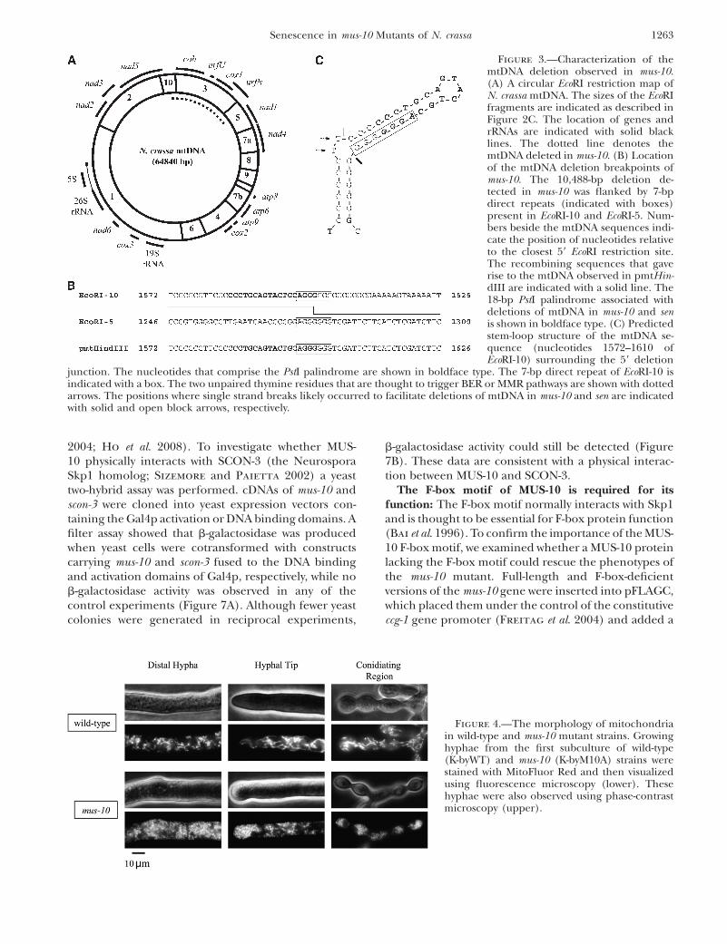
Galagan et al. 2003). This implied that the deletionbreakpoint occurred further into the cloned mtDNAfragment. Consequently, a 2.2-kbp region of mtDNA fromthe cloned ApaI restriction fragment was subcloned intopBluescript using XbaI. Sequence analysis was performedas described above, but the deletion breakpoints couldnot be identified. Subsequent sequencing of pmtHindIII,which contained 1.2 kbp of mtDNA obtained fromdigestion of the 6.6-kbp ApaI restriction fragment withHindIII, finally revealed a 10,488-bp deletion in themtDNA of mus-10. This deletion completely removedEcoRI-3 as well as parts of EcoRI-5 and EcoRI-10, therebyeliminating two unidentified reading frames (urfU andurfN), cox1 (cytochrome c oxidase subunit 1), cob (apoc-ytochrome b), and part of nad1 (NADH dehydrogenasesubunit 1) (Figure 3A).
Further examination revealed that the deleted regionwas flanked by 7-bp direct repeats (AGGGGGG; Figure3B), resembling sequences associated with system II,the site-specific mtDNA recombination pathway. In-terestingly, the first four nucleotides of the 59 repeatoverlapped an 18-bp PstI palindrome (59-CCCTGCAGTACTGCAGGG-39) that occurs in mtDNA of N. crassa 67times (Cahan and Kennell 2005) and is thought toform a stem-loop structure (Figure 3C; Yin et al. 1981;Nargang et al. 1983; de Vries et al. 1986).
Mitochondrial morphology of the mus-10 mutant:Our restriction digest analysis of mtDNA suggested thatloss of the mus-10 gene had an adverse effect on mito-chondria. To explore this hypothesis, we used fluores-cence microscopy to examine the morphology ofmitochondria within mus-10. Growing hyphae werestained with a mitochondria-specific fluorescent dye andthen visualized under a microscope. In wild-type mycelia,long tubular mitochondria were observed in both distaland conidiating hyphae (Figure 4). Conversely, mus-10had abnormal mitochondria that exhibited a sphericalmorphology (Figure 4). Surprisingly, the occurrence ofthis phenotype was not age related, as spherical mito-chondria were observed in earlier subcultures duringwhich hyphae were growing normally (Figure 4). Thisimplies that changes to mitochondrial structure occurprior to the formation of abnormal mtDNAs.
Identification of the mus-10 gene: The mus-10 genewas previously mapped to a site on LG VII near met-7(7%; Kafer and Perlmutter 1980). To identify themus-10 gene, we used a gene-rescue approach in whichmus-10 conidia were transformed with digested cosmidsthat spanned a 27.7-kbp region defined by our linkageand RFLP analyses (data not shown). In this manner, wediscovered an 8.5-kbp NotI fragment derived from thepLORIST cosmid H013 B4 that could rescue the MMSsensitivity of mus-10. This fragment was cloned intopHS to produce pH 13-N8 (Figure 5). The sequencein this fragment contained three annotated ORFs:NCU02379.3, NCU02378.3, and NCU02377.3 (Figure5). Removal of the latter two ORFs from pH 13-N8 was
Figure 1.—Apical growth rates of wild-type and mus-10mutant strains. Conidia from age-matched wild-type(K-byWT) and mus-10 (K-byM10A) were used to inoculaterace tubes containing Vogel’s minimal agar medium supple-mented with 0.5% sucrose. Hyphal growth was recorded onceor twice a day. Once the mycelia had traversed the tube, apiece of the growth front was transferred to a race tubecontaining fresh medium and the entire process was repeated.All race tubes were incubated at 25� under constant light.
Senescence in mus-10 Mutants of N. crassa 1261

achieved through SacII digestion and subsequent re-circularization of the desired fragment to generate theplasmid pH 13-SS. The 3.1-kbp insert in this plasmidcontained a single ORF (NCU02379.3) that conferredwild-type MMS resistance to mus-10. To confirm theidentity of the mus-10 gene, we isolated genomic DNAfrom the original mus-10 mutant and employed PCR toamplify the candidate gene. Sequence analysis of thisPCR product revealed that mus-10 carries a single G-to-Atransition at position 984 of the predicted ORF se-quence, producing a nonsense mutation (Figure 5).
The mus-10 gene was predicted to encode a 637residue polypeptide with a molecular weight of 73.7kDa. The ORF sequence and intron–exon boundarieswere confirmed through sequence analysis of cDNAgenerated through RT–PCR. An F-box was identified atthe N terminus of the MUS-10 protein (Figure 5),suggesting that it belongs to the F-box protein familywhose members are normally components of SCF E3ubiquitin ligase complexes that direct proteins to the26S proteasome, ultimately leading to their degrada-tion. These findings are concurrent with the hypothesisthat SCF complexes can regulate mitochondrial func-tion (Cohen et al. 2008; Deng et al. 2008). A YccV-likedomain, which has been shown to bind hemimethylatedDNA (D’Alencon et al. 2003), was also observed inMUS-10, suggesting that it may be capable of interactingwith DNA (Figure 5).
Localization of MUS-10: To help elucidate thefunction of MUS-10, we wanted to establish its sub-cellular location. Western blot analysis was performedon cytosolic and purified mitochondrial proteins iso-lated from a wild-type strain (C1-T10-37A) and a mus-10knockout (KTO-10H-10A). To ensure the absence ofcytosolic proteins in the mitochondrial fraction and viceversa, blots were also examined using antibodies againsta-tubulin and COX3, which are found in cytosol andmitochondria, respectively (Figure 6). Use of an anti-MUS-10 antibody revealed a band in the cytosolic fractionof wild type that was absent in mus-10 (Figure 6, lanes 1and 3). Conversely, MUS-10 was not observed in mito-chondria from either strain (Figure 6, lanes 5 and 7).
Since MUS-10 appears to influence mitochondrialmorphology and also protects cells against the effects ofMMS, we reasoned that exposure to MMS may inducemovement of MUS-10 from the cytosol to mitochondria.However, MUS-10 localization was not altered in cellsgrown in the presence of MMS (Figure 6). Takentogether, these results suggest that MUS-10 may func-tion in the cytosol.
MUS-10 is part of an SCF complex: Proteins with anF-box motif (F-box proteins) generally function in SCFE3 ubiquitin ligase complexes along with two additionalcore components, Cullin and Skp1, although they arethought to physically interact only with the latter (Bai
et al. 1996; Jackson and Eldridge 2002; Willems et al.
Figure 2.—Restriction analysisof mtDNA. (A) mtDNAs were iso-lated from five sequential subcul-tures (labeled 1–5) of wild-typeand mus-10 mutant strains. ThesemtDNAs were digested with EcoRIor KpnI, and subjected to agarosegel electrophoresis. Bandsemerging or disappearing in thefourth and fifth subcultures ofmus-10 are indicated with anopen circle (�) and asterisk (*),respectively. (B) mtDNAs fromthe fifth subculture of wild-typeand mus-10 mutant strains weredigested with ApaI or ClaI. Re-striction digest fragments thatdiffered between the two strainsare indicated as described in A.(C) Restriction maps of N. crassamtDNA digested with ApaI, ClaI,EcoRI, or KpnI. Position 1 in thisfigure corresponds to the nucleo-tide at the 59 end of the largestEcoRI fragment. Numbers locatedwithin the mtDNA fragments in-dicate their size relative to theother fragments generated fromdigestion with a given restriction
enzyme. The number ‘‘1’’ is used to describe the largest fragment, while ‘‘a’’ and ‘‘b’’ are employed when two fragments of similarsize are produced, where ‘‘a’’ specifies the larger fragment. The shaded area shows the predicted location of a large mtDNAdeletion (�10–12 kbp) observed in latter cultures of mus-10.
1262 A. Kato et al.
2004; Ho et al. 2008). To investigate whether MUS-10 physically interacts with SCON-3 (the NeurosporaSkp1 homolog; Sizemore and Paietta 2002) a yeasttwo-hybrid assay was performed. cDNAs of mus-10 andscon-3 were cloned into yeast expression vectors con-taining the Gal4p activation or DNA binding domains. Afilter assay showed that b-galactosidase was producedwhen yeast cells were cotransformed with constructscarrying mus-10 and scon-3 fused to the DNA bindingand activation domains of Gal4p, respectively, while nob-galactosidase activity was observed in any of thecontrol experiments (Figure 7A). Although fewer yeastcolonies were generated in reciprocal experiments,
b-galactosidase activity could still be detected (Figure7B). These data are consistent with a physical interac-tion between MUS-10 and SCON-3.
The F-box motif of MUS-10 is required for itsfunction: The F-box motif normally interacts with Skp1and is thought to be essential for F-box protein function(Bai et al. 1996). To confirm the importance of the MUS-10 F-box motif, we examined whether a MUS-10 proteinlacking the F-box motif could rescue the phenotypes ofthe mus-10 mutant. Full-length and F-box-deficientversions of the mus-10 gene were inserted into pFLAGC,which placed them under the control of the constitutiveccg-1 gene promoter (Freitag et al. 2004) and added a
Figure 3.—Characterization of themtDNA deletion observed in mus-10.(A) A circular EcoRI restriction map ofN. crassa mtDNA. The sizes of the EcoRIfragments are indicated as described inFigure 2C. The location of genes andrRNAs are indicated with solid blacklines. The dotted line denotes themtDNA deleted in mus-10. (B) Locationof the mtDNA deletion breakpoints ofmus-10. The 10,488-bp deletion de-tected in mus-10 was flanked by 7-bpdirect repeats (indicated with boxes)present in EcoRI-10 and EcoRI-5. Num-bers beside the mtDNA sequences indi-cate the position of nucleotides relativeto the closest 59 EcoRI restriction site.The recombining sequences that gaverise to the mtDNA observed in pmtHin-dIII are indicated with a solid line. The18-bp PstI palindrome associated withdeletions of mtDNA in mus-10 and senis shown in boldface type. (C) Predictedstem-loop structure of the mtDNA se-quence (nucleotides 1572–1610 ofEcoRI-10) surrounding the 59 deletion
junction. The nucleotides that comprise the PstI palindrome are shown in boldface type. The 7-bp direct repeat of EcoRI-10 isindicated with a box. The two unpaired thymine residues that are thought to trigger BER or MMR pathways are shown with dottedarrows. The positions where single strand breaks likely occurred to facilitate deletions of mtDNA in mus-10 and sen are indicatedwith solid and open block arrows, respectively.
Figure 4.—The morphology of mitochondriain wild-type and mus-10 mutant strains. Growinghyphae from the first subculture of wild-type(K-byWT) and mus-10 (K-byM10A) strains werestained with MitoFluor Red and then visualizedusing fluorescence microscopy (lower). Thesehyphae were also observed using phase-contrastmicroscopy (upper).
Senescence in mus-10 Mutants of N. crassa 1263

3xFLAG-tag to the C terminus of each protein (Figure8A). Transformation of these constructs into a mus-10knockout produced strains that were revealed throughWestern blot analysis to contain relatively high levels ofFLAG-tagged full-length or truncated MUS-10 proteinwithin the cytosol (Figure 8B, lanes 2 and 3). Un-fortunately, the anti-FLAG antibody also recognized anonspecific protein similar in size to full-length MUS-10(lanes 1 and 3). However, given the intensity of the bandobserved in mus-10 transformed with the full-lengthconstruct (lane 2), it is clear that FLAG-tagged MUS-10is being produced in this strain. Surprisingly, a smallamount of full-length and DF-box-deficient MUS-10were detected in mitochondria with the relative levelsof the two proteins being similar to that observed in thecytoplasm (Figure 8B, lanes 2 and 3 vs. lanes 5 and 6).This may suggest that overexpression of the two MUS-10forms leads to or increases their association withmitochondria. While this result will be addressedfurther in discussion, it is important to note that theDF-box MUS-10 was also observed in mitochondria andthus, removal of the first 53 amino acids did not removean N-terminal mitochondrial signal sequence.
After we confirmed expression of both FLAG-taggedMUS-10 proteins, we performed spot test analysis todetermine whether these proteins could rescue theMMS sensitivity of mus-10. As anticipated, strains ex-pressing full-length MUS-10 protein displayed wild-typegrowth on MMS plates while those carrying the trun-cated form of MUS-10 lacking the F-box motif remainedsusceptible to MMS (Figure 9A).
To examine whether these transformants exhibited ashortened life span, their growth rates were monitoredin race tubes. Transformants that expressed full-lengthMUS-10 displayed wild-type growth, while those harbor-ing the F-box deficient form senescence prematurely(Figure 9B). Although the strain expressing the trun-cated form of MUS-10 appeared to undergo senescenceearlier than the original mus-10 mutant (Figure 9B), it
should be noted that deficiencies leading to senescencelikely continued to accumulate during the transforma-tion procedure, and thus conidia from this transform-ant are likely ‘‘older’’ than those of the original mus-10mutant. Fluorescence microscopy revealed that tubularmitochondria could be restored in mus-10 throughtransformation with a construct encoding wild-type,but not truncated, MUS-10 protein (Figure 9C). Takentogether, these data confirm that the F-box domain ofMUS-10 is essential for its function in vivo.
Figure 5.—Identification of the mus-10 gene.NotI digestion of pLORIST H013 B4 producedan 8.5-kbp fragment that could restore growthof mus-10 on medium containing MMS. This frag-ment was cloned into pHS to produce pH 13-N8.Sequence analysis of pH 13-N8 revealed threepredicted ORFs, NCU02379.3, NCU02378.3,and NCU02377.3. To identify the mus-10 gene,a portion of pH 13-N8 was removed with SacIIproducing pH 13-SS. This plasmid contained asingle ORF, NCU02379.3, and could complementthe mus-10 mutant phenotype. Sequencing revealedthat the mus-10 mutant carried a G-to-A transition atposition 984 of this ORF, which generated a non-sense mutation. ORF NCU02379.3 is predicted toencode a 637-amino acid polypeptide with a molec-ular weight of 73.7 kDa. Analysis of the amino acidsequence revealed an F-box domain (codons 5–53)and a YccV-like domain (codons 503–526).
Figure 6.—MUS-10 localization. Western blot analysis wasperformed using cytosolic proteins (200 mg) and sucrose gra-dient-purified mitochondria (50 mg) isolated from a wild-type(C1-T10-37A) and mus-10 mutant strain (KTO-10H-10A)grown in the presence (1) or absence (�) of MMS. Anti-a-tubulin and anti-COX3 were used as controls to identifycytosolic and mitochondrial marker proteins, respectively. Atotal of 250 ng of recombinant MUS-10 (MUS-10) was loadedin the gel to verify functionality of the MUS-10 antibody. Sinceuse of the anti-MUS-10 antibody produced several nonspecificbands, the location of MUS-10 is indicated with a solid arrow.
1264 A. Kato et al.

DISCUSSION
Although sensitivity of mus-10 to MMS and UV lightimplied a deficiency in one or more DNA repair path-ways, the precise function of the mus-10 gene wasunclear. In this present work we report that MUS-10belongs to the F-box protein family and is thus likelyinvolved in proteasome-mediated protein turnover.This finding suggests that even though mus-10 is moresusceptible to MMS than wild type, the mus-10 geneproduct may not have a direct role in DNA repair.Indeed, other instances of this phenomenon have beenreported in other organisms. In S. cerevisiae, an in-creased sensitivity to MMS was observed in strainsdeficient for Tim13p, whose role in mitochondrialprotein import is well established (Hanway et al.2002). It was hypothesized that the heightened suscep-tibility of the tim13D strain to MMS resulted from thereduced import of mtDNA repair proteins. Conversely,overexpression of a nuclear BER protein in the mito-chondria of human cells was thought to cause animbalance of mitochondrial DNA repair proteins mak-ing them more vulnerable to MMS (Fishel et al. 2003).Since MUS-10 is likely to function in ubiquitin-mediatedproteolysis, it is conceivable that MUS-10 deficiencypromotes accumulation of one or more mitochondrial
proteins that affect mtDNA repair, replication, or re-combination, leading to an elevated sensitivity to MMS.
Similar to nd and sen, strains deficient for mus-10displayed a shortened life span and accumulatedmtDNA deletions (Seidel-Rogol et al. 1989; Bertrand
et al. 1993; Navaraj et al. 2000; D’Souza et al. 2005).Analysis of the mtDNA deletion observed in lattersubcultures of mus-10 revealed the absence of EcoRI-3and parts of EcoRI-5 and EcoRI-10. This deletion re-moved at least three components of the electron trans-port chain, cob, cox1, and nad1, which likely impairedrespiration leading to senescence and subsequent deathof mus-10. Similarly, the stop–start growth phenotype ofthe E35 stopper mutant was attributed to loss of nad2and nad3 (de Vries et al. 1986; Alves and Videira
1998), while the ER-3 stopper mutant was shown toharbor a �25-kbp deletion of mtDNA that removedseveral genes including cob and cox1 (Niagro and
Figure 7.—A physical interaction between MUS-10 andSCON-3. Yeast two-hybrid analysis was performed to deter-mine whether the MUS-10 protein interacted with a core com-ponent of N. crassa SCF complexes, SCON-3. (A) Yeast cells(Y187) were cotransformed with two plasmids, one of whichcontained the GAL4 activation domain (AD) of pACT2 whilethe other included a GAL4 DNA binding domain (BD) frompGBKT7. While empty vectors (�) were used in some of theseexperiments, transformations were also performed with deriv-atives of pACT2 and pGBKT7 that contained in-frame scon-3(1 scon-3) and mus-10 (1 mus-10) cDNAs, respectively. Selec-tion of cotransformants was achieved using nutritionalmarkers present within pACT2 and pGBKT7, which conferthe ability to grow on medium lacking leucine and trypto-phan, respectively. Colonies emerging after a 3-day incuba-tion at 30� were subjected to a filter assay capable ofdetecting b-galactosidase activity, which would only be ob-served if the two proteins being examined could physically in-teract. (B) Reciprocal experiments performed as described inA. In these experiments, pAS2-1, which contains the GAL4DNA binding domain and the TRP1 gene, was used in placeof pGBKT7.
Figure 8.—FLAG-tagged forms of MUS-10. (A) Diagram offull-length (codons 1–637) and truncated (codons 54–637)MUS-10 used in our experiments. Insertion of cDNAs encod-ing the two forms of MUS-10 into pFLAGC placed their ex-pression under the control of the constitutive ccg-1 genepromoter and added a 3xFLAG tag on the C terminus of eachprotein. Since pFLAGC is derived from pMF272, it contains aportion of the N. crassa his-3 gene, which can be used for tar-geted integration and reversion at the his-3 locus. (B) Westernblot analysis was performed as described in Figure 6. Plasmidsencoding the two different FLAG-tagged versions of MUS-10(shown in A) were used to transform mus-10 (KRA-m10his3-5)conidia. Two resulting his1 transformants, KRA-m10M10F-2and KRA-m10dFM10F-1, expressed full-length and DF-boxMUS-10, respectively. Background levels were determinedusing untransformed mus-10. Full-length MUS-10 protein isindicated with a solid arrow, while the DF-box form is shownwith a dotted arrow. To facilitate viewing of MUS-10 in themitochondrial fraction, a second, longer exposure was alsoincluded (Anti-FLAG, lower).
Senescence in mus-10 Mutants of N. crassa 1265

Mishra 1989; Niagro and Mishra 1990). Senescencein these and other N. crassa strains, including nd, sen,and mus-10, correlated with the accumulation ofmtDNAs that harbor large deletions, which may incura replicative advantage over wild-type molecules due totheir smaller size. In a recent report, quantitative real-time PCR was used to demonstrate that in mice,mitochondrial genomes that contain large deletions(�10 kb) accumulated faster than those carrying smalldeletions (�3.8 kb) (Fukui and Moraes 2009). Whilethese data support the notion that smaller mtDNAs havea replicative advantage over larger ones, it does notexclude the possibility that the number and/or natureof the genes present in a mitochondrial genome caninfluence copy number.
The PstI palindrome observed at the 59 flank of themus-10 mtDNA deletion is capable of forming a GC-richimperfect stem loop, a structure which has been
hypothesized to stall DNA replication and/or act assubstrates for mismatch repair (MMR) or base excisionrepair (BER) systems (D’Souza et al. 2005; Hausner
et al. 2006). Either scenario could lead to single and/ordouble strand breaks that are thought to promotemtDNA recombination. Indeed, the stem-loop struc-ture formed by the PstI palindrome flanking the mus-10mtDNA deletion contains two unpaired nucleotidesthat could potentially trigger BER or MMR pathwaysresulting in endonucleolytic cleavage of the phospho-diester bond following the AGGGGGG repeat (Figure3C). Interestingly, the same PstI palindrome and un-paired nucleotides were implicated in the generation ofmtDNA deletions in sen (recombination junction J2;D’Souza et al. 2005), but in this case, the cleavage eventoccurred on the opposite strand (Figure 3C), implyingthat mtDNA repair pathways can target either strand ofthe mismatched and/or unpaired sequence.
Figure 9.—The F-box of MUS-10 is essen-tial for its function. (A) Spot test analysis wasperformed using conidia from C1-T10-37A(wild type), KTO-10H-10A (mus-10), KRA-m10M10F-2 (mus-10 1 full-length) andKRA-m10dFM10F-1 (mus-10 1 DF-box). Co-nidial suspensions were adjusted to a concen-trationof13 106 conidia/mlandsubjectedtofive 1:4 serial dilutions. A 10-ml aliquot of eachmixture was spotted onto agar plates contain-ing Vogels’s minimal sorbose medium lacking(control) or supplemented with 0.015%MMS (MMS). Plates were photographedafter 2 days at 30�. (B) Measurements of apicalgrowthwereperformedasdescribedinFigure1. In these experiments, race tubes contained1.2% sucrose instead of 0.5%. (C) Mitochon-drial morphology. Live hyphae were stainedwith MitoTracker Green and visualized usinga confocal laser-scanning microscope.
Figure 10.—Models to explain the relation-ship between mus-10 and senescence. (A) In thismodel, loss of MUS-10 prevents operation of amitochondrial E3 ubiquitin ligase leading to ac-cumulation and/or deficiency of numerous mi-tochondrial proteins. The resulting proteinimbalance leads to defects in both mtDNA main-tenance and mitochondrial morphology throughindependent mechanisms. Defective mtDNA re-combination, repair, and/or replication leadsto increased MMS/UV-light sensitivity andmtDNA rearrangements. Mitochondrial frag-mentation and altered mtDNA promote furthermitochondrial dysfunction, which ultimately re-sults in impaired respiration and eventual senes-cence. (B) This model is similar to the oneproposed in A except that in this case, dysfunc-tion of mtDNA recombination, repair, and/orreplication results from a mitochondrial proteinimbalance caused by mitochondrial fragmenta-tion and is thus not a direct result of MUS-10 de-ficiency.
1266 A. Kato et al.

Our examination of mitochondrial morphology andmtDNA rearrangements in mus-10 suggests that frag-mented mitochondria arise prior to modification in themtDNA restriction digest profile. While we cannotexclude the possibility that mtDNA rearrangementsarise in mus-10 much earlier than they can be visualizedthrough restriction digest and agarose gel electropho-resis, our observations do raise the question of whethermitochondrial fragmentation leads to altered mtDNAor if these events occur through independent mecha-nisms. We propose two models that address the relation-ship between mitochondrial morphology and mtDNAintegrity, both of which rely on MUS-10 functioning asan E3 ubiquitin ligase. In our first model (Figure 10A),MUS-10 deficiency is proposed to promote the accu-mulation of a wide variety of mitochondrial proteinsincluding, but not limited to, mediators of fission and/or fusion, components of import machinery, and pro-teases. Accumulation of mitochondrial proteases wouldsubsequently lead to lower levels of their substrates. Theresulting mitochondrial protein imbalance has twoconcurrent effects: (i) deficiencies in mtDNA replica-tion, recombination, and/or repair, leading to mtDNArearrangements and sensitivity to MMS and UV, and (ii)disruption of the fission/fusion equilibrium, resultingin mitochondrial fragmentation. The persistence ofsuch problems promotes further mitochondrial defects,which initiates a vicious cycle leading to the accumula-tion of dysfunctional mitochondria that ultimatelycause senescence. Our second model (Figure 10B)proposes that loss of MUS-10 leads to the productionof fragmented mitochondria through inhibited mito-chondrial fusion and/or accelerated division. Thismodel predicts that smaller mitochondria are morelikely to harbor an imbalanced protein complementand are thus prone to defective maintenance of mtDNA.This compromises respiration, which in turn leads tosenescence.
Regardless of whether there is a causal relationshipbetween defective mtDNA and mitochondrial morphol-ogy in mus-10, the fragmented mitochondria observedin this strain likely arise through abnormally highamounts of fission or by inhibited fusion. Mitochondrialfusion and fission are complicated processes involvingmany proteins and are thought to enable mixing ofmetabolites and mtDNA thereby allowing optimalmitochondrial function (Cerveny et al. 2007; Hoppins
et al. 2007; Knott and Bossy-Wetzel 2008; Hoppins
and Nunnari 2009). Therefore circumstances thatdisrupt the balance between these opposing forces canbe detrimental to the cell. Given that the mus-10 geneproduct is a mediator of proteolysis, the altered mito-chondrial morphology may result from the accumula-tion of one or more proteins that promote division orhinder fusion. Indeed, links between E3 ubiquitin ligasecomplexes and mitochondrial structure have beenreported. Mutations in Parkin, an E3 ubiquitin ligase,
have been associated with defective mtDNA repair andprogression of Parkinson’s disease in humans (Dawson
2006; Dodson and Guo 2007). Recent evidence sug-gests that Parkin functions to promote mitochondrialfission and/or inhibit fusion (Deng et al. 2008; Poole
et al. 2008). In Saccharomyces cerevisiae, the F-box proteinMdm30p is involved in ubiquitylation and subsequentdegradation of the mitochondrial fusion mediatorFzo1p (Fritz et al. 2003; Escobar-Henriques et al.2006; Cohen et al. 2008). The mdm30 mutant was shownto accumulate Fzo1p and exhibit aggregated mitochon-dria. A physical interaction between Mdm30p andFzo1p has also been observed (Escobar-Henriques
et al. 2006). Interestingly, Mdm30p of yeast has a weakhomology to N. crassa MUS-10, and thus it is tempting tospeculate that MUS-10 can interact with FZO-1 and/orother mitochondrial outer membrane proteins. Such aninteraction might explain why we observed full-lengthand truncated forms of MUS-10 in mitochondria upontheir overexpression as elevated levels of these proteinsmay promote binding of MUS-10 to its outer membranetargets. However, we cannot exclude the possibility thatMUS-10 is imported into mitochondria and that ourdetection of the FLAG-tagged MUS-10 proteins inmitochondria stems from increased import arising fromhigh levels of expression. In this case, mitochondrialimport would have to rely on internal mitochondrialsignal sequences as the N-terminal truncated form ofMUS-10 also associated with mitochondria.
There are many unanswered questions regarding thefunction of MUS-10 and its relationship with mitochon-dria. However, the N. crassa mus-10 mutant couldprovide great insight into the role of ubiquitin-mediatedproteolysis in the maintenance of mitochondrial mor-phology and mtDNA and thus may be used as a modelfor studying ageing and mitochondrial diseases inhumans.
We thank Niji Ohta for her help with our sequencing experiments.We also express our gratitude to Yosuke Morishima and ShigeyukiKawano who helped photograph mitochondria. This work was fundedby grants-in-aid for scientific research 11640619, 08F08756, and20570001. The Rational Evolutionary Design of Advanced Biomole-cules, Saitama Prefecture Collaboration of Regional Entities for theAdvancement of Technological Excellence, Japan Science and Tech-nology Agency also supported this work. M.C. was given a grant-in-aidfrom the Japan Society for the Promotion of Science.
LITERATURE CITED
Akins, R. A., R. L. Kelley and A. M. Lambowitz, 1989 Characteriza-tion of mutant mitochondrial plasmids of Neurospora spp. thathave incorporated tRNAs by reverse transcription. Mol. Cell. Bi-ol. 9: 678–691.
Alves, P. C., and A. Videira, 1998 The membrane domain of com-plex I is not assembled in the stopper mutant E35 of Neurospora.Biochem. Cell. Biol. 76: 139–143.
Antignani, A., and R. J. Youle, 2006 How do Bax and Bak lead topermeabilization of the outer mitochondrial membrane? Curr.Opin. Cell Biol. 18: 685–689.
Senescence in mus-10 Mutants of N. crassa 1267

Bai, C., P. Sen, K. Hofmann, L. Ma, M. Goebl et al., 1996 SKP1 con-nects cell cycle regulators to the ubiquitin proteolysis machinerythrough a novel motif, the F-box. Cell 86: 263–274.
Bartlett, K., and S. Eaton, 2004 Mitochondrial beta-oxidation.Eur. J. Biochem. 271: 462–469.
Bertrand, H., Q. Wu and B. L. Seidel-Rogol, 1993 Hyperactiverecombination in the mitochondrial DNA of the natural deathnuclear mutant of Neurospora crassa. Mol. Cell. Biol. 13: 6778–6788.
Boulet, L., G. Karpati and E. A. Shoubridge, 1992 Distributionand threshold expression of the tRNA(Lys) mutation in skeletalmuscle of patients with myoclonic epilepsy and ragged-red fibers(MERRF). Am. J. Hum. Genet. 51: 1187–1200.
Cahan, P., and J. C. Kennell, 2005 Identification and distributionof sequences having similarity to mitochondrial plasmids inmitochondrial genomes of filamentous fungi. Mol. Genet.Genomics 273: 462–473.
Carroll, A. M., J. A. Sweigard and B. Valent, 1994 Improvedvectors for selecting resistance to hygromycin. Fungal Genet.Newsl. 41: 22.
Cerveny, K. L., Y. Tamura, Z. Zhang, R. E. Jensen and H. Sesaki,2007 Regulation of mitochondrial fusion and division. TrendsCell Biol. 17: 563–569.
Chae, M. S., and F. E. Nargang, 2009 Investigation of regulatoryfactors required for alternative oxidase production in Neuros-pora crassa. Physiol. Plant 137: 407–418.
Chatterjee, A., E. Mambo and D. Sidransky, 2006 MitochondrialDNA mutations in human cancer. Oncogene 25: 4663–4674.
Chomyn, A., A. Martinuzzi, M. Yoneda, A. Daga, O. Hurko et al.,1992 MELAS mutation in mtDNA binding site for transcrip-tion termination factor causes defects in protein synthesisand in respiration but no change in levels of upstream anddownstream mature transcripts. Proc. Natl. Acad. Sci. USA89: 4221–4225.
Cohen, M. M., G. P. Leboucher, N. Livnat-Levanon, M. H. Glickman
and A. M. Weissman, 2008 Ubiquitin-proteasome-dependentdegradation of a mitofusin, a critical regulator of mitochondrialfusion. Mol. Biol. Cell 19: 2457–2464.
Corral-Debrinski, M., T. Horton, M. T. Lott, J. M. Shoffner, M. F.Beal et al., 1992 Mitochondrial DNA deletions in human brain:regional variability and increase with advanced age. Nat. Genet.2: 324–329.
Cortopassi, G. A., and N. Arnheim, 1990 Detection of a specificmitochondrial DNA deletion in tissues of older humans. NucleicAcids Res. 18: 6927–6933.
Cortopassi, G. A., D. Shibata, N. W. Soong and N. Arnheim,1992 A pattern of accumulation of a somatic deletion of mito-chondrial DNA in aging human tissues. Proc. Natl. Acad. Sci.USA 89: 7370–7374.
Court, D. A., A. J. Griffiths, S. R. Kraus, P. J. Russell and H.Bertrand, 1991 A new senescence-inducing mitochondriallinear plasmid in field-isolated Neurospora crassa strains fromIndia. Curr. Genet. 19: 129–137.
d’Alencon, E., A. Taghbalout, C. Bristow, R. Kern, R. Aflalo
et al., 2003 Isolation of a new hemimethylated DNA bindingprotein which regulates dnaA gene expression. J. Bacteriol.185: 2967–2971.
D’Souza, A. D., H. Bertrand and R. Maheshwari, 2005 Intramole-cular recombination and deletions in mitochondrial DNA ofsenescent, a nuclear-gene mutant of Neurospora crassa exhibit-ing ‘‘death’’ phenotype. Fungal Genet. Biol. 42: 178–190.
Davis, R. H., and F. J. De Serres, 1970 Genetic and microbiologicalresearch techniques for Neurospora crassa. Meth. Enzymol. 17A:79–143.
Dawson, T. M., 2006 Parkin and defective ubiquitination in Parkin-son’s disease. J. Neural Transm. 70(Suppl.): 209–213.
de Serres, F. J., 1980 Mutagenesis at the ad-3A and ad-3B loci inhaploid UV-sensitive strains of Neurospora crassa. II. Compari-son of dose-response curves for inactivation and mutation in-duced by UV. Mutat. Res. 71: 181–191.
de Vries, H., B. Alzner-DeWeerd, C. A. Breitenberger, D. D.Chang, J. C. de Jonge et al., 1986 The E35 stopper mutantof Neurospora crassa: precise localization of deletion endpointsin mitochondrial DNA and evidence that the deleted DNA codesfor a subunit of NADH dehydrogenase. EMBO J. 5: 779–785.
Deng, H., M. W. Dodson, H. Huang and M. Guo, 2008 The Parkin-son’s disease genes pink1 and parkin promote mitochondrialfission and/or inhibit fusion in Drosophila. Proc. Natl. Acad.Sci. USA 105: 14503–14508.
Dodson, M. W., and M. Guo, 2007 Pink1, Parkin, DJ-1 andmitochondrial dysfunction in Parkinson’s disease. Curr. Opin.Neurobiol. 17: 331–337.
Escobar-Henriques, M., B. Westermann and T. Langer,2006 Regulation of mitochondrial fusion by the F-box proteinMdm30 involves proteasome-independent turnover of Fzo1. J.Cell Biol. 173: 645–650.
Fishel, M. L., Y. R. Seo, M. L. Smith and M. R. Kelley,2003 Imbalancing the DNA base excision repair pathway inthe mitochondria; targeting and overexpressing N-methylpurineDNA glycosylase in mitochondria leads to enhanced cell killing.Cancer Res. 63: 608–615.
Freitag, M., P. C. Hickey, N. B. Raju, E. U. Selker and N. D. Read,2004 GFP as a tool to analyze the organization, dynamics andfunction of nuclei and microtubules in Neurospora crassa.Fungal Genet. Biol. 41: 897–910.
Fritz, S., N. Weinbach and B. Westermann, 2003 Mdm30 is anF-box protein required for maintenance of fusion-competentmitochondria in yeast. Mol. Biol. Cell. 14: 2303–2313.
Fukui, H., and C. T. Moraes, 2009 Mechanisms of formation andaccumulation of mitochondrial DNA deletions in aging neurons.Hum. Mol. Genet. 18: 1028–1036.
Galagan, J. E., S. E. Calvo, K. A. Borkovich, E. U. Selker, N. D.Read et al., 2003 The genome sequence of the filamentousfungus Neurospora crassa. Nature 422: 859–868.
Gerber, J., and R. Lill, 2002 Biogenesis of iron-sulfur proteins ineukaryotes: components, mechanism and pathology. Mitochon-drion 2: 71–86.
Good, A. G., and W. L. Crosby, 1989 Anaerobic induction of alanineaminotransferase in barley root tissue. Plant Physiol. 90: 1305–1309.
Green, D. R., 2005 Apoptotic pathways: ten minutes to dead. Cell121: 671–674.
Gunter, T. E., D. I. Yule, K. K. Gunter, R. A. Eliseev and J. D. Salter,2004 Calcium and mitochondria. FEBS Lett. 567: 96–102.
Handa, N., Y. Noguchi, Y. Sakuraba, P. Ballario, G. Macino et al.,2000 Characterization of the Neurospora crassa mus-25 mutant:the gene encodes a protein which is homologous to the Saccharo-myces cerevisiae Rad54 protein. Mol. Gen. Genet. 264: 154–163.
Hanway, D., J. K. Chin, G. Xia, G. Oshiro, E. A. Winzeler et al.,2002 Previously uncharacterized genes in the UV- and MMS-induced DNA damage response in yeast. Proc. Natl. Acad. Sci.USA 99: 10605–10610.
Hausner, G., K. A. Nummy, S. Stoltzner, S. K. Hubert andH. Bertrand, 2006 Biogenesis and replication of small plas-mid-like derivatives of the mitochondrial DNA in Neurosporacrassa. Fungal Genet. Biol. 43: 75–89.
Higuchi, M., 2007 Regulation of mitochondrial DNA content andcancer. Mitochondrion 7: 53–57.
Ho, M. S., C. Ou, Y. R. Chan, C. T. Chien and H. Pi, 2008 The utilityF-box for protein destruction. Cell. Mol. Life Sci. 65: 1977–2000.
Hoppins, S., and J. Nunnari, 2009 The molecular mechanism ofmitochondrial fusion. Biochim. Biophys. Acta 1793: 20–26.
Hoppins, S., L. Lackner and J. Nunnari, 2007 The machines thatdivide and fuse mitochondria. Annu. Rev. Biochem. 76: 751–780.
Jackson, P. K., and A. G. Eldridge, 2002 The SCF ubiquitin ligase:an extended look. Mol. Cell 9: 923–925.
Kafer, E., 1981 Mutagen sensitivities and mutator effects of MMS-sensitive mutants in Neurospora. Mutat. Res. 80: 43–64.
Kafer, E., 1983 Epistatic grouping of repair-deficient mutants inNeurospora: comparative analysis of two uvs-3 alleles, uvs-6 andtheir mus double mutant strains. Genetics 105: 19–33.
Kafer, E., and E. Perlmutter, 1980 Isolation and genetic analysisof MMS-sensitive mus mutants of neurospora. Can. J. Genet.Cytol. 22: 535–552.
Kelkar, H. S., J. Griffith, M. E. Case, S. F. Covert, R. D. Hall et al.,2001 The Neurospora crassa genome: cosmid libraries sortedby chromosome. Genetics 157: 979–990.
Knott, A. B., and E. Bossy-Wetzel, 2008 Impairing the mitochon-drial fission and fusion balance: a new mechanism of neurode-generation. Ann. N Y Acad. Sci. 1147: 283–292.
1268 A. Kato et al.

Kothe, G. O., M. Kitamura, M. Masutani, E. U. Selker andH. Inoue, 2010 PARP is involved in replicative aging in Neuros-pora crassa. Fungal Genet. Biol. 47: 297–309.
Krishnan, K. J., A. K. Reeve and D. M. Turnbull, 2007 Do mito-chondrial DNA mutations have a role in neurodegenerativedisease? Biochem. Soc. Trans. 35: 1232–1235.
Laemmli, U. K., 1970 Cleavage of structural proteins during theassembly of the head of bacteriophage T4. Nature 227: 680–685.
Lambowitz, A. M., 1979 Preparation and analysis of mitochondrialribosomes. Methods Enzymol. 59: 421–433.
Lill, R., and U. Muhlenhoff, 2005 Iron-sulfur-protein biogenesisin eukaryotes. Trends Biochem. Sci. 30: 133–141.
Maheshwari, R., and A. Navaraj, 2008 Senescence in fungi: theview from Neurospora. FEMS Microbiol. Lett. 280: 135–143.
Myers, C. J., A. J. Griffiths and H. Bertrand, 1989 Linear kaliloDNA is a Neurospora mitochondrial plasmid that integrates intothe mitochondrial DNA. Mol. Gen. Genet. 220: 113–120.
Nargang, F. E., J. B. Bell, L. L. Stohl and A. M. Lambowitz,1983 A family of repetitive palindromic sequences found inNeurospora mitochondrial DNA is also found in a mitochondrialplasmid DNA. J. Biol. Chem. 258: 4257–4260.
Navaraj, A., A. Pandit and R. Maheshwari, 2000 Senescent: a newNeurospora crassa nuclear gene mutant derived from natureexhibits mitochondrial abnormalities and a ‘‘death’’ phenotype.Fungal Genet. Biol. 29: 165–173.
Newmeyer, D., and D. R. Galeazzi, 1978 A meiotic UV-sensitivemutant that causes deletion of duplications in Neurospora.Genetics 89: 245–269.
Newmeyer, D., A. L. Schroeder and D. R. Galeazzi, 1978 Anapparent connection between histidine, recombination, andrepair in Neurospora. Genetics 89: 271–279.
Niagro, F. D., and N. C. Mishra, 1989 An ethidium bromide in-duced mutant of Neurospora crassa defective in mitochondrialDNA. Curr. Genet. 16: 303–305.
Niagro, F. D., and N. C. Mishra, 1990 Biochemical, genetic andultrastructural defects in a mitochondrial mutant (ER-3) ofNeurospora crassa with senescence phenotype. Mech. AgeingDev. 55: 15–37.
Ninomiya, Y., K. Suzuki, C. Ishii and H. Inoue, 2004 Highly efficientgene replacements in Neurospora strains deficient for nonhomol-ogous end-joining. Proc. Natl. Acad. Sci. USA 101: 12248–12253.
Orbach, M. J., 1994 A cosmid with a HyR marker for fungal libraryconstruction and screening. Gene 150: 159–162.
Perkins, D. D., J. A. Kinsey, D. K. Asch and G. D. Frederick,1993 Chromosome rearrangements recovered following trans-formation of Neurospora crassa. Genetics 134: 729–736.
Poole, A. C., R. E. Thomas, L. A. Andrews, H. M. McBride,A. J. Whitworth et al., 2008 The PINK1/Parkin pathway reg-ulates mitochondrial morphology. Proc. Natl. Acad. Sci. USA105: 1638–1643.
Reeve, A. K., K. J. Krishnan and D. Turnbull, 2008 MitochondrialDNA mutations in disease, aging, and neurodegeneration. Ann.N Y Acad. Sci. 1147: 21–29.
Rimessi, A., C. Giorgi, P. Pinton and R. Rizzuto, 2008 The versa-tility of mitochondrial calcium signals: from stimulation of cellmetabolism to induction of cell death. Biochim. Biophys. Acta1777: 808–816.
Rouault, T. A., and W. H. Tong, 2005 Iron-sulphur cluster biogen-esis and mitochondrial iron homeostasis. Nat. Rev. Mol. Cell.Biol. 6: 345–351.
Rowley, N., C. Prip-Buus, B. Westermann, C. Brown, E. Schwarz
et al., 1994 Mdj1p, a novel chaperone of the DnaJ family, is in-volved in mitochondrial biogenesis and protein folding. Cell 77:249–259.
Ryan, F. J., G. W. Beadle and E. L. Tatum, 1943 The tube method ofmeasuring the growth rate of Neurospora. Am. J. Bot. 30: 784–799.
Sakuraba, Y., A. L. Schroeder, C. Ishii and H. Inoue, 2000 A Neu-rospora double-strand-break repair gene, mus-11, encodes aRAD52 homologue and is inducible by mutagens. Mol. Gen.Genet. 264: 392–401.
Schroeder, A. L., 1970 Ultraviolet-sensitive mutants of Neurospora.I. Genetic basis and effect on recombination. Mol. Gen. Genet.107: 291–304.
Schroeder, A. L., 1986 Chromosome instability in mutagen sensi-tive mutants of Neurospora. Curr. Genet. 10: 381–387.
Sciacco, M., E. Bonilla, E. A. Schon, S. DiMauro and C. T. Moraes,1994 Distribution of wild-type and common deletion forms ofmtDNA in normal and respiration-deficient muscle fibers frompatients with mitochondrial myopathy. Hum. Mol. Genet. 3:13–19.
Seidel-Rogol, B. L., J. King and H. Bertrand, 1989 Unstablemitochondrial DNA in natural-death nuclear mutants of Neuros-pora crassa. Mol. Cell. Biol. 9: 4259–4264.
Sheng, T. C., 1951 A gene that causes natural death in Neurosporacrassa. Genetics 36: 199–212.
Simonetti, S., X. Chen, S. DiMauro and E. A. Schon, 1992 Accumu-lation of deletions in human mitochondrial DNA duringnormal aging: analysis by quantitative PCR. Biochim. Biophys. Ac-ta 1180: 113–122.
Sizemore, S. T., and J. V. Paietta, 2002 Cloning and characteriza-tion of scon-31, a new member of the Neurospora crassa sulfurregulatory system. Eukaryot. Cell 1: 875–883.
Tamaru, H., and H. Inoue, 1989 Isolation and characterization of alaccase-derepressed mutant of Neurospora crassa. J. Bacteriol.171: 6288–6293.
Watanabe, K., Y. Sakuraba and H. Inoue, 1997 Genetic and molec-ular characterization of Neurospora crassa mus-23: a gene in-volved in recombinational repair. Mol. Gen. Genet. 256: 436–445.
Willems, A. R., M. Schwab and M. Tyers, 2004 A hitchhiker’sguide to the cullin ubiquitin ligases: SCF and its kin. Biochim.Biophys. Acta 1695: 133–170.
Xu, G., and Y. Shi, 2007 Apoptosis signaling pathways and lympho-cyte homeostasis. Cell Res. 17: 759–771.
Yin, S., J. Heckman and U. L. RajBhandary, 1981 Highly conservedGC-rich palindromic DNA sequences flank tRNA genes in Neu-rospora crassa mitochondria. Cell 26: 325–332.
Zheng, L., V. L. Cash, D. H. Flint and D. R. Dean, 1998 Assemblyof iron-sulfur clusters. Identification of an iscSUA-hscBA-fdxgene cluster from Azotobacter vinelandii. J. Biol. Chem. 273:13264–13272.
Communicating editor: E. U. Selker
Senescence in mus-10 Mutants of N. crassa 1269