Embed Size (px)
Citation preview
Eur. J. Biochem. 194, 161 -165 (1990) 0 FEBS 1990
Delineation of the minimal portion of the Bacillus sphaericus 1593M toxin required for the expression of larvicidal activity Peter SEBO, Therbse BENNARDO, FranCoise de la TORRE and Jekisiel SZULMAJSTER
Laboratoire d'Enzymologie du Centre National de la Recherche Scientifique, Gif-sur-Yvette, France
(Received April 26/July 19, 1990) - EJB 90 0477
The two genes of Bacillus sphaericus 1953M coding for the 51.4-kDa and 41.9-kDa proteins are both required for the expression of the active larvicidal toxin in Escherichia coli. The minimal size of the active peptide of the 41.9-kDa toxin was defined by in vitro deletion analysis of the gene and found to consist of 338 amino acids (38.3 kDa). N-terminal deletions past the Ile18 residue and C-terminal deletions past the His352 residue result in the loss of toxic activity and rapid degradation of such modified toxins by host proteases. The minimal active 38.3-kDa peptide produced in E. coli seems to mimick the stable processed form of the toxin found in larval midguts. However, it still requires the action of the synergistic 51.4-kDa protein for the larvicidal activity.
The entomopathogenic strains of Bacillus sphaericus ac- cumulate in the course of sporulation a larvicidal toxin, mainly active towards Culex and Anopheles species (Davidson 1984; Yousten 1984). These strains are now considered as promising agents for biological control of vectors transmitting human and animal diseases in tropical areas.
The genetic determinants of toxicity were cloned in several laboratories. Their nucleotide sequences revealed the presence of two genes coding for 51.4-kDa and 41.9-kDa proteins (Arapinis et al. 1988; Hindley and Berry 1987; Baumann et al. 1988). These genes were expressed in the heterologous hosts Escherichia coli, Bacillus subtilis and Anacystis nidulans, conferring on them toxicity for mosquito larvae (Tandeau de Marsac et al. 1987; de la Torre et al. 1989).
In the larvicidal B. sphaericus strains (1953, 2362) both proteins are present. Following purification, toxicity was found to be associated with the 41.9-kDa protein (Baumann et al. 1985; Sgarella and Szulmajster 1987). Upon expression of the toxin genes in Escherichia coli, the recombinant-pro- duced toxin is active only when the 51.4-kDa and 41.9-kDa proteins are either co-expressed in the same cell or produced in separate cultures and mixed prior to testing (Baumann et al. 1987; de la Torre et al. 1989). In B. subtilis sporulating cells, however, the toxin activity is expressed in the absence of the 51.4-kDa protein which indicates that the activation of the 41.9-kDa protein can be accomplished by host enzyme(s).
In the present study, we have delineated the portion of the toxin protein required for larvicidal activity. We further show that the truncated protein, produced in E. coli, still requires the action of the 51.4-kDa polypeptide for expression of activity. The significance of this observation will be dis- cussed.
Correspondence to P. Sebo, Institut Pasteur, Unit6 de Biochimie des Regulations Cellulaires, 28, rue du Dr. Roux, F-75724 Paris Cedex 15, France
Dedication. Dedicated to the memory of J. Szulmajster who died on August 31,1990.
MATERIALS AND METHODS
Bacterial strains
The E. coli strains GM2199 (F- thr leu ara14 tonA31 lacy1 tsx78 supE44 galK2 his4 rps1136 xy15 mtll thi dam-13:: Tn9), CSR603 (Sancar et al. 1979) and BNN103 (Young and Davis 1983) were used as hosts for recombinant plasmids.
Medium and growth conditions
The clones of E. coli GM2199 or BNN103, bearing the respective recombinant plasmids were grown on LB medium (Miller 1972) with 150 pg/ml of ampicillin. To express the toxin proteins the precultures of the individual clones were grown overnight and 200 pl used to inoculate 5 ml fresh broth. The cultures were grown for 3 h at 37°C and 1 ml mixed with 4 ml fresh LB/ampicillin containing 2.5 mM isopropyl p-D- thiogalactopyranoside. The cells were harvested after over- night growth at 28"C, washed in 0.1 M Tris/HCl pH 7.5 and used for toxicity assays or immunoblot analysis.
Plasmids, enzymes and reagents
The T7 sequencing kitrM and pKK233-2 DNA were pur- chased from Pharmacia LKB, the mung bean nuclease was from New England Biolabs. The pUC18 plasmid, the BamHI linker and restriction enzymes were from Boehringer Mannheim. The radiolabelled compounds were from Amersham. The stop-codon linker was obtained by annealing two synthetic oligonucleotides, d(TAACTAACTAA) and d(AGCTTTAGTTAGTTA).
General procedures
The DNA manipulations were performed as described by Sambrook et al. (1989). The universal restriction enzyme buffer (potassium glutamate buffer) was used at concen- trations recommended by McClelland et al. (1988). The Ba131 exonuclease digestions were performed at 16 "C in potassium

162
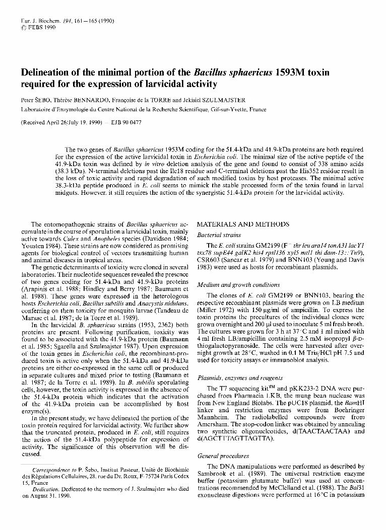
p G s p l O
'"51 kD gene" "42 kD gene"
r'- -
p G S p l O A N l 5 _ _ _
N
NC-deleted
p G s p l O A N 1 5 C 1 7 - -
Bs NC-deleted
pSEP AN15C17 ----
Fig. 1. Schematic representation of the modified toxin genes subcloned in thepUCI8 vector. Restriction sites: B, BamHI; Bc, BclI; Bg, BglII; E, EcoRI; EV, EcoRV; F, FspI; H, HindIII; N, NcoI; X, XhaI. Plac, Ptrc, promoters; RBS, ribosome-binding site of lacZ; 5s TIT2, transcriptional terminators of rmB. The shaded boxes indicate the modified sequences in the different constructs compared to the original pGsplO recombinant plasmid (see text for details)
glutamate buffer with 12.5 mM calcium chloride. Local DNA sequencing of the plasmids was performed according to the instructions of the T7 sequencing kit supplier. SDSjPAGE under denaturing conditions and immunoblotting were performed by standard procedures (Sambrook et al. 1989). For protein quantification in cell extracts the method of Bradford (1976) was used.
Maxicell preparation E. coli CSR603 maxicells were prepared as described
(Sancar et al. 1979). The cells were labelled for 1 h with ~-[~'S]methionine (6.4 pCi/mg protein) diluted in 1.5 pM L-methionine. The labelled material was used in parallel for toxicity assays and SDS/PAGE.
Toxicity assu,vs The bioassay were carried out with second instar larvae
of Culexpipiens, in triplicate samples in plastic caps containing 10 larvae in 5 ml water, tested E. colicells (expressed in protein concentration), and 1 mgiml of baker yeast. The tests were performed at 28 C with 14-h/12-h day/night cycle. The tox- icity was expressed in LC50 (50% of mortality in 48 h . ng protein-' . ml- ' and the limit values from independent tests are given in the tables.
RESULTS
To increase the toxin expression in E. coli, we recloned the 3.08-kb KpnI -Hind111 DNA fragment, bearing both toxicity determining genes, from pGsp04 (Tandeau de Marsac et al. 1987) into the polylinker of the pUC18 vector. From the resulting plasmid, pGsp1O (Fig. l), the synergistic (51 .4-kDa) and toxin (41.9-kDa) proteins are expressed after induction of the lac promoter by isopropyl P-D-thiogalactopyranoside. The expression of toxicity in the E. coli GM2199/pGsP30 cells (LC50 z 10 ng/ml) allowed us to monitor more efficiently the effects of modifications in the toxin protein.
Deletion analysis of the C-terminal part of the toxin protein
Construction oj3'-deletions
The pGsplO plasmid was linearized at the unique BclI site three codons upstream of the natural stop codon of the toxin gene. The resulting ends were progressively digested by Bal31 exonuclease, repaired by the Klenow fragment of DNA poly- merase 1 and the DNA was further digested by HindIII. To introduce a stop codon into the toxin gene, the linear DNA molecules were ligated with an asymetric oligonucleotide linker carrying three consecutive stop codons in different reading frames.
We thus obtained a set of constructs that are further referred to as pGsplOdC plasmids. One of them is shown in Fig. 1.
Expression and biological activity o j the C-terminal truncated toxins
In a subset of pGsplOdC plasmids we localized the precise positions of the inserted linkers by local DNA-sequencing of the modified toxin genes. For testing the toxicity we selected only the clones with toxin genes having inserted in frame the first or the second linker-coded stop codon. The deduced C- terminal amino acid sequences of a set of such truncated toxin proteins and the expression of their toxicity in E. coli are shown in Table 1. Deletion of up to 17 C-terminal amino acid residues has no discernable effect on the toxicity displayed by the corresponding clones.
Furthermore a 100-fold lower biological activity was found with the clones expressing the toxin lacking 18 C-ter- minal residues and no toxicity at all was detected after deletion of 22 or more residues. This strongly suggested that the pro- teins from which more than 18 C-terminal residues had been removed are inactive. Whereas the toxin expressed from pGsplO plasmid could be detected by immunoblotting exper- iments, very low levels of the corresponding protein could be observed from clones expressing the C-terminal truncated proteins. This could be explained by a low affinity of the antibodies towards the modified protein.
To show the direct relation between the loss of toxicity and the size of C-terminal deletions in the toxin molecule, we expressed the different proteins in the E. coli maxicell system. The autoradiogram of SDSjPAGE analysis of the different maxicell preparations is shown in Fig. 2. This figure clearly shows that, in all constructs tested, the introduced stop codons are recognized as translational termination signals, resulting in the synthesis of truncated toxin proteins. It can further be seen, that all modified toxins are accumulated in almost equal amounts in this expression system, the accumulation of wild- type toxin being the lowest with respect to the total quantity

163
Table I . Toxicity related to C-terminal modification The experimental details are described in Materials and Methods. In the sequences, amino acid substitutions are represented in underlined italics. Modification indicates the number of amino acid residues missing in the truncated protein. LCso values are given in ng cell protein of the respective clones/ml (E. coli GM2199); nt = non-toxic at 10’ ng/ml
Plasmid C-terminal sequences of the modified toxin proteins Modification LC50
340 350 360 370
pGsplO . . . LNDNYTTIARYPHFASEPLLGNTKIITDDQN pGsplOdC15 . . . LNDNYTTIARYPHFAN pGspl OdCl6 pGspl Od C17 pGsplOdC18 . . . LNDNYTTIARYPN pGsplOdC22 . . . LNDNYTTIA pGsplOdC27 . . . LNDN
. . . LNDNYTTIARYPHFA
. . . LNDNYTTIARYPHF
0 -15 - 16 -17 - 18 - 22 - 27
ng/ml < 100 < 100 < 100 < 100 w 1000 nt nt
1 2 3 4 5 6 7 8 9 10 kDa 1
- 92
- 69
- 46
- 30
-14.3
Fig. 2. Expression of the C-terminal-deleted toxins in maxicells. The maxicells were prepared as described under Materials and Methods. The proteins were separated on SDSjPAGE (12% acrylamide) and the autoradiogram of the gel is shown. Lane 1, pGsplO; lane 2, pGsplOdC15; lane 3, pGsplOdC16; lane 4, pGsplOdC17; lane 5 , pGsplOdC18; lane 6, pGsplOdC22; lane 7, pGsplOdC27; lane 8, pGsplOdC30; lane 9, molecular mass standards (values shown on the right); lane 10, pUC18 (control)
of maxicell protein. This enabled us to perform toxicity assays with the maxicell material using the same relative amounts of the different toxin proteins. The distribution of toxicity found in these assays and its relation to the extent of the C-terminal deletions was identical to that found in normal E. coli cells expressing the modified toxins.
These data clearly show that the C-terminal truncated proteins are expressed in E. coli and that the removal of more than 18 residues results in an inactive toxin. This indicates that the region between Ala348 - His352 of the toxin polypeptide plays an important role either directly in the activity or in- directly affecting the overall structure and stability of the toxin in the larval midguts.
Deletion analysis of the N-terminal part of the toxin protein
Construction of 5‘-deletions To create deletions in the 5’-part of the toxin gene, we first
introduced a BamHI restriction site downstream of the ATG initiation codon. This was achieved by linearization of the pGsplO DNA with EcoRV endonuclease, followed by Ba131 exonuclease digestion to delete about 130 base pairs and recir- cularization of the molecules by ligation with a BamHI linker d(CGGATCCG). By local DNA sequencing, we found that in one of the constructs obtained the linker was inserted five codons downstream of ATG. On this plasmid, designated pGsplOa, the original translation initiation site of the toxin gene is preserved. We used this plasmid for subcloning of the 5’-deleted toxin gene fragments, in the following way.
The pGsplO plasmid was linearized at the BglII site, digested by Ba131 exonuclease to delete more than 530 bp and ligated with the BamHI linker. The deleted genes were excised by a BamHI/HindIII digestion, and ligated with the BamHI/ HindIII-digested pGsplOa plasmid. We thus obtained a set of pGsplOdN plasmids having the 5’-deleted gene fragments inserted downstream of the original translation initiation sig- nals. The constructs with inserted gene fragments in frame to the ATG codon were selected after local DNA sequencing. The relevant part of one of these plasmids, pGsplOdN9, is illustrated in Fig. 1.
The BamHI site introduced in this plasmid allowed us to extend the 5’-deletion and to eliminate the linker-coded resi- dues from the N-terminal part of the modified toxin. First the BamHI - NcoI fragment, coding for the trc promoter and lacZ translation initiation signals, was excised from the pKK233- 2 vector and blunt-ended by Klenow repair. This fragment was ligated into the BamHI-cut pGsplOdN9 DNA which had been blunt-ended by a mung bean nuclease digestion. The recombinant plasmid obtained was further modified by inser- tion of rrnB transcription terminators (on a HindIII-BglI frag- ment of the pKK233-2 vector) downstream of the toxin gene. The scheme of the plasmid obtained, pGsplOdN15, is shown in Fig. 1.
Expression and biological activity of the N-terminal truncated toxins
The relationship between the N-terminal sequences of a set of the truncated proteins and the toxicity of the respective

164
Table 2. Toxicit?, in relation to N-terminal modification The experimental details are described in Materials and Methods. In the sequences, amino acid substitutions are represented in underlined italics. Modification indicates the number of amino acid residues added or deleted in the modified toxins. LCs0 values are given in ng cell protein of the respective clones/ml (E. coli (iM2199); nt = non-toxic at 10' ng/ml
Plasmid N-terminal sequences of the modified toxin proteins Modification LCSO
1 10 20 30
pGspl0 MRNLDFIDSFIPTEGKYIRVMDFYNSEYPFCIHAPSAPN. . . 0 pGsp1 ON + 4 MRNLDRIRDFIDSFIPTEGKYIRVMDFYNSEYPFCIHAPSAPN.. . + 4 pGsplOdN2 MRNLDRAFFIPTEGKYIRVMDFYNSEYPFCIHAPSAPN. . . - 2 pGspl 0d N 9 MRNLDKRIRVMDFYNSEYPFCIHAPSAPN. . . - 9 pGsplOdN15 MIRVMDFYNSEYPFCIHAPSAPN. . . -15 pGsplOAN13 MRNLDXl lFYNSEYPFCIHAPSAPN. . . -13 pGspl OAN28 MRNLDaRAPN. . . - 28
ng/ml <I00 < 100 < 100 < 100 <I00 nt nt
-9
-9
-9 -9
t
> *
1 2 3 4 5 6 7 8 kDa
- 43
-25.7
- 18.4
Fig. 3. Immunohlo: unal.i'sis of the truncated toxin proteins expressed in E. coli GM2199. Equal amounts of cell protein of individual clones were submitted to SDSjPAGE separation (12% acrylamide). The proteins were electrotransferred to nitrocellulose paper and probed with antiserum against the purified B. sphaericus 1593M toxin. The arrows and arrowheads indicate the positions of the truncated toxins and degradation products, respectively. Lane 1, pUCl8 (control); lane2,pGsplO;larie3,pGsplOdN15; lane4,pGsplOdN9C17;lane 5 , pSEPlOAN9C17; ianc 6, pCspl0; lane 7, pGsplOdNlSC17; lane 8, pSEPlOdN15C17; molecular mass standards are shown on the right
E. coli clones are summarized in Table 2. It can be seen that the insertion of linker-coded amino acid residues or the replacement of the internal peptide Phe6 - Tyrl7 by linker- coded tripeptide Arg-Ile-Arg (pGsplOd N9) has only a minor effect on the toxicity of the modified proteins. In contrast, no toxicity was found with the clones expressing the proteins modified after residue Ilel8. Such N-terminal truncated pro- teins are not detectable on immunoblots which indicates their rapid degradation in E. coli (see below).
The introduction of a strong promoter and of an E. coli- like ribosome-binding site in front of the 5'-deleted toxin gene (in pGsplOdNl5) results in increased expression of the truncated toxin. as shown in Fig. 3. This modified protein (- 15 residues) is still active, but is clearly more susceptible to proteolytic breakdown by E. coli proteases than the wild- type toxin. As a result, an increased proportion of a 24-kDa proteolytic C-terminal fragment accumulates in the cells as shown in Fig. 3 (lanes 3,7,8). This degradation is independent of lon-protease, since it is also observed in the lon-protease- deficient strain BNNl03 (not shown). A similar fragment of the wild-type toxin accumulates also upon expression in B. subtilis (Baumann and Baumann 1989).
These results suggest that a deletion in the region between Tyrl7 and Asp22 of the toxin protein decreases protease resis- tance of the toxin in E. coli. Most likely this region plays an important structural role in the toxin molecule.
Activity of toxin truncated at both ends Construction of plasmids coding for toxin truncated at C- and N-terminals
The XbaI -Hind111 fragment of pGsplOdC17, coding for the C-terminal part of the toxin truncated in the last 17 residues, was inserted into the XbaI/HindIII-digested pGsplOdN9 and pGsplOdN15 plasmids. The resulting con- structs, designated pGsplOdN9C17 and pGsplOdN15C17, code for toxins with deletions at both C- and N-terminals (see Fig. 1).
These plasmids, as well as pGspl0, were further modified by deletion of a substantial part of the coding sequence for the 51.4-kDa protein, excised as an EcoRI fragment. Thus, from the resulting pSEP, pSEPdN9Cl7 and pSEPdNl5C17 plasmids, the 51.4-kDa protein is not co-expressed with the toxin proteins.
Biological activity of toxins with C- and N-terminal deletions The toxins coded on the pGsplOAN9C17 and
pGsplOdNlSC17 plasmids lack the 17 C-terminal amino acids as well as 9 or 15 residues in the N-terminal part. The corre- sponding clones are toxic at an LCs0 z 100 ng/ml. When the same toxins are expressed from the derivative plasmids pSEPdN9C17 and pSEPdN15C17, in the absence of the 51.4-kDa protein, no toxicity is found at protein concen- trations up to lo5 ng/ml. However, the immunoblot (Fig. 3) shows that the amount of the proteins expressed from these plasmids is slightly higher than in the toxic clones.
As in the case of the wild-type toxin (de la Torre et al. 1989), the activity of the truncated toxin is restored when the 51.4-kDa protein is added prior to testing (not shown). Thus, the truncated toxin still requires the presence of the synergistic 51 .CkDa protein to exhibit larvicidal activity.
DISCUSSION By deletion analysis of the toxin genes, we found that the
minimal active toxin consists of 338 amino acid residues.

165
This truncated toxin, missing the last 17 residues (Ala353 - Asn370) in the C-terminal part and the 15 residues (Am3 - Tyrl7) in the N-terminal part, has a deduced molecular mass of 38.3 kDa. It is interesting to note that this molecular mass corresponds to that of the active toxin previously isolated and purified from B. sphaericus 1593 M (Sgarella and Szulmajster, 1987).
The N-terminal deletions behind the Ilel8 residue, as well as C-terminal deletions upstream of the His352 residue, result in loss of toxicity of the respective clones and the N-terminal- truncated toxins are not detected by immunoblot analysis, presumably due to their rapid degradation by host proteases. These data suggest that the Ile18 and His352 residues flank important structural elements of the toxin molecule required for expression of its activity.
In the experiments reported here we mimicked the proteo- lytic processing of the toxin at the gene level. It is noteworthy that the clones bearing pGsplOdN9 and pGsplOdC17 plasmids exhibit a level of toxicity comparable to that of the wild-type toxin producing clone (pGsplO), although the expressed N-terminal truncated toxin (pGsplOAN9) seems to be present in low amounts in the cell material since it is almost undetectable on immunoblots. This cannot be explained by a drop of affinity of the antibodies, since the same antibodies easily recognize the more truncated toxin expressed from pGsplOdNl5 (Fig. 3, lane 3). This could mean that the specific activity of pGsplOdN9 is higher than that of the wild-type toxin and that the processing leads to a further activation of the toxin in vivo.
In a recent study Broadwell and Baumann (1987) have found that in the larval midguts the 43-kDa protein previously isolated (Baumann et al. 1985) is processed to a stable form missing 10 N-terminal and about 20 C-terminal residues.
Although the 42-kDa and 51-kDa proteins seem to be present in approximately equivalent amounts in the two most potent strains of B. sphaericus (2362 and 1593) so far examined (Broadwell and Baumann 1987; Charles et al. 1988), in the E. coli maxicell expression system reported here the 51-kDa protein is barely detectable on the radioautogram (Fig. 2), yet both genes are under the control of the same heterologous promoter.
A similar observation was reported by Baumann and Baumann (1989) in the B. subtilis expression system where the 51-kDa protein appeared to be accumulated in considerably lower amounts than the 42-kDa protein without affecting the level of toxicity compared to that of the original B. sphaericus 2362 cells. Furthermore, the fact that the proteins correspond- ing to the 42-kDa purified toxin from two different potent strains are each active by itself (Baumann et al. 1985; Sgarella and Szulmajster 1987) indicates that a transient exposure of
the 42-kDa protein to the 51-kDa one may be sufficient to stimulate the toxin activity.
Altogether these results, added to those previously report- ed (de la Torre et al. 1989), lead to the assumption that the role of the 51-kDa protein is not only to participate in a proteolytic activation of the 42-kDa protein but may, in ad- dition, be involved at the post-translational level in a covalent modification of the 41-kDa protein required for expression of its activity.
We are grateful to Marc Mirande for his expert advice on the construction of the various recombinant plasmids described in this work. This work was supported by a grant from the Foundation pour la Recherche Mkdicule Francaise.
REFERENCES Arapinis, C., de la Torre, F. & Szulmajster, J. (1988) Nucleic Acids
Res. 16, 7731. Baumann, P., Untermann, B. M., Baumann, L., Broadwell, A. H.,
Abbene, S. J. & Bowditch, R. D. (1985) J . Bacteriol. 163, 738 - 747.
Baumann, P., Baumann, L., Bowditch, R. D. & Broadwell, A. H. (1987) J . Bacteriol. 169, 4061 -4067.
Baumann, L., Broadwell, A. H. & Baumann, P. (1988) J . Bacteriol.
Baumann, L. & Baumann, P. (1989) Appl. Environ. Microbiol. 55,
Bradford, M. M. (1976) Anal. Biochem. 72,248-254. Broadwell, A. H. & Baumann, P. (1987) Appl. Environ. Microbiol. 53,
Charles, J. F., Kalfon, A,, Bourgouin, C. & de Barjac, H. (1988) Ann.
Davidson, E. W. (1984) Mosquito News 44, 147-152. Hindley, J. & Berry, C. (1987) Mol. Microbiol. 1, 187-194. McClelland, M., Hanish, J., Nelson, M. & Patel, Y. (1988) Nucleic
Acids Res. 16, 364. Miller, J. M. (1972) in Experiments in moleculur genetics, Cold Spring
Harbor Laboratory Press, Cold Spring Harbor NY. Sambrook, J., Fritsch, E. F. & Maniatis, T. (1989) in Molecular
cloning. A laboratory manual, Cold Spring Harbor Laboratory Press, Cold Spring Harbor, NY.
Sancar, A., Hack, A. M. & Rupp, W. D. (1979) J . Bacteriol. 137,
Sgarrella, F. & Szulmajster, J . (1987) Biochem. Biophys. Res. Commun.
Tandeau de Marsac, N., de la Torre, F., Szulmajster, J . (1987) Mol.
de la Torre, F., Bennardo, T., Sebo, P. & Szulmajster, J. (1989)
Young, R. A. & Davis, R. W. (1983) Proc. Nut1 Acad. Sci. USA 80,
Yousten, A. A. (1984) Adv. Biotechnol. Processes 3, 315-343.
170,2045 - 2050.
252-2253,
1333- 1337.
Inst. PasteurlMicrobiol. 139, 243 - 259.
692 - 693.
143,901 -907.
Gen. Genet. 209,396-398.
Biochem. Biophys. Res. Commun. 164, 141 7 - 1422.
1194-1198.