Embed Size (px)
Citation preview

BRIEF REPORT Open Access
Dengue associated choroiditis: a rare entityHarshali Manish Yadav1, Parthopratim Dutta Majumder2 and Jyotirmay Biswas2,3*
Abstract
Background: We report a case of choroiditis during dengue fever.
Results: A 35-year-old female presented with blurring of vision during dengue fever. Her fundus examinationrevealed yellow deep choroidal lesions at right macula, multiple small, yellowish subretinal macular dots alongpapillomacular bundle, and hyperemic disc. There was a small retinal hemorrhage at temporal margin of disc. Theleft eye had similar small discrete lesion superonasal and inferotemporal to macula.
Conclusions: We report a hitherto undescribed case of choroiditis in a dengue patient.
Keywords: Dengue, Choroiditis
Dengue fever is a mosquito-borne Flavivirus infection inhumans. The disease is endemic in the tropics, warmtemperate regions of the world such as such as India,Southeast Asia, America, and the Western Pacific [1].Dengue eye disease has plethora of presentations and in-cludes subconjunctival hemorrhage, anterior uveitis,intermediate uveitis, maculopathy, chorioretinitis, neu-roretinitis, and panuveitis [1]. We describe here an inter-esting case of a young lady who presented withchoroidal involvement during dengue fever.
Case reportA 35-year-old female, already a known case of seropositiverheumatoid arthritis, presented with high-grade fever withchills (temperature as high as 39.4 °C [103 °F]), malaise,and headache for 1 week. She was on leflunomide (20 mg)prescribed by her rheumatologist for last 6 months. Shegave a history of acute onset of blurring of vision associ-ated with mild ocular pain in her right eye for 2 days. Shewas diagnosed positive for NS1 antigen with high dengueserology (IgM). Her serology for chikungunya virus wasnegative. Her platelet count was at its nadir of 10000/cu.mm. She was transfused with 8 units of platelet andstarted on other supportive medications. On examination,her best-corrected visual acuity in the right eye was 6/9
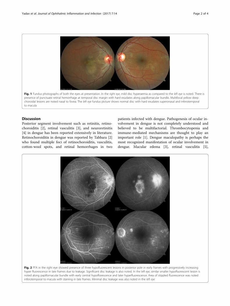
and 6/6 in the left eye. Anterior segment examination re-vealed quiet anterior chamber and anterior vitreous. Fun-dus examination of the right eye showed yellow deepchoroidal lesions at macula, multiple small, hard exudatesalong papillomacular bundle, and hyperemic disc. Therewas a small retinal hemorrhage at the temporal margin ofdisc. The left eye also had hard exudates superonasal andinferotemporal to macula (Fig. 1). Periphery of the fundusin both eyes was normal. There was no relative afferentpupillary defect (RAPD), and her color vision was withinnormal limits. Fundus fluorescein angiography (FFA)demonstrated these lesions to be early hypofluorescentwith late hyperfluorescent staining of the borders. FFAalso displayed leakage of the dye from right optic disc andstaining of both the discs (Fig. 2). Optical computerizedtomography (OCT) revealed disruption and thickening ofthe inner segment/outer segment (IS/OS) junction withsubretinal fluid and intraretinal edema (Fig. 3). She was in-vestigated extensively, and all investigations were non-contributory to the cause of ocular involvement includingtuberculin skin test, interferon gamma release assay, ser-ology for syphilis, and high-resolution computed chesttomography (HRCT). She was started on oral prednisol-one 60 mg/day after obtaining necessary clearance fromphysician. She felt subjective improvement in vision after2 weeks, however, complained of persistence of scotomata,which was more prominent while reading. Fundus exam-ination of her both eyes showed resolved choroiditis(Fig. 4). Her BCVA in both eyes improved to 6/6.
* Correspondence: [email protected] and Vision Research Foundations, Sankara Nethralaya, 18, CollegeRoad, Chennai, Tamil Nadu 600 006, India3Uveitis & Ocular Pathology Department, Sankara Nethralaya, 41, CollegeRoad, Nungambakkam, Chennai, Tamil Nadu 600 006, IndiaFull list of author information is available at the end of the article
Journal of OphthalmicInflammation and Infection
© The Author(s). 2017 Open Access This article is distributed under the terms of the Creative Commons Attribution 4.0International License (http://creativecommons.org/licenses/by/4.0/), which permits unrestricted use, distribution, andreproduction in any medium, provided you give appropriate credit to the original author(s) and the source, provide a link tothe Creative Commons license, and indicate if changes were made.
Yadav et al. Journal of Ophthalmic Inflammation and Infection (2017) 7:14 DOI 10.1186/s12348-017-0132-5
DiscussionPosterior segment involvement such as retinitis, retino-choroiditis [2], retinal vasculitis [3], and neuroretinitis[4] in dengue has been reported extensively in literature.Retinochoroiditis in dengue was reported by Tabbara [2]who found multiple foci of retinochoroiditis, vasculitis,cotton-wool spots, and retinal hemorrhages in two
patients infected with dengue. Pathogenesis of ocular in-volvement in dengue is not completely understood andbelieved to be multifactorial. Thrombocytopenia andimmune-mediated mechanisms are thought to play animportant role [1]. Dengue maculopathy is perhaps themost recognized manifestation of ocular involvement indengue. Macular edema [3], retinal vasculitis [5],
Fig. 1 Fundus photographs of both the eyes at presentation. In the right eye, mild disc hyperaemia as compared to the left eye is noted. There ispresence of punctuate retinal hemorrhage at temporal disc margin with hard exudates along papillomacular bundle. Multifocal yellow deepchoroidal lesions are noted nasal to fovea. The left eye fundus picture shows normal disc with hard exudates superonasal and inferotemporalto macula
Fig. 2 FFA in the right eye showed presence of three hypofluorescent lesions in posterior pole in early frames with progressively increasinghyper fluorescence in late frames due to leakage. Significant disc leakage is also noted. In the left eye, similar smaller hypofluorescent lesion isnoted along papillomacular bundle with early central hypoflorescence and later hyperfluorescence. Area of stippled fluorescence was notedinferotemporal to macula with staining in late frames. Minimal disc leakage was also noted in the left eye
Yadav et al. Journal of Ophthalmic Inflammation and Infection (2017) 7:14 Page 2 of 4

macular hemorrhage, and exudative retinal detachmenthave been frequently described in association with den-gue maculopathy. Foveolitis is characterized by adiscrete, well-defined yellow-orange subretinal lesions inthe fovea, which usually corresponds to disruption of theouter neurosensory retina and the inner segment/outersegment (IS/OS) junction. Hallmark feature described inthese patients was the presence of central scotomata [3,6], which was reported in absence of macular oedemaalso. Optic nerve involvement in dengue is not uncom-mon. In a study of 41 patients of dengue-associatedmaculopathy by Bacsal et al. [7], 11% of the patients haddisc oedema.
The key clinical feature in this case is the presence ofchoroidal inflammation. The choroiditis in our patientwas multifocal, and the patient was investigated to ruleout serpiginous-like choroiditis. Her laboratory investi-gations were non-contributory to the cause of inflamma-tion including tuberculin skin test, interferon gammarelease assay and high-resolution computed tomography(HRCT) chest. Presence of hard exudates along papillo-macular bundle and OCT findings suggest involvementof overlying retina with subretinal fluid and retinaledema. Unfortunately, we could not perform indocya-nine angiography due to systemic condition of the pa-tient. There was no relative afferent pupillary defect, and
Fig. 4 Shown fundus image 2 weeks after presentation. The right eye disc hyperaemia appears to have resolved. Presence of hard exudates is asbefore. The subretinal lesion now appears well defined with marginal reduction in size with pigmentation. The left eye disc and macula appearnormal. Previously noted lesions now appear as patches of chorioretinal atrophy
Fig. 3 Pre and post-treatment OCT picture of the right eye. Pre-treatment OCT showing disruption and thickening of IS-OS junction with subretinalfluid and intraretinal edema
Yadav et al. Journal of Ophthalmic Inflammation and Infection (2017) 7:14 Page 3 of 4

color vision of the patient was normal. However, depos-ition of hard exudates in the form of a partial, nasalmacular star configuration in the right eye may also bedue to optic nerve involvement. The retinal hemorrhagenoticed at the temporal margin of the optic disc in righteye can be due to the endothelial cell damage by thedengue virus.Nevertheless, we believe that our case represents a
rare clinical manifestation in wide-spectrum of dengueeye disease. Tabbara [2] reported two patients who de-veloped multiple foci of retinochoroiditis, vasculitis, cot-ton wool spots, and retinal hemorrhages followingdengue fever that resolved spontaneously. Our patienthad predominantly choroiditis, and we did not observeany cotton wool spot or vasculitis. One of the patientsreported by Tabbara [2], developed macular scarringresulting in poor vision in that eye. Our patientresponded well to oral steroid, though she complainedof persistence of scotoma even after resolution of her le-sions. Exact mechanism of choroiditis in our patient isnot clear. Direct cell damage by the dengue virus leadingto the apoptosis and dysfunction of the affected cells hasbeen described in literature [8]. Also immune-mediatedinjury may have played an important role in denguefever associated choroiditis, similar to foveolitis [6] anddengue-associated maculopathy [7]. Our case highlightsthe importance of oral steroid in patients with maculathreatening lesion in dengue-associated eye disease. Inconclusion, our case presents a rare clinical variant ofocular involvement in dengue fever.
AcknowledgementsNil.
Authors’ contributionsHMY, PDM, and JB contributed to patient management, literature review,and preparation of the manuscript. PDM and JB provided the concept anddesign, intellectual content, and critical review of the manuscript. All authorsread and approved the final manuscript.
Competing interestsThe authors declare that they have no competing interests.
Consent for publicationAn informed consent was taken from patient.
Publisher’s NoteSpringer Nature remains neutral with regard to jurisdictional claims inpublished maps and institutional affiliations.
Author details1Department of Ophthalmology, Kerala Institute of Medical Sciences, AnayaraP.O, Trivandrum, Kerala 695029, India. 2Medical and Vision ResearchFoundations, Sankara Nethralaya, 18, College Road, Chennai, Tamil Nadu 600006, India. 3Uveitis & Ocular Pathology Department, Sankara Nethralaya, 41,College Road, Nungambakkam, Chennai, Tamil Nadu 600 006, India.
Received: 24 January 2017 Accepted: 16 May 2017
References1. Ng AW, Teoh SC (2015) Dengue eye disease. Surv Ophthalmol 60:106–1142. Tabbara K (2012) Dengue retinochoroiditis. Ann Saudi Med 32:530–5333. Teoh SC et al (2010) Optical coherence tomography patterns as predictors
of visual outcome in dengue-related maculopathy. Retina 30:390–3984. de Amorim Garcia CA, Gomes AHB, de Oliveira AGF (2006) Bilateral stellar
neuroretinitis in a patient with dengue fever. Eye (Lond) 20:1382–13835. Chan DPL et al (2006) Ophthalmic complications of dengue. Emerg Infect
Dis 12:285–2896. Loh BK, Bacsal K, Chee SP, Cheng BCL, Wong D (2008) Foveolitis associated
with dengue fever: a case series. Ophthalmologica 222:317–3207. Bacsal KE, Chee SP, Cheng CL, Flores JVP (2007) Dengue-associated
maculopathy. Arch Ophthalmol 1960(125):501–5108. Lei HY et al (2001) Immunopathogenesis of dengue virus infection. J
Biomed Sci 8:377–388
Yadav et al. Journal of Ophthalmic Inflammation and Infection (2017) 7:14 Page 4 of 4