Embed Size (px)
Citation preview
CLINICAL MICROBIOLOGY REVIEWS, Oct. 2009, p. 564–581 Vol. 22, No. 40893-8512/09/$08.00�0 doi:10.1128/CMR.00035-09Copyright © 2009, American Society for Microbiology. All Rights Reserved.
Dengue Virus Pathogenesis: an Integrated ViewByron E. E. Martina,* Penelope Koraka, and Albert D. M. E. OsterhausErasmus MC, Department of Virology, P.O. Box 2040, 3000 CA Rotterdam, The Netherlands
INTRODUCTION .......................................................................................................................................................564THE PATHOGENESIS OF DENV INFECTIONS: CURRENT HYPOTHESES...............................................565
DENV Tropism........................................................................................................................................................565Cells of the immune system ..............................................................................................................................565Organ pathology..................................................................................................................................................565EC .........................................................................................................................................................................566
Virus Virulence........................................................................................................................................................566Activation of the Complement System.................................................................................................................567Transient Autoimmunity........................................................................................................................................567Host Genetic Factors..............................................................................................................................................567Antibody-Dependent Enhancement ......................................................................................................................568Cross-Reactive T-Cell Response ...........................................................................................................................569Soluble Factors........................................................................................................................................................569
THE INTERGRATED VIEW.....................................................................................................................................571DISCUSSION ..............................................................................................................................................................573ACKNOWLEDGMENTS ...........................................................................................................................................574REFERENCES ............................................................................................................................................................574
INTRODUCTION
Dengue virus (DENV) belongs to the family Flaviviridae,genus Flavivirus, and is transmitted to humans by Aedes mos-quitoes, mainly Aedes aegypti. Based on neutralization assaydata, four serotypes (DENV-1, DENV-2, DENV-3, andDENV-4) can be distinguished. DENV infection is a majorcause of disease in tropical and subtropical areas, with anestimated 50 million infections occurring each year and morethan 2.5 billion people being at risk of infection (75). Infectionwith any of the DENV serotypes may be asymptomatic in themajority of cases or may result in a wide spectrum of clinicalsymptoms (87), ranging from a mild flu-like syndrome (knownas dengue fever [DF]) to the most severe forms of the disease,which are characterized by coagulopathy, increased vascularfragility, and permeability (dengue hemorrhagic fever [DHF]).The latter may progress to hypovolemic shock (dengue shocksyndrome [DSS]). In Asia the risk of developing severe diseaseis greater in DENV-infected children (�15 years) than inadults (30, 80, 109, 172). In contrast, in the Americas mainlythe adult population is affected, resulting in mild disease (84,186, 188), although an increasing trend of cases progressingtoward DHF/DSS has also been observed in adults there (75,79, 87, 126, 186). DF is manifested as an incapacitating diseasein older children, adolescents, and adults. It is characterized bythe rapid onset of fever in combination with severe headache,retro-orbital pain, myalgia, arthralgia, gastrointestinal discom-fort, and usually rash. Minor hemorrhagic manifestations mayoccur in the form of petechiae, epistaxis, and gingival bleeding.Leukopenia is a common finding, whereas thrombocytopenia
may occasionally be observed in DF, especially in those withhemorrhagic signs (76, 109). The World Health Organization(WHO) classifies DHF in four grades (I to IV). DHF grades Iand II represent relatively mild cases without shock, whereasgrade III and IV cases are more severe and accompanied byshock. DHF is characterized by all the symptoms of DF, incombination with hemorrhagic manifestations (positive tour-niquet test or spontaneous bleeding), thrombocytopenia, andevidence of increased vascular permeability (increased hemocon-centration or fluid effusion in chest or abdominal cavities). Thelife-threatening DSS stage occurs at the time of or shortly afterdefervescence, which is characterized by a rapid, weak pulse(�20 mm Hg) or hypotension with cold, clammy skin in theearly stage of shock (grade III). If patients do not receiveprompt and appropriate treatment, a stage of profound shockmay set in, in which pulse and blood pressure become unde-tectable (grade IV), resulting in death within 12 to 36 h afteronset of shock (262a). It is important to realize that the WHOcase definition was originally proposed as a tool for clinicaldiagnosis using the results of repeated clinical tests. The WHOclassification system poses a problem for everyday clinicalpractice, because it may be not sufficiently accurate in correctlyclassifying disease severity and may lack good agreement withclinical practice (213). Consequently, the WHO classificationsystem is currently being reconsidered, and a new classificationsystem is to be expected soon. The stage of prolonged shockmay trigger or accelerate the development of disseminatedintravascular coagulation (DIC) (228). Data that support orrefute the occurrence of DIC in severe dengue are inconclu-sive, so better studies using prospective cohorts are needed toshow the frequency of DIC in DHF/DSS patients and its as-sociation with clinical outcome (30, 152, 262). Massive loss ofblood is rare in DHF and DSS and if present it is largelyrestricted to the gastrointestinal tract. This is usually due toprolonged shock resulting in blood being shunted away from
* Corresponding author. Mailing address: Erasmus Medical Center,Institute of Virology, P.O. Box 2040, 3000 CA Rotterdam, The Neth-erlands. Phone: 3110 704 4279. Fax: 3110 704 4760. E-mail: [email protected].
564
on February 17, 2019 by guest
http://cmr.asm
.org/D
ownloaded from

the gastrointestinal tract, leading to anoxia, cell death, andgastrointestinal bleeding. In contrast, the mild forms of hem-orrhages seen early in infection, such as petechiae, result fromdifferent mechanisms related to virus infection in combinationwith the release of vasculogenic cytokines. Understanding themechanism underlying the development of shock is crucial forthe development of novel strategies to improve patient man-agement. It is worth noting that patients classified as havingDHF and DSS have no generalized edema; rather, a selectiveplasma leakage tends to occur in the pleural and abdominalcavities (12, 230, 246, 252, 256), which is detectable by meansof radiology or sonography. Ultrasonographic examinationshave revealed that plasma leakage occurs before defervescenceor changes in hemoconcentration become apparent (12, 230,252). Attempts to explain the pathogenesis of dengue in all itscomplexity must consider all the clinical, immunological,pathological, and epidemiological features of DENV infection.The aim of this review is to outline the current views of DHF/DSS pathogenesis and to identify the gaps in our knowledgethat represent critical challenges for the future.
THE PATHOGENESIS OF DENV INFECTIONS:CURRENT HYPOTHESES
DENV Tropism
Cell and tissue tropism of DENV may have a major impacton the outcome of DENV infections. The absence of an ap-propriate animal disease model largely hampers our under-standing of the role played by DENV tropism. In vitro data andautopsy studies suggest that three organ systems play an im-portant role in the pathogenesis of DHF/DSS: the immunesystem, the liver, and endothelial cell (EC) linings of bloodvessels. The tropism of DENV for cells of the respective sys-tems, the corresponding pathological effects of DENV infec-tion of these systems, and the relevance of these events for theoverall pathogenesis of DENV infection will be described.
Cells of the immune system. During the feeding of mosqui-toes on humans, DENV is presumably injected into the blood-stream, with spillover in the epidermis and dermis, resulting ininfection of immature Langerhans cells (epidermal dendriticcells [DC]) (136, 263), and keratinocytes (136). Infected cellsthen migrate from site of infection to lymph nodes, wheremonocytes and macrophages are recruited, which become tar-gets of infection. Consequently, infection is amplified and virusis disseminated through the lymphatic system. As a result ofthis primary viremia, several cells of the mononuclear lineage,including blood-derived monocytes (59), myeloid DC (20, 91,92, 123, 133), and splenic and liver macrophages (18, 54d, 96,101, 117) are infected. DENV has also been shown to havetropism for circulating mononuclear cells in blood and for cellsresiding in the spleen, lymph nodes, and bone marrow of in-fected AG129 mice (124). Leukocytes also have been shown tobe infected with DENV in experimentally infected nonhumansprimates (156). It should be noted that during secondary in-fections with heterologous DENV, high concentrations ofDENV-specific immunoglobulin G (IgG) will complex newlyproduced virus that adheres to and is taken up by mononuclearcells. Following infection, mononuclear cells predominantlydie by apoptosis (61, 182), while abortively infected or by-
stander DC are stimulated to produce the bulk of mediatorsthat are involved in inflammatory (22, 47, 91, 133, 145) andhemostatic (48, 60, 97, 120, 236) responses of the host. In thisregard, factors that influence the amount of target cells in-fected, and consequently the levels of viremia, may determinethe ratio of different proinflammatory and anti-inflammatorycytokines, chemokines, and other mediators, as well as the wayin which the inflammatory response affects the hemostatic sys-tem (35, 59). Bone marrow stromal cells have also been shownto be susceptible to infection with DENV (124, 171, 202).
Organ pathology. Although thousands of patients with con-firmed dengue have been recognized in Southeast Asia and theAmericas in the past 60 years, autopsies have been performedon only a small number of these patients, and whether thosecases are representative in reflecting the viral tropism in theacute phase of infection is unclear. Histopathological researchis difficult to perform because fatal cases of DHF/DSS are rareand occur mainly in remote parts of the world where appro-priate laboratory technology is largely lacking and thus fresh orfrozen patient materials are rare. In addition, due to culturaland religious practices, autopsy is not conducted on the ma-jority of fatal cases, and usually families opt for rapid burial orcremation. The interpretation of the pathological findings infatal cases of DHF/DSS in relation to viral tropism describedin the literature is complicated by a skewed age distribution,different times of sample collection, and the range of differenttechniques used to confirm the presence of virus in affectedtissues. DENV cell tropism can be inferred from studies thathad used in situ hybridization, immunohistochemistry, or acombination of PCR and virus isolation techniques. A reviewof the literature describing findings on autopsy samples from atotal of 160 fatal cases, mostly children or young adolescents (4to 18 years old) who died within 36 h of developing shock,revealed, in order of frequency, the presence of DENV in cellsin the skin (104), liver (13, 14, 53, 54d, 69, 96, 101, 104, 137,164, 173, 199, 208), spleen (13, 14, 101, 164, 199, 208), lymphnode (13, 14, 101, 104, 173, 199, 208), kidney (14, 78, 101),bone marrow (13, 78, 101, 173), lung (13, 78, 101, 137, 164,173), thymus (106), and brain (164). The presence of infectiousvirus in these samples was not always investigated, but in gen-eral virus could be isolated only from liver and peripheralblood mononuclear cells. The failure to isolate virus from mostorgan samples may indicate that those tissues contained pri-marily degraded virus or virus complexed with antibodies thatprevent infection of cells in vitro. In general, the presence ofDENV in several organs was not associated with gross ormicroscopic evidence of severe organ pathology (17), which isin agreement with the pathogenesis of DHF/DSS. Similar or-gan tropism has been observed in the primate model, with highconcentrations of virus isolated from the skin and gastrointes-tinal tract whereas low concentrations of virus were recoveredfrom the spleen, thymus, and several peripheral lymph nodes(157). DENV has been recovered from the spleen, liver, pe-ripheral lymph nodes, and central nervous system in alpha/betainterferon (IFN-�/�)-deficient mice (13, 267). One notabledifference between humans and the mouse model is the tro-pism of DENV for neuronal cells.
It is interesting to note that a generally used argument in theliterature is that when shock sets in, virus is no longer detect-able in blood and therefore the host response should play a key
VOL. 22, 2009 DENGUE VIRUS PATHOGENESIS 565
on February 17, 2019 by guest
http://cmr.asm
.org/D
ownloaded from
role in pathogenesis (134, 166). Most autopsy data did notspecifically compare the presence of viral antigens or nucleicacid in blood and autopsy samples, but the limited evidencesuggests that DENV replication may occur in some organs,while viremia is no longer detectable (199). In agreement withthese findings it was shown in the rhesus macaque model thatDENV could be recovered from some autopsy samples but notfrom blood.
The liver is commonly involved in DENV infections in hu-mans and mouse models (181, 212), with some reports suggest-ing an association between elevated liver enzyme levels andspontaneous bleeding tendencies (54e, 124, 261). Cases of den-gue-associated hepatitis have been described, which were char-acterized by moderate midzonal hepatocyte necrosis, microve-sicular steatosis, and councilman bodies (63, 78, 124, 181, 254).Although DENV was found in a significant proportion of hu-man hepatocytes and Kupffer cells, little inflammation wasseen within the liver, indicating that much of the observedapoptosis and necrosis was virally induced. The higher preva-lence of apoptosis over necrosis could explain the limited in-flammation seen in the liver, a picture similar to what is ob-served in the early phase of yellow fever or Rift Valley fever(57, 190, 191). It has been proposed that the severe hepaticdamage seen, for instance, in yellow fever, Rift Valley fever,and late Ebola virus infections results in decreased liver func-tion, which could account for the decreased synthesis of coag-ulation factors and development of coagulopathy (43, 269).Although severe hepatic damage is not common in DENVinfections, elevated liver enzymes suggest that the liver is af-fected, but the role of hepatic damage in coagulopathy anddisease severity remains to be established.
EC. EC play an important role in the coagulation responseupon severe systemic inflammation. The integrity of the ECbed is physiologically regulated by many factors. The tropismof DENV for EC in vivo remains controversial. Early studies ofskin biopsy specimens indicated that the microvasculature lo-cated in the dermal papillae is the main site affected, althoughDENV antigen was not detected in EC but was detected incells surrounding the microvasculature (21, 203). In contrast,there is evidence for the presence of DENV antigen in thepulmonary vascular endothelium (101). It is important to re-alize, however, that the mere presence of viral RNA or antigenin EC is no proof for viral replication. In contrast to mononu-clear cells, EC do not carry Fc receptors and thus will not takeup immune-complexed virus. Therefore, the presence of viralRNA in these cells would more likely be explained by a mech-anism of pinocytosis (101). In vitro studies have shown that allDENV serotypes can actively replicate in EC (7, 19, 95), andinfection results in functional rather than morphological dam-age. It is not clear whether EC of different vascular-bed sys-tems have different susceptibilities to DENV infection. In thisregard, it has been proposed that the coagulation responsesupon severe systemic inflammation by EC in different parts ofthe vascular-bed system are not the same (200, 201). Similarly,DENV infection patterns in microvascular cells in vitro suggestthat EC from different tissues have different activation patterns(184). Although increased peripheral microvascular perme-ability has been shown to occur in both DHF and DSS patients(16), it is conceivable that EC from the pulmonary and abdom-inal territories react in a specific way to either infection with or
the response to DENV infection (28, 39), resulting in theselective vascular leakage syndrome characteristic of DHF/DSS. Several studies suggest that vascular damage or dysfunc-tion is central in the pathogenesis of DHF/DSS (26, 29, 39, 115,168). It is interesting to note that selective apoptosis of themicrovascular EC in pulmonary and intestinal tissues has beendetected in fatal cases of DHF/DSS (137), providing a possibleexplanation for the profound plasma leakage seen in pleuraland peritoneal cavities. In this regard, it is worth mentioningthat the major nonstructural protein 1 (NS1) of DENV hasbeen shown to bind preferentially to EC of lung and livertissues (9). It has been hypothesized that recognition of NS1 byanti-NS1 antibodies could then contribute to the selective pul-monary vascular leakage.
Virus Virulence
According to the virus virulence hypothesis, certain DENVstrains are responsible for more severe disease. DENV sero-types can be further classified into different genotypes on thebasis of nucleotide variations. Viral genetic differences havebeen associated with differences in virulence (51, 131, 206,248). Remarkably, the first outbreak of DHF in the Americasoccurred in 1981, which coincided with the introduction of thepossibly more virulent DENV-2 Southeast Asian genotype,while the less virulent indigenous DENV-2 genotype was al-ready circulating in the region (118, 195–197). It has also beenproposed that intraepidemic evolution of the circulatingDENV might be responsible for increased severity of disease.During the 1981 DENV-2 epidemic in Cuba, it was noted thatseverity of disease manifestations and case-fatality rates wereincreased toward the end of the epidemic (118, 119), suggest-ing that the circulating DENV-2 might have become morevirulent through passage in hosts during the epidemic. A sim-ilar situation was observed in the 1992 DENV epidemic inTownsville, Australia (234), and again in Cuba during the 1997epidemic (81). Analysis of DENV genomes has shown thatDENV indeed evolves during an epidemic (42, 198); however,more data are needed to establish an association betweenintraepidemic virus evolution and increased disease severity.Epidemiological observations in the Americas and in Singa-pore suggested that the sequence of infection with particularserotypes and the time interval between primary infection andsecondary infection may play an important role in the devel-opment of DHF. Epidemics with high incidences of DHF havebeen linked to primary infection with DENV-1 followed byinfection with DENV-2 or DENV-3 (79, 83, 178). Further-more, these studies indicated that the longer the interval be-tween primary and secondary infections, the higher the risk ofdeveloping severe disease. In addition, age has been shown toinfluence the outcome of disease following a secondary infec-tion with heterologous DENV (80). In Asia, the risk of severedisease is greater in children than in adults, in contrast to theAmericas, where the adult population is mainly affected andinfection results in milder disease. This difference in diseaseseverity caused by Asian and American genotypes correlatedwith structural differences in the two strains of DENV (51,131). It has also been shown that different geographical DENVstrains or different serotypes may vary in their ability to infectdifferent cell types or cause severe disease (56, 251). However,
566 MARTINA ET AL. CLIN. MICROBIOL. REV.
on February 17, 2019 by guest
http://cmr.asm
.org/D
ownloaded from
the observation that DHF/DSS is seen primarily in a relativesmall percentage of secondary DENV infections and to a muchlesser extent in primary infections even with allegedly virulentstrains suggests that host factors must be crucial determinantsof severe disease development. It is important to realize thatvirulence has traditionally been considered a microbial prop-erty, evaluated independently of the host or only in vitro or inoften inbred animals. However, an increasing body of evidenceincriminates the host immune response in the pathogenesis ofmany microbial infections (31). Therefore, in studying DENVvirulence, both host and viral factors should be considered.
Activation of the Complement System
The complement system is one of the main humoral com-ponents of the innate immunity and interacts closely with thehemostatic system to provide the first line of defense againstpathogens. These innate immune mechanisms provide the hostwith the time needed to maximally induce the more slowlydeveloping adaptive immunity. With regard to DENV, inves-tigators noticed that around the time of defervescence, whenplasma leakage may become apparent, high levels of the acti-vation products C3a and C5a are measured in the plasma,followed by an accelerated consumption and a marked reduc-tion of the complement components in patients with DSS (50,174, 214). Therefore, it was hypothesized that complementactivation plays an important role in the pathogenesis of den-gue. Comparison of global gene expression profiles in periph-eral blood mononuclear cells of DF and DHF/DSS patientsalso suggests the involvement of the complement system indisease severity (249). However, many aspects of complementactivation and its role in DENV pathogenesis remain to beinvestigated.
It has been proposed that NS1 is an important trigger forcomplement activation (122). Binding of heterotypic antibod-ies to NS1 expressed on infected cells may result in comple-ment activation (8, 142). In addition, it is believed that NS1released from infected cells can directly activate complementfactors present in the fluid phase (122). Production of theC5b-C9 complex could then trigger cellular reactions and stim-ulate the production of inflammatory cytokines that are asso-ciated with development of DHF/DSS (8). Alternatively, theC5b-C9 complex could independently trigger other local andsystemic effects (158), which may be implicated in intravascularcoagulation. It is important to realize that coagulation enzymescan also activate the complement system, illustrating the ex-tensive interaction that exists between the complement and thecoagulation system.
Several groups have shown that both IgG1 and IgG3 werethe predominant subclasses involved in the specific antibodyresponse in human DENV infections (116, 242). Both IgGsubclasses can fix and activate the complement system effec-tively, whereas IgG2 and IgG4 are less effective in this respect(90). Although IgG1, IgG2, and IgG4 are able to activate theclassical complement pathway, they require the unlikely eventof two IgG molecules binding close to the antigen in order topromote C1q binding (90). IgG3, on the other hand, has thecapacity to self-associate into multivalent complexes, therebyincreasing functional affinity and the likelihood of C1q binding.The presence of sialic acid in the glycans of IgG subclasses
could also affect their complement-fixing properties (100) andpossibly their infection-enhancing activity (65, 163, 266). It isimportant to understand what determines the threshold ofactivation needed and how activation participates in develop-ment of DHF/DSS.
Transient Autoimmunity
Antibodies produced during a DENV infection have beenshown to cross-react with some self-antigens, but it is not clearif production of these antibodies is associated with secondaryDENV infections. For instance, antibodies recognizing a linearepitope in the E protein have been shown to bind humanplasminogen and inhibit plasmin activity (49, 64, 94, 159). Thepresence of serum antibodies specific to NS1 also has beenshown to correlate with disease severity (134, 218). Cross-reaction of anti-NS1 with cells of the liver, EC, and platelets(33, 140, 177, 237) could be at the basis of this observation.Anti-NS1 antibodies cross-reactive with EC could trigger thesecells to express nitric oxide (NO) and undergo apoptosis (141).Although NO has been shown to inhibit DENV replication(239), its overproduction could also lead to cell damage (141,215). It is worth reiterating that morphological damage is nota common observation in lethal cases of DHF/DSS. Anti-NS1antibodies have also been shown to enhance expression ofinterleukin-6 (IL-6), IL-8, and intracellular adhesion molecule1 (ICAM-1) (138). Further studies are needed to see if cross-reactivity of anti-NS1 with EC could lead to the increasedpermeability that is characteristic of DSS. In addition, anti-NS1 antibodies were also shown to cross-react with human andmouse platelets and were able to cause transient thrombocy-topenia and hemorrhage in mice (142, 237), indicating thatsuch cross-reactive antiplatelet antibodies are pathogenic. Thisobservation may have implications for vaccine development,especially for live-attenuated vaccines. Therefore, it is impor-tant to understand why the autoimmune phenomenon ob-served in some DENV-infected patients does not persist. Sincethe kinetics of anti-NS1 antibodies is difficult to reconcile withthe short duration of the sudden hyperpermeabilty event thatleads to shock, the autoimmune hypothesis has remained con-troversial. Although it is likely that the cross-reactive antibod-ies to self-antigens are of the short-lived IgM isotype (139,204), vaccination strategies should not result in memory IgGresponses to such antigens. Clearly, more efforts should bedeployed in identifying the putative self-antigens that are rec-ognized by anti-DENV antibodies and in understanding theirrole, if any, in dengue pathogenesis.
Host Genetic Factors
Differences in disease severity can be seen at both the indi-vidual and population levels. Several epidemiological studiesindicated that genetic factors constitute important componentsin disease susceptibility. Several human HLA class I and IIalleles are associated with development of DHF (Table 1).Polymorphism in the tumor necrosis factor alpha (TNF-�),Fc� receptor, vitamin D receptor, CTLA-4, and transforminggrowth factor � (TGF-�) genes has been associated with de-velopment of DHF/DSS. Certain host factors, such as glucose-6-phosphate dehydrogenase (G6PD) deficiency, may also con-
VOL. 22, 2009 DENGUE VIRUS PATHOGENESIS 567
on February 17, 2019 by guest
http://cmr.asm
.org/D
ownloaded from
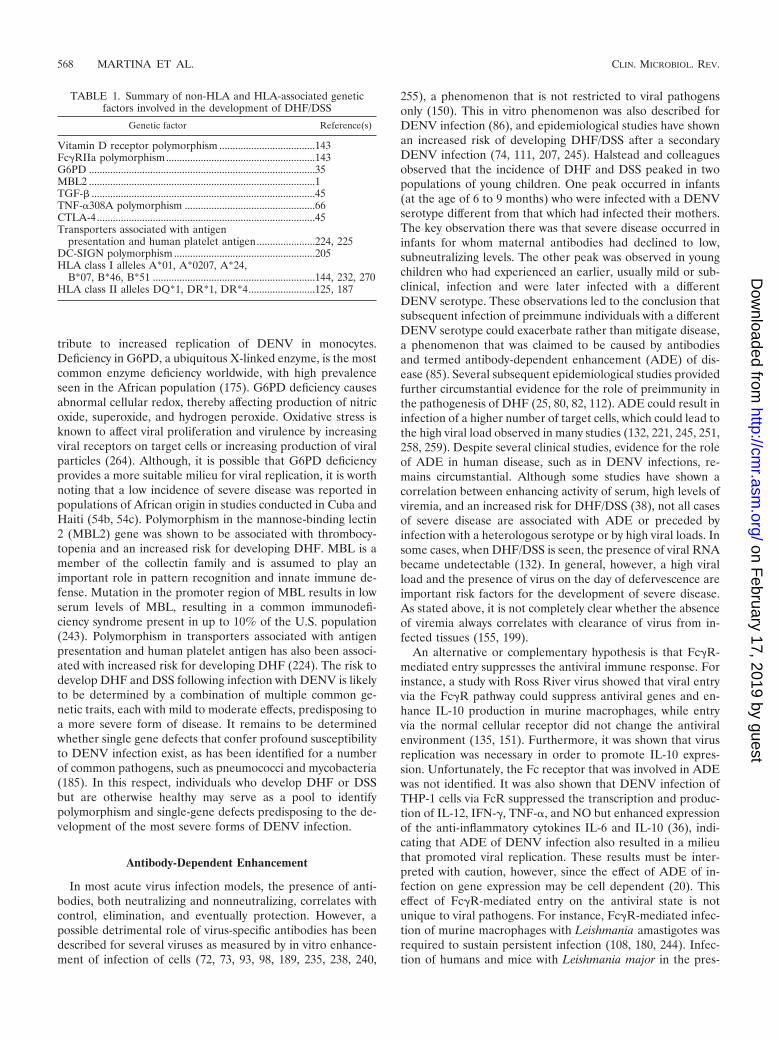
tribute to increased replication of DENV in monocytes.Deficiency in G6PD, a ubiquitous X-linked enzyme, is the mostcommon enzyme deficiency worldwide, with high prevalenceseen in the African population (175). G6PD deficiency causesabnormal cellular redox, thereby affecting production of nitricoxide, superoxide, and hydrogen peroxide. Oxidative stress isknown to affect viral proliferation and virulence by increasingviral receptors on target cells or increasing production of viralparticles (264). Although, it is possible that G6PD deficiencyprovides a more suitable milieu for viral replication, it is worthnoting that a low incidence of severe disease was reported inpopulations of African origin in studies conducted in Cuba andHaiti (54b, 54c). Polymorphism in the mannose-binding lectin2 (MBL2) gene was shown to be associated with thrombocy-topenia and an increased risk for developing DHF. MBL is amember of the collectin family and is assumed to play animportant role in pattern recognition and innate immune de-fense. Mutation in the promoter region of MBL results in lowserum levels of MBL, resulting in a common immunodefi-ciency syndrome present in up to 10% of the U.S. population(243). Polymorphism in transporters associated with antigenpresentation and human platelet antigen has also been associ-ated with increased risk for developing DHF (224). The risk todevelop DHF and DSS following infection with DENV is likelyto be determined by a combination of multiple common ge-netic traits, each with mild to moderate effects, predisposing toa more severe form of disease. It remains to be determinedwhether single gene defects that confer profound susceptibilityto DENV infection exist, as has been identified for a numberof common pathogens, such as pneumococci and mycobacteria(185). In this respect, individuals who develop DHF or DSSbut are otherwise healthy may serve as a pool to identifypolymorphism and single-gene defects predisposing to the de-velopment of the most severe forms of DENV infection.
Antibody-Dependent Enhancement
In most acute virus infection models, the presence of anti-bodies, both neutralizing and nonneutralizing, correlates withcontrol, elimination, and eventually protection. However, apossible detrimental role of virus-specific antibodies has beendescribed for several viruses as measured by in vitro enhance-ment of infection of cells (72, 73, 93, 98, 189, 235, 238, 240,
255), a phenomenon that is not restricted to viral pathogensonly (150). This in vitro phenomenon was also described forDENV infection (86), and epidemiological studies have shownan increased risk of developing DHF/DSS after a secondaryDENV infection (74, 111, 207, 245). Halstead and colleaguesobserved that the incidence of DHF and DSS peaked in twopopulations of young children. One peak occurred in infants(at the age of 6 to 9 months) who were infected with a DENVserotype different from that which had infected their mothers.The key observation there was that severe disease occurred ininfants for whom maternal antibodies had declined to low,subneutralizing levels. The other peak was observed in youngchildren who had experienced an earlier, usually mild or sub-clinical, infection and were later infected with a differentDENV serotype. These observations led to the conclusion thatsubsequent infection of preimmune individuals with a differentDENV serotype could exacerbate rather than mitigate disease,a phenomenon that was claimed to be caused by antibodiesand termed antibody-dependent enhancement (ADE) of dis-ease (85). Several subsequent epidemiological studies providedfurther circumstantial evidence for the role of preimmunity inthe pathogenesis of DHF (25, 80, 82, 112). ADE could result ininfection of a higher number of target cells, which could lead tothe high viral load observed in many studies (132, 221, 245, 251,258, 259). Despite several clinical studies, evidence for the roleof ADE in human disease, such as in DENV infections, re-mains circumstantial. Although some studies have shown acorrelation between enhancing activity of serum, high levels ofviremia, and an increased risk for DHF/DSS (38), not all casesof severe disease are associated with ADE or preceded byinfection with a heterologous serotype or by high viral loads. Insome cases, when DHF/DSS is seen, the presence of viral RNAbecame undetectable (132). In general, however, a high viralload and the presence of virus on the day of defervescence areimportant risk factors for the development of severe disease.As stated above, it is not completely clear whether the absenceof viremia always correlates with clearance of virus from in-fected tissues (155, 199).
An alternative or complementary hypothesis is that Fc�R-mediated entry suppresses the antiviral immune response. Forinstance, a study with Ross River virus showed that viral entryvia the Fc�R pathway could suppress antiviral genes and en-hance IL-10 production in murine macrophages, while entryvia the normal cellular receptor did not change the antiviralenvironment (135, 151). Furthermore, it was shown that virusreplication was necessary in order to promote IL-10 expres-sion. Unfortunately, the Fc receptor that was involved in ADEwas not identified. It was also shown that DENV infection ofTHP-1 cells via FcR suppressed the transcription and produc-tion of IL-12, IFN-�, TNF-�, and NO but enhanced expressionof the anti-inflammatory cytokines IL-6 and IL-10 (36), indi-cating that ADE of DENV infection also resulted in a milieuthat promoted viral replication. These results must be inter-preted with caution, however, since the effect of ADE of in-fection on gene expression may be cell dependent (20). Thiseffect of Fc�R-mediated entry on the antiviral state is notunique to viral pathogens. For instance, Fc�R-mediated infec-tion of murine macrophages with Leishmania amastigotes wasrequired to sustain persistent infection (108, 180, 244). Infec-tion of humans and mice with Leishmania major in the pres-
TABLE 1. Summary of non-HLA and HLA-associated geneticfactors involved in the development of DHF/DSS
Genetic factor Reference(s)
Vitamin D receptor polymorphism ....................................143Fc�RIIa polymorphism........................................................143G6PD .....................................................................................35MBL2 .....................................................................................1TGF-� ....................................................................................45TNF-�308A polymorphism .................................................66CTLA-4..................................................................................45Transporters associated with antigen
presentation and human platelet antigen......................224, 225DC-SIGN polymorphism .....................................................205HLA class I alleles A*01, A*0207, A*24,
B*07, B*46, B*51 .............................................................144, 232, 270HLA class II alleles DQ*1, DR*1, DR*4.........................125, 187
568 MARTINA ET AL. CLIN. MICROBIOL. REV.
on February 17, 2019 by guest
http://cmr.asm
.org/D
ownloaded from
ence of antibodies resulted in development of chronic infec-tion. In this regard, increased levels of IL-10 have beenassociated with visceral and cutaneous leishmaniases, andIL-10 has been flagged as having an important role in theregulation of the immune response to this parasite, therebylinking ADE to Th2 immune responses. This immunosuppres-sive effect of Fc�R-mediated infection is in agreement with thedecrease in the proliferative responses to mitogen and recallantigens that can be measured during acute DENV infection,which is associated with both quantitative and qualitative de-fects in the antigen-presenting cell (APC) population (148,161).
The role of preimmunity conferred by vaccination againstDENV or other flaviviruses in the pathogenesis of DENVinfections has been studied in limited numbers of subjects.Most clinical trials with candidate DENV vaccines were con-ducted in areas where the disease is not endemic and thechance of acquiring a natural infection is limited. One studyconducted in Thailand reported no differences in the incidenceof DHF in children who had received a live-attenuated DENVvaccine and unvaccinated controls at 6 to 8 years after vacci-nation (34). Experimental infection of monkeys with DENV-1or DENV-4 followed by a secondary infection with DENV-3 ayear later did not result in increased viremia or disease (113).The efficacy of DENV candidate vaccines was also not influ-enced by preimmunity to DENV or other flaviviruses (77, 114).The results from these experimental studies should be inter-preted with caution, however, since the interval between pri-mary and secondary infection may have been too short.
Cross-Reactive T-Cell Response
Although memory T cells cross-reactive with a heterologousvirus can provide partial protective immunity, they can alsocause substantial immunopathology (210). The role of CD8� Tcells during DENV infection is not entirely clear, but they mayplay a role in clearing infection as well as in immunopathogen-esis (3, 166). It is worth noting that a consistent finding in allexamples of T-cell-mediated pathology during acute or persis-tent viral infections is morphological tissue damage as a resultof cytolysis or inflammation induced by the high numbers ofeffector T cells. The efficiency of activated T cells in clearingvirus-infected cells is dependent on the avidity of the T-cellreceptor (TCR) for the HLA-peptide complex (222), and it isassumed that cross-reactive T cells of low avidity for heterol-ogous virus are not protective (110). Yet, there are only ahandful of virus-animal models where heterologous immunityhas been shown to cause pathology. These include the combi-nations of infections with lymphocytic choriomeningitis virus(LCMV) and vaccinia virus (VV), as well as influenza A virus(IAV) and murine cytomegalovirus (MCMV) (209). In onestudy, peripheral VV infection of LCMV-immune mice re-sulted in immune-mediated panniculitis (211), whereas respi-ratory VV challenge of LCMV-immune mice resulted in re-cruitment of LCMV-specific CD8� T cells into the lung,causing bronchiolitis obliterans (40). In the IAV-MCMVmodel it was shown that IAV-immune mice challenged withMCMV developed severe consolidating mononuclear pneu-monia as a result of increased viral replication in the lungs (41,209). One example of cross-reactivity leading to disease in
humans has also been described (250, 260). The authors ofthose studies reported two cases of fulminant hepatitis C virusinfection associated with an unusually high frequency of CD8�
T cells. These T cells were shown to recognize a single epitopewithin the hepatitis C virus NS3 that also cross-reacted with anepitope in the IAV neuraminidase protein. These findings sug-gest that cross-reactive memory T cells can modify the primaryimmune response and modulate the immunopathologic re-sponse to subsequent infection with other pathogens.
During the acute phase of a secondary infection of humanswith heterologous DENV, highly cross-reactive CD8� T cellswith high avidity for the infecting virus are preferentially acti-vated (58, 99). The majority of these cross-reactive T cellsproduce high concentrations of pro- and anti-inflammatorycytokines such as IFN-�, TNF-�, and IL-13 but somewhatlower levels of IL-10. These high-avidity cross-reactive CD8�
T cells die through apoptosis, but it is not clear whether cellsdie as a result of activation-induced cell death or whetherapoptosis is selectively induced by cross-reactive epitopes.Other studies have also suggested that epitopes can regulatethe level of proinflammatory cytokines produced by T cells(146, 147). Alternatively, low-avidity cross-reactive CD8� Tcells would be preferentially expanded (165, 166). These cross-reactive T cells react differently to the heterologous epitopesthan to homologous epitopes by producing high levels of proin-flammatory cytokines, but they lose their cytolytic activity. De-layed virus clearance would prolong activation of such cross-reactive CD8� T cells, which then results in the production ofhigh levels of cytokines such as TNF-�, IL-6, or other solublefactors that affect vascular permeability. The phenomenonwhere cross-reactive memory T cells for the primary infectingvirus are more efficiently activated, due to the increased fre-quency and higher activation state of memory cells, has beencalled original antigenic sin (OAS). This phenomenon has alsobeen described for LCMV in mice (110). During a secondaryinfection with a heterologous serotype, cross-reactive epitopespreferentially reactivate the larger number of memory T cellsagainst the priming virus more effectively than they activatenaïve T cells. However, in accordance to what has been de-scribed for several other systems, it is possible that during aheterologous DENV infection, only a very small subset ofcross-reactive memory T cells will be stimulated to expandbecause of a narrowing TCR repertoire. This narrowing of theTCR repertoire in combination with the fact that each indi-vidual has a unique TCR specificity (private TCR [52, 107])would result in dominant responses that are unique for indi-viduals. This could explain the variability seen in disease out-come upon secondary infection with heterologous DENV.Much less is known about the CD4� T-cell response duringDENV infection. However, evidence exists that sequential in-fection with different DENV serotypes may also alter the cy-tokine response of cross-reactive CD4� T cells, resulting inproduction of proinflammatory cytokines (154) that may con-tribute, together with the CD8� T-cell response, to a detri-mental cytokine release.
Soluble Factors
It is strongly believed by many scientists studying denguepathogenesis that a high viral load and activation of high num-
VOL. 22, 2009 DENGUE VIRUS PATHOGENESIS 569
on February 17, 2019 by guest
http://cmr.asm
.org/D
ownloaded from
bers of nonprotective T cells result in a “storm” of inflamma-tory cytokines and other mediators, leading to the increasedplasma leakage characteristic of DHF/DSS. One of the mostdaunting challenges in DENV research is the identification ofsoluble factors that can mediate, either alone or in combina-tion, the functional changes induced in EC that are associatedwith the increased plasma leakage. Several studies have shownthat concentrations of multiple cytokines and other mediators,as well as soluble receptors, are significantly increased duringsevere dengue infections (reviewed in reference 15). Higherplasma levels of IL-1�, IL-2, IL-4, IL-6, IL-7, IL-8, IL-10,IL-13, IL-18, TGF-1�, TNF-�, and IFN-� have been found inpatients with severe DENV infections, in particular in patientswith DSS (10, 23, 32, 103, 128, 169, 172, 183, 192, 194, 236).These studies analyzed samples from infants, children, andadults infected with different DENV serotypes. It is reasonableto assume that synergistic interactions between these cytokineswill occur. Other mediators and soluble factors found to beincreased in severe disease include vascular endothelial growthfactor (VEGF), granulocyte-macrophage colony-stimulatingfactor, monocyte chemoattractant protein 1 (MCP-1), macro-phage migration inhibitory factor, thrombopoietin, soluble vas-cular cell adhesion molecule 1 (VCAM-1), soluble ICAM-1,von Willebrand factor antigen, thrombomodulin, E-selectin,tissue factor (TF), plasminogen activator inhibitor 1 (PAI-1),and tissue plasminogen activator (23, 26, 27, 29, 44, 46, 115,130, 153, 223, 229). Analyses of several studies reveal conflict-ing results, as some studies report increased levels of plasmacytokines while others do not. These discrepancies are attrib-uted mainly to the study design and experimental setup, thelaboratory tests used to measure cytokines, and the statisticaltests applied for the analyses of the results. Furthermore, thetime of sampling during infection is difficult to standardize, andthis may explain some of the discrepant results. The observa-tion that plasma leakage occurs mainly in the pulmonary andperitoneal cavities justifies the question whether levels of cy-tokines and mediators in plasma indeed reflect concentrationsin different compartments. Infection of humans and animalswith seasonal IAV (H1N1 or H3N2) indicated that levels ofcytokines and chemokines were much higher locally, at the siteof infection, than in plasma or serum (68, 71, 88). In contrast,H5N1 causes fulminant disease in humans, characterized bydiffuse alveolar damage and progression to multiorgan dys-function. In this case disease severity correlates strongly withcytokine levels, both locally and systemically (54a, 129). Forinstance, hemophagocytic syndrome has been proposed as acause of the multiorgan dysfunction observed in patients withconfirmed H5N1 fatal infections (247, 268). Notably, hemo-phagocytic syndrome in fulminant virus infections or autoim-mune diseases as well as macrophage activation syndrome inhematopoietic cell transplantation has been associated withexcessive cytokine production (127, 193, 227). The productionand actions of cytokines are thus critically dependent on thecontext in which they occur. More specifically designed studiesare needed to dissect the relationships between levels of sev-eral cytokines in the pulmonary (pleural fluid) and peritoneal(ascites) regions compared to plasma or serum in severeDENV infections. It is conceivable that differences in viralreplication and damage to selective EC in vivo may account for
a differential cytokine profile, resulting in different vascularpermeability patterns.
Some evidence to support a role for cytokines comes fromanimal models of increased vascular permeability and hemor-rhage during DENV infections (39, 217). TNF-�, IL-1�, IL-6,and IL-10 levels have been shown to be high in sera of DENV-infected mice (6). In addition, several in vitro experiments havedemonstrated high levels of cytokines in culture supernatantsof DENV-infected (primary) DC (20), monocytic cells (47,162), and EC (11). In the presence of anti-NS1 antibodies, ECproduce MCP-1, IL-6, and IL-8 in vitro (138). T cells interact-ing with DENV-infected cells may also produce TNF-�, IFN-�,IL-4, and/or IL-10 (91).
The biological roles of some cytokines and soluble factorsand the implication in the development of hemorrhage havebeen inferred and are summarized in Table 2. The cytokinesTNF-� and IL-10 are of particular interest. For instance, inone study a positive correlation between soluble TNF-� con-centrations and thrombocytopenia was found (23). Further-more, the TNF-�308A allele, which leads to overproduction ofthe cytokine, is more commonly found in DHF patients (66).These observations, together with experiments showing thatTNF-� is capable of increasing EC permeability in vitro (55),suggest its possible role in pathogenesis of DHF. In a mousemodel of DENV-induced hemorrhage, high levels of TNF-� intissues correlated with EC apoptosis and hemorrhage (39). It isworth noting that in most models of immunopathology, pathol-ogy is mediated via direct lysis of infected cells by TNF-�, andit has been shown that CD8� T-cell cytotoxicity can be medi-ated exclusively by TNF-�. Plasma levels of IL-10 were shownto correlate with platelet decay in DENV-infected patients (10,132) and may modulate the activation of coagulation. IL-6 is amajor mediator of fever and acute-phase reactions and is pro-duced by macrophages and activated EC. Furthermore, IL-6and IL-8 mediate derangement of coagulation and fibrinolysis,whereas TNF-� and VEGF act synergistically to induce ex-pression of TF on EC (216). TNF-� has a direct effect onproduction of IL-6 and thus an indirect effect on coagulationand fibrinolysis. IL-2 plays a central role in the regulation ofthe immune response, as it induces potent proliferation of Tcells and to a lesser extent of B cells, stimulates synthesis ofIFN-� and TNF-�, and may damage the integrity of EC. IL-8has an effect on the expression of adhesion molecules such asICAM-1 and VCAM-1. MCP-1 causes EC tight-junction open-ings in vitro (231) and elevates endothelial permeabilitychanges in vivo (265). IL-13 is a pleiotropic type 2 cytokine,and its receptor is expressed on vascular EC. IL-13 downregu-lates expression of proinflammatory cytokines IL-1, IL-6, IL-8,and IL-12. Furthermore, IL-13 is a potent stimulator of matrixmetalloproteinase 9 (MMP-9) and cathepsins and plays impor-tant roles in the pathogenesis of emphysema in animal modelsof respiratory infections. Interestingly, DENV-infected cellshave been shown to produce MMP-9, which increases vascularpermeability in vitro (145). IL-18 is a type 1 cytokine, producedmainly by monocytes, macrophages, and DC, and has a strongproinflammatory activity. In vitro, IL-18 upregulates expres-sion of adhesion molecules (E- and P-selectins) on EC, whichmay contribute to a procoagulant state (167). High levels ofIL-18 may be associated with neutropenia, thrombocytopenia,and elevated levels of liver enzymes. During DENV infection,
570 MARTINA ET AL. CLIN. MICROBIOL. REV.
on February 17, 2019 by guest
http://cmr.asm
.org/D
ownloaded from

CD4� T cells have been shown to produce a unique cytokinecalled the cytotoxic factor, with peak amounts measured inDHF/DSS cases (2, 37). The validity of these observations hasto be confirmed by independent experiments.
Clearly, there is a substantial redundancy between cytokines(i.e., the lack of one specific cytokine may be compensated forby another cytokine with overlapping activities), making it dif-ficult to explain DHF/DSS pathogenesis on the basis of a singlecytokine. It is more likely that multiple cytokines contributesimultaneously in a complex way to the development of DHF/DSS. Apart from any other considerations, it is reasonable toassume that cytokines and other soluble mediators of the func-tional, and to a lesser degree the morphological, pathologycharacteristic of DHF/DSS are also essential for efficient viral
clearance. The fact that DSS patients recover extremely rapidlyafter appropriate fluid therapy suggests that cytokines do notcause tissue destruction like in many immunopathology modelsbut rather cause a reversible EC dysfunction.
THE INTERGRATED VIEW
The mechanisms leading to the severe manifestations ofDENV infections are still not completely understood but arelikely to be multifactorial (Fig. 1). The genetic background ofthe host influences the way that the immune response reacts toDENV infection. Upon inoculation of DENV into the dermis,Langerhans cells and keratinocytes will primarily be infected.The virus subsequently spreads via the blood (primary viremia)
TABLE 2. Summary of soluble factors that are or are likely to be associated with development of DHF/DSS
Soluble factor Biological function in relation to pathogenesis
Thrombin ..............................................Thrombin is thought to act near the site at which it is produced. Thrombin converts circulating fibrinogento fibrin and triggers platelet activation, which results in platelet aggregation. Thrombin activates ECand increases EC permeability, leading to plasma leakage and edema formation. Thrombin ischemotactic for monocytes and is mitogenic for lymphocytes and mesenchymal cells. Activated plateletsrelease several soluble factors with inflammatory, antimicrobial, and immune modulating activity, suchas MMP-9, which enhances EC permeability. Activated platelets also secrete soluble CD40 ligand,which can induce EC to produce reactive oxygen species, adhesion molecules, chemokines, and TF.Thrombin also inhibits IL-12 production by mononuclear cells.
C3a and C5a .........................................C3a activates platelets and enhances their activation and adhesion properties. C5a enhances bloodthrombogenicity by upregulating TF and PAI-1 expression on various cell types. C5a stimulatesmonocytes to produce IL-1, IL-6, IL-8, and TNF-�. Activation of these complement factors isenhanced by thrombin, which cleaves C3 and C5 to C3a/b and C5a/b, respectively. Activated plateletsare also involved in C3 cleavage, which induces activation of the classical complement pathway.
C4b.........................................................C4b binds to protein S and thereby inhibit the anticoagulant properties of activated protein C-protein Scomplexes.
IL-1 ........................................................IL-1� is major mediator of platelet-induced activation of EC, causing enhanced chemokine release andupregulation of VCAM-1. VCAM-1 promotes adhesion of monocytes to the endothelium. IL-1increases the expression of TF on EC and suppresses the cell surface anticoagulant activity of EC.Depending on its concentration, it may upregulate TNF-� production or downregulate TNF-receptors.IL-1 stimulates the hypothalamus and, as a consequence, the pituitary gland to produce anti-inflammatory mediators such as endorphins, melanocyte-stimulating hormone, and adrenocorticotropichormone.
IL-6 ........................................................Together with other proinflammatory cytokines, IL-6 potentiates the coagulation cascade. It candownregulate production of TNF-� and TNF receptors. IL-6, together with IL-1, is a potent inducer offever.
IL-8 ........................................................IL-8 is a chemokine that is abundantly produced by monocytes, EC, and hepatocytes. EC damage in theliver may elevate systemic concentrations. Activation of the coagulation system results in increasedexpression of IL-6 and IL-8 by monocytes, while the APC-PS anticoagulation pathway downregulatesproduction of IL-8 by EC.
IL-10 ......................................................IL-10 is produced by monocytes and regulatory T helper cells and may cause platelet decay. Thrombincan stimulate IL-10 production by monocytes. The cytokine downregulates the inflammatory responseand creates a proviral survival milieu. IL-10 promotes OAS by inhibiting development of effector Tcells to new epitopes. IL-10 also inhibits the expression of TF and inhibits fibrinolysis.
TNF-�....................................................TNF-� in a potent activator of EC and enhances capillary permeability. TNF-� upregulates expression ofTF on monocytes and EC and downregulates expression of thrombomodulin on EC. It also activatesthe fibrinolysis system. TNF-� enhances expression of NO and mediates activation-induced death of Tcells, and it has therefore been implicated in peripheral T-cell deletion.
TGF-�....................................................TGF-� may act as a proinflammatory or anti-inflammatory cytokine, depending on its concentration.Early in infection, low levels of TGF-� may trigger secretion of IL-1 and TNF-�. However, later ininfection, the cytokine inhibits the Th1 response and enhances production of Th2 cytokines such as IL-10. TGF-� increases expression of TF on EC and upregulates expression and release of PAI-1.
NO .........................................................NO has a multifaceted role in inflammatory reactions. It enhances vasodilatation and formation ofedema. It upregulates TNF-� production in monocytes. At low concentrations it protects cells fromapoptosis, while at high concentrations it induces apoptosis. NO downregulates expression of MHCclass II and suppresses expansion of Th1 cells. Maintenance of the EC barrier requires a basal level ofNO. Both a lack of NO and high NO levels destabilize EC junctions.
VEGF ....................................................VEGF is a key driver of vascular permeability. It reduces EC occludins, claudins, and VE-cadherincontent, all of which are components of EC junctions. Upon activation, VEGF stimulates expression ofICAM-1, VCAM-1, and E-selectin in EC.
VOL. 22, 2009 DENGUE VIRUS PATHOGENESIS 571
on February 17, 2019 by guest
http://cmr.asm
.org/D
ownloaded from
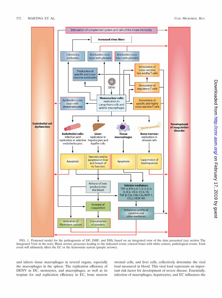
and infects tissue macrophages in several organs, especiallythe macrophages in the spleen. The replication efficiency ofDENV in DC, monocytes, and macrophages, as well as itstropism for and replication efficiency in EC, bone marrow
stromal cells, and liver cells, collectively determine the viralload measured in blood. This viral load represents an impor-tant risk factor for development of severe disease. Essentially,infection of macrophages, hepatocytes, and EC influences the
FIG. 1. Proposed model for the pathogenesis of DF, DHF, and DSS, based on an integrated view of the data presented (see section TheIntegrated View in the text). Black arrows, processes leading to the indicated event; colored boxes with white centers, pathological events. Eachevent will ultimately affect the EC or the hemostatic system (purple arrows).
572 MARTINA ET AL. CLIN. MICROBIOL. REV.
on February 17, 2019 by guest
http://cmr.asm
.org/D
ownloaded from
hemostatic and the immune responses to DENV. Infected cellsdie predominantly through apoptosis and to a lesser extentthrough necrosis. Necrosis results in release of toxic products,which activate the coagulation and fibrinolytic systems. De-pending on the extent of infection of bone marrow stromalcells and the levels of IL-6, IL-8, IL-10, and IL-18, hemopoiesisis suppressed, resulting in decreased blood thrombogenicity.Platelets interact closely with EC, and a normal number offunctioning platelets is necessary to maintain vascular stability.A high viral load in blood and possibly viral tropism for EC,severe thrombocytopenia, and platelet dysfunction may resultin increased capillary fragility, clinically manifested as pete-chiae, easy bruising, and gastrointestinal mucosal bleeding(170), which is characteristic of DHF. At the same time, infec-tion stimulates development of specific antibody and cellularimmune responses to DENV. When IgM antibodies that cross-react with EC, platelets, and plasmin are produced, the loopthat results in increased vascular permeability and coagulopa-thy is amplified. In addition, enhancing IgG antibodies bindheterologous virus during secondary infection and enhanceinfection of APCs, thereby contributing to the increased viralload that is seen during secondary viremia in some patients.Furthermore, a high viral load overstimulates both low- andhigh-avidity cross-reactive T cells. In the context of certainHLA haplotypes, cross-reactive T cells delay virus clearance,while producing high levels of proinflammatory cytokines andother mediators. Ultimately, these high levels of soluble fac-tors, many of which still remain to be identified, inducechanges in EC leading to the coagulopathy and plasma leakagecharacteristic of DSS.
DISCUSSION
Here we review and discuss the plethora of hypotheses aboutDHF and DSS pathogenesis presented in the literature. Mostof these hypotheses are not mutually exclusive, and togetherthey harbor multiple elements that collectively may explainmost of the phenomena observed in the multiple presentationsof DENV infection. Table 3 addresses major challenges to thehypotheses described in this review. The pathogenesis of bothDHF grades I/II and DSS is complicated and multifactorial,involving both viral and host factors. However, necessaryand/or sufficient factors have still not been identified. It may bequestioned whether factors that explain the pathogenesis ofDHF and DSS in all patients do exist. Genetic predispositionmay have a significant effect on disease outcome. Only a fewstudies have studied host genetics with regard to the severity ofDENV infection. Most commonly, clinically significant geneticvariations consist of single-nucleotide polymorphisms withingenes that affect disease pathways. Many polymorphisms havesmall and independent effects on disease outcome and oftenact in concert with other polymorphisms and environmentalrisk factors (24). This eventually results in complex and vari-able disease outcomes. Studies using a combination of geno-mics (transcriptomics, proteomics, and metabolomics), single-nucleotide polymorphism genotyping, and careful phenotypicdisease characterization in well-defined cohorts should beadopted to identify individual molecular markers of DHF/DSS.
As discussed in this review, the pathways to DHF involve
both viral and host-specific elements. The following points canbe summarized.
(i) In contrast to what is generally believed, the involvementof liver and of EC in different organ systems seems to be animportant factor in the pathogenesis of dengue. In this respect,it is of paramount importance to understand how EC lining thethoracic and peritoneal cavities are affected. It is worth notingthat despite the increased vascular permeability measured inboth DHF and DSS patients (16), plasma leakage is highlyrestricted to the pleural and peritoneal cavities, while no gen-eralized edema is seen. No specific vascular lesions are foundin fatal cases of DENV infections. Viruses known to causehemorrhagic fever, such as viruses belonging to the familiesArenaviridae (Junin virus and Lassa virus), Filoviridae (Ebolavirus and Marburg virus), Bunyaviridae (Hanta virus and RiftValley virus), and Flaviviridae (yellow fever virus), are also notassociated with EC damage (43, 70). In these cases, the patho-genesis of hemorrhagic diathesis is the result of severe liverdamage leading to decreased production of coagulation pro-teins and albumin. Increased vascular permeability as a result
TABLE 3. Challenges for DENV research
Challenge
Developing an animal model of dengue diseaseUnderstanding the role of several DENV strains and serotypes in
DENV tropism in vivoElucidating the role of intrahost evolution of DENVs in emergence
of virulent strains and the role of genetic variants and differentserotypes in promoting OAS, ADE, transient autoimmunity, andunbalanced T-cell responses
Elucidating the role of DENV proteins, especially NS1 inpathogenesis
Understanding the interplay between the hemostatic and thecomplement systems in DENV infection
Elucidating the mechanisms by which innate immunity regulatesmemory B- and T-cell generation, maintenance, and activationduring primary and secondary DENV infections
Elucidating the role of autoimmunity in dengue pathogenesisIdentifying serotype-specific linear and conformational B-cell
epitopes and understanding their role in primary and anamnesticB-cell responses
Understanding the role of DENV immune complexes in thesignaling network within target cells, especially APCs, and how itinfluences virus replication and priming of immune responses
Elucidating the role of the T-helper and T-cytotoxic cell repertoire,especially the private repertoire, on evolution of T-cell responsesduring primary and sequential heterologous DENV infections
Understanding the role of OAS in T-cell-mediated immunity inheterologous DENV infections and elucidating the factors thatdetermine occurrence of OAS for T cells and its role inpathogenesis
Identifying soluble factors that are necessary or sufficient to induceendothelial cell dysfunction and coagulopathy seen in DHF/DSS
Identifying critical components of tight junctions and adherentjunctions present in the vascular beds affected during DHF/DSSand elucidating the role that different soluble factors play inexpression and functions of the several components of thesejunctions
Investigating the effect of demographic history of infection on thetype of B- and T-cell responses elicited during primary andsecondary DENV infections and on pathogenesis
Elucidating the role of host gene polymorphism, such as FcR,cytokines, chemokines, etc., in DHF/DSS pathogenesis
Understanding race- and age-dependent susceptibility to severeDENV infection
VOL. 22, 2009 DENGUE VIRUS PATHOGENESIS 573
on February 17, 2019 by guest
http://cmr.asm
.org/D
ownloaded from
of hypercytokinemia and a reduction of plasma osmotic pres-sure due to severe liver damage contribute to edema forma-tion, as is seen in severe cases of Lassa fever (67). In addition,replication of most of these viruses in the adrenal gland con-tributes to hypotension and sodium loss, which collectivelyresult in hypovolemic shock. The shock syndrome associatedwith DSS, however, is unique to DENV infection, and it is ofparamount importance to understand how the EC lining thethoracic and peritoneal cavities are affected.
(ii) Hemorrhagic manifestations as seen in DF and DHFgrades I/II are usually mild and manifested as petechiae dis-seminated in the skin, with an increased tendency of bleedingupon bruising. These manifestations are found early after on-set of fever and coincide with the window of viremia anddegree of thrombocytopenia (121). The pathogenesis of mildhemorrhages seen in DF and DHF grades I/II may be ex-plained by increased capillary fragility as a result of thrombo-cytopenia or platelet dysfunction, virus infection of EC, andhigh concentrations of cytokines that disrupt vascular integrity(54, 170, 257). Most mediators that increase vascular perme-ability affect the organization of the adherens junctions (AJ), acomplex network of adhesion proteins that are linked to intra-cellular cytoskeleton (54), causing retraction of EC and open-ing of intercellular gaps. Alternatively, the stability of the junc-tions may be weakened, resulting in vascular fragility.Weakness of the junctions is not reflected by morphologicalchanges, as is exemplified by the fact that internalization ofVE-caherin or phosphorylation of AJ proteins reduces junc-tion stability without the opening of intercellular gaps (4, 62).It is noteworthy that the major anatomical sites of bleeding inpatients with severe thrombocytopenia are the intercellulargaps in the postcapillary venules, which are rich in AJ (170).Platelets play a role in maintaining the integrity of AJ byconstitutively releasing an array of factors such as platelet-activating factor and sphingosine-1-phosphate, which are re-leased as a result of fluid shear stress (5). Therefore, interrup-tion of platelet-EC interaction by severe thrombocytopenia orplatelet dysfunction may lead to increased vascular fragility,resulting in hemorrhages or increased tendencies for hemor-rhages.
(iii) Several studies have shown that plasma leakage occursbefore defervescence or hemoconcentration. As mentionedbefore, the WHO case definition has mainly a clinical diagnos-tic purpose and is not an appropriate selection criterion forpathogenesis studies. Pathogenesis studies should be designedto understand the differences between (i) no bleeding tenden-cies, (ii) increased manifestations of bleeding or tendencies forbleeding, or (iii) plasma leakage as measured by sonographyand hemoconcentration. It is crucial to study the pleural fluidin order to understand the pathophysiological cause of plasmaleakage and why it is restricted to or more pronounced inthoracic and peritoneal areas.
(iv) Antibody-mediated enhancement of infection either re-sults in a high viral load or represents the link to type 2cytokine responses.
(v) In some viral systems, cross-reactive CD8� T-cell re-sponses are involved in the development of pathologicalchanges during secondary infection with a related but heterol-ogous virus. DENV cross-reactive T cells, however, lose theircytolytic activity, while producing high levels of proinflamma-
tory cytokines and other mediators. The role that preexistingimmunity to other flaviviruses plays in the development ofsevere dengue should be investigated.
(vi) Soluble host factors seem to be central in the pathogen-esis of both DHF grades I/II and DSS. Although several ofthese have been associated with severe disease, their concen-trations are also elevated in other viral infections without re-sulting in plasma leakage. High concentrations of cytokinessuch TNF-�, IL-6, and IL-8 have been implicated in capillaryleakage and development of hypovolemic shock in patientswith anaphylaxis, meningococcal sepsis, and Jarish Herxheimerreaction (105, 176, 233). Similarly, high concentrations of cy-tokines have been associated with unfavorable outcomes offilovirus (253), arenavirus (89, 160), and yellow fever virus(241) infections. However, the shock syndrome associated withDSS does not occur in these conditions. Therefore, it may bespeculated that soluble factors incriminated in the increasedvascular permeability seen in DSS must be qualitatively andquantitatively different from those involved in the conditionsdescribed above.
ACKNOWLEDGMENTS
We thank Peter Fraaij, Rik de Swart, and Bart Haagmans for pro-viding valuable comments.
REFERENCES
1. Acioli-Santos, B., L. Segat, R. Dhalia, C. A. Brito, U. M. Braga-Neto, E. T.Marques, and S. Crovella. 2008. MBL2 gene polymorphisms protectagainst development of thrombocytopenia associated with severe denguephenotype. Hum. Immunol. 69:122–128.
2. Agarwal, R., U. C. Chaturvedi, A. Misra, R. Mukerjee, S. Kapoor, R. Nagar,R. Tandon, and A. Mathur. 1998. Production of cytotoxic factor by periph-eral blood mononuclear cells (PBMC) in patients with dengue haemor-rhagic fever. Clin. Exp. Immunol. 112:477–481.
3. An, J., D. S. Zhou, J. L. Zhang, H. Morida, J. L. Wang, and K. Yasui. 2004.Dengue-specific CD8� T cells have both protective and pathogenic roles indengue virus infection. Immunol. Lett. 95:167–174.
4. Andriopoulou, P., P. Navarro, A. Zanetti, M. G. Lampugnani, and E.Dejana. 1999. Histamine induces tyrosine phosphorylation of endothelialcell-to-cell adherens junctions. Arterioscler. Thromb. Vasc. Biol. 19:2286–2297.
5. Aoki, S., M. Osada, M. Kaneko, Y. Ozaki, and Y. Yatomi. 2007. Fluid shearstress enhances the sphingosine 1-phosphate responses in cell-cell interac-tions between platelets and endothelial cells. Biochem. Biophys. Res. Com-mun. 358:1054–1057.
6. Atrasheuskaya, A., P. Petzelbauer, T. M. Fredeking, and G. Ignatyev. 2003.Anti-TNF antibody treatment reduces mortality in experimental denguevirus infection. FEMS Immunol. Med. Microbiol. 35:33–42.
7. Avirutnan, P., P. Malasit, B. Seliger, S. Bhakdi, and M. Husmann. 1998.Dengue virus infection of human endothelial cells leads to chemokineproduction, complement activation, and apoptosis. J. Immunol. 161:6338–6346.
8. Avirutnan, P., N. Punyadee, S. Noisakran, C. Komoltri, S. Thiemmeca, K.Auethavornanan, A. Jairungsri, R. Kanlaya, N. Tangthawornchaikul, C.Puttikhunt, S. N. Pattanakitsakul, P. T. Yenchitsomanus, J. Mongkolsa-paya, W. Kasinrerk, N. Sittisombut, M. Husmann, M. Blettner, S. Vasana-wathana, S. Bhakdi, and P. Malasit. 2006. Vascular leakage in severedengue virus infections: a potential role for the nonstructural viral proteinNS1 and complement. J. Infect. Dis. 193:1078–1088.
9. Avirutnan, P., L. Zhang, N. Punyadee, A. Manuyakorn, C. Puttikhunt, W.Kasinrerk, P. Malasit, J. P. Atkinson, and M. S. Diamond. 2007. SecretedNS1 of dengue virus attaches to the surface of cells via interactions withheparan sulfate and chondroitin sulfate E. PLoS Pathog. 3:e183.
10. Azeredo, E. L., S. M. Zagne, M. A. Santiago, A. S. Gouvea, A. A. Santana,P. C. Neves-Souza, R. M. Nogueira, M. P. Miagostovich, and C. F. Kubelka.2001. Characterisation of lymphocyte response and cytokine patterns inpatients with dengue fever. Immunobiology 204:494–507.
11. Azizan, A., J. Sweat, C. Espino, J. Gemmer, L. Stark, and D. Kazanis. 2006.Differential proinflammatory and angiogenesis-specific cytokine productionin human pulmonary endothelial cells, HPMEC-ST1.6R infected with den-gue-2 and dengue-3 virus. J. Virol. Methods 138:211–217.
12. Balasubramanian, S., L. Janakiraman, S. S. Kumar, S. Muralinath, and S.
574 MARTINA ET AL. CLIN. MICROBIOL. REV.
on February 17, 2019 by guest
http://cmr.asm
.org/D
ownloaded from
Shivbalan. 2006. A reappraisal of the criteria to diagnose plasma leakage indengue hemorrhagic fever. Indian Pediatr. 43:334–339.
13. Balsitis, S. J., J. Coloma, G. Castro, A. Alava, D. Flores, J. H. McKerrow,P. R. Beatty, and E. Harris. 2009. Tropism of dengue virus in mice andhumans defined by viral nonstructural protein 3-specific immunostaining.Am. J. Trop. Med. Hyg. 80:416–424.
14. Basilio-de-Oliveira, C. A., G. R. Aguiar, M. S. Baldanza, O. M. Barth, W. A.Eyer-Silva, and M. V. Paes. 2005. Pathologic study of a fatal case ofdengue-3 virus infection in Rio de Janeiro, Brazil. Braz. J. Infect. Dis.9:341–347.
15. Basu, A., and U. C. Chaturvedi. 2008. Vascular endothelium: the battlefieldof dengue viruses. FEMS Immunol. Med. Microbiol. 53:287–299.
16. Bethell, D. B., J. Gamble, P. L. Pham, M. D. Nguyen, T. H. Tran, T. H. Ha,T. N. Tran, T. H. Dong, I. B. Gartside, N. J. White, and N. P. Day. 2001.Noninvasive measurement of microvascular leakage in patients with denguehemorrhagic fever. Clin. Infect. Dis. 32:243–253.
17. Bhamarapravati, N., P. Tuchinda, and V. Boonyapaknavik. 1967. Pathologyof Thailand haemorrhagic fever: a study of 100 autopsy cases. Ann. Trop.Med. Parasitol. 61:500–510.
18. Blackley, S., Z. Kou, H. Chen, M. Quinn, R. C. Rose, J. J. Schlesinger, M.Coppage, and X. Jin. 2007. Primary human splenic macrophages, but not Tor B cells, are the principal target cells for dengue virus infection in vitro.J. Virol. 81:13325–13334.
19. Bonner, S. M., and M. A. O’Sullivan. 1998. Endothelial cell monolayers asa model system to investigate dengue shock syndrome. J. Virol. Methods71:159–167.
20. Boonnak, K., B. M. Slike, T. H. Burgess, R. M. Mason, S. J. Wu, P. Sun, K.Porter, I. F. Rudiman, D. Yuwono, P. Puthavathana, and M. A. Marovich.2008. Role of dendritic cells in antibody-dependent enhancement of denguevirus infection. J. Virol. 82:3939–3951.
21. Boonpucknavig, S., V. Boonpucknavig, N. Bhamarapravati, and S. Nim-mannitya. 1979. Immunofluorescence study of skin rash in patients withdengue hemorrhagic fever. Arch. Pathol. Lab. Med. 103:463–466.
22. Bosch, I., K. Xhaja, L. Estevez, G. Raines, H. Melichar, R. V. Warke, M. V.Fournier, F. A. Ennis, and A. L. Rothman. 2002. Increased production ofinterleukin-8 in primary human monocytes and in human epithelial andendothelial cell lines after dengue virus challenge. J. Virol. 76:5588–5597.
23. Bozza, F. A., O. G. Cruz, S. M. Zagne, E. L. Azeredo, R. M. Nogueira, E. F.Assis, P. T. Bozza, and C. F. Kubelka. 2008. Multiplex cytokine profile fromdengue patients: MIP-1beta and IFN-gamma as predictive factors for se-verity. BMC. Infect. Dis. 8:86.
24. Bracken, M. B. 2005. Genomic epidemiology of complex disease: the needfor an electronic evidence-based approach to research synthesis. Am. J.Epidemiol. 162:297–301.
25. Burke, D. S., and S. Kliks. 2006. Antibody-dependent enhancement indengue virus infections. J. Infect. Dis. 193:601–603. (Author reply, 193:603–604.)
26. Butthep, P., S. Chunhakan, K. Tangnararatchakit, S. Yoksan, K. Pattan-apanyasat, and A. Chuansumrit. 2006. Elevated soluble thrombomodulinin the febrile stage related to patients at risk for dengue shock syndrome.Pediatr. Infect. Dis. J. 25:894–897.
27. Cardier, J. E., V. Balogh, C. Perez-Silva, E. Romano, B. Rivas, N. Bosch,and A. L. Rothman. 2006. Relationship of thrombopoietin and interleu-kin-11 levels to thrombocytopenia associated with dengue disease. Cytokine34:155–160.
28. Cardier, J. E., E. Marino, E. Romano, P. Taylor, F. Liprandi, N. Bosch, andA. L. Rothman. 2005. Proinflammatory factors present in sera from patientswith acute dengue infection induce activation and apoptosis of humanmicrovascular endothelial cells: possible role of TNF-alpha in endothelialcell damage in dengue. Cytokine 30:359–365.
29. Cardier, J. E., B. Rivas, E. Romano, A. L. Rothman, C. Perez-Perez, M.Ochoa, A. M. Caceres, M. Cardier, N. Guevara, and R. Giovannetti. 2006.Evidence of vascular damage in dengue disease: demonstration of highlevels of soluble cell adhesion molecules and circulating endothelial cells.Endothelium 13:335–340.
30. Carlos, C. C., K. Oishi, M. T. Cinco, C. A. Mapua, S. Inoue, D. J. Cruz,M. A. Pancho, C. Z. Tanig, R. R. Matias, K. Morita, F. F. Natividad, A.Igarashi, and T. Nagatake. 2005. Comparison of clinical features and he-matologic abnormalities between dengue fever and dengue hemorrhagicfever among children in the Philippines. Am. J. Trop. Med. Hyg. 73:435–440.
31. Casadevall, A., and L. A. Pirofski. 1999. Host-pathogen interactions: rede-fining the basic concepts of virulence and pathogenicity. Infect. Immun.67:3703–3713.
32. Chakravarti, A., and R. Kumaria. 2006. Circulating levels of tumour ne-crosis factor-alpha & interferon-gamma in patients with dengue & denguehaemorrhagic fever during an outbreak. Indian J. Med. Res. 123:25–30.
33. Chang, H. H., H. F. Shyu, Y. M. Wang, D. S. Sun, R. H. Shyu, S. S. Tang,and Y. S. Huang. 2002. Facilitation of cell adhesion by immobilized dengueviral nonstructural protein 1 (NS1): arginine-glycine-aspartic acid structuralmimicry within the dengue viral NS1 antigen. J. Infect. Dis. 186:743–751.
34. Chanthavanich, P., C. Luxemburger, C. Sirivichayakul, K. Lapphra, K.
Pengsaa, S. Yoksan, A. Sabchareon, and J. Lang. 2006. Immune responseand occurrence of dengue infection in Thai children three to eight yearsafter vaccination with live attenuated tetravalent dengue vaccine. Am. J.Trop. Med. Hyg. 75:26–28.
35. Chao, Y. C., C. S. Huang, C. N. Lee, S. Y. Chang, C. C. King, and C. L. Kao.2008. Higher infection of dengue virus serotype 2 in human monocytes ofpatients with G6PD deficiency. PLoS One 3:e1557.
36. Chareonsirisuthigul, T., S. Kalayanarooj, and S. Ubol. 2007. Dengue virus(DENV) antibody-dependent enhancement of infection upregulates theproduction of anti-inflammatory cytokines, but suppresses anti-DENV freeradical and pro-inflammatory cytokine production, in THP-1 cells. J. Gen.Virol. 88:365–375.
37. Chaturvedi, U. C., E. A. Elbishbishi, R. Agarwal, and A. S. Mustafa. 2001.Cytotoxic factor-autoantibodies: possible role in the pathogenesis of den-gue haemorrhagic fever. FEMS Immunol. Med. Microbiol. 30:181–186.
38. Chau, T. N., N. T. Quyen, T. T. Thuy, N. M. Tuan, D. M. Hoang, N. T. Dung,B. Lien le, N. T. Quy, N. T. Hieu, L. T. Hieu, T. T. Hien, N. T. Hung, J.Farrar, and C. P. Simmons. 2008. Dengue in Vietnamese infants—xresultsof infection-enhancement assays correlate with age-related disease epide-miology, and cellular immune responses correlate with disease severity.J. Infect. Dis. 198:516–524.
39. Chen, H. C., F. M. Hofman, J. T. Kung, Y. D. Lin, and B. A. Wu-Hsieh.2007. Both virus and tumor necrosis factor alpha are critical for endothe-lium damage in a mouse model of dengue virus-induced hemorrhage. J. Vi-rol. 81:5518–5526.
40. Chen, H. D., A. E. Fraire, I. Joris, M. A. Brehm, R. M. Welsh, and L. K.Selin. 2001. Memory CD8� T cells in heterologous antiviral immunity andimmunopathology in the lung. Nat. Immunol. 2:1067–1076.
41. Chen, H. D., A. E. Fraire, I. Joris, R. M. Welsh, and L. K. Selin. 2003.Specific history of heterologous virus infections determines anti-viral im-munity and immunopathology in the lung. Am. J. Pathol. 163:1341–1355.
42. Chen, H. L., S. R. Lin, H. F. Liu, C. C. King, S. C. Hsieh, and W. K. Wang.2008. Evolution of dengue virus type 2 during two consecutive outbreakswith an increase in severity in southern Taiwan in 2001–2002. Am. J. Trop.Med. Hyg. 79:495–505.
43. Chen, J. P., and T. M. Cosgriff. 2000. Hemorrhagic fever virus-inducedchanges in hemostasis and vascular biology. Blood Coagul. Fibrinolysis11:461–483.
44. Chen, L. C., H. Y. Lei, C. C. Liu, S. C. Shiesh, S. H. Chen, H. S. Liu, Y. S.Lin, S. T. Wang, H. W. Shyu, and T. M. Yeh. 2006. Correlation of serumlevels of macrophage migration inhibitory factor with disease severity andclinical outcome in dengue patients. Am. J. Trop. Med. Hyg. 74:142–147.
45. Chen, R. F., L. Wang, J. T. Cheng, H. Chuang, J. C. Chang, J. W. Liu, I. C.Lin, and K. D. Yang. 2009. Combination of CTLA-4 and TGFbeta1 genepolymorphisms associated with dengue hemorrhagic fever and virus load ina dengue-2 outbreak. Clin. Immunol. 131:404–409.
46. Chen, R. F., K. D. Yang, L. Wang, J. W. Liu, C. C. Chiu, and J. T. Cheng.2007. Different clinical and laboratory manifestations between dengue hae-morrhagic fever and dengue fever with bleeding tendency. Trans. R. Soc.Trop. Med. Hyg. 101:1106–1113.
47. Chen, Y. C., and S. Y. Wang. 2002. Activation of terminally differentiatedhuman monocytes/macrophages by dengue virus: productive infection, hi-erarchical production of innate cytokines and chemokines, and the syner-gistic effect of lipopolysaccharide. J. Virol. 76:9877–9887.
48. Choi, G., M. J. Schultz, M. Levi, and T. van der Poll. 2006. The relationshipbetween inflammation and the coagulation system. Swiss Med. Wkly. 136:139–144.
49. Chungue, E., L. Poli, C. Roche, P. Gestas, P. Glaziou, and L. J. Markoff.s.1994. Correlation between detection of plasminogen cross-reactive antibod-ies and hemorrhage in dengue virus infection. J. Infect. Dis. 170:1304–1307.
50. Churdboonchart, V., N. Bhamarapravati, and P. Futrakul. 1983. Crossedimmunoelectrophoresis for the detection of split products of the thirdcomplement in dengue hemorrhagic fever. I. Observations in patients’plasma. Am. J. Trop. Med. Hyg. 32:569–576.
51. Cologna, R., and R. Rico-Hesse. 2003. American genotype structures de-crease dengue virus output from human monocytes and dendritic cells.J. Virol. 77:3929–3938.
52. Cornberg, M., A. T. Chen, L. A. Wilkinson, M. A. Brehm, S. K. Kim, C.Calcagno, D. Ghersi, R. Puzone, F. Celada, R. M. Welsh, and L. K. Selin.2006. Narrowed TCR repertoire and viral escape as a consequence ofheterologous immunity. J. Clin. Investig. 116:1443–1456.
53. Couvelard, A., P. Marianneau, C. Bedel, M. T. Drouet, F. Vachon, D.Henin, and V. Deubel. 1999. Report of a fatal case of dengue infection withhepatitis: demonstration of dengue antigens in hepatocytes and liver apop-tosis. Hum. Pathol. 30:1106–1110.
54. Dejana, E., E. Tournier-Lasserve, and B. M. Weinstein. 2009. The controlof vascular integrity by endothelial cell junctions: molecular basis andpathological implications. Dev. Cell 16:209–221.
54a.de Jong, M. D., C. P. Simmons, T. T. Thanh, V. M. Hien, G. J. Smith, T. N.Chau, D. M. Hoang, N. V. Chau, T. H. Khanh, V. C. Dong, P. T. Qui, B. V.Cam, Q. Ha do, Y. Guan, J. S. Peiris, N. T. Chinh, T. T. Hien, and J. Farrar.
VOL. 22, 2009 DENGUE VIRUS PATHOGENESIS 575
on February 17, 2019 by guest
http://cmr.asm
.org/D
ownloaded from
2006. Fatal outcome of human influenza A (H5N1) is associated with highviral load and hypercytokinemia. Nat. Med. 12:1203–1207.
54b.de la C Sierra, B., G. Garcia, A. B. Perez, L. Morier, M. Alvarez, G. Kouri,and M. G. Guzman. 2006. Ethnicity and difference in dengue virus-specificmemory T cell responses in Cuban individuals. Viral Immunol. 19:662–668.
54c.de la C Sierra, B., G. Kouri, and M. G. Guzman. 2007. Race: a risk factorfor dengue hemorrhagic fever. Arch. Virol. 152:533–542.
54d.de Macedo, F. C., A. F. Nicol, L. D. Cooper, M. Yearsley, A. R. Pires, andG. J. Nuovo. 2006. Histologic, viral, and molecular correlates of denguefever infection of the liver using highly sensitive immunohistochemistry.Diagn. Mol. Pathol. 15:223–228.
54e.de Souza, L. J., R. M. Nogueira, L. C. Soares, C. E. Soares, B. F. Ribas, F. P.Alves, F. R. Vieira, and F. E. Pessanha. 2007. The impact of dengue on liverfunction as evaluated by aminotransferase levels. Braz. J. Infect. Dis. 11:407–410.
55. Dewi, B. E., T. Takasaki, and I. Kurane. 2004. In vitro assessment of humanendothelial cell permeability: effects of inflammatory cytokines and denguevirus infection. J. Virol. Methods 121:171–180.
56. Diamond, M. S., D. Edgil, T. G. Roberts, B. Lu, and E. Harris. 2000.Infection of human cells by dengue virus is modulated by different cell typesand viral strains. J. Virol. 74:7814–7823.
57. Ding, X., F. Xu, H. Chen, R. B. Tesh, and S. Y. Xiao. 2005. Apoptosis ofhepatocytes caused by Punta Toro virus (Bunyaviridae: Phlebovirus) and itsimplication for Phlebovirus pathogenesis. Am. J. Pathol. 167:1043–1049.
58. Dong, T., E. Moran, N. Vinh Chau, C. Simmons, K. Luhn, Y. Peng, B. Wills,N. Phuong Dung, L. Thi Thu Thao, T. T. Hien, A. McMichael, J. Farrar,and S. Rowland-Jones. 2007. High pro-inflammatory cytokine secretion andloss of high avidity cross-reactive cytotoxic T-cells during the course ofsecondary dengue virus infection. PLoS One 2:e1192.
59. Durbin, A. P., M. J. Vargas, K. Wanionek, S. N. Hammond, A. Gordon, C.Rocha, A. Balmaseda, and E. Harris. 2008. Phenotyping of peripheralblood mononuclear cells during acute dengue illness demonstrates infectionand increased activation of monocytes in severe cases compared to classicdengue fever. Virology 376:429–435.
60. Esmon, C. T. 2005. The interactions between inflammation and coagula-tion. Br. J. Haematol. 131:417–430.
61. Espina, L. M., N. J. Valero, J. M. Hernandez, and J. A. Mosquera. 2003.Increased apoptosis and expression of tumor necrosis factor-alpha causedby infection of cultured human monocytes with dengue virus. Am. J. Trop.Med. Hyg. 68:48–53.
62. Esser, S., M. G. Lampugnani, M. Corada, E. Dejana, and W. Risau. 1998.Vascular endothelial growth factor induces VE-cadherin tyrosine phosphor-ylation in endothelial cells. J. Cell Sci. 111:1853–1865.
63. Fabre, A., A. Couvelard, C. Degott, C. Lagorce-Pages, F. Bruneel, E. Bou-vet, and F. Vachon. 2001. Dengue virus induced hepatitis with chroniccalcific changes. Gut 49:864–865.
64. Falconar, A. K. 1997. The dengue virus nonstructural-1 protein (NS1)generates antibodies to common epitopes on human blood clotting, inte-grin/adhesin proteins and binds to human endothelial cells: potential im-plications in haemorrhagic fever pathogenesis. Arch. Virol. 142:897–916.
65. Feng, J. Q., K. Mozdzanowska, and W. Gerhard. 2002. Complement com-ponent C1q enhances the biological activity of influenza virus hemaggluti-nin-specific antibodies depending on their fine antigen specificity andheavy-chain isotype. J. Virol. 76:1369–1378.
66. Fernandez-Mestre, M. T., K. Gendzekhadze, P. Rivas-Vetencourt, and Z.Layrisse. 2004. TNF-alpha-308A allele, a possible severity risk factor ofhemorrhagic manifestation in dengue fever patients. Tissue Antigens 64:469–472.
67. Frame, J. D. 1989. Clinical features of Lassa fever in Liberia. Rev. Infect.Dis. 11(Suppl. 4):S783–S789.
68. Fritz, R. S., F. G. Hayden, D. P. Calfee, L. M. Cass, A. W. Peng, W. G.Alvord, W. Strober, and S. E. Straus. 1999. Nasal cytokine and chemokineresponses in experimental influenza A virus infection: results of a placebo-controlled trial of intravenous zanamivir treatment. J. Infect. Dis. 180:586–593.
69. Gasperino, J., J. Yunen, A. Guh, K. E. Tanaka, V. Kvetan, and H. Doyle.2007. Fulminant liver failure secondary to haemorrhagic dengue in aninternational traveller. Liver Int. 27:1148–1151.
70. Geisbert, T. W., and P. B. Jahrling. 2004. Exotic emerging viral diseases:progress and challenges. Nat. Med. 10:S110–S121.
71. Gentile, D., W. Doyle, T. Whiteside, P. Fireman, F. G. Hayden, and D.Skoner. 1998. Increased interleukin-6 levels in nasal lavage samples follow-ing experimental influenza A virus infection. Clin. Diagn. Lab. Immunol.5:604–608.
72. Ghiasi, H., G. C. Perng, A. B. Nesburn, and S. L. Wechsler. 2000. Antibody-dependent enhancement of HSV-1 infection by anti-gK sera. Virus Res.68:137–144.
73. Girn, J., M. Kavoosi, and J. Chantler. 2002. Enhancement of coxsackievirusB3 infection by antibody to a different coxsackievirus strain. J. Gen. Virol.83:351–358.
74. Graham, R. R., M. Juffrie, R. Tan, C. G. Hayes, I. Laksono, C. Ma’roef,Erlin, Sutaryo, K. R. Porter, and S. B. Halstead. 1999. A prospective
seroepidemiologic study on dengue in children four to nine years of age inYogyakarta, Indonesia I. studies in 1995–1996. Am. J. Trop. Med. Hyg.61:412–419.
75. Guha-Sapir, D., and B. Schimmer. 2005. Dengue fever: new paradigms fora changing epidemiology. Emerg. Themes Epidemiol. 2:1.
76. Guilarde, A. O., M. D. Turchi, J. B. Siqueira, Jr., V. C. Feres, B. Rocha,J. E. Levi, V. A. Souza, L. S. Boas, C. S. Pannuti, and C. M. Martelli. 2008.Dengue and dengue hemorrhagic fever among adults: clinical outcomesrelated to viremia, serotypes, and antibody response. J. Infect. Dis. 197:817–824.
77. Guirakhoo, F., S. Kitchener, D. Morrison, R. Forrat, K. McCarthy, R.Nichols, S. Yoksan, X. Duan, T. H. Ermak, N. Kanesa-Thasan, P. Bedford,J. Lang, M. J. Quentin-Millet, and T. P. Monath. 2006. Live attenuatedchimeric yellow fever dengue type 2 (ChimeriVax-DEN2) vaccine: phase Iclinical trial for safety and immunogenicity: effect of yellow fever pre-immunity in induction of cross neutralizing antibody responses to all 4dengue serotypes. Hum. Vaccin. 2:60–67.
78. Guzman, M. G., M. Alvarez, R. Rodriguez, D. Rosario, S. Vazquez, L.Valds, M. V. Cabrera, and G. Kouri. 1999. Fatal dengue hemorrhagic feverin Cuba, 1997. Int. J. Infect. Dis. 3:130–135.
79. Guzman, M. G., and G. Kouri. 2003. Dengue and dengue hemorrhagicfever in the Americas: lessons and challenges. J. Clin. Virol. 27:1–13.
80. Guzman, M. G., G. Kouri, J. Bravo, L. Valdes, S. Vazquez, and S. B.Halstead. 2002. Effect of age on outcome of secondary dengue 2 infections.Int. J. Infect. Dis. 6:118–124.
81. Guzman, M. G., G. Kouri, and S. B. Halstead. 2000. Do escape mutantsexplain rapid increases in dengue case-fatality rates within epidemics? Lan-cet 355:1902–1903.
82. Guzman, M. G., G. Kouri, L. Valdes, J. Bravo, M. Alvarez, S. Vazques, I.Delgado, and S. B. Halstead. 2000. Epidemiologic studies on dengue inSantiago de Cuba, 1997. Am. J. Epidemiol. 152:793–799. (Discussion, 152:804.)
83. Halstead, S. B. 2007. Dengue. Lancet. 370:1644–1652.84. Halstead, S. B. 2006. Dengue in the Americas and Southeast Asia: do they
differ? Rev. Panam. Salud Publica 20:407–415.85. Halstead, S. B. 1970. Observations related to pathogensis of dengue hem-
orrhagic fever. VI. Hypotheses and discussion. Yale J. Biol. Med. 42:350–362.
86. Halstead, S. B., S. Nimmannitya, and S. N. Cohen. 1970. Observationsrelated to pathogenesis of dengue hemorrhagic fever. IV. Relation of dis-ease severity to antibody response and virus recovered. Yale J. Biol. Med.42:311–328.
87. Harris, E., E. Videa, L. Perez, E. Sandoval, Y. Tellez, M. L. Perez, R.Cuadra, J. Rocha, W. Idiaquez, R. E. Alonso, M. A. Delgado, L. A. Campo,F. Acevedo, A. Gonzalez, J. J. Amador, and A. Balmaseda. 2000. Clinical,epidemiologic, and virologic features of dengue in the 1998 epidemic inNicaragua. Am. J. Trop. Med. Hyg. 63:5–11.
88. Hayden, F. G., R. Fritz, M. C. Lobo, W. Alvord, W. Strober, and S. E.Straus. 1998. Local and systemic cytokine responses during experimentalhuman influenza A virus infection. Relation to symptom formation and hostdefense. J. Clin. Investig. 101:643–649.
89. Heller, M. V., M. C. Saavedra, R. Falcoff, J. I. Maiztegui, and F. C. Molinas.1992. Increased tumor necrosis factor-alpha levels in Argentine hemor-rhagic fever. J. Infect. Dis. 166:1203–1204.
90. Hjelm, F., F. Carlsson, A. Getahun, and B. Heyman. 2006. Antibody-mediated regulation of the immune response. Scand. J. Immunol. 64:177–184.
91. Ho, L. J., M. F. Shaio, D. M. Chang, C. L. Liao, and J. H. Lai. 2004.Infection of human dendritic cells by dengue virus activates and primes Tcells towards Th0-like phenotype producing both Th1 and Th2 cytokines.Immunol. Investig. 33:423–437.
92. Ho, L. J., J. J. Wang, M. F. Shaio, C. L. Kao, D. M. Chang, S. W. Han, andJ. H. Lai. 2001. Infection of human dendritic cells by dengue virus causescell maturation and cytokine production. J. Immunol. 166:1499–1506.
93. Hober, D., W. Chehadeh, A. Bouzidi, and P. Wattre. 2001. Antibody-de-pendent enhancement of coxsackievirus B4 infectivity of human peripheralblood mononuclear cells results in increased interferon-alpha synthesis.J. Infect. Dis. 184:1098–1108.
94. Huang, Y. H., B. I. Chang, H. Y. Lei, H. S. Liu, C. C. Liu, H. L. Wu, andT. M. Yeh. 1997. Antibodies against dengue virus E protein peptide bind tohuman plasminogen and inhibit plasmin activity. Clin. Exp. Immunol. 110:35–40.
95. Huang, Y. H., H. Y. Lei, H. S. Liu, Y. S. Lin, C. C. Liu, and T. M. Yeh. 2000.Dengue virus infects human endothelial cells and induces IL-6 and IL-8production. Am. J. Trop. Med. Hyg. 63:71–75.
96. Huerre, M. R., N. T. Lan, P. Marianneau, N. B. Hue, H. Khun, N. T. Hung,N. T. Khen, M. T. Drouet, V. T. Huong, D. Q. Ha, Y. Buisson, and V.Deubel. 2001. Liver histopathology and biological correlates in five cases offatal dengue fever in Vietnamese children. Virchows Arch. 438:107–115.
97. Huerta-Zepeda, A., C. Cabello-Gutierrez, J. Cime-Castillo, V. Monroy-Martinez, M. E. Manjarrez-Zavala, M. Gutierrez-Rodriguez, R. Izaguirre,
576 MARTINA ET AL. CLIN. MICROBIOL. REV.
on February 17, 2019 by guest
http://cmr.asm
.org/D
ownloaded from
and B. H. Ruiz-Ordaz. 2008. Crosstalk between coagulation and inflamma-tion during dengue virus infection. Thromb. Haemost. 99:936–943.
98. Iankov, I. D., M. Pandey, M. Harvey, G. E. Griesmann, M. J. Federspiel,and S. J. Russell. 2006. Immunoglobulin G antibody-mediated enhance-ment of measles virus infection can bypass the protective antiviral immuneresponse. J. Virol. 80:8530–8540.
99. Imrie, A., J. Meeks, A. Gurary, M. Sukhbataar, P. Kitsutani, P. Effler, andZ. Zhao. 2007. Differential functional avidity of dengue virus-specific T-cellclones for variant peptides representing heterologous and previously en-countered serotypes. J. Virol. 81:10081–10091.
100. Jassal, R., N. Jenkins, J. Charlwood, P. Camilleri, R. Jefferis, and J. Lund.2001. Sialylation of human IgG-Fc carbohydrate by transfected rat al-pha2,6-sialyltransferase. Biochem. Biophys. Res. Commun. 286:243–249.
101. Jessie, K., M. Y. Fong, S. Devi, S. K. Lam, and K. T. Wong. 2004. Local-ization of dengue virus in naturally infected human tissues, by immunohis-tochemistry and in situ hybridization. J. Infect. Dis. 189:1411–1418.
102. Reference deleted.103. Juffrie, M., G. M. Meer, C. E. Hack, K. Haasnoot, Sutaryo, A. J. Veerman,
and L. G. Thijs. 2001. Inflammatory mediators in dengue virus infection inchildren: interleukin-6 and its relation to C-reactive protein and secretoryphospholipase A2. Am. J. Trop. Med. Hyg. 65:70–75.
104. Kangwanpong, D., N. Bhamarapravati, and H. L. Lucia. 1995. Diagnosingdengue virus infection in archived autopsy tissues by means of the in situPCR method: a case report. Clin. Diagn. Virol. 3:165–172.
105. Kaplanski, G., B. Granel, T. Vaz, and J. M. Durand. 1998. Jarisch-Herx-heimer reaction complicating the treatment of chronic Q. fever endocardi-tis: elevated TNFalpha and IL-6 serum levels. J. Infect. 37:83–84.
106. Killen, H., and M. A. O’Sullivan. 1993. Detection of dengue virus by in situhybridization. J. Virol. Methods 41:135–146.
107. Kim, S. K., M. Cornberg, X. Z. Wang, H. D. Chen, L. K. Selin, and R. M.Welsh. 2005. Private specificities of CD8 T cell responses control patternsof heterologous immunity. J. Exp. Med. 201:523–533.
108. Kima, P. E., S. L. Constant, L. Hannum, M. Colmenares, K. S. Lee, A. M.Haberman, M. J. Shlomchik, and D. McMahon-Pratt. 2000. Internalizationof Leishmania mexicana complex amastigotes via the Fc receptor is re-quired to sustain infection in murine cutaneous leishmaniasis. J. Exp. Med.191:1063–1068.
109. Kittigul, L., P. Pitakarnjanakul, D. Sujirarat, and K. Siripanichgon. 2007.The differences of clinical manifestations and laboratory findings in chil-dren and adults with dengue virus infection. J. Clin. Virol. 39:76–81.
110. Klenerman, P., and R. M. Zinkernagel. 1998. Original antigenic sin impairscytotoxic T lymphocyte responses to viruses bearing variant epitopes. Na-ture 394:482–485.
111. Kliks, S. C., S. Nimmanitya, A. Nisalak, and D. S. Burke. 1988. Evidencethat maternal dengue antibodies are important in the development ofdengue hemorrhagic fever in infants. Am. J. Trop. Med. Hyg. 38:411–419.
112. Kliks, S. C., A. Nisalak, W. E. Brandt, L. Wahl, and D. S. Burke. 1989.Antibody-dependent enhancement of dengue virus growth in human mono-cytes as a risk factor for dengue hemorrhagic fever. Am. J. Trop. Med. Hyg.40:444–451.
113. Koraka, P., S. Benton, G. van Amerongen, K. J. Stittelaar, and A. D.Osterhaus. 2007. Characterization of humoral and cellular immune re-sponses in cynomolgus macaques upon primary and subsequent heterolo-gous infections with dengue viruses. Microbes Infect. 9:940–946.
114. Koraka, P., S. Benton, G. van Amerongen, K. J. Stittelaar, and A. D.Osterhaus. 2007. Efficacy of a live attenuated tetravalent candidate denguevaccine in naive and previously infected cynomolgus macaques. Vaccine25:5409–5416.
115. Koraka, P., B. Murgue, X. Deparis, E. C. Van Gorp, T. E. Setiati, A. D.Osterhaus, and J. Groen. 2004. Elevation of soluble VCAM-1 plasma levelsin children with acute dengue virus infection of varying severity. J. Med.Virol. 72:445–450.
116. Koraka, P., C. Suharti, T. E. Setiati, A. T. Mairuhu, E. Van Gorp, C. E.Hack, M. Juffrie, J. Sutaryo, G. M. Van Der Meer, J. Groen, and A. D.Osterhaus. 2001. Kinetics of dengue virus-specific serum immunoglobulinclasses and subclasses correlate with clinical outcome of infection. J. Clin.Microbiol. 39:4332–4338.
117. Kou, Z., M. Quinn, H. Chen, W. W. Rodrigo, R. C. Rose, J. J. Schlesinger,and X. Jin. 2008. Monocytes, but not T or B cells, are the principal targetcells for dengue virus (DV) infection among human peripheral bloodmononuclear cells. J. Med. Virol. 80:134–146.
118. Kouri, G. P., M. G. Guzman, and J. R. Bravo. 1987. Why dengue haemor-rhagic fever in Cuba? 2. An integral analysis. Trans. R. Soc. Trop. Med.Hyg. 81:821–823.
119. Kouri, G. P., M. G. Guzman, J. R. Bravo, and C. Triana. 1989. Denguehaemorrhagic fever/dengue shock syndrome: lessons from the Cuban epi-demic, 1981. Bull. W. H. O. 67:375–380.
120. Krishnamurti, C., and B. Alving. 1989. Effect of dengue virus on proco-agulant and fibrinolytic activities of monocytes. Rev. Infect. Dis. 11 Suppl.4:S843–S846.
121. Krishnamurti, C., S. Kalayanarooj, M. A. Cutting, R. A. Peat, S. W. Roth-well, T. J. Reid, S. Green, A. Nisalak, T. P. Endy, D. W. Vaughn, S.
Nimmannitya, and B. L. Innis. 2001. Mechanisms of hemorrhage in denguewithout circulatory collapse. Am. J. Trop. Med. Hyg. 65:840–847.
122. Kurosu, T., P. Chaichana, M. Yamate, S. Anantapreecha, and K. Ikuta.2007. Secreted complement regulatory protein clusterin interacts with den-gue virus nonstructural protein 1. Biochem. Biophys. Res. Commun. 362:1051–1056.
123. Kwan, W. H., A. M. Helt, C. Maranon, J. B. Barbaroux, A. Hosmalin, E.Harris, W. H. Fridman, and C. G. Mueller. 2005. Dendritic cell precursorsare permissive to dengue virus and human immunodeficiency virus infec-tion. J. Virol. 79:7291–7299.
124. Kyle, J. L., P. R. Beatty, and E. Harris. 2007. Dengue virus infects macro-phages and dendritic cells in a mouse model of infection. J. Infect. Dis.195:1808–1817.
125. LaFleur, C., J. Granados, G. Vargas-Alarcon, J. Ruiz-Morales, C. Villar-real-Garza, L. Higuera, G. Hernandez-Pacheco, T. Cutino-Moguel, H. Ran-gel, R. Figueroa, M. Acosta, E. Lazcano, and C. Ramos. 2002. HLA-DRantigen frequencies in Mexican patients with dengue virus infection: HLA-DR4 as a possible genetic resistance factor for dengue hemorrhagic fever.Hum. Immunol. 63:1039–1044.
126. Lancet. 1997. Dengue in the Americas—time to talk. Lancet 350:455.127. Larroche, C., and L. Mouthon. 2004. Pathogenesis of hemophagocytic
syndrome (HPS). Autoimmun. Rev. 3:69–75.128. Laur, F., B. Murgue, X. Deparis, C. Roche, O. Cassar, and E. Chungue.
1998. Plasma levels of tumour necrosis factor alpha and transforminggrowth factor beta-1 in children with dengue 2 virus infection in FrenchPolynesia. Trans. R. Soc. Trop. Med. Hyg. 92:654–656.
129. Lee, N., C. K. Wong, P. K. Chan, S. W. Lun, G. Lui, B. Wong, D. S. Hui,C. W. Lam, C. S. Cockram, K. W. Choi, A. C. Yeung, J. W. Tang, and J. J.Sung. 2007. Hypercytokinemia and hyperactivation of phospho-p38 mito-gen-activated protein kinase in severe human influenza A virus infection.Clin. Infect. Dis. 45:723–731.
130. Lee, Y. R., M. T. Liu, H. Y. Lei, C. C. Liu, J. M. Wu, Y. C. Tung, Y. S. Lin,T. M. Yeh, S. H. Chen, and H. S. Liu. 2006. MCP-1, a highly expressedchemokine in dengue haemorrhagic fever/dengue shock syndrome patients,may cause permeability change, possibly through reduced tight junctions ofvascular endothelium cells. J. Gen. Virol. 87:3623–3630.
131. Leitmeyer, K. C., D. W. Vaughn, D. M. Watts, R. Salas, I. Villalobos, C. de,C. Ramos, and R. Rico-Hesse. 1999. Dengue virus structural differencesthat correlate with pathogenesis. J. Virol. 73:4738–4747.
132. Libraty, D. H., T. P. Endy, H. S. Houng, S. Green, S. Kalayanarooj, S.Suntayakorn, W. Chansiriwongs, D. W. Vaughn, A. Nisalak, F. A. Ennis,and A. L. Rothman. 2002. Differing influences of virus burden and immuneactivation on disease severity in secondary dengue-3 virus infections. J. In-fect. Dis. 185:1213–1221.
133. Libraty, D. H., S. Pichyangkul, C. Ajariyakhajorn, T. P. Endy, and F. A.Ennis. 2001. Human dendritic cells are activated by dengue virus infection:enhancement by gamma interferon and implications for disease pathogen-esis. J. Virol. 75:3501–3508.
134. Libraty, D. H., P. R. Young, D. Pickering, T. P. Endy, S. Kalayanarooj, S.Green, D. W. Vaughn, A. Nisalak, F. A. Ennis, and A. L. Rothman. 2002.High circulating levels of the dengue virus nonstructural protein NS1 earlyin dengue illness correlate with the development of dengue hemorrhagicfever. J. Infect. Dis. 186:1165–1168.
135. Lidbury, B. A., and S. Mahalingam. 2000. Specific ablation of antiviral geneexpression in macrophages by antibody-dependent enhancement of RossRiver virus infection. J. Virol. 74:8376–8381.
136. Limon-Flores, A. Y., M. Perez-Tapia, I. Estrada-Garcia, G. Vaughan, A.Escobar-Gutierrez, J. Calderon-Amador, S. E. Herrera-Rodriguez, A. Bri-zuela-Garcia, M. Heras-Chavarria, A. Flores-Langarica, L. Cedillo-Bar-ron, and L. Flores-Romo. 2005. Dengue virus inoculation to human skinexplants: an effective approach to assess in situ the early infection and theeffects on cutaneous dendritic cells. Int. J. Exp. Pathol. 86:323–334.
137. Limonta, D., V. Capo, G. Torres, A. B. Perez, and M. G. Guzman. 2007.Apoptosis in tissues from fatal dengue shock syndrome. J. Clin. Virol.40:50–54.
138. Lin, C. F., S. C. Chiu, Y. L. Hsiao, S. W. Wan, H. Y. Lei, A. L. Shiau, H. S.Liu, T. M. Yeh, S. H. Chen, C. C. Liu, and Y. S. Lin. 2005. Expression ofcytokine, chemokine, and adhesion molecules during endothelial cell acti-vation induced by antibodies against dengue virus nonstructural protein 1.J. Immunol. 174:395–403.
139. Lin, C. F., H. Y. Lei, C. C. Liu, H. S. Liu, T. M. Yeh, S. T. Wang, T. I. Yang,F. C. Sheu, C. F. Kuo, and Y. S. Lin. 2001. Generation of IgM anti-plateletautoantibody in dengue patients. J. Med. Virol. 63:143–149.
140. Lin, C. F., H. Y. Lei, A. L. Shiau, C. C. Liu, H. S. Liu, T. M. Yeh, S. H. Chen,and Y. S. Lin. 2003. Antibodies from dengue patient sera cross-react withendothelial cells and induce damage. J. Med. Virol. 69:82–90.
141. Lin, C. F., H. Y. Lei, A. L. Shiau, H. S. Liu, T. M. Yeh, S. H. Chen, C. C.Liu, S. C. Chiu, and Y. S. Lin. 2002. Endothelial cell apoptosis induced byantibodies against dengue virus nonstructural protein 1 via production ofnitric oxide. J. Immunol. 169:657–664.
142. Lin, C. F., S. W. Wan, M. C. Chen, S. C. Lin, C. C. Cheng, S. C. Chiu, Y. L.Hsiao, H. Y. Lei, H. S. Liu, T. M. Yeh, and Y. S. Lin. 2008. Liver injury
VOL. 22, 2009 DENGUE VIRUS PATHOGENESIS 577
on February 17, 2019 by guest
http://cmr.asm
.org/D
ownloaded from
caused by antibodies against dengue virus nonstructural protein 1 in amurine model. Lab. Investig. 88:1079–1089.
143. Loke, H., D. Bethell, C. X. Phuong, N. Day, N. White, J. Farrar, and A. Hill.2002. Susceptibility to dengue hemorrhagic fever in Vietnam: evidence ofan association with variation in the vitamin d receptor and Fc gammareceptor IIa genes. Am. J. Trop. Med. Hyg. 67:102–106.
144. Loke, H., D. B. Bethell, C. X. Phuong, M. Dung, J. Schneider, N. J. White,N. P. Day, J. Farrar, and A. V. Hill. 2001. Strong HLA class I-restricted Tcell responses in dengue hemorrhagic fever: a double-edged sword? J. In-fect. Dis. 184:1369–1373.
145. Luplertlop, N., D. Misse, D. Bray, V. Deleuze, J. P. Gonzalez, V. Leardka-molkarn, H. Yssel, and F. Veas. 2006. Dengue-virus-infected dendritic cellstrigger vascular leakage through metalloproteinase overproduction. EMBORep. 7:1176–1181.
146. Ma, H., and J. A. Kapp. 2000. Antigenic epitopes regulate the phenotype ofCD8� CTL primed by exogenous antigens. J. Immunol. 164:5698–5703.
147. Ma, H., and J. A. Kapp. 2001. Peptide affinity for MHC influences thephenotype of CD8(�) T cells primed in vivo. Cell. Immunol. 214:89–96.
148. Mabalirajan, U., T. Kadhiravan, S. K. Sharma, A. Banga, and B. Ghosh.2005. Th(2) immune response in patients with dengue during deferves-cence: preliminary evidence. Am. J. Trop. Med. Hyg. 72:783–785.
149. Reference deleted.150. Mahalingam, S., and B. A. Lidbury. 2003. Antibody-dependent enhance-
ment of infection: bacteria do it too. Trends Immunol. 24:465–467.151. Mahalingam, S., and B. A. Lidbury. 2002. Suppression of lipopolysaccha-
ride-induced antiviral transcription factor (STAT-1 and NF-kappa B) com-plexes by antibody-dependent enhancement of macrophage infection byRoss River virus. Proc. Natl. Acad. Sci. USA 99:13819–13824.
152. Mairuhu, A. T., M. R. Mac Gillavry, T. E. Setiati, A. Soemantri, H. tenCate, D. P. Brandjes, and E. C. van Gorp. 2003. Is clinical outcome ofdengue-virus infections influenced by coagulation and fibrinolysis? A crit-ical review of the evidence. Lancet Infect. Dis. 3:33–41.
153. Mairuhu, A. T., T. E. Setiati, P. Koraka, C. E. Hack, A. Leyte, S. M. Faradz,C. H. ten, D. P. Brandjes, A. D. Osterhaus, P. H. Reitsma, and E. C. vanGorp. 2005. Increased PAI-1 plasma levels and risk of death from dengue:no association with the 4G/5G promoter polymorphism. Thromb. J. 3:17.
154. Mangada, M. M., T. P. Endy, A. Nisalak, S. Chunsuttiwat, D. W. Vaughn,D. H. Libraty, S. Green, F. A. Ennis, and A. L. Rothman. 2002. Dengue-specific T cell responses in peripheral blood mononuclear cells obtainedprior to secondary dengue virus infections in Thai schoolchildren. J. Infect.Dis. 185:1697–1703.
155. Marchette, N. J., and S. B. Halstead. 1973. Survival of dengue virus in postmortem samples of tissues from experimentally infected rhesus monkeys.Am. J. Trop. Med. Hyg. 22:242–243.
156. Marchette, N. J., S. B. Halstead, W. A. Falkler, Jr., A. Stenhouse, and D.Nash. 1973. Studies on the pathogenesis of dengue infection in monkeys. 3.Sequential distribution of virus in primary and heterologous infections.J. Infect. Dis. 128:23–30.
157. Marchette, N. J., S. B. Halstead, D. R. Nash, and A. C. Stenhouse. 1972.Recovery of dengue viruses from tissues of experimentally infected rhesusmonkeys. Appl. Microbiol. 24:328–333.
158. Markiewski, M. M., B. Nilsson, K. N. Ekdahl, T. E. Mollnes, and J. D.Lambris. 2007. Complement and coagulation: strangers or partners incrime? Trends Immunol. 28:184–192.
159. Markoff, L. J., B. L. Innis, R. Houghten, and L. S. Henchal. 1991. Devel-opment of cross-reactive antibodies to plasminogen during the immuneresponse to dengue virus infection. J. Infect. Dis. 164:294–301.
160. Marta, R. F., V. S. Montero, C. E. Hack, A. Sturk, J. I. Maiztegui, and F. C.Molinas. 1999. Proinflammatory cytokines and elastase-alpha-1-antitrypsinin Argentine hemorrhagic fever. Am. J. Trop. Med. Hyg. 60:85–89.
161. Mathew, A., I. Kurane, S. Green, D. W. Vaughn, S. Kalayanarooj, S.Suntayakorn, F. A. Ennis, and A. L. Rothman. 1999. Impaired T cellproliferation in acute dengue infection. J. Immunol. 162:5609–5615.
162. Medin, C. L., K. A. Fitzgerald, and A. L. Rothman. 2005. Dengue virusnonstructural protein NS5 induces interleukin-8 transcription and secre-tion. J. Virol. 79:11053–11061.
163. Mehlhop, E., C. Ansarah-Sobrinho, S. Johnson, M. Engle, D. H. Fremont,T. C. Pierson, and M. S. Diamond. 2007. Complement protein C1q inhibitsantibody-dependent enhancement of flavivirus infection in an IgG subclass-specific manner. Cell Host Microbes 2:417–426.
164. Miagostovich, M. P., R. G. Ramos, A. F. Nicol, R. M. Nogueira, T. Cuzzi-Maya, A. V. Oliveira, R. S. Marchevsky, R. P. Mesquita, and H. G. Schatz-mayr. 1997. Retrospective study on dengue fatal cases. Clin. Neuropathol.16:204–208.
165. Mongkolsapaya, J., W. Dejnirattisai, X. N. Xu, S. Vasanawathana, N.Tangthawornchaikul, A. Chairunsri, S. Sawasdivorn, T. Duangchinda, T.Dong, S. Rowland-Jones, P. T. Yenchitsomanus, A. McMichael, P. Malasit,and G. Screaton. 2003. Original antigenic sin and apoptosis in the patho-genesis of dengue hemorrhagic fever. Nat. Med. 9:921–927.
166. Mongkolsapaya, J., T. Duangchinda, W. Dejnirattisai, S. Vasanawathana,P. Avirutnan, A. Jairungsri, N. Khemnu, N. Tangthawornchaikul, P. Choti-yarnwong, K. Sae-Jang, M. Koch, Y. Jones, A. McMichael, X. Xu, P.
Malasit, and G. Screaton. 2006. T cell responses in dengue hemorrhagicfever: are cross-reactive T cells suboptimal? J. Immunol. 176:3821–3829.
167. Morel, J. C., C. C. Park, J. M. Woods, and A. E. Koch. 2001. A novel rolefor interleukin-18 in adhesion molecule induction through NF kappa B andphosphatidylinositol (PI) 3-kinase-dependent signal transduction pathways.J. Biol. Chem. 276:37069–37075.
168. Murgue, B., O. Cassar, and X. Deparis. 2001. Plasma concentrations ofsVCAM-1 and severity of dengue infections. J. Med. Virol. 65:97–104.
169. Mustafa, A. S., E. A. Elbishbishi, R. Agarwal, and U. C. Chaturvedi. 2001.Elevated levels of interleukin-13 and IL-18 in patients with dengue hem-orrhagic fever. FEMS Immunol. Med. Microbiol. 30:229–233.
170. Nachman, R. L., and S. Rafii. 2008. Platelets, petechiae, and preservationof the vascular wall. N. Engl. J. Med. 359:1261–1270.
171. Nakao, S., C. J. Lai, and N. S. Young. 1989. Dengue virus, a flavivirus,propagates in human bone marrow progenitors and hematopoietic celllines. Blood 74:1235–1240.
172. Nguyen, T. H., H. Y. Lei, T. L. Nguyen, Y. S. Lin, K. J. Huang, B. L. Le, C. F.Lin, T. M. Yeh, Q. H. Do, T. Q. Vu, L. C. Chen, J. H. Huang, T. M. Lam,C. C. Liu, and S. B. Halstead. 2004. Dengue hemorrhagic fever in infants:a study of clinical and cytokine profiles. J. Infect. Dis. 189:221–232.
173. Nisalak, A., S. B. Halstead, P. Singharaj, S. Udomsakdi, S. W. Nye, and K.Vinijchaikul. 1970. Observations related to pathogenesis of dengue hem-orrhagic fever. 3. Virologic studies of fatal disease. Yale J. Biol. Med.42:293–310.
174. Nishioka, K. 1974. Serum complement level in dengue hemorrhagic fever.Allerg. Immunol. (Leipz) 21:385–392.
175. Nkhoma, E. T., C. Poole, V. Vannappagari, S. A. Hall, and E. Beutler. 2009.The global prevalence of glucose-6-phosphate dehydrogenase deficiency: asystematic review and meta-analysis. Blood Cells Mol. Dis. 42:267–278.
176. Ogawa, Y., and J. A. Grant. 2007. Mediators of anaphylaxis. Immunol.Allergy Clin. N. Am. 27:249–260, vii.
177. Oishi, K., S. Inoue, M. T. Cinco, E. M. Dimaano, M. T. Alera, J. A. Alfon,F. Abanes, D. J. Cruz, R. R. Matias, H. Matsuura, F. Hasebe, S. Tanimura,A. Kumatori, K. Morita, F. F. Natividad, and T. Nagatake. 2003. Correla-tion between increased platelet-associated IgG and thrombocytopenia insecondary dengue virus infections. J. Med. Virol. 71:259–264.
178. Ong, A., M. Sandar, M. I. Chen, and L. Y. Sin. 2007. Fatal dengue hem-orrhagic fever in adults during a dengue epidemic in Singapore. Int. J. In-fect. Dis. 11:263–267.
179. Reference deleted.180. Padigel, U. M., and J. P. Farrell. 2005. Control of infection with Leishma-
nia major in susceptible BALB/c mice lacking the common gamma-chainfor FcR is associated with reduced production of IL-10 and TGF-beta byparasitized cells. J. Immunol. 174:6340–6345.
181. Paes, M. V., A. T. Pinhao, D. F. Barreto, S. M. Costa, M. P. Oliveira, A. C.Nogueira, C. M. Takiya, J. C. Farias-Filho, H. G. Schatzmayr, A. M. Alves,and O. M. Barth. 2005. Liver injury and viremia in mice infected withdengue-2 virus. Virology 338:236–246.
182. Palmer, D. R., P. Sun, C. Celluzzi, J. Bisbing, S. Pang, W. Sun, M. A.Marovich, and T. Burgess. 2005. Differential effects of dengue virus oninfected and bystander dendritic cells. J. Virol. 79:2432–2439.
183. Perez, A. B., G. Garcia, B. Sierra, M. Alvarez, S. Vazquez, M. V. Cabrera,R. Rodriguez, D. Rosario, E. Martinez, T. Denny, and M. G. Guzman. 2004.IL-10 levels in dengue patients: some findings from the exceptional epide-miological conditions in Cuba. J. Med. Virol. 73:230–234.
184. Peyrefitte, C. N., B. Pastorino, G. E. Grau, J. Lou, H. Tolou, and P.Couissinier-Paris. 2006. Dengue virus infection of human microvascularendothelial cells from different vascular beds promotes both common andspecific functional changes. J. Med. Virol. 78:229–242.
185. Picard, C., J. L. Casanova, and L. Abel. 2006. Mendelian traits that conferpredisposition or resistance to specific infections in humans. Curr. Opin.Immunol. 18:383–390.
186. Pinheiro, F. P., and S. J. Corber. 1997. Global situation of dengue anddengue haemorrhagic fever, and its emergence in the Americas. WorldHealth Stat. Q. 50:161–169.
187. Polizel, J. R., D. Bueno, J. E. Visentainer, A. M. Sell, S. D. Borelli, L. T.Tsuneto, M. M. Dalalio, M. T. Coimbra, and R. A. Moliterno. 2004. Asso-ciation of human leukocyte antigen DQ1 and dengue fever in a whiteSouthern Brazilian population. Mem. Inst. Oswaldo Cruz 99:559–562.
188. Pongsumpun, P., S. Yoksan, and I. M. Tan. 2002. A comparison of the agedistributions in the dengue hemorrhagic fever epidemics in Santiago deCuba (1997) and Thailand (1998). Southeast Asian J. Trop. Med. PublicHealth 33:255–258.
189. Ponnuraj, E. M., J. Springer, A. R. Hayward, H. Wilson, and E. A. Simoes.2003. Antibody-dependent enhancement, a possible mechanism in aug-mented pulmonary disease of respiratory syncytial virus in the Bonnetmonkey model. J. Infect. Dis. 187:1257–1263.
190. Quaresma, J. A., V. L. Barros, C. Pagliari, E. R. Fernandes, H. F. Andrade,Jr., P. F. Vasconcelos, and M. I. Duarte. 2007. Hepatocyte lesions andcellular immune response in yellow fever infection. Trans. R. Soc. Trop.Med. Hyg. 101:161–168.
191. Quaresma, J. A., V. L. Barros, C. Pagliari, E. R. Fernandes, F. Guedes,
578 MARTINA ET AL. CLIN. MICROBIOL. REV.
on February 17, 2019 by guest
http://cmr.asm
.org/D
ownloaded from

C. F. Takakura, H. F. Andrade, Jr., P. F. Vasconcelos, and M. I. Duarte.2006. Revisiting the liver in human yellow fever: virus-induced apoptosis inhepatocytes associated with TGF-beta, TNF-alpha and NK cells activity.Virology. 345:22–30.
192. Raghupathy, R., U. C. Chaturvedi, H. Al-Sayer, E. A. Elbishbishi, R. Agar-wal, R. Nagar, S. Kapoor, A. Misra, A. Mathur, H. Nusrat, F. Azizieh, M. A.Khan, and A. S. Mustafa. 1998. Elevated levels of IL-8 in dengue hemor-rhagic fever. J. Med. Virol. 56:280–285.
193. Ravelli, A. 2002. Macrophage activation syndrome. Curr. Opin. Rheumatol.14:548–552.
194. Restrepo, B. N., D. M. Isaza, C. L. Salazar, R. Ramirez, M. Ospina, andL. G. Alvarez. 2008. Serum levels of interleukin-6, tumor necrosis factor-alpha and interferon-gamma in infants with and without dengue. Rev. Soc.Bras. Med. Trop. 41:6–10.
195. Rico-Hesse, R. 1990. Molecular evolution and distribution of dengue vi-ruses type 1 and 2 in nature. Virology 174:479–493.
196. Rico-Hesse, R., L. M. Harrison, A. Nisalak, D. W. Vaughn, S. Kalayana-rooj, S. Green, A. L. Rothman, and F. A. Ennis. 1998. Molecular evolutionof dengue type 2 virus in Thailand. Am. J. Trop. Med. Hyg. 58:96–101.
197. Rico-Hesse, R., L. M. Harrison, R. A. Salas, D. Tovar, A. Nisalak, C.Ramos, J. Boshell, M. T. de Mesa, R. M. Nogueira, and A. T. da Rosa. 1997.Origins of dengue type 2 viruses associated with increased pathogenicity inthe Americas. Virology 230:244–251.
198. Rodriguez-Roche, R., M. Alvarez, T. Gritsun, S. Halstead, G. Kouri, E. A.Gould, and M. G. Guzman. 2005. Virus evolution during a severe dengueepidemic in Cuba, 1997. Virology 334:154–159.
199. Rosen, L., M. T. Drouet, and V. Deubel. 1999. Detection of dengue virusRNA by reverse transcription-polymerase chain reaction in the liver andlymphoid organs but not in the brain in fatal human infection. Am. J. Trop.Med. Hyg. 61:720–724.
200. Rosenberg, R. D. 2001. Vascular-bed-specific hemostasis and hypercoagu-lable states: clinical utility of activation peptide assays in predicting throm-botic events in different clinical populations. Thromb. Haemost. 86:41–50.
201. Rosenberg, R. D., and W. C. Aird. 1999. Vascular-bed-specific hemostasisand hypercoagulable states. N. Engl. J. Med. 340:1555–1564.
202. Rothwell, S. W., R. Putnak, and V. F. La Russa. 1996. Dengue-2 virusinfection of human bone marrow: characterization of dengue-2 antigen-positive stromal cells. Am. J. Trop. Med. Hyg. 54:503–510.
203. Sahaphong, S., S. Riengrojpitak, N. Bhamarapravati, and T. Chira-chariyavej. 1980. Electron microscopic study of the vascular endothelial cellin dengue hemorrhagic fever. Southeast Asian J. Trop. Med. Public Health11:194–204.
204. Saito, M., K. Oishi, S. Inoue, E. M. Dimaano, M. T. Alera, A. M. Robles,B. D. Estrella, Jr., A. Kumatori, K. Moji, M. T. Alonzo, C. C. Buerano, R. R.Matias, K. Morita, F. F. Natividad, and T. Nagatake. 2004. Association ofincreased platelet-associated immunoglobulins with thrombocytopenia andthe severity of disease in secondary dengue virus infections. Clin. Exp.Immunol. 138:299–303.
205. Sakuntabhai, A., C. Turbpaiboon, I. Casademont, A. Chuansumrit, T.Lowhnoo, A. Kajaste-Rudnitski, S. M. Kalayanarooj, K. Tangnararatcha-kit, N. Tangthawornchaikul, S. Vasanawathana, W. Chaiyaratana, P. T.Yenchitsomanus, P. Suriyaphol, P. Avirutnan, K. Chokephaibulkit, F. Mat-suda, S. Yoksan, Y. Jacob, G. M. Lathrop, P. Malasit, P. Despres, and C.Julier. 2005. A variant in the CD209 promoter is associated with severity ofdengue disease. Nat. Genet. 37:507–513.
206. Sanchez, I. J., and B. H. Ruiz. 1996. A single nucleotide change in the Eprotein gene of dengue virus 2 Mexican strain affects neurovirulence inmice. J. Gen. Virol. 77:2541–2545.
207. Sangkawibha, N., S. Rojanasuphot, S. Ahandrik, S. Viriyapongse, S. Jata-nasen, V. Salitul, B. Phanthumachinda, and S. B. Halstead. 1984. Riskfactors in dengue shock syndrome: a prospective epidemiologic study inRayong, Thailand. I. The 1980 outbreak. Am. J. Epidemiol. 120:653–669.
208. Sariol, C. A., J. L. Pelegrino, A. Martinez, E. Arteaga, G. Kouri, and M. G.Guzman. 1999. Detection and genetic relationship of dengue virus se-quences in seventeen-year-old paraffin-embedded samples from Cuba.Am. J. Trop. Med. Hyg. 61:994–1000.
209. Selin, L. K., M. A. Brehm, Y. N. Naumov, M. Cornberg, S. K. Kim, S. C.Clute, and R. M. Welsh. 2006. Memory of mice and men: CD8� T-cellcross-reactivity and heterologous immunity. Immunol. Rev. 211:164–181.
210. Selin, L. K., M. Cornberg, M. A. Brehm, S. K. Kim, C. Calcagno, D. Ghersi,R. Puzone, F. Celada, and R. M. Welsh. 2004. CD8 memory T cells:cross-reactivity and heterologous immunity. Semin. Immunol. 16:335–347.
211. Selin, L. K., S. M. Varga, I. C. Wong, and R. M. Welsh. 1998. Protectiveheterologous antiviral immunity and enhanced immunopathogenesis medi-ated by memory T cell populations. J. Exp. Med. 188:1705–1715.
212. Seneviratne, S. L., G. N. Malavige, and H. J. de Silva. 2006. Pathogenesisof liver involvement during dengue viral infections. Trans. R. Soc. Trop.Med. Hyg. 100:608–614.
213. Setiati, T. E., A. T. Mairuhu, P. Koraka, M. Supriatna, M. R. Mac Gillavry,D. P. Brandjes, A. D. Osterhaus, J. W. van der Meer, E. C. van Gorp, andA. Soemantri. 2007. Dengue disease severity in Indonesian children: an
evaluation of the World Health Organization classification system. BMCInfect. Dis. 7:22.
214. Shaio, M. F., F. Y. Chang, and S. C. Hou. 1992. Complement pathwayactivity in serum from patients with classical dengue fever. Trans. R. Soc.Trop. Med. Hyg. 86:672–675.
215. Sharma, J. N., A. Al-Omran, and S. S. Parvathy. 2007. Role of nitric oxidein inflammatory diseases. Inflammopharmacology 15:252–259.
216. Shen, B. Q., D. Y. Lee, K. M. Cortopassi, L. A. Damico, and T. F. Zion-check. 2001. Vascular endothelial growth factor KDR receptor signalingpotentiates tumor necrosis factor-induced tissue factor expression in endo-thelial cells. J. Biol. Chem. 276:5281–5286.
217. Shresta, S., K. L. Sharar, D. M. Prigozhin, P. R. Beatty, and E. Harris.2006. Murine model for dengue virus-induced lethal disease with increasedvascular permeability. J. Virol. 80:10208–10217.
218. Shu, P. Y., L. K. Chen, S. F. Chang, Y. Y. Yueh, L. Chow, L. J. Chien, C.Chin, T. H. Lin, and J. H. Huang. 2000. Dengue NS1-specific antibodyresponses: isotype distribution and serotyping in patients with dengue feverand dengue hemorrhagic fever. J. Med. Virol. 62:224–232.
219. Reference deleted.220. Reference deleted.221. Simmons, C. P., T. N. Chau, T. T. Thuy, N. M. Tuan, D. M. Hoang, N. T.
Thien, B. Lien le, N. T. Quy, N. T. Hieu, T. T. Hien, C. McElnea, P. Young,S. Whitehead, N. T. Hung, and J. Farrar. 2007. Maternal antibody and viralfactors in the pathogenesis of dengue virus in infants. J. Infect. Dis. 196:416–424.
222. Snyder, J. T., M. A. Alexander-Miller, J. A. Berzofskyl, and I. M. Belyakov.2003. Molecular mechanisms and biological significance of CTL avidity.Curr. HIV Res. 1:287–294.
223. Sosothikul, D., P. Seksarn, S. Pongsewalak, U. Thisyakorn, and J. Lusher.2007. Activation of endothelial cells, coagulation and fibrinolysis in childrenwith dengue virus infection. Thromb. Haemost. 97:627–634.
224. Soundravally, R., and S. L. Hoti. 2007. Immunopathogenesis of denguehemorrhagic fever and shock syndrome: role of TAP and HPA gene poly-morphism. Hum. Immunol. 68:973–979.
225. Soundravally, R., and S. L. Hoti. 2008. Polymorphisms of the TAP 1 and 2gene may influence clinical outcome of primary dengue viral infection.Scand. J. Immunol. 67:618–625.
226. Reference deleted.227. Sreedharan, A., S. Bowyer, C. A. Wallace, M. J. Robertson, K. Schmidt,
A. E. Woolfrey, and R. P. Nelson, Jr. 2006. Macrophage activation syn-drome and other systemic inflammatory conditions after BMT. Bone Mar-row Transplant. 37:629–634.
228. Srichaikul, T., and S. Nimmannitya. 2000. Haematology in dengue anddengue haemorrhagic fever. Baillieres Best Pract. Res. Clin. Haematol.13:261–276.
229. Srikiatkhachorn, A., C. Ajariyakhajorn, T. P. Endy, S. Kalayanarooj, D. H.Libraty, S. Green, F. A. Ennis, and A. L. Rothman. 2007. Virus-induceddecline in soluble vascular endothelial growth receptor 2 is associated withplasma leakage in dengue hemorrhagic fever. J. Virol. 81:1592–1600.
230. Srikiatkhachorn, A., A. Krautrachue, W. Ratanaprakarn, L. Wongtapradit,N. Nithipanya, S. Kalayanarooj, A. Nisalak, S. J. Thomas, R. V. Gibbons,M. P. Mammen, Jr., D. H. Libraty, F. A. Ennis, A. L. Rothman, and S.Green. 2007. Natural history of plasma leakage in dengue hemorrhagicfever: a serial ultrasonographic study. Pediatr. Infect. Dis. J. 26:283–290.(Discussion, 26:291–292.)
231. Stamatovic, S. M., R. F. Keep, S. L. Kunkel, and A. V. Andjelkovic. 2003.Potential role of MCP-1 in endothelial cell tight junction ‘opening’: signal-ing via Rho and Rho kinase. J. Cell Sci. 116:4615–4628.
232. Stephens, H. A., R. Klaythong, M. Sirikong, D. W. Vaughn, S. Green, S.Kalayanarooj, T. P. Endy, D. H. Libraty, A. Nisalak, B. L. Innis, A. L.Rothman, F. A. Ennis, and D. Chandanayingyong. 2002. HLA-A and -Ballele associations with secondary dengue virus infections correlate withdisease severity and the infecting viral serotype in ethnic Thais. TissueAntigens 60:309–318.
233. Strait, R., S. C. Morrist, and F. D. Finkelman. 2004. Cytokine enhancementof anaphylaxis. Novartis Found Symp 257:80–91. (Discussion, 257:91–100,276–285.)
234. Streatfield, R., G. Bielby, and D. Sinclair. 1993. A primary dengue 2epidemic with spontaneous haemorrhagic manifestations. Lancet 342:560–561.
235. Subbramanian, R. A., J. Xu, E. Toma, R. Morisset, E. A. Cohen, J. Men-ezes, and A. Ahmad. 2002. Comparison of human immunodeficiency virus(HIV)-specific infection-enhancing and -inhibiting antibodies in AIDS pa-tients. J. Clin. Microbiol. 40:2141–2146.
236. Suharti, C., E. C. van Gorp, T. E. Setiati, W. M. Dolmans, R. J. Djokomoel-janto, C. E. Hack, C. H. ten, and J. W. van der Meer. 2002. The role ofcytokines in activation of coagulation and fibrinolysis in dengue shocksyndrome. Thromb. Haemost. 87:42–46.
237. Sun, D. S., C. C. King, H. S. Huang, Y. L. Shih, C. C. Lee, W. J. Tsai, C. C.Yu, and H. H. Chang. 2007. Antiplatelet autoantibodies elicited by denguevirus non-structural protein 1 cause thrombocytopenia and mortality inmice. J. Thromb. Haemost. 5:2291–2299.
VOL. 22, 2009 DENGUE VIRUS PATHOGENESIS 579
on February 17, 2019 by guest
http://cmr.asm
.org/D
ownloaded from
238. Takada, A., H. Feldmann, T. G. Ksiazek, and Y. Kawaoka. 2003. Antibody-dependent enhancement of Ebola virus infection. J. Virol. 77:7539–7544.
239. Takhampunya, R., R. Padmanabhan, and S. Ubol. 2006. Antiviral action ofnitric oxide on dengue virus type 2 replication. J. Gen. Virol. 87:3003–3011.
240. Tamura, M., R. G. Webster, and F. A. Ennis. 1994. Subtype cross-reactive,infection-enhancing antibody responses to influenza A viruses. J. Virol.68:3499–3504.
241. ter Meulen, J., M. Sakho, K. Koulemou, N. Magassouba, A. Bah, W.Preiser, S. Daffis, C. Klewitz, H. G. Bae, M. Niedrig, H. Zeller, M. Heinzel-Gutenbrunner, L. Koivogui, and A. Kaufmann. 2004. Activation of thecytokine network and unfavorable outcome in patients with yellow fever.J. Infect. Dis. 190:1821–1827.
242. Thein, S., J. Aaskov, T. T. Myint, T. N. Shwe, T. T. Saw, and A. Zaw. 1993.Changes in levels of anti-dengue virus IgG subclasses in patients withdisease of varying severity. J. Med. Virol. 40:102–106.
243. Thiel, S., P. D. Frederiksen, and J. C. Jensenius. 2006. Clinical manifesta-tions of mannan-binding lectin deficiency. Mol. Immunol. 43:86–96.
244. Thomas, B. N., and L. U. Buxbaum. 2008. FcgammaRIII mediates immu-noglobulin G-induced interleukin-10 and is required for chronic Leishma-nia mexicana lesions. Infect. Immun. 76:623–631.
245. Thomas, L., O. Verlaeten, A. Cabie, S. Kaidomar, V. Moravie, J. Martial,F. Najioullah, Y. Plumelle, C. Fonteau, P. Dussart, and R. Cesaire. 2008.Influence of the dengue serotype, previous dengue infection, and plasmaviral load on clinical presentation and outcome during a dengue-2 anddengue-4 co-epidemic. Am. J. Trop. Med. Hyg. 78:990–998.
246. Thulkar, S., S. Sharma, D. N. Srivastava, S. K. Sharma, M. Berry, andR. M. Pandey. 2000. Sonographic findings in grade III dengue hemorrhagicfever in adults. J. Clin. Ultrasound. 28:34–37.
247. To, K. F., P. K. Chan, K. F. Chan, W. K. Lee, W. Y. Lam, K. F. Wong, N. L.Tang, D. N. Tsang, R. Y. Sung, T. A. Buckley, J. S. Tam, and A. F. Cheng.2001. Pathology of fatal human infection associated with avian influenza AH5N1 virus. J. Med. Virol. 63:242–246.
248. Ubol, S., T. Chareonsirisuthigul, J. Kasisith, and C. Klungthong. 2008.Clinical isolates of dengue virus with distinctive susceptibility to nitric oxideradical induce differential gene responses in THP-1 cells. Virology 376:290–296.
249. Ubol, S., P. Masrinoul, J. Chaijaruwanich, S. Kalayanarooj, T. Charoensi-risuthikul, and J. Kasisith. 2008. Differences in global gene expression inperipheral blood mononuclear cells indicate a significant role of the innateresponses in progression of dengue fever but not dengue hemorrhagicfever. J. Infect. Dis. 197:1459–1467.
250. Urbani, S., B. Amadei, P. Fisicaro, M. Pilli, G. Missale, A. Bertoletti, andC. Ferrari. 2005. Heterologous T cell immunity in severe hepatitis C virusinfection. J. Exp. Med. 201:675–680.
251. Vaughn, D. W., S. Green, S. Kalayanarooj, B. L. Innis, S. Nimmannitya, S.Suntayakorn, T. P. Endy, B. Raengsakulrach, A. L. Rothman, F. A. Ennis,and A. Nisalak. 2000. Dengue viremia titer, antibody response pattern, andvirus serotype correlate with disease severity. J. Infect. Dis. 181:2–9.
252. Venkata Sai, P. M., B. Dev, and R. Krishnan. 2005. Role of ultrasound indengue fever. Br. J. Radiol 78:416–418.
253. Villinger, F., P. E. Rollin, S. S. Brar, N. F. Chikkala, J. Winter, J. B.Sundstrom, S. R. Zaki, R. Swanepoel, A. A. Ansari, and C. J. Peters. 1999.Markedly elevated levels of interferon (IFN)-gamma, IFN-alpha, interleu-kin (IL)-2, IL-10, and tumor necrosis factor-alpha associated with fatalEbola virus infection. J. Infect. Dis. 179(Suppl. 1):S188–S191.
254. Wahid, S. F., S. Sanusi, M. M. Zawawi, and R. A. Ali. 2000. A comparisonof the pattern of liver involvement in dengue hemorrhagic fever with classicdengue fever. Southeast Asian J. Trop. Med. Public Health 31:259–263.
255. Wallace, M. J., D. W. Smith, A. K. Broom, J. S. Mackenzie, R. A. Hall, G. R.Shellam, and P. C. McMinn. 2003. Antibody-dependent enhancement of
Murray Valley encephalitis virus virulence in mice. J. Gen. Virol. 84:1723–1728.
256. Wang, C. C., C. C. Wu, J. W. Liu, A. S. Lin, S. F. Liu, Y. H. Chung, M. C.Su, I. K. Lee, and M. C. Lin. 2007. Chest radiographic presentation inpatients with dengue hemorrhagic fever. Am. J. Trop. Med. Hyg. 77:291–296.
257. Wang, L., and S. M. Dudek. 2009. Regulation of vascular permeability bysphingosine 1-phosphate. Microvasc. Res. 77:39–45.
258. Wang, W. K., D. Y. Chao, C. L. Kao, H. C. Wu, Y. C. Liu, C. M. Li, S. C.Lin, S. T. Ho, J. H. Huang, and C. C. King. 2003. High levels of plasmadengue viral load during defervescence in patients with dengue hemor-rhagic fever: implications for pathogenesis. Virology 305:330–338.
259. Wang, W. K., H. L. Chen, C. F. Yang, S. C. Hsieh, C. C. Juan, S. M. Chang,C. C. Yu, L. H. Lin, J. H. Huang, and C. C. King. 2006. Slower rates ofclearance of viral load and virus-containing immune complexes in patientswith dengue hemorrhagic fever. Clin. Infect. Dis. 43:1023–1030.
260. Wedemeyer, H., E. Mizukoshi, A. R. Davis, J. R. Bennink, and B. Reher-mann. 2001. Cross-reactivity between hepatitis C virus and Influenza Avirus determinant-specific cytotoxic T cells. J. Virol. 75:11392–11400.
261. Wichmann, O., J. Gascon, M. Schunk, S. Puente, H. Siikamaki, I. Gjorup,R. Lopez-Velez, J. Clerinx, G. Peyerl-Hoffmann, A. Sundoy, B. Genton, P.Kern, G. Calleri, M. de Gorgolas, N. Muhlberger, T. Jelinek, and theEuropean Network on Surveillance of Imported Infectious Diseases. 2007.Severe dengue virus infection in travelers: risk factors and laboratory indi-cators. J. Infect. Dis. 195:1089–1096.
262. Wills, B. A., E. E. Oragui, A. C. Stephens, O. A. Daramola, N. M. Dung,H. T. Loan, N. V. Chau, M. Chambers, K. Stepniewska, J. J. Farrar, and M.Levin. 2002. Coagulation abnormalities in dengue hemorrhagic fever: serialinvestigations in 167 Vietnamese children with dengue shock syndrome.Clin. Infect. Dis. 35:277–285.
262a.World Health Organization. 1997. Dengue haemorrhagic fever: diagnosis,treatment, prevention and control, 2nd ed. WHO, Geneva, Switzerland.
263. Wu, S. J., G. Grouard-Vogel, W. Sun, J. R. Mascola, E. Brachtel, R.Putvatana, M. K. Louder, L. Filgueira, M. A. Marovich, H. K. Wong, A.Blauvelt, G. S. Murphy, M. L. Robb, B. L. Innes, D. L. Birx, C. G. Hayes,and S. S. Frankel. 2000. Human skin Langerhans cells are targets of denguevirus infection. Nat. Med. 6:816–820.
264. Wu, Y. H., C. P. Tseng, M. L. Cheng, H. Y. Ho, S. R. Shih, and D. T. Chiu.2008. Glucose-6-phosphate dehydrogenase deficiency enhances humancoronavirus 229E infection. J. Infect. Dis. 197:812–816.
265. Yamada, M., S. Kim, K. Egashira, M. Takeya, T. Ikeda, O. Mimura, and H.Iwao. 2003. Molecular mechanism and role of endothelial monocyte che-moattractant protein-1 induction by vascular endothelial growth factor.Arterioscler. Thromb. Vasc. Biol. 23:1996–2001.
266. Yamanaka, A., S. Kosugi, and E. Konishi. 2008. Infection-enhancing and-neutralizing activities of mouse monoclonal antibodies against dengue type2 and 4 viruses are controlled by complement levels. J. Virol. 82:927–937.
267. Yauch, L. E., and S. Shresta. 2008. Mouse models of dengue virus infectionand disease. Antivir. Res. 80:87–93.
268. Yuen, K. Y., P. K. Chan, M. Peiris, D. N. Tsang, T. L. Que, K. F. Shortridge,P. T. Cheung, W. K. To, E. T. Ho, R. Sung, and A. F. Cheng. 1998. Clinicalfeatures and rapid viral diagnosis of human disease associated with avianinfluenza A H5N1 virus. Lancet 351:467–471.
269. Zampieri, C. A., N. J. Sullivan, and G. J. Nabel. 2007. Immunopathology ofhighly virulent pathogens: insights from Ebola virus. Nat. Immunol. 8:1159–1164.
270. Zivna, I., S. Green, D. W. Vaughn, S. Kalayanarooj, H. A. Stephens, D.Chandanayingyong, A. Nisalak, F. A. Ennis, and A. L. Rothman. 2002. Tcell responses to an HLA-B*07-restricted epitope on the dengue NS3protein correlate with disease severity. J. Immunol. 168:5959–5965.
580 MARTINA ET AL. CLIN. MICROBIOL. REV.
on February 17, 2019 by guest
http://cmr.asm
.org/D
ownloaded from
Byron Martina studied biomedical sciencesand graduated as a Master in Immunologyof Infectious Diseases at De Montfort Uni-versity in Leicester (United Kingdom) in1998. He then joined the Department ofVirology at Erasmus Medical Center inRotterdam (The Netherlands), to obtain hisPh.D. degree in 2003. During this period heworked on the development and testing ofvaccine candidates against herpesviruses.He subsequently joined the “Emerging Vi-ruses” team of the same department, where he worked on the patho-genesis of severe acute respiratory syndrome coronavirus. In doing sohe developed an animal model for severe acute respiratory syndrome,in which he evaluated several intervention strategies, including antivi-ral treatment and new generations of vaccines. He has a broad interestin virus pathogenesis and intervention strategies, but his current re-search focuses on the pathogenesis of vector-borne diseases and thedevelopment of vaccines against these viruses.
Penelope Koraka obtained her B.Sc. in lab-oratory technology in the Technological Ed-ucational Institute, Athens (Greece), andsubsequently her M.Sc. in Immunology ofInfectious Diseases in Leicester (UnitedKingdom). She obtained her Ph.D. degreeat the Department of Virology at ErasmusMedical Center in Rotterdam (The Nether-lands) in 2007 studying dengue virus infec-tions in humans and nonhuman primates.As a junior postdoc she joined the “VirusDiscovery” team at the same department, focusing on the identifica-tion of new respiratory pathogens. Recently, she joined the “EmergingViruses” team of the same department, with a main focus on dengueviruses. During the past 10 years, she has gained broad experienceworking on emerging pathogens, being involved in the characterizationof immune responses and development of animal models and vaccines.Currently, her research focuses on virulence factors of vector-borneviruses.
Albert Osterhaus graduated as a veterinar-ian in 1974 and obtained a Ph.D. degree invirology in 1978, both at Utrecht University(The Netherlands). After working at theNational Institute of Health of The Nether-lands, he became Professor of Environmen-tal Virology at Utrecht University and Pro-fessor/Head of Department of Virology atErasmus MC in Rotterdam in 1992 and1994, respectively. He is particularly inter-ested in viruses that affect animals and hu-mans and cross species barriers. He and his team have identified morethan a dozen “new” viruses (e.g., human metapneumovirus and a novelhuman coronavirus) and proved that the severe acute respiratory syn-drome coronavirus is the cause of severe acute respiratory syndrome.His studies focused on virus outbreaks in wildlife, mechanisms oftransmission and pathogenesis of zoonotic viruses, natural and vac-cine-induced immune responses, and antiviral drugs. He acted asPh.D. supervisor for more than 40 students, authored about 800 aca-demic articles, created biotech companies, and held several editorialpositions.
VOL. 22, 2009 DENGUE VIRUS PATHOGENESIS 581
on February 17, 2019 by guest
http://cmr.asm
.org/D
ownloaded from