Embed Size (px)
Citation preview

M. Kelleş et al. Tumor of mandible416
Dicle Tıp Derg / Dicle Med J www.diclemedj.org Cilt / Vol 39, No 3, 416-418
1 Sütçü İmam Üniversitesi Tıp Fakültesi, KBB Anabilim Dalı, Kahramanmaraş, Türkiye2 İnönü Üniversitesi Tıp Fakültesi KBB Anabilim Dalı, Malatya, Türkiye
3 İnönü Üniversitesi Tıp Fakültesi, Patoloji Anabilim Dalı, Malatya, TürkiyeYazışma Adresi /Correspondence: Dr. Mehmet Kelleş,
KSÜ University Medical faculty, Otolaryngology Department, Kahramanmaraş, Turkey Email: [email protected]ş Tarihi / Received: 17.01.2012, Kabul Tarihi / Accepted: 27.08.2012
Copyright © Dicle Tıp Dergisi 2012, Her hakkı saklıdır / All rights reserved
Dicle Tıp Dergisi / 2012; 39 (3): 416-418Dicle Medical Journal doi: 10.5798/diclemedj.0921.2012.03.0169
CASE REPORT / OLGU SUNUMU
Dentinogenic ghost cell tumour of mandible: Case report
Mandibulada Dentinojenik Ghost Hücreli Tümör: Olgu sunumu
Mehmet Kelleş1, Ahmet Kızılay2, Nasuhi Engin Aydın3
ÖZET
Altı aydır gingivada kitlesi olan 67 yaşındaki erkek hasta aspirasyon sitolojisi ve eksizyonel biyopsi ile değerlendi-rildi. Sitolojik bulgularda benign odontojenik keratositleri anımsatan dejenere benign epitelyal hücreler görüldü. Fakat eksizyonel biyopsi, dentinojenik ghost hücreli tümör özelliklerinden olan aberant keratinizasyon, ghost hücre-leri ve displastik dentin görülen odontejenik epitel açısın-dan oldukça belirgindi. Malignite belirtisi yoktu. Kitlenin lokal rezeksiyonundan sonraki 6 aylık dönemde herhangi bir komplikasyon veya rekürrens izlenmedi.
Anahtar kelimeler: Odontojenik tümör, gingiva kitlesi, dentinojenik ghost hücreli tümör
ABSTRACT
A 67 year old man with a gingival mass of six months duration was evaluated by aspiration cytology and ex-cisional biopsy. Cytologic findings showed degenerated benign epithelial cells reminiscent of benign odontogenic keratocyst. However, the excisional biopsy was quite re-markable with odontogenic epithelium showing aberrant keratinization, i.e., ghost cells and dysplastic dentin that were features of dentinogenic ghost cell tumor. There was no sign of malignancy. After local resection of the mass, there was not any complication or recurrence six months later.
Key words: Odontogenic tumor, gingiva mass, dentino-genic ghost cell tumor
INTRODUCTION
Dentinogenic ghost cell tumor (DGCT) also known as odontogenic ghost cell tumor is a rare neoplas-tic counterpart of the calcifying odontogenic cyst.1 Dentinogenic ghost cell tumor is a locally invasive neoplasm caracterised by ameloblastoma-like is-lands of epithelial cells in a mature connective tis-sue stroma.2 Dentinogenic ghost cell tumor can be seen in any age group from 10 to 90 years and there is no significant difference between genders.3 Den-tinogenic ghost cell tumor occuring in the mandible and maxilla is called central or intraosseous, where-as in the alveolar mucosa and gingival soft tissues is called peripheral or extraosseous.3 The great major-ity of these tumors are benign and these are treated by local resection.1
In this paper, a patient with dentinogenic ghost cell tumor in the mandibular gingiva is presented and the diagnosis and treatment is put forward.
CASE REPORT
A 67 year old man without any remarkable past medical history noticed a painless mass at his right lower anterior mandibular gingival region near the lateral incisive tooth. The mass had a contini-ous growth over a six month period. Upon physical examination a nodular mass of 2 cm with bulging smooth surface was seen on right anterior man-dibular gingival sulcus. Radiograph showed a well-defined radiolucency in close approximation to the tooth-bearing area of right lower anterior mandibu-lar region (Figure 1).
Fine needle aspiration of the mass yielded one milliliter of turbid brown fluid. Cytologic exami-nation revealed groups of degenerating epithelial cells with prominent cytoplasm with foamy his-tiocytes (Figure 2). The cytology was interperated as a degenerating epithelial cyst. Combining the radiographic image and the cytology the mass was

M. Kelleş et al. Tumor of mandible 417
Dicle Tıp Derg / Dicle Med J www.diclemedj.org Cilt / Vol 39, No 3, 416-418
thought to be a possible odontogenic cyst with pos-sible proliferative activity. Excisional biopsy was planned. Total excision of the mass was done under local anesthesia. The mass did not have an intraos-seous component. There was no complication.
Figure 1. Radiolucent lesion and cortical defect in the anterior parasagittal aspect of mandible near right lateral incisive tooth (Panoramic mandible radiograph)
Figure 2. Cytologic examination revealing groups of de-generating epithelial cells with prominent cytoplasm with foamy histiocytes (H.E.x200)
Figure 3. Histopathologic examination of the excised mass yielded cyst inner wall formed by stellate reticu-lum type, i.e., odontogenic, epithelium besides hyaline masses of dentin like tissue in a mature connective tissue background (H.E.x200)
Figure 4. There was prominent eosinophilic transfor-mation of anucleated epithelial cells, i.e. ghost cells (H.E.x200)
Figure 5. There was not any mitotic activity or invasive epithelial cell groups. In close approximation to these odontogenic epithelium eosinophilic hyaline masses of dysplastic dentine (dentinoid) without calcification was prominent (H.E.x200)
Histopathologic examination of the excised mass yielded cyst inner wall formed by stellate re-ticulum type, i.e., odontogenic, epithelium besides hyaline masses of dentin like tissue in a mature con-nective tissue background (Figure 3). There was prominent eosinophilic transformation of anucleat-ed epithelial cells, i.e. ghost cells, (Figure 4). There was not any mitotic activity or invasive epithelial cell groups. In close approximation to these odon-togenic epithelium eosinophilic hyaline masses of dysplastic dentine (dentinoid) without calcification was prominent (Figure 5). In other areas mono-nuclear inflammatory cells with some multinucle-ated foreign body giant cells could be seen. Based
M. Kelleş et al. Tumor of mandible418
Dicle Tıp Derg / Dicle Med J www.diclemedj.org Cilt / Vol 39, No 3, 416-418
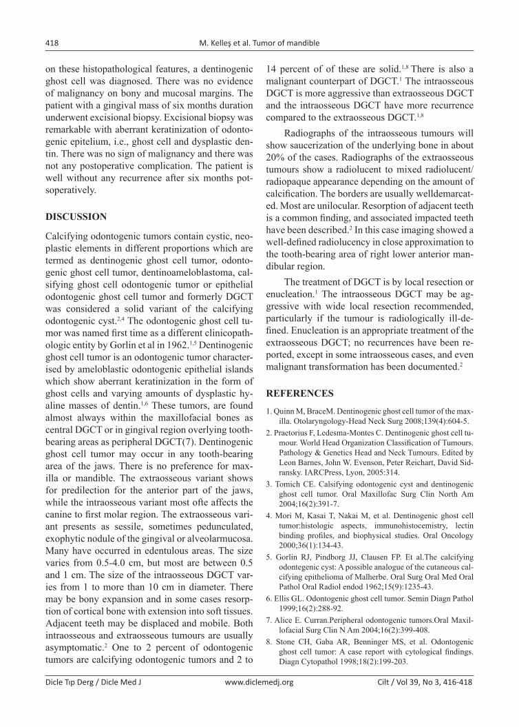
on these histopathological features, a dentinogenic ghost cell was diagnosed. There was no evidence of malignancy on bony and mucosal margins. The patient with a gingival mass of six months duration underwent excisional biopsy. Excisional biopsy was remarkable with aberrant keratinization of odonto-genic epitelium, i.e., ghost cell and dysplastic den-tin. There was no sign of malignancy and there was not any postoperative complication. The patient is well without any recurrence after six months pot-soperatively.
DISCUSSION
Calcifying odontogenic tumors contain cystic, neo-plastic elements in different proportions which are termed as dentinogenic ghost cell tumor, odonto-genic ghost cell tumor, dentinoameloblastoma, cal-sifying ghost cell odontogenic tumor or epithelial odontogenic ghost cell tumor and formerly DGCT was considered a solid variant of the calcifying odontogenic cyst.2,4 The odontogenic ghost cell tu-mor was named first time as a different clinicopath-ologic entity by Gorlin et al in 1962.1,5 Dentinogenic ghost cell tumor is an odontogenic tumor character-ised by ameloblastic odontogenic epithelial islands which show aberrant keratinization in the form of ghost cells and varying amounts of dysplastic hy-aline masses of dentin.1,6 These tumors, are found almost always within the maxillofacial bones as central DGCT or in gingival region overlying tooth-bearing areas as peripheral DGCT(7). Dentinogenic ghost cell tumor may occur in any tooth-bearing area of the jaws. There is no preference for max-illa or mandible. The extraosseous variant shows for predilection for the anterior part of the jaws, while the intraosseous variant most ofte affects the canine to first molar region. The extraosseous vari-ant presents as sessile, sometimes pedunculated, exophytic nodule of the gingival or alveolarmucosa. Many have occurred in edentulous areas. The size varies from 0.5-4.0 cm, but most are between 0.5 and 1 cm. The size of the intraosseous DGCT var-ies from 1 to more than 10 cm in diameter. There may be bony expansion and in some cases resorp-tion of cortical bone with extension into soft tissues. Adjacent teeth may be displaced and mobile. Both intraosseous and extraosseous tumours are usually asymptomatic.2 One to 2 percent of odontogenic tumors are calcifying odontogenic tumors and 2 to
14 percent of of these are solid.1,8 There is also a malignant counterpart of DGCT.1 The intraosseous DGCT is more aggressive than extraosseous DGCT and the intraosseous DGCT have more recurrence compared to the extraosseous DGCT.1,8
Radiographs of the intraosseous tumours will show saucerization of the underlying bone in about 20% of the cases. Radiographs of the extraosseous tumours show a radiolucent to mixed radiolucent/radiopaque appearance depending on the amount of calcification. The borders are usually welldemarcat-ed. Most are unilocular. Resorption of adjacent teeth is a common finding, and associated impacted teeth have been described.2 In this case imaging showed a well-defined radiolucency in close approximation to the tooth-bearing area of right lower anterior man-dibular region.
The treatment of DGCT is by local resection or enucleation.1 The intraosseous DGCT may be ag-gressive with wide local resection recommended, particularly if the tumour is radiologically ill-de-fined. Enucleation is an appropriate treatment of the extraosseous DGCT; no recurrences have been re-ported, except in some intraosseous cases, and even malignant transformation has been documented.2
REFERENCES
1. Quinn M, BraceM. Dentinogenic ghost cell tumor of the max-illa. Otolaryngology-Head Neck Surg 2008;139(4):604-5.
2. Praetorius F, Ledesma-Montes C. Dentinogenic ghost cell tu-mour. World Head Organization Classification of Tumours. Pathology & Genetics Head and Neck Tumours. Edited by Leon Barnes, John W. Evenson, Peter Reichart, David Sid-ransky. IARCPress, Lyon, 2005:314.
3. Tomich CE. Calsifying odontogenic cyst and dentinogenic ghost cell tumor. Oral Maxillofac Surg Clin North Am 2004;16(2):391-7.
4. Mori M, Kasai T, Nakai M, et al. Dentinogenic ghost cell tumor:histologic aspects, immunohistocemistry, lectin binding profiles, and biophysical studies. Oral Oncology 2000;36(1):134-43.
5. Gorlin RJ, Pindborg JJ, Clausen FP. Et al.The calcifying odontegenic cyst: A possible analogue of the cutaneous cal-cifying epithelioma of Malherbe. Oral Surg Oral Med Oral Pathol Oral Radiol endod 1962;15(9):1235-43.
6. Ellis GL. Odontogenic ghost cell tumor. Semin Diagn Pathol 1999;16(2):288-92.
7. Alice E. Curran.Peripheral odontogenic tumors.Oral Maxil-lofacial Surg Clin N Am 2004;16(2):399-408.
8. Stone CH, Gaba AR, Benninger MS, et al. Odontogenic ghost cell tumor: A case report with cytological findings. Diagn Cytopathol 1998;18(2):199-203.