Embed Size (px)
Citation preview
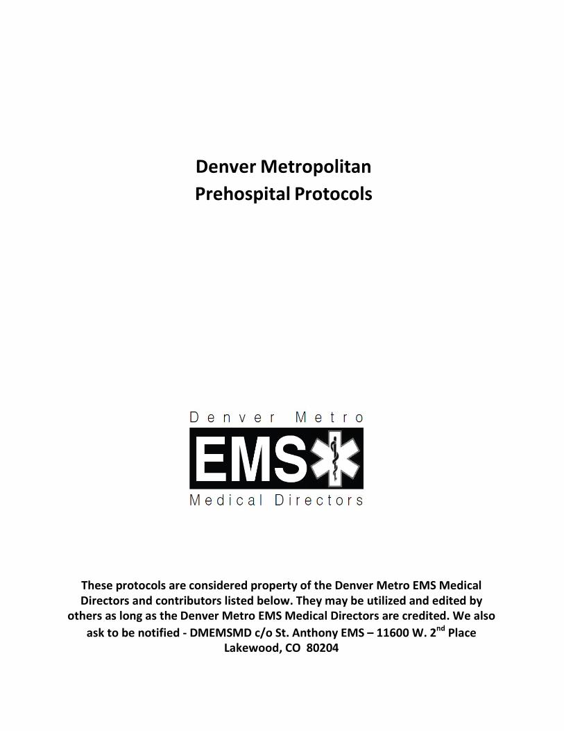
Denver Metropolitan Prehospital Protocols
These protocols are considered property of the Denver Metro EMS Medical Directors and contributors listed below. They may be utilized and edited by
others as long as the Denver Metro EMS Medical Directors are credited. We also ask to be notified -‐ DMEMSMD c/o St. Anthony EMS – 11600 W. 2nd Place
Lakewood, CO 80204
Table of Contents
0001 General Guidelines: Introduction 0002 General Guidelines: Confidentiality 0003 General Guidelines: Consent 0004 General Guidelines: Physician on Scene 0005 General Guidelines: Termination of Resuscitation & field pronouncement 0006 General Guidelines: Advanced medical directives 0007 General Guidelines: Patient determination 0008 General Guidelines: Non-transport/refusal 00090010
General Guidelines: Emergency Department divert and advisoryGeneral Guidelines: Mandatory Reporting of Abuse Patients
Procedural Protocols
0100 Orotracheal Intubation 0110 Nasotracheal Intubation 0120 Percutaneous Cricothyrotomy0121 Bougie Assisted Surgical Cricothyrotomy0130 King Airway 0140 Continuous Positive Airway Pressure (CPAP) 0150 Capnography 0160 Synchronized Cardioversion 0170 Transcutaneous Cardiac Pacing 0180 Therapeutic Induced Hypothermia after Cardiac Arrest 0190 Restraints 0200 Tourniquets 0210 Needle Thoracostomy for Tension Pneumonthorax Decompression 0220 Intraosseus Cathether Placement 0230 Epistaxis Management 0240 TASER Probe Removal
Protocols
1010 Obstructed Airway
2000 Cardiac Arrest: General Principles 2020 ALS Pulseless Arrest 2030 Tachyarrhythmia with Poor Perfusion 2040 Bradyarrhythmia with Poor Perfusion 2050 Adult Chest Pain 2051 Cardiac Alert 2100 Hypertension
3010 Universal Respiratory Distress 3020 Adult Asthma 3030 COPD 3050 CHF/Pulmonary Edema
4011 Stroke 4012 Universal Altered Mental Status 4013 Adult Seizure 4014 Hypoglycemia 4015 Alcohol Intoxication 4020 Allergy and Anaphylaxis 4030 Abdominal Pain/Vomiting

4040 Overdose and Acute Poisoning 4051 High Altitude Illness 4052 Drowning 4053 Hypothermia 4054 Environmental Hyperthermia 4055 Insect/Arachnid Bites and Stings 4056 Snake Bites 4060 Medical Hypotension/Shock 4061 Adrenal Insufficiency 4070 Psychiatric/Behavioral Patient 4075 Agitated/Combative Patient 4076 Transport of the Handcuffed Patient 4080 Childbirth 4081 Obstetrical Complications
5000 General Trauma Care 5005 Special Trauma Scenarios 5006 Trauma in Pregnancy 5010 Adult Traumatic Pulseless Arrest 5015 Traumatic Shock 5020 Amputations 5030 Head Trauma 5040 Face and Neck Trauma 5050 Adult Spinal Trauma 5055 Adult Spinal Immobilization5056 Suspected Spinal Injury with Athletic Equipment in Place 5060 Chest Trauma 5070 Abdominal Trauma 5090 Burns
6000 General Guidelines for Pediatric Patients 6005 Pediatric Seizure 6010 Pediatric Cardiac Arrest – General Principles 6015 Pediatric Pulseless Arrest – BLS 6020 Pediatric Tachycardia with Poor Perfusion 6021 Pediatric Bradycardia with Poor Perfusion 6025 Neonatal Resuscitation 6026 Neonatal Consideration 6030 Pediatric Pulseless Arrest – ALS 6040 Care of the Child with Special Needs 6050 Pediatric Universal Respiratory Distress 6060 Pediatric Apparent Life-Threatening Event (ALTE) 6070 Pediatric Trauma Considerations
Medications
7010 Adenosine (Adenocard) 7010 Albuterol 7010 Amiodarone (Cordarone) 7010 Antiemetics (Ondansetron, Promethazine) 7010 Aspirin 7010 Atropine Sulfate 7010 Benzodiazepines 7010 Calcium Gluconate 7010 Dextrose 50% 7010 Diphenhydramine (Benadryl) 7010 Dopamine 7010 Droperidol 7010 Epinephrine 7010 Furosemide (Lasix) 7010 Glucagon
7010 Haloperidol7010 Hydroxycobalamin (Cyanokit) 7010 Ipratropium Bromide (Atrovent) 7010 Lidocaine 2% Solution 7010 Magnesium Sulfate 7010 Methylprednisolone (Solu-Medrol) 7010 Naloxone (Narcan) 7010 Nitroglycerine (Nitrostat, Nitroquick) 7010 Opiods (Fentanyl, Morphine, Hydromorphone) 7010 Oral Glucose (Glutose, Insta-glucose) 7010 Oxygen 7010 Phenylephrine (Intranasal) 7010 Racemic Epinephrine (Vaponephrine) 7010 Sodium Bicarbonate 7010 Topical Ophthalmic Anesthetics
The process that has been initiated in the construction of this revised set of protocols will remain in place. The authors will continue to edit and revise the protocols to reflect the dynamic role of emergency medical services within the medical care community.
The authors would like to acknowledge the following for their contribution, talent and time during this revision of the Denver Metro EMS protocols.
Denver Metro EMS Medical Directors
Jeff Beckman, M.D. Carl Bonnett, M.D. Christopher Colwell, M.D. Eugene Eby, M.D. Brian Erling, M.D. Josh Heller, M.D. Dylan Luyten, M.D. Maria Mandt, M.D.
Kevin McVaney, M.D. Scott Miner, M.D. Gilbert Pineda, M.D. Lara Rappaport, M.D. John Riccio, M.D. Fred Severyn, M.D. W. Peter Vellman, M.D.
Special thanks to the following:
Nick Boukas, MPA, NREMT-P
Heidi Cabell, EMT-P
Thomas Candlin III, EMT-P
Todd Dorfman, M.D.
Barbara Foster, BA, EMT-P
Paul Fuller, EMT-P
Steve Green, EMT-P
Michelle Loop, BS, NREMT-P
Randy Pennington, EMT-P
Scott Phillips, BS, NREMT-P
Ross Riley, EMT-P
David Sanko, BA, NREMT-P
Erin Selby, BA, EMT-P
Don Tucker, EMT-P
Ralph Vickery, EMT-P
Mark Warth, NREMT-P
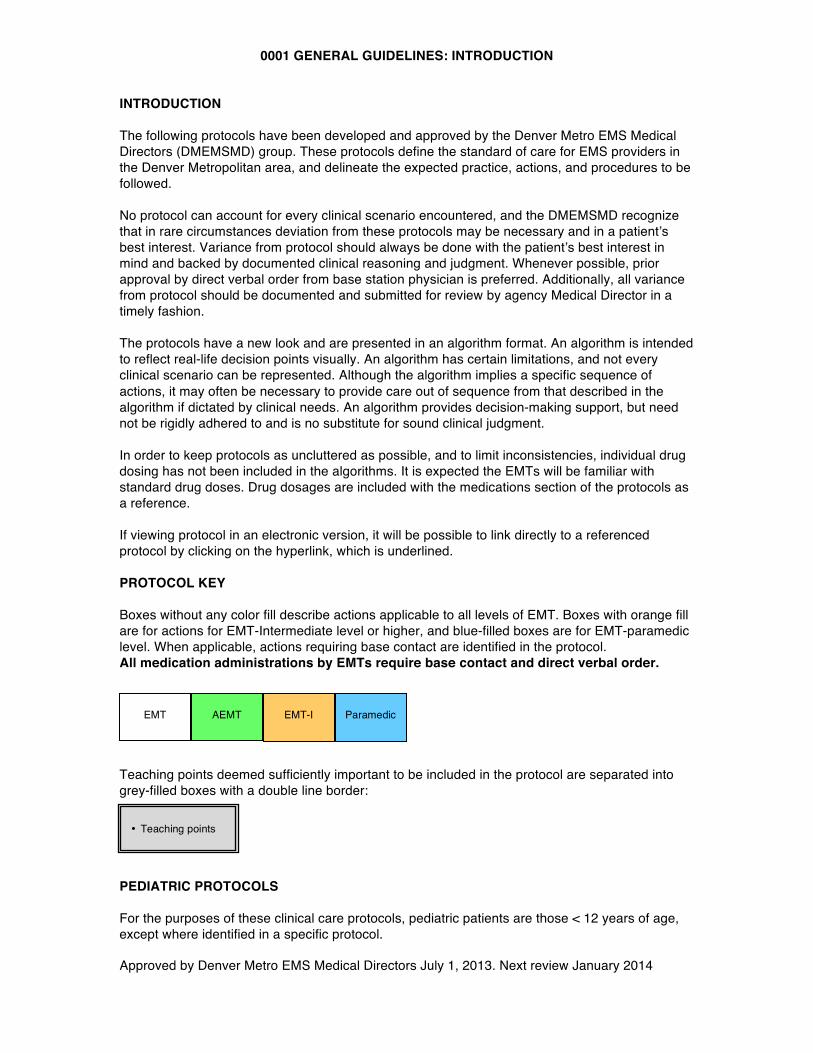
0001 GENERAL GUIDELINES: INTRODUCTION
Approved by Denver Metro EMS Medical Directors July 1, 2013. Next review January 2014
EMT-I Paramedic EMT AEMT
INTRODUCTION The following protocols have been developed and approved by the Denver Metro EMS Medical Directors (DMEMSMD) group. These protocols define the standard of care for EMS providers in the Denver Metropolitan area, and delineate the expected practice, actions, and procedures to be followed. No protocol can account for every clinical scenario encountered, and the DMEMSMD recognize that in rare circumstances deviation from these protocols may be necessary and in a patient’s best interest. Variance from protocol should always be done with the patient’s best interest in mind and backed by documented clinical reasoning and judgment. Whenever possible, prior approval by direct verbal order from base station physician is preferred. Additionally, all variance from protocol should be documented and submitted for review by agency Medical Director in a timely fashion. The protocols have a new look and are presented in an algorithm format. An algorithm is intended to reflect real-life decision points visually. An algorithm has certain limitations, and not every clinical scenario can be represented. Although the algorithm implies a specific sequence of actions, it may often be necessary to provide care out of sequence from that described in the algorithm if dictated by clinical needs. An algorithm provides decision-making support, but need not be rigidly adhered to and is no substitute for sound clinical judgment. In order to keep protocols as uncluttered as possible, and to limit inconsistencies, individual drug dosing has not been included in the algorithms. It is expected the EMTs will be familiar with standard drug doses. Drug dosages are included with the medications section of the protocols as a reference. If viewing protocol in an electronic version, it will be possible to link directly to a referenced protocol by clicking on the hyperlink, which is underlined. PROTOCOL KEY Boxes without any color fill describe actions applicable to all levels of EMT. Boxes with orange fill are for actions for EMT-Intermediate level or higher, and blue-filled boxes are for EMT-paramedic level. When applicable, actions requiring base contact are identified in the protocol. All medication administrations by EMTs require base contact and direct verbal order. Teaching points deemed sufficiently important to be included in the protocol are separated into grey-filled boxes with a double line border:
PEDIATRIC PROTOCOLS For the purposes of these clinical care protocols, pediatric patients are those < 12 years of age, except where identified in a specific protocol.
• Teaching points
0002 GENERAL GUIDELINES: CONFIDENTIALITY
Approved by Denver Metro EMS Medical Directors July 1, 2013. Next review January 2014
CONFIDENTIALITY A. The patient-physician relationship, the patient-registered nurse relationship, and the
patient-EMT relationship are recognized as privileged. This means that the physician, nurse, or EMT may not testify as to confidential communications unless:
1. The patient consents 2. The disclosure is allowable by law (such as Medical Board or Nursing Board
proceedings, or criminal or civil litigation in which the patient's medical condition is in issue)
B. The prehospital provider must keep the patient's medical information confidential. The patient likely has an expectation of privacy, and trusts that personal, medical information will not be disclosed by medical personnel to any person not directly involved in the patient's medical treatment.
1. Exceptions i. The patient is not entitled to confidentiality of information that does not
pertain to the medical treatment, medical condition, or is unnecessary for diagnosis or treatment.
ii. The patient is not entitled to confidentiality for disclosures made publicly. iii. The patient is not entitled to confidentiality with regard to evidence of a
crime. C. Additional Considerations:
1. Any disclosure of medical information should not be made unless necessary for the treatment, evaluation or diagnosis of the patient.
2. Any disclosures made by any person, medical personnel, the patient, or law enforcement should be treated as limited disclosures and not authorizing further disclosures to any other person.
3. Any discussions of prehospital care by and between the receiving hospital, the crewmembers in attendance, or at in-services or audits are done strictly for educational or performance improvement purposes. Further disclosures are not authorized.
4. Radio communications should not include disclosure of patient names. 5. This procedure does not preclude or supersede your agency’s HIPAA policy and
procedures.
0003 GENERAL GUIDELINES: CONSENT
Approved by Denver Metro EMS Medical Directors July 1, 2013. Next review January 2014
CONSENT General Principles: Adults A. An adult in the State of Colorado is 18 years of age or older. B. Every adult is presumed capable of making medical treatment decisions. This includes
the right to make "bad" decisions that the prehospital provider believes are not in the best interests of the patient.
C. A person is deemed to have decision-making capacity if he/she has the ability to provide informed consent, i.e., the patient:
1. Understands the nature of the illness/injury or risk of injury/illness 2. Understands the possible consequences of delaying treatment and/or refusing
transport 3. Given the risks and options, the patient voluntarily refuses or accepts treatment
and/or transport. D. A call to 9-1-1 itself does not prevent a patient from refusing treatment. A patient may
refuse medical treatment (IVs, oxygen, medications), but you should try to inform the patient of the need for therapies, offer again, and treat to the extent possible.
E. The odor of alcohol on a patient’s breath does not, by itself, prevent a patient from refusing treatment.
F. Implied Consent: An unconscious adult is presumed to consent to treatment for life-threatening injuries/illnesses.
G. Involuntary Consent: a person other than the patient in rare circumstances may authorize Consent. This may include a court order (guardianship), authorization by a law enforcement officer for prisoners in custody or detention, or for persons under a mental health hold or commitment who are a danger to themselves or others or are gravely disabled.
Procedure: Adults A. Consent may be inferred by the patient's actions or by express statements. If you are not
sure that you have consent, clarify with the patient or CONTACT BASE. This may include consent for treatment decisions or transport/destination decisions.
B. Determining whether or not a patient has decision-making capacity to consent or refuse medical treatment in the prehospital setting can be very difficult. Every effort should be made to determine if the patient has decision-making capacity, as defined above.
C. For patients who do not have decision-making capacity, CONTACT BASE. D. If the patient lacks decision-making capacity and the patient's life or health is in danger,
and there is no reasonable ability to obtain the patient's consent, proceed with transport and treatment of life-threatening injuries/illnesses. If you are not sure how to proceed, CONTACT BASE.
E. For patients who refuse medical treatment, if you are unsure whether or not a situation of involuntary consent applies, CONTACT BASE.
General Principles: Minors A. A parent, including a parent who is a minor, may consent to medical or emergency
treatment of his/her child. There are exceptions: 1. Neither the child nor the parent may refuse medical treatment on religious
grounds if the child is in imminent danger as a result of not receiving medical treatment, or when the child is in a life-threatening situation, or when the condition will result in serious handicap or disability.
2. The consent of a parent is not necessary to authorize hospital or emergency
0003 GENERAL GUIDELINES: CONSENT
Approved by Denver Metro EMS Medical Directors July 1, 2013. Next review January 2014
health care when an EMT in good faith relies on a minor's consent, if the minor is at least 15 years of age and emancipated or married.
3. Minors may seek treatment for abortion, drug addiction, and venereal disease without consent of parents. Minors > 15 years may seek treatment for mental health.
B. When in doubt, your actions should be guided by what is in the minor's best interests and base contact.
Procedure: Minors
A. A parent or legal guardian may provide consent to or refuse treatment in a non- life-threatening situation.
B. When the parent is not present to consent or refuse: 1. If a minor has an injury or illness, but not a life-threatening medical emergency,
you should attempt to contact the parent(s) or legal guardian. If this cannot be done promptly, transport.
2. If the child does not need transport, they can be left at the scene in the custody of a responsible adult (e.g., teacher, social worker, grandparent). It should only be in very rare circumstances that a child of any age is left at the scene if the parent is not also present.
3. If the minor has a life-threatening injury or illness, transport and treat per protocols. If the parent objects to treatment, CONTACT BASE immediately and treat to the extent allowable, and notify police to respond and assist.
0004 GENERAL GUIDELINES: PHYSICIAN AT THE SCENE/MEDICAL DIRECTION
Approved by Denver Metro EMS Medical Directors July 1, 2013. Next review January 2014
PHYSICIAN AT THE SCENE/MEDICAL DIRECTION Purpose
A. To provide guidelines for prehospital personnel who encounter a physician at the scene
of an emergency
General Principles A. The prehospital provider has a duty to respond to an emergency, initiate treatment, and
conduct an assessment of the patient to the extent possible. B. A physician who voluntarily offers or renders medical assistance at an emergency scene
is generally considered a "Good Samaritan." However, once a physician initiates treatment, he/she may feel a physician-patient relationship has been established.
C. Good patient care should be the focus of any interaction between prehospital care providers and the physician.
Procedure A. See algorithm below and sample note to physician at the scene
Special notes A. Every situation may be different, based on the physician, the scene, and the condition of
the patient. B. CONTACT BASE when any question(s) arise.
0004 GENERAL GUIDELINES: PHYSICIAN AT THE SCENE/MEDICAL DIRECTION
Approved by Denver Metro EMS Medical Directors July 1, 2013. Next review January 2014
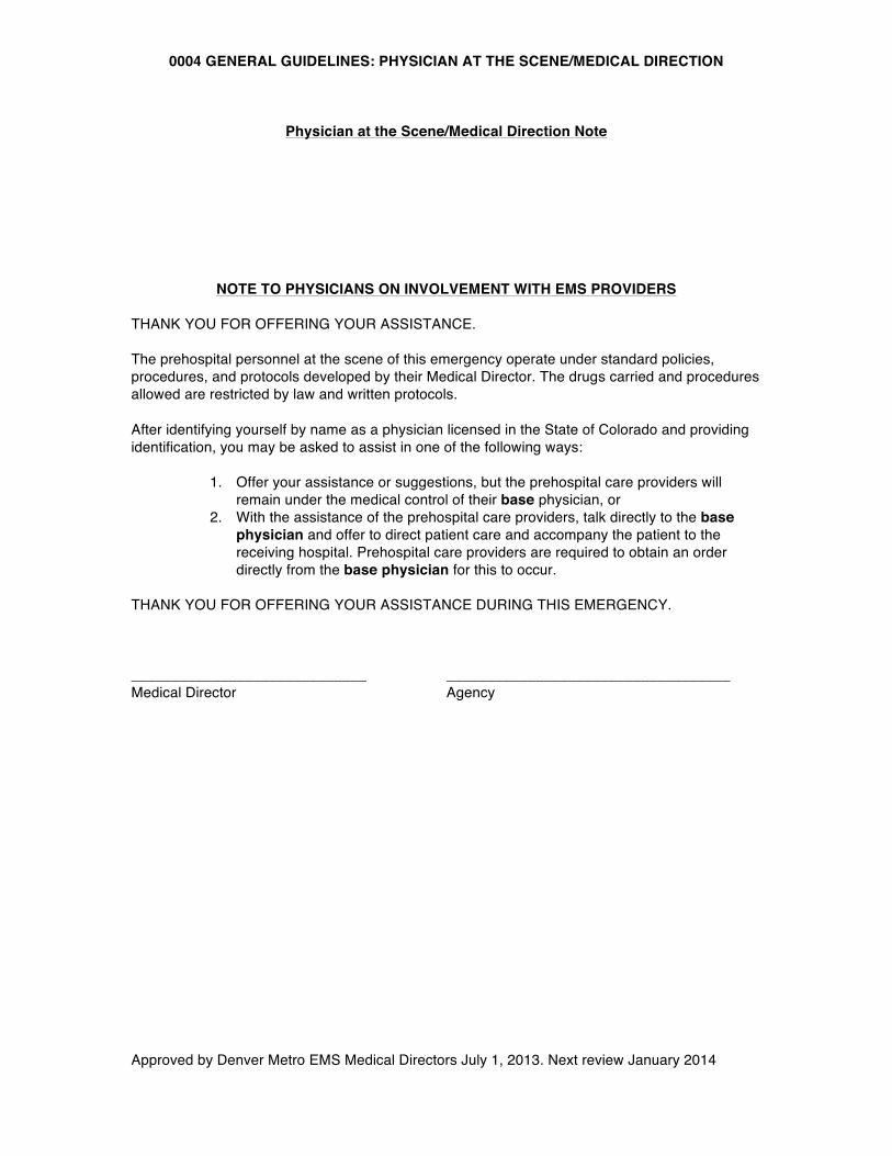
Physician at the Scene/Medical Direction Note
NOTE TO PHYSICIANS ON INVOLVEMENT WITH EMS PROVIDERS
THANK YOU FOR OFFERING YOUR ASSISTANCE. The prehospital personnel at the scene of this emergency operate under standard policies, procedures, and protocols developed by their Medical Director. The drugs carried and procedures allowed are restricted by law and written protocols. After identifying yourself by name as a physician licensed in the State of Colorado and providing identification, you may be asked to assist in one of the following ways:
1. Offer your assistance or suggestions, but the prehospital care providers will remain under the medical control of their base physician, or
2. With the assistance of the prehospital care providers, talk directly to the base physician and offer to direct patient care and accompany the patient to the receiving hospital. Prehospital care providers are required to obtain an order directly from the base physician for this to occur.
THANK YOU FOR OFFERING YOUR ASSISTANCE DURING THIS EMERGENCY. _____________________________ ___________________________________ Medical Director Agency
0004 GENERAL GUIDELINES: PHYSICIAN AT THE SCENE/MEDICAL DIRECTION
Approved by Denver Metro EMS Medical Directors July 1, 2013. Next review January 2014
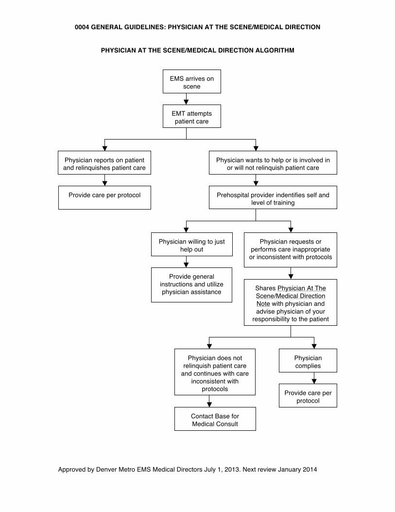
PHYSICIAN AT THE SCENE/MEDICAL DIRECTION ALGORITHM
EMT attempts patient care
EMS arrives on scene
Physician reports on patient and relinquishes patient care
Provide care per protocol
Physician wants to help or is involved in or will not relinquish patient care
Prehospital provider indentifies self and level of training
Physician willing to just help out
Provide general instructions and utilize physician assistance
Physician requests or performs care inappropriate
or inconsistent with protocols
Shares Physician At The Scene/Medical Direction Note with physician and advise physician of your
responsibility to the patient
Physician does not relinquish patient care
and continues with care inconsistent with
protocols
Contact Base for Medical Consult
Physician complies
Provide care per protocol
0005 GENERAL GUIDELINES: TERMINATION OF RESUSCITATION AND FIELD PRONOUNCEMENT GUIDELINES
Approved by Denver Metro EMS Medical Directors July 1, 2013. Next review January 2014
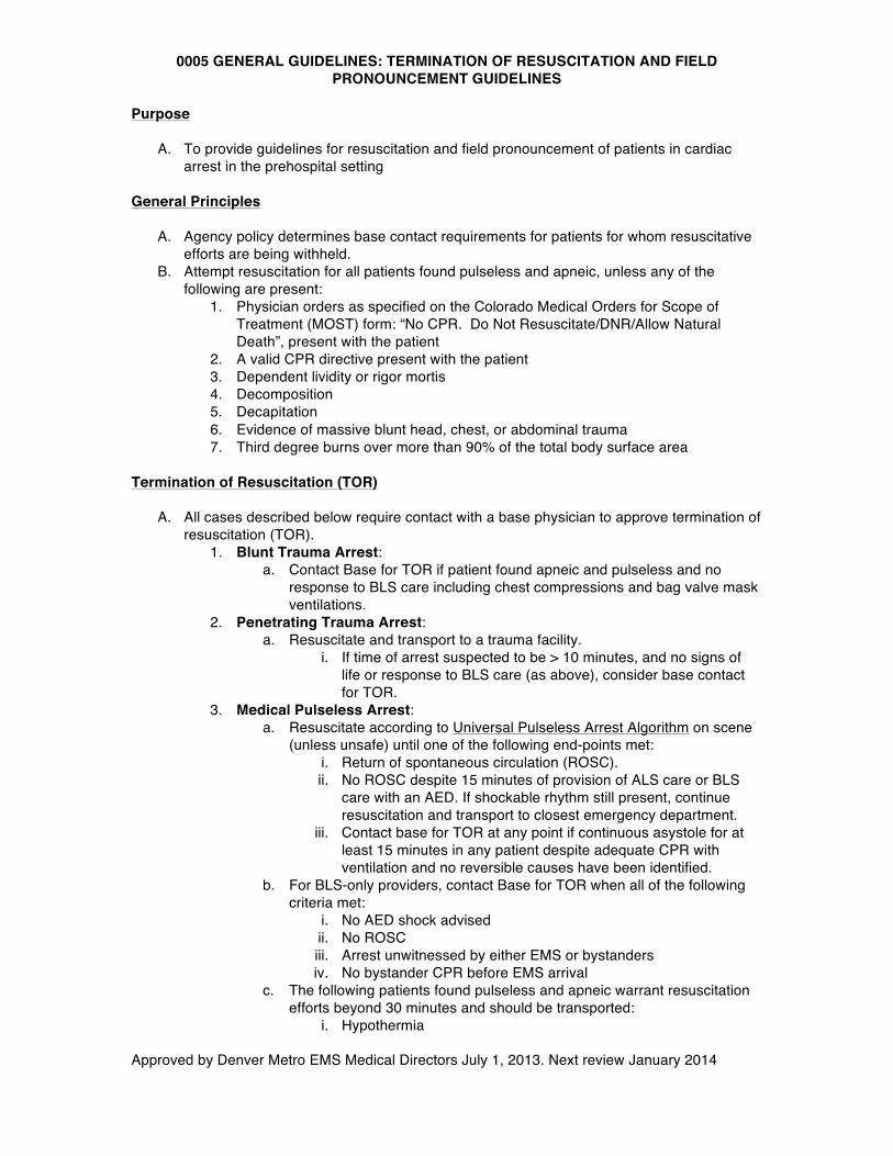
Purpose A. To provide guidelines for resuscitation and field pronouncement of patients in cardiac
arrest in the prehospital setting
General Principles
A. Agency policy determines base contact requirements for patients for whom resuscitative efforts are being withheld.
B. Attempt resuscitation for all patients found pulseless and apneic, unless any of the following are present:
1. Physician orders as specified on the Colorado Medical Orders for Scope of Treatment (MOST) form: “No CPR. Do Not Resuscitate/DNR/Allow Natural Death”, present with the patient
2. A valid CPR directive present with the patient 3. Dependent lividity or rigor mortis 4. Decomposition 5. Decapitation 6. Evidence of massive blunt head, chest, or abdominal trauma 7. Third degree burns over more than 90% of the total body surface area
Termination of Resuscitation (TOR)
A. All cases described below require contact with a base physician to approve termination of
resuscitation (TOR). 1. Blunt Trauma Arrest:
a. Contact Base for TOR if patient found apneic and pulseless and no response to BLS care including chest compressions and bag valve mask ventilations.
2. Penetrating Trauma Arrest: a. Resuscitate and transport to a trauma facility.
i. If time of arrest suspected to be > 10 minutes, and no signs of life or response to BLS care (as above), consider base contact for TOR.
3. Medical Pulseless Arrest: a. Resuscitate according to Universal Pulseless Arrest Algorithm on scene
(unless unsafe) until one of the following end-points met: i. Return of spontaneous circulation (ROSC). ii. No ROSC despite 15 minutes of provision of ALS care or BLS
care with an AED. If shockable rhythm still present, continue resuscitation and transport to closest emergency department.
iii. Contact base for TOR at any point if continuous asystole for at least 15 minutes in any patient despite adequate CPR with ventilation and no reversible causes have been identified.
b. For BLS-only providers, contact Base for TOR when all of the following criteria met:
i. No AED shock advised ii. No ROSC iii. Arrest unwitnessed by either EMS or bystanders iv. No bystander CPR before EMS arrival
c. The following patients found pulseless and apneic warrant resuscitation efforts beyond 30 minutes and should be transported:
i. Hypothermia
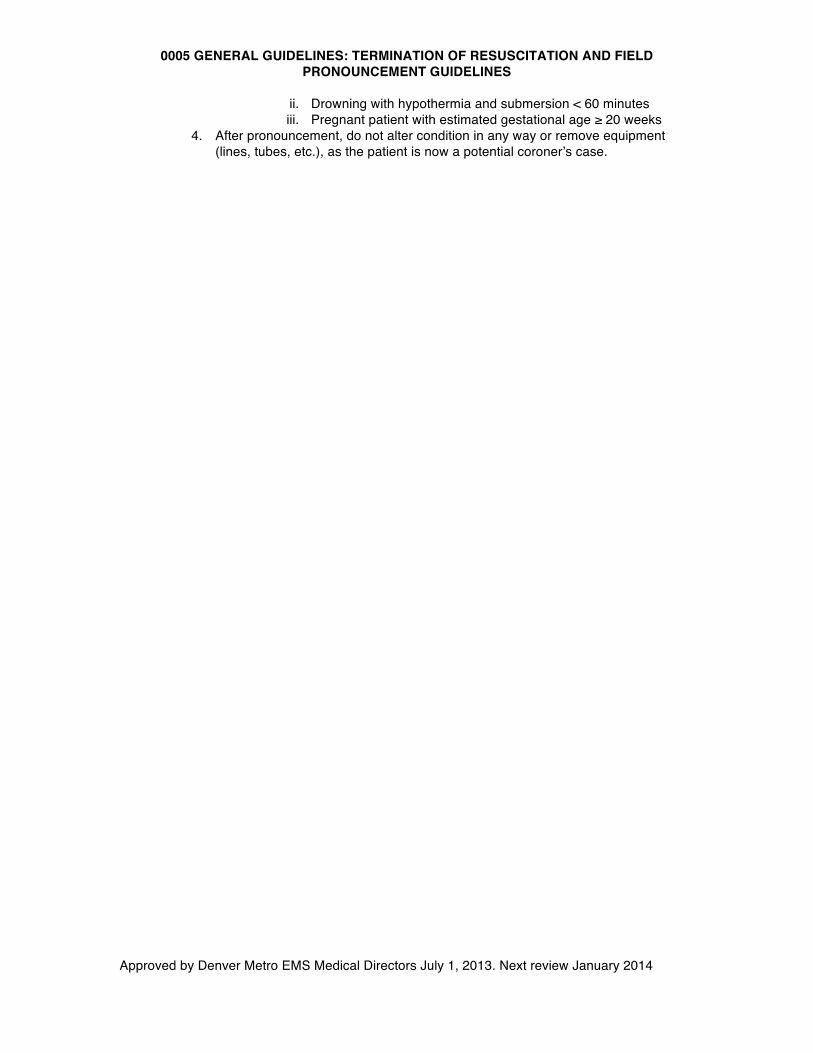
0005 GENERAL GUIDELINES: TERMINATION OF RESUSCITATION AND FIELD PRONOUNCEMENT GUIDELINES
Approved by Denver Metro EMS Medical Directors July 1, 2013. Next review January 2014
ii. Drowning with hypothermia and submersion < 60 minutes iii. Pregnant patient with estimated gestational age ≥ 20 weeks
4. After pronouncement, do not alter condition in any way or remove equipment (lines, tubes, etc.), as the patient is now a potential coroner’s case.
0006 GENERAL GUIDELINES: ADVANCED MEDICAL DIRECTIVES
Approved by Denver Metro EMS Medical Directors July 1, 2013. Next review January 2014
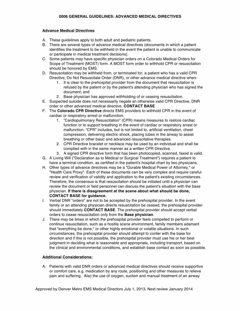
Advance Medical Directives A. These guidelines apply to both adult and pediatric patients. B. There are several types of advance medical directives (documents in which a patient
identifies the treatment to be withheld in the event the patient is unable to communicate or participate in medical treatment decisions).
C. Some patients may have specific physician orders on a Colorado Medical Orders for Scope of Treatment (MOST) form. A MOST form order to withhold CPR or resuscitation should be honored by EMS.
D. Resuscitation may be withheld from, or terminated for, a patient who has a valid CPR Directive, Do Not Resuscitate Order (DNR), or other advance medical directive when:
1. It is clear to the prehospital provider from the document that resuscitation is refused by the patient or by the patient's attending physician who has signed the document; and
2. Base physician has approved withholding of or ceasing resuscitation. E. Suspected suicide does not necessarily negate an otherwise valid CPR Directive, DNR
order or other advanced medical directive. CONTACT BASE F. The Colorado CPR Directive directs EMS providers to withhold CPR in the event of
cardiac or respiratory arrest or malfunction. 1. “Cardiopulmonary Resuscitation” (CPR) means measures to restore cardiac
function or to support breathing in the event of cardiac or respiratory arrest or malfunction. “CPR” includes, but is not limited to, artificial ventilation, chest compression, delivering electric shock, placing tubes in the airway to assist breathing or other basic and advanced resuscitative therapies.
2. CPR Directive bracelet or necklace may be used by an individual and shall be complied with in the same manner as a written CPR Directive.
3. A signed CPR directive form that has been photocopied, scanned, faxed is valid. G. A Living Will ("Declaration as to Medical or Surgical Treatment") requires a patient to
have a terminal condition, as certified in the patient's hospital chart by two physicians. H. Other types of advance directives may be a "Durable Medical Power of Attorney," or
"Health Care Proxy". Each of these documents can be very complex and require careful review and verification of validity and application to the patient's existing circumstances. Therefore, the consensus is that resuscitation should be initiated until a physician can review the document or field personnel can discuss the patient’s situation with the base physician. If there is disagreement at the scene about what should be done, CONTACT BASE for guidance.
I. Verbal DNR "orders" are not to be accepted by the prehospital provider. In the event family or an attending physician directs resuscitation be ceased, the prehospital provider should immediately CONTACT BASE. The prehospital provider should accept verbal orders to cease resuscitation only from the Base physician.
J. There may be times in which the prehospital provider feels compelled to perform or continue resuscitation, such as a hostile scene environment, family members adamant that "everything be done," or other highly emotional or volatile situations. In such circumstances, the prehospital provider should attempt to confer with the base for direction and if this is not possible, the prehospital provider must use his or her best judgment in deciding what is reasonable and appropriate, including transport, based on the clinical and environmental conditions, and establish base contact as soon as possible.
Additional Considerations: A. Patients with valid DNR orders or advanced medical directives should receive supportive
or comfort care, e.g. medication by any route, positioning and other measures to relieve pain and suffering. Also the use of oxygen, suction and manual treatment of an airway
0006 GENERAL GUIDELINES: ADVANCED MEDICAL DIRECTIVES
Approved by Denver Metro EMS Medical Directors July 1, 2013. Next review January 2014
obstruction as needed for comfort. B. Mass casualty incidents are not covered in detail by these guidelines. (See State Trauma
Triage Algorithm). C. If the situation appears to be a potential crime scene, EMS providers should disturb the
scene as little as possible and communicate with law enforcement regarding any items that are moved or removed from the scene.
D. Mechanisms for disposition of bodies by means other than EMS providers and vehicles should be prospectively established in each county or locale.
1. In all cases of unattended deaths occurring outside of a medical facility, the coroner should be contacted immediately.
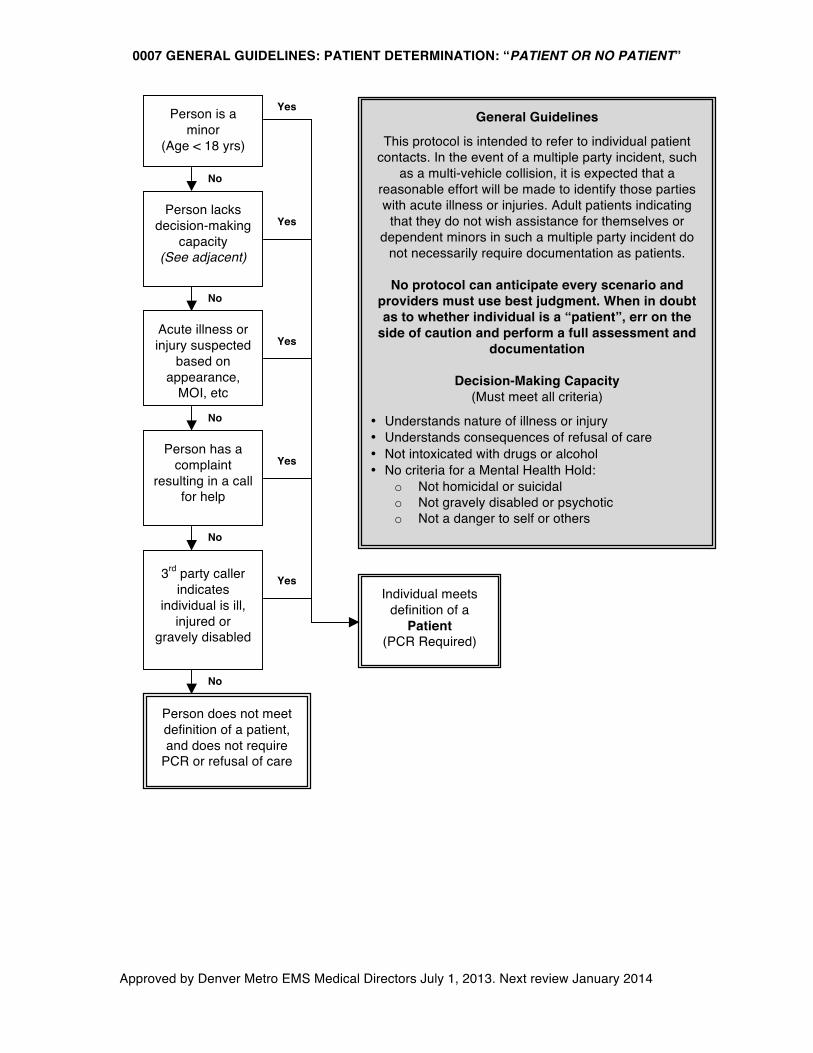
0007 GENERAL GUIDELINES: PATIENT DETERMINATION: “PATIENT OR NO PATIENT”
Approved by Denver Metro EMS Medical Directors July 1, 2013. Next review January 2014
Person has a complaint
resulting in a call for help
Person lacks decision-making
capacity (See adjacent)
Person is a minor
(Age < 18 yrs)
Acute illness or injury suspected
based on appearance,
MOI, etc
3rd party caller indicates
individual is ill, injured or
gravely disabled
No
Yes
No
No
No
Yes
Yes
Yes
Yes
No
General Guidelines
This protocol is intended to refer to individual patient contacts. In the event of a multiple party incident, such
as a multi-vehicle collision, it is expected that a reasonable effort will be made to identify those parties with acute illness or injuries. Adult patients indicating
that they do not wish assistance for themselves or dependent minors in such a multiple party incident do
not necessarily require documentation as patients.
No protocol can anticipate every scenario and providers must use best judgment. When in doubt as to whether individual is a “patient”, err on the
side of caution and perform a full assessment and documentation
Decision-Making Capacity
(Must meet all criteria)
• Understands nature of illness or injury • Understands consequences of refusal of care • Not intoxicated with drugs or alcohol • No criteria for a Mental Health Hold:
o Not homicidal or suicidal o Not gravely disabled or psychotic o Not a danger to self or others
Individual meets definition of a
Patient (PCR Required)
Person does not meet definition of a patient, and does not require
PCR or refusal of care
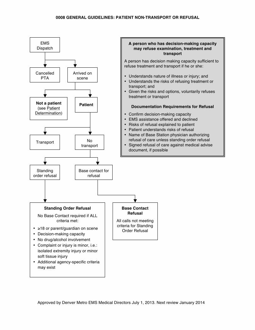
0008 GENERAL GUIDELINES: PATIENT NON-TRANSPORT OR REFUSAL
Approved by Denver Metro EMS Medical Directors July 1, 2013. Next review January 2014
EMS Dispatch
Cancelled PTA
Arrived on scene
Patient Not a patient (see Patient
Determination)
No transport
Transport
Standing order refusal
Base contact for refusal
Base Contact Refusal
All calls not meeting criteria for Standing
Order Refusal
Standing Order Refusal
No Base Contact required if ALL criteria met:
• ≥18 or parent/guardian on scene • Decision-making capacity • No drug/alcohol involvement • Complaint or injury is minor, i.e.:
isolated extremity injury or minor soft tissue injury
• Additional agency-specific criteria may exist
A person who has decision-making capacity may refuse examination, treatment and
transport
A person has decision making capacity sufficient to refuse treatment and transport if he or she: • Understands nature of illness or injury; and • Understands the risks of refusing treatment or
transport; and • Given the risks and options, voluntarily refuses
treatment or transport
Documentation Requirements for Refusal
• Confirm decision-making capacity • EMS assistance offered and declined • Risks of refusal explained to patient • Patient understands risks of refusal • Name of Base Station physician authorizing
refusal of care unless standing order refusal • Signed refusal of care against medical advise
document, if possible
0009 GENERAL GUIDELINES: EMERGENCY DEPARTMENT DIVERT AND ADVISORY
Approved by Denver Metro EMS Medical Directors July 1, 2013. Next review January 2014
Purpose
A. To provide a standard approach to ambulance diversion that is practical for field use
B. To facilitate unobstructed access to hospital emergency departments for ambulance patients
C. To allow for optimal destination policies in keeping with general EMS principles and Colorado State Trauma System Rules and Regulations
General Principles
A. EMSystem, an internet-based tracking system, is used to manage diversion in the Denver Metro area
B. The State Trauma Triage Algorithms should be followed C. The only time an ambulance can be diverted from a hospital is when that hospital
is posted on EMSystem as being on official divert (RED) status. D. Overriding factors: the following are appropriate reasons for a paramedic to
override ED Divert and, therefore, deliver a patient to an emergency department that is on ED divert:
1. Cardiopulmonary arrest 2. Imminent cardiopulmonary arrest 3. Unmanageable airway emergencies 4. Unstable trauma and burn patients transported to Level I and Level II
Trauma Centers 5. Patients meeting “Cardiac Alert“ criteria (participating hospitals) 6. Patients meeting “Stroke Alert“ criteria (participating hospitals) 7. Imminent delivery
E. Prehospital personnel should honor advisory categories, when possible, considering patient’s condition, travel time, and weather. Patients with specific problems that fall under an advisory category should be transported to a hospital not on that specific advisory when feasible.
F. There are several categories that are considered advisory (yellow) alert categories. These categories are informational only and should alert field personnel that a hospital listed as being on an advisory alert may not be able to optimally care for a patient that falls under that advisory category.
G. The following are advisory (yellow) categories recognized by the State. Individual facilities may not utilize these categories often, or ever:
1. ICU (Intensive Care Unit) 2. Psych (Psychiatric)
H. Zone saturation exists when all hospitals within that zone are on ED Divert. I. A Zone Master is the designated hospital within a Zone responsible for
determining and tracking hospital assignments when the zone is saturated. J. When an ambulance is transporting a patient that the paramedic feels cannot go
outside the zone due to patient acuity or other concerns, the paramedic should contact the Zone Master and request a destination assignment.
K. In general, patients contacted within a zone should be transported to an appropriate facility within the zone. Patients may be transported out of the primary zone at the paramedic’s discretion, if it is in the patient’s best interest or if the transport to an appropriate facility is shorter.
L. The zones, hospitals in each zone, Zone Masters, and the Zone Master contact phone numbers are listed on EMSystem.
0010 GENERAL GUIDELINES: MANDATORY REPORTING OF ABUSE PATIENTS
Approved by Denver Metro EMS Medical Directors July 1, 2013. Next review January 2014
Purpose
A. To provide guidelines for the reporting of suspected abuse patients.
General Principles
A. At-risk adult or pediatric patients who are suspected to be victims of abuse or exploitation, as defined in State Statute and Rule, should be reported in a manner consistent with agency guidelines/procedures.
0100 PROCEDURE PROTOCOL: OROTRACHEAL INTUBATION
Approved by Denver Metro EMS Medical Directors July 1, 2013. Next review January 2014
Indications:
• Respiratory failure • Absence of protective airway reflexes • Present or impending complete airway obstruction • Anticipated prolonged need for positive pressure ventilation
Contraindications:
• There are no absolute contraindications. However, in general the primary goals of airway management are adequate oxygenation and ventilation, and these should be achieved in the least invasive manner possible
o Orotracheal intubation is associated with worse outcomes among pediatric patients and head injured patients when compared to BLS airway maneuvers. Therefore, it is relatively contraindicated in these populations
o Intubation is associated with interruptions in chest compressions during CPR, which is associated with worse patient outcomes. Additionally, intubation itself has not been shown to improve outcomes in cardiac arrest
Technique:
1. Initiate BLS airway sequence 2. Suction airway and pre-oxygenate with BVM ventilations, if possible 3. Check equipment and position patient:
a. If trauma: have assistant hold in-line spinal immobilization in neutral position b. If no trauma, sniffing position or slight cervical hyperextension is preferred
4. Perform laryngoscopy a. To improve laryngeal view, use right hand to manipulate larynx, or have assistant
apply backwards, upwards, rightward pressure (BURP) 5. Place ETT. Confirm tracheal location and appropriate depth and secure tube
a. Correct tube depth may be estimated as 3 times the internal diameter of tube at teeth or gums (e.g: 7.0 ETT is positioned at 21 cm at teeth)
6. Confirm and document tracheal location by: a. ETCO2 b. Presence and symmetry of breath sounds c. Rising SpO2 d. Other means as needed
7. Ventilate with BVM. Assess adequacy of ventilations 8. During transport, continually reassess ventilation, oxygenation and tube position with
continuous ETCO2 and SpO2
Precautions: • Ventilate at age-appropriate rates. Do not hyperventilate • If the intubated patient deteriorates, think “DOPE”
o Dislodgement o Obstruction o Pneumothorax o Equipment failure (no oxygen)
• Reconfirm and document correct tube position after moving patient and before disconnecting from monitor in ED
• Unsuccessful intubation does not equal failed airway management. Many patients cannot be intubated without paralytics. Use King airway or BVM ventilations if 2 attempts at intubation unsuccessful.
EMT-I Paramedic
0110 PROCEDURE PROTOCOL: NASOTRACHEAL INTUBATION
Approved by Denver Metro EMS Medical Directors July 1, 2013. Next review January 2014
Paramedic Indications:
• Age ≥ 12 years spontaneously breathing patient with indication for intubation who cannot tolerate either supine position or laryngoscopy
• Present or impending airway obstruction • Lack of protective airway reflexes • Anticipated prolonged need for positive pressure ventilation
Contraindications:
• Apnea • Severe mid-face trauma
Technique:
1. Initiate BLS airway sequence 2. Suction airway and pre-oxygenate with BVM ventilations, if possible 3. Check equipment, choose correct ETT size (usually 7.0 in adult, limit is size of naris) 4. Position patient with head in midline, neutral position 5. If trauma: cervical collar may be in place, or assistant may hold in-line stabilization in
neutral position 6. If no trauma, patient may be sitting upright 7. Administer phenylephrine nasal drops in each nostril 8. Lubricate ETT with Lidocaine jelly or other water-soluble lubricant 9. With gentle steady pressure, advance the tube through the nose to the posterior pharynx.
Use the largest nostril. Abandon procedure if significant resistance is felt 10. Keeping the curve of the tube exactly in midline, continue advancing slowly 11. There will be slight resistance just before entering trachea. Wait for an inspiratory effort
before final passage through cords. Listen for loss of breath sounds 12. Continue advancing tube until air is definitely exchanging through tube, then advance 2
cm more and inflate cuff 13. Note tube depth and tape securely 14. Confirm and document endotracheal location by:
a. ETCO2 b. Presence and symmetry of breath sounds c. Rising SpO2 d. Other means as needed
15. Ventilate with BVM. Assess adequacy of ventilations 16. During transport, continually reassess ventilation, oxygenation and tube position with
continuous ETCO2 and SpO2
Precautions:
• Before performing BNTI, consider if patient can be safely ventilated with non-invasive means such as CPAP or BVM
• Ventilate at age-appropriate rates. Do not hyperventilate • If the intubated patient deteriorates, think “DOPE”
o Dislodgement o Obstruction o Pneumothorax o Equipment failure (no oxygen)
• Reconfirm and document correct tube position after moving patient and before disconnecting from monitor in ED
0110 PROCEDURE PROTOCOL: NASOTRACHEAL INTUBATION
Approved by Denver Metro EMS Medical Directors July 1, 2013. Next review January 2014
• Blind nasotracheal intubation is a very gentle technique. The secret to success is perfect positioning and patience.
0120 PROCEDURE PROTOCOL: PERCUTANEOUS CRICOTHYROTOMY
Approved by Denver Metro EMS Medical Directors July 1, 2013. Next review January 2014
Introduction:
• Percutaneous cricothyrotomy is a difficult and hazardous procedure that is to be used only in
extraordinary circumstances as defined below. The reason for performing this procedure must be documented and submitted for review to the EMS Medical Director within 24 hours.
Indications: • A life-threatening condition exists AND advanced airway management is indicated, AND
adequate oxygenation and ventilation cannot be accomplished by other less invasive means. Contraindications:
• Anterior neck hematoma is a relative contraindication • Age < 12 is a relative contraindication Technique:
1. Prepare skin using aseptic solution 2. Position the patient in a supine position, with in-line spinal immobilization if indicated. If
cervical spine injury not suspected, neck extension will improve anatomic view 3. Perform cricothyrotomy according to manufacturer’s instructions for selected device 4. Confirm and document tube placement by:
a. ETCO2 b. Breath sounds c. Rising pulse oximetry d. Other means as needed
5. Ventilate with BVM assessing adequacy of ventilation 6. Observe for subcutaneous air, which may indicate tracheal injury or extra- tracheal tube
position 7. Secure tube with tube ties or device 8. Continually reassess ventilation, oxygenation and tube placement
Precautions: • Success of procedure is dependent on correct identification of cricothyroid membrane • Bleeding will occur, even with correct technique. Straying from the midline is dangerous and
likely to cause hemorrhage
Paramedic
0121 PROCEDURE PROTOCOL: BOUGIE ASSISTED SURGICAL CRICOTHYROTOMY
Approved by Denver Metro EMS Medical Directors July 1, 2013. Next review January 2014
Introduction:
• Surgical cricothyrotomy is a difficult and hazardous procedure that is to be used only in
extraordinary circumstances as defined below. The reason for performing this procedure must be documented and submitted for review to the EMS Medical Director within 24 hours. Surgical cricothyrotomy is to be performed only by paramedics trained in this procedure.
• An endotracheal tube introducer (“bougie”) facilitates this procedure and has the advantage of additional confirmation of tube position and ease of endotracheal tube placement. If no bougie is available the procedure may be performed without a bougie by introducing endotracheal tube or tracheostomy tube directly into cricothyroid membrane.
• Given the rarity and relative unfamiliarity of this procedure it may be helpful to have a medical consult on the phone during the procedure. Consider contacting base for all cricothyroidotomy procedures. Individual Medical Directors may mandate base contact before initiating the procedure. Individual agency policy and procedures apply and providers are responsible for knowing and following these policies.
Indications:
• A life-threatening condition exists AND advanced airway management is indicated AND you are unable to establish an airway or ventilate the patient by any other means.
Contraindications:
• Age < 12 years: for children a percutaneous needle cricothryrotomy with large angiocath
is preferred surgical airway for anatomic reasons Technique:
1. Position the patient supine, with in-line spinal immobilization if indicated. If cervical spine injury not suspected, neck extension will improve anatomic view.
2. Using an aseptic technique (betadine/alcohol wipes), cleanse the area. 3. Standing on the left side of the patient, stabilize the larynx with the thumb and middle
finger of your left hand, and identify the cricothyroid membrane, typically 4 finger-breadths below mandible
4. Using a scalpel, make a 3 cm centimeter vertical incision 0.5 cm deep through the skin and fascia, over the cricothyroid membrane. With finger, dissect the tissue and locate the cricothyroid membrane.
5. Make a horizontal incision through the cricothyroid membrane with the scalpel blade oriented caudal and away from the cords.
6. Insert the bougie curved-tip first through the incision and angled towards the patient’s feet a. If no bougie available, use tracheal hook instrument to lift caudal edge of incision
to facilitate visualization and introduction of ETT directly into trachea and skip to # 9.
7. Advance the bougie into the trachea feeling for “clicks” of tracheal rings and until “hangup” when it cannot be advanced any further. This confirms tracheal position.
8. Advance a 6-0 endotracheal tube over the bougie and into the trachea. It is very easy to place tube in right mainstem bronchus, so carefully assess for symmetry of breath sounds. Remove bougie while stabilizing ETT ensuring it does not become dislodged
9. Ventilate with BVM and 100% oxygen
Paramedic
0121 PROCEDURE PROTOCOL: BOUGIE ASSISTED SURGICAL CRICOTHYROTOMY
Approved by Denver Metro EMS Medical Directors July 1, 2013. Next review January 2014
10. Confirm and document tracheal tube placement as with all advanced airways: ETCO2 as well as clinical indicators e.g.: symmetry of breath sounds, rising pulse oximetry, etc.
11. Secure tube with ties. 12. Observe for subcutaneous air, which may indicate tracheal injury or extra- tracheal tube
position 13. Continually reassess ventilation, oxygenation and tube placement.
Precautions:
• Success of procedure is dependent on correct identification of cricothyroid membrane • Bleeding will occur, even with correct technique. Straying from the midline is dangerous
and likely to cause hemorrhage from the carotid or jugular vessels, or their branches.
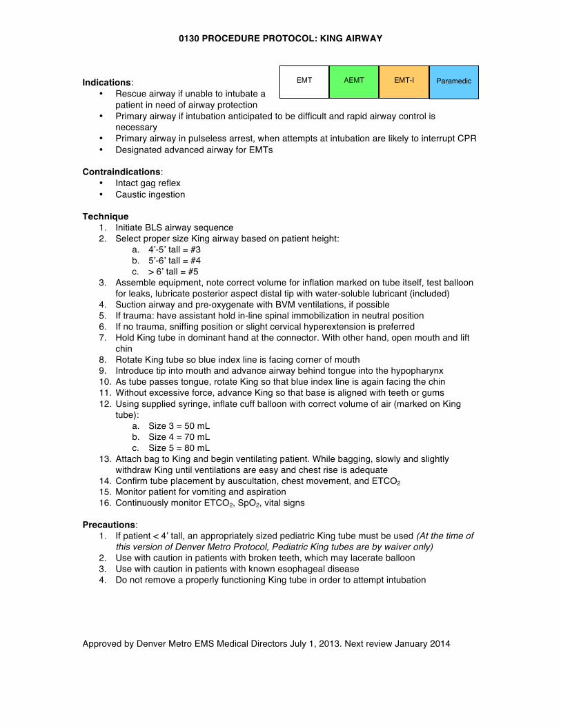
0130 PROCEDURE PROTOCOL: KING AIRWAY
Approved by Denver Metro EMS Medical Directors July 1, 2013. Next review January 2014
Indications:
• Rescue airway if unable to intubate a patient in need of airway protection
• Primary airway if intubation anticipated to be difficult and rapid airway control is necessary
• Primary airway in pulseless arrest, when attempts at intubation are likely to interrupt CPR • Designated advanced airway for EMTs
Contraindications:
• Intact gag reflex • Caustic ingestion
Technique
1. Initiate BLS airway sequence 2. Select proper size King airway based on patient height:
a. 4’-5’ tall = #3 b. 5’-6’ tall = #4 c. > 6’ tall = #5
3. Assemble equipment, note correct volume for inflation marked on tube itself, test balloon for leaks, lubricate posterior aspect distal tip with water-soluble lubricant (included)
4. Suction airway and pre-oxygenate with BVM ventilations, if possible 5. If trauma: have assistant hold in-line spinal immobilization in neutral position 6. If no trauma, sniffing position or slight cervical hyperextension is preferred 7. Hold King tube in dominant hand at the connector. With other hand, open mouth and lift
chin 8. Rotate King tube so blue index line is facing corner of mouth 9. Introduce tip into mouth and advance airway behind tongue into the hypopharynx 10. As tube passes tongue, rotate King so that blue index line is again facing the chin 11. Without excessive force, advance King so that base is aligned with teeth or gums 12. Using supplied syringe, inflate cuff balloon with correct volume of air (marked on King
tube): a. Size 3 = 50 mL b. Size 4 = 70 mL c. Size 5 = 80 mL
13. Attach bag to King and begin ventilating patient. While bagging, slowly and slightly withdraw King until ventilations are easy and chest rise is adequate
14. Confirm tube placement by auscultation, chest movement, and ETCO2 15. Monitor patient for vomiting and aspiration 16. Continuously monitor ETCO2, SpO2, vital signs
Precautions:
1. If patient < 4’ tall, an appropriately sized pediatric King tube must be used (At the time of this version of Denver Metro Protocol, Pediatric King tubes are by waiver only)
2. Use with caution in patients with broken teeth, which may lacerate balloon 3. Use with caution in patients with known esophageal disease 4. Do not remove a properly functioning King tube in order to attempt intubation
EMT EMT-I AEMT Paramedic
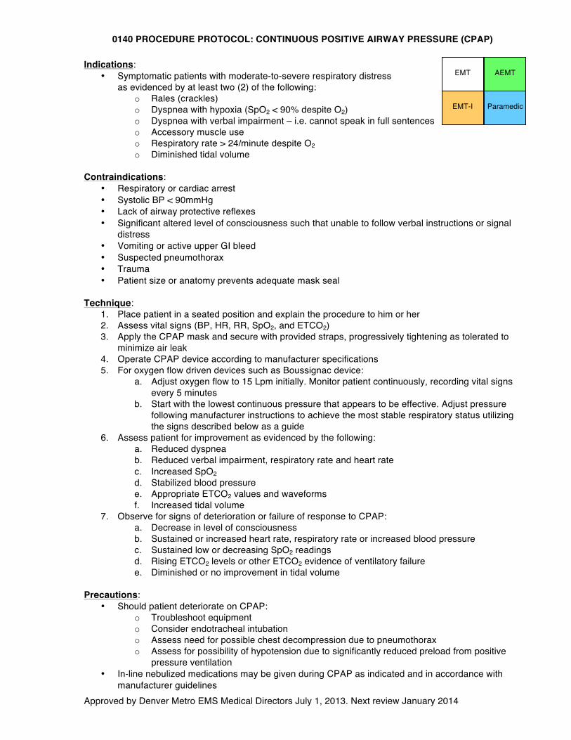
0140 PROCEDURE PROTOCOL: CONTINUOUS POSITIVE AIRWAY PRESSURE (CPAP)
Approved by Denver Metro EMS Medical Directors July 1, 2013. Next review January 2014
Indications: • Symptomatic patients with moderate-to-severe respiratory distress
as evidenced by at least two (2) of the following: o Rales (crackles) o Dyspnea with hypoxia (SpO2 < 90% despite O2) o Dyspnea with verbal impairment – i.e. cannot speak in full sentences o Accessory muscle use o Respiratory rate > 24/minute despite O2 o Diminished tidal volume
Contraindications:
• Respiratory or cardiac arrest • Systolic BP < 90mmHg • Lack of airway protective reflexes • Significant altered level of consciousness such that unable to follow verbal instructions or signal
distress • Vomiting or active upper GI bleed • Suspected pneumothorax • Trauma • Patient size or anatomy prevents adequate mask seal
Technique:
1. Place patient in a seated position and explain the procedure to him or her 2. Assess vital signs (BP, HR, RR, SpO2, and ETCO2) 3. Apply the CPAP mask and secure with provided straps, progressively tightening as tolerated to
minimize air leak 4. Operate CPAP device according to manufacturer specifications 5. For oxygen flow driven devices such as Boussignac device:
a. Adjust oxygen flow to 15 Lpm initially. Monitor patient continuously, recording vital signs every 5 minutes
b. Start with the lowest continuous pressure that appears to be effective. Adjust pressure following manufacturer instructions to achieve the most stable respiratory status utilizing the signs described below as a guide
6. Assess patient for improvement as evidenced by the following: a. Reduced dyspnea b. Reduced verbal impairment, respiratory rate and heart rate c. Increased SpO2 d. Stabilized blood pressure e. Appropriate ETCO2 values and waveforms f. Increased tidal volume
7. Observe for signs of deterioration or failure of response to CPAP: a. Decrease in level of consciousness b. Sustained or increased heart rate, respiratory rate or increased blood pressure c. Sustained low or decreasing SpO2 readings d. Rising ETCO2 levels or other ETCO2 evidence of ventilatory failure e. Diminished or no improvement in tidal volume
Precautions:
• Should patient deteriorate on CPAP: o Troubleshoot equipment o Consider endotracheal intubation o Assess need for possible chest decompression due to pneumothorax o Assess for possibility of hypotension due to significantly reduced preload from positive
pressure ventilation • In-line nebulized medications may be given during CPAP as indicated and in accordance with
manufacturer guidelines
EMT
EMT-I Paramedic
AEMT
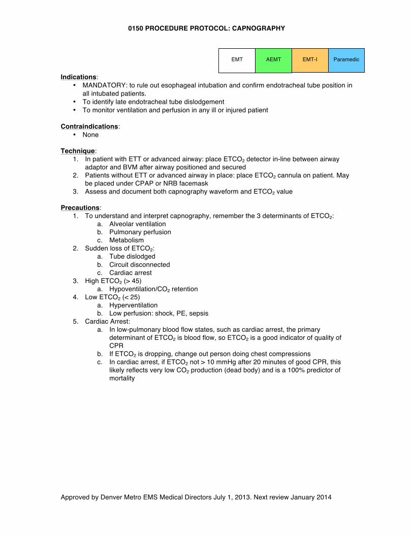
0150 PROCEDURE PROTOCOL: CAPNOGRAPHY
Approved by Denver Metro EMS Medical Directors July 1, 2013. Next review January 2014
EMT-I Paramedic Indications:
• MANDATORY: to rule out esophageal intubation and confirm endotracheal tube position in all intubated patients.
• To identify late endotracheal tube dislodgement • To monitor ventilation and perfusion in any ill or injured patient
Contraindications:
• None Technique:
1. In patient with ETT or advanced airway: place ETCO2 detector in-line between airway adaptor and BVM after airway positioned and secured
2. Patients without ETT or advanced airway in place: place ETCO2 cannula on patient. May be placed under CPAP or NRB facemask
3. Assess and document both capnography waveform and ETCO2 value Precautions:
1. To understand and interpret capnography, remember the 3 determinants of ETCO2: a. Alveolar ventilation b. Pulmonary perfusion c. Metabolism
2. Sudden loss of ETCO2: a. Tube dislodged b. Circuit disconnected c. Cardiac arrest
3. High ETCO2 (> 45) a. Hypoventilation/CO2 retention
4. Low ETCO2 (< 25) a. Hyperventilation b. Low perfusion: shock, PE, sepsis
5. Cardiac Arrest: a. In low-pulmonary blood flow states, such as cardiac arrest, the primary
determinant of ETCO2 is blood flow, so ETCO2 is a good indicator of quality of CPR
b. If ETCO2 is dropping, change out person doing chest compressions c. In cardiac arrest, if ETCO2 not > 10 mmHg after 20 minutes of good CPR, this
likely reflects very low CO2 production (dead body) and is a 100% predictor of mortality
EMT AEMT
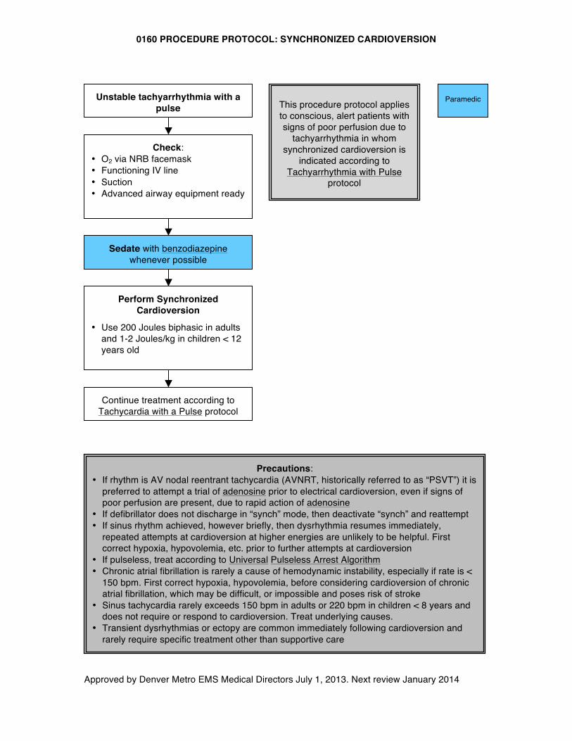
0160 PROCEDURE PROTOCOL: SYNCHRONIZED CARDIOVERSION
Approved by Denver Metro EMS Medical Directors July 1, 2013. Next review January 2014
Unstable tachyarrhythmia with a pulse
Precautions: • If rhythm is AV nodal reentrant tachycardia (AVNRT, historically referred to as “PSVT”) it is
preferred to attempt a trial of adenosine prior to electrical cardioversion, even if signs of poor perfusion are present, due to rapid action of adenosine
• If defibrillator does not discharge in “synch” mode, then deactivate “synch” and reattempt • If sinus rhythm achieved, however briefly, then dysrhythmia resumes immediately,
repeated attempts at cardioversion at higher energies are unlikely to be helpful. First correct hypoxia, hypovolemia, etc. prior to further attempts at cardioversion
• If pulseless, treat according to Universal Pulseless Arrest Algorithm • Chronic atrial fibrillation is rarely a cause of hemodynamic instability, especially if rate is <
150 bpm. First correct hypoxia, hypovolemia, before considering cardioversion of chronic atrial fibrillation, which may be difficult, or impossible and poses risk of stroke
• Sinus tachycardia rarely exceeds 150 bpm in adults or 220 bpm in children < 8 years and does not require or respond to cardioversion. Treat underlying causes.
• Transient dysrhythmias or ectopy are common immediately following cardioversion and rarely require specific treatment other than supportive care
Check: • O2 via NRB facemask • Functioning IV line • Suction • Advanced airway equipment ready
Sedate with benzodiazepine whenever possible
Perform Synchronized Cardioversion
• Use 200 Joules biphasic in adults and 1-2 Joules/kg in children < 12 years old
Paramedic
Continue treatment according to Tachycardia with a Pulse protocol
This procedure protocol applies to conscious, alert patients with signs of poor perfusion due to
tachyarrhythmia in whom synchronized cardioversion is
indicated according to Tachyarrhythmia with Pulse
protocol
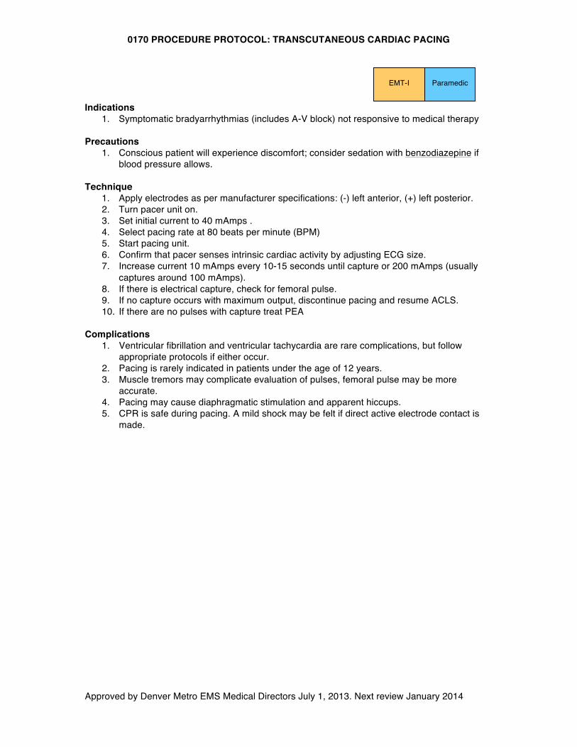
0170 PROCEDURE PROTOCOL: TRANSCUTANEOUS CARDIAC PACING
Approved by Denver Metro EMS Medical Directors July 1, 2013. Next review January 2014
Indications
1. Symptomatic bradyarrhythmias (includes A-V block) not responsive to medical therapy Precautions
1. Conscious patient will experience discomfort; consider sedation with benzodiazepine if blood pressure allows.
Technique
1. Apply electrodes as per manufacturer specifications: (-) left anterior, (+) left posterior. 2. Turn pacer unit on. 3. Set initial current to 40 mAmps . 4. Select pacing rate at 80 beats per minute (BPM) 5. Start pacing unit. 6. Confirm that pacer senses intrinsic cardiac activity by adjusting ECG size. 7. Increase current 10 mAmps every 10-15 seconds until capture or 200 mAmps (usually
captures around 100 mAmps). 8. If there is electrical capture, check for femoral pulse. 9. If no capture occurs with maximum output, discontinue pacing and resume ACLS. 10. If there are no pulses with capture treat PEA
Complications
1. Ventricular fibrillation and ventricular tachycardia are rare complications, but follow appropriate protocols if either occur.
2. Pacing is rarely indicated in patients under the age of 12 years. 3. Muscle tremors may complicate evaluation of pulses, femoral pulse may be more
accurate. 4. Pacing may cause diaphragmatic stimulation and apparent hiccups. 5. CPR is safe during pacing. A mild shock may be felt if direct active electrode contact is
made.
Paramedic EMT-I
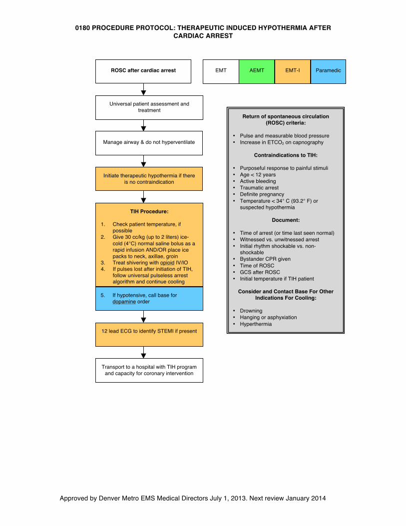
0180 PROCEDURE PROTOCOL: THERAPEUTIC INDUCED HYPOTHERMIA AFTER CARDIAC ARREST
Approved by Denver Metro EMS Medical Directors July 1, 2013. Next review January 2014
ROSC after cardiac arrest
Universal patient assessment and treatment
Manage airway & do not hyperventilate
12 lead ECG to identify STEMI if present
Transport to a hospital with TIH program and capacity for coronary intervention
TIH Procedure:
1. Check patient temperature, if possible
2. Give 30 cc/kg (up to 2 liters) ice-cold (4°C) normal saline bolus as a rapid infusion AND/OR place ice packs to neck, axillae, groin
3. Treat shivering with opioid IV/IO 4. If pulses lost after initiation of TIH,
follow universal pulseless arrest algorithm and continue cooling
Initiate therapeutic hypothermia if there is no contraindication
AEMT EMT-I Paramedic
5. If hypotensive, call base for dopamine order
EMT
Return of spontaneous circulation (ROSC) criteria:
• Pulse and measurable blood pressure • Increase in ETCO2 on capnography
Contraindications to TIH:
• Purposeful response to painful stimuli • Age < 12 years • Active bleeding • Traumatic arrest • Definite pregnancy • Temperature < 34° C (93.2° F) or
suspected hypothermia
Document:
• Time of arrest (or time last seen normal) • Witnessed vs. unwitnessed arrest • Initial rhythm shockable vs. non-
shockable • Bystander CPR given • Time of ROSC • GCS after ROSC • Initial temperature if TIH patient
Consider and Contact Base For Other
Indications For Cooling: • Drowning • Hanging or asphyxiation • Hyperthermia
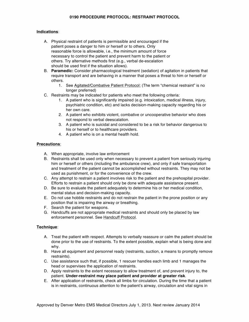
0190 PROCEDURE PROTOCOL: RESTRAINT PROTOCOL
Approved by Denver Metro EMS Medical Directors July 1, 2013. Next review January 2014
Indications: A. Physical restraint of patients is permissible and encouraged if the
patient poses a danger to him or herself or to others. Only reasonable force is allowable, i.e., the minimum amount of force necessary to control the patient and prevent harm to the patient or others. Try alternative methods first (e.g., verbal de-escalation should be used first if the situation allows).
B. Paramedic: Consider pharmacological treatment (sedation) of agitation in patients that require transport and are behaving in a manner that poses a threat to him or herself or others.
1. See Agitated/Combative Patient Protocol: (The term “chemical restraint” is no longer preferred)
C. Restraints may be indicated for patients who meet the following criteria: 1. A patient who is significantly impaired (e.g. intoxication, medical illness, injury,
psychiatric condition, etc) and lacks decision-making capacity regarding his or her own care.
2. A patient who exhibits violent, combative or uncooperative behavior who does not respond to verbal deescalation.
3. A patient who is suicidal and considered to be a risk for behavior dangerous to his or herself or to healthcare providers.
4. A patient who is on a mental health hold. Precautions:
A. When appropriate, involve law enforcement B. Restraints shall be used only when necessary to prevent a patient from seriously injuring
him or herself or others (including the ambulance crew), and only if safe transportation and treatment of the patient cannot be accomplished without restraints. They may not be used as punishment, or for the convenience of the crew.
C. Any attempt to restrain a patient involves risk to the patient and the prehospital provider. Efforts to restrain a patient should only be done with adequate assistance present.
D. Be sure to evaluate the patient adequately to determine his or her medical condition, mental status and decision-making capacity.
E. Do not use hobble restraints and do not restrain the patient in the prone position or any position that is impairing the airway or breathing.
F. Search the patient for weapons. G. Handcuffs are not appropriate medical restraints and should only be placed by law
enforcement personnel. See Handcuff Protocol. Technique:
A. Treat the patient with respect. Attempts to verbally reassure or calm the patient should be
done prior to the use of restraints. To the extent possible, explain what is being done and why.
B. Have all equipment and personnel ready (restraints, suction, a means to promptly remove restraints).
C. Use assistance such that, if possible, 1 rescuer handles each limb and 1 manages the head or supervises the application of restraints.
D. Apply restraints to the extent necessary to allow treatment of, and prevent injury to, the patient. Under-restraint may place patient and provider at greater risk.
E. After application of restraints, check all limbs for circulation. During the time that a patient is in restraints, continuous attention to the patient’s airway, circulation and vital signs in
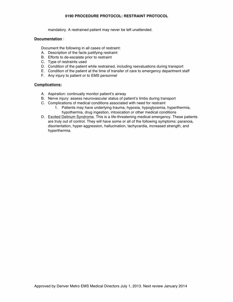
0190 PROCEDURE PROTOCOL: RESTRAINT PROTOCOL
Approved by Denver Metro EMS Medical Directors July 1, 2013. Next review January 2014
mandatory. A restrained patient may never be left unattended. Documentation :
Document the following in all cases of restraint: A. Description of the facts justifying restraint B. Efforts to de-escalate prior to restraint C. Type of restraints used D. Condition of the patient while restrained, including reevaluations during transport E. Condition of the patient at the time of transfer of care to emergency department staff F. Any injury to patient or to EMS personnel
Complications:
A. Aspiration: continually monitor patient’s airway B. Nerve injury: assess neurovascular status of patient’s limbs during transport C. Complications of medical conditions associated with need for restraint
1. Patients may have underlying trauma, hypoxia, hypoglycemia, hyperthermia, hypothermia, drug ingestion, intoxication or other medical conditions
D. Excited Delirium Syndrome. This is a life-threatening medical emergency. These patients are truly out of control. They will have some or all of the following symptoms: paranoia, disorientation, hyper-aggression, hallucination, tachycardia, increased strength, and hyperthermia.
0200 PROCEDURE PROTOCOL: TOURNIQUET PROTOCOL
Approved by Denver Metro EMS Medical Directors July 1, 2013. Next review January 2014
Indications
A. A tourniquet may be used to control potentially fatal hemorrhage only after other means of hemorrhage control have failed.
Precautions
A. A tourniquet applied incorrectly can increase blood loss. B. Applying a tourniquet can cause nerve and tissue damage whether applied correctly or not.
Proper patient selection is of utmost importance. C. Injury due to tourniquet is unlikely if the tourniquet is removed within 1 hour. In cases of life-
threatening bleeding benefit outweighs theoretical risk. D. A commercially made tourniquet is the preferred tourniquet. If none is available, a blood
pressure cuff inflated to a pressure sufficient to stop bleeding is an acceptable alternative. Other improvised tourniquets are not allowed.
Technique
A. First attempt to control hemorrhage by using direct pressure over bleeding area. B. If a discrete bleeding vessel can be identified, point pressure over bleeding vessel is more
effective than a large bandage and diffuse pressure. C. If unable to control hemorrhage using direct pressure, apply tourniquet according to
manufacturer specifications and using the steps below: 1. Cut away any clothing so that the tourniquet will be clearly visible. NEVER obscure a
tourniquet with clothing or bandages. 2. Apply tourniquet proximal to the wound and not across any joints. 3. Tighten tourniquet until bleeding stops. Applying tourniquet too loosely will only increase
blood loss by inhibiting venous return. 4. Mark the time and date of application on the patient’s skin next to the tourniquet. 5. Keep tourniquet on throughout hospital transport – a correctly applied tourniquet should
only be removed by the receiving hospital.
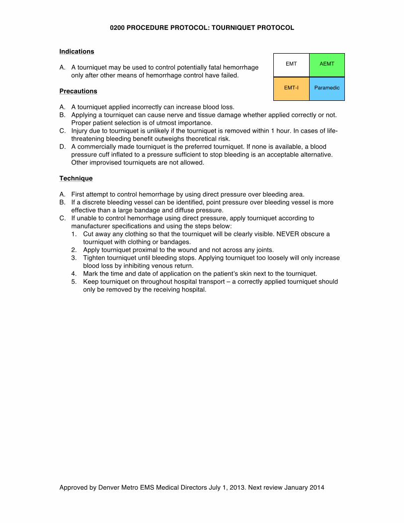
EMT
EMT-I Paramedic
AEMT
0210 PROCEDURE PROTOCOL: NEEDLE THORACOSTOMY FOR TENSION PNEUMOTHORAX DECOMPRESSION
Approved by Denver Metro EMS Medical Directors July 1, 2013. Next review January 2014
Indication: A. Needle decompression of tension pneumothorax is a standing order for EMT-I and
Paramedics. B. All of the following clinical indicators must be present:
1. Severe respiratory distress 2. Hypotension 3. Unilateral absent or decreased breath sounds
Technique: A. Expose entire chest B. Clean skin overlying site with available skin prep C. Insert largest, longest available angiocath either at 2nd intercostal space at midclavicular
line, or 5th intercostal space at midaxillary line 1. Either approach is acceptable, generally the site with the least soft tissue
overlying ribs is preferred D. Notify receiving hospital of needle decompression attempt
Precautions: A. Angiocath may become occluded with blood or by soft tissue B. A simple pneumothorax is NOT an indication for needle decompression
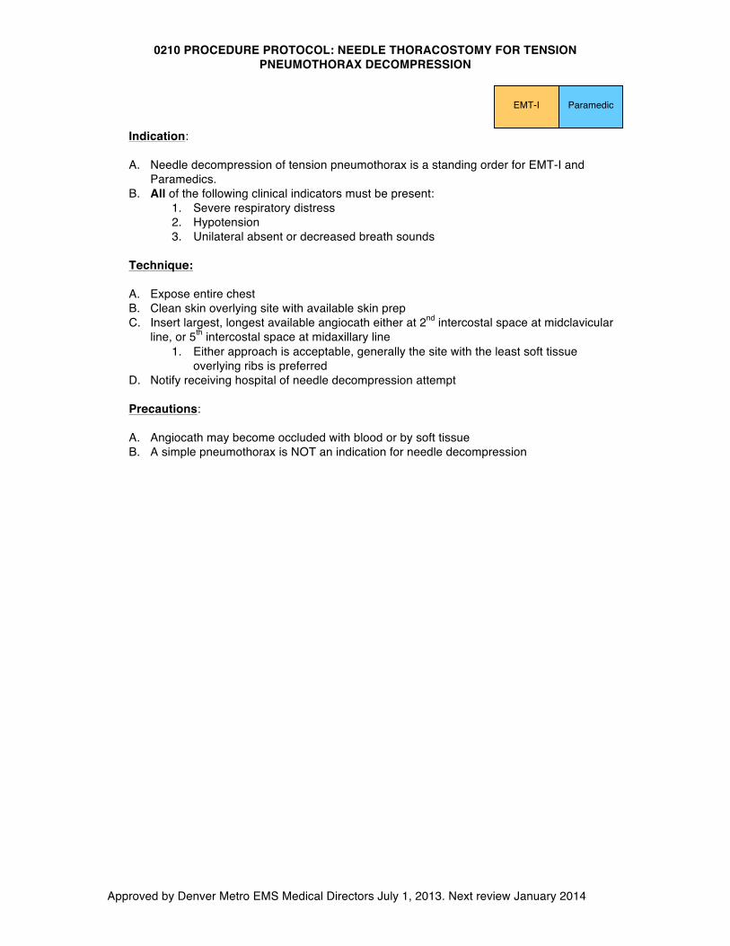
Paramedic EMT-I
0220 PROCEDURE PROTOCOL: INTRAOSSEUS CATHETER PLACEMENT
Approved by Denver Metro EMS Medical Directors July 1, 2013. Next review January 2014
Indications (must meet all criteria):
A. Rescue or primary vascular access device when peripheral IV access not obtainable in a patient with critical illness defined as:
1. Cardiopulmonary arrest or impending arrest 2. Profound shock with severe hypotension and poor perfusion
B. Utilization of IO access for all other patients requires base station contact 1. E.g.: Hypoglycemia with severe symptoms (e.g. unresponsive) and no venous
access C. IO placement may be considered prior to peripheral IV attempts in critical patients without
identifiable peripheral veins
Technique: A. Site of choice – tibial plateau: 2 fingerbreadths below the tibial tuberosity on the
anteromedial surface of tibia. 1. Alternative sites (e.g. humeral head in adults) are device-specific and require
authorization from the agency Medical Director. B. Clean skin with povidone-iodine. C. Place intraosseous needle perpendicular to the bone. D. Follow manufacturer’s guidelines specific to the device being used for insertion. E. Entrance into the bone marrow is indicated by a sudden loss of resistance. F. Flush line with 10 cc saline. Do not attempt to aspirate marrow
a. If patient conscious, administer lidocaine for pain control before infusing any other fluids
G. Secure line 1. Even if properly placed, the needle will not be secure. The needle must be
secured and the IV tubing taped. The IO needle should be stabilized at all times. H. Observe for signs of limb swelling, decreased perfusion to distal extremity that would
indicate a malpositioned IO catheter or other complication. If limb becomes tense or malperfused, disconnect IO tubing immediately and leave IO in place.
I. A person should be assigned to monitor the IV at the scene and en route to the hospital. J. Do not make more than one IO placement attempt per bone. K. Do not remove IO needles in the field. L. Notify hospital staff of all insertion sites/attempts and apply patient wristband included
with kit to identify IO patient.
Complications: A. Fracture B. Compartment syndrome C. Infection
Contraindications:
A. Fracture of target bone B. Cellulitis (skin infection overlying insertion site) C. Osteogenesis imperfecta (rare condition predisposing to fractures with minimal trauma) D. Total knee replacement (hardware will prevent placement)
Side Effects and Special Notes:
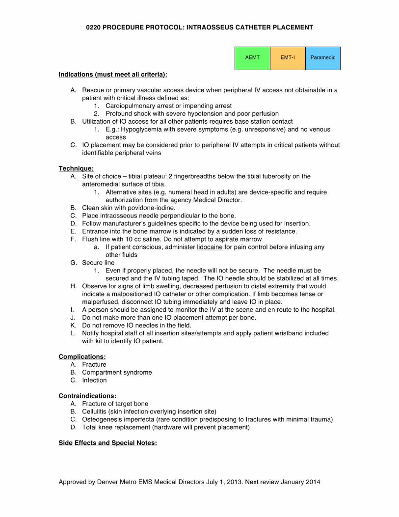
EMT-I Paramedic AEMT
0220 PROCEDURE PROTOCOL: INTRAOSSEUS CATHETER PLACEMENT
Approved by Denver Metro EMS Medical Directors July 1, 2013. Next review January 2014
A. Some authorities recommend aspiration of marrow fluid or tissue to confirm needle location. This is not recommended for field procedures, as it increases the risk of plugging the needle.
B. Expect flow rates to be slower than peripheral IVs. Pressure bags may be needed. Any drug or IV fluid may be infused.
C. Some manufacturers recommend the use of lidocaine for the treatment of pain associated with fluid administration. Check with your manufacturer and Medical Director for further guidance
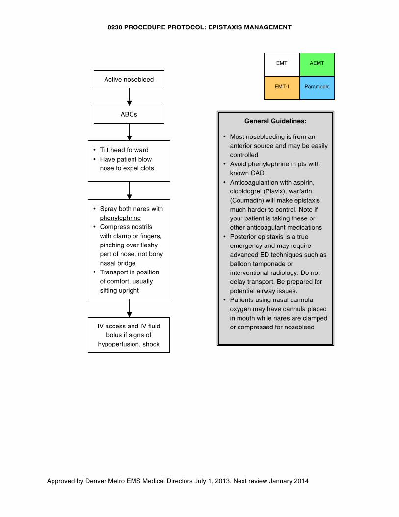
0230 PROCEDURE PROTOCOL: EPISTAXIS MANAGEMENT
Approved by Denver Metro EMS Medical Directors July 1, 2013. Next review January 2014
• Tilt head forward • Have patient blow
nose to expel clots
Paramedic EMT-I
General Guidelines:
• Most nosebleeding is from an anterior source and may be easily controlled
• Avoid phenylephrine in pts with known CAD
• Anticoagulantion with aspirin, clopidogrel (Plavix), warfarin (Coumadin) will make epistaxis much harder to control. Note if your patient is taking these or other anticoagulant medications
• Posterior epistaxis is a true emergency and may require advanced ED techniques such as balloon tamponade or interventional radiology. Do not delay transport. Be prepared for potential airway issues.
• Patients using nasal cannula oxygen may have cannula placed in mouth while nares are clamped or compressed for nosebleed
EMT
Active nosebleed
ABCs
• Spray both nares with phenylephrine
• Compress nostrils with clamp or fingers, pinching over fleshy part of nose, not bony nasal bridge
• Transport in position of comfort, usually sitting upright
IV access and IV fluid bolus if signs of
hypoperfusion, shock
AEMT
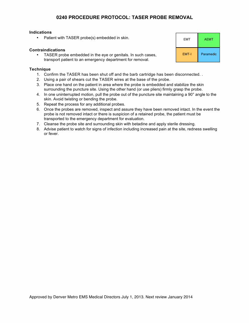
0240 PROCEDURE PROTOCOL: TASER PROBE REMOVAL
Approved by Denver Metro EMS Medical Directors July 1, 2013. Next review January 2014
Indications • Patient with TASER probe(s) embedded in skin.
Contraindications • TASER probe embedded in the eye or genitals. In such cases,
transport patient to an emergency department for removal. Technique
1. Confirm the TASER has been shut off and the barb cartridge has been disconnected. . 2. Using a pair of shears cut the TASER wires at the base of the probe. 3. Place one hand on the patient in area where the probe is embedded and stabilize the skin
surrounding the puncture site. Using the other hand (or use pliers) firmly grasp the probe. 4. In one uninterrupted motion, pull the probe out of the puncture site maintaining a 90° angle to the
skin. Avoid twisting or bending the probe. 5. Repeat the process for any additional probes. 6. Once the probes are removed, inspect and assure they have been removed intact. In the event the
probe is not removed intact or there is suspicion of a retained probe, the patient must be transported to the emergency department for evaluation.
7. Cleanse the probe site and surrounding skin with betadine and apply sterile dressing. 8. Advise patient to watch for signs of infection including increased pain at the site, redness swelling
or fever.
EMT AEMT
EMT-I Paramedic
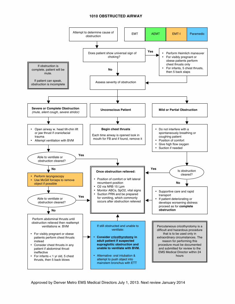
1010 OBSTRUCTED AIRWAY
Approved by Denver Metro EMS Medical Directors July 1, 2013. Next review January 2014
AEMT Paramedic EMT-I
Does patient show universal sign of choking?
• Perform Heimlich maneuver • For visibly pregnant or
obese patients perform chest thrusts only
• For infants, 5 chest thrusts, then 5 back slaps
Assess severity of obstruction
Severe or Complete Obstruction (mute, silent cough, severe stridor)
Mild or Partial Obstruction
• Open airway w. head tilt-chin lift or jaw thrust if craniofacial trauma
• Attempt ventilation with BVM
• Perform laryngoscopy • Use McGill forceps to remove
object if possible
Perform abdominal thrusts until obstruction relieved then reattempt
ventilations w. BVM
• For visibly pregnant or obese patients perform chest thrusts instead
• Consider chest thrusts in any patient if abdominal thrust ineffective
• For infants < 1 yr old, 5 chest thrusts, then 5 back blows
• Do not interfere with a spontaneously breathing or coughing patient
• Position of comfort • Give high flow oxygen • Suction if needed
No
Yes Once obstruction relieved:
• Position of comfort or left lateral
recumbent position • O2 via NRB 15 Lpm • Monitor ABCs, SpO2, vital signs • Suction PRN and be prepared
for vomiting, which commonly occurs after obstruction relieved
No
If still obstructed and unable to ventilate:
• Consider cricothyrotomy in
adult patient if suspected supraglottic obstruction and unable to ventilate with BVM.
• Alternative: oral intubation & attempt to push object into mainstem bronchus with ETT
Yes
No If obstruction is
complete, patient will be mute.
If patient can speak,
obstruction is incomplete
Is obstruction cleared?
Yes
Able to ventilate or obstruction cleared?
No
• Supportive care and rapid transport
• If patient deteriorating or develops worsening distress proceed as for complete obstruction
Unconscious Patient
Begin chest thrusts
Each time airway is opened look in mouth for FB and if found, remove it
Able to ventilate or obstruction cleared?
Yes
Attempt to determine cause of obstruction
Percutaneous cricothyrotomy is a difficult and hazardous procedure
that is to be used only in extraordinary circumstances. The
reason for performing this procedure must be documented and submitted for review to the EMS Medical Director within 24
hours
EMT
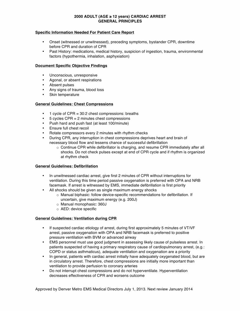
2000 ADULT (AGE ≥ 12 years) CARDIAC ARREST GENERAL PRINCIPLES
Approved by Denver Metro EMS Medical Directors July 1, 2013. Next review January 2014
Specific Information Needed For Patient Care Report
• Onset (witnessed or unwitnessed), preceding symptoms, bystander CPR, downtime before CPR and duration of CPR
• Past History: medications, medical history, suspicion of ingestion, trauma, environmental factors (hypothermia, inhalation, asphyxiation)
Document Specific Objective Findings
• Unconscious, unresponsive • Agonal, or absent respirations • Absent pulses • Any signs of trauma, blood loss • Skin temperature
General Guidelines: Chest Compressions
• 1 cycle of CPR = 30:2 chest compressions: breaths • 5 cycles CPR = 2 minutes chest compressions • Push hard and push fast (at least 100/minute) • Ensure full chest recoil • Rotate compressors every 2 minutes with rhythm checks • During CPR, any interruption in chest compressions deprives heart and brain of
necessary blood flow and lessens chance of successful defibrillation o Continue CPR while defibrillator is charging, and resume CPR immediately after all
shocks. Do not check pulses except at end of CPR cycle and if rhythm is organized at rhythm check
General Guidelines: Defibrillation
• In unwitnessed cardiac arrest, give first 2 minutes of CPR without interruptions for ventilation. During this time period passive oxygenation is preferred with OPA and NRB facemask. If arrest is witnessed by EMS, immediate defibrillation is first priority
• All shocks should be given as single maximum energy shocks o Manual biphasic: follow device-specific recommendations for defibrillation. If
uncertain, give maximum energy (e.g. 200J) o Manual monophasic: 360J o AED: device specific
General Guidelines: Ventilation during CPR
• If suspected cardiac etiology of arrest, during first approximately 5 minutes of VT/VF arrest, passive oxygenation with OPA and NRB facemask is preferred to positive pressure ventilation with BVM or advanced airway
• EMS personnel must use good judgment in assessing likely cause of pulseless arrest. In patients suspected of having a primary respiratory cause of cardiopulmonary arrest, (e.g.: COPD or status asthmaticus), adequate ventilation and oxygenation are a priority
• In general, patients with cardiac arrest initially have adequately oxygenated blood, but are in circulatory arrest. Therefore, chest compressions are initially more important than ventilation to provide perfusion to coronary arteries
• Do not interrupt chest compressions and do not hyperventilate. Hyperventilation decreases effectiveness of CPR and worsens outcome
2000 ADULT (AGE ≥ 12 years) CARDIAC ARREST GENERAL PRINCIPLES
Approved by Denver Metro EMS Medical Directors July 1, 2013. Next review January 2014
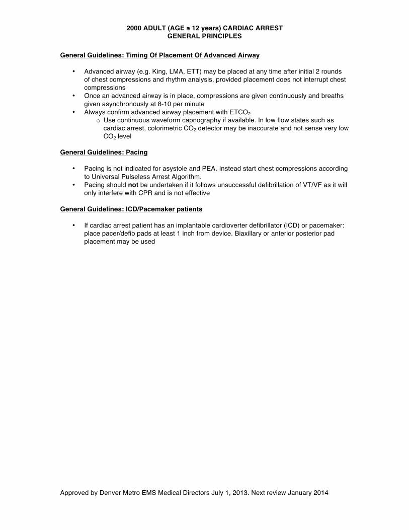
General Guidelines: Timing Of Placement Of Advanced Airway
• Advanced airway (e.g. King, LMA, ETT) may be placed at any time after initial 2 rounds of chest compressions and rhythm analysis, provided placement does not interrupt chest compressions
• Once an advanced airway is in place, compressions are given continuously and breaths given asynchronously at 8-10 per minute
• Always confirm advanced airway placement with ETCO2 o Use continuous waveform capnography if available. In low flow states such as
cardiac arrest, colorimetric CO2 detector may be inaccurate and not sense very low CO2 level
General Guidelines: Pacing
• Pacing is not indicated for asystole and PEA. Instead start chest compressions according to Universal Pulseless Arrest Algorithm.
• Pacing should not be undertaken if it follows unsuccessful defibrillation of VT/VF as it will only interfere with CPR and is not effective
General Guidelines: ICD/Pacemaker patients
• If cardiac arrest patient has an implantable cardioverter defibrillator (ICD) or pacemaker: place pacer/defib pads at least 1 inch from device. Biaxillary or anterior posterior pad placement may be used
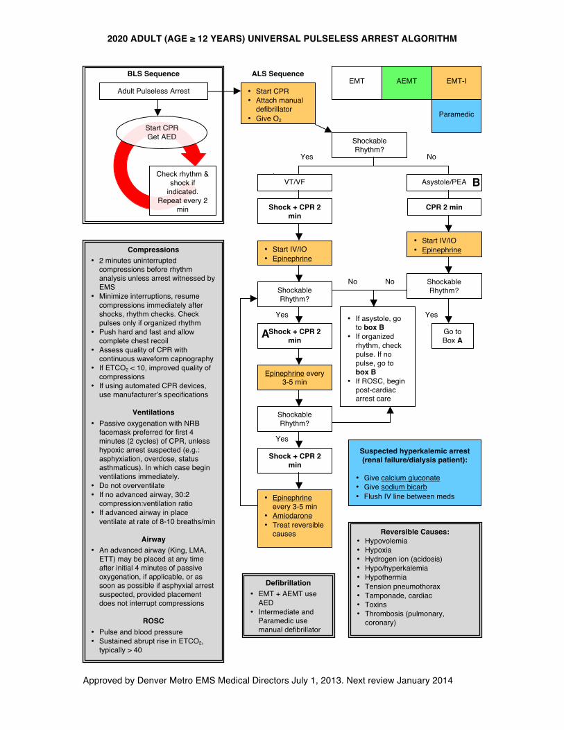
2020 ADULT (AGE ≥ 12 YEARS) UNIVERSAL PULSELESS ARREST ALGORITHM
Approved by Denver Metro EMS Medical Directors July 1, 2013. Next review January 2014
• Start CPR • Attach manual
defibrillator • Give O2
Shockable Rhythm?
VT/VF Asystole/PEA
No Yes
Shock + CPR 2 min
• Start IV/IO • Epinephrine
Shockable Rhythm?
Yes
Shockable Rhythm?
B
No
Epinephrine every 3-5 min
• Start IV/IO • Epinephrine
A
Yes
Go to Box A
No
Shockable Rhythm?
• Epinephrine every 3-5 min
• Amiodarone • Treat reversible
causes
Yes
Shock + CPR 2 min
Shock + CPR 2 min
CPR 2 min
Defibrillation • EMT + AEMT use
AED • Intermediate and
Paramedic use manual defibrillator
Adult Pulseless Arrest
Check rhythm & shock if
indicated. Repeat every 2
min
Start CPR Get AED
BLS Sequence EMT EMT-I
Paramedic
ALS Sequence
• If asystole, go to box B
• If organized rhythm, check pulse. If no pulse, go to box B
• If ROSC, begin post-cardiac arrest care
Suspected hyperkalemic arrest (renal failure/dialysis patient):
• Give calcium gluconate • Give sodium bicarb • Flush IV line between meds
Compressions • 2 minutes uninterrupted
compressions before rhythm analysis unless arrest witnessed by EMS
• Minimize interruptions, resume compressions immediately after shocks, rhythm checks. Check pulses only if organized rhythm
• Push hard and fast and allow complete chest recoil
• Assess quality of CPR with continuous waveform capnography
• If ETCO2 < 10, improved quality of compressions
• If using automated CPR devices, use manufacturer’s specifications
Ventilations • Passive oxygenation with NRB
facemask preferred for first 4 minutes (2 cycles) of CPR, unless hypoxic arrest suspected (e.g.: asphyxiation, overdose, status asthmaticus). In which case begin ventilations immediately.
• Do not overventilate • If no advanced airway, 30:2
compression:ventilation ratio • If advanced airway in place
ventilate at rate of 8-10 breaths/min
Airway • An advanced airway (King, LMA,
ETT) may be placed at any time after initial 4 minutes of passive oxygenation, if applicable, or as soon as possible if asphyxial arrest suspected, provided placement does not interrupt compressions
ROSC
• Pulse and blood pressure • Sustained abrupt rise in ETCO2,
typically > 40
Reversible Causes: • Hypovolemia • Hypoxia • Hydrogen ion (acidosis) • Hypo/hyperkalemia • Hypothermia • Tension pneumothorax • Tamponade, cardiac • Toxins • Thrombosis (pulmonary,
coronary)
AEMT
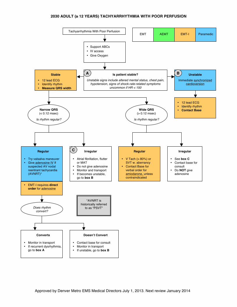
2030 ADULT (≥ 12 YEARS) TACHYARRHYTHMIA WITH POOR PERFUSION
Approved by Denver Metro EMS Medical Directors July 1, 2013. Next review January 2014
• Support ABCs • IV access • Give Oxygen
Is patient stable?
Unstable signs include altered mental status, chest pain, hypotension, signs of shock-rate-related symptoms
uncommon if HR < 150
Stable
• 12 lead ECG • Identify rhythm • Measure QRS width
Unstable
Immediate synchronized cardioversion
Regular
• Try valsalva maneuver • Give adenosine IV If
suspected AV nodal reentrant tachycardia (AVNRT)*
Irregular
• See box C • Contact base for
consult • Do NOT give
adenosine
Regular
• V Tach (> 80%) or SVT w. aberrancy
• Contact Base for verbal order for amiodarone, unless contraindicated
Irregular
• Atrial fibrillation, flutter or MAT
• Do not give adenosine • Monitor and transport • If becomes unstable,
go to box B
Converts
• Monitor in transport • If recurrent dysrhythmia,
go to box A
Doesn’t Convert
• Contact base for consult • Monitor in transport • If unstable, go to box B
Tachyarrhythmia With Poor Perfusion EMT EMT-I Paramedic
Does rhythm convert?
A
B
C
Narrow QRS (< 0.12 msec)
Is rhythm regular?
Wide QRS (> 0.12 msec)
Is rhythm regular?
*AVNRT is historically referred
to as “PSVT”
• EMT-I requires direct order for adenosine
• 12 lead ECG • Identify rhythm • Contact Base
AEMT
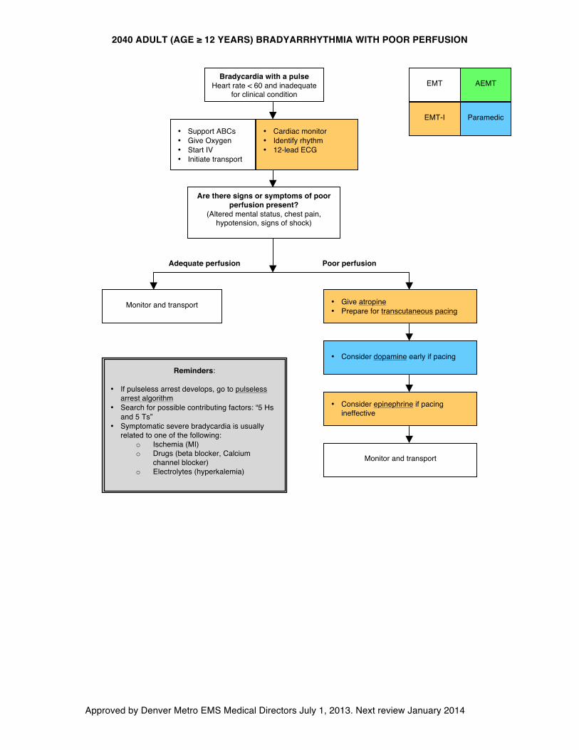
2040 ADULT (AGE ≥ 12 YEARS) BRADYARRHYTHMIA WITH POOR PERFUSION
Approved by Denver Metro EMS Medical Directors July 1, 2013. Next review January 2014
EMT
EMT-I Paramedic
• Support ABCs • Give Oxygen • Start IV • Initiate transport
Bradycardia with a pulse Heart rate < 60 and inadequate
for clinical condition
Are there signs or symptoms of poor perfusion present?
(Altered mental status, chest pain, hypotension, signs of shock)
Adequate perfusion Poor perfusion
Monitor and transport • Give atropine • Prepare for transcutaneous pacing
Monitor and transport
Reminders:
• If pulseless arrest develops, go to pulseless arrest algorithm
• Search for possible contributing factors: “5 Hs and 5 Ts”
• Symptomatic severe bradycardia is usually related to one of the following:
o Ischemia (MI) o Drugs (beta blocker, Calcium
channel blocker) o Electrolytes (hyperkalemia)
• Consider epinephrine if pacing ineffective
• Cardiac monitor • Identify rhythm • 12-lead ECG
• Consider dopamine early if pacing
AEMT
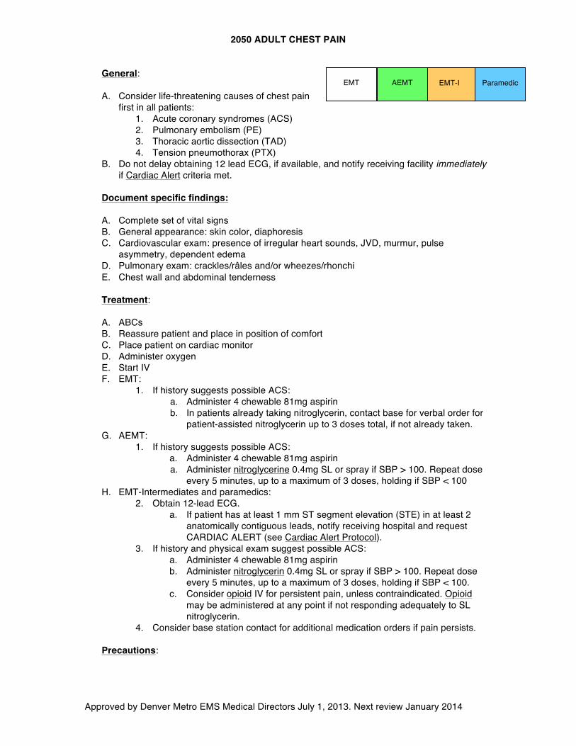
2050 ADULT CHEST PAIN
Approved by Denver Metro EMS Medical Directors July 1, 2013. Next review January 2014
General:
A. Consider life-threatening causes of chest pain first in all patients:
1. Acute coronary syndromes (ACS) 2. Pulmonary embolism (PE) 3. Thoracic aortic dissection (TAD) 4. Tension pneumothorax (PTX)
B. Do not delay obtaining 12 lead ECG, if available, and notify receiving facility immediately if Cardiac Alert criteria met.
Document specific findings:
A. Complete set of vital signs B. General appearance: skin color, diaphoresis C. Cardiovascular exam: presence of irregular heart sounds, JVD, murmur, pulse
asymmetry, dependent edema D. Pulmonary exam: crackles/râles and/or wheezes/rhonchi E. Chest wall and abdominal tenderness
Treatment:
A. ABCs B. Reassure patient and place in position of comfort C. Place patient on cardiac monitor D. Administer oxygen E. Start IV F. EMT:
1. If history suggests possible ACS: a. Administer 4 chewable 81mg aspirin b. In patients already taking nitroglycerin, contact base for verbal order for
patient-assisted nitroglycerin up to 3 doses total, if not already taken. G. AEMT:
1. If history suggests possible ACS: a. Administer 4 chewable 81mg aspirin a. Administer nitroglycerine 0.4mg SL or spray if SBP > 100. Repeat dose
every 5 minutes, up to a maximum of 3 doses, holding if SBP < 100 H. EMT-Intermediates and paramedics:
2. Obtain 12-lead ECG. a. If patient has at least 1 mm ST segment elevation (STE) in at least 2
anatomically contiguous leads, notify receiving hospital and request CARDIAC ALERT (see Cardiac Alert Protocol).
3. If history and physical exam suggest possible ACS: a. Administer 4 chewable 81mg aspirin b. Administer nitroglycerin 0.4mg SL or spray if SBP > 100. Repeat dose
every 5 minutes, up to a maximum of 3 doses, holding if SBP < 100. c. Consider opioid IV for persistent pain, unless contraindicated. Opioid
may be administered at any point if not responding adequately to SL nitroglycerin.
4. Consider base station contact for additional medication orders if pain persists.
Precautions:
EMT EMT-I Paramedic AEMT
2050 ADULT CHEST PAIN
Approved by Denver Metro EMS Medical Directors July 1, 2013. Next review January 2014
A. If inferior MI diagnosed (ST elevation in II, III, aVF), consider possibility of right ventricular infarct. Do not delay transport or receiving hospital contact, however, obtain right-sided ECG leads en route if time and conditions allow in order to identify right ventricular infarct.
B. If RV infarct pattern present (ST elevation in right-sided precordial leads, typically RV4), give nitroglycerin with extreme caution as hypotension common.
C. If hypotension develops following nitroglycerine administration in any patient, treat with 250cc NS boluses.
D. Nitroglycerin is contraindicated in patients taking medication for erectile dysfunction (phosphodiesterase inhibitors, e.g.: Viagra, Cialis).
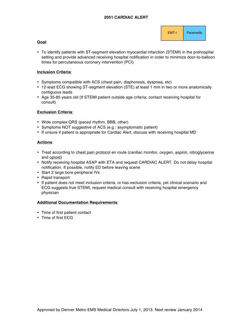
2051 CARDIAC ALERT
Approved by Denver Metro EMS Medical Directors July 1, 2013. Next review January 2014
Paramedic EMT-I Goal: • To identify patients with ST-segment elevation myocardial infarction (STEMI) in the prehospital
setting and provide advanced receiving hospital notification in order to minimize door-to-balloon times for percutaneous coronary intervention (PCI)
Inclusion Criteria: • Symptoms compatible with ACS (chest pain, diaphoresis, dyspnea, etc) • 12-lead ECG showing ST-segment elevation (STE) at least 1 mm in two or more anatomically
contiguous leads • Age 35-85 years old (If STEMI patient outside age criteria, contact receiving hospital for
consult) Exclusion Criteria: • Wide complex QRS (paced rhythm, BBB, other) • Symptoms NOT suggestive of ACS (e.g.: asymptomatic patient) • If unsure if patient is appropriate for Cardiac Alert, discuss with receiving hospital MD Actions: • Treat according to chest pain protocol en route (cardiac monitor, oxygen, aspirin, nitroglycerine
and opioid) • Notify receiving hospital ASAP with ETA and request CARDIAC ALERT. Do not delay hospital
notification. If possible, notify ED before leaving scene • Start 2 large bore peripheral IVs • Rapid transport • If patient does not meet inclusion criteria, or has exclusion criteria, yet clinical scenario and
ECG suggests true STEMI, request medical consult with receiving hospital emergency physician
Additional Documentation Requirements: • Time of first patient contact • Time of first ECG
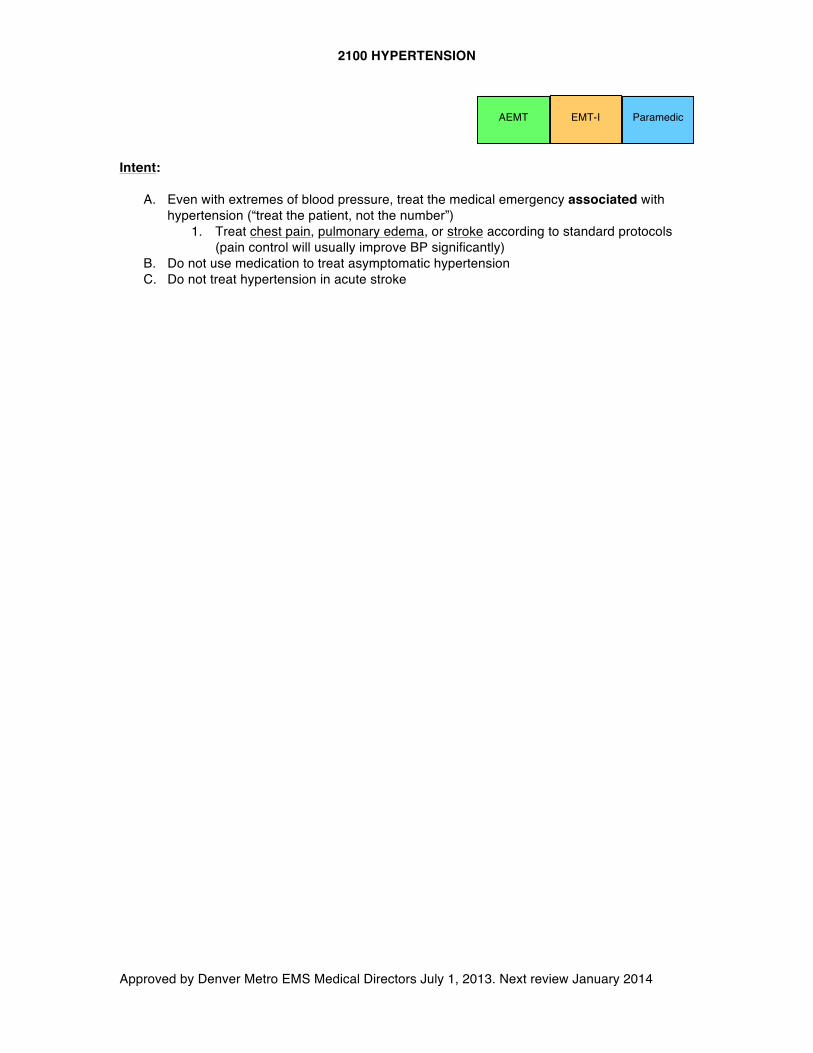
2100 HYPERTENSION
Approved by Denver Metro EMS Medical Directors July 1, 2013. Next review January 2014
EMT-I AEMT
Intent:
A. Even with extremes of blood pressure, treat the medical emergency associated with hypertension (“treat the patient, not the number”)
1. Treat chest pain, pulmonary edema, or stroke according to standard protocols (pain control will usually improve BP significantly)
B. Do not use medication to treat asymptomatic hypertension C. Do not treat hypertension in acute stroke
Paramedic
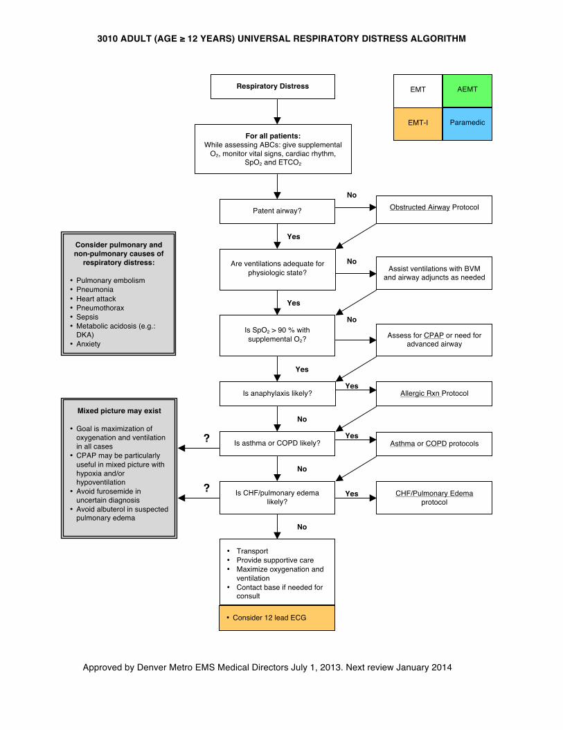
3010 ADULT (AGE ≥ 12 YEARS) UNIVERSAL RESPIRATORY DISTRESS ALGORITHM
Approved by Denver Metro EMS Medical Directors July 1, 2013. Next review January 2014
Consider pulmonary and non-pulmonary causes of
respiratory distress:
• Pulmonary embolism • Pneumonia • Heart attack • Pneumothorax • Sepsis • Metabolic acidosis (e.g.:
DKA) • Anxiety
EMT
EMT-I Paramedic
AEMT
Respiratory Distress
For all patients: While assessing ABCs: give supplemental
O2, monitor vital signs, cardiac rhythm, SpO2 and ETCO2
Patent airway? Obstructed Airway Protocol
Are ventilations adequate for physiologic state?
Is anaphylaxis likely?
Assist ventilations with BVM and airway adjuncts as needed
Yes
Yes
Yes
No
Is SpO2 > 90 % with supplemental O2? Assess for CPAP or need for
advanced airway
No
No
No
Is asthma or COPD likely?
Allergic Rxn Protocol Yes
Asthma or COPD protocols Yes
Is CHF/pulmonary edema likely?
Yes
No
CHF/Pulmonary Edema protocol
Mixed picture may exist
• Goal is maximization of oxygenation and ventilation in all cases
• CPAP may be particularly useful in mixed picture with hypoxia and/or hypoventilation
• Avoid furosemide in uncertain diagnosis
• Avoid albuterol in suspected pulmonary edema
?
?
• Transport • Provide supportive care • Maximize oxygenation and
ventilation • Contact base if needed for
consult
No
• Consider 12 lead ECG
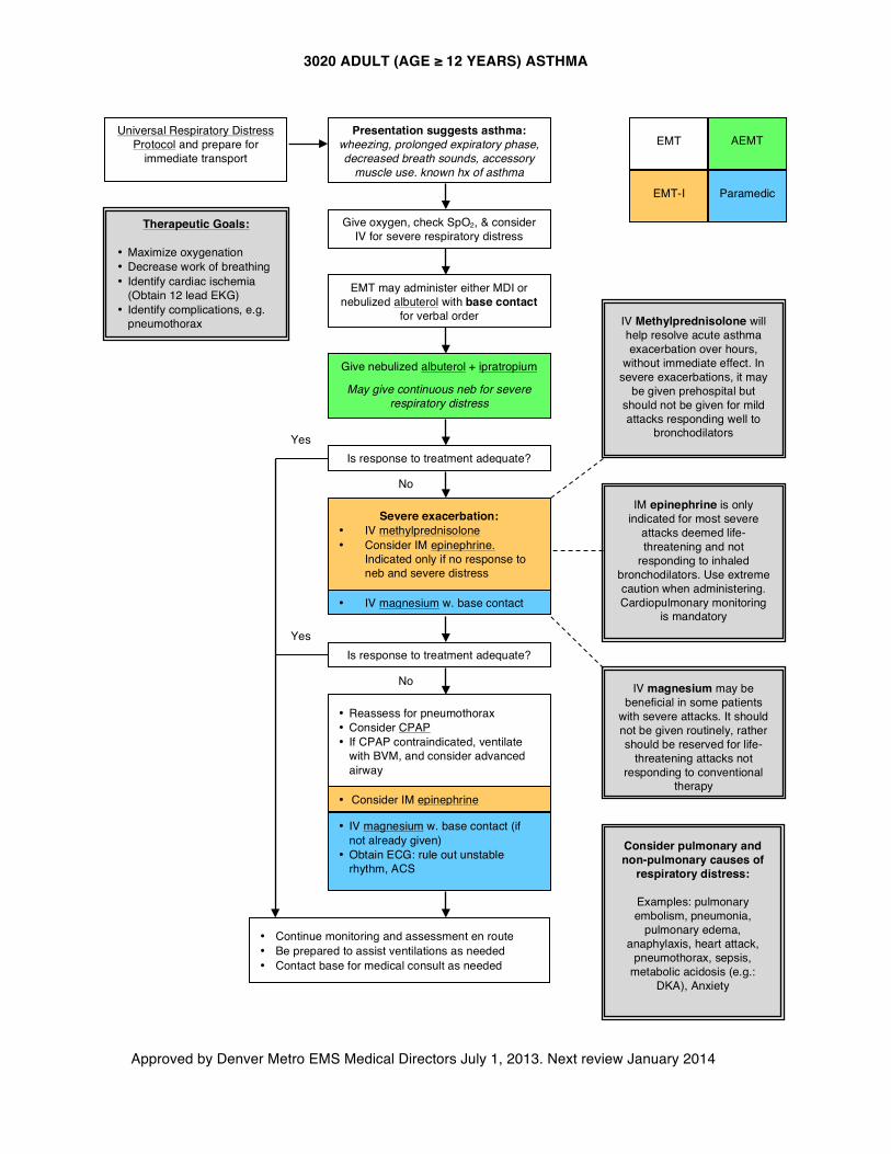
3020 ADULT (AGE ≥ 12 YEARS) ASTHMA
Approved by Denver Metro EMS Medical Directors July 1, 2013. Next review January 2014
Universal Respiratory Distress Protocol and prepare for
immediate transport
Give nebulized albuterol + ipratropium
May give continuous neb for severe respiratory distress
Therapeutic Goals:
• Maximize oxygenation • Decrease work of breathing • Identify cardiac ischemia
(Obtain 12 lead EKG) • Identify complications, e.g.
pneumothorax
Is response to treatment adequate?
• Continue monitoring and assessment en route • Be prepared to assist ventilations as needed • Contact base for medical consult as needed
No
Yes
Yes
No
Presentation suggests asthma: wheezing, prolonged expiratory phase, decreased breath sounds, accessory
muscle use, known hx of asthma
Consider pulmonary and non-pulmonary causes of
respiratory distress:
Examples: pulmonary embolism, pneumonia,
pulmonary edema, anaphylaxis, heart attack,
pneumothorax, sepsis, metabolic acidosis (e.g.:
DKA), Anxiety
• IV magnesium w. base contact (if not already given)
• Obtain ECG: rule out unstable rhythm, ACS
EMT
EMT-I Paramedic
EMT may administer either MDI or nebulized albuterol with base contact
for verbal order
Give oxygen, check SpO2, & consider IV for severe respiratory distress
• IV magnesium w. base contact
Is response to treatment adequate?
IV Methylprednisolone will help resolve acute asthma exacerbation over hours,
without immediate effect. In severe exacerbations, it may
be given prehospital but should not be given for mild attacks responding well to
bronchodilators
IM epinephrine is only indicated for most severe
attacks deemed life-threatening and not
responding to inhaled bronchodilators. Use extreme caution when administering. Cardiopulmonary monitoring
is mandatory
IV magnesium may be beneficial in some patients
with severe attacks. It should not be given routinely, rather should be reserved for life-
threatening attacks not responding to conventional
therapy
• Reassess for pneumothorax • Consider CPAP • If CPAP contraindicated, ventilate
with BVM, and consider advanced airway
Severe exacerbation: • IV methylprednisolone • Consider IM epinephrine.
Indicated only if no response to neb and severe distress
AEMT
• Consider IM epinephrine
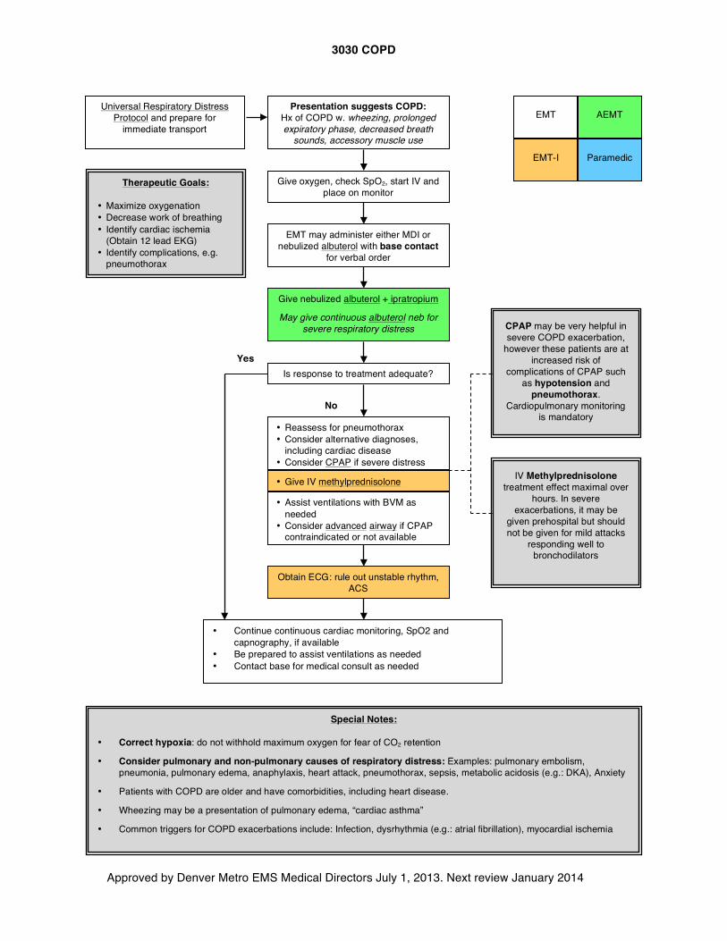
3030 COPD
Approved by Denver Metro EMS Medical Directors July 1, 2013. Next review January 2014
Universal Respiratory Distress Protocol and prepare for
immediate transport
Give nebulized albuterol + ipratropium
May give continuous albuterol neb for severe respiratory distress
Special Notes: • Correct hypoxia: do not withhold maximum oxygen for fear of CO2 retention
• Consider pulmonary and non-pulmonary causes of respiratory distress: Examples: pulmonary embolism, pneumonia, pulmonary edema, anaphylaxis, heart attack, pneumothorax, sepsis, metabolic acidosis (e.g.: DKA), Anxiety
• Patients with COPD are older and have comorbidities, including heart disease.
• Wheezing may be a presentation of pulmonary edema, “cardiac asthma”
• Common triggers for COPD exacerbations include: Infection, dysrhythmia (e.g.: atrial fibrillation), myocardial ischemia
Is response to treatment adequate?
No
Yes
• Reassess for pneumothorax • Consider alternative diagnoses,
including cardiac disease • Consider CPAP if severe distress •
EMT
EMT-I Paramedic
Obtain ECG: rule out unstable rhythm, ACS
• Give IV methylprednisolone
Presentation suggests COPD: Hx of COPD w. wheezing, prolonged expiratory phase, decreased breath
sounds, accessory muscle use
Give oxygen, check SpO2, start IV and place on monitor
• Assist ventilations with BVM as needed
• Consider advanced airway if CPAP contraindicated or not available
• Continue continuous cardiac monitoring, SpO2 and capnography, if available
• Be prepared to assist ventilations as needed • Contact base for medical consult as needed
Therapeutic Goals:
• Maximize oxygenation • Decrease work of breathing • Identify cardiac ischemia
(Obtain 12 lead EKG) • Identify complications, e.g.
pneumothorax
IV Methylprednisolone treatment effect maximal over
hours. In severe exacerbations, it may be
given prehospital but should not be given for mild attacks
responding well to bronchodilators
CPAP may be very helpful in severe COPD exacerbation,
however these patients are at increased risk of
complications of CPAP such as hypotension and
pneumothorax. Cardiopulmonary monitoring
is mandatory
EMT may administer either MDI or nebulized albuterol with base contact
for verbal order
AEMT
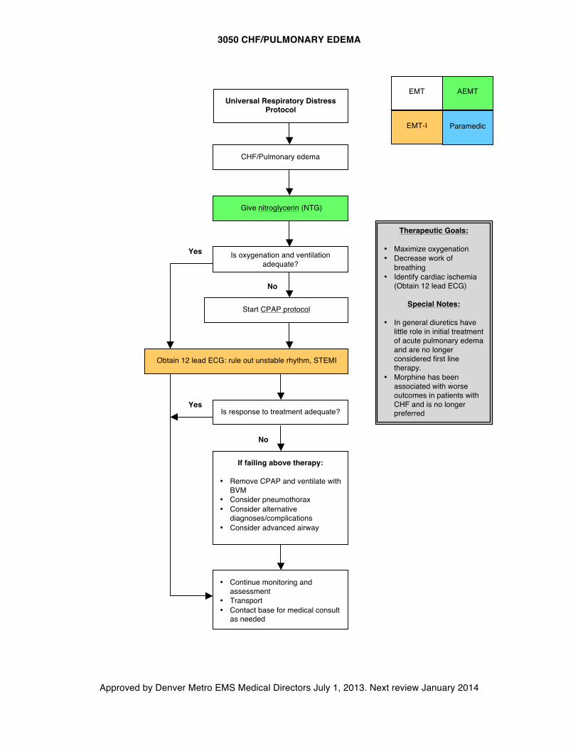
3050 CHF/PULMONARY EDEMA
Approved by Denver Metro EMS Medical Directors July 1, 2013. Next review January 2014
No
No
CHF/Pulmonary edema
Universal Respiratory Distress
Protocol
Give nitroglycerin (NTG)
Start CPAP protocol
Therapeutic Goals:
• Maximize oxygenation • Decrease work of
breathing • Identify cardiac ischemia
(Obtain 12 lead ECG)
Special Notes: • In general diuretics have
little role in initial treatment of acute pulmonary edema and are no longer considered first line therapy.
• Morphine has been associated with worse outcomes in patients with CHF and is no longer preferred
Is oxygenation and ventilation adequate?
• Continue monitoring and assessment
• Transport • Contact base for medical consult
as needed
Yes
If failing above therapy:
• Remove CPAP and ventilate with BVM
• Consider pneumothorax • Consider alternative
diagnoses/complications • Consider advanced airway
Is response to treatment adequate?
Yes
EMT
EMT-I Paramedic
AEMT
Obtain 12 lead ECG: rule out unstable rhythm, STEMI
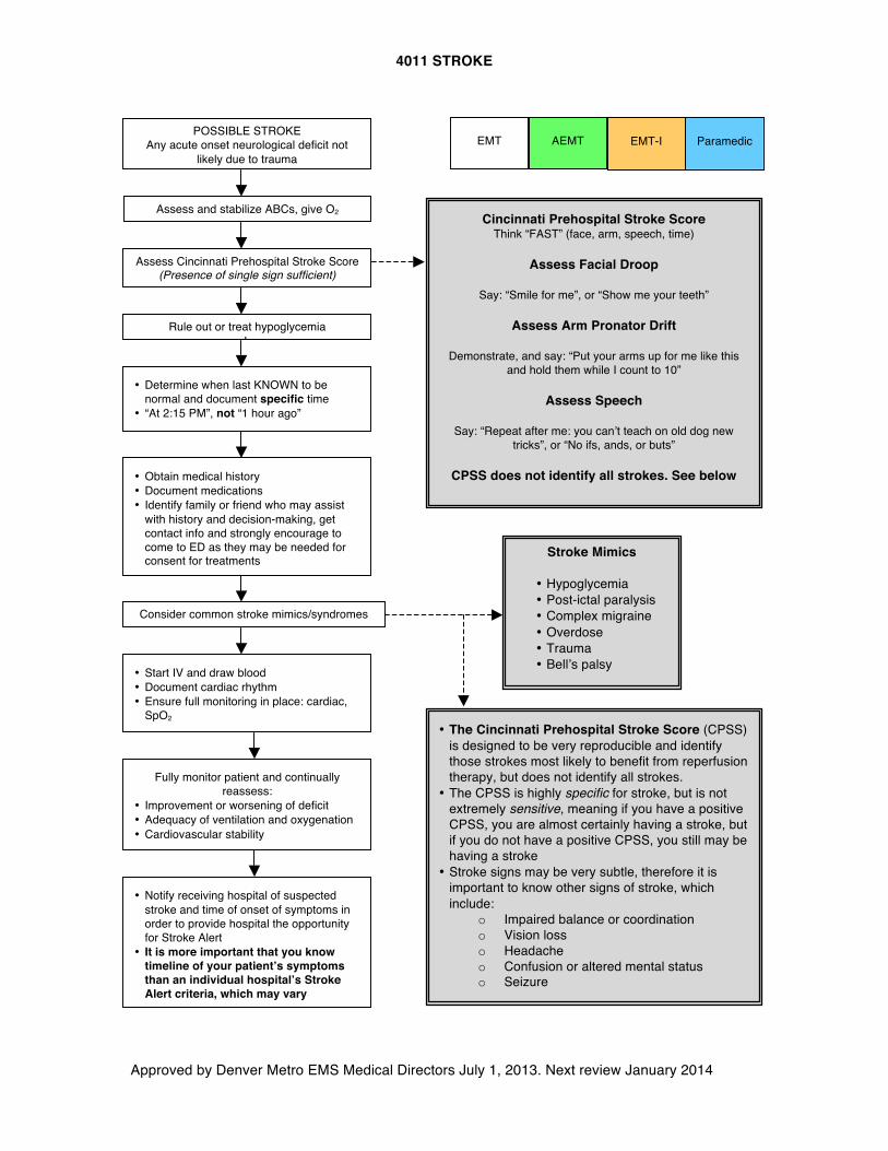
4011 STROKE
Approved by Denver Metro EMS Medical Directors July 1, 2013. Next review January 2014
Rule out or treat hypoglycemia L
• Determine when last KNOWN to be normal and document specific time
• “At 2:15 PM”, not “1 hour ago”
Consider common stroke mimics/syndromes
• Obtain medical history • Document medications • Identify family or friend who may assist
with history and decision-making, get contact info and strongly encourage to come to ED as they may be needed for consent for treatments
Stroke Mimics
• Hypoglycemia • Post-ictal paralysis • Complex migraine • Overdose • Trauma • Bell’s palsy
Fully monitor patient and continually reassess:
• Improvement or worsening of deficit • Adequacy of ventilation and oxygenation • Cardiovascular stability
Cincinnati Prehospital Stroke Score Think “FAST” (face, arm, speech, time)
Assess Facial Droop
Say: “Smile for me”, or “Show me your teeth”
Assess Arm Pronator Drift
Demonstrate, and say: “Put your arms up for me like this
and hold them while I count to 10”
Assess Speech
Say: “Repeat after me: you can’t teach on old dog new tricks”, or “No ifs, ands, or buts”
CPSS does not identify all strokes. See below
POSSIBLE STROKE Any acute onset neurological deficit not
likely due to trauma
• Start IV and draw blood • Document cardiac rhythm • Ensure full monitoring in place: cardiac,
SpO2
Assess and stabilize ABCs, give O2
Assess Cincinnati Prehospital Stroke Score (Presence of single sign sufficient)
• Notify receiving hospital of suspected stroke and time of onset of symptoms in order to provide hospital the opportunity for Stroke Alert
• It is more important that you know timeline of your patient’s symptoms than an individual hospital’s Stroke Alert criteria, which may vary
EMT EMT-I Paramedic
• The Cincinnati Prehospital Stroke Score (CPSS) is designed to be very reproducible and identify those strokes most likely to benefit from reperfusion therapy, but does not identify all strokes.
• The CPSS is highly specific for stroke, but is not extremely sensitive, meaning if you have a positive CPSS, you are almost certainly having a stroke, but if you do not have a positive CPSS, you still may be having a stroke
• Stroke signs may be very subtle, therefore it is important to know other signs of stroke, which include:
o Impaired balance or coordination o Vision loss o Headache o Confusion or altered mental status o Seizure
AEMT
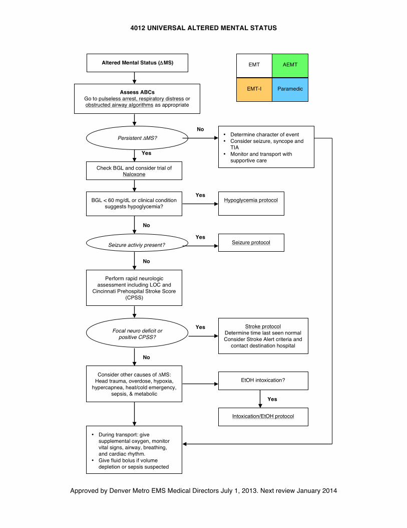
4012 UNIVERSAL ALTERED MENTAL STATUS
Approved by Denver Metro EMS Medical Directors July 1, 2013. Next review January 2014
Persistent ΔMS?
Seizure activiy present?
Focal neuro deficit or positive CPSS?
Altered Mental Status (ΔMS)
Assess ABCs Go to pulseless arrest, respiratory distress or obstructed airway algorithms as appropriate
• Determine character of event • Consider seizure, syncope and
TIA • Monitor and transport with
supportive care
BGL < 60 mg/dL or clinical condition suggests hypoglycemia?
Perform rapid neurologic assessment including LOC and
Cincinnati Prehospital Stroke Score (CPSS)
Seizure protocol
Yes
No
No
Yes
Yes
No
Yes Stroke protocol Determine time last seen normal Consider Stroke Alert criteria and
contact destination hospital
Consider other causes of ΔMS: Head trauma, overdose, hypoxia,
hypercapnea, heat/cold emergency, sepsis, & metabolic
No
• During transport: give supplemental oxygen, monitor vital signs, airway, breathing, and cardiac rhythm.
• Give fluid bolus if volume depletion or sepsis suspected
EtOH intoxication?
Intoxication/EtOH protocol
Yes
Check BGL and consider trial of Naloxone
EMT
EMT-I Paramedic
Hypoglycemia protocol
AEMT
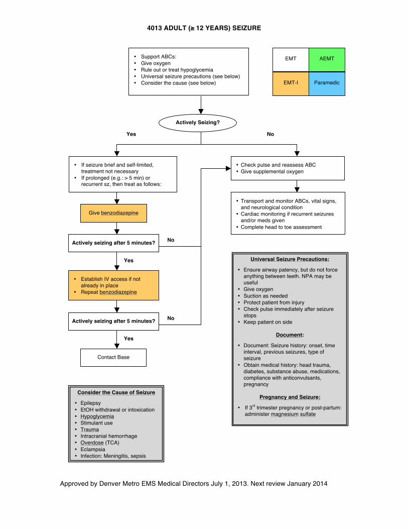
4013 ADULT (≥ 12 YEARS) SEIZURE
Approved by Denver Metro EMS Medical Directors July 1, 2013. Next review January 2014
EMT
EMT-I Paramedic
AEMT
Consider the Cause of Seizure
• Epilepsy • EtOH withdrawal or intoxication • Hypoglycemia • Stimulant use • Trauma • Intracranial hemorrhage • Overdose (TCA) • Eclampsia • Infection: Meningitis, sepsis
Yes No
• Check pulse and reassess ABC • Give supplemental oxygen
No
Yes
Yes
Contact Base
• If seizure brief and self-limited, treatment not necessary
• If prolonged (e.g.: > 5 min) or recurrent sz, then treat as follows:
Actively Seizing?
• Transport and monitor ABCs, vital signs, and neurological condition
• Cardiac monitoring if recurrent seizures and/or meds given
• Complete head to toe assessment
• Support ABCs: • Give oxygen • Rule out or treat hypoglycemia • Universal seizure precautions (see below) • Consider the cause (see below)
Universal Seizure Precautions:
• Ensure airway patency, but do not force anything between teeth. NPA may be useful
• Give oxygen • Suction as needed • Protect patient from injury • Check pulse immediately after seizure
stops • Keep patient on side
Document:
• Document: Seizure history: onset, time interval, previous seizures, type of seizure
• Obtain medical history: head trauma, diabetes, substance abuse, medications, compliance with anticonvulsants, pregnancy
Pregnancy and Seizure:
• If 3rd trimester pregnancy or post-partum: administer magnesium sulfate
No
Actively seizing after 5 minutes?
Give benzodiazepine
Actively seizing after 5 minutes?
• Establish IV access if not already in place
• Repeat benzodiazepine
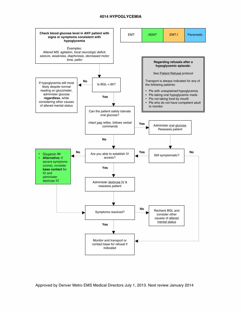
4014 HYPOGLYCEMIA
Approved by Denver Metro EMS Medical Directors July 1, 2013. Next review January 2014
AEMT EMT
Is BGL < 60?
Check blood glucose level in ANY patient with signs or symptoms consistent with
hypoglycemia
Examples: Altered MS, agitation, focal neurologic deficit,
seizure, weakness, diaphoresis, decreased motor tone, pallor
Administer oral glucose. Reassess patient
No
Monitor and transport or contact base for refusal if
indicated
• Glugacon IM • Alternative: If
severe symptoms (coma), consider base contact for IO and administer dextrose IO
Yes
Yes
If hypoglycemia still most likely despite normal
reading on glucometer, administer glucose regardless, while
considering other causes of altered mental status
Symptoms resolved?
Yes
Yes
No
Still symptomatic? Are you able to establish IV access?
Administer dextrose IV & reassess patient
Yes
No
Recheck BGL and consider other
causes of altered mental status
No
Regarding refusals after a hypoglycemic episode:
See Patient Refusal protocol
Transport is always indicated for any of the following patients:
• Pts with unexplained hypoglycemia • Pts taking oral hypoglycemic meds • Pts not taking food by mouth • Pts who do not have competent adult
to monitor Can the patient safely tolerate
oral glucose? intact gag reflex, follows verbal
commands
Paramedic EMT-I
No
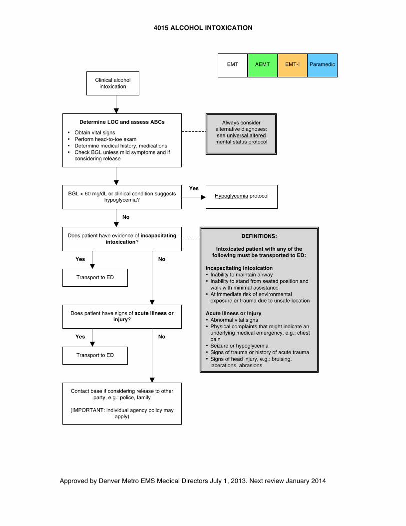
4015 ALCOHOL INTOXICATION
Approved by Denver Metro EMS Medical Directors July 1, 2013. Next review January 2014
BGL < 60 mg/dL or clinical condition suggests hypoglycemia?
Hypoglycemia protocol
Does patient have evidence of incapacitating intoxication?
Transport to ED
DEFINITIONS:
Intoxicated patient with any of the following must be transported to ED:
Incapacitating Intoxication • Inability to maintain airway • Inability to stand from seated position and
walk with minimal assistance • At immediate risk of environmental
exposure or trauma due to unsafe location
Acute Illness or Injury • Abnormal vital signs • Physical complaints that might indicate an
underlying medical emergency, e.g.: chest pain
• Seizure or hypoglycemia • Signs of trauma or history of acute trauma • Signs of head injury, e.g.: bruising,
lacerations, abrasions
No
No
Yes
Yes
Does patient have signs of acute illness or injury?
Transport to ED
No
Contact base if considering release to other party, e.g.: police, family
(IMPORTANT: individual agency policy may
apply)
Clinical alcohol intoxication
Always consider alternative diagnoses: see universal altered
mental status protocol
AEMT Paramedic EMT-I
Yes
EMT
Determine LOC and assess ABCs
• Obtain vital signs • Perform head-to-toe exam • Determine medical history, medications • Check BGL unless mild symptoms and if
considering release
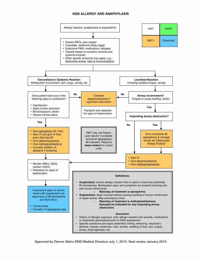
4020 ALLERGY AND ANAPHYLAXIS
Approved by Denver Metro EMS Medical Directors July 1, 2013. Next review January 2014
EMT
Paramedic EMT-I
AEMT
Allergic reaction, anaphylaxis or angioedema
• Assess ABCs, give oxygen • If possible, determine likely trigger • Determine PMH, medications, allergies • Classify based on symptom severity and
systems involved • Other specific protocols may apply: e.g.:
obstructed airway, bites & envenomations
Generalized or Systemic Reaction Multisystem involvement: skin, lungs, airway, etc
Does patient have any of the following signs or symptoms?
• Hypotension • Signs of poor perfusion • Bronchospasm, stridor • Altered mental status
• Give epinephrine IM, then: • Start IV and give IV fluid
bolus 20cc/kg NS • Give diphenhydramine • Give methylprednisolone • Consider addition of
albuterol if wheezing
• Monitor ABCs, SpO2, cardiac rhythm
• Reassess for signs of deterioration
Consider diphenhydramine if
significant discomfort
Localized Reaction Including isolated tongue, airway
Airway involvement? Tongue or uvula swelling, stridor
Impending airway obstruction?
Give immediate IM epinephrine & manage airway per Obstructed
Airway Protocol
• Start IV • Give diphenhydramine • Give methylprednisolone
If persistent signs of severe shock with hypotension not
responsive to IM epinephrine and fluid bolus:
• Contact base • Consider IV epinephrine drip
No
Yes
No
Yes
Yes No
EMT may use Epipen auto injector if available whenever epinephrine IM indicated. Requires
base contact for verbal order
Definitions:
• Anaphylaxis: severe allergic reaction that is rapid in onset and potentially life-threatening. Multisystem signs and symptoms are present including skin and mucus membranes
o Mainstay of treatment is epinephrine • Angioedema: deep mucosal edema causing swelling of mucus membranes
of upper airway. May accompany hives o Mainstay of treatment is methylprednisolone.
Epinephrine indicated for any impending airway obstruction.
Document:
• History of allergen exposure, prior allergic reaction and severity, medications or treatments administered prior to EMS assessment
• Specific symptoms and signs presented: itching, wheezing, respiratory distress, nausea, weakness, rash, anxiety, swelling of face, lips, tongue, throat, chest tightness, etc.
Transport and reassess for signs of deterioration
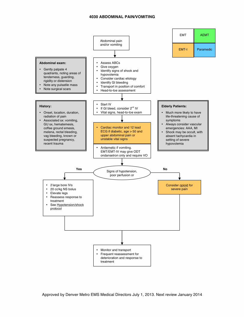
4030 ABDOMINAL PAIN/VOMITING
Approved by Denver Metro EMS Medical Directors July 1, 2013. Next review January 2014
EMT
Paramedic EMT-I
AEMT
Abdominal pain and/or vomiting
• Assess ABCs • Give oxygen • Identify signs of shock and
hypovolemia • Consider cardiac etiology • Identify GI bleeding • Transport in position of comfort • Head-to-toe assessment
• Start IV • If GI bleed, consider 2nd IV • Vital signs, head-to-toe exam
• 2 large bore IVs • 20 cc/kg NS bolus • Elevate legs • Reassess response to
treatment • See Hypotension/shock
protocol
Consider opioid for severe pain
• Monitor and transport • Frequent reassessment for
deterioration and response to treatment
• Cardiac monitor and 12 lead ECG if diabetic, age > 50 and upper abdominal pain or unstable vital signs
Signs of hypotension, poor perfusion or
shock?
No Yes
Abdominal exam:
• Gently palpate 4 quadrants, noting areas of tenderness, guarding, rigidity or distension
• Note any pulsatile mass • Note surgical scars
History:
• Onset, location, duration, radiation of pain
• Associated sx: vomiting, GU sx, hematemesis, coffee ground emesis, melena, rectal bleeding, vag bleeding, known or suspected pregnancy, recent trauma
Elderly Patients:
• Much more likely to have life-threatening cause of symptoms
• Always consider vascular emergencies: AAA, MI
• Shock may be occult, with absent tachycardia in setting of severe hypovolemia
• Antiemetic if vomiting. EMT/EMT-IV may give ODT ondansetron only and require VO
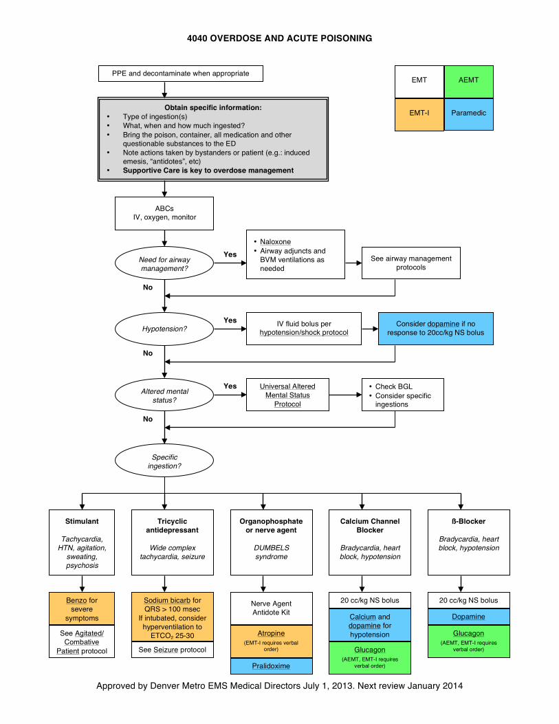
4040 OVERDOSE AND ACUTE POISONING
Approved by Denver Metro EMS Medical Directors July 1, 2013. Next review January 2014
EMT
Paramedic EMT-I
AEMT PPE and decontaminate when appropriate
Obtain specific information: • Type of ingestion(s) • What, when and how much ingested? • Bring the poison, container, all medication and other
questionable substances to the ED • Note actions taken by bystanders or patient (e.g.: induced
emesis, “antidotes”, etc) • Supportive Care is key to overdose management
ABCs IV, oxygen, monitor
• Naloxone • Airway adjuncts and
BVM ventilations as needed
Universal Altered Mental Status
Protocol
IV fluid bolus per hypotension/shock protocol
Stimulant
Tachycardia, HTN, agitation,
sweating, psychosis
Tricyclic antidepressant
Wide complex
tachycardia, seizure
Organophosphate or nerve agent
DUMBELS syndrome
Calcium Channel Blocker
Bradycardia, heart block, hypotension
ß-Blocker
Bradycardia, heart block, hypotension
Benzo for severe
symptoms
Sodium bicarb for QRS > 100 msec
If intubated, consider hyperventilation to
ETCO2 25-30
Nerve Agent Antidote Kit
Atropine
(EMT-I requires verbal order)
Consider dopamine if no response to 20cc/kg NS bolus
20 cc/kg NS bolus
Calcium and dopamine for hypotension
Dopamine
20 cc/kg NS bolus
Yes
No
Yes
No
Yes • Check BGL • Consider specific
ingestions
No
Need for airway management?
Hypotension?
Altered mental status?
Specific ingestion?
Pralidoxime
Glucagon (AEMT, EMT-I requires
verbal order)
See Seizure protocol
See Agitated/ Combative
Patient protocol
See airway management protocols
Glucagon (AEMT, EMT-I requires
verbal order)
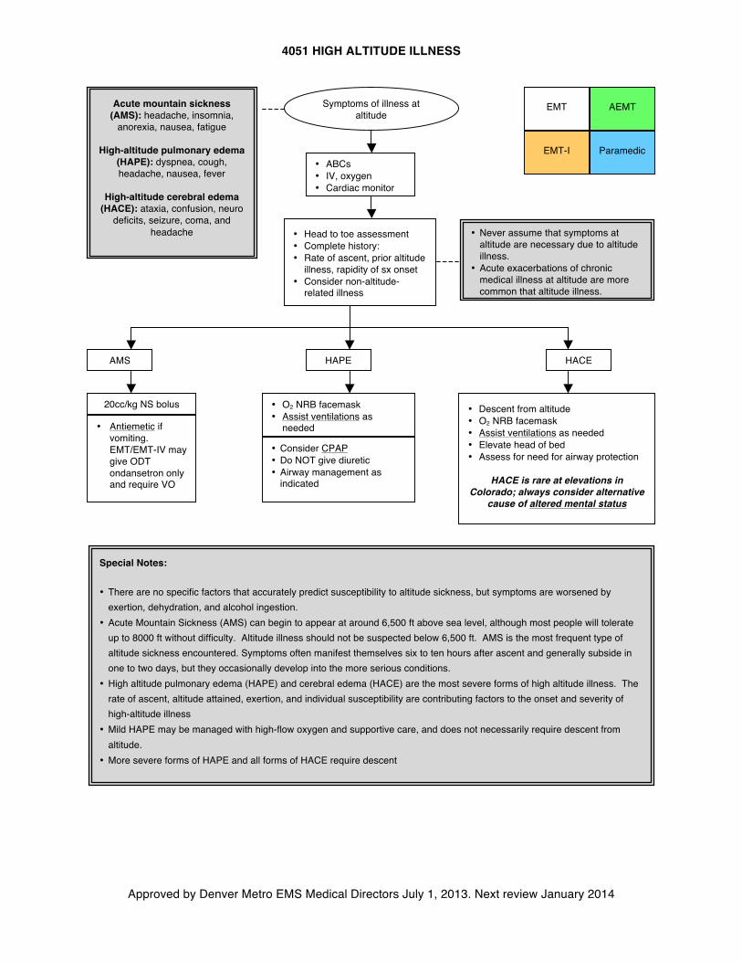
4051 HIGH ALTITUDE ILLNESS
Approved by Denver Metro EMS Medical Directors July 1, 2013. Next review January 2014
AEMT
Special Notes:
• There are no specific factors that accurately predict susceptibility to altitude sickness, but symptoms are worsened by exertion, dehydration, and alcohol ingestion.
• Acute Mountain Sickness (AMS) can begin to appear at around 6,500 ft above sea level, although most people will tolerate up to 8000 ft without difficulty. Altitude illness should not be suspected below 6,500 ft. AMS is the most frequent type of altitude sickness encountered. Symptoms often manifest themselves six to ten hours after ascent and generally subside in one to two days, but they occasionally develop into the more serious conditions.
• High altitude pulmonary edema (HAPE) and cerebral edema (HACE) are the most severe forms of high altitude illness. The rate of ascent, altitude attained, exertion, and individual susceptibility are contributing factors to the onset and severity of high-altitude illness
• Mild HAPE may be managed with high-flow oxygen and supportive care, and does not necessarily require descent from altitude.
• More severe forms of HAPE and all forms of HACE require descent
Acute mountain sickness (AMS): headache, insomnia,
anorexia, nausea, fatigue
High-altitude pulmonary edema (HAPE): dyspnea, cough, headache, nausea, fever
High-altitude cerebral edema
(HACE): ataxia, confusion, neuro deficits, seizure, coma, and
headache
• ABCs • IV, oxygen • Cardiac monitor
AMS HAPE HACE
20cc/kg NS bolus • O2 NRB facemask • Assist ventilations as
needed
• Consider CPAP • Do NOT give diuretic • Airway management as
indicated
• Descent from altitude • O2 NRB facemask • Assist ventilations as needed • Elevate head of bed • Assess for need for airway protection
HACE is rare at elevations in
Colorado; always consider alternative cause of altered mental status
EMT-I
EMT
Paramedic
Symptoms of illness at altitude
e
• Head to toe assessment • Complete history: • Rate of ascent, prior altitude
illness, rapidity of sx onset • Consider non-altitude-
related illness
• Never assume that symptoms at altitude are necessary due to altitude illness.
• Acute exacerbations of chronic medical illness at altitude are more common that altitude illness.
•
• Antiemetic if vomiting. EMT/EMT-IV may give ODT ondansetron only and require VO
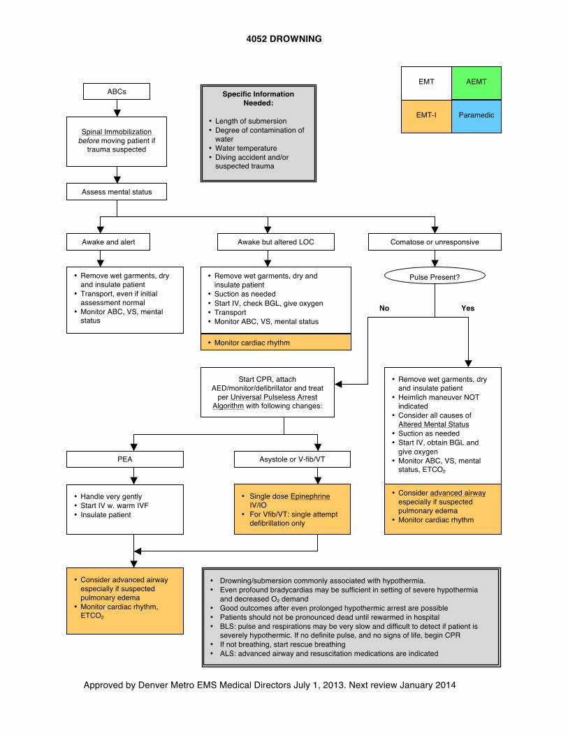
4052 DROWNING
Approved by Denver Metro EMS Medical Directors July 1, 2013. Next review January 2014
AEMT
Specific Information Needed:
• Length of submersion • Degree of contamination of
water • Water temperature • Diving accident and/or
suspected trauma
EMT
Paramedic EMT-I
Spinal Immobilization before moving patient if
trauma suspected
• Monitor cardiac rhythm
• Remove wet garments, dry and insulate patient
• Transport, even if initial assessment normal
• Monitor ABC, VS, mental status
• Remove wet garments, dry and insulate patient
• Heimlich maneuver NOT indicated
• Consider all causes of Altered Mental Status
• Suction as needed • Start IV, obtain BGL and
give oxygen • Monitor ABC, VS, mental
status, ETCO2
• Remove wet garments, dry and insulate patient
• Suction as needed • Start IV, check BGL, give oxygen • Transport • Monitor ABC, VS, mental status
ABCs
Awake and alert
Assess mental status
Awake but altered LOC Comatose or unresponsive
Yes No
• Drowning/submersion commonly associated with hypothermia. • Even profound bradycardias may be sufficient in setting of severe hypothermia
and decreased O2 demand • Good outcomes after even prolonged hypothermic arrest are possible • Patients should not be pronounced dead until rewarmed in hospital • BLS: pulse and respirations may be very slow and difficult to detect if patient is
severely hypothermic. If no definite pulse, and no signs of life, begin CPR • If not breathing, start rescue breathing • ALS: advanced airway and resuscitation medications are indicated
• Consider advanced airway especially if suspected pulmonary edema
• Monitor cardiac rhythm
Pulse Present?
Start CPR, attach AED/monitor/defibrillator and treat
per Universal Pulseless Arrest Algorithm with following changes:
Asystole or V-fib/VT PEA
• Single dose Epinephrine IV/IO
• For Vfib/VT: single attempt defibrillation only
• Handle very gently • Start IV w. warm IVF • Insulate patient
• Consider advanced airway especially if suspected pulmonary edema
• Monitor cardiac rhythm, ETCO2
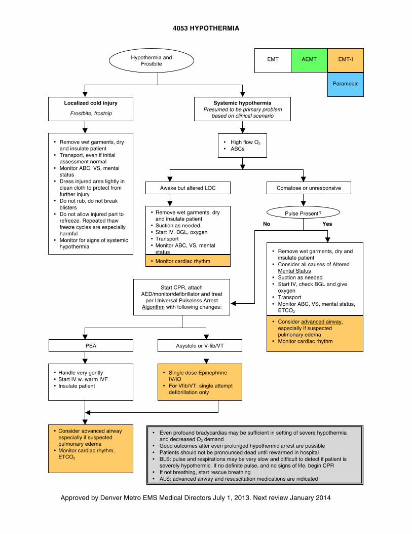
4053 HYPOTHERMIA
Approved by Denver Metro EMS Medical Directors July 1, 2013. Next review January 2014
AEMT
EMT
Paramedic
EMT-I
Systemic hypothermia Presumed to be primary problem
based on clinical scenario
• Monitor cardiac rhythm
• Remove wet garments, dry and insulate patient
• Transport, even if initial assessment normal
• Monitor ABC, VS, mental status
• Dress injured area lightly in clean cloth to protect from further injury
• Do not rub, do not break blisters
• Do not allow injured part to refreeze. Repeated thaw freeze cycles are especially harmful
• Monitor for signs of systemic hypothermia
• Remove wet garments, dry and insulate patient
• Consider all causes of Altered Mental Status
• Suction as needed • Start IV, check BGL and give
oxygen • Transport • Monitor ABC, VS, mental status,
ETCO2
• Remove wet garments, dry and insulate patient
• Suction as needed • Start IV, BGL, oxygen • Transport • Monitor ABC, VS, mental
status
• High flow O2 • ABCs
Awake but altered LOC Comatose or unresponsive
Yes No
PEA Asystole or V-fib/VT
• Handle very gently • Start IV w. warm IVF • Insulate patient
• Consider advanced airway especially if suspected pulmonary edema
• Monitor cardiac rhythm, ETCO2
• Consider advanced airway, especially if suspected pulmonary edema
• Monitor cardiac rhythm
Localized cold injury
Frostbite, frostnip
Hypothermia and Frostbite
Pulse Present?
• Even profound bradycardias may be sufficient in setting of severe hypothermia and decreased O2 demand
• Good outcomes after even prolonged hypothermic arrest are possible • Patients should not be pronounced dead until rewarmed in hospital • BLS: pulse and respirations may be very slow and difficult to detect if patient is
severely hypothermic. If no definite pulse, and no signs of life, begin CPR • If not breathing, start rescue breathing • ALS: advanced airway and resuscitation medications are indicated
Start CPR, attach AED/monitor/defibrillator and treat
per Universal Pulseless Arrest Algorithm with following changes:
• Single dose Epinephrine IV/IO
• For Vfib/VT: single attempt defibrillation only
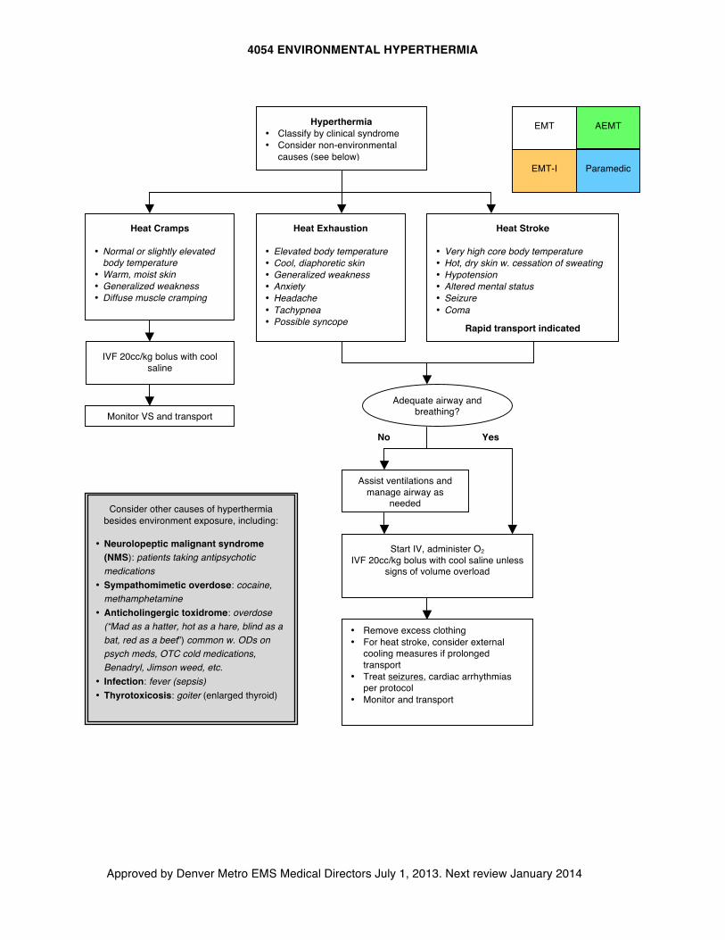
4054 ENVIRONMENTAL HYPERTHERMIA
Approved by Denver Metro EMS Medical Directors July 1, 2013. Next review January 2014
AEMT
EMT
EMT-I Paramedic
Heat Cramps
• Normal or slightly elevated body temperature
• Warm, moist skin • Generalized weakness • Diffuse muscle cramping
Heat Exhaustion
• Elevated body temperature • Cool, diaphoretic skin • Generalized weakness • Anxiety • Headache • Tachypnea • Possible syncope
Heat Stroke
• Very high core body temperature • Hot, dry skin w. cessation of sweating • Hypotension • Altered mental status • Seizure • Coma
Rapid transport indicated
Adequate airway and breathing?
No Yes
Assist ventilations and manage airway as
needed
Start IV, administer O2 IVF 20cc/kg bolus with cool saline unless
signs of volume overload
• Remove excess clothing • For heat stroke, consider external
cooling measures if prolonged transport
• Treat seizures, cardiac arrhythmias per protocol
• Monitor and transport
Consider other causes of hyperthermia besides environment exposure, including:
• Neurolopeptic malignant syndrome
(NMS): patients taking antipsychotic medications
• Sympathomimetic overdose: cocaine, methamphetamine
• Anticholingergic toxidrome: overdose (“Mad as a hatter, hot as a hare, blind as a bat, red as a beet”) common w. ODs on psych meds, OTC cold medications, Benadryl, Jimson weed, etc.
• Infection: fever (sepsis) • Thyrotoxicosis: goiter (enlarged thyroid)
IVF 20cc/kg bolus with cool saline
Monitor VS and transport
Hyperthermia • Classify by clinical syndrome • Consider non-environmental
causes (see below)
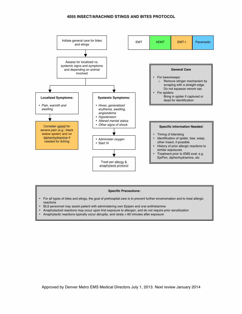
4055 INSECT/ARACHNID STINGS AND BITES PROTOCOL
Approved by Denver Metro EMS Medical Directors July 1, 2013. Next review January 2014
AEMT
Specific Information Needed:
• Timing of bite/sting • Identification of spider, bee, wasp,
other insect, if possible • History of prior allergic reactions to
similar exposures • Treatment prior to EMS eval: e.g.
EpiPen, diphenhydramine, etc
• Administer oxygen • Start IV
General Care • For bees/wasps:
o Remove stinger mechanism by scraping with a straight edge. Do not squeeze venom sac
• For spiders: o Bring in spider if captured or
dead for identification
Assess for localized vs. systemic signs and symptoms
and depending on animal involved
Localized Symptoms:
• Pain, warmth and swelling
Systemic Symptoms:
• Hives, generalized erythema, swelling, angioedema
• Hypotension • Altered mental status • Other signs of shock
EMT-I Paramedic EMT Initiate general care for bites and stings
Specific Precautions:
• For all types of bites and stings, the goal of prehospital care is to prevent further envenomation and to treat allergic reactions
• BLS personnel may assist patient with administering own Epipen and oral antihistamine • Anaphylactoid reactions may occur upon first exposure to allergen, and do not require prior sensitization • Anaphylactic reactions typically occur abruptly, and rarely > 60 minutes after exposure
Treat per allergy & anaphylaxis protocol
Consider opioid for severe pain (e.g.: black widow spider) and /or diphenhydramine if needed for itching
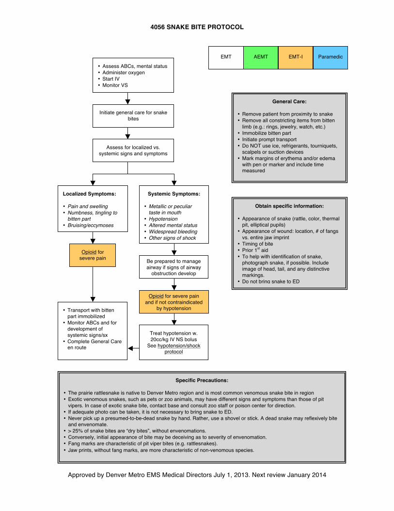
4056 SNAKE BITE PROTOCOL
Approved by Denver Metro EMS Medical Directors July 1, 2013. Next review January 2014
AEMT
Obtain specific information:
• Appearance of snake (rattle, color, thermal pit, elliptical pupils)
• Appearance of wound: location, # of fangs vs. entire jaw imprint
• Timing of bite • Prior 1st aid • To help with identification of snake,
photograph snake, if possible. Include image of head, tail, and any distinctive markings.
• Do not bring snake to ED
• Assess ABCs, mental status • Administer oxygen • Start IV • Monitor VS
General Care: • Remove patient from proximity to snake • Remove all constricting items from bitten
limb (e.g.: rings, jewelry, watch, etc.) • Immobilize bitten part • Initiate prompt transport • Do NOT use ice, refrigerants, tourniquets,
scalpels or suction devices • Mark margins of erythema and/or edema
with pen or marker and include time measured
Assess for localized vs. systemic signs and symptoms
Localized Symptoms: • Pain and swelling • Numbness, tingling to
bitten part • Bruising/eccymoses
Systemic Symptoms:
• Metallic or peculiar taste in mouth
• Hypotension • Altered mental status • Widespread bleeding • Other signs of shock
EMT-I Paramedic EMT
Be prepared to manage airway if signs of airway
obstruction develop
Treat hypotension w. 20cc/kg IV NS bolus
See hypotension/shock protocol
Initiate general care for snake bites
• Transport with bitten part immobilized
• Monitor ABCs and for development of systemic signs/sx
• Complete General Care en route
Specific Precautions:
• The prairie rattlesnake is native to Denver Metro region and is most common venomous snake bite in region • Exotic venomous snakes, such as pets or zoo animals, may have different signs and symptoms than those of pit
vipers. In case of exotic snake bite, contact base and consult zoo staff or poison center for direction. • If adequate photo can be taken, it is not necessary to bring snake to ED. • Never pick up a presumed-to-be-dead snake by hand. Rather, use a shovel or stick. A dead snake may reflexively bite
and envenomate. • > 25% of snake bites are “dry bites”, without envenomations. • Conversely, initial appearance of bite may be deceiving as to severity of envenomation. • Fang marks are characteristic of pit viper bites (e.g. rattlesnakes). • Jaw prints, without fang marks, are more characteristic of non-venomous species.
Opioid for severe pain and if not contraindicated
by hypotension
Opioid for severe pain
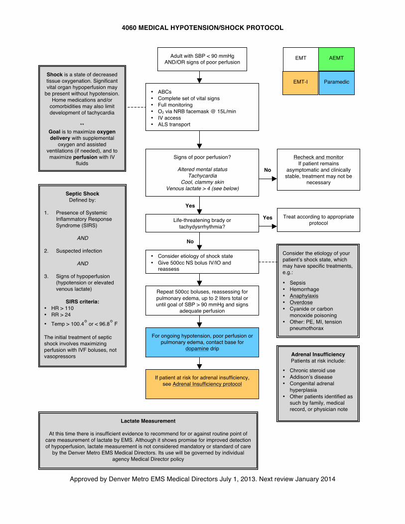
4060 MEDICAL HYPOTENSION/SHOCK PROTOCOL
Approved by Denver Metro EMS Medical Directors July 1, 2013. Next review January 2014
AEMT
Signs of poor perfusion?
Altered mental status Tachycardia
Cool, clammy skin Venous lactate > 4 (see below)
• Consider etiology of shock state • Give 500cc NS bolus IV/IO and
reassess
Adult with SBP < 90 mmHg AND/OR signs of poor perfusion
Consider the etiology of your patient’s shock state, which may have specific treatments, e.g.:
• Sepsis • Hemorrhage • Anaphylaxis • Overdose • Cyanide or carbon
monoxide poisoning • Other: PE, MI, tension
pneumothorax
Repeat 500cc boluses, reassessing for pulmonary edema, up to 2 liters total or until goal of SBP > 90 mmHg and signs
adequate perfusion
Recheck and monitor If patient remains
asymptomatic and clinically stable, treatment may not be
necessary
• ABCs • Complete set of vital signs • Full monitoring • O2 via NRB facemask @ 15L/min • IV access • ALS transport
Life-threatening brady or tachydysrrhythmia?
Treat according to appropriate protocol
Yes
No
Yes
No
For ongoing hypotension, poor perfusion or pulmonary edema, contact base for
dopamine drip
Septic Shock Defined by:
1. Presence of Systemic
Inflammatory Response Syndrome (SIRS)
AND
2. Suspected infection
AND
3. Signs of hypoperfusion
(hypotension or elevated venous lactate)
SIRS criteria: • HR > 110 • RR > 24 • Temp > 100.4° or < 96.8° F
The initial treatment of septic shock involves maximizing perfusion with IVF boluses, not vasopressors
Shock is a state of decreased tissue oxygenation. Significant vital organ hypoperfusion may
be present without hypotension. Home medications and/or
comorbidities may also limit development of tachycardia
**
Goal is to maximize oxygen delivery with supplemental
oxygen and assisted ventilations (if needed), and to maximize perfusion with IV
fluids
EMT
EMT-I Paramedic
Lactate Measurement
At this time there is insufficient evidence to recommend for or against routine point of care measurement of lactate by EMS. Although it shows promise for improved detection of hypoperfusion, lactate measurement is not considered mandatory or standard of care
by the Denver Metro EMS Medical Directors. Its use will be governed by individual agency Medical Director policy
If patient at risk for adrenal insufficiency, see Adrenal Insufficiency protocol
Adrenal Insufficiency Patients at risk include:
• Chronic steroid use • Addison’s disease • Congenital adrenal
hyperplasia • Other patients identified as
such by family, medical record, or physician note
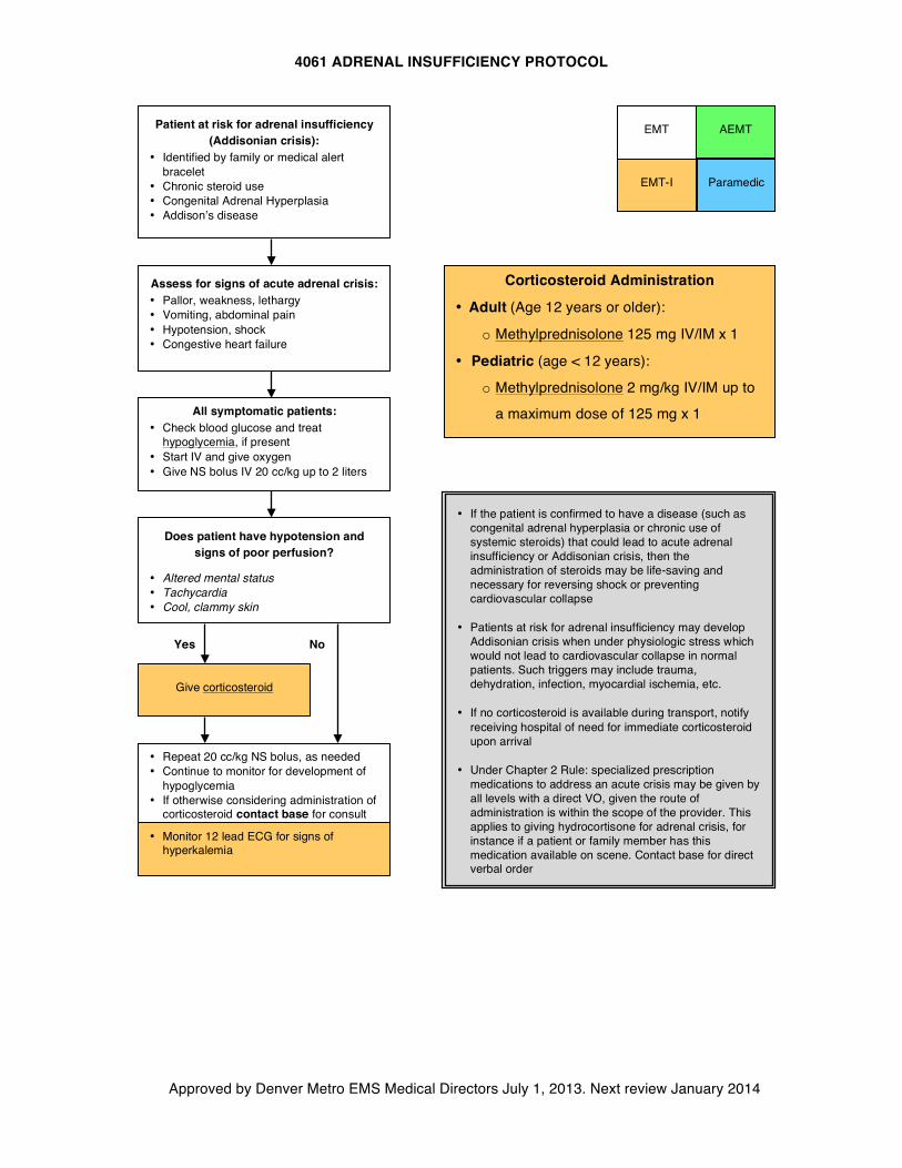
4061 ADRENAL INSUFFICIENCY PROTOCOL
Approved by Denver Metro EMS Medical Directors July 1, 2013. Next review January 2014
AEMT
Does patient have hypotension and signs of poor perfusion?
• Altered mental status • Tachycardia • Cool, clammy skin
All symptomatic patients: • Check blood glucose and treat
hypoglycemia, if present • Start IV and give oxygen • Give NS bolus IV 20 cc/kg up to 2 liters
Patient at risk for adrenal insufficiency (Addisonian crisis):
• Identified by family or medical alert bracelet
• Chronic steroid use • Congenital Adrenal Hyperplasia • Addison’s disease
• If the patient is confirmed to have a disease (such as congenital adrenal hyperplasia or chronic use of systemic steroids) that could lead to acute adrenal insufficiency or Addisonian crisis, then the administration of steroids may be life-saving and necessary for reversing shock or preventing cardiovascular collapse
• Patients at risk for adrenal insufficiency may develop Addisonian crisis when under physiologic stress which would not lead to cardiovascular collapse in normal patients. Such triggers may include trauma, dehydration, infection, myocardial ischemia, etc.
• If no corticosteroid is available during transport, notify
receiving hospital of need for immediate corticosteroid upon arrival
• Under Chapter 2 Rule: specialized prescription
medications to address an acute crisis may be given by all levels with a direct VO, given the route of administration is within the scope of the provider. This applies to giving hydrocortisone for adrenal crisis, for instance if a patient or family member has this medication available on scene. Contact base for direct verbal order
• Repeat 20 cc/kg NS bolus, as needed • Continue to monitor for development of
hypoglycemia • If otherwise considering administration of
corticosteroid contact base for consult
Assess for signs of acute adrenal crisis: • Pallor, weakness, lethargy • Vomiting, abdominal pain • Hypotension, shock • Congestive heart failure
Give corticosteroid
Yes
No
EMT
EMT-I Paramedic
• Monitor 12 lead ECG for signs of hyperkalemia
Corticosteroid Administration
• Adult (Age 12 years or older):
o Methylprednisolone 125 mg IV/IM x 1
• Pediatric (age < 12 years):
o Methylprednisolone 2 mg/kg IV/IM up to
a maximum dose of 125 mg x 1
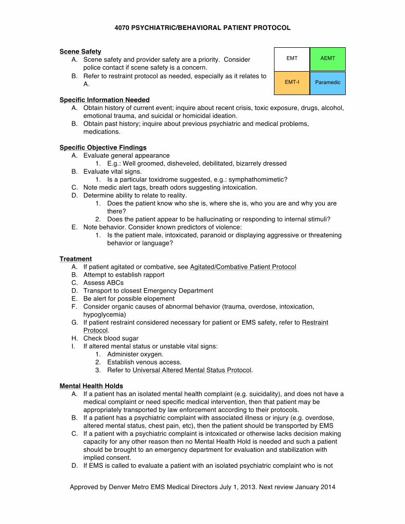
4070 PSYCHIATRIC/BEHAVIORAL PATIENT PROTOCOL
Approved by Denver Metro EMS Medical Directors July 1, 2013. Next review January 2014
AEMT Scene Safety
A. Scene safety and provider safety are a priority. Consider police contact if scene safety is a concern.
B. Refer to restraint protocol as needed, especially as it relates to A.
Specific Information Needed A. Obtain history of current event; inquire about recent crisis, toxic exposure, drugs, alcohol,
emotional trauma, and suicidal or homicidal ideation. B. Obtain past history; inquire about previous psychiatric and medical problems,
medications. Specific Objective Findings
A. Evaluate general appearance 1. E.g.: Well groomed, disheveled, debilitated, bizarrely dressed
B. Evaluate vital signs. 1. Is a particular toxidrome suggested, e.g.: symphathomimetic?
C. Note medic alert tags, breath odors suggesting intoxication. D. Determine ability to relate to reality.
1. Does the patient know who she is, where she is, who you are and why you are there?
2. Does the patient appear to be hallucinating or responding to internal stimuli? E. Note behavior. Consider known predictors of violence:
1. Is the patient male, intoxicated, paranoid or displaying aggressive or threatening behavior or language?
Treatment A. If patient agitated or combative, see Agitated/Combative Patient Protocol B. Attempt to establish rapport C. Assess ABCs D. Transport to closest Emergency Department E. Be alert for possible elopement F. Consider organic causes of abnormal behavior (trauma, overdose, intoxication,
hypoglycemia) G. If patient restraint considered necessary for patient or EMS safety, refer to Restraint
Protocol. H. Check blood sugar I. If altered mental status or unstable vital signs:
1. Administer oxygen. 2. Establish venous access. 3. Refer to Universal Altered Mental Status Protocol.
Mental Health Holds
A. If a patient has an isolated mental health complaint (e.g. suicidality), and does not have a medical complaint or need specific medical intervention, then that patient may be appropriately transported by law enforcement according to their protocols.
B. If a patient has a psychiatric complaint with associated illness or injury (e.g. overdose, altered mental status, chest pain, etc), then the patient should be transported by EMS
C. If a patient with a psychiatric complaint is intoxicated or otherwise lacks decision making capacity for any other reason then no Mental Health Hold is needed and such a patient should be brought to an emergency department for evaluation and stabilization with implied consent.
D. If EMS is called to evaluate a patient with an isolated psychiatric complaint who is not
EMT
EMT-I Paramedic
4070 PSYCHIATRIC/BEHAVIORAL PATIENT PROTOCOL
Approved by Denver Metro EMS Medical Directors July 1, 2013. Next review January 2014
intoxicated, or otherwise lacking decision making capacity, and who refuses treatment or transport, and law enforcement are not willing to transport patient, then EMS should contact agency Base Station for medical consult with BASE PHYSICIAN.
E. If there is a reasonable concern for suicidal or homicidal ideation, or grave disability from another mental health condition, then BASE PHYSICIAN may give a verbal order placing the patient on a Mental Health Hold and direct EMS personnel to transport the patient against his or her will in accordance with Colorado State statutes. The physician’s name, and time and date of the Mental Health Hold must be recorded on the PCR. Effort should be made to obtain consent for transport from the patient, and to preserve the patient’s dignity throughout the process.
F. A patient being transported on a Mental Health Hold may be transported to any appropriate receiving emergency department, as it may not be operationally feasible to transport exclusively to the Base Station hospital, although this is preferred if time and conditions allow.
G. It is expected that receiving facilities will receive such patients and perform an appropriate evaluation to determine if continuation of a Mental Health Hold is indicated at the time of their assessment.
H. Although there is always a risk of accusations of kidnapping or assault in such cases, such accusations are extremely rare, and the Denver Metropolitan EMS Medical Directors feel strongly that the risk of abandonment of a potentially suicidal or otherwise gravely impaired patient far outweigh any theoretical risk of allegations of kidnapping when actions are taken in the interest of patient safety.
Specific Precautions
A. Psychiatric patients often have an organic basis for mental disturbances. Be suspicious of hypoglycemia, hypoxia, head injury, intoxication, or toxic ingestion.
B. If emergency treatment is unnecessary, do as little as possible except to reassure while transporting. Try not to violate the patient's personal space.
C. If the situation appears threatening, consider a show of force involving police before attempting to restrain.
D. Beware of weapons. These patients can become very violent. E. An EMT, AEMT, Intermediate or paramedic may initiate a Mental Health Hold only by
direct verbal order from the BASE PHYSICIAN. F. Document name of BASE PHYSICIAN.
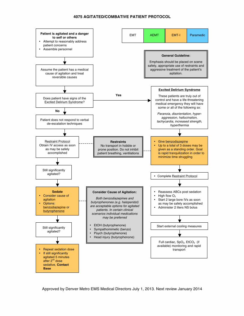
4075 AGITATED/COMBATIVE PATIENT PROTOCOL
Approved by Denver Metro EMS Medical Directors July 1, 2013. Next review January 2014
AEMT
Assume the patient has a medical cause of agitation and treat
reversible causes
Still significantly agitated?
Patient is agitated and a danger to self or others
• Attempt to reasonably address patient concerns
• Assemble personnel
Patient does not respond to verbal de-escalation techniques
Restraint Protocol Obtain IV access as soon
as may be safely accomplished
Sedate • Consider cause of
agitation • Options:
benzodiazepine or butyrophenone
Still significantly agitated?
• Repeat sedation dose • If still significantly
agitated 5 minutes after 2nd dose sedative, Contact Base
Consider Cause of Agitation:
Both benzodiazepines and butyrophenones (e.g. haloperidol) are acceptable options for agitated
patients. In certain clinical scenarios individual medications
may be preferred
• EtOH (butyrophenone) • Sympathomimetic (benzo) • Psych (butyrophenone) • Head injury (butyrophenone)
Does patient have signs of the Excited Delirium Syndrome?
No
Yes
EMT EMT-I Paramedic
General Guideline:
Emphasis should be placed on scene safety, appropriate use of restraints and
aggressive treatment of the patient’s agitation.
Excited Delirium Syndrome
These patients are truly out of control and have a life-threatening medical emergency they will have
some or all of the following sx:
Paranoia, disorientation, hyper-aggression, hallucination,
tachycardia, increased strength, hyperthermia
• Give benzodiazepine • Up to a total of 3 doses may be
given as a standing order. Goal is rapid tranquilization in order to minimize time struggling
• Reassess ABCs post sedation • High flow O2 • Start 2 large bore IVs as soon
as may be safely accomplished • Administer 2 liters NS bolus
Start external cooling measures
Full cardiac, SpO2, EtCO2 (if available) monitoring and rapid
transport
Restraints No transport in hobble or
prone position. Do not inhibit patient breathing, ventilations
• Complete Restraint Protocol
4076 TRANSPORT OF THE HANDCUFFED PATIENT
Approved by Denver Metro EMS Medical Directors July 1, 2013. Next review January 2014
Purpose:
1. Guideline for transport of patients in handcuffs placed by law enforcement Guideline:
1. Handcuffs are only to be placed by law enforcement. EMS personnel are not permitted to use handcuffs.
2. Request that law enforcement remain with the patient in the ambulance, if possible. If not possible, request that police ride behind ambulance so as to be readily available to remove handcuffs if needed in an emergency situation to facilitate medical care of the patient.
3. EMS personnel are not responsible for the law enforcement hold on these patients. 4. Handcuffed patients will not be placed in the prone position. 5. Handcuffs may be used with spinal immobilization. Medical priorities should take priority
in the positioning of the handcuffs.
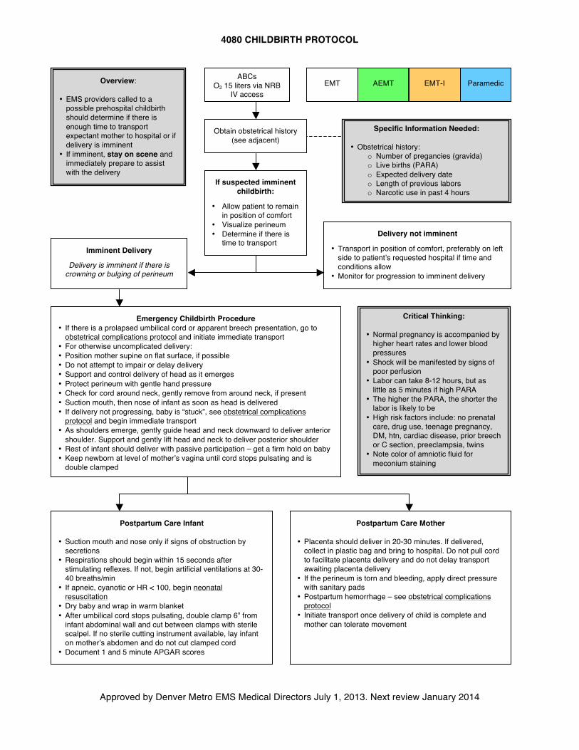
4080 CHILDBIRTH PROTOCOL
Approved by Denver Metro EMS Medical Directors July 1, 2013. Next review January 2014
EMT EMT-I Paramedic Overview:
• EMS providers called to a possible prehospital childbirth should determine if there is enough time to transport expectant mother to hospital or if delivery is imminent
• If imminent, stay on scene and immediately prepare to assist with the delivery
AEMT
Obtain obstetrical history (see adjacent)
ABCs O2 15 liters via NRB
IV access
Specific Information Needed: • Obstetrical history:
o Number of pregancies (gravida) o Live births (PARA) o Expected delivery date o Length of previous labors o Narcotic use in past 4 hours
If suspected imminent childbirth:
• Allow patient to remain in position of comfort
• Visualize perineum • Determine if there is
time to transport Imminent Delivery
Delivery is imminent if there is crowning or bulging of perineum
Emergency Childbirth Procedure • If there is a prolapsed umbilical cord or apparent breech presentation, go to
obstetrical complications protocol and initiate immediate transport • For otherwise uncomplicated delivery: • Position mother supine on flat surface, if possible • Do not attempt to impair or delay delivery • Support and control delivery of head as it emerges • Protect perineum with gentle hand pressure • Check for cord around neck, gently remove from around neck, if present • Suction mouth, then nose of infant as soon as head is delivered • If delivery not progressing, baby is “stuck”, see obstetrical complications
protocol and begin immediate transport • As shoulders emerge, gently guide head and neck downward to deliver anterior
shoulder. Support and gently lift head and neck to deliver posterior shoulder • Rest of infant should deliver with passive participation – get a firm hold on baby • Keep newborn at level of mother’s vagina until cord stops pulsating and is
double clamped
Delivery not imminent
• Transport in position of comfort, preferably on left side to patient’s requested hospital if time and conditions allow
• Monitor for progression to imminent delivery
Postpartum Care Infant
• Suction mouth and nose only if signs of obstruction by secretions
• Respirations should begin within 15 seconds after stimulating reflexes. If not, begin artificial ventilations at 30-40 breaths/min
• If apneic, cyanotic or HR < 100, begin neonatal resuscitation
• Dry baby and wrap in warm blanket • After umbilical cord stops pulsating, double clamp 6” from
infant abdominal wall and cut between clamps with sterile scalpel. If no sterile cutting instrument available, lay infant on mother’s abdomen and do not cut clamped cord
• Document 1 and 5 minute APGAR scores
Postpartum Care Mother
• Placenta should deliver in 20-30 minutes. If delivered, collect in plastic bag and bring to hospital. Do not pull cord to facilitate placenta delivery and do not delay transport awaiting placenta delivery
• If the perineum is torn and bleeding, apply direct pressure with sanitary pads
• Postpartum hemorrhage – see obstetrical complications protocol
• Initiate transport once delivery of child is complete and mother can tolerate movement
Critical Thinking:
• Normal pregnancy is accompanied by higher heart rates and lower blood pressures
• Shock will be manifested by signs of poor perfusion
• Labor can take 8-12 hours, but as little as 5 minutes if high PARA
• The higher the PARA, the shorter the labor is likely to be
• High risk factors include: no prenatal care, drug use, teenage pregnancy, DM, htn, cardiac disease, prior breech or C section, preeclampsia, twins
• Note color of amniotic fluid for meconium staining
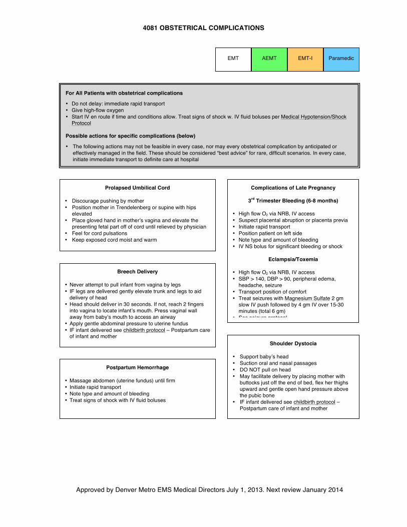
4081 OBSTETRICAL COMPLICATIONS
Approved by Denver Metro EMS Medical Directors July 1, 2013. Next review January 2014
EMT EMT-I Paramedic AEMT
Complications of Late Pregnancy
3rd Trimester Bleeding (6-8 months)
• High flow O2 via NRB, IV access • Suspect placental abruption or placenta previa • Initiate rapid transport • Position patient on left side • Note type and amount of bleeding • IV NS bolus for significant bleeding or shock
Eclampsia/Toxemia
• High flow O2 via NRB, IV access • SBP > 140, DBP > 90, peripheral edema,
headache, seizure • Transport position of comfort • Treat seizures with Magnesium Sulfate 2 gm
slow IV push followed by 4 gm IV over 15-30 minutes (total 6 gm)
• See seizure protocol
Prolapsed Umbilical Cord
• Discourage pushing by mother • Position mother in Trendelenberg or supine with hips
elevated • Place gloved hand in mother’s vagina and elevate the
presenting fetal part off of cord until relieved by physician • Feel for cord pulsations • Keep exposed cord moist and warm
Breech Delivery
• Never attempt to pull infant from vagina by legs • IF legs are delivered gently elevate trunk and legs to aid
delivery of head • Head should deliver in 30 seconds. If not, reach 2 fingers
into vagina to locate infant’s mouth. Press vaginal wall away from baby’s mouth to access an airway
• Apply gentle abdominal pressure to uterine fundus • IF infant delivered see childbirth protocol – Postpartum care
of infant and mother Shoulder Dystocia
• Support baby’s head • Suction oral and nasal passages • DO NOT pull on head • May facilitate delivery by placing mother with
buttocks just off the end of bed, flex her thighs upward and gentle open hand pressure above the pubic bone
• IF infant delivered see childbirth protocol – Postpartum care of infant and mother
Postpartum Hemorrhage
• Massage abdomen (uterine fundus) until firm • Initiate rapid transport • Note type and amount of bleeding • Treat signs of shock with IV fluid boluses
For All Patients with obstetrical complications
• Do not delay: immediate rapid transport • Give high-flow oxygen • Start IV en route if time and conditions allow. Treat signs of shock w. IV fluid boluses per Medical Hypotension/Shock
Protocol Possible actions for specific complications (below)
• The following actions may not be feasible in every case, nor may every obstetrical complication by anticipated or effectively managed in the field. These should be considered “best advice” for rare, difficult scenarios. In every case, initiate immediate transport to definite care at hospital
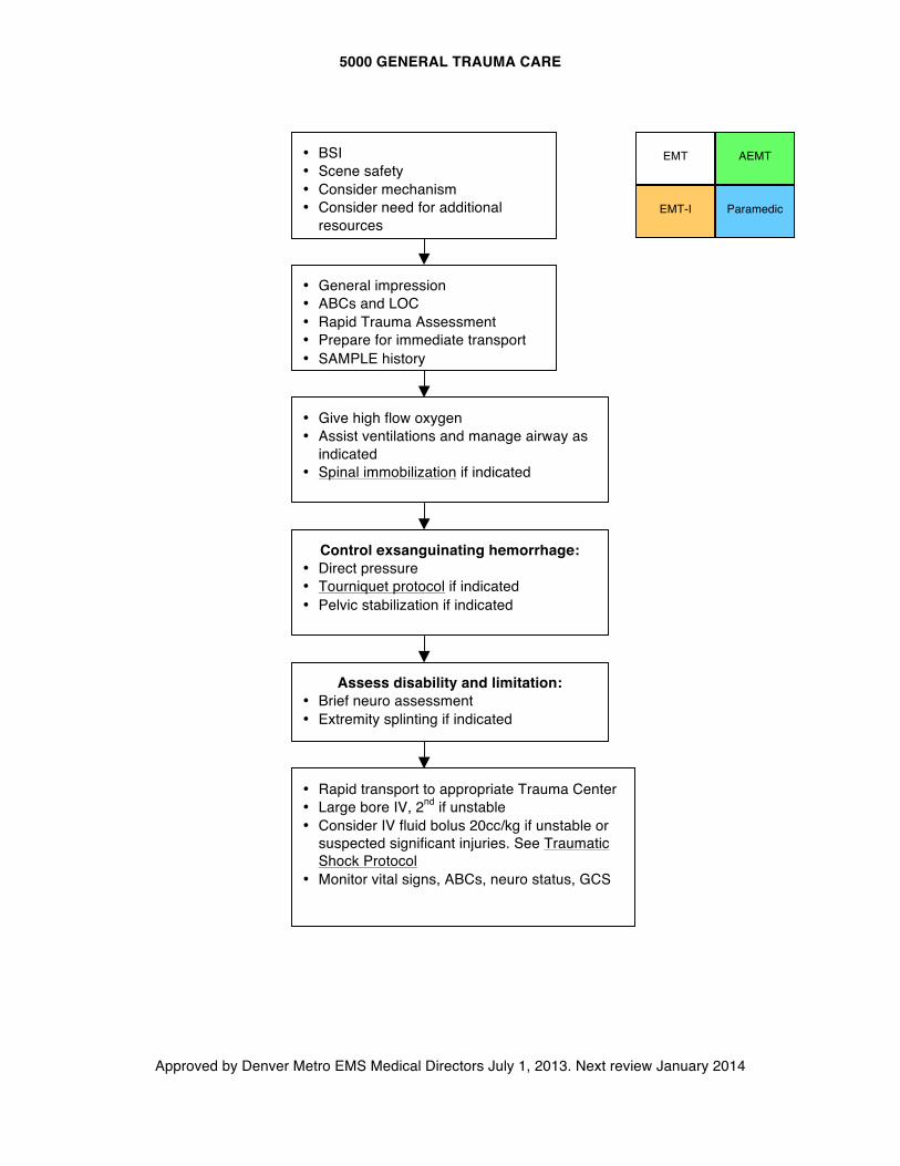
5000 GENERAL TRAUMA CARE
Approved by Denver Metro EMS Medical Directors July 1, 2013. Next review January 2014
AEMT
EMT
Paramedic EMT-I
• BSI • Scene safety • Consider mechanism • Consider need for additional
resources
• Give high flow oxygen • Assist ventilations and manage airway as
indicated • Spinal immobilization if indicated
Control exsanguinating hemorrhage: • Direct pressure • Tourniquet protocol if indicated • Pelvic stabilization if indicated
Assess disability and limitation: • Brief neuro assessment • Extremity splinting if indicated
• Rapid transport to appropriate Trauma Center • Large bore IV, 2nd if unstable • Consider IV fluid bolus 20cc/kg if unstable or
suspected significant injuries. See Traumatic Shock Protocol
• Monitor vital signs, ABCs, neuro status, GCS
• General impression • ABCs and LOC • Rapid Trauma Assessment • Prepare for immediate transport • SAMPLE history
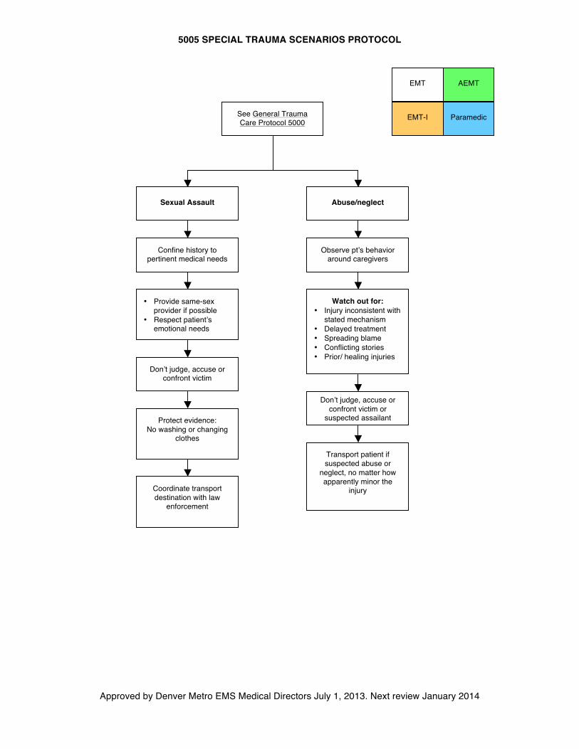
5005 SPECIAL TRAUMA SCENARIOS PROTOCOL
Approved by Denver Metro EMS Medical Directors July 1, 2013. Next review January 2014
AEMT
EMT
Paramedic EMT-I
Sexual Assault Abuse/neglect
Confine history to pertinent medical needs
• Provide same-sex provider if possible
• Respect patient’s emotional needs
Protect evidence: No washing or changing
clothes
Coordinate transport destination with law
enforcement
Observe pt’s behavior around caregivers
Watch out for: • Injury inconsistent with
stated mechanism • Delayed treatment • Spreading blame • Conflicting stories • Prior/ healing injuries
Don’t judge, accuse or confront victim
Don’t judge, accuse or confront victim or
suspected assailant
Transport patient if suspected abuse or
neglect, no matter how apparently minor the
injury
See General Trauma Care Protocol 5000
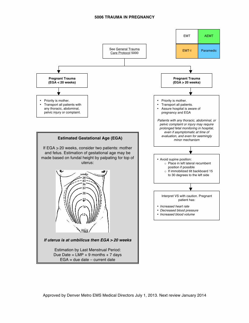
5006 TRAUMA IN PREGNANCY
Approved by Denver Metro EMS Medical Directors July 1, 2013. Next review January 2014
• Avoid supine position: o Place in left lateral recumbent
position if possible o If immobilized tilt backboard 15
to 30 degrees to the left side
AEMT
EMT
Paramedic EMT-I See General Trauma Care Protocol 5000
Pregnant Trauma (EGA > 20 weeks)
Interpret VS with caution. Pregnant patient has:
• Increased heart rate • Decreased blood pressure • Increased blood volume
Estimated Gestational Age (EGA)
If EGA > 20 weeks, consider two patients: mother and fetus. Estimation of gestational age may be
made based on fundal height by palpating for top of uterus:
If uterus is at umbilicus then EGA > 20 weeks
Estimation by Last Menstrual Period: Due Date = LMP + 9 months + 7 days
EGA = due date – current date
Pregnant Trauma (EGA < 20 weeks)
• Priority is mother. • Transport all patients. • Assure hospital is aware of
pregnancy and EGA Patients with any thoracic, abdominal, or
pelvic complaint or injury may require prolonged fetal monitoring in hospital,
even if asymptomatic at time of evaluation, and even for seemingly
minor mechanism
• Priority is mother. • Transport all patients with
any thoracic, abdominal, pelvic injury or complaint.
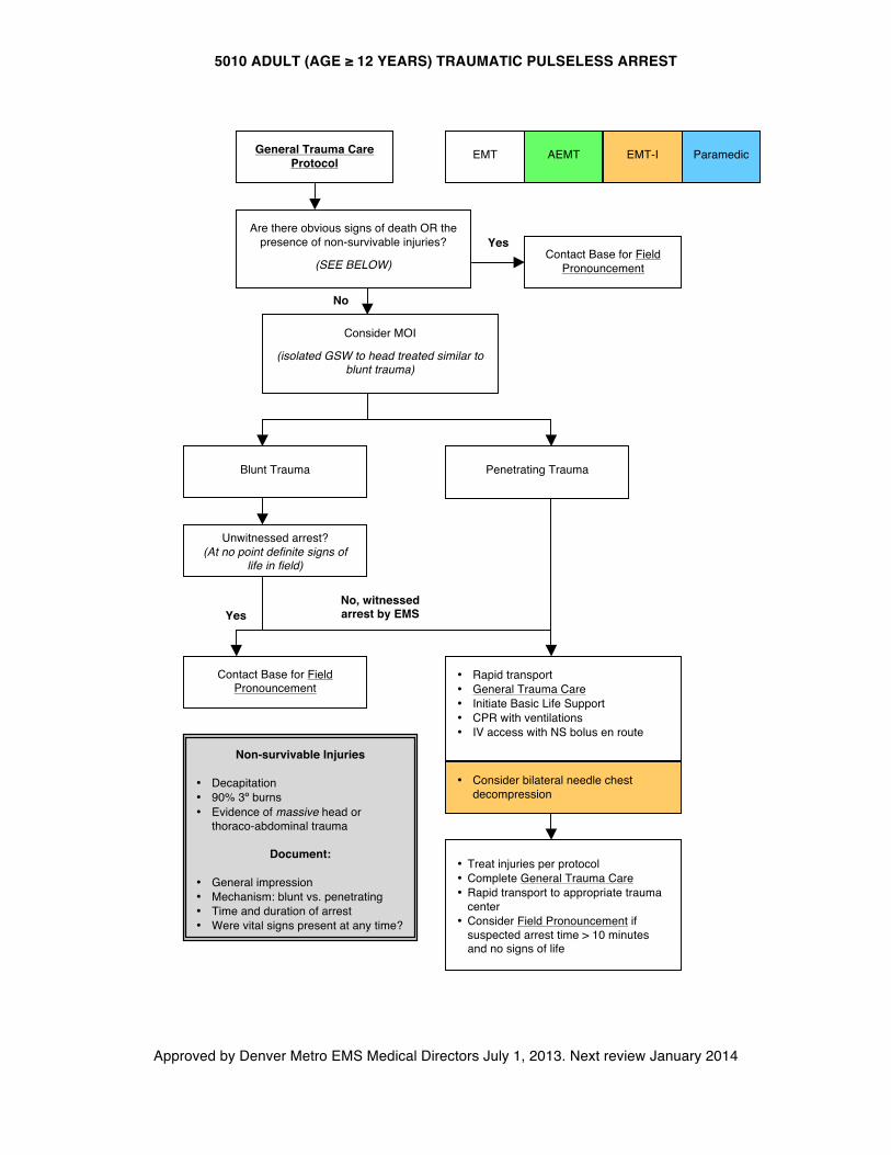
5010 ADULT (AGE ≥ 12 YEARS) TRAUMATIC PULSELESS ARREST
Approved by Denver Metro EMS Medical Directors July 1, 2013. Next review January 2014
AEMT
Blunt Trauma Penetrating Trauma
• Rapid transport • General Trauma Care • Initiate Basic Life Support • CPR with ventilations • IV access with NS bolus en route
Paramedic
EMT EMT-I General Trauma Care Protocol
Are there obvious signs of death OR the presence of non-survivable injuries?
(SEE BELOW)
Non-survivable Injuries • Decapitation • 90% 3º burns • Evidence of massive head or
thoraco-abdominal trauma
Document:
• General impression • Mechanism: blunt vs. penetrating • Time and duration of arrest • Were vital signs present at any time?
Contact Base for Field Pronouncement
Yes
No
• Treat injuries per protocol • Complete General Trauma Care • Rapid transport to appropriate trauma
center • Consider Field Pronouncement if
suspected arrest time > 10 minutes and no signs of life
Unwitnessed arrest? (At no point definite signs of
life in field)
Contact Base for Field Pronouncement
Yes No, witnessed arrest by EMS
Consider MOI
(isolated GSW to head treated similar to blunt trauma)
• Consider bilateral needle chest decompression
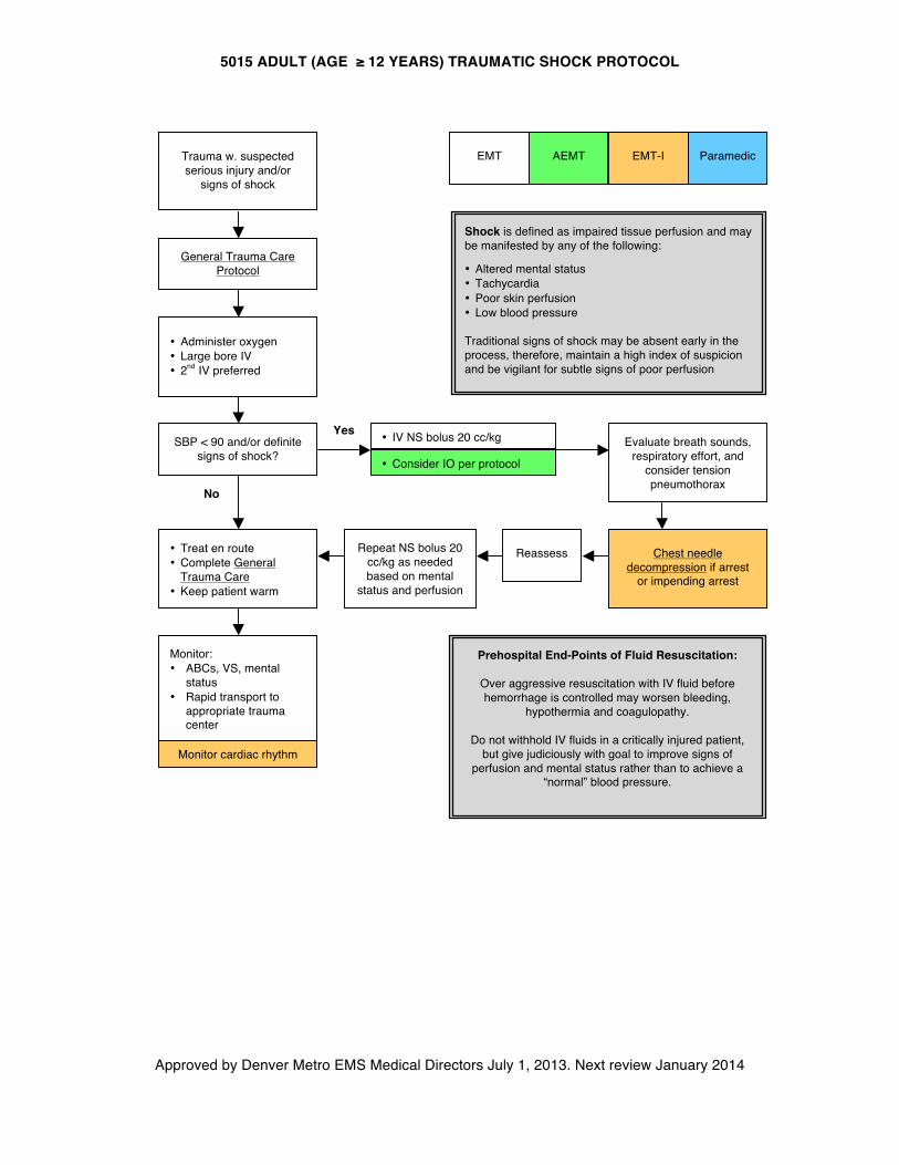
5015 ADULT (AGE ≥ 12 YEARS) TRAUMATIC SHOCK PROTOCOL
Approved by Denver Metro EMS Medical Directors July 1, 2013. Next review January 2014
No
Yes
AEMT
EMT Paramedic EMT-I
General Trauma Care Protocol
• Administer oxygen • Large bore IV • 2nd IV preferred
SBP < 90 and/or definite signs of shock?
Evaluate breath sounds, respiratory effort, and
consider tension pneumothorax
x
Chest needle decompression if arrest
or impending arrest
• Treat en route • Complete General
Trauma Care • Keep patient warm
• Consider IO per protocol
Monitor cardiac rhythm
Monitor: • ABCs, VS, mental
status • Rapid transport to
appropriate trauma center
Prehospital End-Points of Fluid Resuscitation:
Over aggressive resuscitation with IV fluid before hemorrhage is controlled may worsen bleeding,
hypothermia and coagulopathy.
Do not withhold IV fluids in a critically injured patient, but give judiciously with goal to improve signs of
perfusion and mental status rather than to achieve a “normal” blood pressure.
Repeat NS bolus 20 cc/kg as needed based on mental
status and perfusion
Shock is defined as impaired tissue perfusion and may be manifested by any of the following:
• Altered mental status • Tachycardia • Poor skin perfusion • Low blood pressure
Traditional signs of shock may be absent early in the process, therefore, maintain a high index of suspicion and be vigilant for subtle signs of poor perfusion
• IV NS bolus 20 cc/kg
Reassess
Trauma w. suspected serious injury and/or
signs of shock
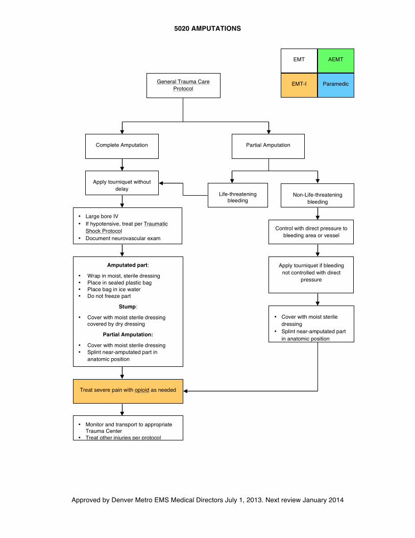
5020 AMPUTATIONS
Approved by Denver Metro EMS Medical Directors July 1, 2013. Next review January 2014
AEMT
General Trauma Care Protocol
EMT
Paramedic EMT-I
Partial Amputation
Control with direct pressure to bleeding area or vessel
Complete Amputation
Apply tourniquet if bleeding not controlled with direct
pressure
• Large bore IV • If hypotensive, treat per Traumatic
Shock Protocol • Document neurovascular exam
Amputated part:
• Wrap in moist, sterile dressing • Place in sealed plastic bag • Place bag in ice water • Do not freeze part
Stump:
• Cover with moist sterile dressing covered by dry dressing
Partial Amputation:
• Cover with moist sterile dressing • Splint near-amputated part in
anatomic position
Treat severe pain with opioid as needed
• Cover with moist sterile dressing
• Splint near-amputated part in anatomic position
• Monitor and transport to appropriate Trauma Center
• Treat other injuries per protocol
Apply tourniquet without delay
Life-threatening bleeding
Non-Life-threatening bleeding
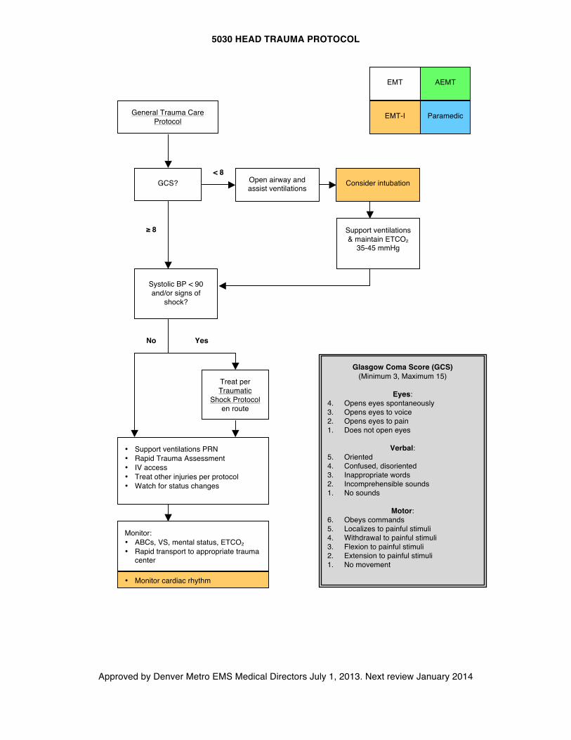
5030 HEAD TRAUMA PROTOCOL
Approved by Denver Metro EMS Medical Directors July 1, 2013. Next review January 2014
< 8
Yes
AEMT
General Trauma Care Protocol
EMT
Paramedic EMT-I
GCS?
≥ 8 Support ventilations & maintain ETCO2
35-45 mmHg
Systolic BP < 90 and/or signs of
shock?
No
Treat per Traumatic
Shock Protocol en route
• Support ventilations PRN • Rapid Trauma Assessment • IV access • Treat other injuries per protocol • Watch for status changes
Open airway and assist ventilations
Glasgow Coma Score (GCS) (Minimum 3, Maximum 15)
Eyes:
4. Opens eyes spontaneously 3. Opens eyes to voice 2. Opens eyes to pain 1. Does not open eyes
Verbal:
5. Oriented 4. Confused, disoriented 3. Inappropriate words 2. Incomprehensible sounds 1. No sounds
Motor:
6. Obeys commands 5. Localizes to painful stimuli 4. Withdrawal to painful stimuli 3. Flexion to painful stimuli 2. Extension to painful stimuli 1. No movement
Consider intubation
• Monitor cardiac rhythm
Monitor: • ABCs, VS, mental status, ETCO2 • Rapid transport to appropriate trauma
center
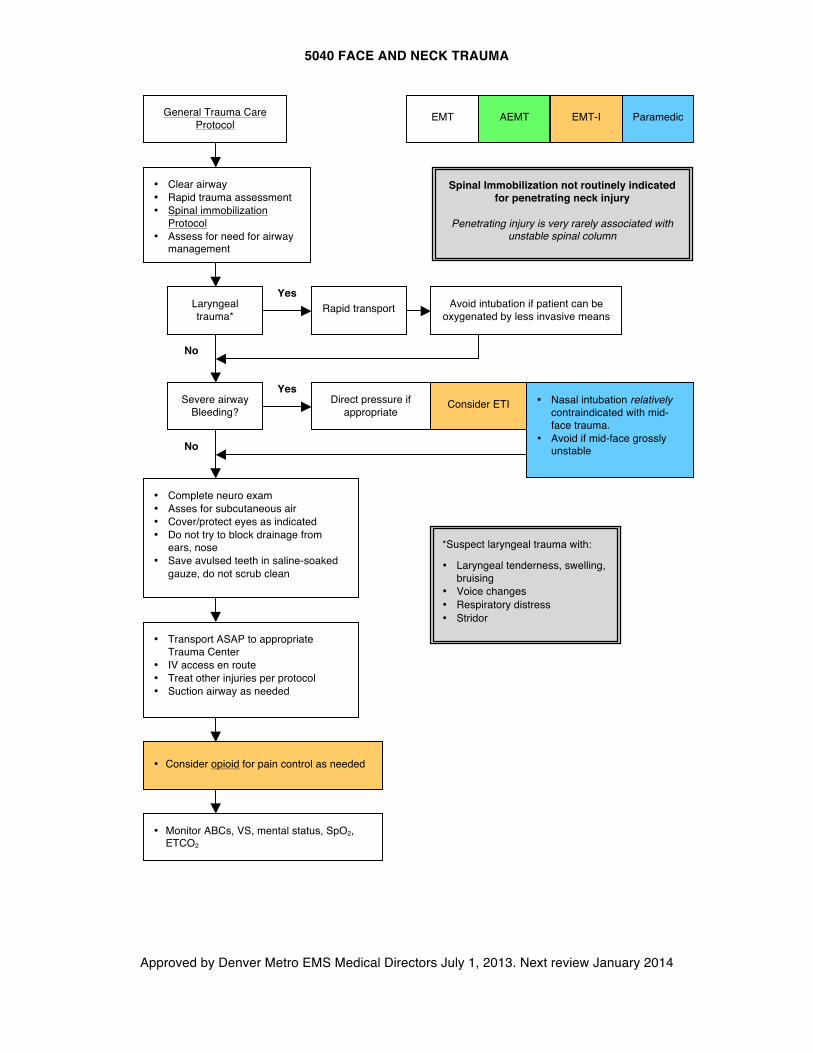
5040 FACE AND NECK TRAUMA
Approved by Denver Metro EMS Medical Directors July 1, 2013. Next review January 2014
AEMT
General Trauma Care Protocol
EMT Paramedic EMT-I
• Clear airway • Rapid trauma assessment • Spinal immobilization
Protocol • Assess for need for airway
management
Laryngeal trauma*
Rapid transport Avoid intubation if patient can be oxygenated by less invasive means
Yes
No
Severe airway Bleeding?
Consider ETI Yes
No
• Complete neuro exam • Asses for subcutaneous air • Cover/protect eyes as indicated • Do not try to block drainage from
ears, nose • Save avulsed teeth in saline-soaked
gauze, do not scrub clean
*Suspect laryngeal trauma with:
• Laryngeal tenderness, swelling, bruising
• Voice changes • Respiratory distress • Stridor
• Transport ASAP to appropriate Trauma Center
• IV access en route • Treat other injuries per protocol • Suction airway as needed
• Nasal intubation relatively contraindicated with mid-face trauma.
• Avoid if mid-face grossly unstable
Spinal Immobilization not routinely indicated for penetrating neck injury
Penetrating injury is very rarely associated with
unstable spinal column
Direct pressure if appropriate
• Monitor ABCs, VS, mental status, SpO2, ETCO2
• Consider opioid for pain control as needed
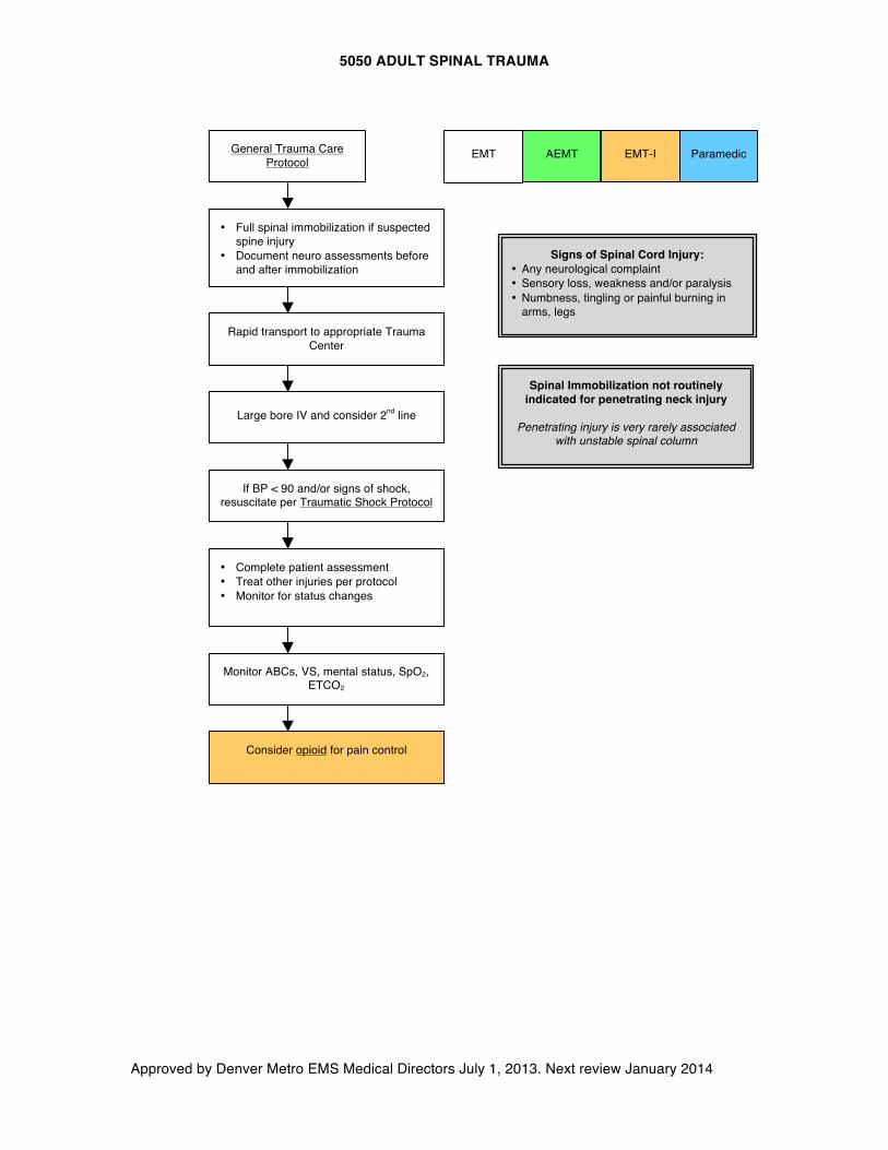
5050 ADULT SPINAL TRAUMA
Approved by Denver Metro EMS Medical Directors July 1, 2013. Next review January 2014
EMT
Signs of Spinal Cord Injury: • Any neurological complaint • Sensory loss, weakness and/or paralysis • Numbness, tingling or painful burning in
arms, legs
AEMT
• Full spinal immobilization if suspected spine injury
• Document neuro assessments before and after immobilization
• Complete patient assessment • Treat other injuries per protocol • Monitor for status changes
Large bore IV and consider 2nd line
Paramedic EMT-I
Rapid transport to appropriate Trauma Center
General Trauma Care Protocol
If BP < 90 and/or signs of shock, resuscitate per Traumatic Shock Protocol
Spinal Immobilization not routinely indicated for penetrating neck injury
Penetrating injury is very rarely associated
with unstable spinal column
Monitor ABCs, VS, mental status, SpO2, ETCO2
Consider opioid for pain control
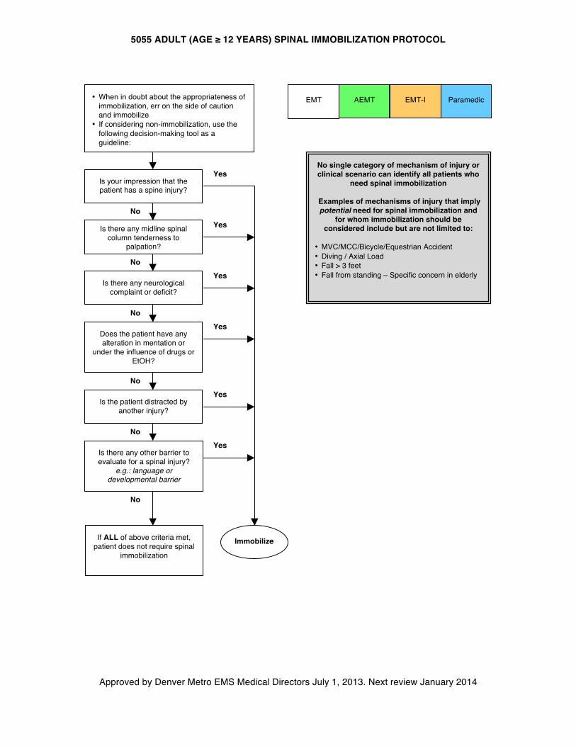
5055 ADULT (AGE ≥ 12 YEARS) SPINAL IMMOBILIZATION PROTOCOL
Approved by Denver Metro EMS Medical Directors July 1, 2013. Next review January 2014
EMT AEMT
Paramedic EMT-I
Is your impression that the patient has a spine injury?
Does the patient have any alteration in mentation or
under the influence of drugs or EtOH?
Is the patient distracted by another injury?
Is there any neurological complaint or deficit?
Yes
Yes
Yes
Yes
Yes
Is there any midline spinal column tenderness to
palpation?
Is there any other barrier to evaluate for a spinal injury?
e.g.: language or developmental barrier
No
Yes
No
No
No
No
No
Immobilize
• When in doubt about the appropriateness of immobilization, err on the side of caution and immobilize
• If considering non-immobilization, use the following decision-making tool as a guideline:
No single category of mechanism of injury or clinical scenario can identify all patients who
need spinal immobilization
Examples of mechanisms of injury that imply potential need for spinal immobilization and
for whom immobilization should be considered include but are not limited to:
• MVC/MCC/Bicycle/Equestrian Accident • Diving / Axial Load • Fall > 3 feet • Fall from standing – Specific concern in elderly
If ALL of above criteria met, patient does not require spinal
immobilization
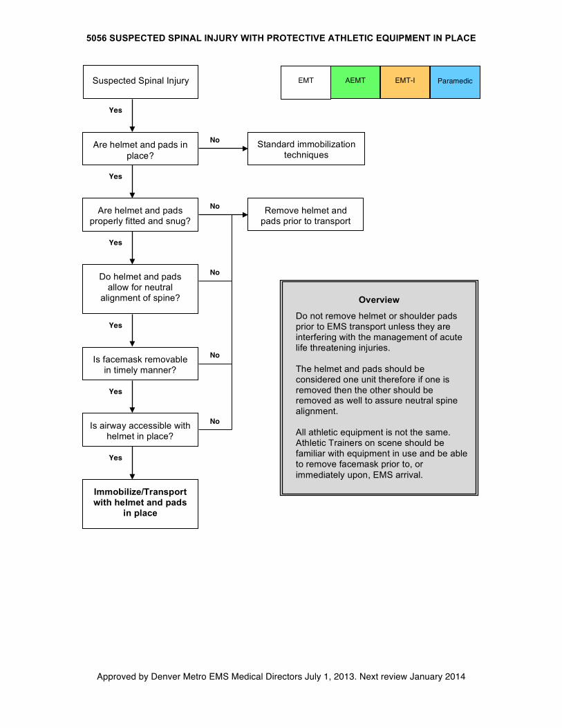
5056 SUSPECTED SPINAL INJURY WITH PROTECTIVE ATHLETIC EQUIPMENT IN PLACE
Approved by Denver Metro EMS Medical Directors July 1, 2013. Next review January 2014
EMT
Paramedic EMT-I
Are helmet and pads in place?
Is airway accessible with helmet in place?
Immobilize/Transport with helmet and pads
in place
Are helmet and pads properly fitted and snug?
Yes
Is facemask removable in timely manner?
Remove helmet and pads prior to transport
No
Yes
No
Suspected Spinal Injury
Overview
Do not remove helmet or shoulder pads prior to EMS transport unless they are interfering with the management of acute life threatening injuries. The helmet and pads should be considered one unit therefore if one is removed then the other should be removed as well to assure neutral spine alignment. All athletic equipment is not the same. Athletic Trainers on scene should be familiar with equipment in use and be able to remove facemask prior to, or immediately upon, EMS arrival.
Standard immobilization techniques
Yes
Yes
Yes
No
No
AEMT
Do helmet and pads allow for neutral
alignment of spine?
Yes
No
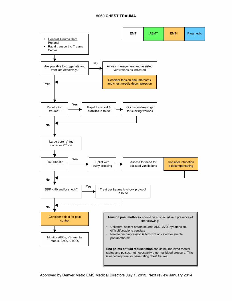
5060 CHEST TRAUMA
Approved by Denver Metro EMS Medical Directors July 1, 2013. Next review January 2014
AEMT
• General Trauma Care Protocol
• Rapid transport to Trauma Center
Penetrating trauma?
SBP < 90 and/or shock?
Occlusive dressings for sucking wounds
Rapid transport & stabilize in route
Large bore IV and consider 2nd line
EMT Paramedic EMT-I
Yes
No
No
Yes
Flail Chest?
Are you able to oxygenate and ventilate effectively?
No Airway management and assisted
ventilations as indicated
Yes
Splint with bulky dressing
Consider intubation if decompensating
No
Yes
Consider tension pneumothorax and chest needle decompression
Tension pneumothorax should be suspected with presence of the following:
• Unilateral absent breath sounds AND: JVD, hypotension, difficult/unable to ventilate
• Needle decompression is NEVER indicated for simple pneumothorax
End points of fluid resuscitation should be improved mental status and pulses, not necessarily a normal blood pressure. This is especially true for penetrating chest trauma.
Treat per traumatic shock protocol in route
Assess for need for assisted ventilations
Consider opioid for pain control
Monitor ABCs, VS, mental status, SpO2, ETCO2
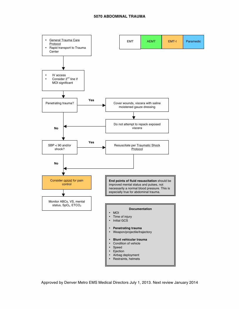
5070 ABDOMINAL TRAUMA
Approved by Denver Metro EMS Medical Directors July 1, 2013. Next review January 2014
EMT AEMT
• IV access • Consider 2nd line if
MOI significant
Penetrating trauma?
SBP < 90 and/or shock?
Cover wounds, viscera with saline moistened gauze dressing
Do not attempt to repack exposed viscera
Resuscitate per Traumatic Shock Protocol
Documentation • MOI • Time of injury • Initial GCS
• Penetrating trauma • Weapon/projectile/trajectory
• Blunt vehicular trauma • Condition of vehicle • Speed • Ejection • Airbag deployment • Restraints, helmets
Yes
No
No
Yes
End points of fluid resuscitation should be improved mental status and pulses, not necessarily a normal blood pressure. This is especially true for abdominal trauma.
EMT-I Paramedic
Monitor ABCs, VS, mental status, SpO2, ETCO2
Consider opioid for pain control
• General Trauma Care Protocol
• Rapid transport to Trauma Center
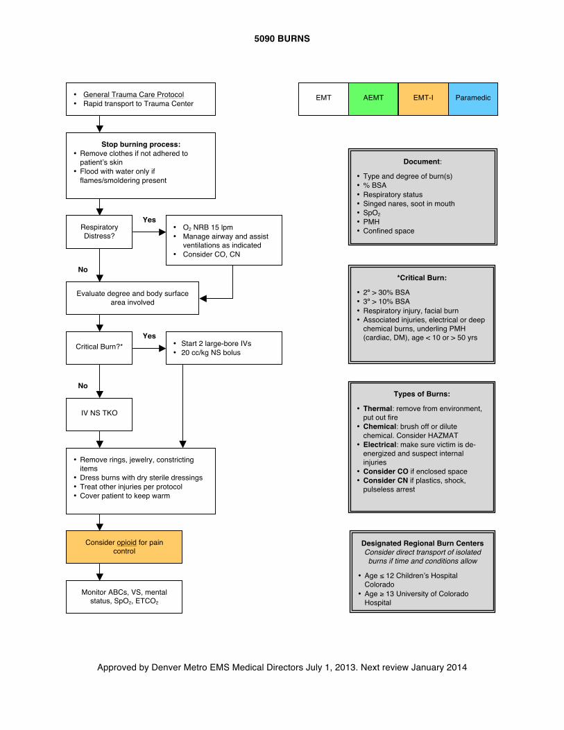
5090 BURNS
Approved by Denver Metro EMS Medical Directors July 1, 2013. Next review January 2014
AEMT
Paramedic EMT EMT-I
Stop burning process: • Remove clothes if not adhered to
patient’s skin • Flood with water only if
flames/smoldering present
Respiratory Distress?
Critical Burn?*
• O2 NRB 15 lpm • Manage airway and assist
ventilations as indicated • Consider CO, CN
Evaluate degree and body surface area involved
• Start 2 large-bore IVs • 20 cc/kg NS bolus
IV NS TKO
• Remove rings, jewelry, constricting items
• Dress burns with dry sterile dressings • Treat other injuries per protocol • Cover patient to keep warm
Types of Burns:
• Thermal: remove from environment, put out fire
• Chemical: brush off or dilute chemical. Consider HAZMAT
• Electrical: make sure victim is de-energized and suspect internal injuries
• Consider CO if enclosed space • Consider CN if plastics, shock,
pulseless arrest
*Critical Burn:
• 2º > 30% BSA • 3º > 10% BSA • Respiratory injury, facial burn • Associated injuries, electrical or deep
chemical burns, underling PMH (cardiac, DM), age < 10 or > 50 yrs
Document:
• Type and degree of burn(s) • % BSA • Respiratory status • Singed nares, soot in mouth • SpO2 • PMH • Confined space
Yes
Yes
No
No
Designated Regional Burn Centers Consider direct transport of isolated burns if time and conditions allow
• Age ≤ 12 Children’s Hospital Colorado
• Age ≥ 13 University of Colorado Hospital
Consider opioid for pain control
Monitor ABCs, VS, mental status, SpO2, ETCO2
• General Trauma Care Protocol • Rapid transport to Trauma Center
6000 GENERAL GUIDELINES FOR PEDIATRIC PATIENTS
Approved by Denver Metro EMS Medical Directors July 1, 2013. Next review January 2014
General Guideline:
A. Pediatric patients, defined as age < 12 years for the purpose of these protocols, have unique anatomy, physiology, and developmental needs that affect prehospital care. Because children make up a small percentage of total calls and few pediatric calls are critically ill or injured, it is important to stay attuned to these differences to provide good care. Therefore, CONTACT BASE early for guidance when treating pediatric patients with significant complaints, including abnormalities of vital signs. Pediatric emergencies are usually not preceded by chronic disease. If recognition of compromise occurs early, and intervention is swift and effective, the child will often be restored to full health.
Specific Considerations:
A. The following should be kept in mind during the care of children in the prehospital setting: 1. Airways are smaller, softer, and easier to obstruct or collapse. 2. Respiratory reserves are small. A minor insult like improper position, vomiting, or airway
narrowing can result in major deficits in ventilation and oxygenation. 3. Circulatory reserves are also small. The loss of as little as one unit of blood can produce
severe shock in an infant. Conversely, it is difficult to fluid overload most children. You can be confident that a good hands-on circulation assessment will determine fluid needs accurately.
4. Assessment of the pediatric patient can be done using your knowledge of the anatomy and physiology specific to infants and children.
5. Listen to the parents' assessment of the patient's problem. They often can detect small changes in their child's condition. This is particularly true if the patient has chronic disease.
6. The proper equipment is very important when dealing with the pediatric patient. A complete selection of pediatric airway management equipment, IV catheters, cervical collars, and drugs has been mandated by the state. This equipment should be stored separately to minimize confusion.
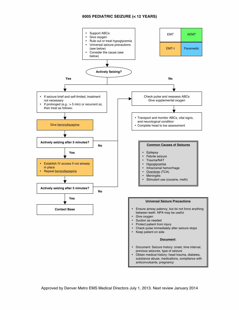
6005 PEDIATRIC SEIZURE (< 12 YEARS)
Approved by Denver Metro EMS Medical Directors July 1, 2013. Next review January 2014
AEMT
Common Causes of Seizures
• Epilepsy • Febrile seizure • Trauma/NAT • Hypoglycemia • Intracranial hemorrhage • Overdose (TCA) • Meningitis • Stimulant use (cocaine, meth)
• Transport and monitor ABCs, vital signs, and neurological condition
• Complete head to toe assessment
• Support ABCs: • Give oxygen • Rule out or treat hypoglycemia • Universal seizure precautions
(see below) • Consider the cause (see
below)
Yes No
Check pulse and reassess ABCs Give supplemental oxygen
No
Yes
Contact Base
• Establish IV access if not already in place
• Repeat benzodiazepine
EMT
EMT-I Paramedic
• If seizure brief and self-limited, treatment not necessary
• If prolonged (e.g.: > 5 min) or recurrent sz, then treat as follows:
Yes
No
Universal Seizure Precautions
• Ensure airway patency, but do not force anything between teeth. NPA may be useful
• Give oxygen • Suction as needed • Protect patient from injury • Check pulse immediately after seizure stops • Keep patient on side
Document:
• Document: Seizure history: onset, time interval,
previous seizures, type of seizure • Obtain medical history: head trauma, diabetes,
substance abuse, medications, compliance with anticonvulsants, pregnancy
Actively Seizing?
Actively seizing after 5 minutes?
Actively seizing after 5 minutes?
Give benzodiazepine
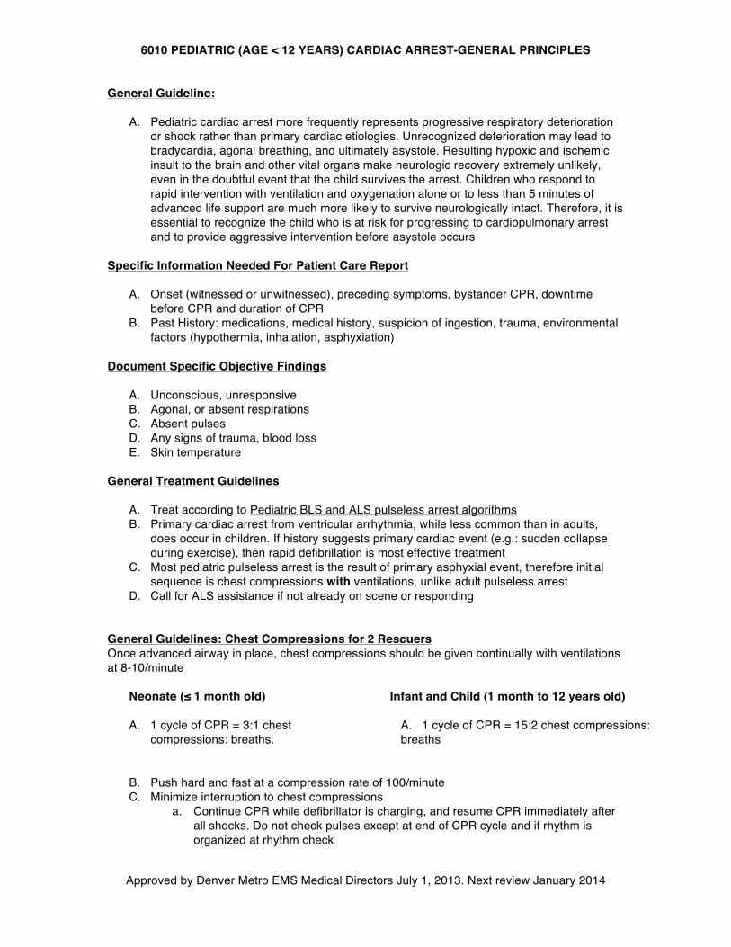
6010 PEDIATRIC (AGE < 12 YEARS) CARDIAC ARREST-GENERAL PRINCIPLES
Approved by Denver Metro EMS Medical Directors July 1, 2013. Next review January 2014
General Guideline:
A. Pediatric cardiac arrest more frequently represents progressive respiratory deterioration or shock rather than primary cardiac etiologies. Unrecognized deterioration may lead to bradycardia, agonal breathing, and ultimately asystole. Resulting hypoxic and ischemic insult to the brain and other vital organs make neurologic recovery extremely unlikely, even in the doubtful event that the child survives the arrest. Children who respond to rapid intervention with ventilation and oxygenation alone or to less than 5 minutes of advanced life support are much more likely to survive neurologically intact. Therefore, it is essential to recognize the child who is at risk for progressing to cardiopulmonary arrest and to provide aggressive intervention before asystole occurs
Specific Information Needed For Patient Care Report
A. Onset (witnessed or unwitnessed), preceding symptoms, bystander CPR, downtime before CPR and duration of CPR
B. Past History: medications, medical history, suspicion of ingestion, trauma, environmental factors (hypothermia, inhalation, asphyxiation)
Document Specific Objective Findings
A. Unconscious, unresponsive B. Agonal, or absent respirations C. Absent pulses D. Any signs of trauma, blood loss E. Skin temperature
General Treatment Guidelines
A. Treat according to Pediatric BLS and ALS pulseless arrest algorithms B. Primary cardiac arrest from ventricular arrhythmia, while less common than in adults,
does occur in children. If history suggests primary cardiac event (e.g.: sudden collapse during exercise), then rapid defibrillation is most effective treatment
C. Most pediatric pulseless arrest is the result of primary asphyxial event, therefore initial sequence is chest compressions with ventilations, unlike adult pulseless arrest
D. Call for ALS assistance if not already on scene or responding General Guidelines: Chest Compressions for 2 Rescuers Once advanced airway in place, chest compressions should be given continually with ventilations at 8-10/minute
Neonate (≤ 1 month old) A. 1 cycle of CPR = 3:1 chest
compressions: breaths.
Infant and Child (1 month to 12 years old)
A. 1 cycle of CPR = 15:2 chest compressions: breaths
B. Push hard and fast at a compression rate of 100/minute C. Minimize interruption to chest compressions
a. Continue CPR while defibrillator is charging, and resume CPR immediately after all shocks. Do not check pulses except at end of CPR cycle and if rhythm is organized at rhythm check
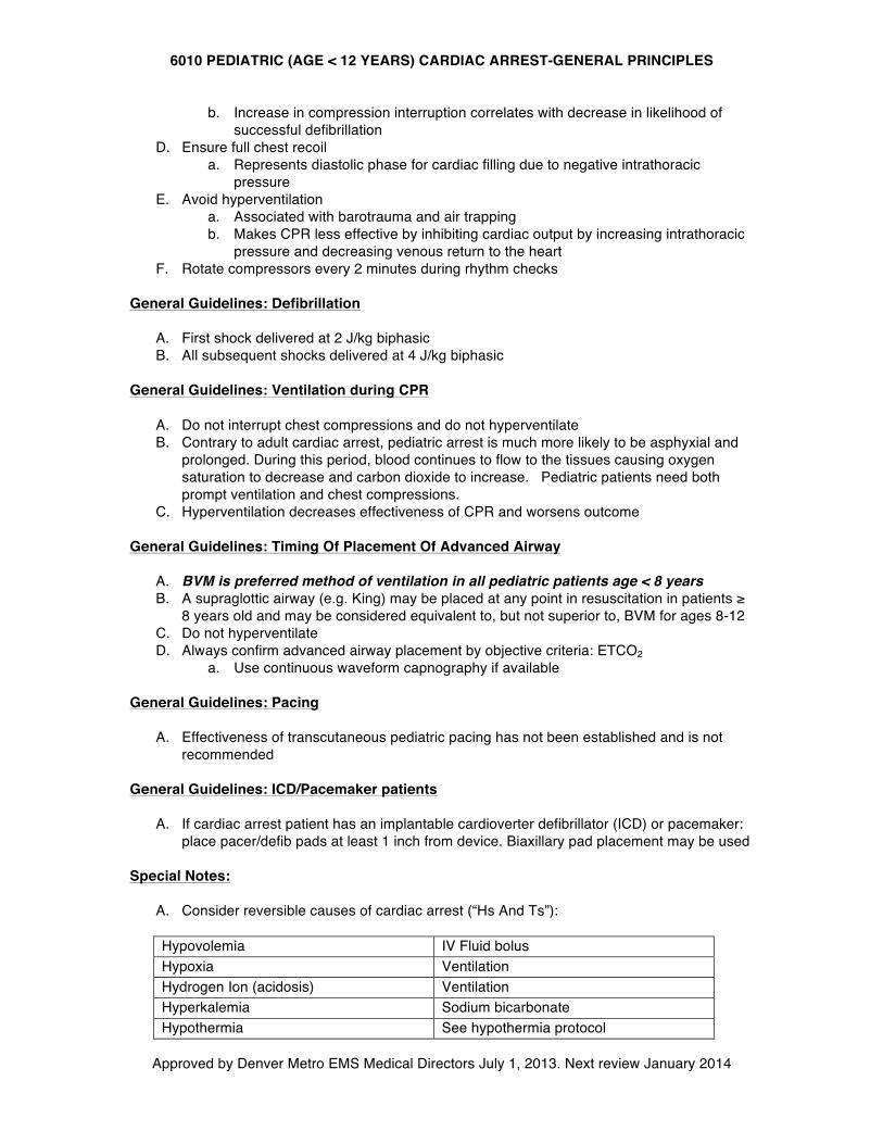
6010 PEDIATRIC (AGE < 12 YEARS) CARDIAC ARREST-GENERAL PRINCIPLES
Approved by Denver Metro EMS Medical Directors July 1, 2013. Next review January 2014
b. Increase in compression interruption correlates with decrease in likelihood of successful defibrillation
D. Ensure full chest recoil a. Represents diastolic phase for cardiac filling due to negative intrathoracic
pressure E. Avoid hyperventilation
a. Associated with barotrauma and air trapping b. Makes CPR less effective by inhibiting cardiac output by increasing intrathoracic
pressure and decreasing venous return to the heart F. Rotate compressors every 2 minutes during rhythm checks
General Guidelines: Defibrillation
A. First shock delivered at 2 J/kg biphasic B. All subsequent shocks delivered at 4 J/kg biphasic
General Guidelines: Ventilation during CPR
A. Do not interrupt chest compressions and do not hyperventilate B. Contrary to adult cardiac arrest, pediatric arrest is much more likely to be asphyxial and
prolonged. During this period, blood continues to flow to the tissues causing oxygen saturation to decrease and carbon dioxide to increase. Pediatric patients need both prompt ventilation and chest compressions.
C. Hyperventilation decreases effectiveness of CPR and worsens outcome General Guidelines: Timing Of Placement Of Advanced Airway
A. BVM is preferred method of ventilation in all pediatric patients age < 8 years B. A supraglottic airway (e.g. King) may be placed at any point in resuscitation in patients ≥
8 years old and may be considered equivalent to, but not superior to, BVM for ages 8-12 C. Do not hyperventilate D. Always confirm advanced airway placement by objective criteria: ETCO2
a. Use continuous waveform capnography if available General Guidelines: Pacing
A. Effectiveness of transcutaneous pediatric pacing has not been established and is not recommended
General Guidelines: ICD/Pacemaker patients
A. If cardiac arrest patient has an implantable cardioverter defibrillator (ICD) or pacemaker: place pacer/defib pads at least 1 inch from device. Biaxillary pad placement may be used
Special Notes:
A. Consider reversible causes of cardiac arrest (“Hs And Ts”): Hypovolemia IV Fluid bolus Hypoxia Ventilation Hydrogen Ion (acidosis) Ventilation Hyperkalemia Sodium bicarbonate Hypothermia See hypothermia protocol
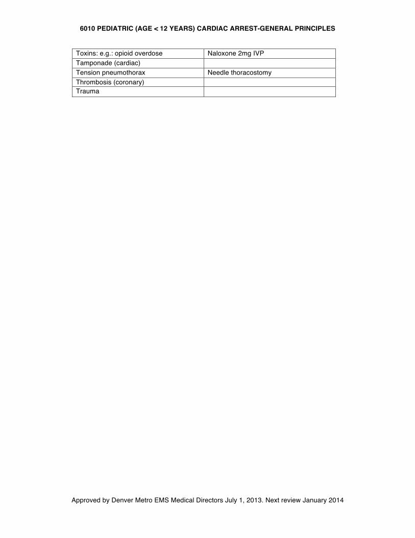
6010 PEDIATRIC (AGE < 12 YEARS) CARDIAC ARREST-GENERAL PRINCIPLES
Approved by Denver Metro EMS Medical Directors July 1, 2013. Next review January 2014
Toxins: e.g.: opioid overdose Naloxone 2mg IVP Tamponade (cardiac) Tension pneumothorax Needle thoracostomy Thrombosis (coronary) Trauma
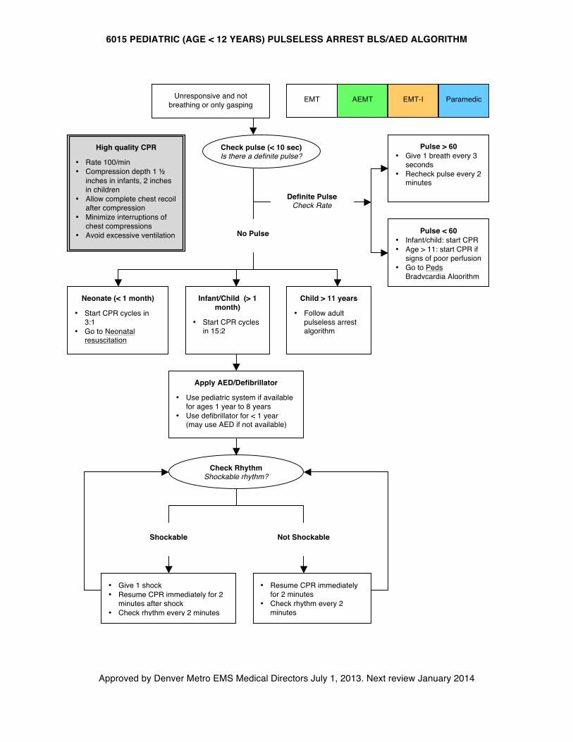
6015 PEDIATRIC (AGE < 12 YEARS) PULSELESS ARREST BLS/AED ALGORITHM
Approved by Denver Metro EMS Medical Directors July 1, 2013. Next review January 2014
AEMT
Unresponsive and not breathing or only gasping
Check pulse (< 10 sec) Is there a definite pulse?
Pulse > 60 • Give 1 breath every 3
seconds • Recheck pulse every 2
minutes
Pulse < 60 • Infant/child: start CPR • Age > 11: start CPR if
signs of poor perfusion • Go to Peds
Bradycardia Algorithm • Recheck pulse every 2
minutes
Definite Pulse Check Rate
No Pulse
EMT EMT-I Paramedic
Neonate (< 1 month)
• Start CPR cycles in 3:1
• Go to Neonatal resuscitation
Infant/Child (> 1 month)
• Start CPR cycles in 15:2
Child > 11 years
• Follow adult pulseless arrest algorithm
Apply AED/Defibrillator
• Use pediatric system if available for ages 1 year to 8 years
• Use defibrillator for < 1 year (may use AED if not available)
Check Rhythm Shockable rhythm?
Shockable Not Shockable
• Resume CPR immediately for 2 minutes
• Check rhythm every 2 minutes
• Give 1 shock • Resume CPR immediately for 2
minutes after shock • Check rhythm every 2 minutes
High quality CPR
• Rate 100/min • Compression depth 1 ½
inches in infants, 2 inches in children
• Allow complete chest recoil after compression
• Minimize interruptions of chest compressions
• Avoid excessive ventilation
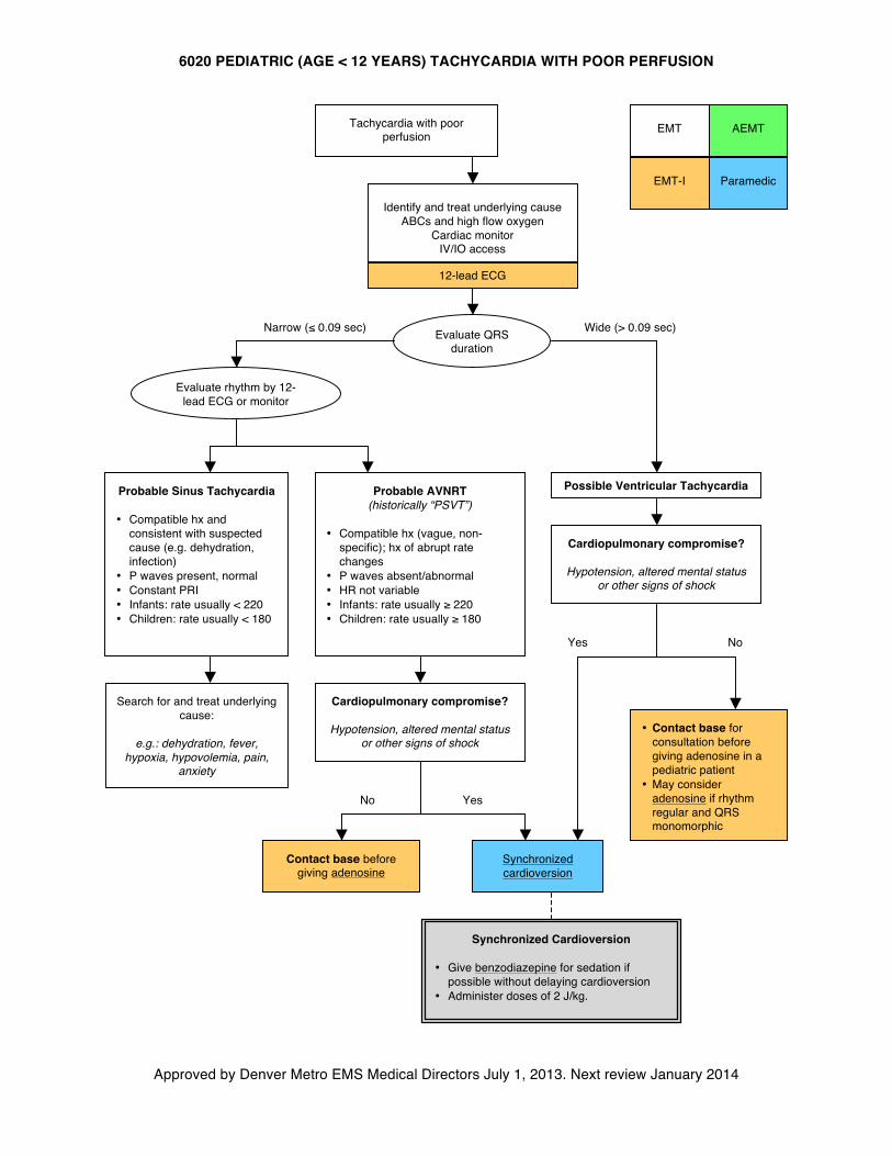
6020 PEDIATRIC (AGE < 12 YEARS) TACHYCARDIA WITH POOR PERFUSION
Approved by Denver Metro EMS Medical Directors July 1, 2013. Next review January 2014
AEMT
Identify and treat underlying cause ABCs and high flow oxygen
Cardiac monitor IV/IO access
Probable AVNRT (historically “PSVT”)
• Compatible hx (vague, non-
specific); hx of abrupt rate changes
• P waves absent/abnormal • HR not variable • Infants: rate usually ≥ 220 • Children: rate usually ≥ 180
Search for and treat underlying cause:
e.g.: dehydration, fever,
hypoxia, hypovolemia, pain, anxiety
Synchronized Cardioversion
• Give benzodiazepine for sedation if possible without delaying cardioversion
• Administer doses of 2 J/kg.
Probable Sinus Tachycardia
• Compatible hx and consistent with suspected cause (e.g. dehydration, infection)
• P waves present, normal • Constant PRI • Infants: rate usually < 220 • Children: rate usually < 180
EMT
EMT-I Paramedic
Tachycardia with poor perfusion
Evaluate rhythm by 12-lead ECG or monitor
12-lead ECG
Evaluate QRS duration
Wide (> 0.09 sec)
Narrow (≤ 0.09 sec)
Possible Ventricular Tachycardia
• Contact base for consultation before giving adenosine in a pediatric patient
• May consider adenosine if rhythm regular and QRS monomorphic
Yes
No
Contact base before giving adenosine
Cardiopulmonary compromise?
Hypotension, altered mental status or other signs of shock
Cardiopulmonary compromise?
Hypotension, altered mental status or other signs of shock
No
Yes
Synchronized cardioversion
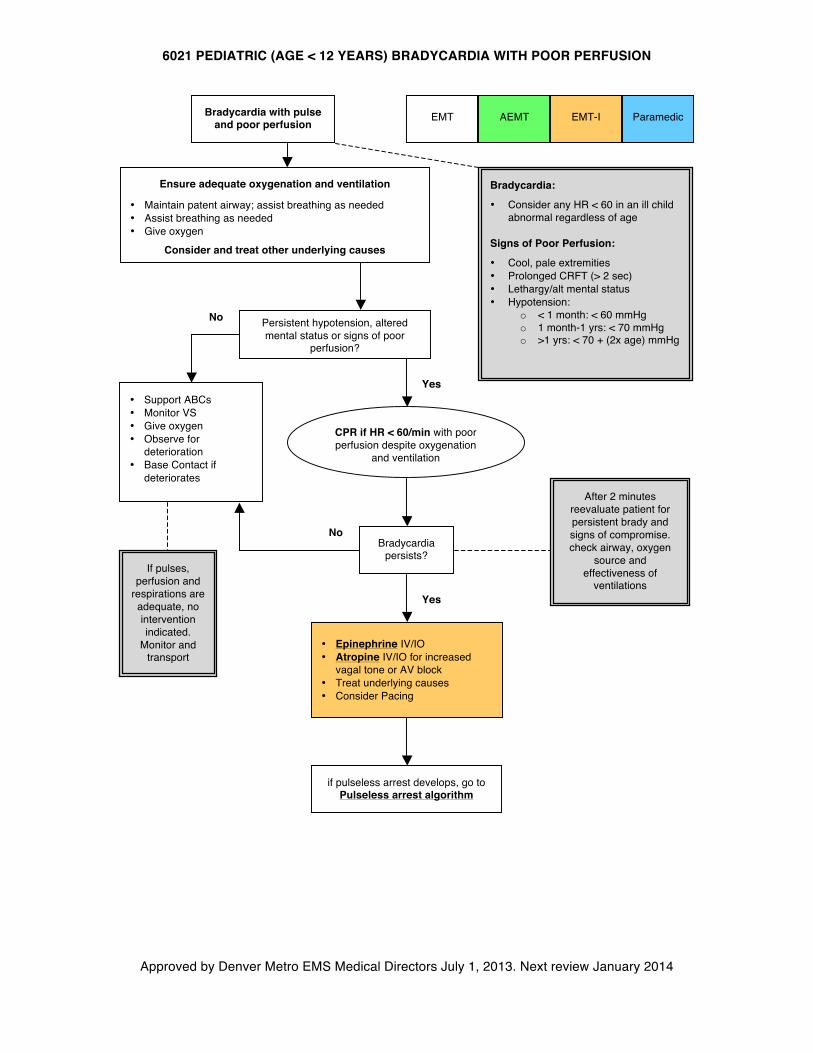
6021 PEDIATRIC (AGE < 12 YEARS) BRADYCARDIA WITH POOR PERFUSION
Approved by Denver Metro EMS Medical Directors July 1, 2013. Next review January 2014
AEMT Bradycardia with pulse
and poor perfusion
Ensure adequate oxygenation and ventilation
• Maintain patent airway; assist breathing as needed • Assist breathing as needed • Give oxygen
Consider and treat other underlying causes
Persistent hypotension, altered mental status or signs of poor
perfusion?
CPR if HR < 60/min with poor perfusion despite oxygenation
and ventilation
Yes
No
• Support ABCs • Monitor VS • Give oxygen • Observe for
deterioration • Base Contact if
deteriorates
• Epinephrine IV/IO • Atropine IV/IO for increased
vagal tone or AV block • Treat underlying causes • Consider Pacing
Bradycardia persists?
Yes
if pulseless arrest develops, go to Pulseless arrest algorithm
If pulses, perfusion and
respirations are adequate, no intervention indicated.
Monitor and transport
After 2 minutes reevaluate patient for persistent brady and signs of compromise. check airway, oxygen
source and effectiveness of
ventilations
EMT Paramedic EMT-I
No
Bradycardia:
• Consider any HR < 60 in an ill child abnormal regardless of age
Signs of Poor Perfusion:
• Cool, pale extremities • Prolonged CRFT (> 2 sec) • Lethargy/alt mental status • Hypotension:
o < 1 month: < 60 mmHg o 1 month-1 yrs: < 70 mmHg o >1 yrs: < 70 + (2x age) mmHg
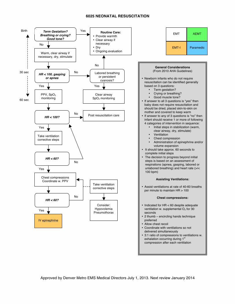
AEMT
6025 NEONATAL RESUSCITATION
Approved by Denver Metro EMS Medical Directors July 1, 2013. Next review January 2014
No
AEMT
EMT
EMT-I Paramedic
General Considerations (From 2010 AHA Guidelines)
• Newborn infants who do not require
resuscitation can be identified generally based on 3 questions:
• Term gestation? • Crying or breathing? • Good muscle tone?
• If answer to all 3 questions is “yes” then baby does not require resuscitation and should be dried, placed skin-to-skin on mother and covered to keep warm
• If answer to any of 3 questions is “no” then infant should receive 1 or more of following 4 categories of intervention in sequence:
• Initial steps in stabilization (warm, clear airway, dry, stimulate)
• Ventilation • Chest compression • Administration of epinephrine and/or
volume expansion • It should take approx. 60 seconds to
complete initial steps • The decision to progress beyond initial
steps is based on an assessment of respirations (apnea, gasping, labored or unlabored breathing) and heart rate (>/< 100 bpm)
Assisting Ventilations:
• Assist ventilations at rate of 40-60 breaths per minute to maintain HR > 100
Chest compressions:
• Indicated for HR < 60 despite adequate ventilation w. supplemental O2 for 30 seconds
• 2 thumb – encircling hands technique preferred
• Allow chest recoil • Coordinate with ventilations so not
delivered simultaneously • 3:1 ratio of compressions to ventilations w.
exhalation occurring during 1st compression after each ventilation
Term Gestation? Breathing or crying?
Good tone?
Routine Care: • Provide warmth • Clear airway if
necessary • Dry • Ongoing evaluation
Yes
No
Warm, clear airway if necessary, dry, stimulate
HR < 100, gasping or apnea
No
PPV, SpO2 monitoring
Labored breathing or persistent cyanosis?
Yes
HR < 100?
No Post resuscitation care
Clear airway SpO2 monitoring
Yes
Take ventilation corrective steps
Yes
HR < 60? Yes
Chest compressions Coordinate w. PPV
HR < 60?
IV epinephrine
Yes
No
No
Take ventilation corrective steps
Consider: Hypovolemia
Pneumothorax
60 sec
30 sec
Birth
6026 NEONATAL CONSIDERATIONS
Approved by Denver Metro EMS Medical Directors July 1, 2013. Next review January 2014
General Considerations:
A. A neonate refers to a newly born child under the age of 30 days. While most neonates transition to post-natal life without difficulty, 10% will require medical assistance. Respiratory insufficiency is the most common complication observed in the newly born.
B. Neonates born precipitously may exhibit signs of stress such as apnea, grunting respirations, lethargy or poor tone
1. Provide warmth, bulb suction mouth and then nose, and dry the infant 2. If breathing spontaneously, HR >100 and infant is vigorous, continue to monitor 3. If apneic, cyanotic, lethargic, or HR <100, provide 100% oxygen via BVM ventilations at a
rate of 40-60 bpm 4. If HR < 60, begin CPR at 3:1 compression:ventilation ratio.
C. For neonates who do not respond to initial interventions as above: 1. Obtain blood glucose level and if < 60, administer dextrose IV/IO (D10 4 mL/kg) 2. Administer epinephrine IV for persistent HR < 60 3. Consider hypovolemia and administer 10-20ml/kg NS over 5-10 minutes
D. Neonates with congenital heart disease may not be detected prior to hospital discharge after delivery. Consider a cardiac cause of shock in the neonate who remains hypoxic or has persistent cyanosis despite 100% oxygen. These neonates may decompensate precipitously and fluid administration should be used judiciously (10ml/kg NS)
E. Newborns are at high risk for hypothermia. Provide early warming measures, keep covered as much as possible (especially the head) and increase the temperature in the ambulance
F. Acrocyanosis (cyanosis of only the hands and feet) is normal in newborns and does not require intervention
G. Prolonged apnea without bradycardia or cyanosis may indicate respiratory depression caused by narcotics. However, naloxone should be avoided in infants of a known or suspected narcotic-addicted mother as this may induce a withdrawal reaction. Respiratory support alone is recommended
H. Obtain pregnancy history, gestational age of the neonate, pregnancy complications, and any illicit drug use during pregnancy.
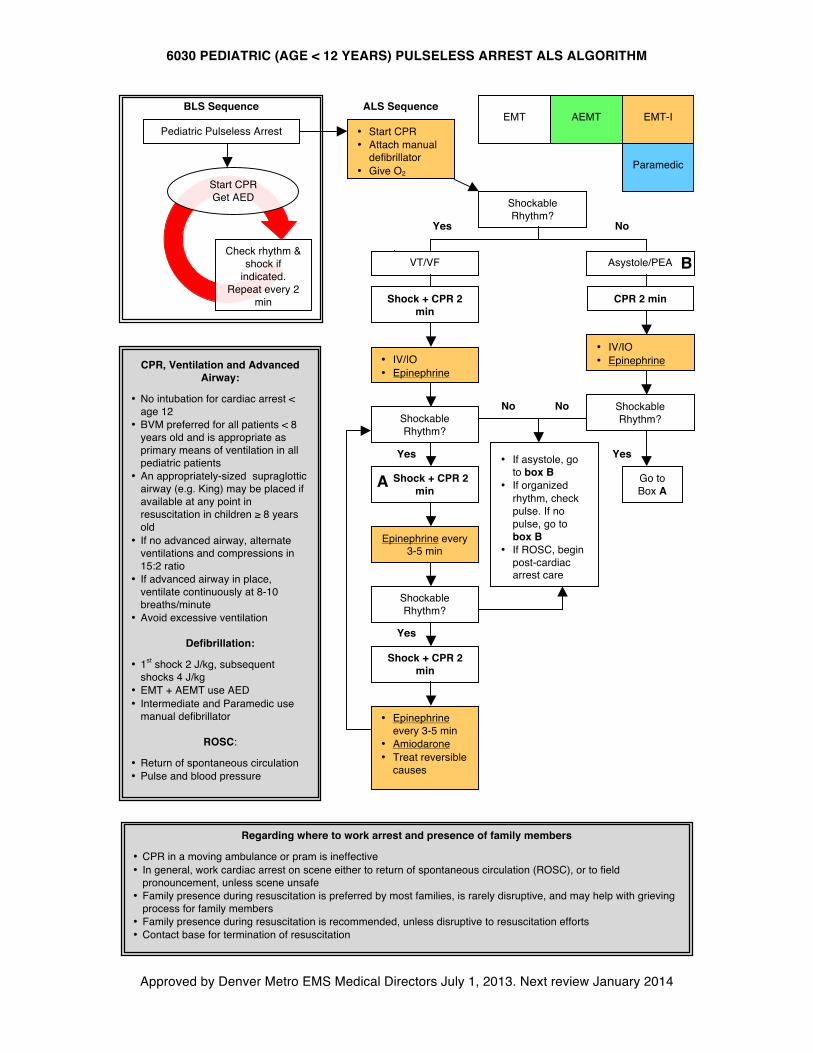
6030 PEDIATRIC (AGE < 12 YEARS) PULSELESS ARREST ALS ALGORITHM
Approved by Denver Metro EMS Medical Directors July 1, 2013. Next review January 2014
• Start CPR • Attach manual
defibrillator • Give O2
Shockable Rhythm?
VT/VF Asystole/PEA
No Yes
Shock + CPR 2 min
• IV/IO • Epinephrine
Shockable Rhythm?
Yes
Shockable Rhythm?
B
No
Epinephrine every 3-5 min
• IV/IO • Epinephrine
A
Yes
Go to Box A
No
Shockable Rhythm?
• Epinephrine every 3-5 min
• Amiodarone • Treat reversible
causes
Yes
Shock + CPR 2 min
Shock + CPR 2 min
CPR 2 min
Pediatric Pulseless Arrest
Check rhythm & shock if
indicated. Repeat every 2
min
Start CPR Get AED
BLS Sequence EMT EMT-I
Paramedic
ALS Sequence
• If asystole, go to box B
• If organized rhythm, check pulse. If no pulse, go to box B
• If ROSC, begin post-cardiac arrest care
AEMT
CPR, Ventilation and Advanced Airway:
• No intubation for cardiac arrest < age 12
• BVM preferred for all patients < 8 years old and is appropriate as primary means of ventilation in all pediatric patients
• An appropriately-sized supraglottic airway (e.g. King) may be placed if available at any point in resuscitation in children ≥ 8 years old
• If no advanced airway, alternate ventilations and compressions in 15:2 ratio
• If advanced airway in place, ventilate continuously at 8-10 breaths/minute
• Avoid excessive ventilation
Defibrillation:
• 1st shock 2 J/kg, subsequent shocks 4 J/kg
• EMT + AEMT use AED • Intermediate and Paramedic use
manual defibrillator
ROSC:
• Return of spontaneous circulation • Pulse and blood pressure
Regarding where to work arrest and presence of family members
• CPR in a moving ambulance or pram is ineffective • In general, work cardiac arrest on scene either to return of spontaneous circulation (ROSC), or to field
pronouncement, unless scene unsafe • Family presence during resuscitation is preferred by most families, is rarely disruptive, and may help with grieving
process for family members • Family presence during resuscitation is recommended, unless disruptive to resuscitation efforts • Contact base for termination of resuscitation
6040 CARE OF THE CHILD WITH SPECIAL NEEDS
Approved by Denver Metro EMS Medical Directors July 1, 2013. Next review January 2014
General Guideline:
A. Children with special health care needs include those with chronic physical, developmental, behavioral or emotional health issues. These children often have complex medical needs and may be technology-dependent. Parents or caregivers for such children can be a wealth of knowledge about their child’s care and may carry a reference care sheet. Contact Base Station for any concerns.
B. Under Chapter 2 Rule: specialized prescription medications to address an acute crisis may be given by all levels with a direct VO, given the route of administration is within the scope of the provider. This applies to giving hydrocortisone for adrenal crisis, for instance if a patient or family member has this medication available on scene. Contact base for direct verbal order
Feeding Tubes:
A. Feedings tubes are used for administration of medications and to provide feeds to children with an impaired ability to take oral feeds. Always ask caretaker the type of feeding tube (does the tube end in the stomach or jejunum?) and when it was placed
B. Tubes may be placed through the nose, mouth or abdomen and end in the stomach or jejunum (upper intestine)
C. Consider venting and/or gently aspirating the feeding tube in a child with respiratory or abdominal distress to allow removal of gastric contents and decompression
D. Feeding tubes that have been placed less than 6 weeks ago are not well established and may close within 1 hour of tube removal. If transport time is prolonged, place an 8 Fr suction catheter tube 2 inches into the stoma to maintain patency. Do NOT use the tube.
Tracheostomy:
A. A tracheostomy is a surgical opening between the trachea and the anterior surface of the neck. Its purpose is to bypass the upper airway for chronically ventilated patients, upper airway obstructions, or to facilitate secretion removal in those with ineffective gag or swallow reflexes.
B. Use bag-valve attached to the tracheostomy to assist ventilations if needed. May also attempt BVM with gloved finger over the tracheostomy
C. Inability to ventilate and/or signs of respiratory distress (nasal flaring, retractions, hypoxia, etc) may indicate tracheostomy obstruction. Suction tracheostomy, passing the suction catheter no further than 6 cm. Limit suctioning time to minimum amount of time necessary to accomplish effective suctioning. Oxygenate between passes with the suction catheter.
D. 0.5ml of saline may be instilled into the tracheostomy to assist suctioning of thick secretions E. If unable to ventilate through the tracheostomy tube and patient is apneic, bradycardic, or in
pulseless arrest, remove tracheostomy tube and pass an appropriately sized endotracheal tube through the stoma approximately 1-2 inches, secure and ventilate. Appropriate depth must be based upon breath sounds, as right mainstem intubation is likely.
F. Remember that caregivers are often the best people to change and suction a tracheostomy tube. Use them as your resource when possible.
Central Venous Catheters (CVCs):
A. Because of their size and location, a much greater risk of serious bacterial infections exist with CVCs compared to peripheral intravenous lines. Special care must be used when accessing such lines
6040 CARE OF THE CHILD WITH SPECIAL NEEDS
Approved by Denver Metro EMS Medical Directors July 1, 2013. Next review January 2014
B. Prior to accessing a CVC, hands should be washed and gloves worn. Vigorously scrub the CVC hub with an alcohol swab. While alcohol possesses some antimicrobial properties, the friction produced by scrubbing is the most effective
C. A port is an implanted venous central venous catheter (below the surface of the skin). These devices require a non-coring (e.g. Huber) needle for accessing and should not be accessed in the field
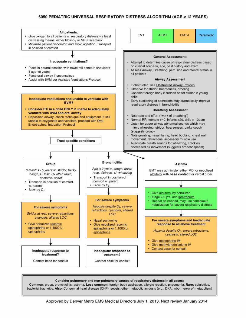
6050 PEDIATRIC UNIVERSAL RESPIRATORY DISTRESS ALGORITHM (AGE < 12 YEARS)
Approved by Denver Metro EMS Medical Directors July 1, 2013. Next review January 2014
AEMT All patients:
• Give oxygen to all patients w. respiratory distress via least distressing means, either blow-by or NRB facemask
• Minimize patient discomfort and avoid agitation. Transport in position of comfort
General Assessment:
• Attempt to determine cause of respiratory distress based on clinical scenario, age, past history and exam
• Assess Airway, Breathing, perfusion and mental status in all patients
Airway Assessment • If obstructed, see Obstructed Airway Protocol • Observe for stridor, hoarseness, drooling • Consider foreign body if sudden onset stridor in young
child • Early suctioning of secretions may dramatically improve
respiratory distress in bronchiolitis Breathing Assessment
• Note rate and effort (“work of breathing”) • Normal RR neonate >40, infants >20, child > 12bpm • Listen for upper airway abnormal sounds which may
mimic wheezing: stridor, hoarseness, barky cough (suggests croup)
• Note grunting, nasal flaring, head bobbing, chest wall movement, retractions, accessory muscle use
• Auscultate breath sounds for wheezing, crackles, decreased air movement (suggests bronchospasm)
EMT
EMT-I
Paramedic
Inadequate ventilations?
• Place in neutral position with towel roll beneath shoulders if age <8 years
• Place oral airway if unconscious • Assist with BVM per Assisted Ventilations Protocol
Inadequate ventilations and unable to ventilate with BVM?
• Consider ETI in a child ONLY if unable to adequately ventilate with BVM and oral airway
• Reposition airway, check technique and equipment. If still unable to oxygenate and ventilate, proceed with Oral Endotracheal Intubation Protocol
Treat specific conditions
Croup
6 months - 5 years w. stridor, barky cough, URI sx. Sx often rapid,
nocturnal onset • Transport in position of comfort
w. parent • Blow-by O2
Bronchiolitis
Age < 2 yrs w. cough, fever, resp. distress, +/- wheezing
• Transport in position of comfort w. parent
• Blow-by O2
For severe symptoms
Stridor at rest, severe retractions, cyanosis, altered LOC
• Give nebulized racemic epinephrine or 1:1000 L-epinephrine
Inadequate response to treatment?
Contact base for consult
For severe symptoms and inadequate response to all above treatment
Hypoxia despite O2, severe retractions, cyanosis, altered LOC
• Give epinephrine IM • Give methylprednisolone IV • Contact base for consult
Consider pulmonary and non-pulmonary causes of respiratory distress in all cases: Common: croup, bronchiolitis, asthma. Less common: foreign body aspiration, allergic reaction, pneumonia. Rare: epiglottitis,
bacterial tracheitis. Also: Congenital heart disease (CHF), sepsis, other metabolic acidosis (e.g.: DKA, inborn error of metabolism)
• Give albuterol by nebulizer • If age > 2 yrs, add ipratropium • Repeat as needed, may use continuous
nebulization for severe respiratory distress
Inadequate response to treatment?
Contact base for consult
For severe symptoms
Hypoxia despite O2, severe retractions, cyanosis, altered
LOC
• Nasal suctioning • Give nebulized racemic
epinephrine or 1:1000 L-epinephrine
Asthma
EMT may administer either MDI or nebulized albuterol with base contact for verbal order
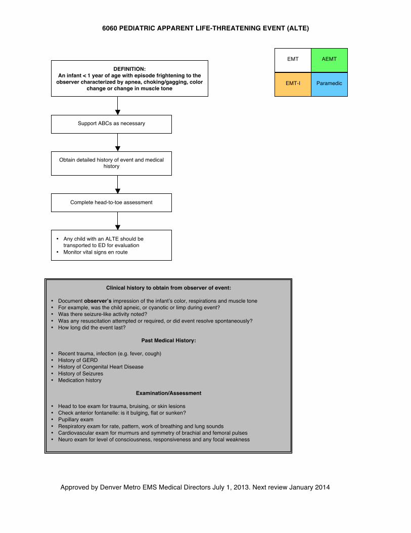
6060 PEDIATRIC APPARENT LIFE-THREATENING EVENT (ALTE)
Approved by Denver Metro EMS Medical Directors July 1, 2013. Next review January 2014
AEMT
DEFINITION: An infant < 1 year of age with episode frightening to the
observer characterized by apnea, choking/gagging, color change or change in muscle tone
Obtain detailed history of event and medical history
Complete head-to-toe assessment
Support ABCs as necessary
• Any child with an ALTE should be transported to ED for evaluation
• Monitor vital signs en route
EMT
EMT-I Paramedic
Clinical history to obtain from observer of event:
• Document observer’s impression of the infant’s color, respirations and muscle tone • For example, was the child apneic, or cyanotic or limp during event? • Was there seizure-like activity noted? • Was any resuscitation attempted or required, or did event resolve spontaneously? • How long did the event last?
Past Medical History:
• Recent trauma, infection (e.g. fever, cough) • History of GERD • History of Congenital Heart Disease • History of Seizures • Medication history
Examination/Assessment
• Head to toe exam for trauma, bruising, or skin lesions • Check anterior fontanelle: is it bulging, flat or sunken? • Pupillary exam • Respiratory exam for rate, pattern, work of breathing and lung sounds • Cardiovascular exam for murmurs and symmetry of brachial and femoral pulses • Neuro exam for level of consciousness, responsiveness and any focal weakness
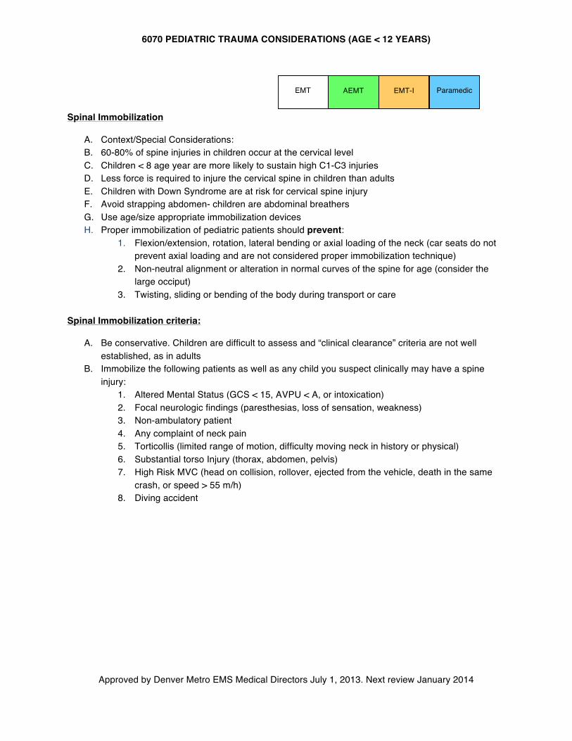
6070 PEDIATRIC TRAUMA CONSIDERATIONS (AGE < 12 YEARS)
Approved by Denver Metro EMS Medical Directors July 1, 2013. Next review January 2014
AEMT
Spinal Immobilization
A. Context/Special Considerations: B. 60-80% of spine injuries in children occur at the cervical level C. Children < 8 age year are more likely to sustain high C1-C3 injuries D. Less force is required to injure the cervical spine in children than adults E. Children with Down Syndrome are at risk for cervical spine injury F. Avoid strapping abdomen- children are abdominal breathers G. Use age/size appropriate immobilization devices H. Proper immobilization of pediatric patients should prevent:
1. Flexion/extension, rotation, lateral bending or axial loading of the neck (car seats do not prevent axial loading and are not considered proper immobilization technique)
2. Non-neutral alignment or alteration in normal curves of the spine for age (consider the large occiput)
3. Twisting, sliding or bending of the body during transport or care
Spinal Immobilization criteria:
A. Be conservative. Children are difficult to assess and “clinical clearance” criteria are not well established, as in adults
B. Immobilize the following patients as well as any child you suspect clinically may have a spine injury:
1. Altered Mental Status (GCS < 15, AVPU < A, or intoxication) 2. Focal neurologic findings (paresthesias, loss of sensation, weakness) 3. Non-ambulatory patient 4. Any complaint of neck pain 5. Torticollis (limited range of motion, difficulty moving neck in history or physical) 6. Substantial torso Injury (thorax, abdomen, pelvis) 7. High Risk MVC (head on collision, rollover, ejected from the vehicle, death in the same
crash, or speed > 55 m/h) 8. Diving accident
EMT EMT-I Paramedic
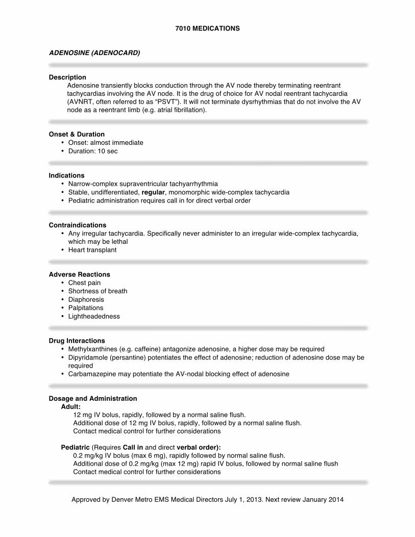
7010 MEDICATIONS
Approved by Denver Metro EMS Medical Directors July 1, 2013. Next review January 2014
ADENOSINE (ADENOCARD) Description
Adenosine transiently blocks conduction through the AV node thereby terminating reentrant tachycardias involving the AV node. It is the drug of choice for AV nodal reentrant tachycardia (AVNRT, often referred to as “PSVT”). It will not terminate dysrhythmias that do not involve the AV node as a reentrant limb (e.g. atrial fibrillation).
Onset & Duration
• Onset: almost immediate • Duration: 10 sec
Indications
• Narrow-complex supraventricular tachyarrhythmia • Stable, undifferentiated, regular, monomorphic wide-complex tachycardia • Pediatric administration requires call in for direct verbal order
Contraindications
• Any irregular tachycardia. Specifically never administer to an irregular wide-complex tachycardia, which may be lethal
• Heart transplant Adverse Reactions
• Chest pain • Shortness of breath • Diaphoresis • Palpitations • Lightheadedness
Drug Interactions
• Methylxanthines (e.g. caffeine) antagonize adenosine, a higher dose may be required • Dipyridamole (persantine) potentiates the effect of adenosine; reduction of adenosine dose may be
required • Carbamazepine may potentiate the AV-nodal blocking effect of adenosine
Dosage and Administration
Adult: 12 mg IV bolus, rapidly, followed by a normal saline flush. Additional dose of 12 mg IV bolus, rapidly, followed by a normal saline flush. Contact medical control for further considerations
Pediatric (Requires Call in and direct verbal order): 0.2 mg/kg IV bolus (max 6 mg), rapidly followed by normal saline flush. Additional dose of 0.2 mg/kg (max 12 mg) rapid IV bolus, followed by normal saline flush Contact medical control for further considerations
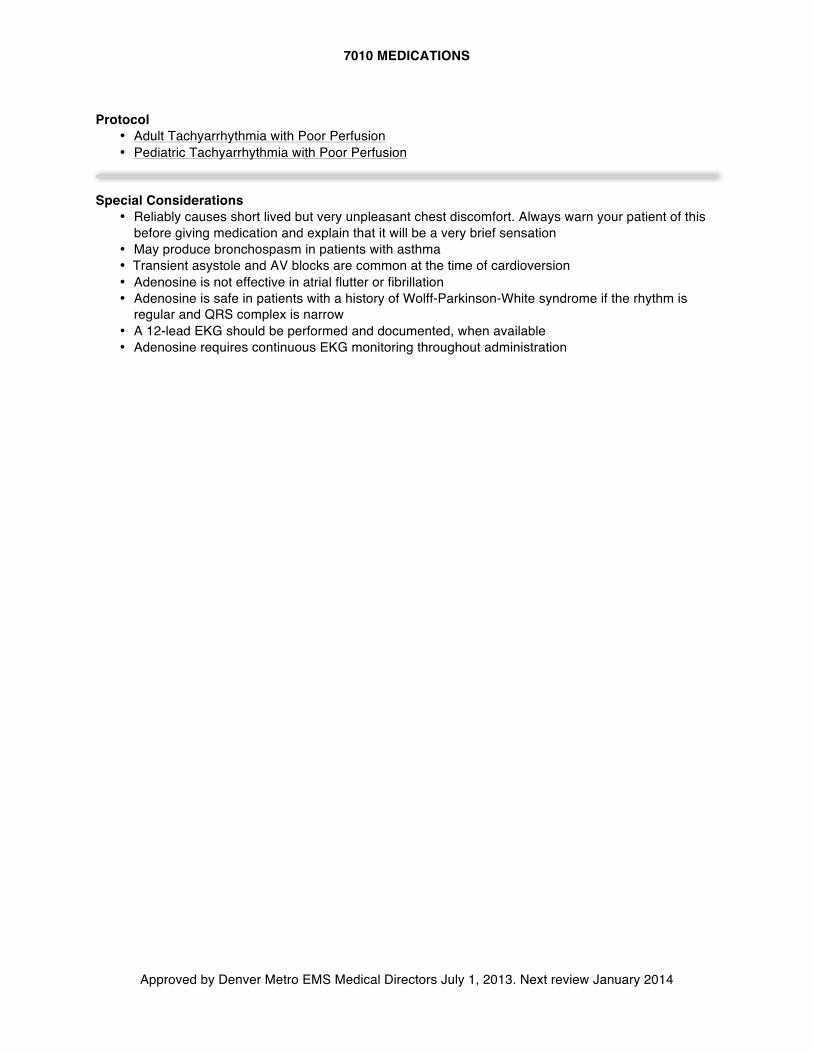
7010 MEDICATIONS
Approved by Denver Metro EMS Medical Directors July 1, 2013. Next review January 2014
Protocol
• Adult Tachyarrhythmia with Poor Perfusion • Pediatric Tachyarrhythmia with Poor Perfusion
Special Considerations
• Reliably causes short lived but very unpleasant chest discomfort. Always warn your patient of this before giving medication and explain that it will be a very brief sensation
• May produce bronchospasm in patients with asthma • Transient asystole and AV blocks are common at the time of cardioversion • Adenosine is not effective in atrial flutter or fibrillation • Adenosine is safe in patients with a history of Wolff-Parkinson-White syndrome if the rhythm is
regular and QRS complex is narrow • A 12-lead EKG should be performed and documented, when available • Adenosine requires continuous EKG monitoring throughout administration
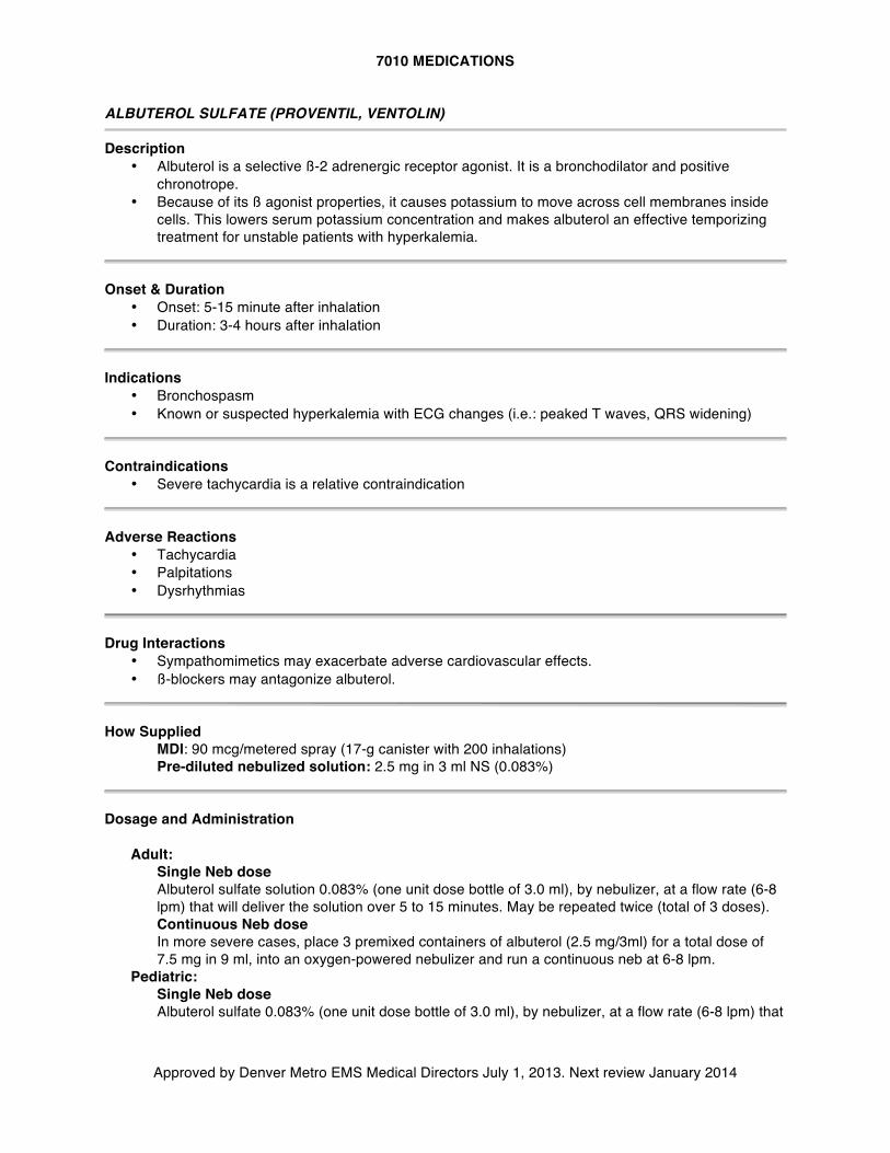
7010 MEDICATIONS
Approved by Denver Metro EMS Medical Directors July 1, 2013. Next review January 2014
ALBUTEROL SULFATE (PROVENTIL, VENTOLIN) Description
• Albuterol is a selective ß-2 adrenergic receptor agonist. It is a bronchodilator and positive chronotrope.
• Because of its ß agonist properties, it causes potassium to move across cell membranes inside cells. This lowers serum potassium concentration and makes albuterol an effective temporizing treatment for unstable patients with hyperkalemia.
Onset & Duration
• Onset: 5-15 minute after inhalation • Duration: 3-4 hours after inhalation
Indications
• Bronchospasm • Known or suspected hyperkalemia with ECG changes (i.e.: peaked T waves, QRS widening)
Contraindications
• Severe tachycardia is a relative contraindication Adverse Reactions
• Tachycardia • Palpitations • Dysrhythmias
Drug Interactions
• Sympathomimetics may exacerbate adverse cardiovascular effects. • ß-blockers may antagonize albuterol.
How Supplied
MDI: 90 mcg/metered spray (17-g canister with 200 inhalations) Pre-diluted nebulized solution: 2.5 mg in 3 ml NS (0.083%)
Dosage and Administration
Adult:
Single Neb dose Albuterol sulfate solution 0.083% (one unit dose bottle of 3.0 ml), by nebulizer, at a flow rate (6-8 lpm) that will deliver the solution over 5 to 15 minutes. May be repeated twice (total of 3 doses). Continuous Neb dose In more severe cases, place 3 premixed containers of albuterol (2.5 mg/3ml) for a total dose of 7.5 mg in 9 ml, into an oxygen-powered nebulizer and run a continuous neb at 6-8 lpm.
Pediatric: Single Neb dose Albuterol sulfate 0.083% (one unit dose bottle of 3.0 ml), by nebulizer, at a flow rate (6-8 lpm) that
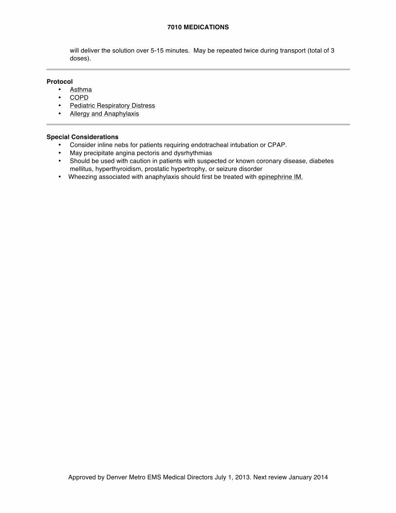
7010 MEDICATIONS
Approved by Denver Metro EMS Medical Directors July 1, 2013. Next review January 2014
will deliver the solution over 5-15 minutes. May be repeated twice during transport (total of 3 doses).
Protocol
• Asthma • COPD • Pediatric Respiratory Distress • Allergy and Anaphylaxis
Special Considerations
• Consider inline nebs for patients requiring endotracheal intubation or CPAP. • May precipitate angina pectoris and dysrhythmias • Should be used with caution in patients with suspected or known coronary disease, diabetes
mellitus, hyperthyroidism, prostatic hypertrophy, or seizure disorder • Wheezing associated with anaphylaxis should first be treated with epinephrine IM.
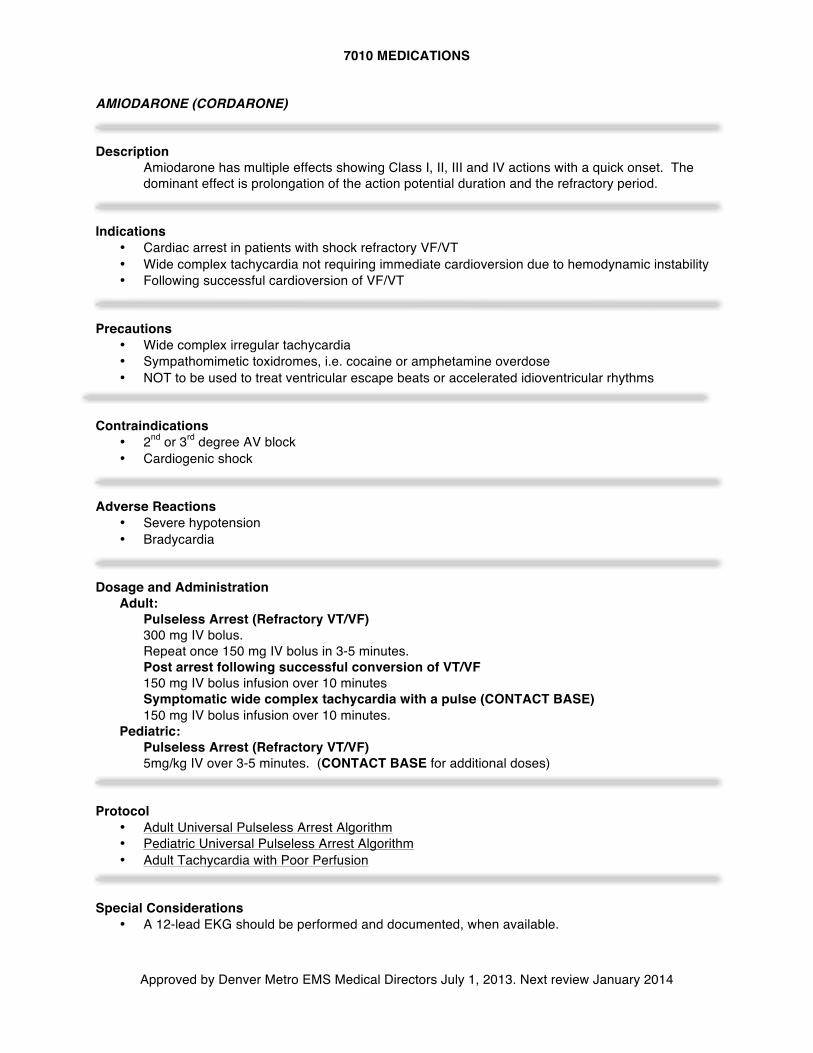
7010 MEDICATIONS
Approved by Denver Metro EMS Medical Directors July 1, 2013. Next review January 2014
AMIODARONE (CORDARONE) Description
Amiodarone has multiple effects showing Class I, II, III and IV actions with a quick onset. The dominant effect is prolongation of the action potential duration and the refractory period.
Indications
• Cardiac arrest in patients with shock refractory VF/VT • Wide complex tachycardia not requiring immediate cardioversion due to hemodynamic instability • Following successful cardioversion of VF/VT
Precautions
• Wide complex irregular tachycardia • Sympathomimetic toxidromes, i.e. cocaine or amphetamine overdose • NOT to be used to treat ventricular escape beats or accelerated idioventricular rhythms
Contraindications
• 2nd or 3rd degree AV block • Cardiogenic shock
Adverse Reactions
• Severe hypotension • Bradycardia
Dosage and Administration
Adult: Pulseless Arrest (Refractory VT/VF) 300 mg IV bolus. Repeat once 150 mg IV bolus in 3-5 minutes. Post arrest following successful conversion of VT/VF 150 mg IV bolus infusion over 10 minutes Symptomatic wide complex tachycardia with a pulse (CONTACT BASE) 150 mg IV bolus infusion over 10 minutes.
Pediatric: Pulseless Arrest (Refractory VT/VF) 5mg/kg IV over 3-5 minutes. (CONTACT BASE for additional doses)
Protocol
• Adult Universal Pulseless Arrest Algorithm • Pediatric Universal Pulseless Arrest Algorithm • Adult Tachycardia with Poor Perfusion
Special Considerations
• A 12-lead EKG should be performed and documented, when available.
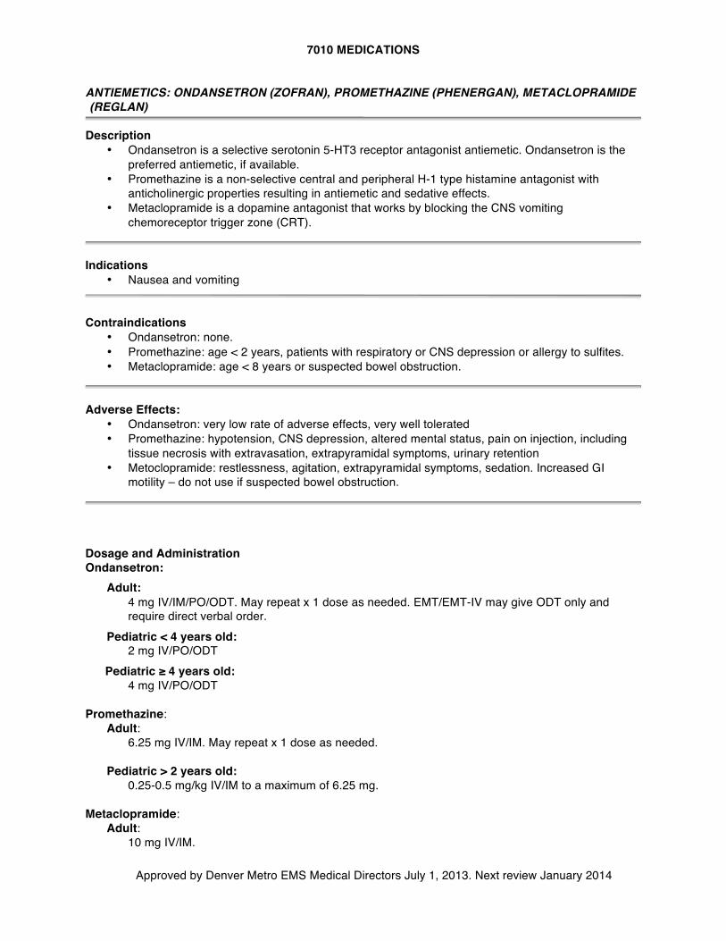
7010 MEDICATIONS
Approved by Denver Metro EMS Medical Directors July 1, 2013. Next review January 2014
ANTIEMETICS: ONDANSETRON (ZOFRAN), PROMETHAZINE (PHENERGAN), METACLOPRAMIDE (REGLAN)
Description
• Ondansetron is a selective serotonin 5-HT3 receptor antagonist antiemetic. Ondansetron is the preferred antiemetic, if available.
• Promethazine is a non-selective central and peripheral H-1 type histamine antagonist with anticholinergic properties resulting in antiemetic and sedative effects.
• Metaclopramide is a dopamine antagonist that works by blocking the CNS vomiting chemoreceptor trigger zone (CRT).
Indications
• Nausea and vomiting Contraindications
• Ondansetron: none. • Promethazine: age < 2 years, patients with respiratory or CNS depression or allergy to sulfites. • Metaclopramide: age < 8 years or suspected bowel obstruction.
Adverse Effects:
• Ondansetron: very low rate of adverse effects, very well tolerated • Promethazine: hypotension, CNS depression, altered mental status, pain on injection, including
tissue necrosis with extravasation, extrapyramidal symptoms, urinary retention • Metoclopramide: restlessness, agitation, extrapyramidal symptoms, sedation. Increased GI
motility – do not use if suspected bowel obstruction.
Dosage and Administration Ondansetron:
Adult: 4 mg IV/IM/PO/ODT. May repeat x 1 dose as needed. EMT/EMT-IV may give ODT only and require direct verbal order.
Pediatric < 4 years old: 2 mg IV/PO/ODT
Pediatric ≥ 4 years old: 4 mg IV/PO/ODT Promethazine:
Adult: 6.25 mg IV/IM. May repeat x 1 dose as needed.
Pediatric > 2 years old: 0.25-0.5 mg/kg IV/IM to a maximum of 6.25 mg.
Metaclopramide:
Adult: 10 mg IV/IM.
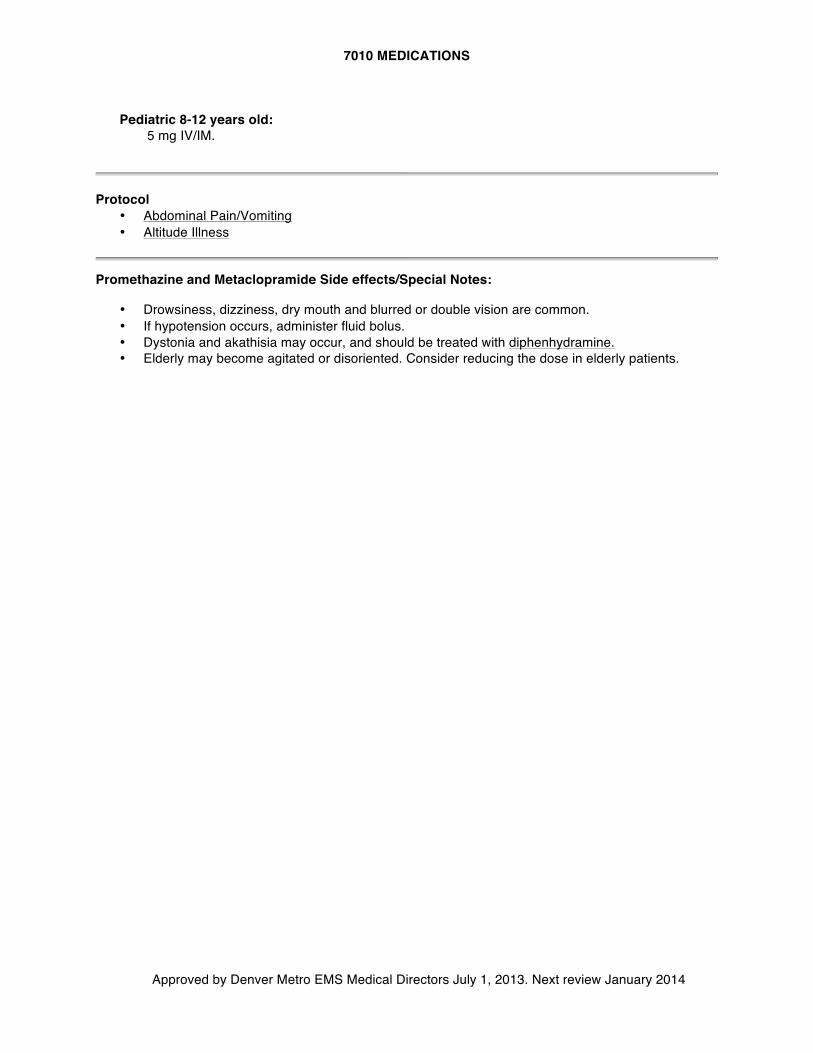
7010 MEDICATIONS
Approved by Denver Metro EMS Medical Directors July 1, 2013. Next review January 2014
Pediatric 8-12 years old:
5 mg IV/IM.
Protocol
• Abdominal Pain/Vomiting • Altitude Illness
Promethazine and Metaclopramide Side effects/Special Notes:
• Drowsiness, dizziness, dry mouth and blurred or double vision are common. • If hypotension occurs, administer fluid bolus. • Dystonia and akathisia may occur, and should be treated with diphenhydramine. • Elderly may become agitated or disoriented. Consider reducing the dose in elderly patients.
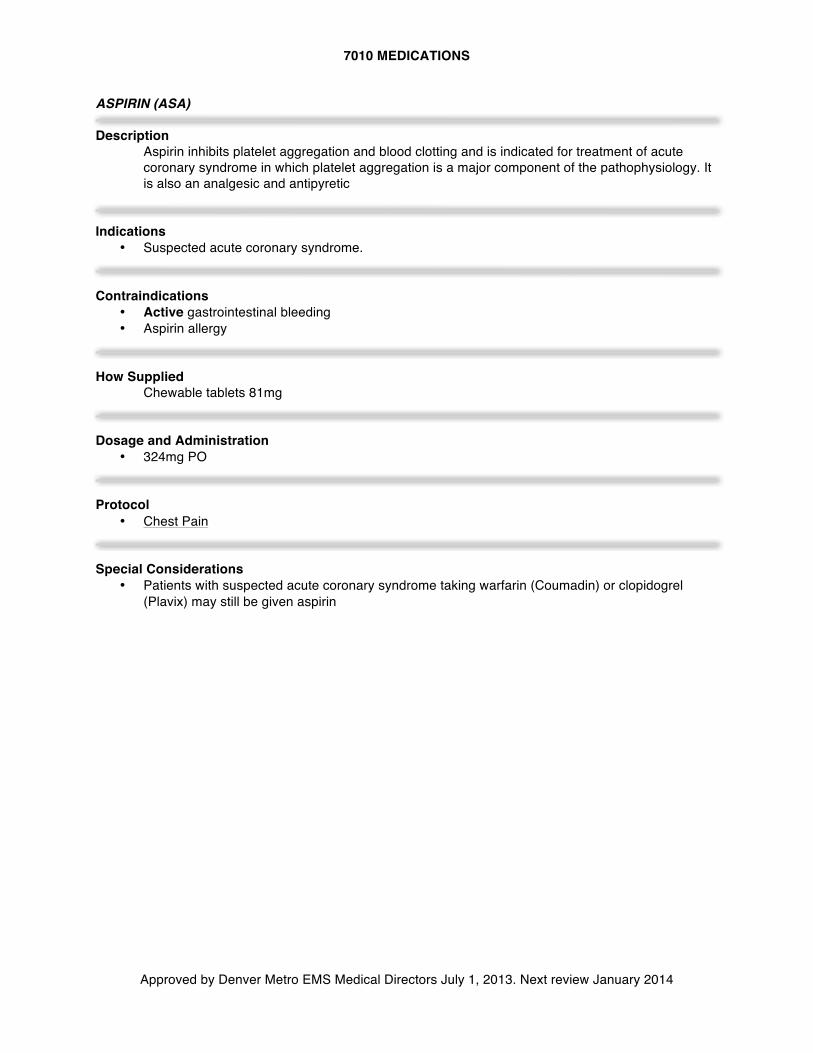
7010 MEDICATIONS
Approved by Denver Metro EMS Medical Directors July 1, 2013. Next review January 2014
ASPIRIN (ASA) Description
Aspirin inhibits platelet aggregation and blood clotting and is indicated for treatment of acute coronary syndrome in which platelet aggregation is a major component of the pathophysiology. It is also an analgesic and antipyretic
Indications
• Suspected acute coronary syndrome. Contraindications
• Active gastrointestinal bleeding • Aspirin allergy
How Supplied
Chewable tablets 81mg Dosage and Administration
• 324mg PO Protocol
• Chest Pain
Special Considerations • Patients with suspected acute coronary syndrome taking warfarin (Coumadin) or clopidogrel
(Plavix) may still be given aspirin
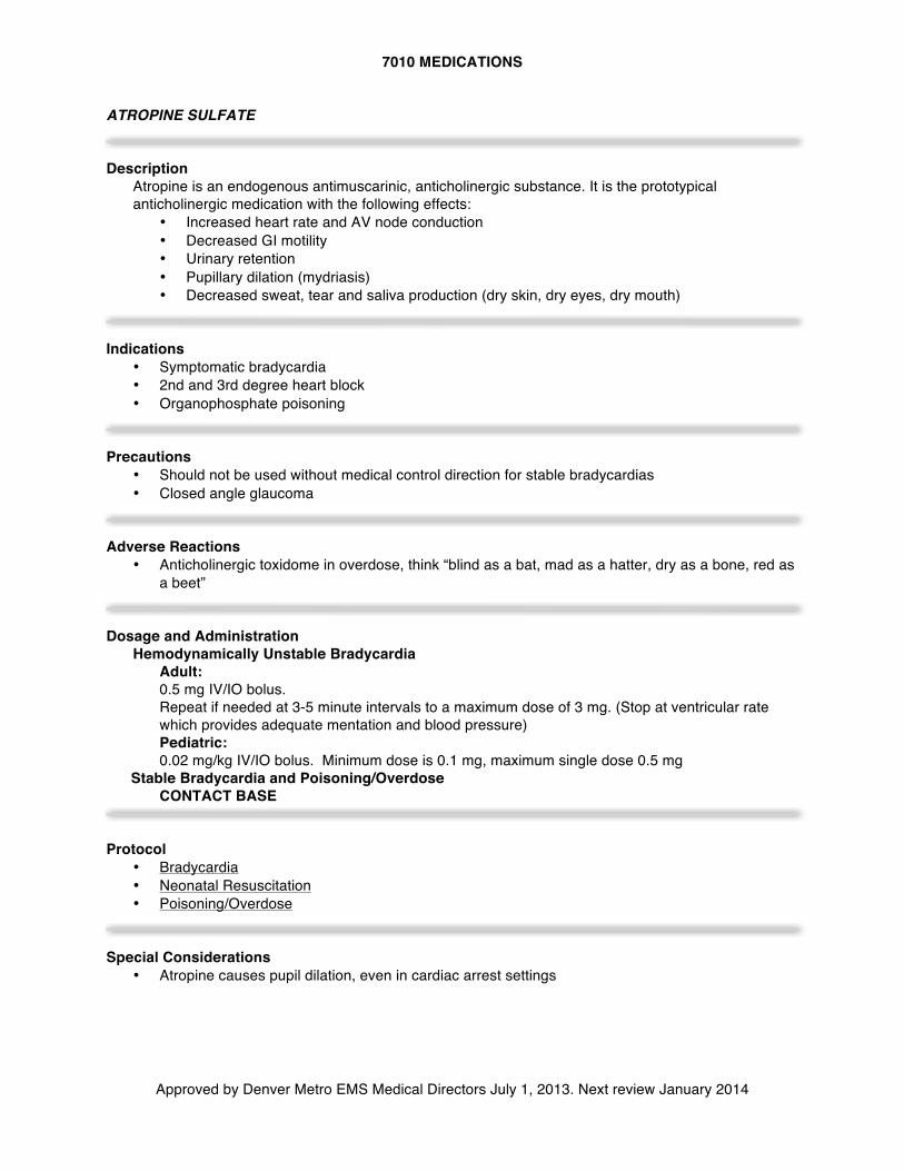
7010 MEDICATIONS
Approved by Denver Metro EMS Medical Directors July 1, 2013. Next review January 2014
ATROPINE SULFATE Description
Atropine is an endogenous antimuscarinic, anticholinergic substance. It is the prototypical anticholinergic medication with the following effects:
• Increased heart rate and AV node conduction • Decreased GI motility • Urinary retention • Pupillary dilation (mydriasis) • Decreased sweat, tear and saliva production (dry skin, dry eyes, dry mouth)
Indications
• Symptomatic bradycardia • 2nd and 3rd degree heart block • Organophosphate poisoning
Precautions
• Should not be used without medical control direction for stable bradycardias • Closed angle glaucoma
Adverse Reactions
• Anticholinergic toxidome in overdose, think “blind as a bat, mad as a hatter, dry as a bone, red as a beet”
Dosage and Administration
Hemodynamically Unstable Bradycardia Adult: 0.5 mg IV/IO bolus. Repeat if needed at 3-5 minute intervals to a maximum dose of 3 mg. (Stop at ventricular rate which provides adequate mentation and blood pressure) Pediatric: 0.02 mg/kg IV/IO bolus. Minimum dose is 0.1 mg, maximum single dose 0.5 mg
Stable Bradycardia and Poisoning/Overdose CONTACT BASE Protocol
• Bradycardia • Neonatal Resuscitation • Poisoning/Overdose
Special Considerations
• Atropine causes pupil dilation, even in cardiac arrest settings
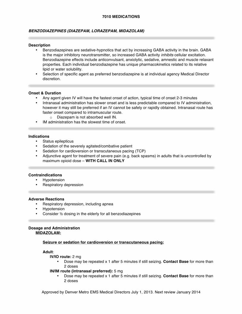
7010 MEDICATIONS
Approved by Denver Metro EMS Medical Directors July 1, 2013. Next review January 2014
BENZODIAZEPINES (DIAZEPAM, LORAZEPAM, MIDAZOLAM) Description
• Benzodiazepines are sedative-hypnotics that act by increasing GABA activity in the brain. GABA is the major inhibitory neurotransmitter, so increased GABA activity inhibits cellular excitation. Benzodiazepine effects include anticonvulsant, anxiolytic, sedative, amnestic and muscle relaxant properties. Each individual benzodiazepine has unique pharmacokinetics related to its relative lipid or water solubility.
• Selection of specific agent as preferred benzodiazepine is at individual agency Medical Director discretion.
Onset & Duration
• Any agent given IV will have the fastest onset of action, typical time of onset 2-3 minutes • Intranasal administration has slower onset and is less predictable compared to IV administration,
however it may still be preferred if an IV cannot be safely or rapidly obtained. Intranasal route has faster onset compared to intramuscular route.
o Diazepam is not absorbed well IN. • IM administration has the slowest time of onset.
Indications
• Status epilepticus • Sedation of the severely agitated/combative patient • Sedation for cardioversion or transcutaneous pacing (TCP) • Adjunctive agent for treatment of severe pain (e.g. back spasms) in adults that is uncontrolled by
maximum opioid dose – WITH CALL IN ONLY Contraindications
• Hypotension • Respiratory depression
Adverse Reactions
• Respiratory depression, including apnea • Hypotension • Consider ½ dosing in the elderly for all benzodiazepines
Dosage and Administration
MIDAZOLAM: Seizure or sedation for cardioversion or transcutaneous pacing: Adult:
IV/IO route: 2 mg • Dose may be repeated x 1 after 5 minutes if still seizing. Contact Base for more than
2 doses IN/IM route (intranasal preferred): 5 mg
• Dose may be repeated x 1 after 5 minutes if still seizing. Contact Base for more than 2 doses
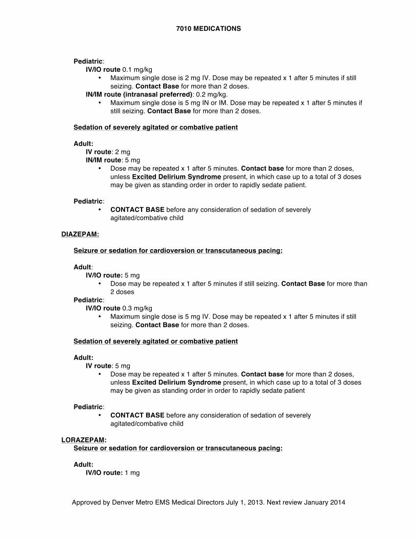
7010 MEDICATIONS
Approved by Denver Metro EMS Medical Directors July 1, 2013. Next review January 2014
Pediatric:
IV/IO route 0.1 mg/kg • Maximum single dose is 2 mg IV. Dose may be repeated x 1 after 5 minutes if still
seizing. Contact Base for more than 2 doses. IN/IM route (intranasal preferred): 0.2 mg/kg.
• Maximum single dose is 5 mg IN or IM. Dose may be repeated x 1 after 5 minutes if still seizing. Contact Base for more than 2 doses.
Sedation of severely agitated or combative patient Adult:
IV route: 2 mg IN/IM route: 5 mg
• Dose may be repeated x 1 after 5 minutes. Contact base for more than 2 doses, unless Excited Delirium Syndrome present, in which case up to a total of 3 doses may be given as standing order in order to rapidly sedate patient.
Pediatric:
• CONTACT BASE before any consideration of sedation of severely agitated/combative child
DIAZEPAM:
Seizure or sedation for cardioversion or transcutaneous pacing: Adult:
IV/IO route: 5 mg • Dose may be repeated x 1 after 5 minutes if still seizing. Contact Base for more than
2 doses Pediatric:
IV/IO route 0.3 mg/kg • Maximum single dose is 5 mg IV. Dose may be repeated x 1 after 5 minutes if still
seizing. Contact Base for more than 2 doses.
Sedation of severely agitated or combative patient Adult:
IV route: 5 mg • Dose may be repeated x 1 after 5 minutes. Contact base for more than 2 doses,
unless Excited Delirium Syndrome present, in which case up to a total of 3 doses may be given as standing order in order to rapidly sedate patient
Pediatric:
• CONTACT BASE before any consideration of sedation of severely agitated/combative child
LORAZEPAM:
Seizure or sedation for cardioversion or transcutaneous pacing: Adult:
IV/IO route: 1 mg
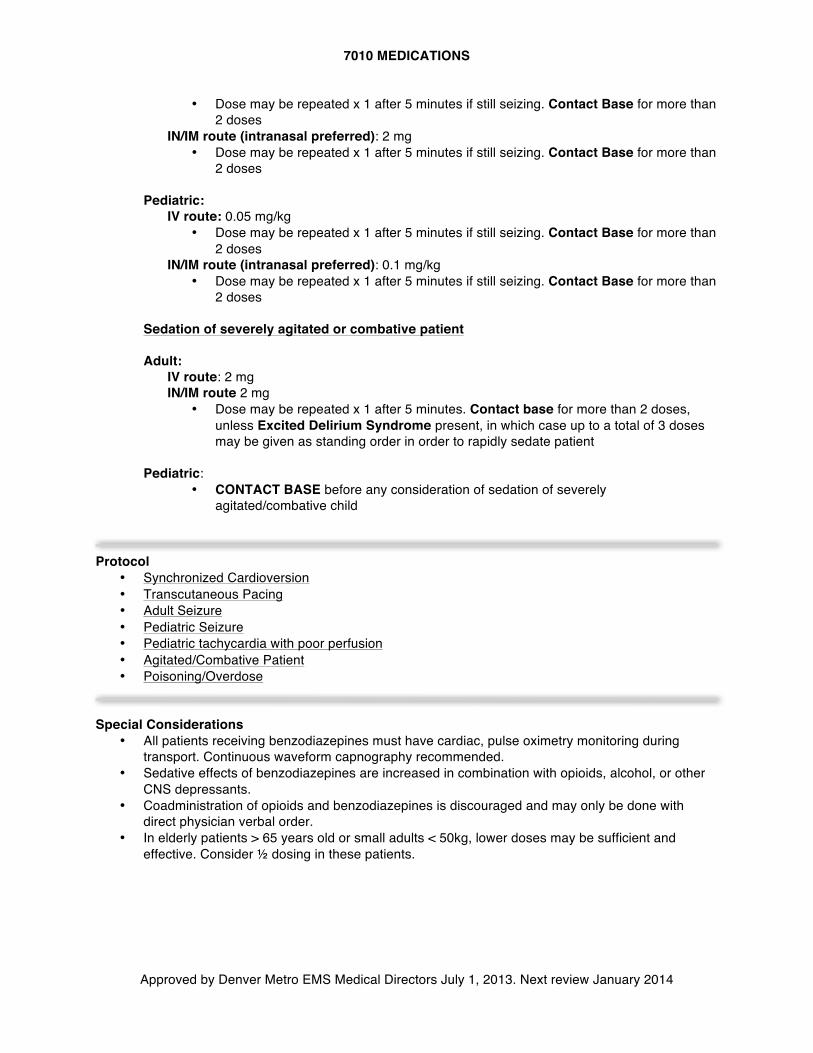
7010 MEDICATIONS
Approved by Denver Metro EMS Medical Directors July 1, 2013. Next review January 2014
• Dose may be repeated x 1 after 5 minutes if still seizing. Contact Base for more than 2 doses
IN/IM route (intranasal preferred): 2 mg • Dose may be repeated x 1 after 5 minutes if still seizing. Contact Base for more than
2 doses Pediatric:
IV route: 0.05 mg/kg • Dose may be repeated x 1 after 5 minutes if still seizing. Contact Base for more than
2 doses IN/IM route (intranasal preferred): 0.1 mg/kg
• Dose may be repeated x 1 after 5 minutes if still seizing. Contact Base for more than 2 doses
Sedation of severely agitated or combative patient Adult:
IV route: 2 mg IN/IM route 2 mg
• Dose may be repeated x 1 after 5 minutes. Contact base for more than 2 doses, unless Excited Delirium Syndrome present, in which case up to a total of 3 doses may be given as standing order in order to rapidly sedate patient
Pediatric:
• CONTACT BASE before any consideration of sedation of severely agitated/combative child
Protocol • Synchronized Cardioversion • Transcutaneous Pacing • Adult Seizure • Pediatric Seizure • Pediatric tachycardia with poor perfusion • Agitated/Combative Patient • Poisoning/Overdose
Special Considerations
• All patients receiving benzodiazepines must have cardiac, pulse oximetry monitoring during transport. Continuous waveform capnography recommended.
• Sedative effects of benzodiazepines are increased in combination with opioids, alcohol, or other CNS depressants.
• Coadministration of opioids and benzodiazepines is discouraged and may only be done with direct physician verbal order.
• In elderly patients > 65 years old or small adults < 50kg, lower doses may be sufficient and effective. Consider ½ dosing in these patients.
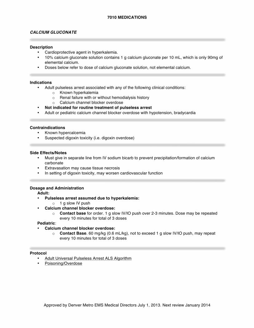
7010 MEDICATIONS
Approved by Denver Metro EMS Medical Directors July 1, 2013. Next review January 2014
CALCIUM GLUCONATE Description
• Cardioprotective agent in hyperkalemia. • 10% calcium gluconate solution contains 1 g calcium gluconate per 10 mL, which is only 90mg of
elemental calcium. • Doses below refer to dose of calcium gluconate solution, not elemental calcium.
Indications
• Adult pulseless arrest associated with any of the following clinical conditions: o Known hyperkalemia o Renal failure with or without hemodialysis history o Calcium channel blocker overdose
• Not indicated for routine treatment of pulseless arrest • Adult or pediatric calcium channel blocker overdose with hypotension, bradycardia
Contraindications
• Known hypercalcemia • Suspected digoxin toxicity (i.e. digoxin overdose)
Side Effects/Notes
• Must give in separate line from IV sodium bicarb to prevent precipitation/formation of calcium carbonate
• Extravasation may cause tissue necrosis • In setting of digoxin toxicity, may worsen cardiovascular function
Dosage and Administration
Adult: • Pulseless arrest assumed due to hyperkalemia:
o 1 g slow IV push • Calcium channel blocker overdose:
o Contact base for order. 1 g slow IV/IO push over 2-3 minutes. Dose may be repeated every 10 minutes for total of 3 doses
Pediatric: • Calcium channel blocker overdose:
o Contact Base. 60 mg/kg (0.6 mL/kg), not to exceed 1 g slow IV/IO push, may repeat every 10 minutes for total of 3 doses
Protocol
• Adult Universal Pulseless Arrest ALS Algorithm • Poisoning/Overdose
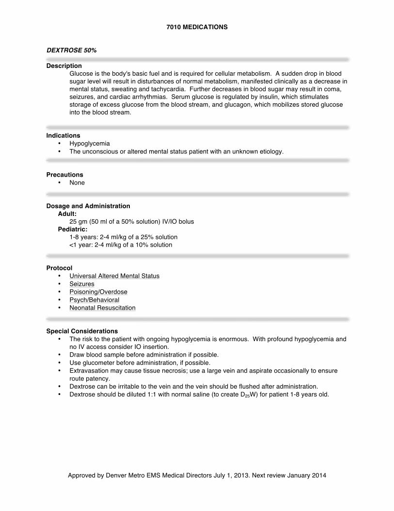
7010 MEDICATIONS
Approved by Denver Metro EMS Medical Directors July 1, 2013. Next review January 2014
DEXTROSE 50% Description
Glucose is the body's basic fuel and is required for cellular metabolism. A sudden drop in blood sugar level will result in disturbances of normal metabolism, manifested clinically as a decrease in mental status, sweating and tachycardia. Further decreases in blood sugar may result in coma, seizures, and cardiac arrhythmias. Serum glucose is regulated by insulin, which stimulates storage of excess glucose from the blood stream, and glucagon, which mobilizes stored glucose into the blood stream.
Indications
• Hypoglycemia • The unconscious or altered mental status patient with an unknown etiology.
Precautions
• None
Dosage and Administration
Adult: 25 gm (50 ml of a 50% solution) IV/IO bolus
Pediatric: 1-8 years: 2-4 ml/kg of a 25% solution <1 year: 2-4 ml/kg of a 10% solution
Protocol
• Universal Altered Mental Status • Seizures • Poisoning/Overdose • Psych/Behavioral • Neonatal Resuscitation
Special Considerations
• The risk to the patient with ongoing hypoglycemia is enormous. With profound hypoglycemia and no IV access consider IO insertion.
• Draw blood sample before administration if possible. • Use glucometer before administration, if possible. • Extravasation may cause tissue necrosis; use a large vein and aspirate occasionally to ensure
route patency. • Dextrose can be irritable to the vein and the vein should be flushed after administration. • Dextrose should be diluted 1:1 with normal saline (to create D25W) for patient 1-8 years old.
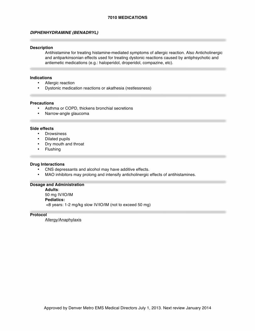
7010 MEDICATIONS
Approved by Denver Metro EMS Medical Directors July 1, 2013. Next review January 2014
DIPHENHYDRAMINE (BENADRYL) Description
Antihistamine for treating histamine-mediated symptoms of allergic reaction. Also Anticholinergic and antiparkinsonian effects used for treating dystonic reactions caused by antiphsychotic and antiemetic medications (e.g.: haloperidol, droperidol, compazine, etc).
Indications
• Allergic reaction • Dystonic medication reactions or akathesia (restlessness)
Precautions
• Asthma or COPD, thickens bronchial secretions • Narrow-angle glaucoma
Side effects
• Drowsiness • Dilated pupils • Dry mouth and throat • Flushing
Drug Interactions
• CNS depressants and alcohol may have additive effects. • MAO inhibitors may prolong and intensify anticholinergic effects of antihistamines.
Dosage and Administration
Adults: 50 mg IV/IO/IM Pediatics: <8 years: 1-2 mg/kg slow IV/IO/IM (not to exceed 50 mg)
Protocol
Allergy/Anaphylaxis
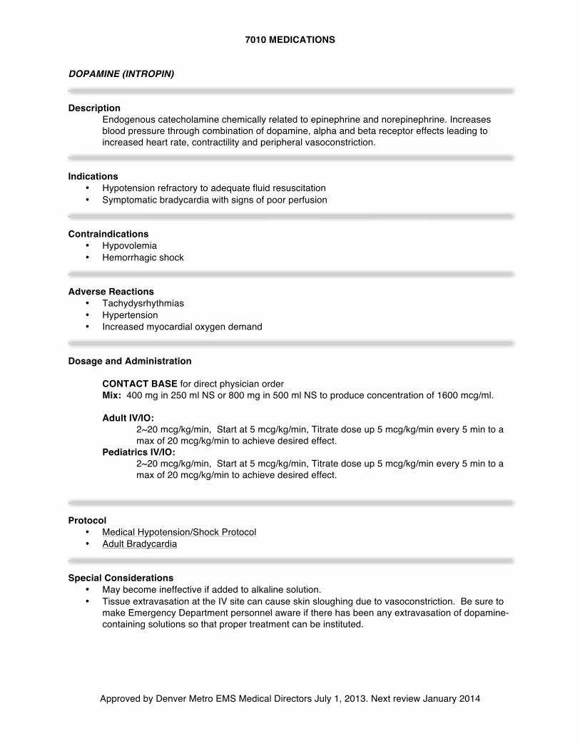
7010 MEDICATIONS
Approved by Denver Metro EMS Medical Directors July 1, 2013. Next review January 2014
DOPAMINE (INTROPIN) Description
Endogenous catecholamine chemically related to epinephrine and norepinephrine. Increases blood pressure through combination of dopamine, alpha and beta receptor effects leading to increased heart rate, contractility and peripheral vasoconstriction.
Indications
• Hypotension refractory to adequate fluid resuscitation • Symptomatic bradycardia with signs of poor perfusion
Contraindications
• Hypovolemia • Hemorrhagic shock
Adverse Reactions
• Tachydysrhythmias • Hypertension • Increased myocardial oxygen demand
Dosage and Administration
CONTACT BASE for direct physician order Mix: 400 mg in 250 ml NS or 800 mg in 500 ml NS to produce concentration of 1600 mcg/ml. Adult IV/IO:
2~20 mcg/kg/min, Start at 5 mcg/kg/min, Titrate dose up 5 mcg/kg/min every 5 min to a max of 20 mcg/kg/min to achieve desired effect.
Pediatrics IV/IO: 2~20 mcg/kg/min, Start at 5 mcg/kg/min, Titrate dose up 5 mcg/kg/min every 5 min to a max of 20 mcg/kg/min to achieve desired effect.
Protocol
• Medical Hypotension/Shock Protocol • Adult Bradycardia
Special Considerations
• May become ineffective if added to alkaline solution. • Tissue extravasation at the IV site can cause skin sloughing due to vasoconstriction. Be sure to
make Emergency Department personnel aware if there has been any extravasation of dopamine-containing solutions so that proper treatment can be instituted.
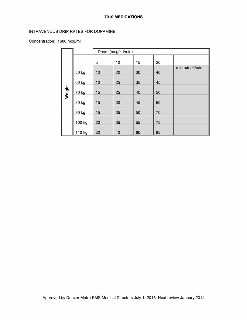
7010 MEDICATIONS
Approved by Denver Metro EMS Medical Directors July 1, 2013. Next review January 2014
INTRAVENOUS DRIP RATES FOR DOPAMINE Concentration: 1600 mcg/ml
Wei
ght
Dose (mcg/kd/min)
5
10
15
20
50 kg
10
20
30
40
microdrips/min
60 kg
10
25
35
45
70 kg
15
25
40
50
80 kg
15
30
45
60
90 kg
15
35
50
70
100 kg
20
35
55
75
110 kg
20
40
60
85
7010 MEDICATIONS
Approved by Denver Metro EMS Medical Directors July 1, 2013. Next review January 2014
DROPERIDOL (INAPSINE) Description
• Droperidol is a butyrophenone derivative closely related to haloperidol. Droperidol produces a dopaminergic blockage, a mild alpha-adrenergic blockage, and causes peripheral vasodilation. Its major actions are sedation, tranquilization, and potent anti-emetic effect.
Onset & Duration
• Onset: 3-10 minutes after IM administration. • Duration: 2-3 hours
Indications
• Primary use for management of agitated/combative patients. • Second line medication for management of intractable vomiting requiring base contact. • Combative head injured patients.
Contraindications
• Any patient with: o Suspected acute myocardial infarction/ACS o Systolic blood pressure under 100 mm/Hg, or the absence of a palpable radial pulse o Signs of respiratory depression
Side Effects
• Due to the vasodilation effect, droperidol can cause a transient hypotension that is usually self limiting and can be treated effectively with leg elevated position and IV fluids. Droperidol may cause tachycardia which usually does not require pharmacologic intervention.
• Some patient’s may experience unpleasant sensations manifested as restlessness, hyperactivity, or anxiety following droperidol administration.
• Extra-pyramidal reactions have been noted hours to days after treatment. • Rare instances of neuroleptic malignant syndrome have been known to occur following treatment
using droperidol. Dosage and Administration
Agitation/Combative
Adult: IV/IM route: 5.0 mg slow IV/IM administration, after 10 minutes if desired effect is not achieved contact base to consider a second dose. Pediatric: Under the age of 12 Contact Base
Antiemetic: Contact base for orders
IV/IM route: Adult: 1.25 mg slow push. Pediatric: 0.05 mg/kg slow push.
Special Considerations
7010 MEDICATIONS
Approved by Denver Metro EMS Medical Directors July 1, 2013. Next review January 2014
• Due to droperidol’s potential effect on QT interval prolongation, all patients receiving droperidol should be placed on the cardiac monitor. Though it is understood that obtaining an ECG on the combative or agitated patient may be difficult, every effort should be made to do so.
• Avoid droperidol in frail or elderly patients due to increased risk of prolonged and over-sedation as well as increased risk of hypotension and prolonged QT. If it must be given, administer ½ typical dose.
Protocol
• Agitated/Combative Patient Protocol
7010 MEDICATIONS
Approved by Denver Metro EMS Medical Directors July 1, 2013. Next review January 2014
EPINEPHRINE (ADRENALIN) Description
Endogenous catecholamine alpha, beta-1, and beta-2 adrenergic receptor agonist. Causes dose-related increase in heart rate, myocardial contractility and oxygen demand, peripheral vasoconstriction and bronchodilation.
Indications
• Pulseless Arrest • Anaphylaxis • Asthma • Bradycardia with poor perfusion
Adverse Reactions
• Tachycardia and tachydysrhythmia • Hypertension • Anxiety • May precipitate angina pectoris
Drug Interactions
• Should not be added to sodium bicarbonate or other alkaloids as epinephrine will be inactivated at higher pH.
Dosage and Administration
Adult: Pulseless Arrest 1 mg (10 ml of a 1:10,000 solution), IV/IO bolus. Repeat every 3-5 minutes. Bradycardia/ hypotension refractory to other interventions (Contact Base): Continuous infusion titrated to effect: 1 mg in 250 ml of Normal Saline IV/IO infused at 2 mcg/min until desired BP of > 90 mmHg systolic achieved. Asthma: 0.3 mg (0.3 ml of a 1:1,000 solution) IM. May repeat dose x 1. Systemic allergic reaction: 0.3 mg (0.3 ml of a 1:1,000 solution) IM. May repeat dose x 1. Severe systemic allergic reaction (Anaphylaxis) refractory to IM epi (Contact Base): Continuous infusion titrated to effect: 1 mg in 250 ml of Normal Saline IV/IO infused at 2 mcg/min until desired BP of > 90 mmHg systolic achieved ALTERNATIVE to racemic epinephrine: (for epiglottitis, miscellaneous causes of stridor) 5 mL of 1:1000 epinephrine via nebulizer x 1
Epinephrine Auto-Injector: requires BASE CONTACT for EMT administration
Systemic allergic reaction: Adult: 0.3 mg IM with autoinjector (adult EpiPen) Pediatric: 0.15 mg IM with autoinjector (EpiPen Jr.)
Pediatric:
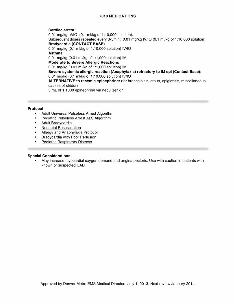
7010 MEDICATIONS
Approved by Denver Metro EMS Medical Directors July 1, 2013. Next review January 2014
Cardiac arrest: 0.01 mg/kg IV/IO (0.1 ml/kg of 1:10,000 solution). Subsequent doses repeated every 3-5min: 0.01 mg/kg IV/IO (0.1 ml/kg of 1:10,000 solution) Bradycardia (CONTACT BASE) 0.01 mg/kg (0.1 ml/kg of 1:10,000 solution) IV/IO Asthma 0.01 mg/kg (0.01 ml/kg of 1:1,000 solution) IM Moderate to Severe Allergic Reactions 0.01 mg/kg (0.01 ml/kg of 1:1,000 solution) IM Severe systemic allergic reaction (Anaphylaxis) refractory to IM epi (Contact Base): 0.01 mg/kg (0.1 ml/kg of 1:10,000 solution) IV/IO ALTERNATIVE to racemic epinephrine: (for bronchiolitis, croup, epiglottitis, miscellaneous causes of stridor) 5 mL of 1:1000 epinephrine via nebulizer x 1
Protocol
• Adult Universal Pulseless Arrest Algorithm • Pediatric Pulseless Arrest ALS Algorithm • Adult Bradycardia • Neonatal Resuscitation • Allergy and Anaphylaxis Protocol • Bradycardia with Poor Perfusion • Pediatric Respiratory Distress
Special Considerations
• May increase myocardial oxygen demand and angina pectoris. Use with caution in patients with known or suspected CAD
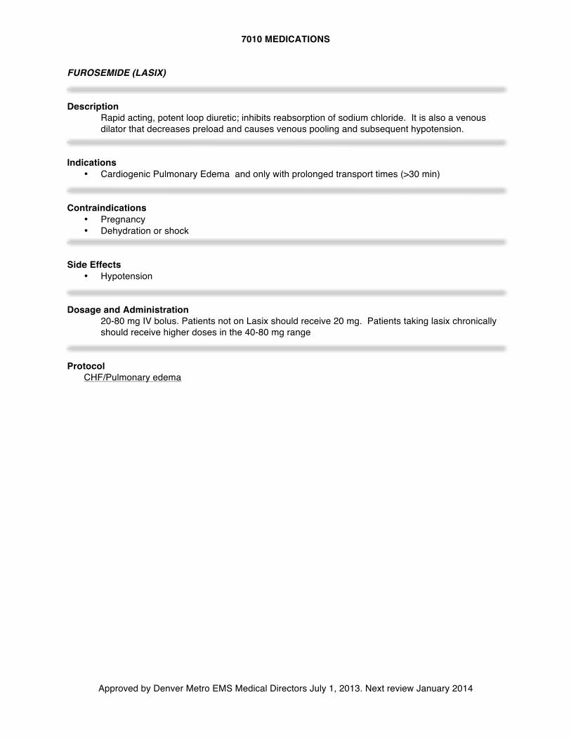
7010 MEDICATIONS
Approved by Denver Metro EMS Medical Directors July 1, 2013. Next review January 2014
FUROSEMIDE (LASIX) Description
Rapid acting, potent loop diuretic; inhibits reabsorption of sodium chloride. It is also a venous dilator that decreases preload and causes venous pooling and subsequent hypotension.
Indications
• Cardiogenic Pulmonary Edema and only with prolonged transport times (>30 min) Contraindications
• Pregnancy • Dehydration or shock
Side Effects
• Hypotension Dosage and Administration
20-80 mg IV bolus. Patients not on Lasix should receive 20 mg. Patients taking lasix chronically should receive higher doses in the 40-80 mg range
Protocol
CHF/Pulmonary edema
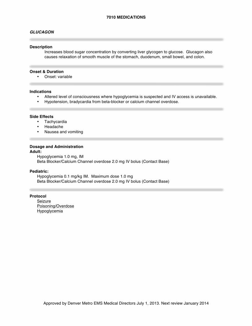
7010 MEDICATIONS
Approved by Denver Metro EMS Medical Directors July 1, 2013. Next review January 2014
GLUCAGON Description
Increases blood sugar concentration by converting liver glycogen to glucose. Glucagon also causes relaxation of smooth muscle of the stomach, duodenum, small bowel, and colon.
Onset & Duration
• Onset: variable Indications
• Altered level of consciousness where hypoglycemia is suspected and IV access is unavailable. • Hypotension, bradycardia from beta-blocker or calcium channel overdose.
Side Effects
• Tachycardia • Headache • Nausea and vomiting
Dosage and Administration Adult:
Hypoglycemia 1.0 mg, IM Beta Blocker/Calcium Channel overdose 2.0 mg IV bolus (Contact Base)
Pediatric:
Hypoglycemia 0.1 mg/kg IM. Maximum dose 1.0 mg Beta Blocker/Calcium Channel overdose 2.0 mg IV bolus (Contact Base)
Protocol
Seizure Poisoning/Overdose Hypoglycemia
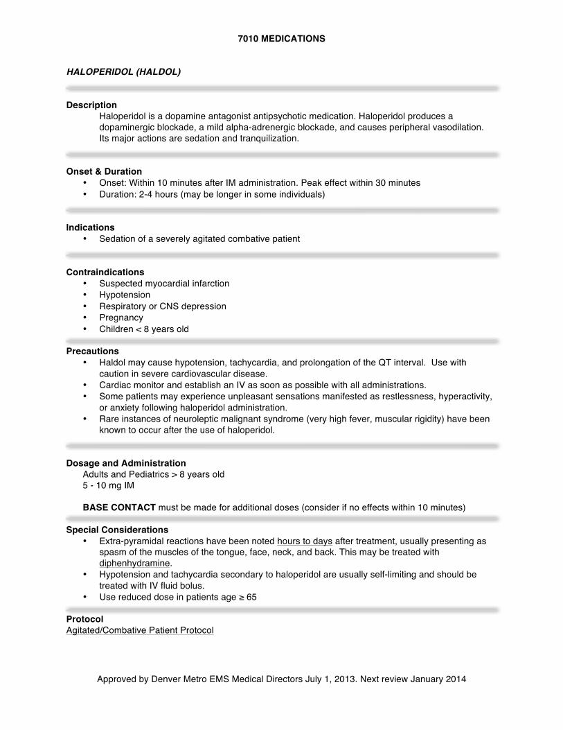
7010 MEDICATIONS
Approved by Denver Metro EMS Medical Directors July 1, 2013. Next review January 2014
HALOPERIDOL (HALDOL) Description
Haloperidol is a dopamine antagonist antipsychotic medication. Haloperidol produces a dopaminergic blockade, a mild alpha-adrenergic blockade, and causes peripheral vasodilation. Its major actions are sedation and tranquilization.
Onset & Duration
• Onset: Within 10 minutes after IM administration. Peak effect within 30 minutes • Duration: 2-4 hours (may be longer in some individuals)
Indications
• Sedation of a severely agitated combative patient Contraindications
• Suspected myocardial infarction • Hypotension • Respiratory or CNS depression • Pregnancy • Children < 8 years old
Precautions
• Haldol may cause hypotension, tachycardia, and prolongation of the QT interval. Use with caution in severe cardiovascular disease.
• Cardiac monitor and establish an IV as soon as possible with all administrations. • Some patients may experience unpleasant sensations manifested as restlessness, hyperactivity,
or anxiety following haloperidol administration. • Rare instances of neuroleptic malignant syndrome (very high fever, muscular rigidity) have been
known to occur after the use of haloperidol. Dosage and Administration
Adults and Pediatrics > 8 years old 5 - 10 mg IM BASE CONTACT must be made for additional doses (consider if no effects within 10 minutes)
Special Considerations • Extra-pyramidal reactions have been noted hours to days after treatment, usually presenting as
spasm of the muscles of the tongue, face, neck, and back. This may be treated with diphenhydramine.
• Hypotension and tachycardia secondary to haloperidol are usually self-limiting and should be treated with IV fluid bolus.
• Use reduced dose in patients age ≥ 65
Protocol Agitated/Combative Patient Protocol
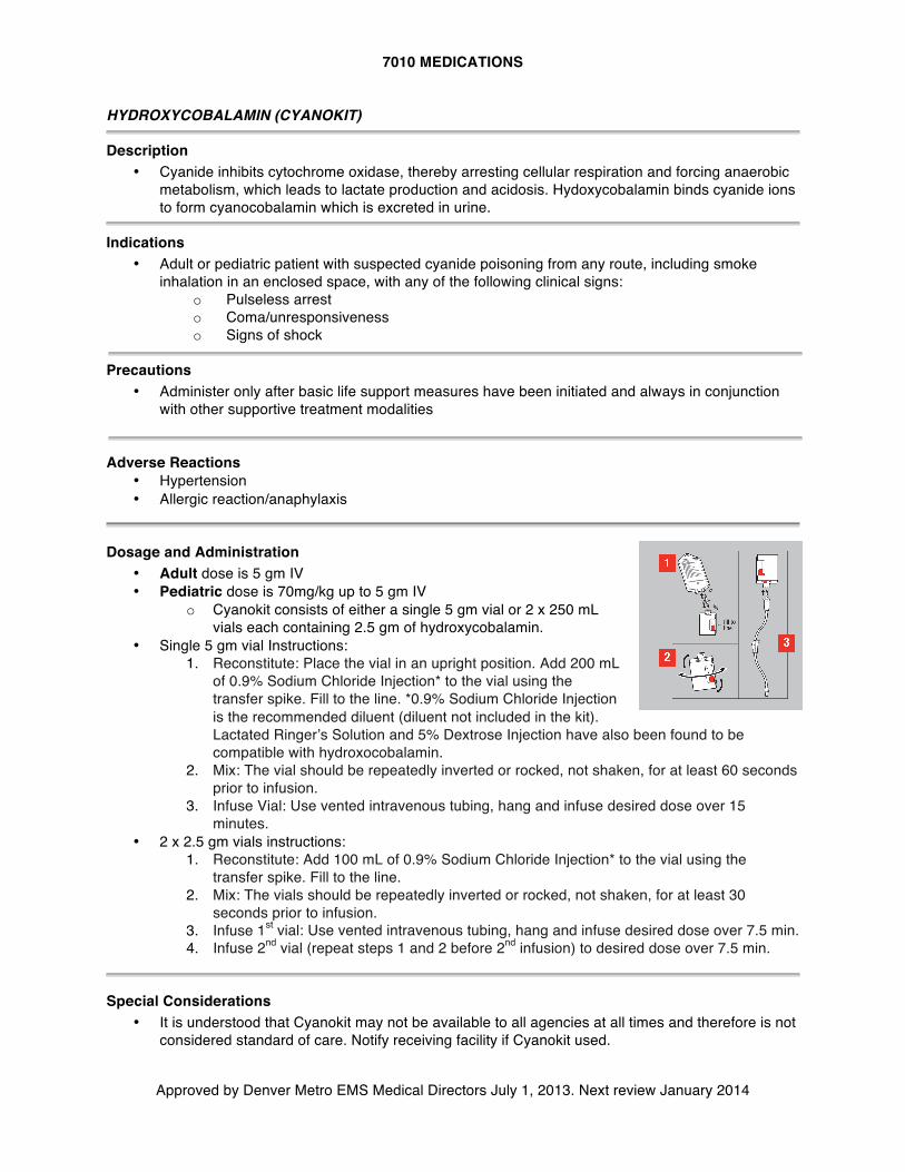
7010 MEDICATIONS
Approved by Denver Metro EMS Medical Directors July 1, 2013. Next review January 2014
HYDROXYCOBALAMIN (CYANOKIT) Description
• Cyanide inhibits cytochrome oxidase, thereby arresting cellular respiration and forcing anaerobic metabolism, which leads to lactate production and acidosis. Hydoxycobalamin binds cyanide ions to form cyanocobalamin which is excreted in urine.
Indications
• Adult or pediatric patient with suspected cyanide poisoning from any route, including smoke inhalation in an enclosed space, with any of the following clinical signs:
o Pulseless arrest o Coma/unresponsiveness o Signs of shock
Precautions
• Administer only after basic life support measures have been initiated and always in conjunction with other supportive treatment modalities
Adverse Reactions
• Hypertension • Allergic reaction/anaphylaxis
Dosage and Administration
• Adult dose is 5 gm IV • Pediatric dose is 70mg/kg up to 5 gm IV
o Cyanokit consists of either a single 5 gm vial or 2 x 250 mL vials each containing 2.5 gm of hydroxycobalamin.
• Single 5 gm vial Instructions: 1. Reconstitute: Place the vial in an upright position. Add 200 mL
of 0.9% Sodium Chloride Injection* to the vial using the transfer spike. Fill to the line. *0.9% Sodium Chloride Injection is the recommended diluent (diluent not included in the kit). Lactated Ringer’s Solution and 5% Dextrose Injection have also been found to be compatible with hydroxocobalamin.
2. Mix: The vial should be repeatedly inverted or rocked, not shaken, for at least 60 seconds prior to infusion.
3. Infuse Vial: Use vented intravenous tubing, hang and infuse desired dose over 15 minutes.
• 2 x 2.5 gm vials instructions: 1. Reconstitute: Add 100 mL of 0.9% Sodium Chloride Injection* to the vial using the
transfer spike. Fill to the line. 2. Mix: The vials should be repeatedly inverted or rocked, not shaken, for at least 30
seconds prior to infusion. 3. Infuse 1st vial: Use vented intravenous tubing, hang and infuse desired dose over 7.5 min. 4. Infuse 2nd vial (repeat steps 1 and 2 before 2nd infusion) to desired dose over 7.5 min.
Special Considerations
• It is understood that Cyanokit may not be available to all agencies at all times and therefore is not considered standard of care. Notify receiving facility if Cyanokit used.
7010 MEDICATIONS
Approved by Denver Metro EMS Medical Directors July 1, 2013. Next review January 2014
IPRATROPIUM BROMIDE (ATROVENT) Description
Ipratropium is a anticholinergic antimuscarinic bronchodilator chemically related to atropine.
Onset & Duration
• Onset: 5-15 min. after inhalation • Duration: 6-8 hr. after inhalation
Indications
• Bronchospasm Contraindications
• Do not administer to children < 2 years • Soy or peanut allergy is a contraindication to use of Atrovent metered dose inhaler, not the
nebulized solution, which does not have the allergen contained in propellant Adverse Reactions
• Palpitations • Tremors • Dry mouth
How Supplied
Premixed Container: 0.5 mg in 2.5ml NS Dosage and Administration Adult Bronchospasm:
Ipratropium (0.5 mg/2.5 ml) along with albuterol in a nebulizer
Child (2yrs – 12yrs) Mod and Severe Bronchospasm
Ipratropium (0.5 mg/2.5 ml) along with albuterol in a nebulizer Not indicated for repetitive dose or continuous neb use
Protocol
• Asthma • COPD • Pediatric Respiratory Distress
7010 MEDICATIONS
Approved by Denver Metro EMS Medical Directors July 1, 2013. Next review January 2014
LIDOCAINE 2% SOLUTION Description
Local anesthetic for relief of pain during intraosseous fluid administration. Indications
• Analgesic for intraosseous infusion Side Effects
• Seizures • Drowsiness • Tachycardia • Bradycardia • Confusion • Hypotension
Precautions
• Lidocaine is metabolized in the liver and therefore, elderly patients and those with liver disease or poor liver perfusion secondary to shock or congestive heart failure are more likely to experience side effects
Dosage and Administration
0.5 mg/kg IO bolus, slowly, maximum dose is 50 mg Protocol
Intraosseous Administration Special Notes
• Seizure from lidocaine toxicity likely to be brief and self-limited. If prolonged, or status epilepticus, treat per seizure protocol
• Treat dysrhythmias according to specific protocol
7010 MEDICATIONS
Approved by Denver Metro EMS Medical Directors July 1, 2013. Next review January 2014
MAGNESIUM SULFATE Description
Magnesium sulfate reduces striated muscle contractions and blocks peripheral neuromuscular transmission by reducing acetylcholine release at the myoneural junction. In cardiac patients, it stabilizes the potassium pump, correcting repolarization. It also shortens the Q-T interval in the presence of ventricular arrhythmias due to drug toxicity or electrolyte imbalance. In respiratory patients, it may act as a bronchodilator in acute bronchospasm due to asthma or other bronchospastic diseases. In patients suffering from eclampsia, it controls seizures by blocking neuromuscular transmission and lowers blood pressure as well as decreases cerebral vasospasm.
Indications
Antiarrhythmic • Torsade de pointes associated with prolonged QT interval Respiratory • Severe bronchospasm unresponsive to continuous albuterol, ipratropium, and IM epinephrine. Obstetrics • Eclampsia: Pregnancy > 20 weeks gestational age or post partum with seizures
Precautions
• Bradycardia • Hypotension • Respiratory depression
Adverse Reactions
• Bradycardia • Hypotension • Respiratory depression
Dosage and Administration
• Torsades de Pointes suspected caused by prolonged QT interval: o 2 gm, IV bolus.
• Refractory Severe Bronchospasm: o 2 gm, IV bolus, over 2 minutes CONTACT BASE for order.
• Eclampsia: o 2 gm, IV bolus slowly o Mix 4 gm, diluted in 50 ml of Normal Saline (0.9 NS), IV drip over 15-30 minutes.
Protocol
• Adult Universal Pulseless Arrest Algorithm • COPD • Asthma • Obstetric Complications
7010 MEDICATIONS
Approved by Denver Metro EMS Medical Directors July 1, 2013. Next review January 2014
METHYLPREDNISOLONE (SOLU-MEDROL) Description
Methylprednisolone is a synthetic steroid that suppresses acute and chronic inflammation and may alter the immune response. In addition, it potentiates vascular smooth muscle relaxation by beta-adrenergic agonists and may alter airway hyperactivity.
Indications
• Anaphylaxis • Severe asthma • COPD • Suspected Addisonian crisis (cardiovascular collapse in patient at risk for adrenal insufficiency)
Contraindications
• Evidence of active GI bleed
Adverse Reactions
Most adverse reactions are a result of long-term therapy and include: • Gastrointestinal bleeding • Hypertension • Hyperglycemia
Dosage and Administration
Adult: 125 mg, IV/IO bolus, slowly, over 2 minutes
Pediatric: 2 mg/kg, IV/IO bolus, slowly, over 2 minutes to max dose of 125 mg
Protocol
• Asthma • Allergy and Anaphylaxis • Chronic Obstructive Pulmonary Disease • Adult hypotension/shock • Adrenal Insufficiency
Special Considerations
• Must be reconstituted and used immediately • The effect of methylprednisolone is generally delayed for several hours. • Methylprednisolone is not considered a first line drug. Be sure to attend to the patient’s primary
treatment priorities (i.e. airway, ventilation, beta-agonist nebulization) first. If primary treatment priorities have been completed and there is time while in route to the hospital, then methylprednisolone can be administered. Do not delay transport to administer this drug
7010 MEDICATIONS
Approved by Denver Metro EMS Medical Directors July 1, 2013. Next review January 2014
NALOXONE (NARCAN) Description
Naloxone is a competitive opioid receptor antagonist
Onset & Duration
Onset: Within 5 minutes Duration: 1-4 hours
Indications
• For reversal of suspected opioid-inducted CNS and respiratory depression • Coma of unknown origin with impaired airway reflexes or respiratory depression
Adverse Reactions
• Tachycardia • Nausea and vomiting • Pulmonary Edema
Dosage and Administration
Adult: 0.5 mg IV/IO/IM/IN and titrate to desired effect, up to 2 mg total In cases of severe respiratory compromise or arrest, 2 mg bolus IV/IO/IM is appropriate, otherwise drug should be titrated
Pediatrics:
0.5 mg IV/IO/IM/IN and titrate to desired effect, up to 2 mg total Protocol
• Universal Altered Mental Status Protocol • Poisoning/Overdose
Special Considerations
• Not intended for use unless respiratory depression or impaired airway reflexes are present. Reversal of suspected mild-moderate opioid toxicity is not indicated in the field as it may greatly complicate treatment and transport as narcotic-dependent patients may experience violent withdrawal symptoms
• Patients receiving naloxone must be transported to a hospital
7010 MEDICATIONS
Approved by Denver Metro EMS Medical Directors July 1, 2013. Next review January 2014
NITROGLYCERINE (NITROSTAT, NITROQUICK, etc) Description
Short-acting peripheral venodilator decreasing cardiac preload and afterload Onset & Duration
Onset: 1-3 min. Duration: 20-30 min.
Indications
• Pain or discomfort due to suspected Acute Coronary Syndrome • Pulmonary edema due to congestive heart failure
Contraindications
• Suspected right ventricular ST-segment elevation MI (Inferior STEMI pattern plus ST elevation in right sided-precordial leads)
• Hypotension SBP < 100 • Recent use of erectile dysfunction (ED) medication (e.g. Viagra, Cialis)
Adverse Reactions
• Hypotension • Headache • Syncope
Dosage and Administration
0.4 mg (1/150 gr) sublingually or spray, every 5 minutes PRN up to a total of 3 doses for persistent CP. Nitropaste: system specific protocol
Protocol
• Adult Chest Pain • CHF/Pulmonary Edema
7010 MEDICATIONS
Approved by Denver Metro EMS Medical Directors July 1, 2013. Next review January 2014
OPIOIDS (FENTANYL, MORPHINE, HYDROMORPHONE) Description
Opioid analgesics with desired effects of analgesia, euphoria and sedation as well as undesired effects of respiratory depression and hypotension. A synthetic opioid, fentanyl is 100 times more potent than morphine, and is less likely to cause histamine release.
Indications
• Treatment of hemodynamically stable patients with moderate to severe pain due to traumatic or medical conditions, including cardiac conditions, abdominal pain, back pain, etc.
• Treatment of shivering after therapeutic induced hypothermia (TIH). Contraindications
• Hypotension, hemodynamic instability or shock • Respiratory depression
Caution/Comments:
• Opioids should only be given to hemodynamically stable patients and titrated slowly to effect. • The objective of pain management is not the removal of all pain, but rather, to make the patient’s
pain tolerable enough to allow for adequate assessment, treatment and transport • Respiratory depression, including apnea, may occur suddenly and without warning, and is more
common in children and the elderly. Start with ½ traditional dose in the elderly. • Coadministration of opioids and benzodiazepines is discouraged and may only be done with
direct physician verbal order. • Chest wall rigidity has been reported with rapid administration of fentanyl
Dosage and Administration
FENTANYL: • Adult doses may be rounded to nearest 25 mcg increment • Initial dose in adults typically 100 mcg • Strongly consider ½ typical dosing in elderly or frail patient
Adult: IV/IO route: 1 mcg/kg.
• Dose may be repeated after 10 minutes and titrated to clinical effect to a maximum cumulative dose of 3 mcg/kg
• Additional dosing requires BASE CONTACT
IN route: 1 mcg/kg. • Dose may be repeated after 10 minutes after initial IN dose to a maximum cumulative
dose of 3 mcg/kg. IV route is preferred for repeat dosing. • Additional dosing requires BASE CONTACT
Pediatric (1-12 years):
IV/IO route: 1 mcg/kg. • Dose may be repeated after 10 minutes and titrated to clinical effect to a maximum
cumulative dose of 3 mcg/kg.
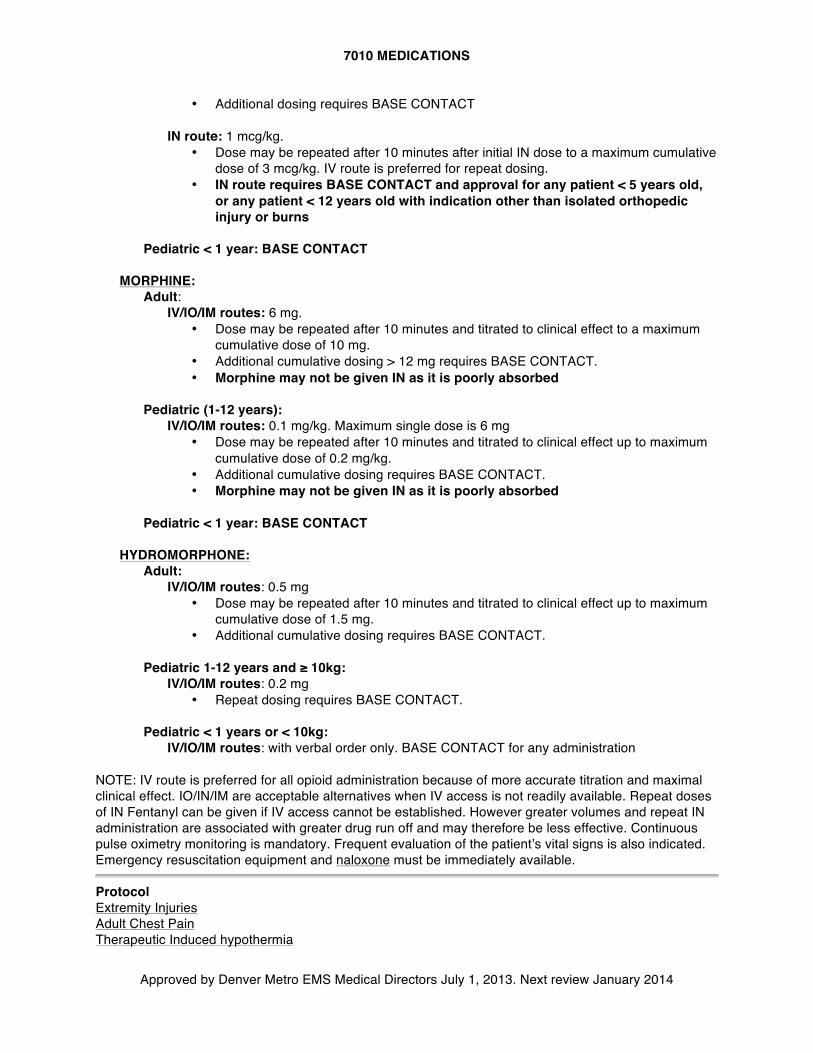
7010 MEDICATIONS
Approved by Denver Metro EMS Medical Directors July 1, 2013. Next review January 2014
• Additional dosing requires BASE CONTACT
IN route: 1 mcg/kg. • Dose may be repeated after 10 minutes after initial IN dose to a maximum cumulative
dose of 3 mcg/kg. IV route is preferred for repeat dosing. • IN route requires BASE CONTACT and approval for any patient < 5 years old,
or any patient < 12 years old with indication other than isolated orthopedic injury or burns
Pediatric < 1 year: BASE CONTACT
MORPHINE:
Adult: IV/IO/IM routes: 6 mg.
• Dose may be repeated after 10 minutes and titrated to clinical effect to a maximum cumulative dose of 10 mg.
• Additional cumulative dosing > 12 mg requires BASE CONTACT. • Morphine may not be given IN as it is poorly absorbed
Pediatric (1-12 years):
IV/IO/IM routes: 0.1 mg/kg. Maximum single dose is 6 mg • Dose may be repeated after 10 minutes and titrated to clinical effect up to maximum
cumulative dose of 0.2 mg/kg. • Additional cumulative dosing requires BASE CONTACT. • Morphine may not be given IN as it is poorly absorbed
Pediatric < 1 year: BASE CONTACT
HYDROMORPHONE:
Adult: IV/IO/IM routes: 0.5 mg
• Dose may be repeated after 10 minutes and titrated to clinical effect up to maximum cumulative dose of 1.5 mg.
• Additional cumulative dosing requires BASE CONTACT.
Pediatric 1-12 years and ≥ 10kg: IV/IO/IM routes: 0.2 mg
• Repeat dosing requires BASE CONTACT.
Pediatric < 1 years or < 10kg: IV/IO/IM routes: with verbal order only. BASE CONTACT for any administration
NOTE: IV route is preferred for all opioid administration because of more accurate titration and maximal clinical effect. IO/IN/IM are acceptable alternatives when IV access is not readily available. Repeat doses of IN Fentanyl can be given if IV access cannot be established. However greater volumes and repeat IN administration are associated with greater drug run off and may therefore be less effective. Continuous pulse oximetry monitoring is mandatory. Frequent evaluation of the patient’s vital signs is also indicated. Emergency resuscitation equipment and naloxone must be immediately available. Protocol Extremity Injuries Adult Chest Pain Therapeutic Induced hypothermia
7010 MEDICATIONS
Approved by Denver Metro EMS Medical Directors July 1, 2013. Next review January 2014
Abdominal Pain Amputations Burns Bites/Stings Snake Bites Face and Neck Trauma Chest Trauma Abdominal Trauma Spinal Trauma
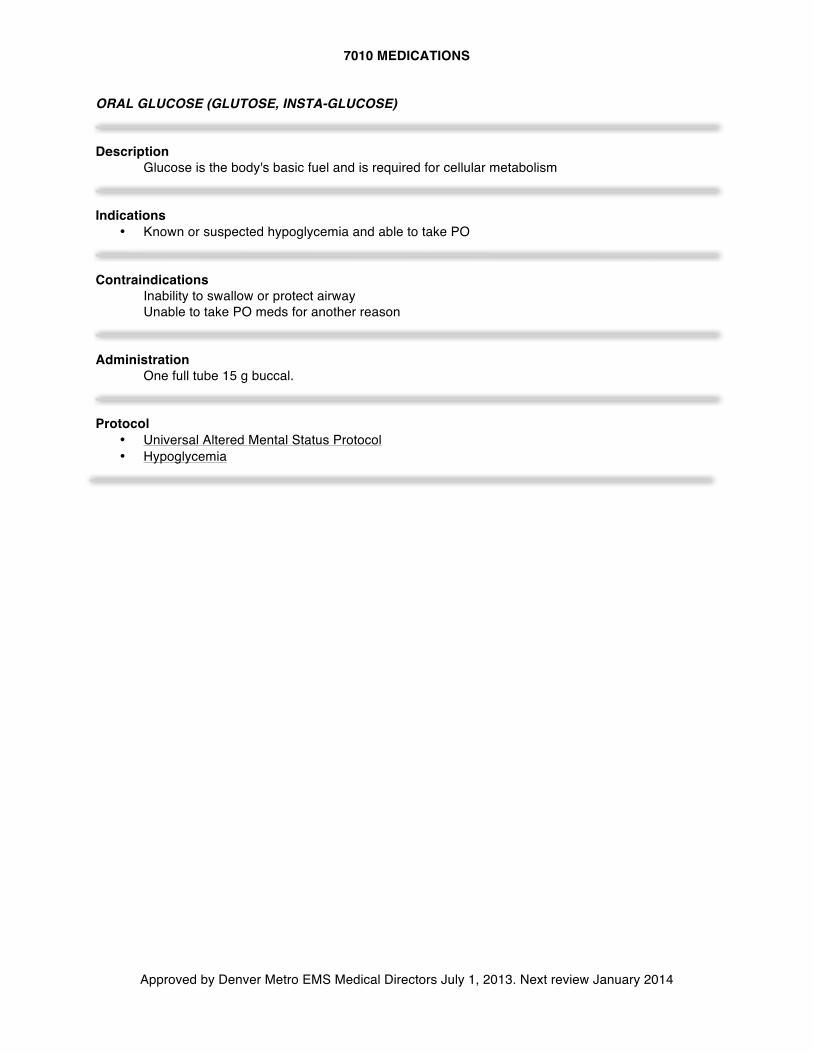
7010 MEDICATIONS
Approved by Denver Metro EMS Medical Directors July 1, 2013. Next review January 2014
ORAL GLUCOSE (GLUTOSE, INSTA-GLUCOSE) Description
Glucose is the body's basic fuel and is required for cellular metabolism Indications
• Known or suspected hypoglycemia and able to take PO Contraindications
Inability to swallow or protect airway Unable to take PO meds for another reason
Administration
One full tube 15 g buccal.
Protocol
• Universal Altered Mental Status Protocol • Hypoglycemia
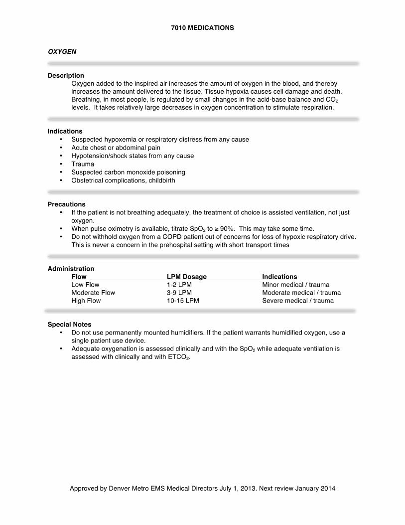
7010 MEDICATIONS
Approved by Denver Metro EMS Medical Directors July 1, 2013. Next review January 2014
OXYGEN Description
Oxygen added to the inspired air increases the amount of oxygen in the blood, and thereby increases the amount delivered to the tissue. Tissue hypoxia causes cell damage and death. Breathing, in most people, is regulated by small changes in the acid-base balance and CO2 levels. It takes relatively large decreases in oxygen concentration to stimulate respiration.
Indications
• Suspected hypoxemia or respiratory distress from any cause • Acute chest or abdominal pain • Hypotension/shock states from any cause • Trauma • Suspected carbon monoxide poisoning • Obstetrical complications, childbirth
Precautions
• If the patient is not breathing adequately, the treatment of choice is assisted ventilation, not just oxygen.
• When pulse oximetry is available, titrate SpO2 to ≥ 90%. This may take some time. • Do not withhold oxygen from a COPD patient out of concerns for loss of hypoxic respiratory drive.
This is never a concern in the prehospital setting with short transport times Administration
Flow LPM Dosage Indications Low Flow 1-2 LPM Minor medical / trauma Moderate Flow 3-9 LPM Moderate medical / trauma High Flow 10-15 LPM Severe medical / trauma
Special Notes
• Do not use permanently mounted humidifiers. If the patient warrants humidified oxygen, use a single patient use device.
• Adequate oxygenation is assessed clinically and with the SpO2 while adequate ventilation is assessed with clinically and with ETCO2.
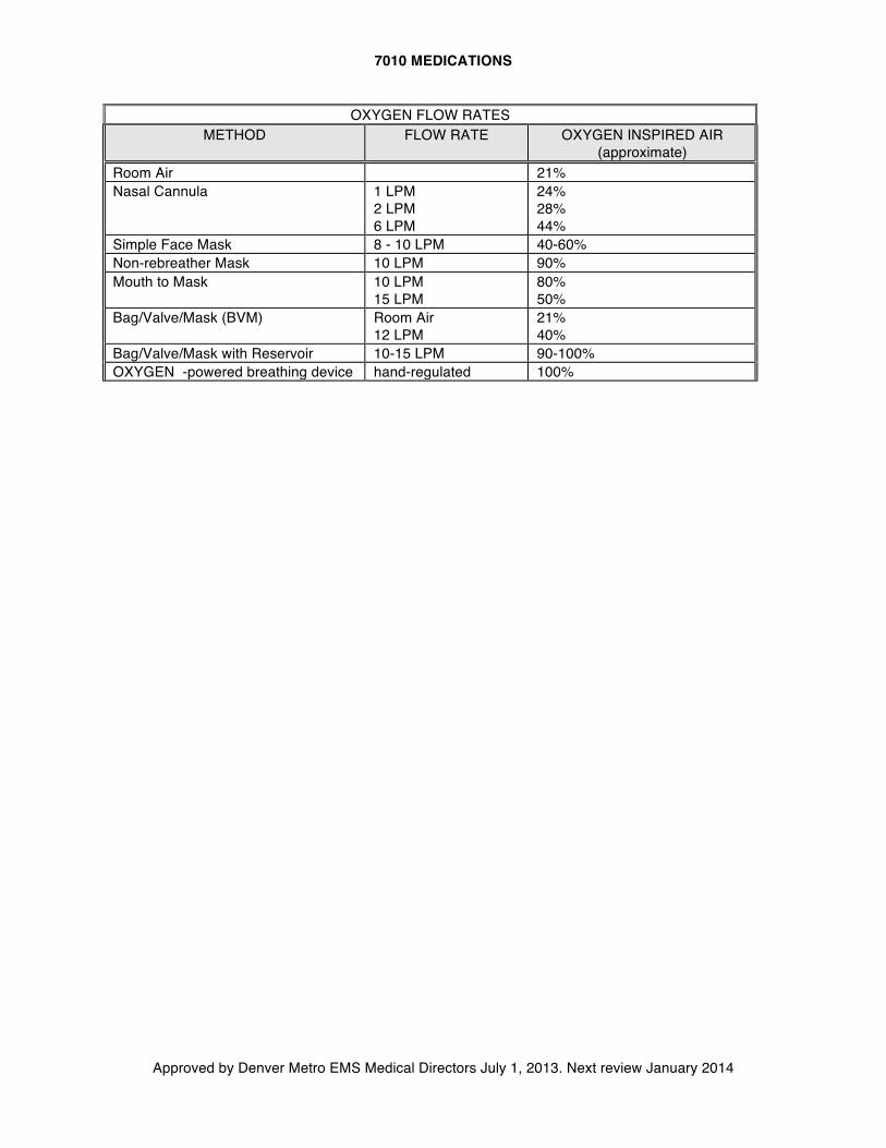
7010 MEDICATIONS
Approved by Denver Metro EMS Medical Directors July 1, 2013. Next review January 2014
OXYGEN FLOW RATES METHOD FLOW RATE OXYGEN INSPIRED AIR
(approximate) Room Air 21% Nasal Cannula
1 LPM 2 LPM 6 LPM
24% 28% 44%
Simple Face Mask 8 - 10 LPM 40-60% Non-rebreather Mask 10 LPM 90% Mouth to Mask 10 LPM
15 LPM 80% 50%
Bag/Valve/Mask (BVM) Room Air 12 LPM
21% 40%
Bag/Valve/Mask with Reservoir 10-15 LPM 90-100% OXYGEN -powered breathing device hand-regulated 100%
7010 MEDICATIONS
Approved by Denver Metro EMS Medical Directors July 1, 2013. Next review January 2014
PHENYLEPHRINE (INTRANASAL) Description
Used for topical nasal administration, phenylephrine primarily exhibits alpha adrenergic stimulation. This stimulation can produce moderate to marked vasoconstriction and subsequent nasal decongestion.
Indications
• Prior to nasotracheal intubation to induce vasoconstriction of the nasal mucosa • Nose bleed
Precautions
• Avoid administration into the eyes, which will dilate pupil Dosage and Administration
• Instill two drops of 1% solution in the nostril prior to attempting nasotracheal intubation • Administer 2 sprays in affected naris in patient with active nosebleed after having patient blow
nose to expel clots. Protocol
• Nasotracheal intubation • Epistaxis
7010 MEDICATIONS
Approved by Denver Metro EMS Medical Directors July 1, 2013. Next review January 2014
RACEMIC EPINEPHRINE Description
Racemic epinephrine 2.25% is an aqueous solution that delivers 11.25 mg of racemic epinephrine per 0.5mL for use by oral inhalation only. Inhalation causes local effects on the upper airway as well as systemic effects from absorption. Vasoconstriction may reduce swelling in the upper airway, and ß effects on bronchial smooth muscle may relieve bronchospasm.
Onset & Duration
• Onset: 1-5 minutes • Duration: 1-3 hours
Indications
• Bronchospasm in bronchiolitis • Stridor at rest in croup • Suspected epiglottitis in adults or children
Side Effects
• Tachycardia • Palpitations • Muscle tremors
Dosage and Administration
0.5 ml racemic epinephrine (acceptable dose for all ages) mixed in 2 ml saline, via nebulizer at 6-8 LPM to create a fine mist and administer over 15 minutes.
Protocol
• Pediatric Respiratory Distress Special Considerations
• Racemic epi is heat and photo-sensitive • Once removed from the refrigerator, the unopened package is stable at room temperature until
the expiration date stated on the package. • Do not confuse the side effects with respiratory failure or imminent respiratory arrest.
7010 MEDICATIONS
Approved by Denver Metro EMS Medical Directors July 1, 2013. Next review January 2014
SODIUM BICARBONATE Description
Sodium bicarbonate is an alkalotic solution, which neutralizes acids found in the body. Acids are increased when body tissues become hypoxic due to cardiac or respiratory arrest.
Indications
• Tricyclic overdose with arrhythmias, widened QRS complex, hypotension, seizures • Suspected hyperkalemic pulseless arrest: consider in patients with renal failure
Contraindications
• Metabolic and respiratory alkalosis • Hypocalcemia • Hypokalemia
Adverse Reactions
• Metabolic alkalosis • Hyperosmolarity may occur, causing cerebral impairment
Drug Interactions
• May precipitate in calcium solutions. • Alkalization of urine may increase half-lives of certain drugs. • Vasopressors may be deactivated.
Dosage and Administration
Adults and children (>10 kg), 8.4% Tricyclic OD with hypotension or prolonged QRS > 0.10 sec or suspected hyperkalemia-related pulseless arrest:
1.0 mEq/kg slow IV push Repeat if needed in 10 minutes.
Protocol
• Adult Universal Pulseless Arrest Algorithm • Poisoning/Overdose
Special Considerations
• Sodium bicarbonate administration increases CO2 which rapidly enters cells, causing a paradoxical intracellular acidosis.
• Sodium bicarb is no longer recommended for routine use in prolonged cardiac arrest. Its use in pulseless arrest should be limited to known or suspected hyperkalemia (e.g. dialysis patient).
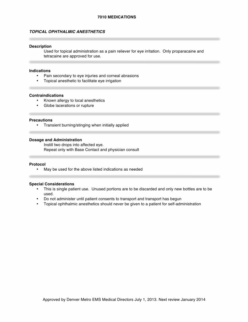
7010 MEDICATIONS
Approved by Denver Metro EMS Medical Directors July 1, 2013. Next review January 2014
TOPICAL OPHTHALMIC ANESTHETICS Description
Used for topical administration as a pain reliever for eye irritation. Only proparacaine and tetracaine are approved for use.
Indications
• Pain secondary to eye injuries and corneal abrasions • Topical anesthetic to facilitate eye irrigation
Contraindications
• Known allergy to local anesthetics • Globe lacerations or rupture
Precautions
• Transient burning/stinging when initially applied
Dosage and Administration
Instill two drops into affected eye. Repeat only with Base Contact and physician consult
Protocol
• May be used for the above listed indications as needed Special Considerations
• This is single patient use. Unused portions are to be discarded and only new bottles are to be used.
• Do not administer until patient consents to transport and transport has begun • Topical ophthalmic anesthetics should never be given to a patient for self-administration