Embed Size (px)
Citation preview

Draft
Kinetics of Pyrite, Pyrrhotite and Chalcopyrite Dissolution
by Acidithiobacillus ferrooxidans
Journal: Canadian Journal of Microbiology
Manuscript ID cjm-2016-0085.R1
Manuscript Type: Article
Date Submitted by the Author: 19-Mar-2016
Complete List of Authors: Kocaman, Ayse Tuba; Yildiz Technical University , Bioengineering Department Cemek, Mustafa; Yildiz Technical University , Bioengineering Department Edwards, Katrina Jane; University of Southern California, Biological Sciences Department
Keyword: A. ferrooxidans, pyrite, pyrrhotite, chalcopyrite, dissolution kinetics
https://mc06.manuscriptcentral.com/cjm-pubs
Canadian Journal of Microbiology

Draft
1
Kinetics of Pyrite, Pyrrhotite and Chalcopyrite Dissolution by 1
Acidithiobacillus ferrooxidans 2
3
4
Ayse Tuba Kocaman a, b, *
, Mustafa Cemek b, Katrina Jane Edwards
a, 1 5
6
7
a Department of Biological Sciences, University of Southern California, Los Angeles, CA 8
90089, USA 9
b Faculty of Chemical and Metallurgical Engineering, Department of Bioengineering, Yildiz 10
Technical University, 34210 Esenler, Istanbul, TURKEY 11
1 Deceased 12
13
14
*Corresponding author 15
Ayse Tuba Kocaman
16
+90(505) 697 7135 18
19
Mustafa Cemek 20
22
23
24
25
Page 1 of 39
https://mc06.manuscriptcentral.com/cjm-pubs
Canadian Journal of Microbiology
Draft
2
Kinetics of Pyrite, Pyrrhotite and Chalcopyrite Dissolution by 26
Acidithiobacillus ferrooxidans 27
28
Abstract 29
The main objective of this study was to investigate the dissolution kinetics of pyrite, 30
pyrrhotite, and chalcopyrite. Crushed minerals were reacted with A. ferrooxidans (25°C). The 31
kinetics of dissolution was investigated by monitoring pH and Fe2+
, Fe3+
ion concentrations in 32
the leaching solutions. Pyrite, pyrrhotite and chalcopyrite dissolution by A. ferrooxidans were 33
found to be chemically controlled processes. The dissolution rates of the minerals with 34
bacteria increased in the order of pyrrhotite, pyrite, and chalcopyrite. The attached cell 35
numbers to mineral surfaces increased in the same order. A. ferrooxidans was found to 36
enhance the dissolution rates of the minerals. The acid-insoluble trait of pyrite and acid-37
soluble trait of the other two minerals affected the pH changes in the leaching solutions. 38
39
Keywords: A. ferrooxidans, pyrite, pyrrhotite, chalcopyrite, dissolution kinetics 40
41
42
43
44
45
46
47
48
49
50
Page 2 of 39
https://mc06.manuscriptcentral.com/cjm-pubs
Canadian Journal of Microbiology
Draft
3
1. Introduction 51
Over the last few decades, the mining industry has had the growing interest in 52
bioleaching of sulphide minerals as a means of metal recovery. “Bioleaching” is described as 53
biological oxidation of metal sulphides (Rawlings and Silver 1995), and the mobilization of 54
metal cations involving biological oxidation from otherwise insoluble ores (Vera et al. 2013). 55
Bioleaching is a vital alternative to traditional extraction methods because of its low cost, 56
environmental benefits, and broad efficiency for low-grade and recalcitrant ores (Rawlings 57
and Silver 1995). Pyrite (FeS2) and pyrrhotite (Fe1-xS) are the examples of sulphide minerals 58
composed entirely of Fe and S, whereas other sulphide minerals such as chalcopyrite 59
(CuFeS2) contain economically valuable metals as well (Bulaev et al. 2012). Although pyrite 60
is not an important economic mineral by itself, many studies on biologically mediated pyrite 61
oxidation have been conducted (Bellenberg et al. 2015; Konhauser et al. 2011; Edwards et al. 62
2003; Singer and Stumm 1970) because it is commonly associated with valuable minerals 63
such as sphalerite, chalcopyrite, and galena (Chandra and Gerson 2010). Pyrrhotite (Fe1-xS) is 64
the second common iron sulphide in nature. Although extracting copper by bioleaching is 65
difficult due to the refractoriness of chalcopyrite, it has been studied intensively because of its 66
high crustal abundance (Brierly and Brierly 2013). 67
Iron-oxidizing bacteria obtain metabolic energy through the oxidation of iron- and 68
sulfur-bearing minerals. In doing so, they dissolve economically valuable metals (Sand et al. 69
1995; Ehrlich and Fox 1967). Acidithiobacillus ferrooxidans (Acidithiobacillus was formerly 70
known as “Thiobacillus”, Kelly and Wood 2000) was the first to be isolated and is the best-71
studied iron and/or sulfur-oxidizing bacterium (Vera et al. 2013). 72
Most studies on bioleaching of sulphide minerals have focused mainly on the effect of 73
mixed bacterial cultures (Romo et al. 2013), mineral morphology (Dong et al. 2013), bacterial 74
attachment (Yang et al. 2014), mineral particle size (Olubambi et al. 2009), pulp density 75
Page 3 of 39
https://mc06.manuscriptcentral.com/cjm-pubs
Canadian Journal of Microbiology
Draft
4
(Pradhan et al. 2010), the presence of various metallic ions in solution (Wong et al. 2015), 76
and temperature on the dissolution kinetics of sulphide minerals (Vilcaez et al. 2008). A 77
significant number of individual studies focused on the bioleaching of pyrite (Edwards et al. 78
1998; Florian et al. 2011), pyrrhotite (Korehi et al. 2014) and chalcopyrite (Zhu et al. 2015).79
To date, more studies on chemical leaching kinetics of sulphide minerals were carried 80
out (Perez and Dutrizac 1991; Dreisinger and Abed 2002; Aydogan et al. 2006; Adebayo et 81
al. 2003; Havlik and Kammel 1995) compared to bioleaching kinetics (Pradhan et al. 2010; 82
Abhilash et al. 2013). On the other hand, very few studies compared the bioleaching rates of 83
pyrite, pyrrhotite, and chalcopyrite (Lei et al. 2007). The objective of this study was to 84
investigate the dissolution kinetic mechanisms of pyrite, pyrrhotite, chalcopyrite, and 85
compare the bioleaching rates in the presence of A. ferrooxidans. 86
2. Materials and Methods 87
2.1. Bacterial Cultivation 88
A. ferrooxidans strain ATCC 23270 was purchased from the American Type Culture 89
Collection (ATCC). Cells were cultured in a medium containing solution A, and solution B. 90
Solution A contained (in g/L): (NH4)2SO4 0.8; MgSO4.7H2O 2.0; K2HPO4 0.4; and 0.5% 91
Wolfe’s mineral solution (V/V) (Wolin et al. 1963). Solution B contained 20 g FeSO4.7H2O 92
per liter of solution. The ratio in which solutions A and B were mixed for preparing the final 93
medium was 4:1. Solution A was prepared as described above, and the pH was adjusted to 2.3 94
with H2SO4, then sterilized by autoclaving at 121°C for 15 minutes. Solution B was prepared 95
separately and filtered through a 0.2 µm membrane filter (Acrodisc). Solution A and solution 96
B were then aseptically combined. Batch cultures of A. ferrooxidans were grown at 120 rpm 97
on a rotary shaker at 25°C for seven days before harvesting for bioleaching experiments. 98
2.2. Minerals 99
The following sulphide minerals were used in the bioleaching experiments: Pyrite 100
Page 4 of 39
https://mc06.manuscriptcentral.com/cjm-pubs
Canadian Journal of Microbiology

Draft
5
(FeS2; Ward’s, Scientific, Huanzala, Peru), pyrrhotite (Fe1-xS, x=0 to 0.2; Ward’s, Scientific, 101
Galax, Virginia, USA), and chalcopyrite (CuFeS2; Ward’s, Scientific, Durango, Mexico). All 102
minerals were crushed using a mortar and pestle and sieved to a grain size of ≤ 355µm. 103
Crushed minerals were cleaned by following three steps: First, they were repeatedly rinsed 104
with distilled water and sonicated in an ultrasonic cleaner for 1-2 minutes to remove fine 105
particles. Second, they were treated with 10% HCl for 2 hours to remove any preexisting 106
oxide layers, then rinsed with 100% ethanol (Koptec, 200 proof) and dried in a shallow layer 107
(Edwards et al. 2001). Finally, 2 grams of the cleaned, crushed minerals were added to each 108
of sterile 250-ml flasks and autoclaved for 8 min. 109
2.3. Experimental Procedures 110
Cells of the A. ferrooxidans grown in the ferrous sulfate medium for one week in 111
shake culture were harvested by centrifugation at 10,000 rpm for 20 minutes and then washed 112
once by centrifugation at 10,000 rpm for 15 min in filter-sterilized distilled water adjusted to 113
pH 1.5 with H2SO4. Cells were re-suspended in salts medium (solution A) at pH 2.3. Two mL 114
of cell suspension (initial cell number ≈ 6.5x10⁶ cells mL
1-) was inoculated into 250 mL 115
flasks containing 2 grams of sterile crushed minerals and 35 mL of salts medium (solution A) 116
(Edwards et al. 2001). Duplicate sets for each mineral were prepared. Bioleaching flasks (A. 117
ferrooxidans + medium + crushed minerals) and control flasks (medium + crushed minerals) 118
were incubated without shaking at 25°C for 30 days. 119
2.4. Analytical Techniques 120
The leaching solutions were analyzed for the concentration of bacteria unattached to 121
mineral particles, total Fe, Fe2+
, Cu2+
ions, and pH. For cell counting, 2µL of culture samples 122
were spread evenly on a 1% agarose-coated fluoroslide printed with 4 mm diameter circles 123
(Emerson and Moyer 2002). After air-drying, the samples were stained with DAPI (200 124
µg/mL) and fifty fields/circle were counted at 100X magnification using an epifluorescent 125
Page 5 of 39
https://mc06.manuscriptcentral.com/cjm-pubs
Canadian Journal of Microbiology

Draft
6
Axiostar Plus microscope. From each flask, a 1-mL sample was removed daily, filtered 126
through a 0.2 µm membrane, and pH values were determined using a pH-meter (Thermo 127
Scientific). For dissolved iron measurements, a 1-mL sample was collected from each flask, 128
filtered through a 0.2µm membrane and diluted ten times with 1 mM HCl when their 129
absorbance measurements with UV- spectrophotometer were higher than 1.0. 1-mL of 130
samples were also collected from the chalcopyrite flasks and filtered through a 0.2µm 131
membrane and diluted 100 times with distilled water to measure Cu2+
ion concentrations. All 132
ion concentrations were measured using the UV-spectrophotometer (Shimadzu UV-1601). 133
Any decrease in volume due to sampling was adjusted to the original volume by addition of 134
an equivalent volume of sterile solution A. 135
The original ferrozine method was modified to measure total Fe and Fe2+
ion 136
concentrations (Stookey 1970). Ferrozine reagent (2 mM Ferrozine in 50 mM Hepes) was 137
used to determine both total Fe and Fe2+
ion concentrations whereas a reducing reagent (6.25 138
mM hydroxylamine hydrochloride) was used to determine total Fe ion concentrations. The 139
absorbance values at 562 nm were used to calculate iron ion concentrations. Standards 140
containing different known concentrations of FeSO4 in 1mM HCl were prepared. Fe3+
141
concentrations were calculated as the difference between total Fe and Fe2+
. 142
To measure Cu2+
ion concentrations, 0.1 mM Bathocuprione disulfonic acid (Aldrich) 143
and reducing reagent (0.3 mM hydroxylamine hydrochloride) were used. The method was 144
modified from that of Moffett et al. (1985). Standards containing different known 145
concentrations of CuSO4 in distilled water were prepared. For each sample, a full spectrum 146
was run between 400-800 nm. A peak for Cu2+
concentrations can be detected at 484 nm. The 147
area of the peak was measured using the Shimadzu UV Probe Version 2.20 software. Cu2+
148
concentrations in the leaching solutions were calculated by using the peak area. 149
The XRD patterns and chemical compositions of original pyrite, pyrrhotite and 150
Page 6 of 39
https://mc06.manuscriptcentral.com/cjm-pubs
Canadian Journal of Microbiology

Draft
7
chalcopyrite, were characterized by X-ray diffraction (XRD, Philips Panalytical X’Pert-Pro, 151
CuKα radiation), and X-ray fluorescence spectrometry (XRF, Philips Panalytical Minipal 4), 152
respectively. 153
3. Results and Discussion 154
Leaching experiments in the presence and absence of A. ferrooxidans were carried out 155
to investigate the dissolution kinetic mechanisms of pyrite, pyrrhotite and chalcopyrite. In the 156
following, we record our findings on pH, Fe2+
, Fe3+
, Cu2+
concentration variations in the 157
leaching solutions, XRD and XRF analysis of untreated minerals. 158
The mineral structure is an important factor, which determines the mechanism and 159
chemistry of dissolution. Sand et al. (2001) reported that metal sulphides are degraded 160
through a chemical attack of Fe3+
ions and/or protons. Most metal sulphides, including pyrite, 161
pyrrhotite, and chalcopyrite, are semiconductors. In semiconductors, the valence band has the 162
highest energy level and is filled with electrons. In pyrite, the valance bands are derived only 163
from orbitals of metal atoms, whereas the valance bands in pyrrhotite and chalcopyrite are 164
derived from both metal and sulfur orbitals. Thus, the bonding between the metal and sulfur 165
atoms in the pyrite crystal lattice can be broken only by Fe3+
ions. In contrast, both Fe3+
ions 166
and protons can break metal-sulfur bonds in many other metal sulphides including pyrrhotite 167
and chalcopyrite. Consequently, pyrite is insoluble in acid while the others are acid-soluble 168
(Sand et al. 2001). Schippers and Sand (1999) proposed a thiosulfate mechanism for the 169
oxidation of acid-insoluble metal sulphides and a polysulfide mechanism for the oxidation of 170
acid-soluble metal sulphides. Thus for acid-insoluble pyrite, the reactions of the thiosulfate 171
mechanism are as follows (Eq. 1-2): 172
FeS2 + 6Fe3+
+ 3H2O → S2O32-
+ 7Fe2+
+ 6H+ (1) 173
S2O32-
+ 8Fe3+
+ 5H2O → 2SO42-
+ 8Fe2+
+ 10H+ (2) 174
For acid-soluble metal sulphides (MS) including pyrrhotite and chalcopyrite, the polysulfide 175
Page 7 of 39
https://mc06.manuscriptcentral.com/cjm-pubs
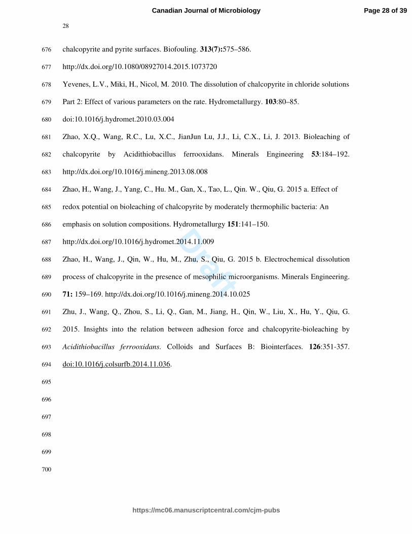
Canadian Journal of Microbiology
Draft
8
mechanism reaction is as follows (Eq. 3): 176
MS + Fe3+
+ 2H+ → M
2+ + H2S
*+ + Fe
2+ (3) 177
Fe3+
ions are the most reactive oxidants in the oxidation of sulphide minerals. The 178
dissolution reactions of pyrite, pyrrhotite and chalcopyrite with Fe3+
are shown in equations 4, 179
5 and 6 (Singer and Stumm 1970; Nicholson and Scharer 1994; Dutrizac and MacDonald 180
1974). 181
FeS2 + 14 Fe3+
+ 8H2O → 15Fe2+
+ 2SO42-
+ 16H+
(4) 182
Fe1-xS + (8-2x) Fe3+
+ 4 H2O → (9-3x) Fe2+
+ SO42-
+ 8H+ (5) 183
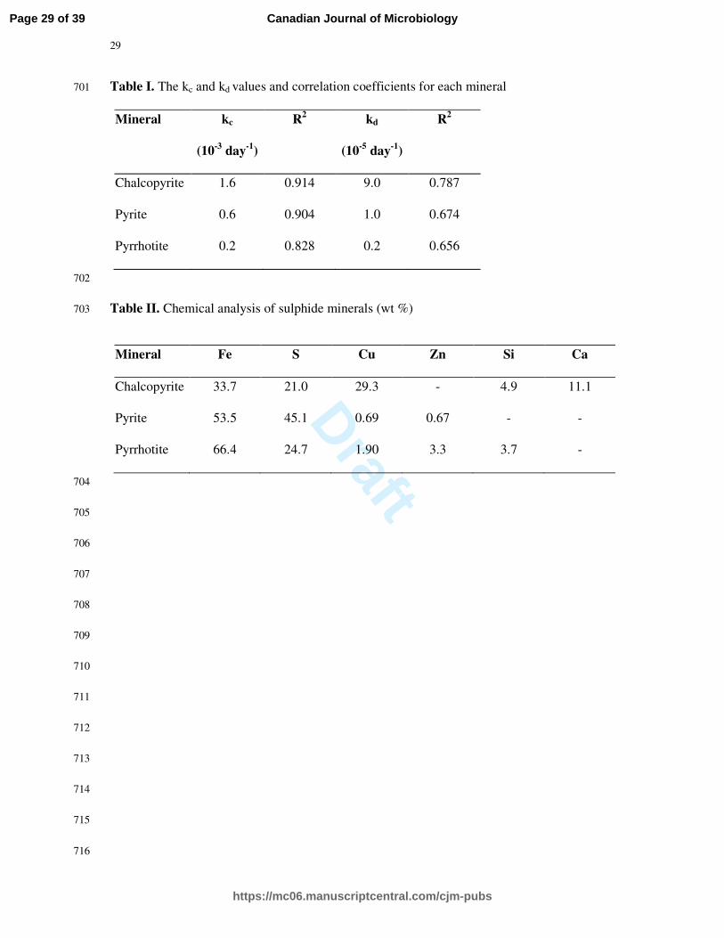
CuFeS2(s) + 16Fe3+
+ 8H2O → Cu2+
+ 17Fe2+
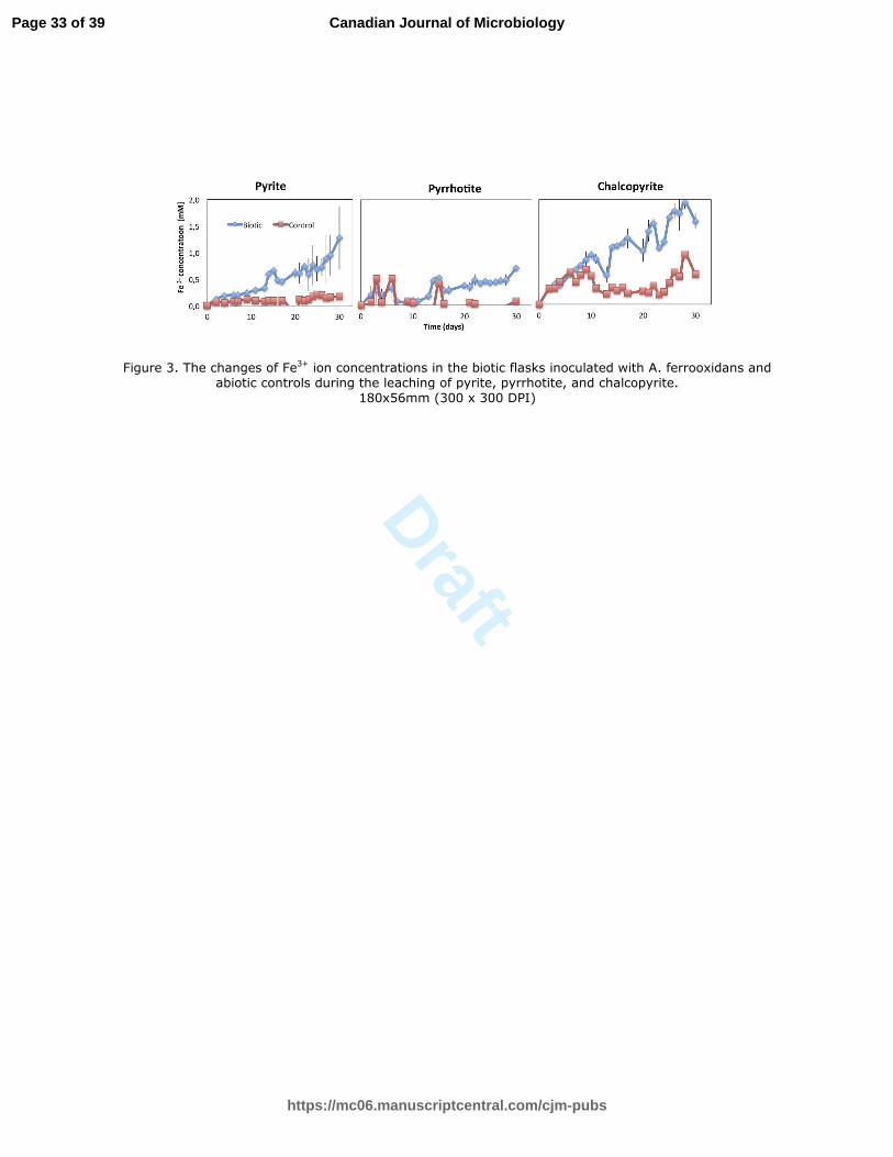
+ 2SO42-
+ 16H+ (6) 184
4Fe2+
+ 4 H+ + O2 → 4 Fe
3+ + 2H2O (Fe-oxidizers) (7) 185
S0 + 3/2 O2 (aq) + H2O → H2SO4 (S-oxidiziers) (8) 186
As the most abundant metal sulphides on Earth, pyrite, pyrrhotite and chalcopyrite are 187
well-studied sulphide minerals. However, the oxidation processes of these minerals are 188
complicated and not yet fully understood (Evangelou 1995; Chirita and Rimstidt 2014; 189
Ahmadi et al. 2011). Because the dissolution mechanisms of chalcopyrite in acidic solutions 190
are still under debate, a general theory could not be proposed. Many researchers have studied 191
the efficient factors on dissolution rates and the possible reaction mechanisms (Li et al. 2010). 192
Stott et al. (2003) studied the bioleaching of chalcopyrite by 11 species of acidophilic bacteria 193
and archaea and investigated the effects of microorganisms, temperature and redox potential 194
on the dissolution rates. The effects of acid and ferric iron concentrations, solid/liquid ratio, 195
and synergistic effect of cupric/ferrous ions on chalcopyrite dissolution behavior were also 196
investigated (Antonijevic and Bogdanovic 2003; Hiroyoshi et al. 2004). In another study, 197
neither an increase in ferric iron nor an increase of bacterial concentration could improve the 198
dissolution rates. A limited bacterial activity and a controlled ferric iron concentration were 199
suggested for the improved bioleaching rates (Third et al. 2000). Sasaki et al. (1995) studied 200
Page 8 of 39
https://mc06.manuscriptcentral.com/cjm-pubs
Canadian Journal of Microbiology
Draft
9
the chemical dissolution of pyrite with Fe3+
ions around pH 2. The results showed that pyrite 201
dissolved nonstoichiometrically in the initial stage, and later a sulfur-rich layer was formed on 202
the mineral surface. Yahya and Johnson (2002) examined the bioleaching of pyrite by two 203
strains of acidophilic bacteria. They discussed the advantages of bioleaching of sulfidic 204
minerals under low pH (<1) and low redox potential. Bhatti et al. (1993) studied the oxidation 205
of pyrrhotite by A. ferrooxidans. They observed an acid-consuming step with the formation of 206
elemental sulphur, following by an acid-producing step, which is a result of the formation of 207
solid products. Janzen at al. (2000) studied on the oxidation of 12 well-characterized 208
pyrrhotite samples by ferric iron and oxygen and reported the incomplete oxidation of 209
pyrrhotite with both oxidants. Harries et al. (2013) investigated the dissolution behavior of 210
pyrrhotite samples, which have different crystal structures, and found that the crystal structure 211
strongly influences the mineral dissolution. Plenty of researches were carried out to 212
investigate the effects of crystal structure, oxygen and ferric iron concentrations, temperature 213
and pH on the dissolution of pyrrhotite (Belzile et al. 2004). Although chalcopyrite is 214
refractory to hydrometallurgical methods, it is studied extensively due to its high crustal 215
abundance. The main factors affecting the dissolution rates of chalcopyrite are the 216
concentration of oxidants, the ferric/ferrous ion ratio in the leaching solutions, and the 217
formation of the passivation layer on the mineral surface (Klauber 2008; Li et al. 2010, 2013). 218
Olvera et al. (2014) studied the dissolution of chalcopyrite electrode in a solution composed 219
of H2SO4, FeSO4.7H2O and Fe2(SO4)3.5H2O at 25°C. According to the results, dissolution 220
of mineral increased with increasing Fe3+
concentrations and with the addition of FeS2 221
particles. Zhao et al (2015a) reported that additional Cu2+
and Fe2+
ions accelerated the 222
dissolution of chalcopyrite initially, but the formation of jarosite inhibited further dissolution. 223
In another study, Zhao et al. (2015b) studied the bioleaching of chalcopyrite with A. 224
ferrooxidans. The results showed that bacteria accelerate the copper extraction rate initially. 225
Page 9 of 39
https://mc06.manuscriptcentral.com/cjm-pubs
Canadian Journal of Microbiology
Draft
10
In the later stage of bioleaching, copper extraction rate decreased due to the formation of 226
jarosite and polysulfide. 227
Fe3+
/Fe2+
ratio is another important parameter affecting the dissolution of sulphide 228
minerals (Cordoba et al. 2008; Hiroyoshi et al. 2000, 2001, 2004, 2008; Kametani and aoki 229
1985; Sandstrom et al. 2004). Microorganisms maintain high ratios of ferric to ferrous ion in 230
bioleaching solutions, thus enhance electron transfer between oxidizing agents and the 231
minerals (Wu et al. 2014). To understand the role of bacteria, Khoshkhoo et al. (2014) 232
studied on the chemical dissolution of chalcopyrite by mimicking the redox conditions of 233
thermophilic bioleaching of the mineral. Surprisingly, copper recoveries were very similar in 234
the presence and the absence of bacteria. Therefore, they concluded that the role of bacteria 235
was only to provide the oxidizing agent for chalcopyrite dissolution, by oxidizing Fe2+
to 236
Fe3+
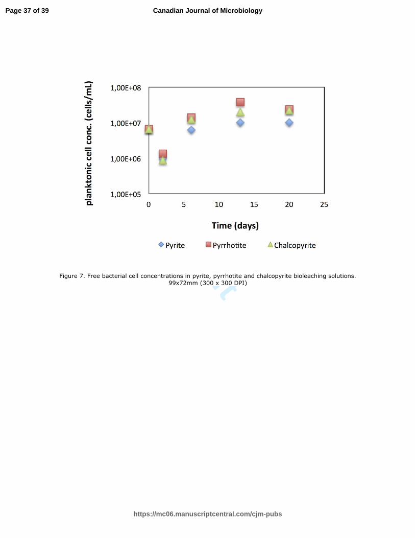
. Similarly, Third et al. (2000) carried out a series of leaching experiments with and 237
without bacteria and found that ferric/ferrous ratio is more relevant for determining 238
chalcopyrite dissolution rates than the concentration of bacteria in the leaching solution. 239
Petersen and Dixon (2006) studied on bioleaching of a copper-gold concentrate by using 240
mesophilic, moderately thermophilic and extremely thermophilic bacteria and concluded that 241
high temperature and low redox potential supported the dissolution of chalcopyrite. In 242
contrast, the opposite conditions were sufficient for obtaining high dissolution rates of pyrite. 243
Gericke et al. (2010) found that redox control is not necessary for achieving high copper 244
extraction rates at high temperatures around 70°C, but it increases the extraction rates around 245
45°C. 246
Attacking agents for dissolution of pyrite are only Fe3+
ions (Eq. 1-2), whereas 247
attacking agents for pyrrhotite and chalcopyrite are both Fe3+
and H+
(Eq. 3). In our study, the 248
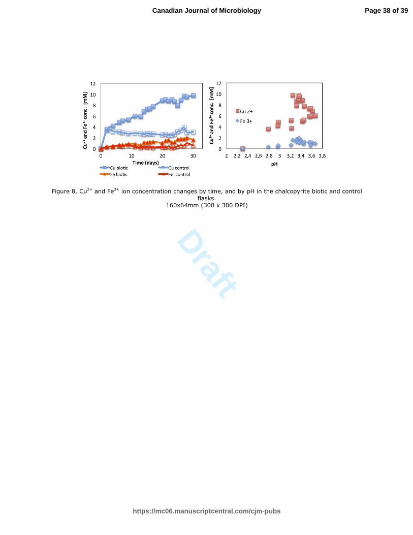
pH values continuously decreased throughout the experiments in pyrite leaching solutions due 249
to its acid-insoluble nature. The pH values in pyrrhotite and chalcopyrite leaching solutions 250
Page 10 of 39
https://mc06.manuscriptcentral.com/cjm-pubs
Canadian Journal of Microbiology

Draft
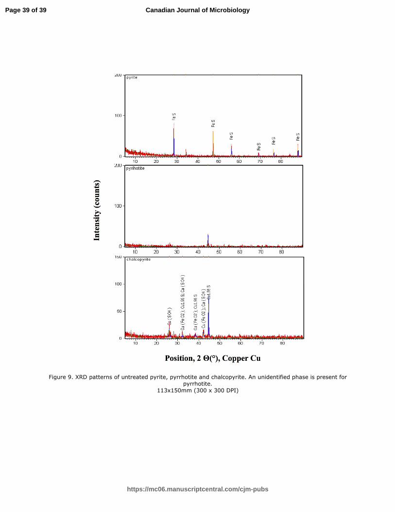
11
increased at the initial stage of bioleaching due to their acid-soluble nature and then 251
decreased. Lei et al. (2007) observed significant amounts of jarosite and elemental sulfur 252
during the bio-oxidation of pyrrhotite and chalcopyrite with A. ferrooxidans while no such 253
deposition was observed for pyrite. Similarly, in our study, no deposition was observed for 254
pyrite during the experiments. For pyrrhotite and chalcopyrite, some deposits were seen after 255
a week, corresponding to the decrease in pH (Fig. 1). 256
Because bacteria both accelerate the oxidation of Fe2+
and sulphur, more Fe3+
ions 257
were involved in dissolution reactions, and more sulfate was produced in bioleaching 258
solutions compared to control solutions (Eq. 4-5). Thus, the pH values in the pyrite and 259
pyrrhotite bioleaching solutions were lower than the corresponding controls (Fig. 1). By 260
contrast, the pH values in chalcopyrite bioleaching solutions were higher than in control 261
solutions during measurements (Fig. 1). Chalcopyrite (bio)leaching processes include 262
complex oxidation-reduction reactions such as the oxidation of mineral, iron, sulphur, and 263
hydrolysis of ferric iron. The pH behavior in chalcopyrite leaching solutions may be a result 264
of combined effect of these reactions. Although chalcopyrite has the lowest %Fe content 265
compared to other minerals (Table II), it is apparent from Fig. 5c that the highest bacterial 266
oxidation of Fe was determined in chalcopyrite bioleaching solutions. Therefore, bacterial 267
oxidation of Fe, an acid-consuming reaction, is very likely to be the dominant reaction in 268
chalcopyrite bioleaching processes and resulted in high pH values of biotic solutions 269
compared to controls. On the other hand, our result is in agreement with that of Zhao et al. 270
(2013). They studied the bioleaching of chalcopyrite with A. ferrooxidans, and the pH values 271
in bioleaching system were found to be higher than the controls during the experiments. 272
Fe2+
released from minerals (Eq. 1-3) is cycled between the oxidized and reduced 273
states by bacterial oxidation of Fe2+
(Eq. 7) and consumption of Fe3+
during mineral 274
dissolution (Eq. 4-6). Because of the bacterial oxidation of Fe2+
, we observed low 275
Page 11 of 39
https://mc06.manuscriptcentral.com/cjm-pubs
Canadian Journal of Microbiology
Draft
12
concentrations of Fe2+
in the bioleaching solutions (Fig. 2), and low levels of Fe3+
in control 276
(abiotic) solutions for each mineral tested (Fig. 3). 277
The state of iron depends on environmental factors, such as pH. Above pH 3, iron is 278
insoluble, and it precipitates (Bird et al. 2011). Because of that, the concentration of free Fe3+
279
ions, which plays an active role in the dissolution of minerals, decrease in the leaching 280
solutions (Eq. 4-6). Figure 4 shows the Fe3+
concentration changes by pH in the bioleaching 281
solutions of the three minerals. Fe3+
concentrations decrease with increasing pH in pyrite 282
bioleaching solution, whereas they increase up to pH values 3.0 and 3.4 for pyrrhotite and 283
chalcopyrite respectively, and then decrease. Eq. (7) explains the Fe3+
behavior in pyrite and 284
pyrrhotite bioleaching solutions. However, the oxidation of ferrous ion might be affected both 285
by bacteria (Eq. 7) and cupric ions (Eq. 9) in chalcopyrite bioleaching solutions (Cher and 286
Davidson 1955). 287
Fe2+
+ Cu2+
↔ Fe3+
+ Cu1+
(9) 288
Figure 5 compares the pH, Fe2+
and Fe3+
concentration changes by time in the 289
bioleaching and control solutions of the three minerals. 290
Mathematical modeling of fluid-solid systems such as leaching of metals from 291
minerals is crucial to gain insight into the reaction mechanisms and to interpret experimental 292
results. During the leaching processes, many dissolution products were found on the mineral 293
surfaces. Researchers suggested the formation of various leaching products on the 294
chalcopyrite surface including jarosite, chalcocite, covellite, and sulfur (Pradhan et al. 2008; 295
Watling 2006; Karimi et al. 2010; Gericke et al. 2010). When pyrrhotite is exposed to moist 296
air, an over-layer of iron (III) oxyhydroxides form on the mineral surface (Buckley and 297
Woods 1985; Pratt et al. 1994; Mycroft et al. 1995). As the oxidation proceeds, a sub-layer of 298
polysulfides forms on the mineral surface. Bhatti et al. (1993) identified the mineral 299
weathering products (elemental sulfur, K-jarosite, goethite, schwertmannite) during the 300
Page 12 of 39
https://mc06.manuscriptcentral.com/cjm-pubs
Canadian Journal of Microbiology
Draft
13
bioleaching of pyrrhotite by A. ferrooxidans. Sasaki et al. (1995) studied the dissolution of 301
pyrite by Fe3+
ions around pH 2. They observed a S-rich layer on pyrite surface. According to 302
Schippers et al. (1996), more elemental sulfur forms on the mineral surfaces, which are 303
oxidized by polysulfide mechanism (such as pyrrhotite) compared to the minerals oxidized by 304
thiosulfate mechanism. 305
If the product layers forming on the mineral surfaces are porous and permeable to 306
attacking agents, mineral dissolution process is controlled by chemical reactions. The kinetic 307
equation can be written as follows: 308
kc t= 1- (1-x)1/3
(10) 309
If the product layers are impermeable to attacking agents, diffusion through the product layer 310
controls the dissolution rate of mineral. The kinetic equation can be written as follows: 311
kd t = 1- (2x/3)-(1-x)2/3
(11) 312
In these equations, kc and kd are the leaching rate constants, and x is the fraction of iron 313
oxidized (Pradhan et al. 2010). 314
Abhilash et al. (2013) studied on the bioleaching of a low-grade chalcopyrite ore by A. 315
ferrooxidans. Their kinetic data was compatible with diffusion-controlled. Pradhan et al. 316
(2010) studied on the bioleaching of complex sulphide minerals including chalcopyrite, 317
sphalerite and galena by A. ferrooxidans. The dissolution of minerals showed a good fit with 318
product diffusion model. Natural pyrite samples were leached under various conditions, and 319
the dissolution found to be controlled by surface chemical reactions (Wiersma and Rimstidt 320
1984; Antonijevic et al. 1997; Dimitrijevic et al. 1999; Dimitrijevic et al. 1996). 321
Of the two kinetic equations, equation (10) has been found to give a straight line. 322
Figure 6 indicates the plot of 1-(1-x)1/3
versus bioleaching time of pyrite, pyrrhotite, and 323
chalcopyrite. The rate constants and the R2
values for both equations (Eq. 10-11) are shown in 324
Table I. Based on the R2 values of the plots, the dissolution of pyrite, pyrrhotite and 325
Page 13 of 39
https://mc06.manuscriptcentral.com/cjm-pubs
Canadian Journal of Microbiology

Draft
14
chalcopyrite by A. ferrooxidans were found to be chemically controlled processes. It is also 326
evident that the bioleaching rate of minerals increases in the order of pyrrhotite, pyrite and 327
chalcopyrite (Table I). On the other hand, we observed very different ferric/ferrous iron ratios 328
for the bioleaching solutions of the three minerals. This might be one of the reasons for 329
various dissolution behaviors. 330
Chemical analysis of sulphide minerals is shown in Table II. Iron percentage of pyrite, 331
pyrrhotite and chalcopyrite are 53.5, 66.4 and 33.7 (wt %), respectively. 332
Three main bioleaching mechanisms are generally accepted: indirect, contact, and 333
cooperative mechanisms. For the latter two, bacterial attachment onto the mineral surface is 334
essential. In the indirect mechanism, planktonic cells make Fe3+
ions, which are present in the 335
bioleaching solutions, available for mineral dissolution (Li et al. 2013). According to 336
Rodriguez et al. (2003), bioleaching process includes three stages. In the first stage, bacterial 337
cells attach to the mineral surface. In the second phase, bacterial attachment decreases due to 338
surface saturation. Therefore, the concentration of planktonic cells in the leaching solution 339
increases. In the last stage, a balance between planktonic and attached cells is reached. 340
Yang et al. (2015) studied the early stage attachment of A. ferrooxidans at pyrite and 341
chalcopyrite surfaces. They did not observe a significant difference in selectivity of 342
attachment between the minerals. Africa et al. (2013) obtained the higher levels of attachment 343
of A. ferrooxidans to pyrite surface compared to chalcopyrite surface. Zhu et al. (2015) 344
studied the relation between bacterial attachment and bioleaching rate of chalcopyrite by A. 345
ferrooxidans. They concluded that as the number of attached cells increased, the dissolution 346
rate of mineral increased. Shrihari et al. (1995) studied on bioleaching of pyrite by T. 347
ferrooxidans in shake flasks. They concluded that bacteria dissolved the mineral primarily 348
through the direct mechanism. 349
In the present study, the planktonic cell concentrations in the bioleaching solutions of 350
Page 14 of 39
https://mc06.manuscriptcentral.com/cjm-pubs
Canadian Journal of Microbiology

Draft
15
each mineral reached stationary phase around the 13th
day (Fig. 7). It seems that A. 351
ferrooxidans cells were effectively oxidizing the minerals during the first two weeks of 352
incubation (Eq. 7-8). The planktonic cell concentrations in the bioleaching solutions including 353
all minerals dropped on the second day and then increased. This decrease was likely due to 354
the bacterial attachment to the mineral surfaces within two days. It is known that most of the 355
leaching bacteria grow attached on the sulphide mineral surfaces. Schippers et al. (2014) 356
reported that more than 80% of inoculated cells were lost from the bioleaching solution 357
within one day. The second day of our experiments, 86%, 84%, and 80% of inoculated cells 358
were lost from the bioleaching solutions including chalcopyrite, pyrite, and pyrrhotite, 359
respectively. Attached cell numbers were determined by the difference between the initial 360
concentration of cells and the concentration of cells remaining in the solution after two days. 361
The results for chalcopyrite, pyrite, and pyrrhotite were 5.64x10⁶, 5.51x10
6, and 5.20x10
6 362
(cells/mL), respectively. These findings are also consistent with the rate constants of the 363
minerals (Table I). 364
The oxidation rate of Cu (0.31 mM Cu2+
day-1
) was almost five times higher than the 365
oxidation rate of Fe (0.054 mM Fe3+
day-1
) in chalcopyrite bioleaching solution (Fig. 8a). It is 366
likely that some Fe3+
precipitated and some underwent further reaction with chalcopyrite. Fe3+
367
and Cu2+
ion concentrations in chalcopyrite bioleaching solution increased as the pH 368
increased up to pH 3.4, after which both ion concentrations decreased as the pH increased 369
(Fig. 8b). 370
The X-ray diffraction (XRD) analysis was conducted for the untreated minerals (Fig. 371
9). FeS2 and FeS were the major and the minor phases of pyrite, respectively. The main peaks 372
for chalcopyrite were djurleite (Cu1.96S), anhydrite (CaSO4), and Cu (FeO2). Pyrrhotite 373
showed an unidentified peak, which might be an evidence of its amorphous structure. 374
Although investigating the effect of some other parameters on the dissolution 375
Page 15 of 39
https://mc06.manuscriptcentral.com/cjm-pubs
Canadian Journal of Microbiology

Draft
16
behaviors of sulphide minerals is out of scope in this paper, they are stated in the following 376
paragraph to gain a broad perspective. 377
In a relatively early study, pyrite, pyrrhotite and chalcopyrite were subjected to 68% 378
humidity in the air at 52°C to ascertain the nature of the oxidation of these minerals. The 379
results showed that pyrite and chalcopyrite were initially oxidized to ferrous/cuprous 380
thiosulphates, and then further oxidized to ferric/cupric sulphates. Pyrrhotite was oxidized to 381
goethite and elemental sulphur (Steger and Desjardins, 1978). Iron sulphides show different 382
dissolution behaviors due to their structures. Thomas et al. (2000, 2003) studied the acidic 383
dissolution of troilite (FeS), pyrite and pyrrhotite. Pyrite was found to dissolve oxidatively, 384
troilite was found to dissolve nonoxidatively, and pyrrhotite was found to dissolve in both 385
ways. They also reported that the temperature and the supply of oxidizing agents have an 386
impact on the dissolution mechanism of pyrrhotite. For further analyzing the effect of 387
different crystal and electronic structures on dissolution behaviors, Schippers and Sand (1999) 388
selected six different metal sulphides for dissolution experiments. The oxidizing agent was Fe 389
(III) chloride, and the pH was 1.9. As a result, they observed the formation of different 390
sulphur compounds for each of the mineral. Yevenes et al. (2010) studied the dissolution of 391
various chalcopyrite concentrates in chloride solutions with additional cupric ions at 35°C. 392
They reported that the cupric ion concentrations below 0.1 g/L increased the chalcopyrite 393
dissolution rate. Another possible factor affecting the dissolution of minerals is the 394
composition of the exopolymeric layer, a special reaction compartment, where the dissolution 395
reactions take place. It can differ concerning the substrate, thus affect the dissolution rates 396
(Sand et al. 2001). 397
In summary, our results showed that bioleaching rates of minerals by A. ferrooxidans 398
increase in the order of pyrrhotite, pyrite and chalcopyrite. Considering the individual pH 399
values of each of the bioleaching solutions (Fig. 5a), different dissolution behaviors of these 400
Page 16 of 39
https://mc06.manuscriptcentral.com/cjm-pubs
Canadian Journal of Microbiology
Draft
17
minerals might be explained. Besides, the difference in crystal structures of minerals (Fig. 9), 401
the mineral compositions (Table II), the number of attached cells, the concentration of 402
oxidizing agents (Fig. 5c), ferric to ferrous ion ratio in bioleaching solutions, the (in)solubility 403
of minerals in acid, and the presence of cupric ions in chalcopyrite bioleaching solutions 404
might be responsible for observing different dissolution rates. 405
4. Conclusion 406
Aspects of dissolution kinetics of pyrite, pyrrhotite, and chalcopyrite by A. 407
ferrooxidans were studied in 30-day experiments using crushed minerals. Plots of 1-(1-x)1/3
408
versus bioleaching time has been found to give a straight line for pyrite, pyrrhotite and 409
chalcopyrite (Fig. 6). These results indicate that the dissolution of pyrite, pyrrhotite and 410
chalcopyrite by A. ferrooxidans were chemically controlled processes. From the results in 411
Table I, it can be observed that the dissolution rates of minerals increase in the order of 412
pyrrhotite, pyrite, and chalcopyrite. The attached cell numbers to mineral surfaces increase in 413
the same order. A. ferrooxidans was found to accelerate the dissolution rates of the three 414
minerals. The pH behavior of the leaching solutions, containing the three minerals, was 415
affected by the acid-insoluble nature of pyrite, and the acid-soluble natures of pyrrhotite and 416
chalcopyrite. 417
418
419
420
Acknowledgements 421
The authors would like to thank Dr. Roman Barco, Dr. Jason B. Sylvan, and Professor Dr. 422
Hanifi Sarac for discussions. The salary was provided to A.T. Kocaman by the Center for 423
Dark Energy Biosphere Investigation Science and Technology Center (C-DEBI), and this 424
research was supported by Yildiz Technical University under grant [2014-07-04-DOP03]. 425
Page 17 of 39
https://mc06.manuscriptcentral.com/cjm-pubs
Canadian Journal of Microbiology
Draft
18
References 426
Abhilash., Mehta, K.D., Pandey, B.D. 2013. Bacterial leaching kinetics for copper dissolution 427
from a low- grade Indian chalcopyrite ore. Rem: Rev. Esc. Minas, Ouro Preto, 66(2):245-250. 428
http://dx.doi.org/10.1590/S0370-44672013000200017 429
Adebayo, A.O., Ipinmoroti, K.O., Ajayi, O.O. 2003. Dissolution Kinetics of Chalcopyrite 430
with Hydrogen Peroxide in Sulphuric acid Medium. Chem. Biochem. Eng. Q. 17(3):213–218. 431
Africa, C.J., van Hille, R.P., Harrison, S.T.L. 2013. Attachment of Acidithiobacillus 432
ferrooxidans and Leptospirillum ferriphilum cultured under varying conditions to pyrite, 433
chalcopyrite, low-grade ore and quartz in a packed column reactor Appl Microbiol 434
Biotechnol. 97:1317–1324. DOI 10.1007/s00253-012-3939-x 435
Ahmadi A, Schaffie M, Petersen J, Schippers A, Ranjbar M. 2011. Conventional and 436
electrochemical bioleaching of chalcopyrite concentrates by moderately thermophilic bacteria 437
at high pulp density. Hydrometallurgy. 106:84–92. doi:10.1016/j.hydromet.2010.12.007 438
Antonijevic, M.M., Dimitrijevic, M., Jankovic, Z. 1997. Leaching of pyrite with hydrogen 439
peroxide in sulphuric acid. Hydrometallurgy. 46:71–83. 440
doi:10.1016/S0304-386X(96)00096-5 441
Antonijevic, M.M. and Bogdanovic, G.D. 2003. Investigation of the leaching of chalcopyritic 442
ore in acidic solutions. Hydrometallurgy. 73:245–256. doi:10.1016/j.hydromet.2003.11.003 443
Aydogan, S., Ucar, G., Canbazoglu, M. 2006. Dissolution kinetics of chalcopyrite in acidic 444
potassium dichromate solution. Hydrometallurgy. 81:45–51. 445
doi:10.1016/j.hydromet.2005.10.003 446
Bellenberg, S., Barthen, R., Boretska, M., Zhang, R., Sand, W., Vera, M. 2015. Manipulation 447
of pyrite colonization and leaching by iron-oxidizing Acidithiobacillus species. Appl 448
Microbiol Biotechnol. 99:1435-1449. doi:10.1007/s00253-014-6180-y. 449
Belzile, N., Chen, Y., Cai, M., Li, Y. 2004. A review on pyrrhotite oxidation. Journal of 450
Page 18 of 39
https://mc06.manuscriptcentral.com/cjm-pubs
Canadian Journal of Microbiology
Draft
19
Geochemical Exploration, 84:65-76. doi:10.1016/j.gexplo.2004.03.003 451
Bhatti, T.M., Bigham, J.M., Carlson, L., Tuovinen, O.H. 1993. Mineral Products of Pyrrhotite 452
Oxidation by Thiobacillus ferrooxidans. Appl. Environ. Microbiol. 59(6):1984-1990. 453
PMCID: PMC182203 454
Bird, L.J., Bonnefoy, V., Newman, D.K. 2011. Bioenergetic challenges of microbial iron 455
metabolisms. Trends in Microbiology. 19(7):330-340. doi:10.1016/j.tim.2011.05.001 456
Brierley, C.L., and Brierley, J.A. 2013. Progress in bioleaching: Part B: Applications of 457
microbial processes by the minerals industries. Appl Microbiol Biotechnol. 97:7543–7552. 458
doi: 10.1007/s00253-013-5095-3. 459
Buckley, A. N., and Woods, R. 1985. X-ray photoelectron spectroscopy of oxidized pyrrhotite 460
surfaces:I. Exposure to air. Appl. Surf. Sci. 22-23(1):280–287. 461
doi:10.1016/0378-5963(85)90061-3 462
Bulaev, A.G., Pivovarova, T.A., Kuznetsov, B.B., Kolganova, T.V., Kondrat’eva, T.F. 2012. 463
Rates of sulfide mineral oxidation by acidophilic Chemolithotrophic microbial communities 464
from various sources. Microbiology. 81(4): 397–404. doi: 10.1134/S0026261712040030. 465
Chandra, A.P., and Gerson, A.R. 2010. The mechanisms of pyrite oxidation and leaching: A 466
fundamental perspective. Surface Science Reports. 65: 293–315. 467
doi:10.1016/j.surfrep.2010.08.003. 468
Cher, M., and Davidson, N. 1955. The kinetics of the oxygenation of ferrous iron in 469
phosphoric acid solution. J. Am. Chem. Soc. 77(3):793-798. doi: 10.1021/ja01608a086. 470
Chirita, P. and Rimstidt, J.D. 2014. Pyrrhotite dissolution in acidic media. Applied 471
Geochemistry. 41:1–10. http://dx.doi.org/10.1016/j.apgeochem.2013.11.013 472
Cordoba, E.M., Munoz, J.A., Blazquez, M.L., Gonzalez, F., Ballester, A. 2008. Leaching of 473
chalcopyrite with ferric ion. Part II: Effect of redox potential. Hydrometallurgy 93:88–96. 474
doi:10.1016/j.hydromet.2008.04.016 475
Page 19 of 39
https://mc06.manuscriptcentral.com/cjm-pubs
Canadian Journal of Microbiology
Draft
20
Dimitrijevic, M., Antonijevic, M.M., Jankovic, Z. 1996. Kinetics of pyrite dissolution by 476
hydrogen peroxide in perchloric acid. Hydrometallurgy. 42:377–386. 477
doi:10.1016/0304-386X(95)00094-W 478
Dimitrijevic, M., Antonijevic, M.M., Dimitrijevic, V. 1999. Investigation of The Kinetics of 479
Pyrite Oxidation By Hydrogen Peroxide In Hydrochloric Acid Solutions. Minerals 480
Engineering. 12(2):165-174. doi:10.1016/S0892-6875(98)00129-0 481
Dong, Y., Lin, H., Fu, K., Xu, X., Zhau, S. 2013. Bioleaching of two different types of 482
chalcopyrite by Acidithiobacillus ferrooxidans. International Journal of Minerals, Metallurgy 483
and Materials. 20 (2):119-124. doi: 10.1007/s12613-013-0702-y. 484
Dreisinger, D., and Abed, N. 2002. A fundamental study of the reductive leaching of 485
chalcopyrite using metallic iron part I: kinetic analysis. Hydrometallurgy. 66:37–57. 486
doi:10.1016/S0304-386X(02)00079-8 487
Dutrizac, J.E., MacDonald, R.J.C. 1974. Ferric ion as a leaching medium. Minerals science 488
and engineering. 6(2):59–100. 489
Edwards, K.J., Schrenk, M.O., Hamers, R.,
Banfield, J.F. 1998. Microbial oxidation of 490
pyrite: Experiments using microorganisms from an extreme acidic environment. American 491
Mineralogist.83:1444–1453. 492
http://www.minsocam.org/MSA/AmMin/toc/Articles_Free/1998/Edwards_p1444-493
1453_98.pdf. 494
Edwards, K.J., Hu, B., Hamers, R.J., Banfield, J.F. 2001. A new look at microbial leaching 495
patterns on sulfide minerals. FEMS Microbiology Ecology. 34:197-206. 496
doi: http://dx.doi.org/10.1111/j.1574-6941.2001.tb00770.x. 497
Edwards, K.J., McCollom, T.M., Konishi, H., Buseck, P.R. 2003. Seafloor bio alteration of 498
sulfide minerals: Results from in situ incubation studies. Geochimica et Cosmochimica Acta. 499
67(15):2843-2856. doi:10.1016/S0016-7037(00)00089-9. 500
Page 20 of 39
https://mc06.manuscriptcentral.com/cjm-pubs
Canadian Journal of Microbiology
Draft
21
Ehrlich, H.L., and Fox, S.I. 1967. Environmental effects on bacterial copper extraction from 501
low-grade copper sulfide ores. Biotech Bioeng. IX:471-485. doi: 10.1002/bit.260090404. 502
Emerson, D., and Moyer, C.L. 2002. Neutrophilic Fe-oxidizing bacteria are abundant at the 503
Loihi seamount hydrothermal vents and play a major role in Fe oxide deposition. Applied and 504
Environmental Microbiology. 68(6):3085-3093. doi: 10.1128/AEM.68.6.3085–3093.2002. 505
Evangelou, V. P. 1995. Pyrite oxidation and its control. CRC Press, Boca Raton, New York. 506
Florian, B., Noël, N., Thyssen, C., Felschau, I., Sand, W. 2011. Some quantitative data on 507
bacterial attachment to pyrite. Minerals Engineering. 24:1132–1138. 508
doi:10.1016/j.mineng.2011.03.008. 509
Gericke, M., Govender, Y., Pinches, A. 2010. Tank bioleaching of low-grade chalcopyrite 510
concentrates using redox control. Hydrometallurgy. 104:414–419. 511
doi:10.1016/j.hydromet.2010.02.024 512
Harries, D., Pollok, K., Langenhorst, F. 2013. Oxidative dissolution of 4C- and NC-513
pyrrhotite: Intrinsic reactivity differences, pH dependence, and the effect of anisotropy. 514
Geochimica et Cosmochimica Acta. 102:23-44. http://dx.doi.org/10.1016/j.gca.2012.10.021. 515
Havlik, T., and Kammel, R. 1995. Leaching of chalcopyrite with acidified ferric chloride and 516
carbon tetrachloride addition. Minerals Engineering. 8(10):1125-1134. 517
doi:10.1016/0892-6875(95)00077-4 518
Hiroyoshi, N., Miki, H., Hirajima, T., Tsunekawa, M. 2000. A model for ferrous-promoted 519
chalcopyrite leaching. Hydrometallurgy. 57:31–38. doi:10.1016/S0304-386X(00)00089-X 520
Hiroyoshi, N., Miki, H., Hirajima, T., Tsunekawa, M. 2001. Enhancement of chalcopyrite 521
leaching by ferrous ions in acidic ferric sulfate solutions. Hydrometallurgy. 60:185–197. 522
doi:10.1016/S0304-386X(00)00155-9 523
Hiroyoshi, N., Kuroiwa, S., Miki, H., Tsunekawa, M., Hirajima, T. 2004. Synergistic effect of 524
cupric and ferrous ions on active–passive behavior in anodic dissolution of chalcopyrite in 525
Page 21 of 39
https://mc06.manuscriptcentral.com/cjm-pubs
Canadian Journal of Microbiology

Draft
22
sulfuric acid solutions. Hydrometallurgy. 74:103–116. doi:10.1016/j.hydromet.2004.01.003 526
Hiroyoshi, N., Tsunekawa, M., Okamoto, H., Nakayama, R., Kuroiwa, S. 2008. Improved 527
chalcopyrite leaching through optimization of redox potential. Can. Metall. Q. 47: 253–258. 528
Janzen, M.P., Nicholson, R.V., Scharer, J.M. 2000. Pyrrhotite reaction kinetics: Reaction 529
rates for oxidation by oxygen, ferric iron, and for non-oxidative dissolution. Geochimica et 530
Cosmochimica Acta. 64(9):1511–1522. doi:10.1016/S0016-7037(99)00421-4. 531
Kametani, H., Aoki, A., 1985. Effect of suspension potential on the oxidation rate of copper 532
concentrate in a sulfuric acid solution. Metall. Mater. Trans. B 16:695–705. doi: 533
10.1007/BF02667506 534
Karimi, G.R., Rowson, N.A., Hewitt, C.J. 2010. Bioleaching of copper via iron oxidation 535
from chalcopyrite at elevated temperatures. Food Bioprod. Process. 88(1):21–25. 536
doi:10.1016/j.fbp.2009.06.005 537
Kelly, D.P., and Wood, A.P. 2000. Reclassification of some species of Thiobacillus to the 538
newly designated genera Acidithiobacillus gen. nov., Halothiobacillus gen. nov. and 539
Thermithiobacillus gen. nov. International Journal of Systematic and Evolutionary 540
Microbiology. 50: 511-516. doi: 10.1099/00207713-50-2-511. 541
Khoshkhoo, M., Dopson, M., Shchukarev, A., Sandstrom, A. 2014. Chalcopyrite leaching and 542
bioleaching: An X-ray photoelectron spectroscopic (XPS) investigation on the nature of 543
hindered dissolution. Hydrometallurgy. 149:220-227. doi:10.1016/j.hydromet.2014.08.012 544
Klauber, C.A. 2008. A critical review of the surface chemistry of acidic ferric sulphate 545
dissolution of chalcopyrite with regards to hindered dissolution. Int. J. Miner. Process. 86(1-546
4):1-17. doi:10.1016/j.minpro.2007.09.003 547
Konhauser, K.O., Lalonde,
S.V., Planavsky, N.J.,
Pecoits, E., Lyons, T.W.,
Mojzsis, S.J., 548
Rouxel, O.J., Barley, M.E., Ros`ıere, C., Phillip, W., Fralick,
P.W., Kump,
L.R., Bekker, A. 549
2011. Aerobic bacterial pyrite oxidation and acid rock drainage during the Great Oxidation 550
Page 22 of 39
https://mc06.manuscriptcentral.com/cjm-pubs
Canadian Journal of Microbiology
Draft
23
Event. Nature. 478:369-373. doi:10.1038/nature10511. 551
Korehi, H., Blöthe, M., Schippers, A. 2014. Microbial diversity at the moderate acidic stage 552
in three different sulfidic mine tailings dumps generating acid mine drainage. Research in 553
Microbiology. 165:713-718. http://dx.doi.org/10.1016/j.resmic.2014.08.007. 554
Lei, J., HuaiYang, Z., XiaoTong, P. 2007. Bio-oxidation of pyrite, chalcopyrite and pyrrhotite 555
by Acidithiobacillus ferrooxidans. Chinese Science Bulletin. 52 (19):2702-2714. 556
doi: 10.1007/s11434-007-0352-4. 557
Li, J., Kawashima, N., Kaplun, K., Absolon, V.J., Gerson, A.R. 2010. Chalcopyrite leaching: 558
The rate controlling factors. Geochim. Cosmochim. Acta 74:2881-2893. 559
doi:10.1016/j.gca.2010.02.029 560
Li, Y., Kawashima, N., Li, J., Chandra, A.P., Gerson, A.R. 2013. A review of the structure, 561
and fundamental mechanisms and kinetics of the leaching of chalcopyrite. Advances in 562
Colloid and Interface Science. 197-198:1-32. http://dx.doi.org/10.1016/j.cis.2013.03.004. 563
Moffett, J.W., Zika, R.G., Petasne, R.G. 1985. Evolution of Bathocuprione for the 564
spectrophotometric determination of copper (I) in copper redox studies with applications in 565
studies of natural waters. Analytica Chimica Acta. 175:171-179. doi:10.1016/S0003-566
2670(00)82729-4. 567
Mycroft, J.R., Nesbitt, H.W., and Pratt, A.R. 1995. X-ray photoelectron and Auger electron 568
spectroscopy of air-oxidized pyrrhotite: Distribution of oxidized species with depth. 569
Geochim. Cosmochim. Acta. 59:721–733. 570
doi:10.1016/0016-7037(94)00352-M 571
Nicholson, R.V., and Scharer, J.M. 1994. Chapter 2. Laboratory studies of pyrrhotite 572
oxidation kinetics. In Environmental Geochemistry of Sulfide Oxidation. ACS Symposium 573
Series, Washington DC. Edited by Alpers, C.N., and Blowes, D.W. 550:14–30. 574
doi: 10.1021/bk-1994-0550.ch002. 575
Page 23 of 39
https://mc06.manuscriptcentral.com/cjm-pubs
Canadian Journal of Microbiology
Draft
24
Olubambi, P.A., Potgieter, J.H., Ndlovu, S., Borode, J.O. 2009. Electrochemical studies on 576
interplay of mineralogical variation and particle size on bioleaching low-grade complex 577
sulphide ores. Trans. Nonferrous Met Soc China. 19: 1312-1325. doi: 10.1016/S1003-578
6326(08)60443-4. 579
Olvera, O.G., Quiroz, D.G., Dixon, D.G., Asselin, E. 2014. Electrochemical dissolution of 580
fresh and passivated chalcopyrite electrodes. Effect of pyrite on the reduction of Fe 3+ ions 581
and transport processes within the passive film. Electrochimica Acta. 127:7–19. 582
http://dx.doi.org/10.1016/j.electacta.2014.01.165 583
Perez, P.I., and Dutrizac, J.E. 1991. The effect of the iron content of sphalerite on its rate of 584
dissolution in ferric sulphate and ferric chloride media. Hydrometallurgy. 26: 211-232. 585
doi:10.1016/0304-386X(91)90032-H 586
Peterson, J. and Dixon, D.G. 2006. Competitive bioleaching of pyrite and chalcopyrite. 587
Hydrometallurgy. 83(1-4):40–9. doi:10.1016/j.hydromet.2006.03.036 588
Pradhan, N., Nathsarma, K.C., Rao, K.S., Sukla, L.B., Mishra, B.K. 2008. Heap bioleaching 589
of chalcopyrite: A review. Minerals Engineering. 21:355–365. 590
doi:10.1016/j.mineng.2007.10.018 591
Pradhan, D., Kim, D.J., Chaudhury, G.R. 2010. Dissolution kinetics of complex sulfides using 592
acidophilic microorganisms. Materials Transactions. 51(2):413-419. 593
http://doi.org/10.2320/matertrans.M2009195. 594
Pratt, A. R, Muir, I. J., and Nesbitt, H. W. 1994. X-ray photoelectron and Auger electron 595
studies of pyrrhotite and mechanism of air oxidation. Geochim. Cosmochim. Acta. 58:827–596
841. doi:10.1016/0016-7037(94)90508-8 597
Rawlings, D.E., and Silver. S. 1995. Mining with microbes. Nature Biotechnology. 13:773-598
778. doi:10.1038/nbt0895-773. 599
Rodríguez, Y., Ballester, A., Blázquez, M.L., González, F., Muñoz, J.A. 2003. Study of 600
Page 24 of 39
https://mc06.manuscriptcentral.com/cjm-pubs
Canadian Journal of Microbiology

Draft
25
Bacterial Attachment During the Bioleaching of Pyrite, Chalcopyrite, and Sphalerite. 601
eomicrobiology Journal. 20:131–141. http://dx.doi.org/10.1080/01490450303880 602
Romo, E., Weinacker, D.F., Zepada, A.B., Figueroa, C.A., Chavez-Crooker, P., Farias, J.G. 603
2013. Bacterial consortium for copper extraction from sulphide ore consisting mainly of 604
chalcopyrite. Brazilian Journal of Microbiology. 44(2): 523-528. 605
http://dx.doi.org/10.1590/S1517-83822013005000043. 606
Sand, W., Gehrke, T., Hallmann, R., Schippers, A. 1995. Sulfur chemistry, biofilm, and the 607
(in) direct attack mechanism-a critical evaluation of bacterial leaching. Applied Microbiology 608
and Biotechnology. 43(6):961–966. doi:10.1007/BF00166909. 609
Sand, W., Gehrke, T., Jozsa, P.G., Schippers, A. 2001. (Bio)chemistry of bacterial leaching-610
direct vs. indirect bioleaching. Hydrometallurgy. 59:159–175. doi:10.1016/S0304-611
386X(00)00180-8. 612
Sandstrom, A., Shchukarev, A., Paul, J., 2004. XPS characterisation of chalcopyrite chemi- 613
cally and bio-leached at high and low redox potential. Miner. Eng. 18(5):505–515. 614
doi:10.1016/j.mineng.2004.08.004 615
Sasaki, K., Tsunekawa, M., Ohtsuka, T., Konno, H. 1995. Confirmation of a sulfur-rich layer 616
on pyrite after oxidative dissolution by Fe(lIl) ions around pH 2. Geochimica et 617
Cosmochimica Acta. 59(15):3155-3158. doi:10.1016/0016-7037(95)00203-C 618
Shippers, A., Jozsa, P.G., Sand, W. 1996. Sulfur Chemistry in Bacterial Leaching of Pyrite, 619
Appl. Environ. Microbiol. 62(9):3424–3431. PMCID: PMC1388944 620
Schippers, A., and Sand, W. 1999. Bacterial leaching of metal sulfides proceeds by two 621
indirect mechanisms via thiosulfate or via polysulfides and sulfur. Appl Environ Microbiol. 622
65:319–321. http://www.ncbi.nlm.nih.gov/pubmed/9872800. 623
Schippers, A., Hedrich, S., Wasters, J., Drobe, M., Sand, W., Willscher, S. 2014. Biomining: 624
Metal recovery from ores with microorganisms. Adv Biochem Eng Biotechnol. 141:1-47. 625
Page 25 of 39
https://mc06.manuscriptcentral.com/cjm-pubs
Canadian Journal of Microbiology

Draft
26
doi: 10.1007/10_2013_216. 626
Shrihari, Modak, J.M., Kumar, R., Gandhi, K.S. 1995. Dissolution of particles of pyrite 627
mineral by direct attachment of Thiobacillus ferrooxidans. Hydrometallurgy. 38:175-187. 628
doi:10.1016/0304-386X(94)00053-6 629
Singer, P.C., and Stumm, W. 1970. Acidic Mine Drainage: The rate-determining step. 630
Science. 167:1121-1123. doi: 10.1007/10_2013_216. 631
Steger, H.F. and Desjardins, L.E. 1978. Oxidation of Sulfide Minerals, 4. Pyrite, Chalcopyrite 632
and Pyrrhotite. Chemical Geology. 23:225-237. doi:10.1016/0009-2541(78)90079-7 633
Stookey, L.L. 1970. Ferrozine-A new spectrophotometric reagent for iron. Analytical 634
Chemistry. 42(7):779-781. doi: 10.1021/ac60289a016. 635
Stott, M.B., Sutton, D.C., Watling, H.R., Franzmann, P.D. 2003. Comparative Leaching of 636
Chalcopyrite by Selected Acidophilic Bacteria and Archaea. Geomicrobiology Journal. 637
20:215–230. DOI: 10.1080/01490450390206692 638
Third, K.A., Cord-Ruwish, R., Watling, H.R. 2000. The role of iron-oxidizing bacteria in 639
stimulation or inhibition of chalcopyrite bioleaching. Hydrometallurgy. 57:225-233. 640
doi:10.1016/S0304-386X(00)00115-8 641
Thomas, J.E., Smart, R.S.C., Skinner, W.M. 2000. Kinetic Factors for Oxidative and Non-642
Oxidative Dissolution of Iron Sulfides. Minerals Engineering. 13(10-11):1149-1159. 643
doi:10.1016/S0892-6875(00)00098-4 644
Thomas, J.E., Skinner, W.M., Smart, R.S.C. 2003. A comparison of the dissolution behavior 645
of troilite with other iron(II) sulfides; implications of structure. Geochimica et Cosmochimica 646
Acta. 67(5): 831–843. doi:10.1016/S0016-7037(02)01146-8 647
Vera, M., Schippers, A., Sand, W. 2013. Progress in bioleaching: Fundamentals and 648
mechanisms of bacterial metal sulfide oxidation—Part A. Appl Microbiol Biotechnol. 649
97:7529–7541. doi:10.1007/s00253-013-4954-2. 650
Page 26 of 39
https://mc06.manuscriptcentral.com/cjm-pubs
Canadian Journal of Microbiology

Draft
27
Vilcaez, J., Suto, K., Inoue, C. 2008. Bioleaching of chalcopyrite with thermophiles: 651
Temperature–pH–ORP dependence. Int. J. Miner. Process. 88:37-44. 652
doi:10.1016/j.minpro.2008.06.002. 653
Watling, H.R. 2006. The bioleaching of sulphide minerals with emphasis on copper sulphide-654
a review. Hydrometallurgy. 84:81–108. doi:10.1016/j.hydromet.2006.05.001 655
Wiersma, C.L., and Rimstidt, J.D. 1984. Rates of reaction of pyrite and marcasite with ferric 656
iron at pH 2. Geochimica et Cosmochimica Acta. 48(1):85-92. 657
doi:10.1016/0016-7037(84)90351-X 658
Wolin, E.A., Wolin, M.J., Wolfe, R.S. 1963. Formation of methane by bacterial extracts. The 659
Journal of Biological Chemistry. 238(8):2882-2886. 660
http://www.ncbi.nlm.nih.gov/pubmed/14063318. 661
Wong, J.W.C., Zhou, J., Kurade, M.B., Murugesan, K. 2015. Influence of ferrous ions on 662
extracellular polymeric substances content and sludge dewaterability during bioleaching. 663
Bioresource Technology. 179:78-83. http://dx.doi.org/10.1016/j.biortech.2014.10.099. 664
Wu, B., Wen, J.K., Yao, G.C., Wang, D.Z. 2014. Control of redox potential by 665
oxygen limitation in selective bioleaching of chalcocite and pyrite. Rare Metals, 666
33(5):622-627. DOI 10.1007/s12598-014-0364-6 667
Yahya, A., Johnson, A.B., 2002, Bioleaching of pyrite at low pH and low redox potentials by 668
novel mesophilic Gram-positive bacteria, Hydrometallurgy, 63:181-188. PII: S0304-669
386X(01)00224-9 670
Yang, H., Feng, S., Xin, Y., Wang, W. 2014. Community dynamics of attached and free cells 671
and the effects of attached cells on chalcopyrite bioleaching by Acidithiobacillus sp. 672
Bioresource Technology. 154:185-191. doi:10.1016/j.biortech.2013.12.036. 673
Yang, Y., Tan, S.N., Glenn, A.M., Harmer, S., Bhargava, S., Chen, M. 2015. A direct 674
observation of bacterial coverage and biofilm formation by Acidithiobacillus ferrooxidans on 675
Page 27 of 39
https://mc06.manuscriptcentral.com/cjm-pubs
Canadian Journal of Microbiology
Draft
28
chalcopyrite and pyrite surfaces. Biofouling. 313(7):575–586. 676
http://dx.doi.org/10.1080/08927014.2015.1073720 677
Yevenes, L.V., Miki, H., Nicol, M. 2010. The dissolution of chalcopyrite in chloride solutions 678
Part 2: Effect of various parameters on the rate. Hydrometallurgy. 103:80–85. 679
doi:10.1016/j.hydromet.2010.03.004 680
Zhao, X.Q., Wang, R.C., Lu, X.C., JianJun Lu, J.J., Li, C.X., Li, J. 2013. Bioleaching of 681
chalcopyrite by Acidithiobacillus ferrooxidans. Minerals Engineering 53:184–192. 682
http://dx.doi.org/10.1016/j.mineng.2013.08.008 683
Zhao, H., Wang, J., Yang, C., Hu. M., Gan, X., Tao, L., Qin. W., Qiu, G. 2015 a. Effect of 684
redox potential on bioleaching of chalcopyrite by moderately thermophilic bacteria: An 685
emphasis on solution compositions. Hydrometallurgy 151:141–150. 686
http://dx.doi.org/10.1016/j.hydromet.2014.11.009 687
Zhao, H., Wang, J., Qin, W., Hu, M., Zhu, S., Qiu, G. 2015 b. Electrochemical dissolution 688
process of chalcopyrite in the presence of mesophilic microorganisms. Minerals Engineering. 689
71: 159–169. http://dx.doi.org/10.1016/j.mineng.2014.10.025 690
Zhu, J., Wang, Q., Zhou, S., Li, Q., Gan, M., Jiang, H., Qin, W., Liu, X., Hu, Y., Qiu, G. 691
2015. Insights into the relation between adhesion force and chalcopyrite-bioleaching by 692
Acidithiobacillus ferrooxidans. Colloids and Surfaces B: Biointerfaces. 126:351-357. 693
doi:10.1016/j.colsurfb.2014.11.036. 694
695
696
697
698
699
700
Page 28 of 39
https://mc06.manuscriptcentral.com/cjm-pubs
Canadian Journal of Microbiology
Draft
29
Table I. The kc and kd values and correlation coefficients for each mineral 701
Mineral kc R2 kd R
2
(10-3
day-1
) (10-5
day-1
)
Chalcopyrite 1.6 0.914 9.0 0.787
Pyrite 0.6 0.904 1.0 0.674
Pyrrhotite 0.2 0.828 0.2 0.656
702
Table II. Chemical analysis of sulphide minerals (wt %) 703
Mineral Fe S Cu Zn Si Ca
Chalcopyrite 33.7 21.0 29.3 - 4.9 11.1
Pyrite 53.5 45.1 0.69 0.67 - -
Pyrrhotite 66.4 24.7 1.90 3.3 3.7 -
704
705
706
707
708
709
710
711
712
713
714
715
716
Page 29 of 39
https://mc06.manuscriptcentral.com/cjm-pubs
Canadian Journal of Microbiology

Draft
30
Figure 1. The changes of pH in the biotic flasks inoculated with A. ferrooxidans and abiotic 717
controls during the leaching of pyrite, pyrrhotite, and chalcopyrite. 718
Figure 2. The changes of Fe2+
ion concentrations in the biotic flasks inoculated with A. 719
ferrooxidans and abiotic controls during the leaching of pyrite, pyrrhotite, and chalcopyrite. 720
Figure 3. The changes of Fe3+
ion concentrations in the biotic flasks inoculated with A. 721
ferrooxidans and abiotic controls during the leaching of pyrite, pyrrhotite, and chalcopyrite. 722
Figure 4. Fe3+
ion concentration changes by pH in the biotic solutions of pyrite, pyrrhotite, 723
and chalcopyrite. 724
Figure 5. The comparison of (a) pH, (b) Fe2+
, and (c) Fe3+
ion concentrations in biotic (data 725
are avarage of duplicates) and control flasks. 726
Figure 6. Plot of (1-(1-x)1/3
) versus leaching time for pyrite, pyrrhotite and chalcopyrite. 727
Figure 7. Free bacterial cell concentrations in pyrite, pyrrhotite and chalcopyrite bioleaching 728
solutions. 729
Figure 8. Cu2+
and Fe3+
ion concentration changes by time, and by pH in the chalcopyrite 730
biotic and control flasks. 731
Figure 9. XRD patterns of untreated pyrite, pyrrhotite and chalcopyrite. An unidentified 732
phase is present for pyrrhotite. 733
Page 30 of 39
https://mc06.manuscriptcentral.com/cjm-pubs
Canadian Journal of Microbiology
Draft
Figure 1. The changes of pH in the biotic flasks inoculated with A. ferrooxidans and abiotic controls during the leaching of pyrite, pyrrhotite, and chalcopyrite.
180x53mm (300 x 300 DPI)
Page 31 of 39
https://mc06.manuscriptcentral.com/cjm-pubs
Canadian Journal of Microbiology
Draft
Figure 2. The changes of Fe2+ ion concentrations in the biotic flasks inoculated with A. ferrooxidans and abiotic controls during the leaching of pyrite, pyrrhotite, and chalcopyrite.
180x59mm (300 x 300 DPI)
Page 32 of 39
https://mc06.manuscriptcentral.com/cjm-pubs
Canadian Journal of Microbiology
Draft
Figure 3. The changes of Fe3+ ion concentrations in the biotic flasks inoculated with A. ferrooxidans and abiotic controls during the leaching of pyrite, pyrrhotite, and chalcopyrite.
180x56mm (300 x 300 DPI)
Page 33 of 39
https://mc06.manuscriptcentral.com/cjm-pubs
Canadian Journal of Microbiology

Draft
Figure 4. Fe3+ ion concentration changes by pH in the biotic solutions of pyrite, pyrrhotite, and chalcopyrite. 99x59mm (300 x 300 DPI)
Page 34 of 39
https://mc06.manuscriptcentral.com/cjm-pubs
Canadian Journal of Microbiology
Draft
Figure 5. The comparison of (a) pH, (b) Fe2+, and (c) Fe3+ ion concentrations in biotic (data are avarage of duplicates) and control flasks. 184x154mm (300 x 300 DPI)
Page 35 of 39
https://mc06.manuscriptcentral.com/cjm-pubs
Canadian Journal of Microbiology

Draft
Figure 6. Plot of 1-(1-x)1/3 versus leaching time for pyrite, pyrrhotite and chalcopyrite. 99x60mm (300 x 300 DPI)
Page 36 of 39
https://mc06.manuscriptcentral.com/cjm-pubs
Canadian Journal of Microbiology
Draft
Figure 7. Free bacterial cell concentrations in pyrite, pyrrhotite and chalcopyrite bioleaching solutions.
99x72mm (300 x 300 DPI)
Page 37 of 39
https://mc06.manuscriptcentral.com/cjm-pubs
Canadian Journal of Microbiology
Draft
Figure 8. Cu2+ and Fe3+ ion concentration changes by time, and by pH in the chalcopyrite biotic and control flasks.
160x64mm (300 x 300 DPI)
Page 38 of 39
https://mc06.manuscriptcentral.com/cjm-pubs
Canadian Journal of Microbiology
Draft
Figure 9. XRD patterns of untreated pyrite, pyrrhotite and chalcopyrite. An unidentified phase is present for pyrrhotite.
113x150mm (300 x 300 DPI)
Page 39 of 39
https://mc06.manuscriptcentral.com/cjm-pubs
Canadian Journal of Microbiology


![Effet des bactéries sur les sols riches en uranium ... · Leptospirillum ferrooxidans Fe2+, Pyrite fonctionne à haut potentiel (forte [Fe3+]) 1.5-1.8 Sulfolobus thermosulfiooxidans](https://img.pdfslide.net/doc/110x75/5b9c5d4409d3f221608d0d33/effet-des-bacteries-sur-les-sols-riches-en-uranium-leptospirillum-ferrooxidans.jpg)