Embed Size (px)
Citation preview

2018
Cairns Hospital
Department of Anaesthesia and
Perioperative Medicine
PROTOCOLS AND PROCEDURES
17/10/2018
1
TABLE OF CONTENTS
INTRODUCTION ............................................................................................................ 3
ANAESTHETIC MACHINE CHECK - AESTIVA ........................................................ 5
ANTIBIOTIC PROPHYLAXIS FOR ELECTIVE SURGERY .................................... 14
MANAGEMENT OF ANAPHYLAXIS ........................................................................ 17
LATEX ALLERGY – GUIDELINES FOR MANAGEMENT ..................................... 19
MANAGEMENT OF LOCAL ANAESTHETIC TOXICITY ...................................... 22
MRI CRITICAL CARE CHECKLIST .......................................................................... 24
OBSTETRIC ANTICONVULSANT THERAPY FOR ECLAMPSIA AND
SEVERE PRE-ECLAMPSIA ......................................................................................... 27
ANALGESIA IN BIRTH SUITE ................................................................................... 29
EPIDURAL DRUG DOSES IN BIRTH SUITE ............................................................ 34
OBSTETRIC - UTEROTONIC AND RELAXANT DRUGS ...................................... 36
ORAL INTAKE IN LABOUR ....................................................................................... 38
OBSTETRIC PATIENTS WITH KNOWN RHEUMATIC HEART DISEASE -
MANAGEMENT OF LABOUR AND DELIVERY ..................................................... 39
PAEDIATRIC DAY SURGERY SUITABILITY CRITERIA ..................................... 42
PAEDIATRIC (ADENO)TONSILLECTOMY DISCHARGE PLANNING ............... 43
PAEDIATRIC VENOUS ACCESS DECISION PATH ................................................ 45
PERIOPERATIVE METARAMINOL INFUSION ...................................................... 47
PERIOPERATIVE MANAGEMENT OF FRACTURED NECK OF FEMUR ........... 49
PERIOPERATIVE MANAGEMENT OF ANTICOAGULANTS ............................... 57
PERIOPERATIVE MANAGEMENT OF DIABETES MELLITUS IN ADULTS ...... 67
ANTIPLATELET AGENTS PERIOPERATIVE MANAGEMENT ............................ 74
POSTOPERATIVE VENOUS THROMBOEMBOLISM (VTE) ................................. 78
PREOPERATIVE ASTHMA AND REFLUX THERAPIES ........................................ 79
PREOPERATIVE FASTING FOR ADULT INPATIENTS ......................................... 80
PREOPERATIVE FASTING FOR PAEDIATRIC INPATIENTS (ELECTIVE
SURGERY) .................................................................................................................... 81
PREOPERATIVE FASTING FOR DAY SURGERY PATIENTS .............................. 82
ROUTINE PREOPERATIVE INVESTIGATIONS ...................................................... 84
IRON DEFICIENCY ANAEMIA PRE-OP ................................................................... 86
2
PREOPERATIVE MEDICATION IN FASTING PATIENTS ..................................... 89
PACU INTRAVENOUS PAIN PROTOCOL ............................................................... 91
PACU ANTIEMETIC STANDING ORDERS .............................................................. 93
PACU DISCHARGE CRITERIA GUIDELINES ......................................................... 94
AIRWAY MANAGEMENT .......................................................................................... 97
TRAUMA TEAM ACTIVATION ............................................................................... 101
MASSIVE TRANSFUSION PROTOCOL (MTP) ...................................................... 103
RED BLANKET PROTOCOL .................................................................................... 107
EMERGENCY BOOKING PROCEDURES ............................................................... 110
OBSTETRIC EMERGENCY LIST MANAGEMENT ............................................... 113

3
INTRODUCTION
Philosophy:
The Department recognises the value of clear protocols, guidelines and policies
(‘protocols’) in a wide range of clinical and administrative situations, for the
following reasons:
1. A superior method, if one exists, should be followed by all clinicians.
2. A checklist, or ‘aide-memoire’, is helpful to avoid embarrassing
oversights.
3. A standard approach minimises confusion and facilitates continuity of
care, even if neither of the above is relevant.
Location of Departmental and other useful ‘protocols’:
i. This Department Protocol Booklet includes a range of ‘protocols’ which
can also be found on the hospital network at:
I:\ANA-PER-ICU\Anaesthesia\Information\PROTOCOL\Protocol_Booklet\Current
And on the website of the Cairns Anaesthetists Association at:
http://cairnsanaesthesia.org
ii. APS Guidelines for Prescriptions and Problems, available as hard copy,
also at:
I:\ANA-PER-ICU\Anaesthesia\Information\PROTOCOL\APS_Guidelines\
iii. Orientation Booklet, available as hard copy only
iv. ICU Clinical Practice Guidelines, available as hard copy in ICU; also at:
I:\ANA-PER-ICU\ICU\Manuals & Policies\ICU Clinical Guidelines
(‘shortcut to ICU’ icon is on the desktop in ICU)
v. A-Z of Policies, Manuals and Documents on the Cairns Health Service
District website, at:
http://qheps.health.qld.gov.au/cairns/A_Z_policies.htm

4
Useful material is available under Anticoagulation Guidelines; Drug Policy Manual
(Sections A and B); and Critical Care Drug Guidelines (Sections A-D)

5
ANAESTHETIC MACHINE CHECK - AESTIVA
This protocol has three levels of testing:
A. READINESS TEST
This is performed immediately before the start of each anaesthetic.
B. PREOPERATIVE TEST
This test confirms that the machine is functional and free of leaks. Any doctor
about to use the machine for one or a series of anaesthetics must perform this test,
and it is an appropriate check for anaesthetic assistants preparing a theatre for use.
C. COMPREHENSIVE TEST
Make a complete check of the machine. This must be performed weekly on every
anaesthetic machine, and on any occasion when a machine is being returned to
use after any alterations, repairs or service.
READINESS TEST
Flowmeters
• Turn oxygen flowmeter to minimum
• Ensure N2O and Air flowmeters are set to zero
Vaporiser
• Ensure the vaporiser is turned off
• Check the anaesthetic fluid level is adequate for the next case
• Anytime a vaporiser is changed the system must be tested using the
Vaporiser and Circle System protocols
Breathing System

6
• Check the circle breathing system is ready to use;
New breathing system filter fitted
Adjustable Pressure Limiting (APL) valve is fully open
Ensure switch is set to “APL”
• If using a circuit other than the circle, check it using a protocol suitable for
that circuit
Suction
• Ensure the suction equipment is operational
Other Equipment
• Ensure the equipment needed for intubation is ready for use
• Ensure any other equipment needed for the case is ready for use

7
PREOPERATIVE TEST
Turn on the machine master switch
Piped Gas Supply
• Check that no Bulk Gas Supply warning lights / alarms are indicating
failure
• Check that piped gas supplies are correctly connected and secure
• Check the pipeline supply gauges read about 400 kPa.
Reserve oxygen supply
• Check that the cylinder is firmly secured in the yoke
• Open and then close the cylinder valve, observing the pressure gauge A
falling pressure indicates a leak from the machine’s internal tubing
• Replace cylinder if less than one quarter full.
Flowmeters
• Turn on all the flowmeters and observe that the bobbins spin freely
• Attempt to create a hypoxic mixture by reducing the oxygen flow and
observe that the N2O flow is reduced in proportion
• Turn off the flowmeters and observe that the bobbins return to approx
100ml for O2 and zero for N2O and air.
Vaporisers
Test each vaporiser in turn:
• Check that the vaporiser is seated correctly and locked in place
• Check that the vaporiser can be turned on
• Check that only one vaporiser can be turned on at a time
• Turn the vaporiser off
• Check that the vaporiser contains enough anaesthetic agent

8
• Check that the vaporiser filling ports are closed.
Flow Sensor Calibration
• Open front of flow sensor cartridge
• Push up on the latch under flow sensor module
• Pull out flow sensor cartridge fractionally. When the ventilator screen
shows ‘No Insp Flow Sensor’ and ‘No Exp Flow Sensor’ the calibration is
complete
• Reinstall flow sensor cartridge
Condensate Drainage
• Push drain button (silver) located in flow sensor cartridge for ≥10 seconds
to remove condensate.
Circle System
Prior to performing the leak test:
• Check that all the breathing hose connections are correct and firm
• Check the absorber-locking lever is locked and that the CO2 bypass warning
is not displayed on the ventilator screen
• Check the condensate bleed plug is closed
• Set selector switch to “APL” mode
• Close the Adjustable Pressure Limiting (APL) valve
• Disconnect the reservoir bag and form a ‘circle’ by attaching the reservoir
bag tubing to the patient “Y” connection (or the breathing system filter)
• Pressurise the circuit to 30 cm H2O using an O2 flow of 1 l /Min
• Turn the O2 flow down to 300 ml/min while observing the pressure gauge
and ensure the pressure in the circuit continues to rise1 slowly
• Turn each vaporiser on and off in turn: the pressure should continue to rise
• Reconnect the reservoir bag
• Attach another 2-litre reservoir bag (as a test lung) onto the patient “Y”
connection and use the oxygen flush button to inflate the reservoir bags
1 The pressure in the circuit has to be seen to rise. This ensures there is no leak greater than 300 ml/min in the circle system and in the machine low-
pressure circuit (“backbar”) back to the oxygen flowmeter. If the pressure rises there is no need to perform any other leak tests. If the pressure falls
or remains static, this indicates a leak in the circle system or the back bar - perform a machine low pressure leak test to distinguish between them.

9
• Visually and audibly check the reservoir bags for leaks by squeezing the
bag and increasing the circuit pressure to >30 cm/H2O
• Check proper functioning of the unidirectional valves.
The Ventilator
• Turn on The ventilator
• Fill the ventilator bellows with oxygen using the oxygen flush
• Set the oxygen flow to 300 ml/min
• Verify that during inspiration the bellows delivers the set tidal volume
(ventilator in volume control mode) or set pressure (ventilator in pressure
control mode) and that during exhalation the bellows fills completely
• Now set the fresh gas flow to 5 L/min
• Ensure the ventilator continues to function without sustained pressure at
the end of expiration
• Remove the test lung and ensure the low airway pressure and apnoea
alarms sound after 30 seconds
• Turn the ventilator off and open APL valve.
Anaesthesia Gas Scavenging System (Active AGSS)
• Verify that the flow indicator ball is in the green zone2.
• Open the APL valve and occlude patient “Y” connection
• Set oxygen flow to minimum
• Ensure the pressure gauge reads about zero
• Activate O2 flush and verify that the pressure gauge does not read above
10 cm H2O.
Soda Lime
• Visually check that the canister contains Soda Lime. You can only check
for exhaustion of the Soda Lime during a case. At rest it may return to a
normal colour but when in use it will change colour, remain cool and the
inspired CO2 will be seen to rise.
Oxygen
• Verify that the low O2 alarm is enabled and functioning by setting it to
above 21% and disconnect the gas-sampling line
2 If the ball is in the upper red zone this indicates that the either the extraction flow rate is too high or that the filter is blocked. If the ball is in the lower red zone this
indicates that the extraction flow rate is too low.

10
• Reconnect the gas-sampling line to the breathing filter, flush with oxygen
and ensure the monitor reads >90%.
Suction System
• Ensure the system is operational.
Intubating Equipment
Check the presence and function of:
• Two laryngoscopes and blades
• Suitable facemasks
• Suitable LMAs
• Guedel airways
• Endotracheal tubes
• Stylet and bougie
• Air syringe
• Magill forceps
• Scissors
Self-Inflating Bag
• Ensure it is present (kept behind the anaesthetic machine)
• Ensure it is properly assembled
• Ensure the one-way valve is working correctly.

11
CHECK LIST:
Gas Supply
Flowmeters
Vaporisers
Circle System
Leak Test
Ventilator
Scavenging System
Suction
Other Apparatus
Monitor.
When you have finished checking the anaesthetic machine you should be able to
safely perform the following for a patient:
Oxygenate
Ventilate
Anaesthetise
Intubate
Suction.

12
COMPREHENSIVE TEST
The following tests are performed in addition to the Preoperative Test.
The machine low-pressure circuit (Backbar) leak test
• With the machine power off, turn all the flow controls one and one half
turns anticlockwise (on) - no gas will flow.
• Attach the suction bulb to the common gas outlet (ensure the lever is
down to access common gas outlet)
• Squeeze the bulb until fully deflated. If the bulb re-inflates in 30 seconds
or less there is a leak in the machine low-pressure circuit
• Turn on a vaporiser, deflate the bulb again and observe that it stays
collapsed. Turn the vaporiser off
• Repeat for each vaporiser
• Flick the lever back up reconnecting the circle system
• Turn the machine back on
• Turn the N2O and air flows off
• Flush the N2O and the inhalational agents out of the system by running
oxygen at 1L/min for one minute and then turn the oxygen flowmeter to
minimum.
Reserve gas supply and oxygen failure warning device
• Disconnect the bulk oxygen supply
• Open the oxygen cylinder and turn on the oxygen flowmeter to 2 l/min
• Check that gas is able to pass from the cylinder through the flowmeter and
that the monitor reads >90% oxygen
• Close the oxygen cylinder
• Turn on the nitrous oxide flowmeter to 2 l/min
• Press the emergency oxygen button to release the oxygen pressure in the
machine. The audible warning device should now operate and the nitrous
oxide should cease to flow
• Restore the bulk oxygen supply and the warning device should cease.
One Gas Test
• Ensure the oxygen cylinder is turned off
• Check that the oxygen hose is connected to the correct wall outlet and to
the oxygen inlet of the machine
• Check that the gas-sampling line from the monitor is connected to the
breathing system

13
• Turn on the oxygen and the nitrous oxide flowmeters to 2 l/min
• Disconnect the nitrous oxide supply from the wall outlet. Nitrous oxide
flow should cease after a short delay. Check that only oxygen flows as
detected by the oxygen analyser
• Reconnect the nitrous oxide hose to the wall outlet
• Check that nitrous oxide flows in the correct flowmeter and that the gas
analyser reads 50% oxygen and 50% nitrous oxide.

14
ANTIBIOTIC PROPHYLAXIS FOR ELECTIVE SURGERY
To decrease the incidence of postoperative wound infection, antibiotic
prophylaxis should be given routinely to adult patients without a history of
allergy. A single dose is usually adequate; up to 24 hours of prophylaxis is
acceptable for vascular and orthopaedic surgery.
Abdominal/Bowel Surgery
- Cephazolin 2g or gentamicin 2mg/kg IV, before induction of anaesthesia
(ideally 15-30 minutes before incision)
PLUS,
- Metronidazole 500mg IV, if the large bowel is to be breached
- If surgery is not completed within 3 hours of the first dose, repeat the
Cephazolin
Caesarean Section
- Cephazolin 2g IV before skin incision.
- Use Clindamycin 600mg IV over 20 minutes OR Lincomycin 600mg IV
over 1 hour if substantial risk of severe adverse reaction to Cephazolin
ENT surgery
- Antibiotics are not indicated for routine tonsillectomy or endoscopic sinus
surgery.
- For repair of facial fractures use Cephazolin 2g IV as above, adding
Metronidazole 500mg IV for incisions through oral or nasal mucosa.
- For complex head and neck surgery use Cephazolin 2g IV
Endoscopies/ERCP
- Routine upper or lower endoscopy or ERCP, including biopsy, do not
require antibiotics
- For obstructed biliary disease use Cephazolin 2g or Gentamicin 2mg/kg
Hernia repair with mesh insertion
- Cephazolin 2g IV before induction of anaesthesia.
Hysterectomy
- Cephazolin 2g IV before induction of anaesthesia
For vaginal hysterectomy, ADD
- Tinidazole 2g orally the night before surgery OR Metronidazole 500 mg
IV before induction of anaesthesia
15
- Use Clindamycin 600mg IV over 20 minutes OR Lincomycin 600mg IV
over 1 hour if risk of severe adverse reaction to Cephazolin
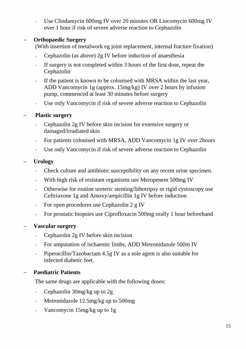
Orthopaedic Surgery
(With insertion of metalwork eg joint replacement, internal fracture fixation)
- Cephazolin (as above) 2g IV before induction of anaesthesia
- If surgery is not completed within 3 hours of the first dose, repeat the
Cephazolin
- If the patient is known to be colonised with MRSA within the last year,
ADD Vancomycin 1g (approx. 15mg/kg) IV over 2 hours by infusion
pump, commenced at least 30 minutes before surgery
- Use only Vancomycin if risk of severe adverse reaction to Cephazolin
Plastic surgery
- Cephazolin 2g IV before skin incision for extensive surgery or
damaged/irradiated skin
- For patients colonised with MRSA, ADD Vancomycin 1g IV over 2hours
- Use only Vancomycin if risk of severe adverse reaction to Cephazolin
Urology
- Check culture and antibiotic susceptibility on any recent urine specimen.
- With high risk of resistant organisms use Meropenem 500mg IV
- Otherwise for routine ureteric stenting/lithotripsy or rigid cystoscopy use
Ceftriaxone 1g and Amoxy/ampicillin 1g IV before induction
- For open procedures use Cephazolin 2 g IV
- For prostatic biopsies use Ciprofloxacin 500mg orally 1 hour beforehand
Vascular surgery
- Cephazolin 2g IV before skin incision
- For amputation of ischaemic limbs, ADD Metronidazole 500m IV
- Piperacillin/Tazobactam 4.5g IV as a sole agent is also suitable for
infected diabetic feet.
Paediatric Patients
The same drugs are applicable with the following doses:
- Cephazolin 30mg/kg up to 2g
- Metronidazole 12.5mg/kg up to 500mg
- Vancomycin 15mg/kg up to 1g

16
Other Procedures, non-elective or complicated Patients
- Advice can be sought from the Antibiotic Guidelines and/or the
Infectious Diseases Physician.

17
MANAGEMENT OF ANAPHYLAXIS
Adapted from Guidelines from Australian & New Zealand Anaesthetic
Allergy Group
& Australian & New Zealand College of Anaesthetist - www.anzaag.com
IMMEDIATE MANAGEMENT
D
R
Danger & Diagnosis
Response to stimulus
Unresponsive Hypotension or
Bronchospasm
Cease triggers including Chlorhexidine &
Colloid
Stop procedure. Use minimal volatile if
GA
S Send for help & organise
team
Call for Help
Assign a designated leader & scribe
Assign a reader of Anaphylaxis Card
A
B
Secure Airway
Breathing with 100%
Oxygen
Intubate: airway oedema or compromise
Confirm FiO2 is 100%
C Circulation: CPR if no
pulse
Give iv Fluid bolus
If no pulse give 1mg Adrenaline iv (Paed
10 μg/kg) & follow ALS protocol
IV Fluid: 20 ml/kg bolus repeat PRN
D Drugs: Adrenaline IV Bolus
repeat if needed 1-2
minutely & Prepare
Infusion
IV Adrenaline Boluses
Draw up 1mg in 10 ml
• 100 μg/ml
Adrenaline infusion:
Adrenaline 6mg in 100ml
(1ml/h = 1 μg/min)
Moderate
Hypotension or
Bronchospasm
Severe
Hypotension or
Bronchospasm
Adult 0.05-0.4 mg/kg/min
Child 0.1-5 μg/kg/min
Adult 5-20 μg
Child 1-5 μg/kg
Adult 100-200 μg
Child 5-10 μg/kg
18
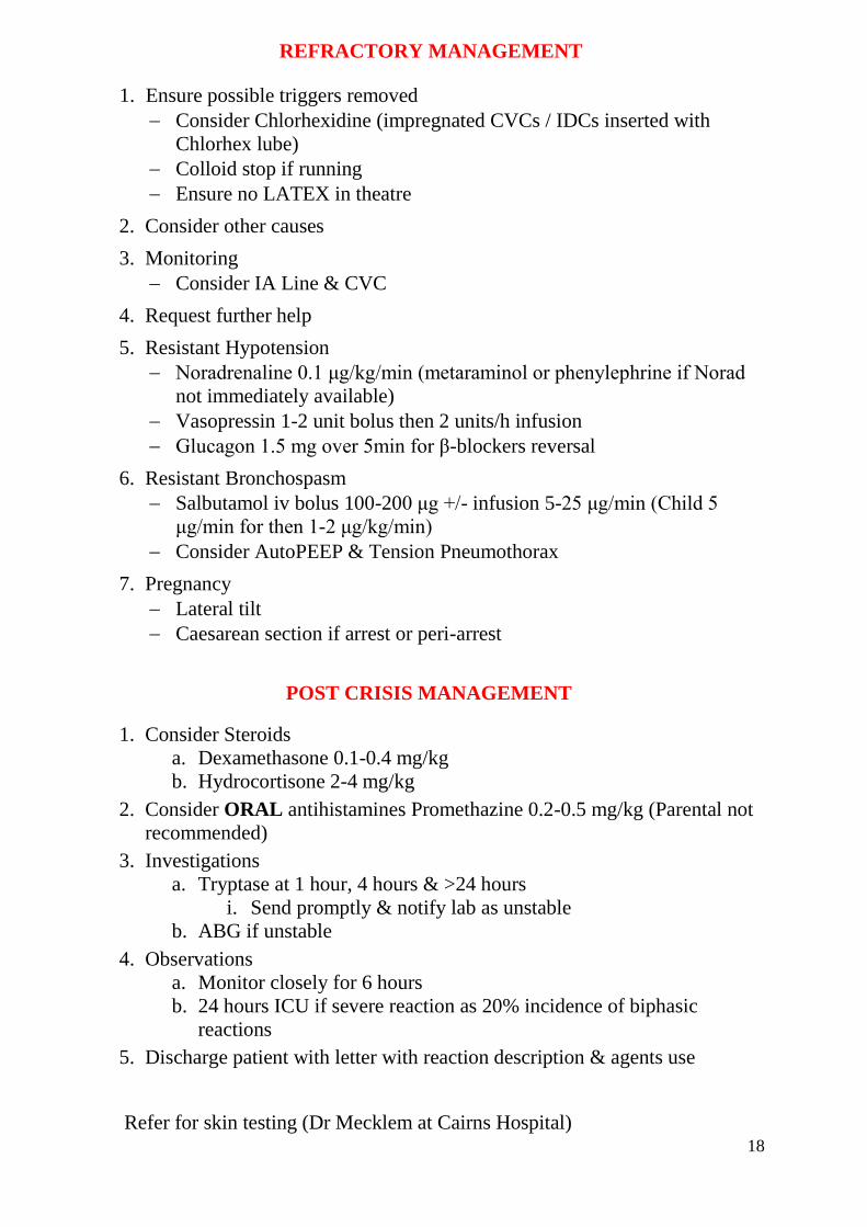
REFRACTORY MANAGEMENT
1. Ensure possible triggers removed
Consider Chlorhexidine (impregnated CVCs / IDCs inserted with
Chlorhex lube)
Colloid stop if running
Ensure no LATEX in theatre
2. Consider other causes
3. Monitoring
Consider IA Line & CVC
4. Request further help
5. Resistant Hypotension
Noradrenaline 0.1 μg/kg/min (metaraminol or phenylephrine if Norad
not immediately available)
Vasopressin 1-2 unit bolus then 2 units/h infusion
Glucagon 1.5 mg over 5min for β-blockers reversal
6. Resistant Bronchospasm
Salbutamol iv bolus 100-200 μg +/- infusion 5-25 μg/min (Child 5
μg/min for then 1-2 μg/kg/min)
Consider AutoPEEP & Tension Pneumothorax
7. Pregnancy
Lateral tilt
Caesarean section if arrest or peri-arrest
POST CRISIS MANAGEMENT
1. Consider Steroids
a. Dexamethasone 0.1-0.4 mg/kg
b. Hydrocortisone 2-4 mg/kg
2. Consider ORAL antihistamines Promethazine 0.2-0.5 mg/kg (Parental not
recommended)
3. Investigations
a. Tryptase at 1 hour, 4 hours & >24 hours
i. Send promptly & notify lab as unstable
b. ABG if unstable
4. Observations
a. Monitor closely for 6 hours
b. 24 hours ICU if severe reaction as 20% incidence of biphasic
reactions
5. Discharge patient with letter with reaction description & agents use
Refer for skin testing (Dr Mecklem at Cairns Hospital)
19
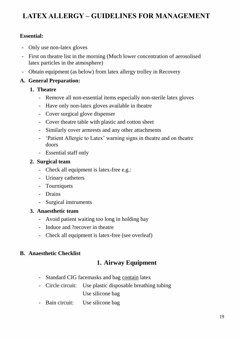
LATEX ALLERGY – GUIDELINES FOR MANAGEMENT
Essential:
- Only use non-latex gloves
- First on theatre list in the morning (Much lower concentration of aerosolised
latex particles in the atmosphere)
- Obtain equipment (as below) from latex allergy trolley in Recovery
A. General Preparation:
1. Theatre
- Remove all non-essential items especially non-sterile latex gloves
- Have only non-latex gloves available in theatre
- Cover surgical glove dispenser
- Cover theatre table with plastic and cotton sheet
- Similarly cover armrests and any other attachments
- ‘Patient Allergic to Latex’ warning signs in theatre and on theatre
doors
- Essential staff only
2. Surgical team
- Check all equipment is latex-free e.g.:
- Urinary catheters
- Tourniquets
- Drains
- Surgical instruments
3. Anaesthetic team
- Avoid patient waiting too long in holding bay
- Induce and ?recover in theatre
- Check all equipment is latex-free (see overleaf)
B. Anaesthetic Checklist
1. Airway Equipment
- Standard CIG facemasks and bag contain latex
- Circle circuit: Use plastic disposable breathing tubing
Use silicone bag
- Bain circuit: Use silicone bag

20
- Ayres circuit: Use disposable circuit
- The Omeda ventilator does not contain latex
- The Laerdel self-inflating bag does not contain latex
- Use (blue) Silicone Masks
- Use PVC ET tubes
- Use clear plastic oropharyngeal airways
- The gum elastic bougie and Satinslip introducer contain no latex
- There is no latex in the LMAs
- There is no latex in the Hudson mask
- Use airway filters as these also filter latex particles
2. IV Equipment
The following do not contain latex:
Syringes - Glass or Terumo
IV cannulae - Insyte
CVP catheters - Arrow or Cook
IA cannulae - Arrow or Insyte
IV tubing - Interlink
Burettes - Interlink
Syringe pump extension sets - Terumo
Reflux-valve - Braun.
- There is latex in the bung of the Haemaccel bottle.
- There is no latex in the Albumex bottle.
- The Gemini infusion tubing does not contain latex.
- There is no latex in the injection port of Baxter IV fluids.
- Remove the rubber stopper from drug vials.
- There is no latex in the stopper or plunger of the Diprifusor syringes.
C. Monitoring

21
- Use Velband under NIBP cuff. Cover tubing and cables with Velband,
as there is latex in bladder and tubing
- The pulse oximeter probe does not contain latex
- The temperature probe does not contain latex
- Baxter transducer sets do not contain latex
- 3M ECG dots do not contain latex
- The balloon of the Swan Ganz catheter does contain latex
D. Other Equipment
- The anaesthetic forceps have rubber shoes
- The Warm Touch hot air blanket does not contain latex
- Suction tubing does not contain latex
- The following do not contain latex:
NG tubes: Indoplas or Sherwood
Epidural catheters: Portex or BD
Tapes and dressings Micropore
Tegaderm
Steri-strips
Velband
Hypofix.
(Sleek does contain latex)
22
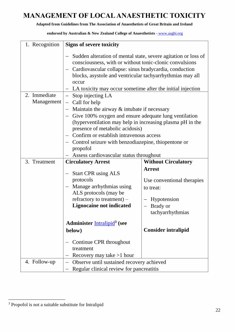
MANAGEMENT OF LOCAL ANAESTHETIC TOXICITY Adapted from Guidelines from The Association of Anaesthetists of Great Britain and Ireland
endorsed by Australian & New Zealand College of Anaesthetists - www.aagbi.org
1. Recognition Signs of severe toxicity
Sudden alteration of mental state, severe agitation or loss of
consciousness, with or without tonic-clonic convulsions
Cardiovascular collapse: sinus bradycardia, conduction
blocks, asystole and ventricular tachyarrhythmias may all
occur
LA toxicity may occur sometime after the initial injection
2. Immediate
Management Stop injecting LA
Call for help
Maintain the airway & intubate if necessary
Give 100% oxygen and ensure adequate lung ventilation
(hyperventilation may help in increasing plasma pH in the
presence of metabolic acidosis)
Confirm or establish intravenous access
Control seizure with benzodiazepine, thiopentone or
propofol
Assess cardiovascular status throughout
3. Treatment Circulatory Arrest
Start CPR using ALS
protocols
Manage arrhythmias using
ALS protocols (may be
refractory to treatment) –
Lignocaine not indicated
Administer Intralipid3 (see
below)
Continue CPR throughout
treatment
Recovery may take >1 hour
Without Circulatory
Arrest
Use conventional therapies
to treat:
Hypotension
Brady or
tachyarrhythmias
Consider intralipid
4. Follow-up Observe until sustained recovery achieved
Regular clinical review for pancreatitis
3 Propofol is not a suitable substitute for Intralipid

23
LIPID EMULSION (INTRALIPID) PROTOCOL
INTRALIPID IS STORED IN THE THEATRE ARREST TROLLEY
Immediately
Give bolus of 20% intralipid – 1.5 ml/kg over 1 minute
Start infusion of 20% intralipid at 15 ml/kg/hour
After 5 minutes reassess
Give a maximum of 2 further boluses (5 minutes between each bolus) and
Double infusion rate to 30 ml/kg/hour if
Cardiovascular stability not restored
Cardiovascular status deteriorates
Continue infusion until stable and adequate circulation restored
Intralipid dose should not exceed a maximum of 12 ml/kg

24
MRI CRITICAL CARE CHECKLIST
STAFF
Staff screening Jewellery, watches, wallets (credit cards), scissors, phones etc should
be removed
- Be guided by MRI staff
PATIENT
Patient Screening Jewellery, metal objects (zippers, bra clips), hearing aids, dentures,
some medication patches should be removed
Contraindications - Pacemakers, implantable defibrillators, metal prosthetic valves, metal
stents, cochlear implants, Portocaths, Hickman lines, neurostimulators,
implanted drug pumps, metal foreign bodies are all contraindications.
- Coronary stents and intracranial aneurysm clips especially if multiple
are contraindicated unless able to check with manufacturer.
- Wound staples, sternal wires and other surgical clips are generally
OK.
- Internal orthopaedic prostheses are OK – external fixation devices
may be a contra-indication (check with hand-held magnet).
- Each case needs individual assessment as it is impossible to cover
every eventuality in a list like this.
Respiratory
guidelines
Need a secure airway (ie awake or LMA/ETT)
Not requiring high PEEP (eg >10cm) or high O2 (> 70%)
Circulatory
guidelines
Not requiring high inotropic support (eg >10μg/min
adrenaline/noradrenaline)

25
EQUIPMENT
THE MAGNET IS ALWAYS ON
Most normal equipment is NOT compatible and must not be taken into the scanning
room.
Bed NOT compatible – must transfer patient to MRI-compatible bed
Drip-stand NOT compatible – must transfer fluids etc to MRI-compatible stand
Oxygen cylinders NOT compatible – require aluminium cylinders
IV pumps and
syringes
Most are NOT compatible – use outside scanning room or use MRI-
compatible units
IV tubing for pumps
and syringes
Need long extensions ie 4.5-6m if pumps outside scanning room
• Take additional pre-primed lines to Medical Imaging
• Avoid coils of tubing in scanner to prevent heating from
magnetic field
Anaesthetic
machine
Must change to specific MRI-compatible machine
Even this machine must not be moved near the magnet
Defibrillator
Cardiac arrest
trolley
NOT compatible – must never be taken into scanning room
Instead, the patient MUST BE REMOVED from the scanning room
Breathing circuits Plastic only; avoid PEEP valves with metal springs
Endotracheal tubes;
pilot balloons
Avoid reinforced tubes (artefact)
Spring of pilot balloon can cause artefact so tape away from scanned
region
• Rarely may have to knot pilot balloon tubing and cut off
balloon

26
LMAs Pilot balloon problems (as per endotracheal tubes)
Laryngoscopes Use MRI-compatible laryngoscope and batteries
Monitor NOT compatible - must change to specific MRI-compatible monitor
• Even this monitor must not be moved near the magnet
NIBP cuffs Tube connections must be plastic not metal
Invasive art lines Transducer may be not compatible so remove it and bung cannula
Pulse oximeter Change to specific MRI-compatible probe
ECG (general) Artefact and other problems common – consider removing altogether
-ECG dots Change to MRI-compatible (to avoid burns)
-ECG leads May cause local heating – avoid loops and protect patient skin
Temperature probes NOT compatible and must be removed
Calf compressors NOT compatible
Forced air warmers NOT compatible

27
OBSTETRIC ANTICONVULSANT THERAPY FOR ECLAMPSIA
AND SEVERE PRE-ECLAMPSIA
The drug of choice is Magnesium Sulphate. It is available in 5ml ampoules each
containing 2.5g of magnesium. It should be given as a loading dose followed by a
maintenance infusion.
Loading dose: Use pre-filled syringes of 5g (if available) from Birth Suite.
Alternatively, add 5g (2 ampoules) to 90ml of normal saline and infuse over 15
minutes (100ml at 400ml/hour). The woman should be warned that she may
experience transient hot flushing. ECG monitoring is not required.
Maintenance infusion: Add 25g (10 ampoules) to 450ml of normal saline to
make 500ml of solution. Infuse at 1g/hour initially (20ml/hour), up to 2g/hour
(40ml/hour) depending on clinical parameters and magnesium levels.
Patient monitoring:
Magnesium may cause muscular weakness, and levels may increase with poor
urine output. Consequently, there should be hourly evaluation of:
- Patellar reflexes (should be present)
- Respiration (should have rate >12)
- Urine output (should exceed 100ml in 4 hours)
and 6-hourly measurement of serum magnesium levels starting 2 hours after
loading dose (2.0-3.5 mmol/l is considered therapeutic). Calcium gluconate 1g
slowly IV can help reverse magnesium toxicity.
In the event of fitting despite magnesium therapy, the patient should be nursed in
the lateral position with attention to Airway, Breathing and Circulation.
An additional “loading” dose of 2.5-5.0g of magnesium (50-100ml of solution) can
be given over 7.5-15 minutes. Other anticonvulsants and tracheal intubation may
occasionally be required.

28
Other features of magnesium therapy:
1. Magnesium is a tocolytic so increased doses of oxytocin may be needed.
2. Intrapartum CTG’s may show decreased variability with therapeutic
magnesium levels.
29
ANALGESIA IN BIRTH SUITE
NOTE: This document is not an exhaustive guide, but describes local practices and
gives some trouble-shooting advice.
RESPONSIBILITY OF THE ANAESTHETIST
The epidural service is provided by the Acute Pain Service during the day, and by the
anaesthetists in theatre after hours.
The anaesthetist inserting the regional block is responsible for the management of it
until it is ceased. When that anaesthetist goes off duty the patient should be ‘handed
over’ to another.
The anaesthetist should attend the mother regularly throughout the duration of the
regional block, so that complications are avoided and analgesia assured. It is not
sufficient to attend the mother only when called by a midwife.
LOW-DOSE ‘EPIDURAL’ TECHNIQUES – ADVANTAGES AND OPTIONS
Low-dose techniques are used routinely in the Birth Suite (BS). They minimise motor
block and reduce the incidence of instrumental deliveries.
There are essentially two ways of achieving good analgesia using low-dose techniques:
1. EPIDURAL ONLY
This is the technique of choice if the woman is not too distressed with pain. The
doses and dilutions are described in the document ‘Epidural Drug Doses in Birth
Suite’.
2. COMBINED SPINAL EPIDURAL (CSE)`
A CSE can be used if the mother is very distressed and in severe pain, as it may be
difficult to establish an epidural block using dilute epidural solutions. Its main
advantage is rapid analgesia without muscle weakness.
Establishing epidural analgesia on the BS with stronger solutions (eg 0.25%-0.5%
bupivacaine) causes muscle weakness and consequently, these solutions should
only be used if low-dose techniques are ineffective.

30
NOTE: The following apply to the use of Combined Spinal Epidurals (CSEs) in
Birth Suite only - they are not intended as a guide to using CSEs in
Theatre.
PREPARATION:
The initial preparation for a CSE is the same as for any regional technique in
obstetrics:
Contraindications (eg thrombocytopenia and sepsis) must be respected, and
informed consent obtained.
An aseptic technique (mask, surgical scrub, gown and glove) must be used.
A filter needle should be used to draw up all drugs from glass ampoules for
injection into the subarachnoid space.
The mother may be in the lateral or (preferably) sitting position.
PROCEDURE:
A needle-through-needle technique is most commonly used.
When the subarachnoid space is located, 1.25mg bupivacaine (0.5ml of 0.25%
plain bupivacaine) plus 25μg of fentanyl (0.5ml) is injected.
The spinal needle is withdrawn and the epidural catheter is threaded and fixed in
place, but no injection of local anaesthetic is made at this stage. However, the
catheter should be flushed with 2ml saline and aspirated (to ensure it is not
kinked; to prevent it being occluded by a clot; and to check for IV or
subarachnoid placement).
If the mother has been sitting she should then lie on her side as soon as the
epidural catheter is taped in place.
See Patient Controlled Epidural Analgesia (PCEA) for subsequent management
of epidural analgesia.
PROBLEMS:
Mother very distressed and unable to cooperate
Firstly, ensure the syntocinon infusion is turned off. Consider giving her 50-75
μg fentanyl IV prior to the block, and/or insert the spinal first (using a 24 or 25g
pencil point needle) and insert the epidural under more favourable circumstances
when the mother is better able to cope.
Failure to locate the CSF with the spinal needle

31
In our experience this occurs in about 2% of cases. In this situation the epidural
catheter should be inserted and the block established using epidural top-ups
only.
Inadequate pain relief after spinal dose
Give first epidural dose - refer to the document ‘Epidural Drug Doses in Birth
Suite’.
Pros and Cons:
The advantages of using a CSE to establish the block (no muscle weakness) must
be weighed against the potential disadvantages in each woman.
Certain clinical circumstances make a CSE less suitable for pain relief in labour:
Infection
As the dura is punctured during a CSE, it may be inadvisable if the mother has
a systemic infection, is pyrexial or has prolonged ruptured membranes and is not
on antibiotics, because of the theoretical risk of meningitis.
Foetal bradycardia
Foetal bradycardia may occur soon after the spinal component of the CSE,
possibly due to increased uterine tone. Consequently it may not be advisable to
insert a CSE for pain relief if the foetus has a non-reassuring CTG trace. It is
also advisable to routinely stop a syntocinon infusion before doing a CSE, and
to monitor the FHR as soon as possible after the spinal block is established.
Imminent Caesarean Section
If a caesarean section is likely in the near future it is better to establish an
epidural block and ensure it is functioning properly rather than doing a CSE.
The doses used subarachnoid in BS are not sufficient for a CS.
Previous CSE in the same labour
The incidence of headache following an uncomplicated CSE is low -
approximately 1:200 at CBH. However, another CSE or spinal increases the
chance of a PDPH and should not be performed without good reason.
TROUBLESHOOTING EPIDURALS IN BIRTH SUITE
Failure to locate the epidural space
If the epidural space cannot easily be located at one level, a different
intervertebral space should be tried. If attempts at two interspaces are
unsuccessful, more experienced assistance should be sought. It is not reasonable
to subject the mother to repeated needling if more experienced help is available.
Dural tap

32
In the event of an accidental dural puncture, it is usually advisable to remove the
Tuohy needle and insert an epidural catheter at an adjacent interspace. The
anaesthetist must then do all the top-ups through the epidural until he/she is
satisfied with the position and function of the epidural catheter (an unexpectedly
high block may occur from retrograde spread into the CSF).
If the dural tap occurred after much difficulty in finding loss-of-resistance, insert
the epidural catheter into the subarachnoid space and label the catheter clearly
as ‘subarachnoid’. Inform the senior anaesthetist on call as well as the mother
and the obstetric staff. The anaesthetic staff must do all the top-ups through this
catheter for the duration of the block. The recommended dose for analgesia in
labour is 1.25mg bupivacaine (0.5ml 0.25% plain bupivacaine) plus 25μg
fentanyl (0.5ml), followed by 1ml NSaline as a flush to account for the dead
space of the filter and catheter.
Blood in the epidural catheter
First pull the catheter back until only 3cm remains in the epidural space, then
flush the catheter with 5ml of saline. Gently aspirate and observe if blood still
fills the catheter.
If the aspirate is heavily blood-stained the catheter should be removed. If there
is no aspirate or it is only slightly blood-stained, an adrenaline-containing test
dose should be given, ie 2ml of 2% lignocaine with adrenaline 1:200,000.
If there is no increase in heart rate (more than 10/min) and the patient does not
develop symptoms of dizziness, tingling or dysphoria, the epidural can be used.
Inadequate pain relief
Refer to the document ‘Epidural Drug Doses in Birth Suite’.
Patient Controlled Epidural Analgesia (PCEA)
PCEA is the standard way of maintaining epidural analgesia on the BS at CBH.
Compared to other techniques it is more effective, dose-sparing and gives the
mother a sense of control.
It is provided through the Go Medical mechanical device, which allows a bolus
of 4ml with a fifteen-minute lockout. In addition, our protocol allows for a
midwife-administered top up of 10ml.
The solution used is 0.125% levobupivacaine with 2μg/ml fentanyl.
It is used after initial analgesia is achieved with either an epidural or a CSE.
After epidural only
PCEA should be commenced once the anaesthetist is happy that the epidural
catheter is correctly placed and the patient is comfortable.
After CSE
Two important issues after the spinal component of a CSE are:
33
the untested epidural catheter, and
the potential for recurrence of severe pain during transition to epidural
analgesia.
After a straightforward CSE insertion:
Follow the protocol on the yellow PCEA order sheet:
The midwife should set up the PCEA device and connect it to the epidural
catheter as soon as possible
The patient should start using PCEA at the first uncomfortable contraction
before they become too painful
The anaesthetist must be informed when the patient starts using PCEA, and
review her
Give additional doses as required (see ‘Epidural Drug Doses in BS’)
After a complicated CSE insertion (eg possible IV puncture or subarachnoid placement of
epidural catheter):
• The anaesthetist must give an epidural test dose before PCEA is commenced
but before severe pain returns
• The anaesthetist should give further top ups to fully establish analgesia and
confirm safe epidural placement before commencing PCE

34
EPIDURAL DRUG DOSES IN BIRTH SUITE
Note: This document is not an exhaustive guide, but describes local practices and
gives some trouble-shooting advice.
1. Low-dose epidural analgesia
Test Dose
Use 4ml 0.25% bupivacaine
OR
Use 8ml 0.125% levobupivacaine in divided doses
After a bloody tap use 2ml 2% lignocaine plus 1:200,000 adrenaline
Top-up
Use 10ml 0.125% levobupivacaine plus 50-100μg fentanyl in divided
doses
If block is not adequate after 15 minutes, give another 5-10ml of 0.125%
levobupivacaine
If this is not adequate see “Persistent Pain” section below.
2. Combined Spinal Epidural
Subarachnoid
Use 1.25mg of bupivacaine (0.5ml of 0.25% plain bupivacaine) plus
25μg of fentanyl (0.5ml)
Epidural first dose
Use 10ml 0.125% levobupivacaine plus 2μg/ml fentanyl (PCEA solution)
as two 5ml doses
OR, if patient has started PCEA (a 4ml dose)
Give further 6ml 0.125% levobupivacaine plus 2μg/ml fentanyl

35
If this is not adequate see “Persistent Pain” section below.
Persistent Pain
Plan:
Ask site of patient’s pain.
Measure extent of the block with ice.
Ensure epidural catheter is still in place.
Give treatment according to the classification below:
i. Patchy Block
Give further 10-15ml 0.125% levobupivacaine ± fentanyl in divided doses
(In this situation, larger volumes of dilute solution work better than smaller
volumes of stronger solutions.)
ii. One-sided Block
Pull the epidural catheter back until 3cm left in the epidural space and give
10-15ml 0.125% levobupivacaine ± fentanyl. If this is not effective, or
catheter is only 3cm in epidural space to start with, the epidural catheter
should be replaced.
(Giving a higher concentration will only make the blocked side even more
numb and weak and is unlikely to provide any analgesia on the unblocked
side.)
iii. Back Pain
Epidural fentanyl 100μg (diluted to 5ml) often works well, if only low
concentration fentanyl has been used before.
iv. Perineal Pain or Breakthrough Abdominal Pain
In this case, with a demonstrable block on both sides but the patient
still complaining of pain, give 10ml 0.25% levobupivacaine ±
fentanyl
If these measures are not adequate, call for senior help.
Ongoing Analgesia
Refer to the document ‘Analgesia in Birth Suite’.

36
OBSTETRIC - UTEROTONIC AND RELAXANT DRUGS
Note:
- This document is a reminder of doses only
- Indications, contraindications and side effects need to be understood
before using these drugs
UTEROTONIC DRUGS
1. Carbetocin (for use in elective LSCS only)
Give one ampule (100ug in 1ml) IV over 1min after delivery of the baby. There is no
need for an oxytocin infusion, as this is a long-acting oxytocic.
2. Oxytocin (Syntocinon)
2-5 Units IV, repeated prn (expect to need higher doses if prolonged labour/grand
multip/ >4kg baby/polyhydramnios) and infusion of 40 Units in 1000ml @ 10-80
Units/hour (or more).
3. Ergometrine
250 ug slow IV, repeated prn
Relatively contraindicated in P.I.H. / Pre-eclampsia
4. Prostaglandins
i. Carboprost (Prostin15M) 250ug/ml
o 250 ug (1 ml) via deep IM injection. If necessary, this dose can be repeated
at intervals of at least 15 minutes for up to 8 total doses (2 mg).
ii. Misoprostol
o 400-800μg (2-4 tablets) per rectum
NB If response to uterotonic drugs and uterine massage is inadequate,
alternative management strategies will be needed, including:
o balloon tamponade
o uterine packing
o B-Lynch suture
o uterine artery and internal iliac artery ligation
o pelvic arterial embolization, or
o hysterectomy.
RELAXANT DRUGS
IV GTN

37
• Put 50mg in 1000 ml to make 50 μg/ml
• Start with 2-3 ml IV (100-150 μg)

38
ORAL INTAKE IN LABOUR
During late pregnancy heartburn is common.
During active labour stomach emptying is slow and food is poorly absorbed.
Opioid drugs (IV, IM or epidural) slow the process further.
Epidural anaesthesia or general anaesthesia may be necessary during labour
(approximately 35% of women) and food in the stomach increases the risk of
aspiration.
It is wiser for a woman not to eat in labour and to choose only clear fluid.
It is CBH hospital policy to discourage eating in labour and to disallow intake
of any solids when the mother is likely to need an operation. However if the
mother is requesting food she should be informed of the following guidelines.
Latent labour / Induction of labour
• Spontaneous onset – Light diet4 as tolerated
• Prostaglandin priming – Light diet as tolerated until active labour
commences then clear fluids5 only
• ARM / Syntocinon induction – Clear fluids only
Active labour
• Women in active labour should have clear fluids only
• Women who insist on eating may have light diet if they have:
o Spontaneous onset of labour
o No medical or obstetric problems
o No known risk of an instrumental or operative delivery
o Not using nor intend to use any form of pharmacological analgesia
(N2O, opioid or epidural)
o There is an approximate 10% chance of an operation (LUSCS,
retained placenta) in this patient group and eating in labour
increases the chance of an adverse outcome for both mother and
baby.
4 Light diet – Tea, coffee, toast, plain biscuits 5 Clear fluids – Water , cordial, sports drinks, NOT milk or fruit juice

Department of Anaesthesia & Perioperative Medicine CHHHS
39
OBSTETRIC PATIENTS WITH KNOWN
RHEUMATIC HEART DISEASE - MANAGEMENT
OF LABOUR AND DELIVERY
Plan: Assess risk according to maternal risk factors
Advise management as below
Document plan in obstetric notes
Maternal Risk factors are based on history, current symptoms and
echocardiography:
• History: prior cardiac event (pulmonary oedema, tachyarrhythmia)
• Current Symptoms: dyspnoea with minimal exertion (NYHA III)
(Dyspnoea at rest (NYHA IV) needs immediate evaluation and
treatment by cardiology ± ICU)
• LV dysfunction (Ejection fraction < 60%)
• Left heart obstruction moderate or severe (mitral or aortic valve area
1.5cm2)
• Pulmonary hypertension (systolic PAP >50mmHg)
Reference: Sartain J, Anderson N, Barry J, Boyd P, Howat P. Rheumatic heart disease in
pregnancy: cardiac and obstetric outcomes. Internal Med Journal 2012; 42:978-98
Low Risk (No risk factors): <5% chance of cardiac complications
Management:
▪ Treat on obstetric indications only
▪ Epidural beneficial but not essential
▪ Avoid excessive IV fluids
▪ Antibiotic prophylaxis indicated with prosthetic valves, history of
endocarditis or obstetric reasons

Department of Anaesthesia & Perioperative Medicine CHHHS
40
Moderate Risk (One risk factor): 25% chance of cardiac complications
Management:
Needs cardiology review and collaboration between obs / anaes / paeds /
possibly ICU
Delivery:
▪ In Cairns or other regional centre with relevant experience
▪ Epidural pain relief early (once labour established)
▪ Avoid excessive IV fluids
▪ Assisted vaginal delivery (Vaginal delivery preferred to CS if obstetric
considerations allow)
▪ Anaesthetist present for delivery
▪ Avoid ergometrine (causes pulmonary oedema)
▪ Syntocinon bolus (NB not syntometrine) 5U IM or 1U slow IV
▪ Syntocinon infusion 10U in 100ml saline @ 50ml/hour initially
▪ Antibiotic prophylaxis indicated with prosthetic valves, history of
endocarditis or obstetric reasons.
Post-Delivery:
▪ IV fluid to replace blood loss only – cease once bleeding stopped
▪ Consider frusemide 10-20mg IV
▪ Observe in obstetric HDU overnight (monitor P, BP, dyspnoea, SpO2
and blood loss)
High Risk (More than 1 risk factor): 50% chance of cardiac
complications
Management:
As for Moderate Risk except for:
▪ Consider transfer eg for balloon valvotomy if severe stenosis or
symptoms not medically controllable
▪ Consider delivery in monitored situation (OT probably best)

Department of Anaesthesia & Perioperative Medicine CHHHS
41
▪ Avoid bolus syntocinon (may cause ↓↓ BP) unless uterus poorly
contracted despite syntocinon infusion
▪ Slow bolus of 1U for poor contraction (ie 10ml of solution)

42
PAEDIATRIC DAY SURGERY SUITABILITY CRITERIA
A. Clinical Requirements
1. Babies should be older than 3 months of age if they require general
anaesthesia. However, ex-premature babies may still be unsuitable for
same day discharge and need individual anaesthetic assessment.
2. Children should have no organic disease or only mild organic disease,
which minimally interferes with their normal activities.
3. Procedures should be less than 1 hour.
4. Procedures should cause minimal blood loss.
5. Postoperative pain can be controlled with oral analgesics.
6. No special nursing care should be required postoperatively and parents
must be able to carry out pre- and post-operative instructions.
B. Social and Logistical Requirements
1. The child can only go home by private vehicle or taxi.
2. Another responsible person (apart from the driver) must be present in
the vehicle to look after the child.
3. The distance to be travelled following discharge should be less than
60km.
4. Parents advised that the child might need to be admitted overnight.
C. Other Procedural Factors
1. Day case patients should be given priority on the operation list
sequence.
The Surgical Team responsible for the patient must be consulted if these guidelines cannot
be met. They will then arrange to admit the child overnight.

43
PAEDIATRIC (ADENO)TONSILLECTOMY
DISCHARGE PLANNING
Day of Surgery discharge is suitable if ALL the following criteria are met:
• Age 4 years
• History of only mild or no OSA (Obstructive Sleep Apnoea)*
• No ongoing desaturations (<95% on RA) when sleeping in Recovery or
DSU
• No significant co-morbidities (including high BMI)
• 4 hours minimum observation since surgery
• Eating and drinking, good pain control
• < 1 hour travel time to hospital (in case of bleeding)
• Good family situation and carer available
ICU Admission if ANY of:
• History of severe OSA
• Marked ongoing desaturations (<80%) postoperatively
• Need for high flow nasal oxygen (eg consider if >2ml//kg)
• Major patient co-morbidities or marked obesity (eg >99th centile)
Paediatric Ward Admission:
• Patients outside of Day of Surgery and ICU categories
• Must be stable in recovery for at least 30 minutes before discharge to the
ward
• Continuous oximetry and direct nursing care for at least 4 hours on the
ward
• Overnight continuous oximetry if likely moderate OSA or higher risk of
OSA (significant co-morbidities, high BMI or age <2 years), or if any
desaturations or obstructed breathing occurs

44
Obstructive Sleep Apnoea (OSA)
• 10-12% of children snore, but only 1-2% have OSA.
• Definitive diagnosis with polysomnography (sleep study) is unusual but
AHI (apnoea/hypopnoea index) of 6-9/hour is moderate OSA; 10 hour is
severe
• Nocturnal oximetry with SpO2 < 80% indicates at least moderate OSA
• STOPBANG OSA acronym checklist (modified for children):
o Snoring
o Tiredness (daytime) and Tonsillar enlargement
o Observed Apnoeas
o Posture – extended neck sleeping; mouth breathing
o BMI increased, Breathing difficulties at night
o Age <3 years
o Neuromuscular disease
o Genetic conditions (eg 40% of Down children have OSA)
➢ Snoring and large Tonsils are sensitive markers, ie absence = OSA
unlikely
➢ Apnoeas, Breathing difficulties and daytime tiredness are specific
markers, i.e. presence = OSA likely
Selected References
• Patino M et al. OSA in children: perioperative considerations. Br J Anaes
2013; 111 (S1):i83-i95
• Certal V et al. Clinical assessment of Pediatric OSA: a systematic review
and meta-analysis. Laryngoscope 2012; 122(9):2105-14

45
PAEDIATRIC VENOUS ACCESS DECISION PATH
No
* As this number is only a recommendation, it is up to the anaesthetist performing the procedure to decide the most appropriate approach on a case-by-case basis
Considerations • Peripheral vs. central venous
access?
• Therapy duration
• Medical history
• Weight
• Age
< 7 days 7-14 days > 14 days
Therapy suitable for
peripheral infusion
Yes No
Potential IV sites
Yes No
Call for early
anaesthetic
assessment
Peripheral
IV cannula Non-tunnelled CVC
Call for early
anaesthetic
assessment
Weight < 20kg* = tunnelled CVC
Weight > 20kg = PICC
Otherwise call for
early anaesthetic
assessment
Weight < 20kg* = tunnelled CVC
Weight > 20kg = PICC
Consider referral to
Tertiary Hospital for
surgical cuffed CVC
PICC lines
Typically inserted into either basilic or brachial vein
PICC lines have high insertion and post insertion
failure rates if:
• children < 20kg
• inserted into cubital fossa
• inserted into cephalic or long saphenous vein
Simple IV cannula dwell time ≈ 3 days
PICC lines dwell time 2-12 weeks
Non-tunnelled CVC dwell time up to 14 days
Tunnelled non-cuffed CVC dwell time 2-12 weeks
Tunnelled cuffed CVC dwell time up to years
On Call Anaesthetist Tel 66910
The majority of the lines will be done on M, W & F
Refer to V. Petkov, N. Naydenov

46
Consent for the insertion of Central Venous Access Device (CVAD)
General considerations
• The child, the child’s parent/guardian should be explained the benefits and
risks associated with the CVAD insertion
• The person inserting the line should ideally be the person gaining the
consent for it and ensure the risks have been understood
• It is inappropriate for a person unfamiliar with the risks of a CVAD to gain
consent
CVAD complications at the time of insertion
• Pain, bruising or bleeding at the insertion site afterwards
• Difficulty inserting the catheter
• Temporary nerve damage or pain
• An irregular of fast heart beat sometimes requiring treatment
• Very rarely damage to nearby structures such as blood vessels or heart
during insertion
CVAD complications whilst the line is in situ
• Life-threatening
o Tamponade
o Pleural effusion
o Pericardial effusion
• Other
o The catheter may block, bend or move out of place, requiring
treatment or removal
o Blood clot blocking the vein requiring treatment or removal
o Very rarely the blood clot can move out of the vein and can travel to
the lungs or brain
o Infection at the skin puncture site requiring antibiotics or further
treatment
o Infection in the catheter requiring antibiotics or removal
o Medications leaking outside the vein causing pain, swelling or tissue
damage, requiring treatment
o Movement of the tip of the catheter requiring repositioning

47
PERIOPERATIVE METARAMINOL INFUSION
Description:
Metaraminol is a sympathomimetic vasopressor that increases blood pressure mainly
through peripheral vasoconstriction (-agonist effect), though it also has some
stimulatory effect on the heart.
Use:
Metaraminol is used as an adjunct in the treatment of hypotension, especially from
vasodilation due to epidural or spinal anaesthesia/analgesia. Other causes of
hypotension (eg hypovolaemia, sepsis, cardiogenic) must also be considered and treated
appropriately.
It should be commenced only on the direction of Anaesthetic or Intensive Care medical
staff, familiar with the use of vasopressor agents. Consideration should be given to
transfer to a High Dependency situation, depending on patient circumstances and
acuity.
Patients receiving a post-operative Metaraminol infusion are the responsibility of the
APS team regardless of whether an APS modality is being used.
Contra-indications:
Allergy to metabisulfite preservative.
Interactions:
Mono-amine oxidase inhibitors and digoxin increase the risk of arrhythmias.
Antihypertensive drugs and diuretics should generally be withheld while on a
metaraminol infusion.
Precautions:
Hypertension, headache, chest pain & pulmonary oedema may occur with excessive
dose.

48
Dose and administration:
Standard: 10 mg metaraminol to 100ml 0.9% saline.
Infuse via infusion pump. To prevent reflux and inconsistent drug delivery, other fluids
must be given either via an infusion pump or a giving set with a non-return valve.
Rate is usually prescribed as 0-20ml hour (0-2mg/hour), with adjustments (eg of 2-
5ml/hour) according to blood pressure. A target level must be stipulated (eg >90
mmHg). A common starting dose is 5 or 10ml/hour; it takes about 10 minutes for a
new rate to reach equilibrium.
For marked hypotension, an initial IV bolus of 0.1- 0.25 mg can be given, repeated
every 3-5 minutes if necessary. If a rate of 20ml/h is required higher level care is
needed.
Monitoring:
Enquire about symptoms of hypotension (nausea, dizziness) and of excessive dose (see
precautions above).
Check blood pressure and pulse rate after dose adjustment (whether increasing,
decreasing or stopping): every 15 minutes for 30 minutes, then hourly if stable

49
PERIOPERATIVE MANAGEMENT OF FRACTURED NECK OF
FEMUR
Anaesthetic Factors and Responsibilities
1. Anaesthetic review on Orthopaedic ward
• All patients should be referred to APS pre-op
• Consider Fascia Iliaca block if not already done by ED – generally place a
Fascia Iliaca catheter not a single shot block – See Fascia Iliaca Section
Below
• All patients should have a pre-operative assessment
o APS PM Registrar
o If patient 2nd on trauma list and not yet reviewed Trauma AM Registrar
o Check
▪ FBC / U&E / Coags (if liver disease or on warfarin) / G&H / ECG
o These are often complex patients and should be discussed with the
Trauma list anaesthetist
2. Anti-coagulated patients (general plan - refer also to perioperative
anti-coagulant and antiplatelet protocols)
a. Warfarin:
• Check INR; if > 1.5 give vit K 3mg IV (in 100ml saline)
• Repeat INR next day; consider Prothrombinex
• Surgery (and neuraxial block OK) if INR<1.5
• Clexane 20mg sc daily, start >12 hours post-op
b. NOACs:
• Wait 24h from last dose unless neuraxial anaesthetic considered
optimal
• If neuraxial anaesthetic required follow standard NOAC protocols
• Check renal function
c. Clopidogrel:
• Wait 24h from last dose unless neuraxial anaesthetic considered
optimal
• Wait 5 days if neuraxial anaesthetic required
3. Fasting guidelines
• No food after 0200 day of surgery

50
• No fluids after 0600 day of surgery
• Review emergency booking at lunchtime re fasting (through APS)
4. Trauma list management
• Ideally 2nd patient on Trauma list
• Avoid prolonged & repeated fasting if surgery delayed
5. Anaesthetic Choice
• No recommendation about particular anaesthetic technique
• Tailor to patient requirements
• Senior anaesthetic should be involved
6. Post-op Care
• Hb in PACU before discharge to ward (i-STAT or formal)
• Acute Pain Service Follow-up
• Analgesia
• Fluids

51
FASCIA ILIACA CATHETERS
LANDMARK-BASED INSERTION GUIDE
Introduction
Single shot fascia iliaca or femoral nerve blocks provide good analgesia, however,
the block can wear off long before the patient receives surgery. Instead, a fascia
iliaca catheter facilitates ongoing analgesia until the patient receives surgery. In
contrast to a femoral nerve block, the fascia iliaca catheter is inserted lateral to (not
on) the femoral nerve, thereby reducing the potential for femoral nerve injury.
Indications Contraindications (when in doubt,
discuss with a consultant)
Imaging-confirmed fracture of
neck of femur (all subtypes
except isolated acetabular
fractures)
Patients on clopidogrel (or other
antiplatelets in same class)
INR > 1.5
Previous femoral vascular bypass
surgery (scar + altered anatomy)
Allergy to local anaesthetic
Local infection in groin
Systemic infection (single shot block
is OK)
Very obese patients (impalpable
landmarks. Ultrasound advised)
Local anaesthetic safety
• Max safe dose of ropivacaine is 3mg/kg (up to max 200mg)
• Each 10mL of 0.75% ropivacaine contains 75mg ropivacaine

52
• Patients less than 50kg should be administered reduced concentration or volume of
local anaesthetic
Equipment required
• Take a ready-made Fascia Iliaca kit. If unavailable, refer to the equipment list “drop
sheet”.
Preparing the ropivacaine solution
• In each 20mL syringe draw up 10mL of ropivacaine 0.75% PLUS 10mL of saline
(resulting in 2 syringes, each containing 20mL of 0.375% ropivacaine).
Performing a fascia iliaca catheter insertion (aseptic procedure)
• Palpate landmarks (ASIS and pubic tubercle)
• Mark the insertion point: 2cm caudad to the junction between lateral and middle
1/3rd of line joining the ASIS to the pubic tubercle
• Check that the marked injection point is 1.5 – 2cm lateral to the pulsation of the
femoral artery
• Prepare the skin and apply a windowed drape
• Infiltrate skin and deeper tissues with 5mL of 1% lignocaine along the expected
path of the FI catheter
• Pierce the skin with the Touhy needle at right angles. Once just through the skin,
adjust the needle angle to about 45degrees directing the tip cranially, keep the needle
in the sagittal plane.
• Advance the needle through 2 distinct “pops” as it perforates first the fascia lata,
then the fascia iliaca
• Flatten the angle of the needle and skin to about 30degrees and advance the needle
a further 1-2 millimeters
• Take note of the depth of the needle at skin (“NEEDLE DEPTH”). Black marks the
4th, 6th and 8th cms.
• Secure the needle with your non-dominant hand and remove the trocar from within
the Touhy needle
• Attach the first 20mL syringe of 0.375% ropivacaine to the Touhy needle
• If aspiration is negative for blood, start injecting. There should be very little
resistance to injection.
• Excessive resistance to injection indicates the needle tip may be within iliacus
muscle. In this case, withdraw slightly until injection is easy.
• Inject 20mL slowly, aspirating every 5mL, then detach the syringe from the needle.
You have now created a plane filled with local anaesthetic under the fascia iliaca.
This facilitates passage of the catheter. It is normal to observe some of the injected
53
fluid coming back through the needle during syringe change.
• Thread the catheter through the Touhy needle. The catheter tip will start protruding
from the tip of the Touhy needle once it passes the 10cm marker (double blue line).
You may feel mild resistance at this point, but push through it.
• Continue threading the catheter until you have passed at least 15cm. Stop when
you encounter significant resistance or have reached the 20cm marker (quadruple
blue line), whichever is reached first.
• Withdraw the Touhy needle, continuing to thread the catheter down the needle at
the same rate that the needle is withdrawn, so that the catheter does not come out
with the needle
• Check the CATHETER DEPTH at skin - you will need to record this
• Attach the yellow connector to the catheter, and connect the antibacterial filter to
the yellow connector
• Connect the 2nd 20mL syringe of ropivacaine and aspirate before injecting
• Inject the remaining 20mL, aspirating every 5mL. Since you are using a large
syringe to inject down a long, fine-bore catheter, expect significant resistance to
injection. If you cannot inject at all, the catheter may be kinked or up against tissue.
Withdraw the catheter slowly as you continue to inject. But remember: the catheter
should not be less than 3cm under fascia iliaca.
• Apply a drop of SurgiSeal tissue sealant to the catheter entry point, allow a few
minutes to dry
• Remove the yellow connector (disengage it by inserting the tip a non-Leuer lock
syringe into the connector’s side hole, and lever it open)
• Attach Epi-lock catheter securing patch
• Trim the catheter (using sterile scissors) to around 20cm above skin
• Re-attach the yellow connector
• Cover the filter-connector complex with a Tegaderm “Taco” dressing
Documentation: fill out these forms
1. APS referral form (place in APS tray)
2. Yellow non-neuraxial nerve block audit form (place in APS tray)
3. Regional block catheter infusion order form (place in patient’s notes – Ward/PACU
nurses will initiate)
Re-dosing of local anaesthetic in the Emergency Department
If a fascia iliaca catheter is inserted in the Emergency Department, as long as the
patient remains in the Emergency Department, they will require manual re-dosing
by a Medical Practitioner as per the protocol in the infusion order form (re-dose
using 20mL of 0.2% ropivacaine every 3 hours).
Infusion pumps will be established once the patient is admitted to the wards.

54
Useful tips
1. Anatomy: Lateral to medial NAVL (nerve, artery, vein, lymphatics)
2. Create a superficial dermis-level weal of 1% lignocaine at the insertion point to
reduce the pain of subsequent skin entry with the larger Touhy needle
3. When advancing the Touhy needle, rest your wrist on the patient’s thigh and “dart-
grip” the needle at about the 5cm mark to avoid suddenly overshooting with the loss
of resistance
4. Don’t impale the femoral nerve! Stay 1.5-2cm away from femoral pulsation. There
should be no pain or paraesthesia on injection.
5. Don’t shear off the catheter tip! If the catheter has been threaded more than 10cm
but less than 13cm before you encounter significant resistance, YOU MUST remove
the needle and catheter as one unit, and start again. Withdrawing the catheter from
the needle once it has already passed beyond the tip of the Touhy needle can result
in the sharp tip of the Touhy needle cutting off the tip of the catheter, leaving foreign
body within the patient.
6. Success of the block relies on a high volume of local anaesthetic spreading under
the fascia iliaca, to bathe the femoral nerve and other nerves. Note that other nerves
supplying the hip are not blocked, so some opioid requirement is expected.
7. [Catheter depth] - [Needle depth] = [Length of catheter under the fascia iliaca],
which should be between 5 and 10cm (less than 3cm predicts block failure).
8. Obese patient with apron – an assistant can retract the apron cephalad to facilitate
access to the groin
Further Reading
• Anaesthetic tutorial of the week 193. Fascia iliaca compartment block: landmark
and ultrasound approach http://www.frca.co.uk/Documents/193%20Fascia%20Iliaca%20compartment%20block.pdf
• New York School of Regional Anaesthesia. Ultrasound guided fascia iliaca block. https://www.nysora.com/ultrasound-guided-fascia-iliaca-block-2

55
Site of injection (Figure A):
This is 1 cm caudal to the inguinal ligament at the junction of the lateral one third
and the medial two thirds of a line that joins the pubic spine (1) to the anterior
superior iliac spine (2).
Transverse view at the mid-inguinal ligament level. (Figure B):
(3) Femoral vein; (4) femoral artery; (5) femoral nerve; (6) needle insertion for a
femoral or three-in-one nerve block; (7) fascia lata; (8) fascia iliaca; (9) needle
insertion for a fascia iliaca compartment block; (10) lateral femoral cutaneous
nerve.

56
FASCIA ILIACA CATHETER INSERTION - EQUIPMENT LIST
DROP SHEET
Equipment list
A. Skin marker pen
B. 2x chlorhexidine-alcohol prep sticks (“pink lollipops”)
C. Adhesive drape with a window
D. Basic dressing pack
E. Sterile gloves
F. 5mL syringe
G. 23G (blue) hypodermic needle
H. 18G (pink) blunt drawing up needle
I. 5mL 1% lignocaine
J. 2 x 20mL syringes
K. 20mL of 0.75% ropivacaine
L. 2 x 10mL vials of 0.9% sodium chloride
M. 16G Touhy epidural kit
N. Tissue glue (SurgiSeal or DermaBond)
O. Sterile scissors
P. 2 x large Tegaderm
Q. Documentation:
a. APS referral form (white single A4)
b. Epidural/Regional infusion order form (white double-spread
sheet)
c. Yellow non-neuraxial nerve block audit form

57
PERIOPERATIVE MANAGEMENT OF ANTICOAGULANTS
Changes: Recent studies have shown that bridging anti-coagulation is unwarranted
in patients at low or moderate Thrombo-Embolic (TE) risk, and have further
established the place of more rapid warfarin reversal with Vitamin K and
Prothrombinex. Pre-operative bridging therapy is no longer recommended for
NOACs (New Oral AntiCoagulants), and the time of abstinence has been shortened
for Dabigatran.
Disclaimer: There are inconsistencies between published guidelines, reflecting the lack of good
clinical data. Therefore these should be seen a practical guide rather than a strict protocol. Good
communication, documentation and clinical judgement are imperative.
General Instructions
1. Make a clear plan for each patient:
• If possible, anticoagulated patients should be seen in Anaesthetic clinic at
least 1 week before surgery.
2. Determine the need to stop anticoagulants:
• Continuation may be acceptable for specific procedures such as cataract
extractions and endoscopies without intended biopsy – if any doubt check
with the surgeon.
3. Stratify the Patient’s Thrombo-Embolic (TE) Risk (Table 1):
• Patients with Atrial Fibrillation (AF), Mechanical Heart Valves (MHVs)
or Venous Thrombo-Embolism (VTE) who meet the criteria in Table 1
should be managed as High TE Risk in the perioperative period.
• All other patients with MHVs, AF or VTE may be managed as Low TE
Risk. Some borderline patients may reasonably be managed as High Risk
based on clinical judgement.

58
Table 1. High Thrombo-Embolic (TE) Risk Factors
• AF:
o With CHADS2 Score of 5-6 (see Table 2)
o With recent (<3 months) stroke/TIA/systemic embolus
o attributable to valvular heart disease
• MHVs:
o Mitral valve
o Older aortic valve (caged ball/tilting disc); bi-leaflet aortic valve
with any CHADS2 risk factors
o Any mechanical valve with previous stroke, TIA, or systemic
embolic event
• VTE:
o Recent (<3 months ago) DVT or Pulmonary Embolism
o High Risk Thrombophilia (deficiency in Antithrombin, Protein
C, Protein S, Antiphospholipid syndrome, Homozygous Factor
V Leiden and Prothrombin variant) or VTE due to active cancer
(< 6 months since diagnosis)
Table 2. CHADS2 Score
(establish by totalling points for patients with non-valvular AF)
C Congestive Heart Failure 1
H Hypertension 1
A Age > 75 1
D Diabetes 1
S2 Stroke/TIA 2
4. Implement plan for the patient’s TE Risk for their specific medication and
surgery
• See relevant medication section below
• Consider guidelines for neuraxial anaesthesia (Section 4) if relevant
5. Document the management plan:
• On the Anaesthetic assessment sheet
• In the patient’s notes (ieMR)
• Give written instructions to the patient

59
Specific Anticoagulant Medications
1. WARFARIN – Vitamin K antagonist
Pre-operative Warfarin Management – High TE Risk Patients (Table 3)
• Two days before surgery take last dose of warfarin
• One day before surgery give Vitamin K1 (Konakion) - 5 mg oral or 3 mg IV (eg
at Preadmission Clinic or DSU)
• Check INR on the day of surgery (i-stat). Give Prothrombinex if INR ≥ 1.5
(see Table 5) and repeat INR to ensure < 1.5.
Table 3. Preop Warfarin for High Thrombotic Risk
Pre-op
Day 2
Pre-op
Day 1 Day of op
Normal Warfarin Vitamin K
5mg oral or 3mg IV
-Check INR (i-stat)
-Prothrombinex as required
Post-operative Warfarin Management – High TE Risk Patients
• Recommence regular warfarin dose on the night of surgery unless there are
bleeding concerns (discuss with surgical team).
• Unless there are bleeding concerns, start either IV UFH infusion (without loading
dose) at 12-24 hours post-op aiming for 1.5X APTT; or LMWH at prophylactic
dose for 24-72 hours (depending on bleeding risk), then treatment dose (eg
enoxaparin 1.5mg/kg daily or 1mg/kg bd).
• Continue UFH or LMWH for 2 days after INR is therapeutic
Pre-operative Warfarin Management – Low TE Risk Patients (Table 4)
• Give last warfarin dose 5 days pre-operatively
• Check INR on the day of surgery (i-stat). Give Prothrombinex if INR ≥ 1.5 (see
protocol on reversal of Warfarin) and repeat INR to ensure <1.5.

60
Table 4. Preop Warfarin for Low Thrombotic Risk
Pre-op
Day 5 Pre-op Day 4
Pre-op
Day 3
Pre-op
Day 2
Pre-op
Day 1 Day of op
Normal
Warfarin
Rest Day
(No warfarin)
Rest Day
Rest Day
Rest Day
Check INR (i-stat)
Prothrombinex as required
Post-operative Warfarin Management – Low TE Risk Patients
• Recommence regular warfarin dose on the night of surgery unless there are
bleeding concerns.
• Bridging anticoagulant therapy is not required (but VTE prophylaxis as
indicated).
URGENT SURGERY ON WARFARIN
a. Immediate reversal required, but no active bleeding
• Give Prothrombinex-VF. This contains factor II, IX and X., with only very
small amounts of factor VII. Despite this, adequate reversal of warfarin can
be achieved without the need for additional FFP in most cases.
• Prothrombinex-VF can normalise the INR in 15 minutes in appropriate
doses (see Table 5)
• The infused clotting factors have a half-life of about 24 hours. Therefore,
vitamin K IV eg 1-3 mg will need to be given for sustained effect.
• If Prothrombinex is unavailable administer FFP 15ml/kg
Table 5. Suggested Prothrombinex Dose
to achieve Target INR of < 1.5
Initial INR
1.5-2.5 2.6-3.5 ≥3.6
25 IU/kg 35 IU/kg 50 IU/kg
b. Immediate warfarin reversal for life-threatening bleeding
Give:
• Vitamin K 5-10mg IV

61
• Prothrombinex –VF 50 IU/kg
• FFP 150-300ml
• If Prothrombinex is unavailable administer FFP 15ml/kg

62
2. DABIGATRAN (Pradaxa®) – Direct Thrombin Inhibitor
General Notes
• Popular because of rapid anticoagulation and no need for routine monitoring
• Not currently indicated for Mechanical Heart Valves or Valvular AF
• Mostly (80%) renally excreted; T½ varies accordingly: 14h with normal eGFR, 20
hours with eGFR 30-50ml/min; 30h eGFR <30 ml/min
• Contra-indicated with eGFR < 30ml/min
Pre-operative Dabigatran Management – both Low and High TE Risk
• Assess patient’s renal function. If eGFR < 30, then cease Dabigatran immediately
• Timing of last dose of Dabigatran depends on eGFR (Table 6)
• Check Thrombin Time (TT) on admission or prior to surgery IF considering neuraxial
blockade or very high bleeding risk; not routinely required. If normal, no Dabigatran
present.
• Bridging therapy is not required due to short period of abstinence
Table 6. Pre-op Dabigatran Management
Pre-op
Day 5
Pre-op
Day 4
Pre-op
Day 3
Pre-op
Day 2
Pre-op
Day 1
Day of
surgery
Normal doses
(for most)
Rest Day if:
eGFR <
30ml/min
Normal doses
(for most)
Rest Day if:
eGFR <
50ml/min
Normal doses
(for most)
Rest Day if:
eGFR <
80ml/min
Rest Day
(omit)
Rest Day Rest Day
Post-operative Dabigatran Management – Low and High Thrombotic Risk
• Re-starting must involve discussion with the surgical team re bleeding risk
• Do not restart Dabigatran if eGFR < 30ml/min; discuss with treating physician re
alternative treatment.
• If haemostasis satisfactory then Dabigatran may be started at usual dose 12-24 hours
post-op (low bleeding risk) or 48-72 hours (higher bleeding risk)
• Bridging therapy is generally not required due to rapid onset of anti-coagulant effect.

63
• If commencement of Dabigatran is delayed, and the patient is High TE Risk, then
consider either IV UFH infusion without a bolus dose, or SC LMWH (prophylactic
dose for 24-72 hours, depending on bleeding risk, then treatment dose).
URGENT SURGERY ON DABIGATRAN
• Consider oral activated charcoal if Dabigatran ingested in last 2 hours
• Delay surgery for 4-5 half-lives if possible (eg 3 days, or Table 6)
• Check Thrombin Time, renal function
• Neuraxial anaesthesia contraindicated unless TT normal
• Cross match blood and see advice for Management of Bleeding
Management of Bleeding on Dabigatran
• Supportive measures
• Identification and management of bleeding source
• Consider Tranexamic acid IV 15 mg/kg, followed by infusion of 1mg/kg/hr
• Consider bolus dose of Factor VIIa (50 mcg/kg) and repeat if severe
haemorrhage.
• Haemodialysis for 4-6 hours is effective and can remove up to 60 % of
Dabigatran
• Prothrombinex, Vitamin K and FFP are NOT effective. The effect of Factor VIIa
is currently unknown.
• (Idarucizumab is a Dabigatran-specific (Fab) antibody which rapidly reverses
the effect of Dabigatran, but is not yet commercially available in Australia)

64
3. RIVAROXABAN (Xarelto®) (t ½ 8h) and APIXABAN (Eliquis®) (t ½ 12h) -
Direct Factor Xa Inhibitors
General Notes
• Popular because of rapid dosing and no need for routine monitoring
• Not currently indicated for Mechanical Heart Valves or Valvular AF
• Although the half-lives are slightly different, recommended time intervals
for abstinence are currently the same for the different agents
• Excretion is approximately 30% renal and 70% hepatic.
• Contra-indicated with eGFR <30ml/min or hepatic disease with coagulopathy
Pre-operative Riv/Apixaban Management – both Low and High TE Risk
• Check renal and hepatic function (and coags if likely hepatic disease)
• Give last pre-operative dose Riv/Apixaban according to Table 7.
• A shorter period of abstinence (eg 2 days) is adequate for low-moderate
bleeding risk procedures in patients without hepato-renal disease, esp for
low dose (eg thromboprophylaxis) Riv/Apixaban.
• No pre-op bridging therapy is required due to the short period of abstinence
• Pre-operative coagulation studies are generally NOT recommended.
However, establishing that PT (and esp anti-Xa levels if available) are
normal may be reasonable if absolute haemostasis is required, especially if
the period of abstinence is shorter than desired.
Table 7. Pre-op Riv/Apixaban Management
Pre-op
Day 5
Pre-op
Day 4
Pre-op
Day 3
Pre-op
Day 2
Pre-op
Day 1
Day of
surgery
Normal dose
(for most)
Rest Day if:
Child-Pugh C
Normal dose
(for most)
Rest Day if:
eGFR < 30
OR
Child-Pugh B
Normal dose
(for most)
Rest Day if:
eGFR < 50
OR Age >70
Rest Day
(omit)
Rest Day Rest Day
65
Post-operative Riv/Apixaban Management
• Re-starting must involve discussion with surgical team re bleeding risk
• Do not restart Riv/Apixaban if eGFR < 30ml/min or Child-Pugh B or C.
Discuss with treating physician regarding alternative treatment.
• If haemostasis is satisfactory then Riv/Apixaban may be re-started at usual
dose 12-24 hours post-op
• Bridging therapy is generally not required due to rapid onset of anti-coagulant
effect.
• If higher bleeding risk, re-start Riv/Apixaban 48-72 hours postoperatively
• If commencement of Riv/Apixaban is delayed (eg ileus), AND the patient is
High TE Risk, then consider either IV UFH infusion without a bolus dose,
or SC LMWH (prophylactic dose for 24-72 hours, depending on bleeding
risk, then treatment dose).
URGENT SURGERY ON RIV/APIXABAN
• Consider oral activated charcoal if Riv/Apixaban was ingested in the last 2
hours
• Consider delaying surgery for 4-5 half-lives (eg 2-3 days, or Table 7)
• Cross match blood, check PT +/- anti-Xa levels, renal and hepatic function
• Neuraxial anaesthesia contraindicated unless anti-Xa levels normal
Management of Bleeding in Riv/Apixaban
• Supportive measures
• Identification and management of bleeding source
• In severe bleeding consider:
o Prothrombinex 50 IU/kg (shows benefit in experimental studies)
o Tranexamic acid 1g then infusion 1mg/kg/hr
o Factor VIIa 50mcg/kg and repeat if critical bleeding
• (Andexanet alpha is a binding reversal agent for Xa inhibitors that is under
development but not yet commercially available)

66
4. Neuraxial Blockade
• Neuraxial blockade should only be undertaken:
o with a normal (<1.5) INR – Warfarin
o after the time period in Table 6 or normal TT – Dabigatran
o after the time period in Table 7 or normal PT/anti-Xa – Riv/Apixaban.
• Epidural catheters are contraindicated with treatment dose Dabigatran or
Riv/Apixaban. If treatment doses are given inadvertently, epidural catheter
removal should be delayed for a time period according to Tables 6 and 7.
• Removal of an epidural catheter should be delayed for >24 hours after low
dose once daily (DVT prophylaxis) Dabigatran or Riva/Apixaban.
• Dabigatran or Riv/apixaban should not be administered until at least 6 hours
after spinal anaesthesia or epidural catheter removal.
5. References:
1. Tran H et al. (Aust Soc of Thrombosis and Haemostasis). New Oral Anticoagulants:
a practical guide on prescription, laboratory testing and peri-procedural/bleeding
management Int Med J 2014; 44:525-536
2. Douketis J et al. Perioperative Bridging Anticoagulation in Patients with Atrial
Fibrillation. NEJM 2015; 373:823-33
3. UpToDate®. Perioperative thrombotic risk; Perioperative management of direct
thrombin inhibitors and factor Xa inhibitors; Timing of neuraxial anesthesia during
antithrombotic therapy; all accessed Dec 2015
4. Tran H et al. An update of consensus guidelines for warfarin reversal. MJA 2013
5. Queensland Health Guidelines for Warfarin (2015), Dabigatran (2013) and Xa
inhibitors (2014).

67
PERIOPERATIVE MANAGEMENT OF DIABETES
MELLITUS IN ADULTS
General Considerations
• Ensure optimal glycaemic control prior to surgery.
• Frequent monitoring of Blood Glucose Level (BGL) is the key to good
control.
• Patients with diabetes, especially type I (IDDM) and insulin requiring
type II should be first on the list if possible.
• People with type I diabetes require a constant supply of background
insulin to prevent ketoacidosis. However, an insulin infusion can be
temporarily ceased when BGL <7 mmol/l.
• IV Dextrose infusion is required for all patients with IDDM and all
patients on an insulin infusion. A minimum of 100-150g exogenous
glucose (2-3 L of 5% Dextrose) per day is needed for protein sparing
and to prevent ketosis.
• BGL’s >12 mmol/l are associated with poor wound healing, infections
and osmotic diuresis.
• The patient’s fluid and cardiovascular status should be reviewed at least
daily. Additional N/Saline KCl is often required for optimal fluid and
electrolyte balance. Electrolytes should be measured every 24 hours in
patients on a Dextrose infusion.
• The following management plan and insulin regimens are guidelines
only, and experienced medical staff may choose to vary management in
specific situations.
• Cease Metformin 1 day prior to any investigation using potentially
nephrotoxic IV contrast agent, and recommence 1 day after
investigation.
• All treatment orders should be clearly prescribed.
Diabetics normally on drug treatment should be discussed with the relevant
anaesthetist preoperatively. The endocrine unit is available for consultation and
should be notified of all unstable patients.
68
1 DIET TREATED
Blood Glucose Monitoring
• 6 hourly preoperatively
• 1-2 hourly intra-operatively
• 4 hourly postoperatively until stable
Preoperative Management
1 Well Controlled (BG consistently <12 mmol/l)
No insulin or IV Dextrose required.
2 Poorly Controlled (BG consistently >12 mmol/l or fluctuating widely)
Increase BGL monitoring to 4 hourly.
Will need stabilisation preoperatively.
Commence insulin therapy:
• insulin infusion and IV Dextrose infusion
OR
• sliding scale SC insulin with Dextrose 5% at 80-100 ml/hour.
Postoperative Management
Commence or continue insulin therapy (as above) if BG consistently
>12 mmol/l.
Resume usual diet when able.

69
2 OHA TREATED (Oral hypoglycaemic Agent)
Blood Glucose Monitoring
• 6 hourly preoperatively
• 1-2 hourly intra-operatively
• hourly postoperatively until stable
Preoperative Management
Omit all oral hypoglycaemic agents on the morning of surgery
Well Controlled (BG consistently <12 mmol/l)
No insulin or IV Dextrose required initially
However, insulin will often be required for major surgery
Poorly Controlled (BG consistently >12 mmol/l or fluctuating widely)
Increase BGL monitoring to 4 hourly.
Will need stabilisation preoperatively.
Commence insulin therapy:
• insulin infusion and IV Dextrose infusion
OR
• sliding scale SC insulin with Dextrose 5% at 80-100 ml/hour.
Postoperative Management
• Commence or continue insulin therapy (as above) if BG consistently
>12 mmol/l.
• Resume usual OHA treatment once eating resumed or when insulin no
longer required.

70
3 INSULIN TREATED
Blood Glucose Monitoring
• 4-6 hourly preoperatively.
• 1-2 hourly intraoperatively.
• 1-2 hourly postoperatively for at least 6 hours.
• 4-6 hourly once BG stable and insulin infusion not required.
Preoperative Management
Well Controlled (BG consistently >5 and <12 mmol/l)
Minor Surgery
• Give HALF the normal morning dose of intermediate/long acting
insulin SC and OMIT short-acting insulin, and at 8am start IV 5%
Dextrose infusion at 80-100 ml/hour via infusion pump.
[Examples: If on pre-mixed insulin (such as MIXTARD, HUMULIN 30/70) give half the total
daily dose in the form of ISOPHANE insulin (eg. Protaphane or Humulin NPH) SC. Alternatively
for afternoon surgery give half the usual morning pre-mixed insulin dose with a light breakfast.]
OR
• Withhold normal insulin; commence IV insulin infusion and Dextrose
infusion.
Major Surgery
• Withhold normal insulin. At 8am start IV insulin infusion and
Dextrose infusion.
Poorly Controlled (BG consistently >12 mmol/l or fluctuating widely)
▪ Will need stabilisation preoperatively.
▪ Commence insulin therapy:
▪ IV insulin infusion and Dextrose infusion.
Postoperative Management
• Continue Dextrose infusion (and insulin infusion if used
preoperatively) until eating resumed.
• If BGL satisfactory revert to usual therapy.
• If BGL consistently >12 mmol/l
• increase insulin infusion rate
OR
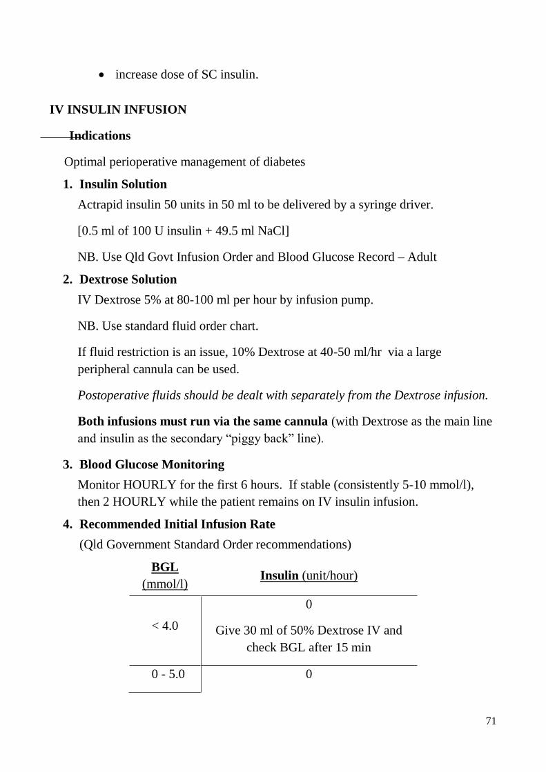
71
• increase dose of SC insulin.
IV INSULIN INFUSION
Indications
Optimal perioperative management of diabetes
1. Insulin Solution
Actrapid insulin 50 units in 50 ml to be delivered by a syringe driver.
[0.5 ml of 100 U insulin + 49.5 ml NaCl]
NB. Use Qld Govt Infusion Order and Blood Glucose Record – Adult
2. Dextrose Solution
IV Dextrose 5% at 80-100 ml per hour by infusion pump.
NB. Use standard fluid order chart.
If fluid restriction is an issue, 10% Dextrose at 40-50 ml/hr via a large
peripheral cannula can be used.
Postoperative fluids should be dealt with separately from the Dextrose infusion.
Both infusions must run via the same cannula (with Dextrose as the main line
and insulin as the secondary “piggy back” line).
3. Blood Glucose Monitoring
Monitor HOURLY for the first 6 hours. If stable (consistently 5-10 mmol/l),
then 2 HOURLY while the patient remains on IV insulin infusion.
4. Recommended Initial Infusion Rate
(Qld Government Standard Order recommendations)
BGL
(mmol/l) Insulin (unit/hour)
< 4.0
0
Give 30 ml of 50% Dextrose IV and
check BGL after 15 min
0 - 5.0 0

72
5.1 - 7.0 0.5
7.1 - 10.0 1
10.1 - 15.0 2
15.1 - 20.0 3
>20 4
If BGL <5 or >15 mmol/l, notify MO
• CONTINUE the Dextrose infusion if insulin infusion is ceased (BGL
<7.0).
• If BGL <5.0 mmol/l, BG measurements must continue at an HOURLY
rate.
• RECOMMENCE insulin infusion once BGL >7.0 mmol/l.
• If BGL consistently <5.0 mmol/l, decrease scale by 0.5-1.0 unit/hour or
increase Dextrose.
• If two consecutive BGL’s >15.0 mmol/l, increase dose by 0.5-1.0
unit/hour.
• If unsure, seek Senior advice or ask Endocrine team.
5. Review of BGL
Should be made by MEDICAL STAFF frequently (at least twice a day) for
safety reasons and to allow review of insulin infusion rate.
6. Insulin Administration
No other form or route of insulin should be given whilst having insulin infusion.
7. Normal Regimen of Insulin
Start approximately 1 hour prior to ceasing the infusion.

73
SLIDING SCALE INSULIN (Subcutaneous)
This may be used peri-operatively when insulin requirements are unpredictable, but
when intensive IV treatment is not required (eg satisfactory control, likely short
duration without normal diet).
It should only be used temporarily, as wide swings in BGL may occur.
Short-acting insulin (Actrapid or Humulin R) should be used 6 hourly and prescribed
in ‘Supplemental Insulin Orders’ on the Queensland Government Insulin
Subcutaneous Order and Blood Glucose Record sheet.
Initial dosing can be weight-based, but adjusted according to BGLs.
Suggested initial dose of short acting insulin
Weight
BGL (mmol/l) 50 - 100kg 100.1 - 150kg
<8.1-12 2 units 3 units
12.1-16 4 6
16.1-20.0 6 9
>20.0 8 12
The above Queensland Government sheet should be consulted for ongoing
management and further information

74
ANTIPLATELET AGENTS PERIOPERATIVE MANAGEMENT
1. General Guidelines
• Antiplatelet Agents (APAs) are used increasingly to prevent arterial thrombosis.
Dual antiplatelet therapy (DAPT) is usual for patients at high risk, especially
after placement of coronary artery stents or after acute coronary syndromes
(ACSs).
• The risk of thrombosis versus haemorrhage should be determined for each
patient.
• At present, platelet function tests are not widely available for routine use.
Consequently, decisions usually need to be based on drug pharmacology and the
available outcome data.
• Table 1 provides a schema for decision making for APAs. Frequently, the final
decision will require a judgement regarding the balance of risks.
• Please be aware:
o For patients at the highest risk of thrombosis (eg new coronary artery stent
plus a recent acute coronary event), the risk of death or further coronary
events is extreme (eg>50%).
o Combinations of anticoagulant drugs with different actions (eg combinations
of APAs and/or heparin) greatly potentiate the anti-thrombotic effect and
risk of bleeding.
o For some surgical patients bleeding from continued APA therapy can
result in death or severe morbidity.
• Considerations re postponing surgery: Depending on the surgical condition,
surgery should be delayed until the thrombotic risk is less, preferably until DAPT
(dual antiplatelet therapy) is no longer required. If this is not possible, delaying
until after 6 months after stent placement or ACS greatly reduces the risk.
Stopping DAPT within 6 weeks of stent placement should be avoided if at all
possible.
• Patient transfer: Depending on the adjudged risk of thrombosis, possible
transfer for surgery to a centre with at least daytime or even 24-hour re-stenting
capacity should be considered. However, as a possible thrombosis is
unpredictable in its timing, the duration of stay in that location (eg for surgical
recovery; 3 or 7 days) is unclear.
• Epidural/spinal anaesthesia and other nerve blocks: Neuraxial blockade is
acceptable on Aspirin and other NSAIDs. However neuraxial blockade, and other
blocks where control of bleeding from vascular injury is difficult, should
generally NOT be performed for 5-7 days after clopidogrel, 7-10 days after
prasugrel and 3-5 days after Ticagrelor. The risk is probably higher for an
epidural (esp with a catheter) than a single shot spinal. An epidural catheter
should be REMOVED at least 2 hours before potent APAs are started.

75
• For patients on both APAs and warfarin (eg for atherosclerosis and AF), try to
determine the major risk factor and manage according to the relevant
perioperative protocol (APA or warfarin).
• APAs should be re-started once the risk of major bleeding is minimal. This may
vary from 1 to 7 days post-op. A loading dose should be considered.
• Multi-disciplinary consultation (and careful discussion with the patient) and clear
documentation is necessary for complex patients.
• As duration of required DAPT is variable depending on patient and stent-related
factors (eg branching, length), consultation with the treating cardiologist is
recommended for all such patients.
2. Different Antiplatelet Agents
• Aspirin and ADP P2Y12 receptor antagonists (Clopidogrel, Prasugrel,
Ticagrelor) are inhibitors of platelet function through different mechanisms. The
P2Y12 antagonists are more potent than Aspirin; Prasugrel and Ticagrelor are
more potent than Clopidogrel. The clinical effect is longest for Prasugrel (7d) and
shortest for Ticagrelor (3d), which is a competitive antagonist.
• Dipyridamole (Persantin) is used in CVA prophylaxis in combination with
aspirin. Its duration of effect is only 12 hours.
• GPIIb/IIIa inhibitors are intravenously administered in acute coronary settings.
The effect of Abciximab lasts 12-24 hours; Tirofiban lasts for only 2-4 hours.
• Non-selective NSAIDs have a reversible effect, with a duration of action ranging
from hours (eg ibuprofen) to a couple of days (eg naproxen).
3. Emergency Surgery
• For an irreversible APA (eg Aspirin, Clopidogrel, Prasugrel) no obvious
improvement in platelet function can be expected within 24 hours, whereas
significant improvement occurs by 48-72 hours from platelet regeneration.
• The use of platelet function tests (PFTs) has been validated in emergency
situations: >50% platelet function indicates that surgical bleeding due to platelet
dysfunction is very unlikely. So consider cessation of APAs for 48-72 hours then
PFTs.
• Platelet transfusion may be helpful for treatment of excessive bleeding due to
Aspirin and P2Y12 antagonists, as is factor VIIa. The place of prophylactic
platelet transfusion is unclear.
• For the GP IIb/IIIa inhibitors, it is preferable to wait 12-24 hours after stopping
Abciximab and 2-4 hours after Tirofiban, rather than transfusing platelets with a
large amount of active drug present.

76
References
• UpToDate. Elective noncardiac surgery after percutaneous coronary intervention.
Cutlip and Windecker. Updated November 2014.
• Aspirin in patients undergoing noncardiac surgery (POISE-2).
Devereaux et al. N Eng J Med 2014; 370:1494-1503
• Cessation of dual antiplatelet treatment and cardiac events after percutaneous
coronary intervention (PARIS): 2 year results from a prospective observational
study. Mehren et al. Lancet 2013; 382:1714-1722
• 2012 Update to the Society of Thoracic Surgeons Guideline on the Use of
Antiplatelet Drugs in Patients Having Cardiac and Non-Cardiac Operations.
Ferraris et al. Ann Thorac Surg 2012; 94:1761-1781
• Double antiplatelet therapy after Drug-eluting stent implantation. Risks
associated with discontinuation in the first year. Ferreira-Gonzalez I et al. JACC
2012; 60:1333-1339
• Perioperative management of antiplatelet therapy in patients with coronary artery
disease. Joint position paper including European Society for Cardiology. Korte et
al. Thrombosis and Haemostasis 2011; 105:743-749.
• Regional Anaesthesia in the patient receiving antithrombotic and antiplatelet
medication. Horlocker T. Br J Anaes 2011;107(S1) i96-i106

77
Table 1: Management of Aspirin and Clopidogrel in Elective Surgical Patients
1. Determine bleeding and thrombosis risk
2. Read management plan from table
3. Complex situations may need discussion with surgeon,
anaesthetist, treating cardiologist and patient
4. Consult cardiologist if on DAPT#
Bleeding Risk of the Procedure
Major:
(Risk of major bleeding >5% or life/limb
threatening)
Intermediate:
(Risk of Major Bleeding 1- 5%)
Minor:
(Risk of Major Bleeding <1%)
Eg: Neuro/ spinal, aortic or major pelvic, major
ENT, prostate surgery
Eg: Abdominal, Major joint, Non-aortic vascular surgery, urology
except TURP, ERCP
Superficial surgery
eg Cataract, minor skin lesions, ECT
Risk of Thrombosis (with cessation of treatment + surgery)
Category Examples Management
Extreme
Death 5-15%
Death + MI/redo stent
25%
• *DES or **BMS with 6 weeks
• ***ACS within 6 weeks
• Postpone surgery1;
If impossible,
• Consider transfer to centre with re-stenting capacity2, AND
• Stop APAs 5-7d pre-op;
AND
• substitute short-acting anti-platelet drug eg
tirofiban infus’n until 4/24 preop
• Postpone surgery1;
If impossible,
• Consider transfer to centre with re-stenting capacity2, AND
• Continue APAs
OR
• continue aspirin, but stop other APAs 5-7d pre-op and substitute short-acting anti-platelet drug eg tirofiban until 4/24 preop
• Postpone surgery1;
If impossible,
• Continue APAs
High
• DES and/or ACS > 6 weeks but within
1 year still requiring DAPT#
• Postpone surgery1;
If impossible,
• Consider transfer to centre with re-stenting capacity2, AND
Stop APAs 5-7 d pre-op
• Postpone surgery1;
If impossible,
• Consider transfer to centre with re-stenting capacity2, AND Continue aspirin only from 5-7 d pre-op
• Postpone surgery1;
If impossible,
1. Continue APAs
Moderate
Death + MI/redo stent 1-
5%; CVA 0.2-1%
• Other DES or BMS patients
• Ischaemic Heart Disease (chronic);
• TIA/CVA (previous)
Stop APAs 7-10d pre-op Stop APAs 5-7d pre-op
(except continue single APA for carotid or peripheral vascular
surgery)
Continue aspirin;
Continue or stop other APAs 5d
preop
Minor
Death + MI/redo <1%
HT/DM etc alone without proven IHD Stop APAs
10d preop
Stop APAs
5-10d preop
Continue or stop APAs (surgical
preference)
#DAPT – dual antiplatelet therapy; ##APAs – antiplatelet agents; *DES – Drug-eluting stent; **BMS – Bare metal stent; ***ACS – acute coronary syndrome;
1see notes re postponing surgery; 2see notes re patient transfer Version 4.1 Dec 2014

78
POSTOPERATIVE VENOUS THROMBOEMBOLISM (VTE)
Introduction:
The management of patients with suspected or proven thromboembolism in the
postoperative period is problematic. The risk of death or major morbidity from
pulmonary embolus or venous thrombosis must be balanced with the risk of
bleeding from the operative site, the gastro-intestinal tract, or at the site of other
invasive procedures (such as epidurals or arterial lines).
Guidelines:
• Confirmation of the diagnosis with appropriate radiological imaging is
essential, as the symptoms and signs of thromboembolism are non-specific.
Urgent imaging is required in unstable patients who are also at high risk of
bleeding complications. There is no place for the measurement of D-dimer,
which is raised in most postoperative patients with or without VTE.
• Heparinisation should not be commenced in the postoperative patient before
the diagnosis is proven, unless there is a high clinical suspicion of pulmonary
embolism. Initial management should focus on simple resuscitative measures
(ABC) and determining the diagnosis.
• Patients who have been significantly hypoxic or haemodynamically unstable
should initially be managed in a Critical Care environment (ICU or CCU).
Early involvement of relevant staff is required for these patients.
• Senior consultation between surgeon, physician and anaesthesia/intensive care
is necessary. The risk of death from pulmonary embolism varies, depending
chiefly on cardiorespiratory reserve and the site and extent of embolism
(determined by imaging). The risk of bleeding will also vary, depending on the
nature of surgery and time elapsed. Management therefore must be
individualised, with consideration of the relative merits of heparinisation or a
vena caval filter.
• If heparinisation is indicated, un-fractionated heparin (and monitoring of
APTT) should be used within the first two weeks of major surgery, as low
molecular weight heparins (eg Clexane) are difficult to reverse in the event of
bleeding.
• Before heparinisation, epidural catheters should be removed, if possible at least
1-2 hours beforehand, to minimise the risk of an epidural haematoma.
References:
1. Tai N, Atwel A and Hamilton G. Modern Management of Pulmonary Embolism.
Br J Surg 1999;86: 853-868.
2. Hambleton J. Anticoagulation in High-Risk Patients: When is this approach not worth the risk?
Am J Med 2000; 109: 335-337.

79
PREOPERATIVE ASTHMA AND REFLUX THERAPIES
Asthma
i. Asthmatics are at higher risk of perioperative respiratory complications,
including bronchospasm.
ii. Optimal control of asthma in the preoperative period is important to minimise
this risk.
iii. Patients should continue their usual therapy (especially ‘preventers’).
iv. Nebulised salbutamol immediately pre-operatively is usually warranted.
v. Consider oral prednisolone 0.5 – 1.0mg/kg for three days preoperatively in the
following asthmatic patients:
• Currently wheezy
• FEV1 <60%
• Admitted to ICU with asthma in the past
• Inpatient with asthma in the past year
• Visit to Emergency Department in the past 6 months
• Normally on oral prednisolone
• Have taken oral prednisolone in the past 6 months.
(Oral prednisolone is available ‘pre-packed’ in the Anaesthetic Clinic)
vi. Explain to the patient the reason for these measures, to improve compliance.
Reflux
i. Patients with gastro-oesophageal reflux are at higher risk of pulmonary
aspiration during anaesthesia.
ii. The following is advised for such patients:
• Attention to fasting guidelines and consideration of regional anaesthesia
• Continuation of any current anti-reflux treatments (ie PPIs or H2 blockers)
• Advise patient to take their PPI/H2 blocker on the morning of surgery
• If symptoms are poorly controlled despite treatment, prescribe sodium citrate
(30ml of 0.3M solution), to be taken just before coming to theatre
• If not on treatment, give famotidine 40mg tablet the night before and the
morning of surgery (‘pre-packed’ in anaes clinic), or prescribe a PPI (eg
omeprazole 20 mg).
iii. Patients for elective CS should receive famotidine as above; sodium citrate
should be given for emergency CS.
iv. Explanation of the reason for these measures (‘to avoid food or acid going into
the lungs’) seems to improve compliance.
80
PREOPERATIVE FASTING FOR ADULT INPATIENTS
• General anaesthesia (GA) depresses protective upper airway reflexes,
predisposing patients with significant residual gastric contents to the dangerous
complication of pulmonary aspiration.
• Patients for GA should therefore be fasted for sufficient time to minimise gastric
contents.
• Patients for sedation are also vulnerable, and patients for regional anaesthesia
sometimes require conversion to GA. Consequently, both of these groups should
also be fasted.
• Lengthy fasting is, however, uncomfortable for many patients, and can result in
dehydration, ketosis and hypoglycaemia.
• This protocol is intended to minimise both the risk of aspiration and the
physiological impact of fasting.
Recommendations for Patients Undergoing Elective Surgery
• Water may be consumed up to 2 hours prior to the scheduled surgery.
• Solids, or fluid other than water may be consumed up to 6 hours preoperatively
• After a particularly heavy or fatty meal an overnight fast is warranted.
• It is strongly suggested that surgery should be postponed for 12 hours in patients
who have recently drunk alcohol, taken hallucinogenic drugs, or smoked.
• Routine medication should usually be taken at the prescribed time, with a sip of
water if needed.
• Each patient should be individually assessed to exclude co-morbidities that may
necessitate variation in fasting times, or the use of prokinetic or alkalinising
agents. (see below)
Special Situations
The following specific groups of patients may be at increased risk of aspiration
and their fasting requirements should be discussed with the attending anaesthetist:
Those who are: or those with: and those who are:
Morbidly obese
Pregnant
Diabetic
Ileus or gastric stasis
Significant oesophageal reflux
A potentially difficult airway
Receiving opioids
Having an emergency
procedure

81
PREOPERATIVE FASTING FOR PAEDIATRIC INPATIENTS
(ELECTIVE SURGERY)
General Rules:
• Children must fast for six hours from food or milk drinks before their operation.
• Babies less than six months of age (breast or bottle-fed) must fast for four hours.
• Clear fluids (cordial or water) may be given until two hours before the scheduled
time of the operation.
Further Points:
• Parents or ward staff should time the fasting period for 8.00 am surgery if the
child is on a morning or all day operating list, and 1.00 pm surgery if the child is
on an afternoon list.
• If surgery is in the AFTERNOON then a light breakfast of toast and milk fluids
may be given until 7.00 am.
• Regular medications, analgesics and pre-medication ordered by the Anaesthetist
should be given, even within 2 hours before the procedure. IF THERE IS ANY
UNCERTAINTY, the attending Anaesthetist (or the Anaesthetist on call)
SHOULD BE CONTACTED for advice.
• Any variation to these guidelines can only be authorised by the Anaesthetist.
• FAILURE TO OBSERVE THESE GUIDELINES MAY RESULT IN THE
PROCEDURE BEING DEFERRED OR CANCELLED.

82
PREOPERATIVE FASTING FOR DAY SURGERY PATIENTS
• General anaesthesia (GA) depresses protective upper airway reflexes,
predisposing patients with significant residual gastric contents to the dangerous
complication of pulmonary aspiration.
• Patients for GA should therefore be fasted for sufficient time to minimise gastric
contents.
• Patients for sedation are also vulnerable, and patients for regional anaesthesia
sometimes require conversion to GA. Consequently, both of these groups should
also be fasted.
• Lengthy fasting is, however, uncomfortable for many patients, and can result in
dehydration, ketosis and hypoglycaemia.
• Patients are occasionally called to theatre earlier than anticipated due to changes
in list order, shorter than planned operating times and unexpected cancellations.
When these patients are not fasted when called to theatre it results in disruption
to the theatre schedule. This can lead to delays and/or cancellations to the elective
list. Fasted patients from other lists (e.g. emergency list) will be called for to
avoid wasting theatre time and resources.
• This protocol is intended to minimise both the risk of aspiration and the
physiological impact of fasting.
General Rules:
• Patients must fast for SIX hours from food and all fluids other than water
before their operation.
• Patients booked on MORNING and ALL-DAY operating lists must fast from
MIDNIGHT.
• ONLY patients booked on operating lists STARTING AFTER 12:30 PM may
have a light breakfast BEFORE 06:30 AM. A light breakfast may include
toast/cereal/milk products, and excludes fried food/meat/egg.
• Patients should be ENCOURAGED TO DRINK WATER up until 2 hours prior
to surgery. Day surgery staff should consult with the team leader for each theatre
to determine how long it will be before the patient is called to theatre.
• Patients who have persistent symptoms or signs related to prolonged fasting
should receive treatment. The attending anaesthetist should be consulted, and
will make necessary arrangements.
• Patients who have a reduced capacity to fast prior to surgery should be identified
prior to booking and scheduled at the beginning of the operating list. This
includes children, the elderly, diabetics, pregnant and mentally disabled patients.

83
Patients who have fasted on successive days should also be scheduled at the
beginning of the list.
Further Points:
• Regular medications, analgesics and pre-medication ordered by the Anaesthetist
should be given, even within 2 hours before the procedure. IF THERE IS ANY
UNCERTAINTY, the attending Anaesthetist (or the Anaesthetist on call)
SHOULD BE CONTACTED for advice.
• Any variation to these guidelines can only be authorised by the Anaesthetist.
• FAILURE TO OBSERVE THESE GUIDELINES MAY RESULT IN THE
PROCEDURE BEING DEFERRED OR CANCELLED.
Special Situations:
The following specific groups of patients may be at increased risk of aspiration and
their fasting requirements should be discussed with the attending anaesthetist.
Those who are: or those with: and those who are:
Morbidly obese
Pregnant
Diabetic
Ileus or gastric stasis
Significant oesophageal
reflux
A potentially difficult
airway
Receiving opioids
Having an emergency
procedure

84
ROUTINE PREOPERATIVE INVESTIGATIONS
AGE
Over 60 ECG, FBC, UEC
PROPOSED OPERATION
These investigations should be ordered no matter how young or fit the
patient
Bowel Surgery, Hysterectomy,
Major Joint Surgery, Major Back
Surgery, TURP, Major Vascular
Surgery
FBC, Iron Studies (IS), UEC
HEALTH QUESTIONNAIRE
These investigations should be ordered no matter how minor the surgery
Heart Disease (including HTN) ECG, FBC, UEC
Respiratory Disease
(except mild asthma)
ECG (>40 years old), FBC, UEC
Chest X-ray or PFT are not routine
investigations for patients with respiratory
disease, these should only be ordered
depending on the clinical findings
Diabetes mellitus ECG (>40), FBC, UEC, Glu, HbA1C
Renal Disease ECG (>40), FBC, UEC, Mg, Ca, PO4
Liver Disease ECG (>40), FBC, UEC, LFT, Coags
Thyroid Disease ECG (>40), FBC, UEC, TFT
Rheumatoid Arthritis Flex/Ext Neck X-ray
Bleeding disorder including
anticoagulants
FBC, Coags

85
NOTE
Organising a group and hold or a cross match is the responsibility of the
surgical team.

86
IRON DEFICIENCY ANAEMIA PRE-OP
Determine cause.
• If no obvious cause consider gastroscopy / colonoscopy.
• Consider deferring operation.
• Treat with oral iron.
Consider IV iron transfusion pre-op:
• If a patient is anaemic (Males <130g/L, Females <120g/L)
• And iron deficient (Ferritin <30mcg/L )
• And the operation is not deferrable and has the potential for
substantial blood loss (and thus transfusion) eg: Colectomy.
IV Iron therapy:
• Prescribe Ferric Carboxymaltose 1000mg (in 250ml NSaline,
over 15 mins), on a fluid prescription chart
• Refer to Patient Blood Management (PBM) Anaemia Clinic (Tel
69868)
• They will organise for the patient to be admitted through the
Minor Procedures Unit and organise the infusion.
• They will repeat the iron studies and will come to the anaesthetic
clinic for another prescription if the patient would benefit from
further iron transfusion.

87
Iron Therapy
Oral iron in divided daily doses. Evaluate response after 1 month. Provide
patient information material.
IV iron if oral iron contraindicated, is not tolerated or effective; and consider if
rapid iron repletion is clinically important (e.g. <2 months to non deferrable
surgery).
NOTE: 1 mcg/L of ferritin is equivalent to 810 mg of storage iron. It will take
approximately 165 mg of storage iron to reconstitute 10 g/L of Hb in a 70 kg
adult. If preoperative ferritin is <100 mcg/L, blood loss resulting in a
postoperative Hb drop of ≥30 g/L would deplete iron stores. In patients not
receiving preoperative iron therapy, if unanticipated blood loss is encountered,
150 mg IV iron per 10g/L Hb drop may be given to compensate for bleeding
related iron loss (1 ml blood contains ~0.5 mg elemental iron)
See http://www.blood.gov.au/pbm-guidelines-preoperative-optimisation-
template for more information

88
Is the patient anaemic?
Hb <130 g/L (male) or
Hb <120 g/L (female)
Preoperative tests
• Full blood count
• Iron studies2 including ferritin
• CRP and renal function
Preoperative haemoglobin assessment and optimisation template
This template1 is for patients undergoing procedures in which substantial blood loss is anticipated such as cardiac surgery, major orthopaedic, vascular
and general surgery. Specific details, including reference ranges and therapies, may need adaptation for local needs, expertise or patient groups.
Ferritin <30 mcg/L2,3
NO YES
Ferritin >100 mcg/L
Possible anaemia of chronic
disease or inflammation, or
other cause5
• Consider clinical context
• Review renal function,
MCV/MCH and blood film
• Check B12/folate levels and
reticulocyte count
• Check liver and thyroid function
• Seek haematology advice or, in
the presence of chronic kidney
disease, renal advice
Possible iron deficiency
• Consider clinical context
• Consider haematology advice or,
in the presence of chronic kidney
disease, renal advice
• Discuss with gastroenterologist
regarding GI investigations and
their timing in relation to surgery3
• Commence iron therapy#
Iron deficiency anaemia
• Evaluate possible causes based
on clinical findings
• Discuss with gastroenterologist
regarding GI investigations and
their timing in relation to surgery3
• Commence iron therapy#
No anaemia: ferritin
<100 mcg/L
•Consider iron therapy# if
anticipated postoperative Hb
decrease is ≥30 g/L
•Determine cause and need for GI
investigations if ferritin is
suggestive of iron deficiency <30
mcg/L2,3
Raised Normal
CRP4
Ferritin 30–100 mcg/L2,3

89
PREOPERATIVE MEDICATION IN FASTING PATIENTS
Introduction
It is important that most prescribed medication is given to patients, even during a
designated period of fasting before an operation. Surgery may be delayed if appropriate
medication has not been given.
Unless instructions to the contrary are given for a particular patient, the following
guidelines apply.
A. Medication to Give:
1. Premedication prescribed for the operation;
2. Analgesia;
3. An insulin regimen specifically prescribed for the operation;
4. The patient’s usual medication. This is particularly important in the case of:
a) Cardiovascular drugs, ie. for hypertension, angina, arrhythmias and heart
failure, and
b) Respiratory drugs (including inhalers), to prevent deterioration in the
patient’s condition over the operative period,
and
c) Anti-reflux therapy, to minimise the risk of pulmonary aspiration of
gastric acid.
Points to note:
i. Patients with vomiting or peritonitis should receive medication by a non-oral route.
ii. It is reasonable to give important tablets with a small sip of water up to half an
hour before theatre, though an hour or more beforehand is preferable.

90
B. Medication to Withhold:
1. Oral hypoglycaemic drugs, which may cause hypoglycaemia in fasting
patients.
2. Anticoagulants such as aspirin, clopidogrel, warfarin and heparin. Non-
steroidal anti-inflammatory drugs (eg. Brufen), may also result in increased
haemorrhage during surgery or epidural/spinal anaesthesia.
These drugs should initially be withheld, pending discussion with the surgeon and
anaesthetist responsible for the patient.
Points to note:
i. A plan of management should be made for each diabetic patient.
ii. All patients on anticoagulants require specific management plans. They should
all be referred to the Anaesthetic Clinic or the Anaesthetist on call, preferably at
least 1 week prior to surgery, to minimise the chance of cancellation or delay.
iii. Heparin DVT prophylaxis should generally be withheld if a spinal or epidural
anaesthetic is intended. Low dose (ie 20 – 40mg daily) Clexane can be given at
1800 hrs the day before surgery.
iv. On occasions it may be preferable to withhold other medications, such as oral
contraceptives or anti-depressants, but this should be done in consultation with
the treating medical staff.

91
PACU INTRAVENOUS PAIN PROTOCOL
Sedation Score
0 = Nil Awake, alert
1 = Mild Sometimes drowsy, easily roused
2 = Moderate Very drowsy, easy to rouse
3 = Severe Somnolent, difficult to rouse.
A = Asleep Normal sleep, easy to rouse
Opioid Order as per Anaesthetist
Dilute to 10ml with saline:
Morphine 10mg,
Oxycodone 10mg,
Fentanyl 200μg,
Tramadol 100mg as ordered

92
Paediatric IV Pain Protocol FOR PACU (Children <40kg)
1. Ensure that patient has an Opioid Pain Protocol ordered.
2. First make up the Recovery Pain Protocol Adult Opioid Solution:
a. Morphine 10 mg into 10ml with saline, to make 1mg/ml, or
b. Fentanyl 200 mcg into 10ml with saline, to make 20mcg/ml, or
3. Round off the patient’s weight to the nearest 5kg
(eg 20 kg for a 22 kg child)
4. Discard some of the Adult Opioid Solution to leave the Residual Amount
in the syringe according to the table below, then
5. Add further saline to make up to 10 ml (Final concentration is given in the
table).
Step 3
Round off
Patient’s weight
Step 4
Discard some
of Adult Opioid
Solution
Step 5
Add further saline to make up to 10ml
Final Concentration per ml
Approx
Weight
Residual
Amount
Morphine
(mg)
Fentanyl
(mcg)
Pethidine
(mg)
10 kg 2 ml 0.2 4 2
15 kg 3 ml 0.3 6 3
20 kg 4 ml 0.4 8 4
25 kg 5 ml 0.5 10 5
30 kg 6 ml 0.6 12 6
35 kg 7 ml 0.7 14 7
40 kg 8 ml 0.8 16 8
>40 kg 10 ml 1.0 20 10
6. Give 1 or 2 ml increments according to the Recovery IV Pain Protocol,
monitoring Pain and Sedation scores

93
PACU ANTIEMETIC STANDING ORDERS
Introduction
• Nausea in the postoperative period is common, and effective treatment often
requires more than one drug.
• The Antiemetic Protocol is designed to facilitate this process, which can
usually follow a standard format.
• Attention must first be paid to more critical aspects of the recovery process,
specifically care of the airway, breathing and circulation.
Management (for adults 18-70 years)
A. Verify that the patient has an “Antiemetic Protocol” prescription
(either on Winchart or paper Anaesthetic Record).
B. Give drugs in the order below until nausea is relieved.
C. If nausea persists after 10 minutes, give the next drug.
D. Record each drug and dose given on the Recovery record sheet.
E. If all four drugs are ineffective, or nausea recurs, notify the anaesthetist.
Antiemetic
Protocol Drugs
1. Metoclopramide 10mg IV
2. Ondansetron 4mg IV
3. Droperidol# 1mg IV
4. Cyclizine 50mg IV
#Droperidol preparation: dilute 2.5mg (1 amp) into 2.5ml total with saline and
give 1ml

94
PACU DISCHARGE CRITERIA GUIDELINES
GENERAL REMARKS
- The Post-Operative Care Unit (PACU) is a Critical Care Area with a
high potential for immediate life-threatening problems.
- The relevant Anaesthetist is ultimately responsible for his/her patients in
the PACU.
- Care may be delegated, when appropriate, to the care of nursing staff
experienced and skilled in the care of unconscious and surgical patients.
- The Discharge Criteria Guidelines are intended to ensure that patients
are in a safe and satisfactory condition before returning to a general
postoperative ward.
- Variations from the stated parameters may be allowable for many
reasons. However, patients who do not meet the guideline criteria
must not be discharged from the PACU without consultation with
the relevant anaesthetic medical staff.
- The PACU nurse should always contact the anaesthetist:
a. if there is doubt regarding a patient’s fitness for discharge, or
b. if the anaesthetist has asked to be contacted prior to the patient’s
discharge.
DISCHARGE CRITERIA
1. Airway and Breathing
- Airway must be clear without assistance.
- Respiratory rate: adults >8/min; children: <6 months >30/min;
6-12 mo > 25/min; >1 year: >20 – age/2.
- Able to deep breathe on command (adults).
- Oxygen saturation 95%.
2. Circulation
- Pulse regular.
- Heart Rate appropriate for age:
Adult or large child: 60-100/min

95
Small child: 80-120/min
Infant (< 1 year): 100-140/min
- Systolic Blood Pressure:
Adult or large child > 100 mmHg and within 30% of pre-op
level.
Small child (if req’d) > 85 mmHg
Infant < 1 year (if req’d) > 70 mmHg
- Perfusion: hands and feet should be warm and pink.
- IV cannula (IV fluids not running):
If required on the ward it must be flushed in the RR.
3. Neurological
4. Sedation score: 2 or better.
- Able to lift head off pillow and/or firmly squeeze hand.
- Epidurals and spinals
Spinal anaesthetic: Block receded by 2 dermatomes and
below T4.
Lumbar epidural: Block below T6.
Thoracic epidural: Block below T4.
5. Metabolic
- Temperature on discharge > 36 0C and <38 0C.
- Diabetic patients: BGL between 5 and 10 mmol/l.
6. Renal
- Fluid input and output must be documented.
- If IDC in situ output > 1ml/kg/hour.
7. Comfort
- Pain must be adequately controlled (moderate or less).
- Nausea/vomiting must be adequately controlled.
- The patient should be clean and dry.
- Cot bumpers are required for all small children and restless patients.
8. Specific Surgical Observations
- As appropriate, such as:
Wound dry
Drains secure and functioning
Minimal blood loss

96
Limb perfusion and movement adequate
9. Minimum Length of Stay
a. All other discharge criteria must be met before discharge is considered
b. Many patients will therefore require a longer period in the PACU
c. Patients must not be discharged before this length of time unless
signed out by an anaesthetist
- LA only (no sedation):
Discuss with the surgeon if PACU observation is required
- Minor surgery:
GA, Sedation, Bier’s Block, major nerve blocks: 20 minutes
- Major surgery:
One hour. Knee replacement with a tourniquet: Two hours
- After opioid pain relief:
30 minutes after IM or epidural bolus; 10 minutes after IV bolus
- Children less than 1 year of age:
All must be reviewed by an anaesthetist before discharge from the
PACU
10. Documentation is complete
- Most observations should be recorded on the WinChart record
- Fluids should also be recorded on the fluid balance sheet
- Orders for oxygen, fluids, analgesia and anti-emetics as appropriate
11. Transportation
- Nurse and wardsperson/USO present
- Portable oxygen, Hudson mask, pocket mask, tubing, sucker and
suction with patient
- Additional equipment for ventilated patient

97
AIRWAY MANAGEMENT
A. General responsibilities.
1. Airway assessment
• Look for suspected problems with
1. Pre- oxygenating
• Difficult mask seal or cooperation
• Reduced size of FRC
• Increased oxygen consumption
2. BMV
• BMI > 30
• Edentulous
• Facial hair
• OSA or history of snoring
• Mallampati III or IV
3. Laryngoscopy
• History
• Mallampati III or IV
• Reduced thyromental distance
• Limited neck extension
• Restricted mouth opening (and unfavourable dentition)
• Gross face or neck abnormalities
4. LMA placement
• Male
• Poor dentition
• BMI > 30
5. Anterior neck anatomy
2. Communication
• If you are in any doubt about your airway plan or your ability to execute
it, you should communicate with your consultant.
• You are not expected to manage situations that are beyond your level of
experience or expertise.
• You will not be criticised if you ask for help.
3. Trauma and MET calls
• As the duty anaesthetic registrar you will not be included in MET calls.

98
• The duty anaesthetist is included in Trauma Team Activation calls.
Primarily your role in Airway Management for Trauma is supportive to
the Emergency Department staff as described in the Trauma Team
Chapter
• If there is an anticipated need for anaesthetic airway assistance you will
be phoned directly.
• You may occasionally be asked to provide airway assistance in ICU, ED,
radiolology, cath lab, or the ward. If you are able to do so you should
attend as requested.
• Your primary responsibility is always with the patient you are currently
looking after. If you get asked to assist somewhere else in the hospital
while you are busy in theatre, you simply can’t go.
• If you can’t attend or are out of your depth, the other options for help are
the ICU registrar or the consultant on for ICU or anaesthetics.
B. Unexpected airway emergencies.
• Ultimately there are only four ways to oxygenate a patient:
1. Through the face – BMV.
2. Supraglottic – LMA.
3. Infraglottic from above – ETT.
4. Infraglottic through the neck.
• Have an initial attempt, optimise, have another go, then abandon – you
must move on.
• 1, 2 and 3 can be done in any order.
• Have no more than three attempts at each of 1, 2 and 3 (preferably only
two).
• Don’t keep repeating unsuccessful things.
• If necessary the patient ends up with a hole in their neck, and you should
do this BEFORE they die.
• A useful model for emergencies is the Airway Vortex (Nicholas Chrimes
2013).

99
• Imagine you are looking
down into a funnel and rolling
a marble around the edge. You
can start in any of the three
regions and go in either
direction, but if they all fail
you put a hole in the patient’s
neck.
• http://vortexapproach.com/Vortex_Approach/Vortex.html
C. Advanced airway equipment.
1. Video-laryngoscopes
• Each operating theatre has a video-laryngoscope with size 3 and 4
Macintosh shaped blades, as well as “difficult” blades.
• The blades are cheap and disposable, so there are no restrictions on
their use.
2. Cricothyroidotomy kit
▪ A Cook Enk Flow Modulator kit can be found on the right hand side
of the second drawer of all anaesthetic drug trolleys.
▪ They attach directly to any oxygen outlet. The flow should be
1L/min/year of age (up to 15 L/min).
▪ The needle can be placed through the cricothyroid membrane or
trachea.
3. Difficult airway trolley
• This lives in the technician storage area.
• Items found on this trolley include:
o Drugs and equipment for airway topicalisation.
o Adjuncts for awake intubation.
o Sugammadex 8 x 200mg ampoules for reversal of rocuronium
(and vecuronium)

100
▪ Doses are 2, 4 and 16 mg/kg for standard, deep and
immediate reversal respectively.
▪ Recovery also stocks a small supply of sugammadex.
▪ If you use it you must fill out the form to have it
replaced.
o McCoy, Miller, Kessel, left handed and short handle
laryngoscopes.
o Cricothyroidotomy/tracheostomy gear.
o Manujet jet ventilator.
o Airway exchange catheter, Aintree catheter, staged extubation
kit, endobronchial blocker and bougies.
4. Bronchoscopes
• All our bronchoscopes are the disposable “AMBU-scope” video-
bronchoscope system.
• The “Ambu-scopes” connect to a small rechargeable monitor that is
attached to a drip pole. This system is compact, cheap and mobile
(doesn’t require electricity). If you are inexperienced with
bronchoscopy you should use this scope in preference to our Olympus
ones.
D. Airway grab bag
• This bag lives at the bottom of our difficult airway trolley, and
contains standard equipment for managing airways in off site
locations.
E. Difficult airway documentation
• The general consensus is that should a patient require specialised
equipment or techniques to safely manage their airway this needs to be
clearly documented and communicated.
• QHEPS has a form that should be completed in this instance
o https://clinicalexcellence.qld.gov.au/sites/default/files/docs/clini
cal-networks/difficult-airway-alert.pdf
• Copies should go to the patient, their general practitioner and into the
notes.
• An alert should be entered into ieMR and AARK
101
TRAUMA TEAM ACTIVATION
Criteria for Trauma Team Activation
Trauma Teams are activated by ED when a patient presents or is expected (QAS
or Lifeflight) with:
• Significant penetrating injuries (head, neck, chest, abdomen)
• Major head injuries (dilated pupil(s), open injury, severe facial injury)
• Systolic BP <90 mmHg
• Request by Retrieval Physician
• Discretion of ED Team Leader
Trauma Team Members (Medical)
1. ED
a. Team Leader
b. Airway
c. Circulation & Procedure
d. Primary Survey
2. Duty Anaesthetist
3. ICU Registrar
4. Surgical Registrar
Role of Duty Anaesthetist
1. Provide airway assistance as required by the ED Team Leader. Usually
airway management will be undertaken by ED Airway Doc in the first
instance. The anaesthetist provides assistance and oversight. Out-of-
hours the anaesthetist may be asked to be the primary airway doctor if ED
skill mix reduced.
2. Facilitate movement to OT if required expediently. The anaesthetist will
liaise with the Team Leader in theatre - 66924 and the Emergency OT
Anaesthetic Technician – 67162. The Anaesthetist will transfer the
patient from ED to OT.
3. Timely communication with Consultant Anaesthetist on-call if not
already present.
4. Procedures as required by ED Team Leader (e.g. vascular access)

102
Practical Points:
1. Unable to attend If you are unable to attend ED immediately contact the
ICU access Registrar - 66979 to inform them you are indisposed. A line
of communication should be then maintained between ICU &
Anaesthetics and Anaesthetics should make every effort to attend ED as
soon as possible. DO NOT Call the ED Consultant.
2. Attending ED Make sure you introduce yourself to the ED Team Leader
on arrival. Await their instructions before entering the Trauma Bay.
Remain behind the Red Line until asked to cross.
Roles of other Team Members:
1. ICU Registrar
a. Provide procedural assistance to the ED Team Leader
b. If skills appropriate may provide airway assistance as per
Anaesthetist especially if Anaesthesia unable to attend
c. Assist with advanced transfusion management (Massive
Transfusion Protocol)
d. Clear line of communications
i. ICU Consultant
ii. ICU TL regarding bed management
iii. Anaesthesia as overleaf
2. Surgical Registrar
a. Provide procedural assistance to the ED Team Leader
b. May be required to undertake primary survey if ED skill mix
reduced
c. Clear line of communication with Surgical Consultant to make
time critical decisions about movement to theatre

103
MASSIVE TRANSFUSION PROTOCOL (MTP)
Introduction
Massive transfusion is defined by the Australian Red Cross as
“in adults, Replacement of >1 blood volume in 24 hours or >50% of blood volume
in 4 hours (adult blood volume is approximately 70ml/kg)…In children it is defined
as transfusion of >40ml/kg”
A Massive transfusion protocol should be initiated if there in anticipated requirement for a
massive transfusion and involves multiple team members. Where possible ROTEM
analysis should be used to guide the delivery of blood component therapy.
ROTEM
Criteria for the use of ROTEM analysis are:
• Any patient with a massive transfusion protocol activated
o ROTEM results will guide product use according the ROTEM-guided MTP
• Trauma patient
o If a request to perform ROTEM comes from OT or ED and it is stated that
the patient has critical bleeding
• Obstetrics
o If a request to perform a ROTEM comes from OT and it is stated that the
patient is bleeding excessively
• Actively bleeding patients
o Anticipated to need >3units of blood in the next hour
o Haemodynamically unstable due to active bleeding requiring blood
transfusion eg PPH / upper GI bleed
Use of ROTEM should be discussed with the on-call Intensivist and in general the patient
will need to be critically bleeding.
Once ROTEM approved
• Contact ICU Registrar (66979) to coordinate ROTEM use
• Notify Pathology that ROTEM is being used to guide the MTP
• Collect 5ml blood in a Citrate blood tube (Blue top)
• Hand deliver sample to ICU Nursing Team Leader with 2 patient identification
stickers. DO NOT SEND in the Lamson tube system
• Results can be monitored real-time on the ROTEM Secure View accessed via most
PC desktops in the OT
ROTEM should not be used to monitor antiplatelet agents and anticoagulants routinely.
See below for ROTEM analysis guidance. The ICU Registrar will be available to liaise
with the treating time on appropriate blood product use. It is the anaesthetist’s
responsibility to communicate with Blood Bank about requirements.

104
If treatment is given ROTEM should be repeated after 10 minutes to assess response.
If ROTEM confirms hypofibrinoginaemia Fibrinogen concentrate (RiaSTAP) is available
in ICU. 2 specialists are required to approve the use of RiaSTAP; the on-call intensivist
and usually the lead clinician involved in the resuscitation.

105
ROTEM ALGORITHM FOR INTERPRETATION

106
CAIRNS HOSPITAL CRITICAL BLEEDING ALGORITHM

107
RED BLANKET PROTOCOL
Introduction
Patients who remain haemodynamically unstable despite resuscitation,
with uncontrollable or non-compressible bleeding, may need to be
expediently transferred to the operating theatre for life saving surgery.
- A Trauma Call must already be activated
- Staff will attend the trauma call as required by their role – Trauma Call
- Massive transfusion protocol should be activated
- Case may be discussed with the general surgical consultant prior to
patient arrival in ED if there is sufficient pre-hospital concern
- Upon receipt of a trauma call the duty anaesthetist should, in liaison
with the theatre team leader, identify potential staff and theatre space
for a potential red blanket patient requiring emergency surgery
Decision for ‘Red Blanket’ activation
- The Surgical Consultant may make the decision to approve activation
of a ‘Red Blanket’
- The Emergency Department Consultant or surgical registrar may
recommend a red blanket to the Surgical Consultant
- The decision should occur in agreement between the Emergency and
Surgical consultant
Process for ‘Red Blanket’ activation
- Call Duty Anaesthetist on 66910 and state ‘Red Blanket Activation’
- The duty anaesthetist will liaise with the theatre team leader who will
organise additional staff and theatre space
Emergency Consultant responsibilities
- The most senior emergency department doctor will accept full
resuscitation responsibility until a formal handover to an anaesthetic
team occurs
- After hours, this may necessitate the emergency doctor continuing to
assist in theatre until the anaesthetist has the resources to assume full
responsibility
- Ensure that massive transfusion protocol has been activated
- Inform ICU registrar of need for post-operative bed
- May be required to transfer the patient to theatre once notified theatre is
ready

108
Surgical Consultant/team responsibilities
- Inform the operating theatre of specific surgical requirements
- Inform the anaesthetist of any specific surgical requirements/surgical
plan
- Proceed to the theatre immediately
- Ensure patient or next of kin has informed consent, or emergency
consent process has occurred
- Inform the theatre team leader of patient details, operation plan
Anaesthetist responsibilities
- Liaise with theatre team leader to organise theatre staff and space
- Notify the emergency consultant (66100) of the operating theatre
readiness, and number
- Liaise with anaesthetic technician/assistant regarding specific
anaesthetic requirements
- Organise extra anaesthetic staff if required
- Assume responsibility of the patient’s resuscitation when clinically safe
to do so
- Delegate tasks to other staff as appropriate (i.e. organise ‘blood team
leader’)

109
RED BLANKET ACTIVATION FLOWSHEET
ANAESTHETIST DUTIES
➢ Arrange operating theatre & staffing
➢ Liaise with Emergency consultant
(66100) about theatre availability
➢ Organize additional anaesthesia staff
➢ Discuss plan with anaesthesia assistant
➢ Take over resuscitation lead from
Emergency consultant when possible
➢ Delegate resuscitation roles as
appropriate: Circulation, procedures
ACTIVATION TRIGGERS
➢ UNCONTROLLABLE BLEEDING
➢ HAEMODYNAMIC INSTABILITY DESPITE
RESUSCITATION
SURGICAL & EMERGENCY CONSULTANTS to
activate RED BLANKET if indicated
CALL DUTY ANAESTHETIST – 66910
Declare: “RED BLANKET ACTIVATION”
ED CONSULTANT DUTIES
➢ Take charge of resuscitation duties
➢ Continue resuscitation until handover
to anaesthesia team is complete
➢ This may include transfer to operating
theatre and ongoing care if
anaesthetist is initially unavailable
➢ Inform ICU registrar (66979) to
arrange post-op ICU bed
SURGICAL DUTIES
➢ Inform operating theatre of specific
surgical & equipment requirements
➢ Discuss surgical plan with
anaesthesia and OT teams
➢ Ensure consent via next of kin, or
emergency consent is completed
Activate Massive Transfusion Protocol
BLOOD BANK – 66314
110
EMERGENCY BOOKING PROCEDURES
1. At the beginning of the shift divert 66910 to emergency list anaesthetist’s
DECT phone (at Anaesthetic Secretary’s desk) – usually Consultant’s phone
during daytime and Reg’s phone after hours.
2. Receive call on 66910 from surgeon – write down in the Emergency List Red
Book all relevant clinical and patient details (including patient name, URN,
location, fasting status, urgency code, expected duration of surgery, surgeon
details), and add your name.
3. Advise surgeon of expected start time and remind them to call the nursing staff
booking phone 66940, or transfer the call from your DECT phone:
Transferring from DECT Handsets:
- Press 66940 then the green dial button– wait for ringing tone
- Press centre OK button to complete transfer
- Press red phone button to end your call
4. Tell anaesthetic tech and nursing staff in your theatre about the case.
5. At the end of your shift, hand over folder and information to next anaesthetic
Consultant (or night Registrar), who then MUST divert 66910 to their phone.
6. During working hours no Orthopaedic Trauma should be booked on the
Emergency List without approval by the Anaesthetic Consultant responsible for
the Emergency List (see below)
7. Out of hours (including weekends) Orthopaedic Trauma are booked as per other
Emergency Cases with list order determined with negotiation between all
relevant surgical teams & Emergency List Anaesthetist.
8. Orthopaedic Trauma – Lists are currently Sunday to Friday. The Orthopaedic
Department determines list order & priority though some negotiation with the
Trauma List Anaesthetist may be necessary.
9. If either the Emergency Theatre or Orthopaedic Trauma theatre is un-utilised,
other brief (eg <30 min) operating time cases may be placed into the empty
theatre. This must be approved by the Emergency List Anaesthetic consultant
prior to arranging.

111
EMERGENCY SURGERY PRIORITY CODES
Category Description
A Immediate – First Available Operating Theatre (Time frame < 1
hour)
Patient at immediate risk of loss of life or limb
B Critical – Next reasonably available theatre (Time frame < 4
hours)
Patient stable currently but imminent risk of deterioration or organ
survival
C Priority – Same day (Time frame < 24 hours; may need to be
done after hours)
Surgical problem at substantial risk of deterioration if untreated
D Desirable Non-Critical – Time frame < 10 days
Inpatient. Do if time within reasonable hours, plan to finish by 10
pm.
E Elective Non-Critical – Time frame < 10 days - Outpatient. Plan to
finish 10 pm.
If the choice of Code is unclear or disputed, both Consultant
Surgeon/Obstetrician and Consultant Anaesthetist must be contacted

112
Notification of the Consultant
As a registrar you are never expected to administer and anaesthetic without consultation,
supervision and assistance from your consultant in a timely manner whenever you require
it. The decision as to when to call will vary according to the experience of the registrar and
/ or the level of complexity of the case. If you are unsure it is always better to call than not.
As a rule of thumb – if you think “Should I call the consultant?” call your consultant.
The following protocol details which cases, as a minimum, should be discussed with the
supervising consultant.
1. Trainees with less than one year’s anaesthetic experience at Registrar level, not
including ICU, must notify consultant about all cases
2. Trainees with less than two year’s anaesthetic experience at Registrar level, must
notify the consultant about all:
a. Children under 10-years-old
b. All seriously ill patients (e.g. ASA 3-5, multi-trauma)
c. All unfamiliar situations
d. All GA LSCSs
3. ALL trainees must notify the consultant about:
a. Children less than 5-years-old
b. Any medico-legal or political concerns
c. Any compromised airway
d. Any complications of the anaesthetic
e. Any patients requiring ICU admission
f. Patients requiring the presence of 2 anaesthetists
g. Potential or actual death of the patient
4. For all post-fellowship registrars, consultation and supervision is available at
all time

113
OBSTETRIC EMERGENCY LIST MANAGEMENT
Caesarean Sections The following categories are to be used when booking an emergency caesarean
section. The obstetric team must inform both the anaesthetist and the theatre staff
- this will allow them to organise theatre appropriately. The category may change
if the maternal or fetal condition deteriorates; ongoing discussion is essential
and should involve consultant staff if there are any problems or delays.
Category Description Examples
Crash Critical obstetric emergency requiring
immediate access to the first available
operating theatre and delivery as fast as
possible.
Uncontrolled haemorrhage
Severe fetal distress
Cord prolapse
1 Requires access to theatre within 15
minutes and delivery within 30 minutes.
Haemorrhage
Fetal distress
2 Requires access to theatre within 30
minutes and delivery within 60 minutes
Maternal or fetal compromise
that is urgent but not
immediately life threatening
3 Requires access to theatre within 60
minutes and delivery within 90 minutes
Failure to progress with no
fetal compromise
4 Requires delivery that day but no maternal
or fetal compromise
Previous LSCS with ruptured
membranes but not in labour
5 Desirable to do that day but can be
deferred
Elective LSCS but no space on
list
If the choice of Code is unclear or disputed,
both Consultant Obstetrician and Consultant Anaesthetist
must be contacted

114
Please Direct any queries to:
Dr Andy Potter or Dr James Sartain
Department of Anaesthesia & Perioperative Medicine
Cairns Hospital
PO Box 902, Cairns QLD 4870
Ph: 07 4226 6960 / Fax: 07 4226 6854
Email: [email protected]
Updated 17/10/18
The information in this document was up to date at the time of publication.
For further updates, please visit
The Cairns Anaesthetists’ Association website:
www.cairnsanaesthesia.org

115