Embed Size (px)
Citation preview

Department of Physiology
University of Veterinary Medicine Hannover
Germany
Influence of Lidocaine on the Equine Small Intestin e
Contractile Function
after an Ischaemia and Reperfusion Injury:
Effects and Mechanisms – Therapy of the Postoperative Paralytic Ileus in Hor ses
Thesis
Submitted in partial fulfilment of the requirements
For the degree
DOCTOR OF PHILOSOPHY
(PhD)
at the University of Veterinary Medicine Hannover
by
Mag.a med.vet. Maria GUSCHLBAUER from Vienna, Austria
Hannover, 2010

____________________________________________________________________
1
Supervisor: Prof. Dr.a K. Huber
Advisory Committee: Prof. Dr.a K. Huber
Prof. Dr. K. Feige
Prof. Dr. F. Ungemach († December 2009)
Prof. Dr. M. Kietzmann
1st Evaluation:
Prof. Dr.a K. Huber, Department of Physiology, University of Veterinary Medicine,
Hannover, Germany
Prof. Dr. K. Feige, Clinic for Horses, University of Veterinary Medicine, Hannover,
Germany
Prof. Dr. M. Kietzmann, Department of Pharmacology, University of Veterinary
Medicine, Hannover, Germany
2nd Evaluation:
Prof. Dr. G. Schusser, Large Animal Clinic for Internal Medicine, University of
Veterinary Medicine, Leipzig, Germany
Date of final examination: 10.08.2010

____________________________________________________________________
2
DEN PFERDEN

____________________________________________________________________
3
Parts of this thesis have already been published or communicated:
GUSCHLBAUER M. et al. (2008): In vitro Effects of Electrolytes and Physiological
Transmitters on the Contractile Function of Smooth Muscle in Ischemic and
Reoxygenated Small Intestines of Horses. Abstract, 9th International Equine Colic
Research Symposium, BEVA, Liverpool, England
GUSCHLBAUER M. et al. (2008): In vitro Effekte von Lidocain auf das durch
Ischämie und Reperfusion geschädigte Jejunum des Pferdes – Ansätze zur Therapie
des postoperativen paralytischen Ileus. Klinische Forschung, 57-61. TiHo –
Forschungsmagazin, Germany
GUSCHLBAUER M. et al. (2010): Wirkungen von Lidocain auf die durch Ischämie
und Reperfusion geschädigte glatte Muskulatur des Darmes – Eine in vivo - in vitro
Studie am Jejunum des Pferdes. Extended Abstract, Tagungsband des 19.
Symposiums der Fachgruppe Physiologie und Biochemie der Deutschen
Veterinärmedizinischen Gesellschaft 2010, Hannover, Germany; ISBN 978-3-
941703-55-1
GUSCHLBAUER M. et al. (2010): Intraoperative Lidocain-Infusion: Wirkung auf die
durch Ischämie und Reperfusion verminderte Motilität glatter Muskulatur des
Pferdejejunums Abstract, Tagungsband, der Arbeitstagung der Fachgruppe
Pferdekrankheiten der Deutschen Veterinärmedzinischen Gesellschaft, 2010,
Hannover, Germany
GUSCHLBAUER, M., S. HOPPE, F. GEBUREK, K. FEIGE and K. HUBER (2010): In
vitro effects of lidocaine on the contractility of equine jejunal smooth muscle
challenged by ischaemia-reperfusion injury, Equine Vet. J. 42, 53-58

____________________________________________________________________
4
GUSCHLBAUER M. et al. (2010): Intraoperative Lidocain-Infusion: Wirkung auf die
durch Ischämie und Reperfusion verminderte Motilität glatter Muskulatur des
Pferdejejunums, Vet-MedReport V01, 34, 12-13
GUSCHLBAUER, M., J. SLAPA, K. HUBER and F. FEIGE (2010): Lidocaine reduces
tissue oedema formation in equine gut wall challenged by ischaemia and reperfusion.
Pferdeheilkunde, 26, (4) (submitted19.04.2010, accepted May, 2010), Germany
GUSCHLBAUER, M., K. FEIGE, F. GEBUREK, S. HOPPE, K. HOPSTER, M.J.
PRÖPSTING and K. HUBER (2010): In vivo lidocaine administration at the time of
ischemia and reperfusion protects equine jejunal smooth muscle contractility in vitro.
Am. J. Vet. Res. (submitted 15.04.2010), United States of America

____________________________________________________________________
5
List of Figures in Text:
Figure 1 Mechanisms of ROS formation (CASSUTO and GFELLNER, 2003)
Figure 2 Concentration of lidocaine, MEGX and GX in serum during continuous
lidocaine infusion (NAVAS de SOLIS et al., 2007)
Figure 3 Photomicrograph of a histological section of equine jejunum
Figure 4 Schematic overview of intestinal gut wall layers
____________________________________________________________________
7
Index of Contents
1 INTRODUCTION................................................................................................. 1
1.1 Physiology of Intestinal Motility – A Short Backgro und ............................ 1
1.1.1 The Enteric Nervous System (ENS) ......................................................... 2
1.1.2 The Interstitial Cells of Cajal (ICC)............................................................ 4
1.2 Pathophysiology of Intestinal Motility Disorders af ter Ischaemia and
Reperfusion Injury – Development of a Postoperative Paralytic Ileus (POI) ....... 5
1.2.1 Ischaemia and Reperfusion (IR) in the Equine Small Intestine................. 5
1.2.2 The Postoperative Paralytic Ileus - POI .................................................. 10
2 LIDOCAINE.......................................... ............................................................. 14
2.1 General Information................................ ..................................................... 14
2.1.1 Chemical Structure ................................................................................. 14
2.1.2 Local Anaesthetic Effects and Use ......................................................... 14
2.1.3 Systemic Effects and Use....................................................................... 15
2.2 Lidocaine - A Prokinetic Agent ..................... .............................................. 18
2.2.1 Possible Pathways of Lidocaine Action .................................................. 19
2.2.2 Lidocaine Affects Intestinal Motility ......................................................... 21
3 STUDY DESIGN AND AIMS OF THE STUDY................. ................................. 25
4 PAPER 1 ........................................................................................................... 28
4.1 In vitro effects of lidocaine on the contractility of equin e jejunal smooth
muscle challenged by ischaemia-reperfusion injury .. ........................................ 28

____________________________________________________________________
8
5 PAPER 2 ........................................................................................................... 31
5.1 In vivo lidocaine administration at the time of ischemia a nd reperfusion
protects equine jejunal smooth muscle contractility in vitro ............................. 31
5.1.1 Abstract .................................................................................................. 32
5.1.2 Introduction............................................................................................. 33
5.1.3 Material and Methods ............................................................................. 34
5.1.4 Results.................................................................................................... 39
5.1.5 Discussion .............................................................................................. 41
5.1.6 References ............................................................................................. 45
5.1.7 Figures and Legends.............................................................................. 49
6 HISTOLOGY ..................................................................................................... 52
6.1 Histology of the Equine Small Intestine............ ......................................... 52
6.1.1 Figure 3 .................................................................................................. 54
6.1.2 Figure 4 .................................................................................................. 55
6.2 Morphological Changes in the Intestine ............. ....................................... 56
6.2.1 Morphological Changes of Colic Horses................................................. 56
6.2.2 Morphological Changes of Horses with Artificially Induced Ischaemia and
Reperfusion Injury................................................................................................. 57
6.3 Aims of the Study .................................. ...................................................... 59
6.4 PAPER 3 ....................................................................................................... 60
6.4.1 Lidocaine reduces tissue oedema formation in equine gut wall challenged
by ischaemia and reperfusion ............................................................................... 60
7 LITERATURE......................................... ........................................................... 63

____________________________________________________________________
9
8 SUMMARY........................................................................................................ 81
8.1 Current State of Research.......................... ................................................. 81
8.2 Hypothesis ......................................... .......................................................... 82
8.3 Aims of the Study .................................. ...................................................... 83
8.4 Animals, Materials and Method ...................... ............................................ 83
8.5 Results and Discussion ............................. ................................................. 84
8.6 Conclusion and clinical relevance .................. ........................................... 86
9 ZUSAMMENFASSUNG .................................... ................................................ 87
9.1 Gründe für die Studie .............................. .................................................... 87
9.2 Hypothese .......................................... .......................................................... 88
9.3 Ziele.............................................. ................................................................. 89
9.4 Material und Methode ............................... ................................................... 90
9.5 Ergebnisse und Diskussion.......................... .............................................. 90
9.6 Schlussfolgerung und klinische Relevanz ............ .................................... 92
10 ACKNOWLEDGEMENT.................................... ............................................ 94

____________________________________________________________________
10
Abbreviations
ACVS American College of Veterinary Surgeons
ADP adenosine diphosphate
ATP adenosine 5`-triphosphate
Ca2+ calcium
C14H22N2O lidocaine
CK creatine kinase
CNS central nervous system
CP creatine phosphate
CRI constant rate infusion
DNA desoxyribonucleic acid
ENS enteric nervous system
GI gastrointestinal tract
GX glyclyxylidide
H2O2 hydrogen peroxide
HOCL hypochlorous acid
HPLC high performance liquid chromatography
LDH lactate dehydrogenase
ICC interstitial cells of Cajal
IR ischaemia and reperfusion
IUPAC International Union of Pure and Applied Chemistry
IV intravenous
IPAN intrinsic primary afferent neurons
K+ potassium
KG Körpergewicht
MAC minimal alveolar concentration
MEGX monoethylglycylxylidide
MMC migrating myoelectric complex
MODS multiple organ dysfunction syndrome
Na+ sodium

____________________________________________________________________
11
PAF platelet activating factor
PGE1 prostaglandin E1
PGE2 prostaglandin E2
PLA2 phospholipase A2
PMN polymorphonuclear leukocytes
POI postoperative paralytic ileus
ROS reactive oxygen species
SIRS systemic inflammatory response syndrome
TNF tumor necrosis factor
TTX tetrodotoxin
XD xanthine dehydrogenase
XO xanthine oxidase


____________________________________________________________________
1
1 Introduction
This PhD project was operated in cooperation and collaboration of the Department of
Physiology and the Clinic for Horses (University of Veterinary Studies, Foundation,
Hannover, Germany).
1.1 Physiology of Intestinal Motility – A Short Bac kground
For exact understanding of the pathogenesis and development of the postoperative
paralytic ileus (POI) in the equine small intestine it is of important necessity to have
an outline about physiological functions of intestinal smooth muscle contractility. The
postoperative paralytic ileus (POI) is a very common and severe complication after
equine small intestinal colic surgery. It was defined as a loss of gastrointestinal
coordination and failure of intestinal propulsive contractile activity followed by
intestinal distention because of accumulations of fluid and ingesta within the lumen of
intestine (GERRING et al. 1986).
SAZAKI et al. (2003) reviewed that proper smooth muscle contractility was essential
for gastrointestinal movement and physiological functions. They maintain that
“intestinal motility is a crucial function in mechanical digestion for the intake of
nutrients, for separating these nutrients and for their mixing, transportation and
excretion”. Furthermore SAZAKI et al. (2003) reported that in dogs (FLECKENSTEIN
et al., 1982; SZURSZEWSKI et al., 1969) and other mammals, gastrointestinal
motility was cyclic and therefore showed a digestive as well as an interdigestive
period (ITOH et al., 1977; PRATHER et al., 2000). This so called interdigestive
period had been reported to be intersected into three self-contained phases showing
different motility patterns (phase 1 – 3) (ITOH et al., 1977; SASAKI et al., 1999;
SZURSZEWSKI et al., 1969). SAZAKI et al. (2003) summarised that phase 1
represented the resting period during which sparse contractions are detectable,
whereas phase 2 was the contraction period showing irregular contraction patterns.
____________________________________________________________________
2
The period of the strongest contractions within the small intestine occurred in phase
3 (ITOH et al., 1977; PRATHER et al., 2000; SZURSZEWSKI et al., 1969).
SZURSZEWSKI et al. (1969) furthermore reported that this phase 3 was initiated in
the proximal jejunum and thereafter propagated to the distal jejunum and the ileum.
Hence, they stated that this propagation of phase 3 was the so called “migrating
myoelectric complex (MMC)” (SZURSZEWSKI et al., 1969). The same findings and
explanation of motility patterns and MMC were described by GERRING and HUNT
(1986), discovering the same observations in small intestines of ponies.
SAZAKI et al. (2003) concluded that physiological intestinal motility was caused by
constriction of bowel lumen, to propel and separate ingesta and fluids, bringing them
anally. This was taking place in phase 3, continuously showing wave types with large
amplitudes, which meant a strong force of contractions of the smooth intestinal
muscle (SAZAKI et al., 2003).
KUNZE and FURNESS (1999) published a review evaluating the regulations of
intestinal motility in animals and reported about the function and mechanism of the
enteric nervous system (ENS) (see 1.1.1), which played a highly important role in the
process of physiologic intestinal transportation and digestion. They stated that in
“continuously eating animals, such as sheep and guinea pigs, the MMC passes down
the intestine at regular intervals” (KUNZE and FURNESS, 1999).
1.1.1 The Enteric Nervous System (ENS)
GOYAL and HIRANO (1996) constituted in their review that the ENS had the over all
function to be the “brain of the gut”. The ENS is responsible for the autonomic
regulations of all the basic physiological functions according the gastrointestinal tract.
GERSHON et al. (1994), also describing the functional anatomy of the ENS,
maintained that this is because of the fact that the ENS is self-contained and not
dependent of the central nervous system (CNS), too. GOYAL and HIRANO (1996)
shortly summarised the functions of the ENS as follows: it regulates and controls the
intestinal motility (COSTA and BROOKES, 1994; FURNESS and BORNSTEIN,
1995), it is responsible for exocrine and endocrine secretions according the

____________________________________________________________________
3
gastrointestinal tract (COOKE, 1994), and it influences the microcirculation within the
gastrointestinal tract (SURPRENANT, 1994). LUNDGREN et al. (1989) stated that
the ENS also had a participation in “regulating immune and inflammatory processes”.
Hence, the enteric nervous system (ENS) regulates intestinal motility and has
therefore the control over mixing and transporting motions, with the objective of
stirring the chymus within the small intestine (KUNZE and FURNESS, 1999).
Furthermore, KUNZE and FURNESS (1999) reported in their studies, researching on
the field of the regulation of intestinal motility that “the smooth muscle cells form an
electrical syncytium that is innervated by about 300 excitatory and 400 inhibitory
motor neurons per mm length”. This was an interesting finding showing clearly the
complexity of intestinal motility and its difficult and interrelated pathways, as neuronal
and hormonal ways, which always have to function physiologically and often work in
collaboration. Though, KUNZE and FURNESS (1999) maintained that there is a lot of
missing knowledge concerning the neuronal pathways by which motility patterns
were generated.
KUNZE and FURNESS (1999) stated that the propulsion of contents had been
referred to as “peristalsis or peristaltic reflex”. However, BAYLISS and STARLING
(1899) were the first who defined the movements and the innervations of the small
intestine more precisely. They described intestinal peristalsis as contractions “of the
circular muscle oral to a bolus in the lumen (the ascending excitatory reflex) and
relaxation on the anal side (the descending inhibitory reflex)” (BAYLISS and
STARLING, 1899). Distention of intestinal gut wall, alterations and irritations of the
mucosa as well as shifts in luminal chemistry, evoked special neural responses like
“oral excitation and anal relaxation” in the small intestine (KUNZE and FURNESS,
1999).
The muscle layers of the intestine are innervated by excitatory and inhibitory motor
neurons. GABELLA et al. (1972) described that the axons of these neurons were
located “circumferentially” in order to follow the direction of the intestinal smooth
muscle cells. They maintained that “many of the muscle fibres are embedded in a
dense layer, the deep muscular plexus”, near to the transition of the circular muscle
to the submucosa (GABELLA et al., 1972; KUNZE and FURNESS, 1999).

____________________________________________________________________
4
1.1.2 The Interstitial Cells of Cajal (ICC)
Raimond V. CAJAL (1893; 1911) firstly characterized and entitled these intestinal
cells as the “interstitial cells of CAJAL”, with the main function being the intestinal
pacemaker cells and therefore playing a highly considerable role in motility disorders.
They are inserted between the autonomic nerves and the smooth muscle cells of the
organ. SARNA et al. (2007) wrote an informative review about all the exact
mechanisms of ICC in the small intestine. She summarised the functions of the ICCs
as follows: ICCs were required to pace the slow waves and therefore regulate
intestinal propagation. As another main duty they reported that they were responsible
to communicate enteric neuronal signals to intestinal smooth muscle cells and
provided the ability to operate as mechanosensors within the gut lumen (SARNA et
al., 2007).
There is a lot of literature concerning the existence, morphology and physiological as
well as pathophysiological functions of the ICC, the intestinal pacemaker cells,
provided (CHANG et al., 2001; FINTL et al., 2004; HOROWITZ et al., 1999;
HUIZINGA et al., 1995; HUIZINGA et al., 1998; HUIZINGA et al., 2002; KLÜPPEL et
al., 1998; SANDERS et al., 1999; SARNA et al., 2008; SAZAKI et al., 2003).
HOROWITZ et al. (1999) stated in a physiological review that gastrointestinal motility
was mainly influenced by three different parameters: intestinal pacemaker cells
(ICC), the enteric nervous system (ENS) and the vegetative nervous system.
HUIZINGA et al. (2002) more closely defined the ICCs to be responsible for the
“rhythmic, peristaltic, slow, wave-driven motor patterns (HUIZINGA et al., 1995;
THUNEBERG, 1982; MAEDA et al., 1992; WARD et al., 1994 ), developing in the
small intestine”.
KUNZE et al. (1999) reported that ICCs had the assignment to transfer the incoming
effects on the smooth muscle from both the excitatory and inhibitory motor neurons.
Furthermore they explained that the ICCs were “electrically coupled” to the small
intestinal muscle (HOROWITZ et al., 1999; KUNZE et al., 1999).
HOROWITZ et al. (1999) more precisely stated that ICCs “possess unique ionic
conductance” which was responsible for activating slow wave patterns in intestinal

____________________________________________________________________
5
smooth muscle cells. This fact was fundamental for the coordination of
gastrointestinal motility (HOROWITZ et al., 1999). Though there is a lot of information
concerning ICCs and its functions and its relevance for physiological gastrointestinal
motility available, HUIZINGA et al. (1995) reported that “the cellular basis for this
intrinsic activity” was still not sufficiently detected.
FINTL et al. (2004) found out, evaluating samples from 44 horses undergoing
abdominal surgery because of colic symptoms, that there was a reduction in ICC
density in horses with impactions of the large intestine. They suggested that a
reduction and attenuation of ICC integrity entailed the physiological intestinal function
and may therefore had severe implications on diverse equine intestinal motility
disorders (FINTL et al., 2004).
1.2 Pathophysiology of Intestinal Motility Disorder s after
Ischaemia and Reperfusion Injury – Development of a
Postoperative Paralytic Ileus (POI)
1.2.1 Ischaemia and Reperfusion (IR) in the Equine Small Intestine
During equine small intestinal colic events, due to strangulations and obstructions,
gastrointestinal structures often suffer from a lack of oxygen supply leading to
ischaemia in strangulated parts of the intestine. Surgeons are going to reoxygenate
the intestine by manual reposition of displaced gut and therefore reconstruct
intestinal blood flow. In the early 1980ies scientists found out that this phenomenon
called “ischaemia and reperfusion injury” was leading to severe clinical postoperative
complications. Strangulations and obstructions cause distention of intestinal lumen
and gut wall, leading to a decrease in intestinal blood flow (GRANGER et al., 1980;
RHODIN 1981; OHMAN 1984), often resulting in intestinal motility disorders.
DABAREINER et al. (2001) more closely defined that after small intestinal
obstructions and strangulations in the equine patient a lack of oxygen supply and an
____________________________________________________________________
6
increased membrane permeability of smooth muscle cells as well as a leakage of the
mucosal barrier of the small intestine were observed (DABAREINER et al., 2001).
Intestinal studies reported that morphological mucosal damage and destruction of
smooth muscle cell integrity were highly important factors in association with
ischaemia and reperfusion injury and the consecutive clinical consequences
(COHEN et al., 2004; FRENCH et al., 2002; MAIR et al., 2003).
These findings according severe mucosal damage in context with intestinal motility
disorders were affirmed by other working groups researching in the field of
gastrointestinal motility (WHITE et al., 1989; SULLINS et al., 1985; FREEMAN et al.,
1988). Mucosal damage led to increased membrane permeability which provoked
intestinal bacterial translocation and endotoxaemia (KONG et al., 1998). Ischaemia-
reperfusion injury was discussed to be of essential relevance for the accruement of
POI, as POI was reported to be an “iatrogenic condition that follows abdominal
surgery” (BAUER et al., 2004).
The exact pathophysiological accruement of ischaemia and reperfusion injury is
complex, often in context with discussions whether the ischaemic event or the
postischaemic reperfusion is responsible for severe tissue damage. As mentioned
before ischaemia is the restriction in blood supply with resulting in damage of tissue
leading to motility dysfunctions (COLLARD and GELMAN. 2001).
MOORE et al. (1995) stated in their review about possible mechanisms of
gastrointestinal ischaemia and reperfusion injury in animals that after a period of
ischaemia, when reoxygenation by return of blood supply took place because of
mechanical manipulation through surgeons, the typical clinical signs of a reperfusion
injury could be found. Exactly ischaemia and reperfusion injury was defined as “a
cellular damage” after reperfusion of a forerun ischaemic event, bringing the
emphasis on severe changes in physiological cell metabolism (COLLARD and
GELMAN, 2001). The cellular effects after ischaemia had different consequences on
cell functionality and therefore intestinal motility disorders: the membrane potential
and the ion distribution was altered and cellular swelling and damage to due cellular
acidosis was observed (COLLARD and GELMAN, 2001), which was leading to an
impairment of structures involved in intestinal motility.

____________________________________________________________________
7
CASSUTTO and GFELLNER (2003) published an interesting state-of-the-art article
reviewing the use of lidocaine in the prevention of reperfusion injury. They gave an
overview about how the cellular damage is occurred, bringing up the open-end
question of the exact mechanisms of lidocaine affecting GI motility. They presumed
that because of the oxygen deficiency the ATP-dependent Ca2+/ Na+ cotransporter
did not work properly, which increased the influx of calcium (Ca2+), sodium (Na+) and
water (H2O) into the cell (CASSUTO and GFELLNER, 2003). An increase in cellular
Ca2+ led to activation of the enzyme calpain which converted xanthine
dehydrogenase (XD) into xanthine oxidase (XO). Under physiological circumstances
hypoxanthine would be oxidized into xanthine and uric acid which was metabolised in
the liver (EMSTER et al., 1988; CASSUTO and GFELLNER, 2003; COHEN, 1989).
This was also reported by ROCHAT et al. (1991). They stated that Ca2+ release from
the mitochondria to the cytosol during ischaemia was possibly activated by calpain
(ROCHAT et al., 1991). The conversion of XD in to XO by calpain can be seen in
Figure 1 (Figure from CASSUTTO et al. 2003).
CASSUTTO and GFELLNER (2003) stated that the intracellular accumulated
hypoxanthine induced the production the so called “reactive oxygen species” (ROS),
which were highly toxic, when they were not metabolised. XO needed oxygen and
was therefore during ischaemia unable to catalyse the conversion of hypoxanthine in
to xanthine. This resulted in an excessive high level of hypoxanthine within the cell.
The ROS were supposed to harm cell membrane integrity by lipid peroxidation and
therefore were responsible for increase of cell membrane permeability (CASSUTTO
and GFELLNER, 2003; COLLARD and GELMAN, 2001; ROWE et al., 2002), leading
to severe changes in cell metabolism and proper function of smooth intestinal muscle
cells.
As a further consequence ROS stimulated leukocyte activation and leukocyte-
endothelial adherence after ischaemia and reperfusion (COLLARD et al., 2001;
MOORE et al., 1995; ROWE et al., 2002), leading to inflammation of intestinal tissue
which may also be a contributing factor in the development of motility disorders. This
was affirmed by COLLARD and GELMAN (2001) proposing that the ROS would
stimulate leukocyte activation and chemotaxis through the release of the enzyme

____________________________________________________________________
8
phospholipase A2 to form arachidonic acid. This was known to lead to the secretion
of different inflammatory mediators like prostaglandins, leukotrienes, thromboxanes,
tumor necrosis factor (TNF) as well as the platelet activating factor (PAF) (COLLARD
and GELMAN, 2001). This was considered to be a further conducive factor for
impairment of smooth cell metabolism leading to dysmotility (Figure 1).
CASSUTO and GFELLNER (2003) stated that from their point of view “the formation
of superoxide radical after calcium influx quickly leads to the formation of other toxic
radicals such as hydroxylradical (HO-), hypochlorous acid (HOCl), hydrogen peroxide
(H2O2), and peroxynitrite radicals, which are released into the systemic circulation”
(CASSUTO and GFELLNER, 2003). In 1934 F. HABER in collaboration with J.
WEISS reported that the most toxic of these radicals was the HO-, which could be
generated from an interaction of superoxide (O2-) and H2O2 (HABER and WEISS.
1934). KEHRER (2000) also described this HO- as the most toxic one, also finding
the explanation for the formation through the HABER-WEISS reaction (Figure 1). In
publications dealing with the pathophysiology of ischaemia and reperfusion injury the
HO- and other ROS were often described as potent oxidizing agents that directly led
to destruction of cellular membranes by oxidizing and/or denaturing proteins and
lipids (CASSUTO and GFELLNER, 2003; ROCHAT, 1991) and therefore being in
discussion as further potential causes for GI motility disorders.
Ischaemia and reperfusion injury activated an increase in the expression of different
endothelial adhesion molecules, provoking a firm leukocyte adherence and
aggregation. This was resulting in increased cellular oedema, vascular permeability,
thrombosis, and cell death (COLLARD and GELMAN, 2001; CASSUTO and
GFELLNER, 2003).

____________________________________________________________________
9
Figure 1
Mechanisms of ROS formation: XD = xanthine dehydrogenase; XO = xanthine
oxidase; H2O2 = hydrogen peroxide; PLA2 = phospholipase A2; PMN =
polymorphonuclear leukocytes, PAF = platelet-activating factor. TNF = tumor
necrosis factor (Figure adapted from CASSUTO and GFELLNER, 2003).
As already mentioned before, clinical signs of an ischaemia and reperfusion injury
are severe and diverse and may result in developing a multiple organ dysfunction
syndrome (MODS). COLLARD and GELMAN (2001) stated a general clinical
observation that blood flow to an ischaemic organ e.g. jejunum after an obstruction,
was often not fully restored after release of the vascular occlusion which further led to
severe membrane permeability dysfunctions.
After 70 minutes of experimentally induced ischaemia DABAREINER et al. (2001)
could demonstrate that motility of the intestine was completely interrupted. Intestinal
wall thickness was increased and severe changes in the physiological colour of the
involved intestinal tissue. Physiological intestinal colour and an apparently
macroscopically intact motility returned after about one hour of reperfusion. By
evaluating seromuscular biopsies they found out that ischaemic jejunal parts showed
Mechanical reposition Damage of cell
membrane integrity

____________________________________________________________________
10
a decreased vascular density in the submucosa and seromuscular layer compared
with the reperfused tissue. DABAREINER et al. (2001) proposed that this
experimentally induced ischaemia provoked comparable effects on colour and wall
thickness as it would have been observed after a strangulation obstruction of
intestine under in vivo situations (DABAREINER et al., 2001).
The direct influence of the consequences of ischaemia and reperfusion injury on the
motility of equine small intestine is not fully understood yet. Ischaemia and
reperfusion and the accruement ROS within the equine small intestine was
associated with a lot of pathologic consequences. Breakdown of the intestinal barrier
function and increased intestinal permeability was often seen and was known to be
one of the most severe side effects. Normally this mucosal barrier function protected
the mammalian from the hostile environment within the bowel lumen. Increased
intestinal permeability allowed microbial invasion because of bacterial translocation
(COLLARD and GELMAN, 2001; OLANDERS et al., 2000).
KONG et al. (1998) confirmed this thesis also stating that there was an increased
intestinal permeability and thus bacterial translocation into the portal and systemic
circulation occurred. The bacterial translocation and the following activation of
inflammatory cells like cytokines may led to another severe affliction, the so called
“systemic inflammatory response syndrome (SIRS)” (KONG et al., 1998). Hence,
both, intestinal permeability and cell membrane permeability were from essential
relevance for physiologic intestinal function, metabolism and motility.
1.2.2 The Postoperative Paralytic Ileus - POI
GERRING and HUNT (1986) and KING and GERRING (1989) defined the ileus as
an “obstruction of the gastrointestinal tract”. They also published that an ileus
following gastrointestinal surgery in the horse was characterised by a loss of
“coordinated propulsive motility of the stomach and the intestine”, leading to the
failure of transportation of fluid and ingesta. This failure of intestinal propulsive
contractile activity followed by intestinal distention due to accumulation of fluid and

____________________________________________________________________
11
ingesta was proposed to be a problem mainly of the small intestine in horses (DART
and HODGSON, 1998; GERRING and HUNT, 1986; KING and GERRING, 1989).
Particularly after intestinal surgical manipulation and resection of parts of the small
intestine, a dysfunction of gastrointestinal motility was likely to be observed in the
early postoperative period. POI was supposed to be caused multifactorial always
leading to a great discomfort for the horses. Additionally owners were confronted with
concerns about the increased costs for the clinical stay. There were a lot of factors
which were discussed to increase the risk of developing POI. Ischaemia and
reperfusion injury, shock, electrolyte imbalances, hypoalbuminaemia, peritonitis,
endotoxaemia, distention of gut wall, manipulation of surgeons and inflammation of
the intestinal tract were debated to be involved in the pathogenesis of POI in the
horse (BLIKSLAGER, 1994; DART and HODGSON, 1998; EDWARDS and HUNT,
1985; GERRING et al., 1986; KING and GERRING, 1989; KING and GERRING,
1991).
DART and HODGSON (1998) stated that in the horse intestinal motility disorders,
following gastrointestinal surgery should be categorized in three groups:
1. Affected horses showing a clinically uncomplicated recovery (group 1)
2. Affected horses requiring an intensive, tedious postoperative therapy (group 2)
3. Affected and therapy-resistant horses (group 3)
The argued that some of the horses which received colic surgery appeared to had a
“clinically uncomplicated recovery with routine treatment” in the post operative period
(group 1). Most of the remaining horses developed enduring clinical signs of
postoperative motility disorders. Even though horses received a special and routine
postoperative prokinetic treatment some of them initiated an affliction from a transient
period of ileus. This was characterized by reduced gastrointestinal motility detected
by abdominal auscultation resulting in an absence of borborygmy, subsequently
leading to a delayed intestinal transit of ingesta and mild gastric distention. Small
amounts of reflux were observed. After removing the reflux using a nasogastric tube,

____________________________________________________________________
12
horses often felt relieved and discontinued showing symptoms of colic (BLIKSLAGER
et al., 1992; DART and HODGSON, 1998; MacDONALD et al., 1989).
This clinical symptom related to gut dysmotility or non-motility always required
repeated removal of reflux of small intestinal contents into the stomach by
nasogastric tube. Horses showed mild to severe colic symptoms and heart rates over
40 beats per minute were measured in all of these cases. Transabdominal ultrasound
and rectal examination demonstrate multiple loops of fluid-distended small intestine
showing dysmotility or complete loss of motility (BLIKSLAGER et al., 1992; COHEN
et al., 2004; ROUSSEL et al., 2001).
Those affected horses showed a mostly transient, reversible period of decreased
intestinal motility. This was also described by GERRING et al. (1998) as the
common, uncomplicated type of equine postoperative ileus (group 1). As mentioned
before there were several pathways as developing factors for POI discussed. This
included the sympathetic inhibition of intestinal motility as well as dopamine,
endotoxin and PGE1 and PGE2 production. Several authors suggested these factors
as to be mainly involved in the development of this severe postoperative complication
(GERRING and HUNT, 1986; HUNT and GERRING, 1985; KING and GERRING,
1989; KING and GERRING, 1991). These factors were essential for the adequate
postoperative therapy.
DART and HODGSON (1998) published that most of the horses of group 2 were
going to undergo full recovery with an intensive routine peri- and postoperative
treatment, showing very low mortality rates.
The last group of patients developing signs of an ileus suffered seriously and seemed
to be therapy-resistant. A failure of return of propulsive intestinal motility was
accompanied with severe colic symptoms, resulting in high mortality rates (group 3).
They required intensive medical support and removal of persistent and high volumes
of gastric reflux. Group 3 was associated with high mortality rates (13 – 86 %) and
showed a prevalence of 10 – 47 % depending on the different risk factors
(BLIKSLAGER et al., 1992; FRENCH et al., 2002; MAIR et al., 2003).
There were different activating factors discussed which are overlapping with the
factors of group 2 and 3 of affected horses, but differing in severity. Persistent

____________________________________________________________________
13
endotoxaemia, severe shock and electrolyte imbalances, intestinal gut wall distention
and severe ischaemia and reperfusion injury as well as inflammation had been listed
as underlying causes (TELFORD et al., 1993; HUNT et al., 1986).
A distribution of patients into one of the three mentioned groups is important for the
choice of adequate postoperative intensive care and medical treatment. Based on
experience of veterinary clinicians and the knowledge about the location of the
intestinal lesion, some authors described the possibility to classify these patients into
one of the three groups (ALLEN et al., 1986; EDWARDS and HUNT, 1985; WHITE,
1990), in order to find an adequate therapy.
TELFORD et al. (1993) could show a similarity to the clinical setting of the condition
reported as adynamic ileus in humans. There the primary location for decreased gut
motility was also the small intestine, but as a consequence, in contrary to the horse, it
involved the motility patterns of stomach and large intestine (TELFORD et al., 1993).
Reviewing the role of prokinetic drugs for the treatment of the post operative ileus
(POI) in horses, DART and HODGSON (1998) reported that all horses undergoing
colic surgery, because of acute abdominal pathologies, were at risk of developing an
ileus in the postoperative period. Horses should therefore receive a prokinetic
therapy aiming a promotion of gastrointestinal propulsive function and additionally
restoring fluid and electrolyte balance. They advised that “adequate analgesia and
prevention against peritonitis, bacteraemia and endotoxaemia should be provided”
(DART and HODGSON, 1998).
Currently used prokinetic agents for the treatment of equine postoperative motility
disorders are: adrenergic receptor agonists (propranolol, yohimbine), cholinergic
agonists (bethanacol, neostigmine), benzamides (metoclopramide, cisapride),
dopamine receptor antagonists (domperidone) and macrolide antibiotics
(erythromycin) (DART and HODGSON, 1998). But, the most commonly used agent
for prokinetic treatment is a local anaesthetic: lidocaine hydrochloride (VAN
HOOGMOED et al., 2004).

____________________________________________________________________
14
2 Lidocaine
2.1 General Information
2.1.1 Chemical Structure
In human and veterinary medicine lidocaine, formerly known as lignocaine, is a
common local anaesthetic with muscle relaxant properties, but also used as an
antiarrhythmic and prokinetic drug. Lidocaine has the chemical formula: C14H22N2O
and a molecular mass of 234.34 g/mol. Its IUPAC name is 2-(diethylamino)-N-(2,6-
dimethylphenyl)-acetamide (DULLENKOPF and BORGEAT, 2003; HONDEGHEM
and RODEN,1998).
LOEFGREN (1943) was the first who synthesised lidocaine under the name
xylocaine and classified lidocaine to be an amino amide-type local anaesthetic.
2.1.2 Local Anaesthetic Effects and Use
CATTERAL et al. (2002) summarised that “lidocaine alters signal conduction in
neurons by blocking the fast voltage gated Na+-channels in the neuronal cell
membrane”. This mechanism seemed to be responsible for lack of signal
propagation. The membrane of the postsynaptic neuron would not depolarize and
therefore transmission of an action potential was interrupted. This mechanism was
leading to the local anaesthetic effects of lidocaine (CATTERALL et al., 2002).
In general local anaesthetics are classified into two groups: amino-esters and amino-
amides. The attribution into one of the groups depends on the link between an
aromatic molecule and their tertiary amine. ADAMS et al. (2005) described amino-
amide local anaesthetics, like lidocaine, mepivacaine, and bupivacaine as local
anaesthetics which all share an amide linkage. ADAMS et al. (2005) summarised that
all local anaesthetics inhibited the transmission of nerve impulses by binding to Na+
channel in the nerve membrane. They inhibited the transmission by slowing the rate
of depolarization and therefore prevented the propagation of action potentials.

____________________________________________________________________
15
Lidocaine was classified as a class B1 antiarrhythmic agent according the heart. It
did so by binding to fast Na+ - channels and affects the duration of action potentials
(ADAMS et al., 2005).
2.1.3 Systemic Effects and Use
2.1.3.1 Pharmacokinetics
PLUMB (2002) stated no effectiveness of lidocaine when applied orally because of a
high first-pass effect. After two minutes of intravenous infusion of therapeutically
doses of lidocaine a steady-state level was reached (PLUMB, 2002).
HONDEGHEM and RODEN (1998) and THOMSON et al. (1973) calculated the
elimination half-life of lidocaine with 1.5–2 hours in human patients, which did not
show hepatic or cardiac impactions. In those patients half-life time was prolonged.
They firstly reported a half-life time of 0.9 hours in the dog (HONDEGHEM et al.,
1998; THOMSON et al., 1973).
In another study measuring lidocaine concentrations during an infusion of 1.3 mg/kg
intravenously over 15 minutes, followed by a 50 µg/kg/minute intravenous CRI,
serum values of lidocaine ranged from 722 to 1222 ng/ml, whereas 30 minutes after
discontinuing the infusion, the serum lidocaine concentration was 204.8±72.6 ng/ml.
This was also indicating a quite short half-life of lidocaine (ROBERTSON et al.,
2005).
A former study of FEARY et al. (2005) comparing the disposition of lidocaine in
healthy awake and anaesthetized horses, using the standard prokinetic dose (1.3
mg/kg intravenous bolus infusion over 15 minutes, followed by a 50 µg/kg/minute
intravenous constant rate infusion (CRI) (VAN HOOGMOED et al., 2003)), reported a
lidocaine half-life of 79±41 minutes, a volume of distribution of 0.79±0.16 l/kg, and a
clearance of 29±7.6 ml/min/kg in fasted awake horses. Under general anaesthesia
they demonstrated that horses exhibited differences in lidocaine pharmacokinetics. In
anesthetized horses they found a smaller volume of distribution and a lower

____________________________________________________________________
16
clearance. Furthermore a shorter half-life could be measured (FEARY et al., 2005;
FEARY et al., 2006).
2.1.3.2 Catabolism and Elimination
MAMA et al. (2001) published some general information about lidocaine and its
pharmacological characteristics: In the liver lidocaine was metabolised by the
cytochrome P450 system into the two major active metabolites
monoethylglycinexylidide (MEGX) and glycinexylidide (GX). Cytochrome P450 was
involved in the metabolism of xenobiotics in the human and mammalian body
(FONTANA et al. 1999). Metabolism of lidocaine occurred mainly by oxidative
reactions as dealkylation, hydrolysis and hydroxylation. This was done by certain
microsomal oxidases in the liver (MAMA et al., 2001). There is no information about
accumulation of lidocaine and its metabolites in body tissues, as in fat and muscle,
available.
2.1.3.3 Horses and Lidocaine Treatment
Great efforts were made in a study by NAVAS de SOLIS et al. (2007), which tested
the serum concentrations of lidocaine and its two major metabolites in ten horses.
After infusion of 1.3 mg/kg intravenously over 15 minutes, followed by a 50
µg/kg/minute intravenous CRI (VAN HOOGMOED, 2003), the mean serum lidocaine
concentration increased over the duration of treatment. The recommended
therapeutic range was maintained. Concentrations of MEGX and GX increased
gradually, and lidocaine and metabolite concentrations exceeding 1000 ng/ml were
observed frequently after 72 hours of infusion (NAVAS de SOLIS et al., 2007).
The serum concentrations during the CRI infusion published by NAVAS de SOLIS et
al. (2007) are demonstrated in figure 2. Furthermore NAVAS de Solis et al. (2007)
published that none of the horses, which were treated with this dosage of lidocaine,
developed severe signs of toxicity. Serum concentrations between 452.6 ng/ml after

____________________________________________________________________
17
the bolus and 1636.2 ng/ml 96 hours after initiation of the CRI, with concentrations
over the toxic limit (1850 ng/ml) after prolonged infusion time could be demonstrated.
This may be a severe clinical problem in postoperative colic patients receiving a
prolonged lidocaine therapy (NAVAS de SOLIS et al., 2007). The serum
concentrations showed substantial interindividual variability (NAVAS de SOLIS et al.,
2007; MEYER et al., 2001).
Figure 2
In this figure serum concentrations of lidocaine and of the metabolites (MEGX and
GX) during continuous lidocaine infusion (1.3 mg/kg intravenously over 15 minutes,
followed by a 50 µg/kg/minute intravenous CRI infusion) can be seen. The group
denoted by <96 received lidocaine for less than 96 hours, while the group denoted by
>96 received a prolonged lidocaine infusion for more than 96 hours (Figure from
NAVAS de SOLIS et al., 2007).
____________________________________________________________________
18
Besides possible accumulation of lidocaine in body tissues, high plasma
concentrations of lidocaine after prolonged lidocaine infusion in horses may result in
clinical signs of intoxication. The CNS as well as the cardiovascular and
musculoskeletal system was mostly prone to respond to lidocaine toxic doses. The
most common side effects were dose related and rapidly disappear when
discontinuing the intravenous infusion of lidocaine. Drowsiness, depression, ataxia,
muscle tremors, nausea and vomiting could be observed (MEYER et al., 2001). If the
intravenous bolus was given too rapidly hypotension may occur (VALVERDE et al.,
2005). The most commonly observed signs of toxicity reported in horses included
“alterations in visual function, rapid and intermittent eye blinking, attempts to inspect
objects closely, anxiety, mild sedation, ataxia, collapse, seizures, and death”
(MEYER et al., 2001; VALVERDE et al., 2005).
Other side effects of lidocaine reported in the horses were delayed detection of pain
resulting from laminitis, increased incisional infection rates, and lower quality of
anaesthetic recovery after intraoperative infusion (MALONE et al., 1999; VALVERDE
et al., 2005). On account of VALVERDE et al. (2005) advised to stop intraoperative
lidocaine infusion at least 30 minutes before the end of surgery. This reduced the
possible incidence of developing ataxic problems during the recovering period,
leading to severe problems when horses have to get up.
2.2 Lidocaine - A Prokinetic Agent
As described in chapter 2.1, lidocaine is widely used as a local anesthetic drug. In
horses it was administered systemically in the postoperative period as a prokinetic
agent to treat the POI (BRIANCEAU et al., 2002; COHEN et al., 2004; VAN
HOOGMOED et al., 2004; MALONE et al., 2006). Many pharmacological agents had
been used in the postoperative period to prevent POI or to ameliorate disturbed gut
motility (VAN HOOGMOED, 2003) (Chapter 1.2.2).
VAN HOOGMOED et al. (2004), conducted a survey among surgeons of the
American College of Veterinary Surgeons and found out that lidocaine is the most

____________________________________________________________________
19
commonly prokinetic drug used in equine postoperative medical care in in colic
patients. Horses were usually treated postoperatively with an intravenous lidocaine
bolus infusion (1.3 mg/kg bwt) followed by a CRI of 0.05 mg/kg/min bwt for 24 hours
or longer (VAN HOOGMOED et al., 2004; VAN HOOGMOED, 2003).
The exact mechanisms of action of lidocaine in reducing ileus by increased reduced
intestinal motility are still not known so far. NIETO et al. (2002) and MILLIGAN et al.
(2007) suggested that the “mechanisms of action appear to lack a direct prokinetic
effect”, suggesting that other mechanisms probably contribute to its prokinetic
therapeutic effect. Though, other affecting mechanisms remain unknown.
2.2.1 Possible Pathways of Lidocaine Action
As mentioned before in chapter 2.1., lidocaine is a local anaesthetic agent also used
in human medicine for the treatment of ventricular dysrhythmias associated with
cardiac trauma and myocardial ischaemia. The effectiveness of treatment of POI by
intravenous lidocaine infusion was investigated by RIMBÄCK et al. (1990) in a
human double-blind study. When used postoperatively as a prokinetic drug lidocaine
infusion had the ability to shorten the duration of the POI in humans (RIMBÄCK et al.,
1990). The results of human studies must be extrapolated to the horse with caution
because POI in humans is a problem of the large intestine and in horses clinically
recognized POI is attributed to be a problem of the small intestine (MILLIGAN et al.,
2007; NIETO et al., 2000).
In equine veterinary medicine lidocaine had been shown to be effective in decreasing
the duration of post operative refluxing and in shortening the time to first defecation
after colic surgery in horses (BRIANCEAU et al., 2002; GROUDINE et al., 1998;
MALONE et al., 2006; RUSIECKI et al., 2008).
The different effects of lidocaine on intestinal function were believed to be the result
of blockade of inhibitory sympathetic and parasympathetic effects and anti-
inflammatory properties doing this by inhibiting the prostaglandin synthesis. Further
effects were the inhibition of free radical formation and the reduction in circulating
catecholamines. An inhibition of the migration of granulocytes in to the inflamed

____________________________________________________________________
20
intestinal area was also discussed (HAMMER et al., 1985; HORROBIN and MANKU,
1977; MacGREGOR, 1980; RIMBÄCK et al., 1990; SINCLAIR et al., 1987).
Lidocaine has shown to reduce the secretion of inflammatory cytokines (LAHAV et
al., 2002) and inhibits neutrophil function (LAN et al., 2004). In studies evaluating
lidocaine effects on ischaemia and reperfusion injury in other organs than in the
intestine, reduces lipid peroxidation (LANTOS et al., 1996), indicating a cell
membrane protective effect and inhibits neutrophil adhesion and migration (SCHMID
et al., 1996).
COOK et al. (2008) published results about the effects of lidocaine in context with
attenuation of ischaemic injury in the jejunum of horses. They stated that systemically
infused lidocaine had the ability to ameliorate the inhibitory effects of flunixin
meglumine on recovery of the mucosal barrier from ischaemic injury. This effect was
only seen when the two treatments were combined. Though, the exact effects of
lidocaine improving mucosal repair could not be elucidated (COOK et al., 2008).
As mentioned before the exact mechanisms regarding direct cellular effects of
lidocaine, resulting in promoting gut motility, are not known.
A clinical trial with 32 horses suffering from POI revealed that lidocaine decreased
the duration of POI in postoperative colic patients. Treated horses produced reflux to
a lesser extent than the control group (MALONE et al., 2006). MILLIGAN et al. (2007)
reported that in gastrointestinal unaffected horses lidocaine had no influence on
duration of migrating myoelectric complex (MMC) postoperatively. Also jejunal
spiking activity and number of phase III events remained unaffected. They discussed
that this results may differ in clinically affected horses (MILLIGAN et al., 2007).
In 1988 TAKEO et al. (1988) published an interesting study on ischaemic and
reperfused isolated rabbit hearts and the effects of a lidocaine treatment. They stated
in their introduction that ischaemia provoked functional and metabolic disturbances
like reduction of contractile force of myocardial muscle, loss of myocardial high
energy phosphates as adenosine triphosphate (ATP) and creatine phosphate (CP)
(HEARSE et al., 1979; KÜBLER and KATZ, 1977), reduction or inability of
mitochondrial ATP synthesis (JENNINGS and GANOTE, 1976; TRUMP et al., 1976),
intracellular acidosis (GARLICK et al., 1979) as well as changes in the Ca2+

____________________________________________________________________
21
homeostasis (NAYLER et al., 1979). As a very important fact TAKEO et al. (1988)
reported previously detected changes in cell membrane permeability (BURTON et
al., 1977; JENNINGS, 1976) and a higher release of enzymes (CK) (HEARSE and
HUMPHREY, 1975), ions like potassium and sodium (POLIMENI, 1975), and
metabolites such as ATP products (SCHRADER et al., 1977). In the study, during
artificially induced ischaemia, lidocaine was infused and the infusion was
discontinued before starting reoxygenation of the isolated hearts (TAKEO et al.,
1988). They could demonstrate a beneficial effect of lidocaine on cardiac contractile
force. Administration of 69 µM lidocaine after the onset of oxygen deficiency “resulted
in a significant suppression of hypoxia, induced rise in resting tension, tissue calcium
accumulation and release of creatine kinase and ATP metabolites” (TAKEO et al.,
1988).
This study clearly showed for the first time that lidocaine may have a stabilizing effect
on membrane permeability and an ameliorating effect on the recovery of heart
muscle contractility and on the energy metabolism of heart muscle. These effects
were only seen after a forerun ischaemia followed by a period of reoxygenation
(TAKEO et al., 1988). Though, exact mechanisms of action of lidocaine could not be
evaluated and are still unknown. Decreasing membrane permeability in smooth
muscle cells, like in rabbit cardiac muscle cells, may be a possible pathway of action
regarding the property of lidocaine acting as a prokinetic agent (GUSCHLBAUER et
al., 2010).
2.2.2 Lidocaine Affects Intestinal Motility
Some information about lidocaine mechanisms on smooth muscle contractility is
available in literature (see chapter 2.2.2.1 and chapter 2.2.2.2). Great efforts were
made to perform in vivo and in vitro studies to get to know more and novel
information about its exact pathways of action regarding its motility enhancing
properties. Nevertheless, mechanisms of lidocaine prokinetic effects could not be
identified.

____________________________________________________________________
22
2.2.2.1 Lidocaine Effects on Intestinal Motility: In vivo Studies
Some studies report contradictory results about the effects of lidocaine on intestinal
contractility when measured in in vivo studies in different species. RIMBÄCK et al.
(1990) and GROUDINE et al. (1998) reported that intravenous lidocaine infusion,
after abdominal surgery, undergoing retropubic prostatectomy and cholecystectomy,
in humans, provoked a significantly earlier return of bowel function.
RIMBÄCK et al. (1990) measured postoperative human colonic motility using radio
labelled markers and serial abdominal radiographs. The results showed that the
markers in the intestine in the lidocaine group were propelled significantly earlier from
the caecum and the ascending colon than in saline treated human patients. The
mean time for the first postoperative defecation was 17 hours earlier in lidocaine-
treated patients (RIMBÄCK et al., 1990).
GROUDINE et al. (1998) furthermore demonstrated that lidocaine significantly
decreased postoperative pain and shortened the hospital stay in human patients.
They maintained that lidocaine when infused intravenously “speeds the return of
bowel function”, examining patients undergoing radical retropubic prostatectomy
(GROUDINE et al., 1998).
Similar results were shown in different studies measuring motility parameters in
horses. In a clinical trial of MALONE et al. (2006), horses with the diagnosis of an
intestinal disorder requiring surgical intervention were either administered lidocaine
(1.3 mg/kg bwt lidocaine IV as a bolus followed by a 0.05 mg/kg/min CRI)
intravenously or saline solution. Affected horses included in this study postoperatively
showed typical signs of ileus as gastric reflux for more than 24 hours and reflux
volumes of more than 20 litres. MALONE et al. (2006) could demonstrate that 65 %
of the lidocaine-treated horses stopped refluxing within 30 hours compared to 27 %
of the saline-treated horses. Faecal passage was significantly correlated with the
treatment resulting in significantly improving the clinical course, leading to shorter
hospitalization time and therefore lower costs for clinical stay.
BRIANCEAU et al. (2002) found out that lidocaine may had effects after jejunal
distention and peritoneal fluid accumulation but could not find some significant effects

____________________________________________________________________
23
of lidocaine regarding gastrointestinal sounds, time to passage of first faeces or
gastric reflux. They maintained the difficulty to assess the effectiveness of lidocaine
in the prevention of postoperative ileus (BRIANCEAU et al., 2002).
In normal, clinically unaffected horses the effects of lidocaine seemed to be not as
apparent as in horses with small intestines showing dysmotility or an ileus. Using
electrointestinography OKAMURA et al. (2009) could demonstrate that 1.3 mg/kg
lidocaine intravenously did not significantly promote gastric emptying, small intestinal
or caecum motility in the normal, clinically healthy horse. RUSIECKI et al. (2008)
measured the effects of lidocaine by administration of barium-filled microspheres to
horses by nasogastric tube. Their results revealed that continuous lidocaine
administration in normal horses may prolong the intestinal transit time and may
decrease the faecal output (RUSIECKI et al., 2008).
In another study MILLIGAN et al. (2007) found out, by direct measurement of the
muscular contractions within the intestine, that continuous intravenous lidocaine had
no effect on the duration of MMC and did not shorten or restore the MMC.
Furthermore the spiking activity of the jejunum in normal horses did not show any
changes due to lidocaine infusion (MILLIGAN et al., 2007). The mechanisms by
which the beneficial prokinetic effects of lidocaine were mediated remain unclear. Is a
dysfunction of intestinal smooth muscle contractility required that lidocaine is able to
develop its full prokinetic potential?
2.2.2.2 Lidocaine Effects on Intestinal Motility: In vitro Studies
A few studies described the effects of lidocaine on gut motility in horses (ADAMS et
al., 1995; CASSUTTO et al., 2003) and its in vitro effects on uninfluenced motility of
small intestine of healthy horses (MESCHTER et al., 1986; MILLIGAN et al., 2007;
NIETO et al., 2000; VAN HOOGMOED et al., 2004).
In vitro lidocaine increased contractile activity in the circular smooth muscle of the
proximal duodenum, but had no effects in the pyloric antrum or jejunum of normal
horses without gastrointestinal disorders. However, the concentrations of lidocaine
required were 10 times the levels obtained with the recommended dose used in

____________________________________________________________________
24
clinical cases in vivo (NIETO et al. 2000). NIETO et al. (2000) could also not support
the use of lidocaine as a prokinetic agent in normal, clinically unaffected horses.
However, results may differ in clinically affected horses. Extrapolation from in vitro
results on in vivo circumstances were not satisfactorily because of severe dose
dependent differences and the absence of systemic consequences and influences
(COOK et al., 2008; GUSCHLBAUER et al., 2010; NIETO et al., 2000).
The in vitro effects of lidocaine, directly on the intestinal smooth muscle cells, from
ischaemic and reperfused injured small intestine of horses have not been evaluated
yet. Strong reduction of muscular function in ischaemic and reperfused injured
tissues might be based on changes in membrane permeability of smooth muscle
cells. Well designed clinical studies involving artificial ischaemia, reperfusion and the
perioperative use of lidocaine in the equine colic patient, to find out more about
lidocaine direct prokinetic effects, have not been performed yet.

____________________________________________________________________
25
3 Study Design and Aims of the Study
To study the direct effects of lidocaine on the ischaemic and reperfused smooth
muscle tissue a modified artificial injury model according to DABAREINER et al.
(2001) was used. An artificial in vivo ischaemia and reperfusion injury was set in the
distal jejunum of horses. An artificial damage of equine jejunum was produced; not a
complete damage of jejunal tissue was required. The resected segment of small
intestine was defined to be the residual which would usually stay in the abdominal
cavity after colic surgery and not a segment which would normally have been
resected. That resected segment represented an intestinal tissue which is prone to
develop POI under in vivo conditions. The surgical procedure provided reproducible
results and comparable artificial injuries in equine smooth muscle.
An undamaged (control) and an ischaemic and reperfused (IR) section of distal
jejunum were resected in the first part of the study (Paper 1). Thereafter they were
treated in vitro applying lidocaine. In a second study jejunal segments, artificially
damaged in the same way as in the first part of the study, were in vivo treated with a
lidocaine bolus infusion (during surgery) before reperfusion (IRL) (Paper 2). The
loading bolus infusion lasted 10 minutes (1.3 mg/kg bwt IV lidocaine) and was
followed by a constant rate infusion (CRI) of 0.05 mg/kg bwt/min lidocaine. For in
vitro studies, immediately after resection, the isolated intestinal smooth muscle tissue
was transferred an oxygenated physiological buffer solution. The tissue samples
were transferred into a dissecting dish, prepared and cut into muscle strips under a
light microscope. Thereafter they were suspended in the measuring apparatus,
where the contraction patterns could be evaluated using isometric force transducers.
From important necessity was the evaluation of different contraction qualities:
amplitude of contraction (force of contraction), frequency of contractions (activity of
ICC) and calculation of area under curve, which was representing the contractility of
the intestinal tissue. The addition of the neuronal blocker tetrodotoxin (TTX), a Na+–
channel blocker, allowed a differentiation between neuronal and myogenic functional
pathways of action. The addition of TTX and subsequent lidocaine administration
allowed predictions of lidocaine mechanisms directly on intestinal smooth muscle

____________________________________________________________________
26
cells and ICC. After recording basic dynamic contractile patterns, lidocaine effects
were evaluated in vitro. To assess the property of lidocaine to affect membrane
permeability, release of cell membrane viability markers were evaluated (CK, LDH) in
in vitro incubations.
1. Therefore the aim of the first step of this study was to investigate the in vitro
effects of lidocaine on the motility of ischaemic and reperfused injured jejunum
of horses. It was hypothesised that treatment with lidocaine is able to restore
small intestinal contractile performance (Chapter 4, Paper 1).
2. Thereafter it was hypothesised that intraoperative in vivo application of
lidocaine during ischaemia and reperfusion results in effective lidocaine
concentrations in jejunal smooth muscle to prevent smooth muscle from the
negative consequences of ischaemia-reperfusion injury (Chapter 5, Paper 2).
There is no information about availability and accumulation of lidocaine and its
metabolites in body tissues as fat, heart muscle or, which is from great interest
afflicting its prokinetic properties, in smooth muscle tissue of the equine small
intestine. For further understanding of lidocaine distribution, accumulation of
lidocaine in blood samples and jejunal smooth muscle tissues was measured.
3. To study the effects of lidocaine on morphological parameters, lidocaine was
infused during surgery before reperfusion (IRL). To gain information about the
extent of the artificially created ischaemia and reperfusion injury used in our
studies and lidocaine effects histological specimens were collected and
morphological parameters evaluated (control, IR and IRL) (Chapter 6,
Histology, Paper 3).

____________________________________________________________________
27
Artificial Ischaemia and Reperfusion Injury
Lidocaine : in vitro prokinetic effects (Chapter 4, Paper 1 )
Lidocaine : in vivo prokinetic effects (Chapter 5, Paper 2 )
Lidocaine : effects on membrane permeability: in vitro (Chapter 4,5; Paper 1,2 )
Equine distal Jejunum
Lidocaine : effects on intestinal morphology: Histology (Chapter 6, Paper 3 )

____________________________________________________________________
28
4 PAPER 1
4.1 In vitro effects of lidocaine on the contractility of equin e
jejunal smooth muscle challenged by ischaemia-reper fusion
injury
GUSCHLBAUER, M., S. HOPPE, F. GEBUREK, K. FEIGE and K. HUBER
Abstract
Reasons for performing study
Postoperative ileus (POI) in horses is a severe complication after colic surgery. A
commonly used prokinetic drug is lidocaine, which has been shown to have
stimulatory effects on intestinal motility. The cellular mechanisms through which
lidocaine affects smooth muscle activity are not known yet.
Objectives
The aim of the study was to examine the effects of lidocaine on smooth muscle in
vitro and to identify mechanisms by which lidocaine may affect the contractility of
intestinal smooth muscle.
Hypothesis
Ischaemia and reperfusion (IR) associated with intestinal strangulation can cause
smooth muscle injury. Consequently, muscle cell functionality and contractile

____________________________________________________________________
29
performance is decreased. Lidocaine can improve basic cell functions and thereby
muscle cell contractility especially in IR-challenged smooth muscle.
Methods
To examine the effects of lidocaine on smooth muscle function directly, isometric
force performance was measured in vitro in non-injured (Control) and in vivo IR
injured smooth muscle tissues. Dose-dependent response of lidocaine was
measured in both samples. To assess membrane permeability as a marker of basic
cell function, release of creatine kinase (CK) was measured in in vitro incubations.
Results
Lidocaine stimulated contractility of IR injured smooth muscle more pronounced than
that of Control smooth muscle. A three-phasic dose-dependency was observed with
an initial recovery of contractility especially in IR injured smooth muscle followed by a
plateau phase where contractility was maintained over a broad concentration range.
CK release was decreased by lidocaine.
Conclusion
Lidocaine may improve smooth muscle contractility and basic cell function by cellular
repair mechanisms which are still unknown. Improving contractility of smooth muscle
after IR injury is essential in recovery of propulsive intestinal motility.

____________________________________________________________________
30
4.1.1 Potential Relevance
Characterisation of the cellular mechanisms of effects of lidocaine especially on
ischaemia-reperfusion injured smooth muscle may lead to improved treatment
strategies for horses with POI.
The full text is available under:
http://onlinelibrary.wiley.com/doi/10.2746/042516409X475454/pdf
Guschlbauer et al., 2010, Equine Veterinary Journal, 42, 53-58

____________________________________________________________________
31
5 PAPER 2
5.1 In vivo lidocaine administration at the time of ischemia a nd
reperfusion protects equine jejunal smooth muscle c ontractility
in vitro
Maria Guschlbauer, Mag.med.vet.1, Karsten Feige, Prof., Dr.med.vet., DiplECEIM3,
Florian Geburek Dr.med.vet3, Susanne Hoppe1, Klaus Hopster, Dr.med.vet3, Marcus
J. Pröpsting, Dr.rer.nat.2, Korinna Huber, Prof., Dr.med.vet1,*
1Department of Physiology, University of Veterinary Medicine, Hannover 2Department of Physiological Chemistry, University of Veterinary Medicine, Hannover 3Clinic for Horses, University of Veterinary Medicine, Hannover
Keywords: lidocaine; equine jejunum; ischemia; reperfusion; contractility
Acknowledgement
We would like to thank Dr. Rohwedder for his excellent technical assistance with the
HPLC measurement. We would also like to thank Ass. Prof. Jeremy S. Wasser and
Francis Sherwood for constructive proof reading the manuscript.

____________________________________________________________________
32
5.1.1 Abstract
Objective- Lidocaine is commonly used as a prokinetic drug in the postoperative
ileus treatment, although cellular mechanisms are still not fully understood. It was
hypothesized that application of lidocaine during ischemia and reperfusion results in
effective lidocaine concentrations in smooth muscle to protect smooth muscle motility
from the negative consequences of ischemia-reperfusion injury.
Animals- 12 horses
Procedures- Artificial ischemia and reperfusion injury of jejunal segments was
performed in vivo with either application of lidocaine during ischemia (IRL) or without
any treatment (IR). Isometric force performance was measured in vitro in IRL and IR
smooth muscle samples without and with further in vitro lidocaine supplementation.
Lidocaine concentrations in smooth muscle were determined by HPLC. To assess
the influence of lidocaine on membrane permeability, activity of creatine kinase (CK)
and lactate dehydrogenase (LDH) released by in vitro incubated tissues was
determined biochemically.
Results- In vivo application of lidocaine allowed maintenance of contractile
performance after an ischemia and reperfusion injury. Basic contractility and
frequency of contractions were significantly increased in IRL smooth muscle tissues
in vitro. Additional, in vitro supplementation of lidocaine achieved a further
improvement of contractility of both, IR and IRL. Only in vitro-applied lidocaine was
able to ameliorate membrane permeability in smooth muscles of IR and IRL.
Lidocaine accumulation could be measured in all treated tissue samples and serum.
Conclusions and Clinical Relevance - In vivo lidocaine application during ischemia
and reperfusion has beneficial effects on smooth muscle motility. Commencing
lidocaine treatment during colic surgery in horses may improve its prokinetic features

____________________________________________________________________
33
by protecting smooth muscle from the consequences of ischemia and reperfusion
injury.
5.1.2 Introduction
Equine colic events often have major influences on gastrointestinal motility. Ischemia
and reperfusion injury is a serious problem in the colic process, especially after
strangulating obstructions, frequently leading to a decreased intestinal propulsive
motility.1 Ischemia and reperfusion injury damages intestinal tissues. When
oxygenated blood returns to the affected area after a period of ischemia, this may
lead to cell injury caused by an increase in reactive oxygen species (ROS) and
subsequently by an increase in cell membrane permeability.1-5 Horses suffering from
ischemia and reperfusion injury are prone to develop a postoperative ileus (POI). In
equine postoperative colic management lidocaine is the most commonly used
prokinetic agent.6
In an in vitro study, an ameliorating effect of lidocaine on jejunal motility after an in
vivo ischemia and reperfusion injury was demonstrated.7 Impaired smooth muscle
contractility recovered to the level of undamaged control tissues. In in vitro smooth
muscle incubations lidocaine decreased cell membrane permeability resulting in a
diminished release of the marker enzyme creatine kinase (CK).These results indicate
that lidocaine can improve smooth muscle contractility and basic cell functions by
cellular repair mechanisms.7 The exact pathways of repair mechanisms on a cellular
level are still unknown. However, improving decreased contractility of small intestinal
smooth muscle after ischemia and reperfusion injury is essential for the recovery of
coordinated propulsive intestinal motility.
It was hypothesized that not only repair of ischemic and reperfused injured cells
could be supported by lidocaine but that the injury could also be ameliorated by
simultaneous application of lidocaine during ischemia and reperfusion. To test this
hypothesis lidocaine was applied by intravenous infusion during an artificially induced
ischemia and reperfusion injury in vivo. Immediate lidocaine presence in smooth
muscle during reperfusion after ischemia was proposed to attenuate the negative

____________________________________________________________________
34
consequences of ischemia and reperfusion on contractility protecting smooth muscle
tissues. After measurement of basic contractile performance of jejunal smooth
muscle samples to assess the effect of in vivo application of lidocaine, it was also
added to the organ bath buffer to assess further improvement of smooth muscle
motility. Accumulation of lidocaine in the tissues is essential for the formation of
lidocaine effects. Therefore, to assess lidocaine distribution and accumulation in
body tissues lidocaine concentrations were measured in blood samples and in jejunal
smooth muscle.
5.1.3 Material and Methods
5.1.3.1 Surgical procedure of ischemia-reperfusion injury
Twelve adult warmblood horses of various breeds were used in this study. The 6
mares and 6 geldings aged from 3 to 22 years (450 – 555 kg bwt), were clinically
healthy and showed no gastrointestinal disorders. Two weeks before surgery horses
received anthelmintic treatment and were kept in individual stalls with free access to
hay and water. A modified jejunal ischemia and reperfusion injury model was used as
described previously.7 Prior to surgery horses were fasted for 6 h. For premedication
horses received 0.8 – 1.1 mg/kg bwt xylazine (i.v.). Anesthesia was induced by 0.05
mg/kg bwt diazepam and 2.2 mg/kg bwt ketamine (i.v.). Balanced anesthesia was
maintained with isoflurane in 100% oxygen and continuous rate infusion of ketamine
at 1 mg/kg bwt/h. Dobutamine, lactated Ringer’s solution and hydroxyethyl starch
were administered to maintain a mean arterial blood pressure above 60 mmHg
during anesthesia. Following induction of anesthesia and tracheal intubation the
horses were positioned in dorsal recumbency. After aseptic preparation a routine
laparatomy was performed. To create an ischemia and reperfusion injury, mesenteric
vessels of a 25 cm segment (IR), located in the distal jejunum about 1.5 m orally to
the end of the ileocaecal fold were ligated using Penrose drains to maintain the
integrity of the vessels. Thus a hemorrhagic strangulating obstruction was simulated.

____________________________________________________________________
35
The jejunal lumen was also closed with Penrose drains. The intestinal segment was
distended by infusion of body-warm Ringer’s solution to 20 mm Hg intraluminal
pressure. Ischemia was maintained for 15 min. Meanwhile jejunum was replaced into
the abdominal cavity. Afterwards, ligations of mesenteric vessels and Penrose drains
were removed and the intraluminal fluid was emptied once into the caecum manually.
For reperfusion, the segment was replaced into the abdominal cavity again for
another 15 min. Thereafter this first segment was resected.
5.1.3.2 In vivo infusion of lidocaine
Immediately after resection of the first segment a second jejunal segment (IRL) was
challenged in the same horse exactly as described above for the ischemia and
reperfusion segment (IR). Directly after ligation of mesenteric vessels and lumen
closure, a loading bolus infusion lasting 10 minutes (1.3 mg/kg bwt IV lidocaine)
during the ischemic period was initiated. This was followed by a constant rate
infusion (CRI) of 0.05 mg/kg bwt/min lidocaine. For reperfusion, the segment was
replaced into the abdominal cavity again for 15 min. Thereafter the segment (IRL)
was resected about 35 min after the first segment. Lidocainea was always used as a
2% solution.
All horses were euthanized applying 60 mg/kg bwt pentobarbitalb without regaining
consciousness after surgery. Procedures were approved by the State Office for
Consumer Protection and Food Safety in accordance with the German Animal
Welfare Law.
5.1.3.3 Tissue preparation
Immediately after resection, jejunal samples were transferred into a modified Krebs-
Henseleit buffer (KHB) (in mmol/l: 117.0 NaCl, 4.7 KCL, 2.5 CaCl2, 1.2 MgCl2, 1.2
NaH2PO4, 25.0 NaHCO3, 11.0 glucose, gassed with 95% O2 and 5% CO2 (pH 7.4,
38oC)) and prepared as previously published.7 Briefly, strips of the circular smooth

____________________________________________________________________
36
muscle of equal size and weight were prepared and mounted into a force
measurement apparatus with 8 isometric force transducersc (4 strips of each sample
at the same time). To simulate the effect of a CRI 25 mg/l lidocaine was added to the
transport buffer and organ baths of IRL samples. This concentration was assessed
as effective dose by studies regarding dose-dependency of lidocaine effect on
smooth muscle contractility in vitro. Furthermore, preliminary studies showed that the
basic contractility was equal in IRL samples without and with further supplementation
of lidocaine in the transport buffer and the organ bath. However, a rapid wash-out
effect was observed in samples without further lidocaine supplementation. To avoid
this wash-out supplementation of lidocaine to the transport buffer and organ bath
buffer was necessary. Furthermore, to study the effects of lidocaine on smooth
muscle cells and interstitial cells of Cajal (ICC) directly, IR and IRL tissues were
pretreated with tetrodotoxin (TTX) (1µmol/l) to deactivate the enteric nervous system
(ENS) neurons.8 Successful inhibition was documented by lack of response to
electric field stimulation.
5.1.3.4 Basal contractile activity and lidocaine responses
Smooth muscles express spontaneous contractions which can be defined by their
amplitude (= isometric force of contractions) and by their frequency, determined by
the activity of ICC. From these two parameters we calculated the area under curve
(AUC) for all contractions within one minute (= contractility). All three parameters
were used to define the isometric force performance of equine intestinal smooth
muscle. Each strip was mounted into an organ bath filled with 10 ml KHB and fitted
with an isometric force transducerc. The initial tension of muscle strips was adjusted
to 2 g. Preliminary own studies and previous published data7,24 showed that 2 g of
tension resulted in optimal muscle length for isometric force development in the
equine jejunum. Isometric contractile forces of smooth muscle samples were
measured with a Spider 8 chart recorder (4.8 kHz/DC)c and data were collected with
Catman Easy software (version 1.01).c

____________________________________________________________________
37
After equilibration of IR and IRL smooth muscle strips the basic contractile activity
was recorded for 30 min. Isometric force performance was determined by amplitude
(mN), the frequency of contractions (f/min) and by contractility (mN/min). Thereafter,
the responses of a further supplementation of lidocaine were measured in IR and IRL
tissues supplementing 25 mg/l lidocaine in a volume of 12.5µl by pipetting the
solution directly into the organ bath. The most in vitro effective lidocaine
concentration was identified by preliminary studies determining the dose-dependency
of lidocaine effect.7,24 Effects of diluents containing methylparaben, propylparaben
and sodiumedetat were tested. No effects of the diluents or time were observed on
concentrations used in this study.
5.1.3.5 Membrane permeability
To study cell membrane permeability the release of specific markers (CK, LDH) was
measured after in vitro incubation of jejunal IR and IRL smooth muscle tissues. Two
segments of muscle (size 1x1cm) of each sample were prepared and incubated in 5
ml of gassed KHB at 38 oC for 5 min. One segment of each sample was treated with
lidocaine (25 mg/l) respectively. After incubation, the buffer was aliquoted and stored
at -20 °C until analysis. LDH and CK activities wer e determined using an automated
analyzer.d The enzyme assays were validated for equine samples.
5.1.3.6 Lidocaine concentrations in serum and jejunal tissues
IR and IRL jejunal segments (n=12) were obtained immediately after exenteration
and the mucosa was removed from the smooth muscle layer. About 10 g of muscle
was snap-frozen in liquid nitrogen and stored at -80 °C for lidocaine concentration
measurements. Venous blood samples were collected before and 15 min after
initiation of the bolus infusion of lidocaine. Blood was sampled from an intravenous
catheter, different from the catheter used for lidocaine infusion, placed in the jugular
____________________________________________________________________
38
vein. Twenty ml were drawn and discharged. Nine ml were collected in vacuum blood
tubes. The blood was centrifuged (2000 g, 10 min, 7 °C), serum aliquoted and
thereafter stored at -80 °C for lidocaine concentra tion measurements.
Concentrations of lidocaine in blood and tissue samples were measured using high
performance liquid chromatography (HPLC) method.9
5.1.3.7 Lipid extraction
The samples were extracted with methanol-chloroform according to a commonly
used extraction procedure according to Bligh and Dyer.10 500 µl of serum (500 µg of
jejunal tissue) and 300 µl (800 µl) ultra pure H2O were homogenized with 2 ml
methanol and 1 ml chloroform. After 30 min of mixing by rotation tissue samples were
centrifuged (2000 g, 3 min, 7 °C) and the supernata nt was transferred into a new
reaction tube. The residual pellet was stored at -20 °C until quantifying and
determining the protein amount with the Bradford method.11 After a further 10 min of
centrifugation of the supernatant (2000 g, 10 min, 7°C) the upper phase was drawn
off and centrifuged again. The entire lower phase was collected and concentrated by
evaporation to dryness in an analytical high-speed rotary vacuum evaporator.e After
removing all final traces of solvent and water the extract was mixed with 2 ml
methanol and vortexed (120 sec).
5.1.3.8 HPLC measurement
50 µl of prepared sample was injected onto the column of the chromatograph. A flow
rate of 0.5 was used; the column temperature was kept at 25°C. The absorbance of
the effluent was measured at 205 nm. The mobile phase for elution of lidocaine was
prepared as follows: 300 ml methanol, 200 ml 10 mmol/l NH4HCO3 (pH 10.5). All
samples were extracted and lidocaine contents were quantified in duplicate.
Quantitative assessment of lidocaine concentrations in tissues and serum was

____________________________________________________________________
39
validated by recovery experiments. Limit of detection for serum and smooth muscle
lipid extracts was 5 ng/ml and limit of quantification was 25 ng/ml.
5.1.3.9 Statistical analysis
Data were given as mean ± SEM of N = 12 horses, n = 4 IR and IRL, respectively.
The lower case letter “n” denotes the number of IR and IRL tissues obtained from
each horse. Significance of difference of basic contractility, frequency of contractions
and amplitude was statistically tested with paired Student´s t-test. Significance of
differences of basic and lidocaine supplemented contractile responses were
calculated by Two Way ANOVA for matched observations regarding the factors “type
of injury” (IR, IRL) and “in vitro lidocaine supplementation” (IR+L, IRL+L) with
Bonferroni post-test to assess significant differences of IR and IRL versus IR+L and
IRL+L. The level of significance was set at *p<0.05, **p<0.01, ***p<0.001. All
statistical analyses were performed using graph.pad.prism 4.0.f
5.1.4 Results
5.1.4.1 Basal contractile activity and lidocaine responses
All circular smooth muscle strips of the equine distal jejunum of each horse showed
spontaneous activity. Basic contractility (t-test, p<0.001; Figure 1A ) and basic mean
frequency of contractions (t-test, p<0.001, Figure 1B ) were significantly increased in
IRL tissues. The basic force of contractions (amplitude) did not show any differences
between IRL and IR samples (Figure 1C ).
By adding 25 mg/l lidocaine in vitro contractility of IR and IRL tissues increased
significantly (Two Way ANOVA, factor “in vitro lidocaine supplementation” p<0.001).
This was more pronounced in IR samples (Bonferroni post test IR vs. IR+L p<0.01:
Figure 1A ). In IR samples this increase was based on a significant stimulation of the
frequency of contractions (Bonferroni post test IR vs. IR+L p<0.001: Figure 1B ),

____________________________________________________________________
40
while in IRL samples only a slight change of frequency was observed. Because
extent of effect of lidocaine supplementation depended on the “type of injury” a
significant interaction was detected (Two Way ANOVA, interaction p<0.05). The force
of contractions (amplitude) was significantly increased in both, IR and IRL tissues
(Two Way ANOVA, factor “in vitro lidocaine supplementation” p<0.001; Figure 1C ),
again more pronounced in lidocaine supplemented IR samples (Bonferroni post test
IR vs. IR+L p<0.0001).
5.1.4.2 Membrane permeability
Basically no significant difference of CK and LDH release in untreated IRL and IR
samples was observed (Figure 2A,B ). After supplementing 25 mg/l lidocaine CK and
LDH activities of IR+L and IRL+L were significantly decreased (Two Way ANOVA,
factor “in vitro lidocaine supplementation” p<0.001; Figure 2A,B ). The decrease in
CK release was more pronounced in IRL+L (Bonferroni post test IRL vs. IRL+L
p<0.01: Figure 2A) , while the release of LDH was stronger diminished in IR+L
(Bonferroni post test IR vs. IR+L p<0.001: Figure 2B).
5.1.4.3 Lidocaine concentrations in serum and jejunal tissues
In all samples taken during lidocaine infusion measurable lidocaine concentrations
above the limits of detection could be obtained. Untreated samples were used as
negative controls in HPLC measurements. Mean lidocaine serum levels were
97.17±17.74 ng/ml and ranged from 10-155 ng/ml. Mean lidocaine concentrations in
jejunal smooth muscle tissues were 133.9±24.49 ng/mg, ranging from 37-309 ng/mg.
The accumulation of lidocaine in serum and in tissue samples after short-term IV
infusion was strongly variable between the horses. Comparing the means of
lidocaine concentrations in serum and tissues, no differences were observed as
assessed by paired Student´s t-test. However, looking at individual data of each
horse, the interindividual variation was extremely high and did not seem to reflect a
relationship between serum and tissue lidocaine concentrations (Figure 3 ).

____________________________________________________________________
41
5.1.5 Discussion
This study aimed to elucidate how lidocaine is able to influence smooth muscle
contractility and to characterize possible mechanisms of its effects. In vivo application
by intravenous lidocaine infusion protects smooth muscle function against ischemia-
reperfusion injury by maintaining frequency of contractions. Dose-dependent
improvement of the force of contraction was only observed after in vitro
supplementation of lidocaine and was associated with a decrease in membrane
permeability as assessed by in vitro measuring the release of CK and LDH from
smooth muscle. However, even if the experimental approach was aimed to simulate
conditions in the gut wall of horses suffering from colic the results of the study cannot
be used to extrapolate to the in vivo situation directly.
Frequency and force of contractions – physiological background- Exclusive
increase in frequency of contractions after an early initiation of lidocaine indicates a
beneficial effect on the interstitial cells of Cajal (ICC), the gastrointestinal basic
pacemaker cells. In contrast, participation of lidocaine-stimulated signals from the
enteric nervous system could be excluded because TTX, a blocker of the ENS, was
present throughout the in vitro incubations. ICC propagate membrane potential
variations, slow waves, and spontaneous action potentials, spikes, which are
transmitted to the intestinal smooth muscle cells. This coordinated cell-cell signal
transmission results in smooth muscle contractions and is fundamentally important
for the gastrointestinal motility. Spikes trigger the influx of extracellular Ca2+ into the
smooth muscle cells and the extent of Ca2+ influx determines the force of
contractions. Additionally, adequate energy production is an essential prerequisite for
development of maximal force of contraction. The harmonic concert of these
physiological processes on the smooth muscle cell level is an essential prerequisite
for coordinated propulsive motility.12-15
Potential mechanisms of lidocaine effects on freque ncy of contraction and
force development- In vivo application of lidocaine significantly improved the
____________________________________________________________________
42
frequency of contractions while ischemia reperfusion injury decreased frequency
indicating influences on ICC function. These ICC are susceptible to hypoxia, as
induced by ischemia-reperfusion injury, subsequently developing cell alterations and
damage leading to electrical dysfunction.16 Artificial short-term ischemia followed by
15 min of reperfusion is likely to induce cellular damage which could cause
dysfunction of ICC and thereby decrease frequency of contractions. Additionally, the
function of smooth muscle cells was disturbed, resulting in a lack of adequate
response to ICC signals. The force of contractions was also diminished in IR and IRL
associated with increased membrane permeability as indicated by the release of CK
and LDH in both.
Ischemia and reperfusion injury causes a general cellular damage by excessive Ca2+
influx and ROS production after reoxygenation of ischemic tissues. ROS interact with
cell membranes, damage proteins and cause lipid peroxidation resulting in
perturbations in membrane permeability leading to cell death.3-5,16,17 Increased
intracellular Ca2+ in ischemic tissue is known to lead to dissolution of lipid
membranes.2,18,19 Both pathophysiological pathways result in an increase in
membrane permeability causing cellular dysfunction. Lidocaine was able to decrease
membrane permeability in IR und IRL samples and subsequently this improved
smooth muscle force performance in vitro.
The underlying mechanism of decreasing membrane permeability in smooth muscle
cells and probably, ICC by lidocaine is unclear. Hypothetically it could be an effective
mechanism to protect smooth muscle cell and ICC function by inhibiting intracellular
Ca2+ accumulation. That helps to prevent the formation of ROS and therefore to
decrease membrane lipid peroxidation and subsequent cell injury. In vivo application
of an initial bolus of lidocaine maintained only frequency of contractions indicating a
dose-dependent effect on ICC.
IR and IRL samples expressed a comparable high membrane permeability which
could only be decreased by an additional supplementation of lidocaine in the in vitro
incubation. Improvement of membrane stability could prevent losses of
macromolecules like ATP for maintaining energy metabolism of the smooth muscle
cell and could stabilize membrane potential and subsequently, cellular functions. In

____________________________________________________________________
43
equine intestinal smooth muscle cells, contractile performance was increased.
Originally, this hypothesis is confirmed by analogous studies in hypoxic and
reperfused isolated rabbit hearts. Takeo et al.20 could demonstrate that lidocaine was
able to decrease the release of adenosine, other ATP metabolites and CK from heart
muscle when applied during the hypoxic period thereby increasing the contractile
performance of heart muscle. Higher membrane stability of blood vessel endothelial
cells was also assessed in a study about morphological changes in IR injured
intestinal wall as influenced by lidocaine.21 In vivo lidocaine-treated gut wall
expressed less oedema in all tissue layers supporting the idea that alterations in
membrane permeability could contribute to lidocaine effects.
Availability of lidocaine in smooth muscle tissue- An essential prerequisite of
lidocaine effect is supposed to be its presence in jejunal smooth muscle after a 15-
minute bolus infusion. Under CRI conditions as used in POI therapy, serum lidocaine
levels increase up to about 1000 ng/ml after 3 h and remain stable at about 950
ng/ml over a 96 h infusion period.6,22 Navas de Solis et al. showed a mean serum
concentration of lidocaine of 891.1 ng/ml after 3 h after a lidocaine infusion.23 Hence
under our experimental conditions, about a tenth of the serum concentrations of CRI
could be achieved after short-term infusion of lidocaine. To assess intramuscular
lidocaine concentrations HPLC tissue measurements were performed. Lidocaine
presence in smooth muscle could be confirmed even after short-term application.
However, short-term application of lidocaine during ischemia and reperfusion
resulted in serum and tissue lidocaine concentrations which were highly variable
among the horses.
Further on, the smooth muscle samples were incubated with 25 mg/l lidocaine in
vitro. High doses of lidocaine used for in vitro studies were assessed as being non-
toxic for isolated smooth muscle tissues.7,24 It is well known that plasma lidocaine
concentrations in vivo are much lower than these in vitro effective concentrations.
However, to our knowledge there is no information about lidocaine accumulation after
CRI in equine intestinal smooth muscle available yet. Large amounts (about 36 g per
24 h) of lidocaine are routinely infused over days. Accumulation of lidocaine in

____________________________________________________________________
44
tissues is likely since serum lidocaine concentrations remain in a steady state after 3
h of infusion. Certainly, the quantitative extent of hepatic biotransformation and renal
elimination of lidocaine metabolites have an impact on tissue accumulation.
However, studies in neuronal and artificial membrane models incubated with
lidocaine resulted in changes of physical properties like membrane fluidity of
membrane models by inserting lidocaine into the cell membrane.25 This could be one
of the underlying mechanism provoking changes in membrane permeability and
could explain that lidocaine can be protective for cells. Additionally, if this insertion of
lidocaine into membranes is stable, large amounts of lidocaine can be accumulated
in tissue even if plasma concentrations are already decreased. Pharmacokinetic
studies are necessary to assess lidocaine bioavailability especially under CRI
conditions. Lack of knowledge regarding lidocaine pharmacokinetics also causes
difficulties in extrapolating from in vitro concentrations to in vivo therapeutically
relevant dosages. In previous studies high in vitro dosages of lidocaine were
necessary to get ameliorating effects on contractility of smooth muscle.7,24 Cook et al.
also reported difficulties in the extrapolation of in vitro effective doses of lidocaine
and of in vivo therapeutically concentrations.26 Detailed knowledge of the
pharmacokinetics of lidocaine is needed to extrapolate the results of this study to in
vivo therapeutic conditions.
To summarize, lidocaine might be able to interfere with cell membrane and thereby
influences cellular metabolism of intestinal pacemaker cells and of smooth muscle
cells. This is dependent on the dose of lidocaine. Basically, in vivo infused lidocaine
prevents the decrease in frequency (IRL) while the force of contractions was low due
to increased membrane permeability in both IR and IRL samples. This indicates a
protective effect on ICC cell function. Enhancing lidocaine concentrations resulted in
a rise in the force of contractions in both IR and IRL, thereby restoring contractility to
equal performance. Therefore, repair of cellular functions by decreasing membrane
permeability was also necessary to restore full contractile performance. Hence,
protective and repair mechanisms were induced by lidocaine and were both effective
in maintaining frequency of contractions and restoring contractile performance of
intestinal smooth muscle.

____________________________________________________________________
45
5.1.5.1 Manufacturer’s addresses
a. bela pharm, Germany.
b. Release, WDT, Germany.
c. Hottinger Baldwin Messtechnik, Germany.
d. Vitro System Chemistry DT60ii, Johnson&Johnson Ortho Clinical Diagnostics,
USA
e. Eppendorf, Germany.
f. Graph Pad Software (www.graphpad.prism), USA.
5.1.6 References
1. Taggart MJ, Wray S. Hypoxia and smooth muscle function: key regulator events
during metabolic stress. J Physiol 1998;509:315-325.
2. Cassutto BH, Gfellner RW. Use of intravenous lidocaine to prevent reperfusion
injury and subsequent multiple organ dysfunction syndrome. J Vet Emerg Crit Care
2003;13:137-148.
3. Snyder JR. The pathophysiology of intestinal damage: effects of luminal distention
and ischemia. Vet Clin North Am Equine Pract 1989;5:247-270.
4. Collard CD, Gelman S. Pathophysiology, clinical manifestations, and prevention of
ischemia-reperfusion injury. Anesthesiol 2001;94:1133-8.
5. Rochat MC. An introduction to reperfusion injury. Compend Contin Educ Pract Vet
1991;6:923-930.
6. Van Hoogmoed LM, Nieto JE, Snyder JR, et al. Survey of prokinetic use in horses
with gastrointestinal injury. Vet Surg 2004;33:279-285.

____________________________________________________________________
46
7. Guschlbauer M, Hoppe S, Geburek F, et al. In vitro effects of lidocaine on the
contractility of equine jejunal smooth muscle challenged by ischemia-reperfusion
injury. Equine vet J 2010;42:53-58.
8. Boddy G, Bong A, Cho WJ, et al. ICC pacing mechanisms in intact mouse
intestine differ from those in cultured or dissected intestine. Am J Physiol
Gastrointest Liver Physiol 2004;286:653-662.
9. Proelss HF, Townsend TB. Simultaneous liquid-chromatographic determination of
five antiarrhythmic drugs and their major active metabolites in serum. Clin Chem
1986;32:1311-7.
10. Bligh EG, Dyer WJ. A rapid method for total lipid extraction and purification. Can
J Biochem Physiol 1959;37:911-917.
11. Bradford M. A rapid and sensitive method for the quantitation of microgram
quantities of protein utilizing the principle of protein-dye binding. Anal Biochem
1976;72:248-254.
12. Huizinga JD, Thuneberg L, Klüppel M, et al. W/kit gene required for interstitial
cells of Cajal and for intestinal pacemaker activity. Nature 1995;373:347-349.
13. Maeda H, Yamagata A, Nishikawa S, et al. Requirement of c-kit for development
of intestinal pacemaker system. Development 1992;116:369-375.
14. Sanders KM, Ördög T, Koh SD, et al. Development and plasticity of interstitial
cells of Cajal. Neurogastroenterol Mot 1999;11:311-338.
15. Takaki M. Gut pacemaker cells: the interstitial cell of Cajal (ICC). J Smooth
Muscle Res 2003;39:137-161.

____________________________________________________________________
47
16. Laws EG, Freeman DE. Significance of reperfusion injury after venous
strangulation obstruction of equine jejunum. J Investig Surg 1995;8:263-270.
17. Moore RM, Bertone AL, Bailey MQ, et al. Neutrophil accumulation in the large
colon of horses during low-flow ischemia and reperfusion. Am J Vet Res
1994;55:1454-1463.
18. Kamiyam T, Tanonaka K, Harada H. Mexiteline and lidocaine reduce post-
ischemic and biochemical dysfunction of perfused hearts. Eur J Pharmacol
1995;272:151-158.
19. Van Emous JG, Nederhoff MGJ, Ruigrok TJC, et al. The role of the Na+ channel
in the accumulation of intracellular Na+ during myocardial ischemia: consequences
for postischemic recovery. J Mol Cell Cardiol 1997;29:85-96.
20. Takeo S, Tanonaka K, Shimizu K, et al. Beneficial effects of lidocaine and
disopyramide on oxygen-defiency-induced contractile failure in isolated rabbit hearts.
J Pharm experiment Therap 1988;248:306-314.
21. Guschlbauer M, Slapa J, Huber K, et al. Lidocaine reduces tissue oedema
formation in equine gut wall challenged by ischaemia and reperfusion.
Pferdeheilkunde 2010;26:4 in press.
22. Dickey EJ, McKenzie HC, Brown JA, et al. Serum concentrations of lidocaine and
its metabolites after prolonged infusion in healthy horses. Equine vet J 2008;40:348-
352.
23. Navas de Solis C, McKenzie III HC. Serum concentrations of lidocaine and its
metabolites MEGX and GX during and after prolonged intravenous infusion of
lidocaine in horses after colic surgery. J Equine Vet Sci 2007;27:398-404.

____________________________________________________________________
48
24. Nieto JE, Rakestraw PC, Snyder JR, et al. In vitro effects of erythromycin,
lidocaine, and metoclopramide on smooth muscle from the pyloric antrum, proximal
portion of the duodenum, and middle portion of the jejunum of horses. Am J Vet Res
2000;61:413-419.
25. Yun I, Cho ES, Jang HO, et al. Amphiphilic effects of local anesthetics on
rotational mobility in neuronal and model membranes. Biochem Biophys Acta
2002;1564:123-132.
26. Cook LV, Neuder LE, Blikslager AT, et al. The effect of lidocaine on in vitro
adhesion and migration of equine neutrophils. Vet Immun Immunopathology
2009;129:137-142.

____________________________________________________________________
49
5.1.7 Figures and Legends
5.1.7.1 Figure 1
Figure 1: Basic contractility (A), frequency (B) and force of contractions
(amplitude)(C) of ischemic and reperfused (IR □) and ischemic and reperfused jejunal
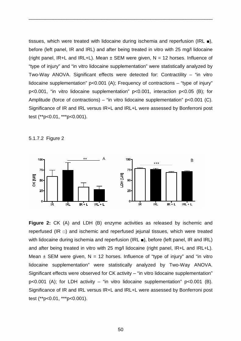
____________________________________________________________________
50
tissues, which were treated with lidocaine during ischemia and reperfusion (IRL ■),
before (left panel, IR and IRL) and after being treated in vitro with 25 mg/l lidocaine
(right panel, IR+L and IRL+L). Mean ± SEM were given, N = 12 horses. Influence of
“type of injury” and “in vitro lidocaine supplementation” were statistically analyzed by
Two-Way ANOVA. Significant effects were detected for: Contractility – “in vitro
lidocaine supplementation” p<0.001 (A); Frequency of contractions – “type of injury”
p<0.001, “in vitro lidocaine supplementation” p<0.001, interaction p<0.05 (B); for
Amplitude (force of contractions) – “in vitro lidocaine supplementation” p<0.001 (C).
Significance of IR and IRL versus IR+L and IRL+L were assessed by Bonferroni post
test (**p<0.01, ***p<0.001).
5.1.7.2 Figure 2
Figure 2: CK (A) and LDH (B) enzyme activities as released by ischemic and
reperfused (IR □) and ischemic and reperfused jejunal tissues, which were treated
with lidocaine during ischemia and reperfusion (IRL ■), before (left panel, IR and IRL)
and after being treated in vitro with 25 mg/l lidocaine (right panel, IR+L and IRL+L).
Mean ± SEM were given, N = 12 horses. Influence of “type of injury” and “in vitro
lidocaine supplementation” were statistically analyzed by Two-Way ANOVA.
Significant effects were observed for CK activity – “in vitro lidocaine supplementation”
p<0.001 (A); for LDH activity – “in vitro lidocaine supplementation” p<0.001 (B).
Significance of IR and IRL versus IR+L and IRL+L were assessed by Bonferroni post
test (**p<0.01, ***p<0.001).

____________________________________________________________________
51
5.1.7.3 Figure 3
Figure 3: Lidocaine concentrations in serum and smooth muscle of individual horses.
Abbreviations
ATP Adenosine triphosphate
AUC Area under curve
CK Creatine kinase
CRI Constant rate infusion
ENS Enteric nervous system
ICC Interstitial cells of Cajal
IR Ischemic and reperfused
IRL Ischemic and reperfused plus lidocaine infusion
KHB Krebs-Henseleit-buffer
LDH Lactate dehydrogenase
POI Postoperative paralytic ileus
ROS Reactive oxygen species
TTX Tetrodotoxine
____________________________________________________________________
52
6 HISTOLOGY
The aim of this part was to get to more information about the extent of the damage of
the artificially induced 15 minutes of ischaemia followed by the 15 minutes of
reperfusion (IR) we used in the studies (GUSCHLBAUER et al., 2010). Does an
intravenous lidocaine infusion starting during surgery (IRL) have any effects on
morphological parameters of intestinal wall? As an introduction it might be useful to
give a short overview about the histological situation of the small intestine (according
to LIEBICH, 1993).
6.1 Histology of the Equine Small Intestine
In this study the equine jejunum was used for histological examinations to evaluate
morphological parameters (Chapter 6.3., Paper 3). The jejunal wall is divided into five
layers: tunica mucosa, lamina propria mucosa, tela submucosa, tunica muscularis
and tunica serosal (Figure 4). The tunica mucosa provides macroscopically and
permanently observable plicas (plica circularis), which are losing height from cranial
to caudal. They are present in the distal duodenum and proximal jejunum and serve
as a platform for the intestinal villi (villi intestinales), which cover the whole small
intestinal mucosal membrane. Villi are finger-like formations of the plica circularis and
have a single-layered columnar epithelium (epithelium mucosae) (Figure 3; 1)
(LIEBICH, 1993).
Those villi (0.5 to 1.5 mm length) are essential for the surface-area amplification of
the intestinal mucosa. The longest villi are observed in the jejunum, whereas they are
shorter in the duodenum and ileum. Within the villi there is a subepithelial capillary
net including a venule, an arteriole and a lymphatic vessel (Figure 3; 2). Special
tubular-like, straight and unbranched glands entail the lamina propria mucosa. Those
glands are named glandulae intestinales, also known as the Lieberkühn-glands or
crypts (Figure 3; 6). The surfaces of those crypts also have a single-layered

____________________________________________________________________
53
epithelium (epithelium mucosae), whose single cell is high-prismatic (Figure 2; 5)
(LIEBICH, 1993).
The mucosal epithelium is a composition of different cells, which also differ in
structure and function. There are enterocytes (epitheliocyti columnares villi), goblet
cells (epitheliocyti calciformes) and Paneth-cells (exocrinocyti cum granulis
acidophilis) (Figure 3; 6). Enterocytes are resorptive cells and goblet cells are
exocrine secretory cells. Both together compose the barrier layer of the villous
surface. Furthermore they are essential components of the wall of glands (crypts).
Paneth-cells (Figure 3; 6) are exocrine secretory cells, producing a serosal,
glycoproteine-enriched fluid. The duodenum submucosal glands (Brunner’s glands)
produce mucus. Those glands are branched and tubular- acinous (LIEBICH, 1993).
The lamina propria mucosa consists of loosely connective tissue and is the
substructure of gut villi (stroma villi). Most of the parts of this subepithelial tissue
display crypts (glandulae intestinales, Lieberkühn-glands) (Figure 2; 7). Between
those glands there are connective tissue, blood and lymphatic vessels as well as
nervous tissue, myofibroblasts and smooth muscle cells. Underneath the lamina
propria mucosa there is the lamina muscularis mucosae (Figure 3; 3) (LIEBICH,
1993).
The tela submucosa has collagenous fibres, fat tissue, vessels and nervous
structures (plexus nervorum submucosus). Two kinds of lymphatic tissues are
located in the tela submucosa: solitary and aggregated lymphatic glands (noduli
lymphatici solitarii and noduli lymphatici aggregate which are also called the Peyer-
plaques) (LIEBICH, 1993).
The tunica muscularis is divided into the stratum circulare (inner layer) and the
stratum longitudinale (outer layer) and contains smooth muscle cells. For nervous
innervations in the tunica muscularis the plexus nervorum myentericus (Auerbach-
plexus) and the plexus nervorum submucosus (Meißner-plexus) are located in this
tissue layer (LIEBICH, 1993).
The ICCs are associated with the neural plexuses within the intestinal musculature.
They are connected to each other via gap junctions. In a review HUIZINGA et al.
(1998) referred that the interstitial cells of Cajal (ICC), the intestinal pacemaker cells,

____________________________________________________________________
54
and the gut smooth muscle cells originate from “mesenchymal precursor cells”
(KLÜPPEL et al., 1998; LECOIN et al., 1996; YOUNG et al., 1996). KLÜPPEL et al.
(1998) reported that they can be found between the external longitudinal and the
circular muscle layers on the level of the Auerbach`s plexus in the small intestine of
animals (KLÜPPEL et al., 1998; HUIZINGA et al., 1998). These cells require specific
immunohistochemistry to detect them.
The tunica serosa has a single-layered squamous epithelium and is connected to the
tunica muscularis via the connective tissue of the tela subserosa (LIEBICH, 1993)
(Overall view of intestinal wall structure Figure 4).
6.1.1 Figure 3

____________________________________________________________________
55
Legend Figure 3: Photomicrograph of a physiological jejunal histological section of a
horse (overview, 4x magnification, GUSCHLBAUER, M., SLAPA, J., 2010).
1. Villi show a single columnar epithelium 2. Subepithelial capillaries are located
within the villi, 3. Lamina muscularis mucosa, 4. Tela submucosa, 5. Longitude of a
villus, 6. Area of location of intestinal crypts (lamina propria), 7. Area of location of
glandulae intestinales (Lieberkühn-glands).
6.1.2 Figure 4
Lumen
Mesentery
Tunica mucosa
Tela submucosa
Muscularis mucosa
Circular muscle
Tunica serosa
Longitudinal muscle
Tunica muscularis
Lumen
Mesentery
Tunica mucosa
Tela submucosa
Muscularis mucosa
Circular muscle
Tunica serosa
Longitudinal muscle
Tunica muscularis
Legend Figure 4: Schematic overview of intestinal gut wall layers.

____________________________________________________________________
56
6.2 Morphological Changes in the Intestine
6.2.1 Morphological Changes of Colic Horses
The integrity of physiological morphology is of vital necessity to guarantee
physiological gastrointestinal functions and motility. The integrity of the lamina
propria mucosa and existence of villi and epithelial cells were very important
indicators for physiological intestinal function and were used for assessment of the
extent of damage after an ischaemia and reperfusion injury (WHITE et al., 1980).
MESCHTER et al. (1986) found out that intestinal lesions could be graded with
regard to severity due to different causes of colic. This might correlate with
postoperative survival or nonsurvival rates of affected horses. For grading the
morphological changes they used a grading system according to CHIU et al. (1970).
Mucosal ulcerations had been attributed to colic due to simple intestinal obstruction
whereas mucosal necrosis, venous stasis, congestion and oedema could be found
due to strangulative infarction of the equine small intestine. Furthermore there was
an inflammatory cell infiltration, epithelial sloughing and haemorrhage of the
underlying lamina propria. MESCHTER et al. (1986) summarised that mucosal
degeneration in colic horses “is characterised by different amounts of epithelial
sloughing of the mucosa, subepithelial cleft formation, and necrolytic vacuolization of
the absorptive epithelium”. A mild distention of crypts and accumulations of necrotic
debris within the subsurface of the lamina propria was observed in affected areas.
Cellular infiltrates occurred with accumulations of inflammatory cells like neutrophils,
lymphocytes, eosinophils, leukocytes and mast cells (MESCHTER et al., 1986).
To examine the morphological and quantitative morphological changes of plexuses
and neurons SCHUSSER and WHITE (1997) evaluated the large colon of horses
with colon diseases. They discovered that horses suffering from chronic obstruction,
which last 24 hours or longer, had significantly lower neuron density in the pelvic
flexure. SCHUSSER and WHITE (1997) reported that “myenteric plexus density in
horses with strangulating large colon torsion/volvulus was significantly less in the
right ventral, right dorsal, and transverse colons, and neuron density in these horses

____________________________________________________________________
57
was significantly less in all segments of colon, except the left ventral colon”
(SCHUSSER et al. 1997). They also compared the findings with the surviving rates of
affected horses and found out that “horses with colon strangulation that survived had
significantly greater neuron density than horses with colon strangulation that died”.
Clinically highly relevant they found out that enteroglial cell numbers were increased
in myenteric plexuses of horses suffering from acute and chronic intestinal
obstruction. Therefore SCHUSSER and WHITE (1997) concluded that an increase of
enteroglial cells may negatively affect bowel function because of inflammation of the
myenteric plexus.
6.2.2 Morphological Changes of Horses with Artifici ally Induced
Ischaemia and Reperfusion Injury
DABAREINER et al. (2001) reported in a study, inducing artificial ischaemia and
reperfusion injury in equine jejunum and colon, that the serosa of the ascending
colon was less sensitive against ischaemia and reperfusion injury than the serosa of
jejunum. Ischaemia, reperfusion and intraluminal distention resulted in severe
morphological changes in the equine jejunum. They stated that intraluminal distention
and subsequent decompression, as it is seen in the equine colic events, caused
profoundly damage in the affected parts of jejunum leading to severe serosal
alterations (DABAREINER et al., 2001).
Additionally, DABAREINER et al. (2001) reported that morphological changes, in the
equine jejunum and colon, after an artificially induced ischaemia and reperfusion
injury, included mesothelial cell loss. In their study jejunal intraluminal distention led
to serosal basement membrane damage. Furthermore, basal membrane disruption
and capillary endothelial cell damage resulted in moderate erythrocyte leakage. After
jejunal distention and decompression, neutrophil infiltration was seen adjacent to the
serosal basal membrane. Many inflammatory cells like neutrophils were observed in
affected areas showing signs of an acute inflammation. DABAREINER et al. (2001)
assumed that mesothelial cell loss was probably caused by mechanical manipulation

____________________________________________________________________
58
because of handling the intestine during colic surgery. Furthermore they stated that
vasoactive substances as histamine, bradykinin, and prostaglandine would increase
capillary permeability. As a consequence serofibrinous exudate diffused within the
tunica serosa and on serosal surfaces (DABAREINER et al., 2001).
After 2 hours of artificially induced ischaemia and 30 minutes of reperfusion of equine
large intestine GROSCHE et al. (2008) reported increased numbers of inflammatory,
calprotectin-positive, neutrophils cells within submucosal venules and within the
colonic mucosa. FREEMAN et al. (1988) published findings which suggest that short
periods of strangulation obstructions were able to induce severe morphological
changes in the jejunal wall of ponies. They found out that after an artificially induced
obstruction in the distal portion of jejunum and after maintaining an ischaemia time
from 2 to 3 hours, necrosis could be seen of the villus tip cells in the strangulated
segment. Furthermore FREEMAN et al. (1998) observed that intestinal “villi were
clubbed and denuded to the base”. This was leading to an exposure of lamina
propria and capillaries. They also stated that further effects of ischaemia were the
development of oedema and haemorrhage (FREEMAN et al., 1998).
In another study by ALLEN et al. (2008) the morphologic effects after inducing
artificial intraluminal pressures, simulating fluid accumulations during colic events,
were evaluated in 33 isolated equine jejunal segments. ALLEN et al. (2008) used
light and transmission electron microscopy for examination of intestinal sections.
They could find oedema of the villi and submucosa as well as separation of the
epithelial cells in all segments. Furthermore they asserted that increases in
intraluminal hydrostatic pressure would reduce blood flow, mainly in the jejunal
mucosa of the small intestine. As a consequence of increased pressure fluid
accumulated in the lamina propria resulting in showing oedema in histological
stainings (ALLEN et al., 2008).
Those studies showed the importance of physiological integrity of gut wall and barrier
function for intestinal motility. Hence, what does the integrity of the small intestine
look like after experimentally induced 15 minutes of ischaemia followed by 15
minutes of reperfusion? Is this short-term ischaemia able to provoke histological
changes in the jejunal intestinal wall structure and does lidocaine have an influence

____________________________________________________________________
59
on the extent of damage? The histology of jejunal wall artificially injured by 15
minutes of ischaemia and 15 minutes of reperfusion has not been evaluated yet.
6.3 Aims of the study
As we previously described a short-term ischaemia and reperfusion injury was able
to decrease jejunal motility in vitro significantly. An addition of lidocaine was able to
restore decreased contractility (GUSCHLBAUER et al., 2010). It was hypothesised
that an in vivo artificially induced short-term ischaemia (15 minutes) followed by
reperfusion (15 minutes) is able to induce histological damages in jejunal tissues. To
study the effects of lidocaine on morphological parameters, lidocaine was infused
during surgery before reperfusion (IRL). To gain information about the extent of the
artificially induced ischaemia and reperfusion injury and lidocaine effects on
morphological parameters, specimens were collected from intestinal tissue (control,
IR, and IRL) immediately after resection and prepared for morphological evaluation
(Chapter 6.4., Paper 3).

____________________________________________________________________
60
6.4 PAPER 3
6.4.1 Lidocaine reduces tissue oedema formation i n equine gut wall
challenged by ischaemia and reperfusion
Lidocain reduziert die Bildung eines Gewebeödemes i n der ischämisch-
reperfusionsgeschädigten Darmwand des Pferdes
Maria Guschlbauer1, Johanna Slapa1, Korinna Huber1 und Karsten Feige2 1Institut für Physiologie und 2Klinik für Pferde der Stiftung Tierärztliche Hochschule,
Hannover
Schlüsselwörter: Lidocain, Histomorphologie, Jejunum, Ischämie, Reperfusion
Keywords: lidocaine, histomorphology, jejunum, ischaemia, reperfusion
Zusammenfassung
Eine reduzierte propulsive Motilität des Dünndarmes tritt oft als Komplikation nach
Kolikoperationen auf. Schäden durch Ischämie und Reperfusion sind
mitverantwortlich für die Entwicklung eines postoperativen Ileus. Das Prokinetikum
der Wahl in der postoperativen Phase ist Lidocain. Durch isometrische
Kraftmessungen mit glatten Muskelproben von in vivo durch Ischämie und
Reperfusion geschädigtem, mit Lidocain behandeltem und unbehandeltem Jejunum
des Pferdes konnte in vitro gezeigt werden, dass Lidocain eine
kontraktilitätssteigernde Wirkung auf die geschädigte Muskulatur hat und die
Freisetzung von CK, einem Marker für die Zellmembranpermeabilität, signifikant
herabsetzt. Ziel dieser Studie war es zu erfassen, ob strukturelle Veränderungen der
Darmwand, die durch kurzzeitig Ischämie und Reperfusion ausgelöst werden, mit der
Motilitätsstörung einhergehen und ob Lidocain das Auftreten von morphologischen
Schäden verhindern oder abschwächen kann. Hierzu wurden von ungeschädigten

____________________________________________________________________
61
und von durch Ischämie und Reperfusion geschädigten, unbehandelten und mit
Lidocain behandelten Darmproben histologische Schnitte angefertigt. Mit einem
Bewertungssystem angelehnt an Snyder et al. (1998) wurde der Einfluss durch die
Schädigung mit und ohne Lidocain auf morphologische Parameter wie Füllung der
Blutgefässe, die Auflockerung des Gewebes, das Vorhandensein von Hämorrhagien
sowie die Integrität der intestinalen Villi erfasst. Die Ergebnisse zeigen, dass schon
eine kurzzeitige Ischämie und Reperfusion ausreichend sind, um eine erhöhten
Blutfüllung der Gefäße, eine Auflockerung des Bindegewebes von Mukosa und
Muskularis sowie milde Hämorrhagien und Degeneration der intestinalen Villi
auszulösen. Lidocain verringerte die Auflockerung der intestinalen Schichten, was für
eine reduzierte Ödembildung in behandeltem Gewebe spricht. Dies könnte auf der
membranstabilisierenden Wirkung des Lidocains beruhen. Neben der bestätigten
prokinetischen Wirkung reduziert Lidocain strukturelle Schäden der Darmwand, die
durch Ischämie und Reperfusion ausgelöst werden.
Summary
Reduction of small intestinal propulsive motility is still a major problem after equine
colic surgery. It is a common side effect after an ischaemia and reperfusion injury,
frequently resulting in development of a postoperative ileus. Lidocaine is commonly
used for prokinetic treatment in the early postoperative period. Determining the
contractile performance of intestinal smooth muscle in vitro, it was shown that
lidocaine increases contractility after an in vivo artificially induced short-term
ischaemia and reperfusion injury. A lidocaine-dependent decrease in membrane
permeability of smooth muscle cells indicated a possible mechanism for cellular
repair effects. Aim of this study was to examine if structural alterations of intestinal
gut wall induced by short-term ischemia and reperfusion accompanied motility
disorders and if lidocaine was able to prevent morphological alterations. Tissue slices
from control and ischaemic and reperfused equine small intestine - untreated and
treated with lidocaine – were prepared, stained and analysed. A classification
protocol according to Snyder et al. (1998) was used to evaluate morphological

____________________________________________________________________
62
alterations like filling of blood vessels, looseness of tissue, haemorrhage in
submucosa and villus degeneration. Short-term ischaemia and reperfusion was able
to induce increased blood vessel filling, enhanced looseness of mucosal and
muscular layer, mild haemorrhage and slight degeneration of intestinal villi. Lidocaine
significantly reduced the looseness of tissue in the submucosal and muscular layer,
indicating prevention of interstitial oedema. This might be due to the membrane
stabilising effect of lidocaine. Besides approved prokinetic features lidocaine could
prevent structural alterations of gut wall induced by ischaemia and reperfusion.
The full text is available in:
Guschlbauer et al., 2010, Pferdeheilkunde, Juli/August

____________________________________________________________________
63
7 Literature
ADAMS, H.R. (1995): Antiarrhythmic agents. In: Adams HR. ed. Veterinary
Pharmacology and Therapeutics. Ames: Iowa State Press, 358-371
ALLEN, D., N.A. WHITE and D.E. TYLER (1988): Morphologic effects of
experimental distention of equine small intestine. Vet. Surg. 17, 10-14
ALLEN, D., N.A. WHITE and D.E. TYLER (1986): Factors for prognostic use in
equine obstructive small intestinal disease. J. Am. Vet. Med. Assoc. 18, 777-779
BAUER, A.J. and G.E. BOECKXSTAENS (2004): Mechanisms of postoperative ileus.
Neurogastroenterol. Motil. 16, 54-60
BAYLISS, W.M. and E.H. STARLING (1899): The movements and innervation of the
small intestine. J. Physiol. 24, 99-143
BLIKSLAGER, A.T., K.F. BOWMAN, J.F. LEVINE, D.G. BRISTOL and M.C.
ROBERTS (1994): Evaluation of risk factors associated with postoperative ileus in
horses: 31 cases (1990-1992). J. Am. Vet. Med. Assoc. 205, 1748-1752
BRIANCEAU, P., H. CHEVALIER, A. KARAS, M.H. COURT, L. BASSAGE, C.
KIRKER-HEAD, P. PROVOST and M.R. PARADIS (2002): Intravenous lidocaine and
small-intestinal size, abdominal fluid, and outcome after colic surgery in horses. J.
Vet. Intern. Med. 16, 736-741
BURTON, K.P., H.K. HAGLER, G.H. TEMPLETON, J.T. WILLERSON and L.M.
BUJA (1977): Lanthanum probe studies of cellular pathology induced by hypoxia in
isolated cardiac muscle. J. Clin. Invest. 60, 1289-1302

____________________________________________________________________
64
BODDY, G., A. BONG, W.J. CHO and E.E. DANIEL (2004): ICC pacing mechanisms
in intact mouse intestine differ from those in cultured or dissected intestine. Am. J.
Physiol. Gastrointest. Liver Physiol. 286, 653-662
CAJAL, R.V. (1893): Sur les ganglions et plexus nerveux d`intestine. C. R. Soc. Biol.
(Paris) 5, 217-223
CAJAL, R.V. (1911): Histologie du systeme` nerveux de l`homme et des vertebres.
(Paris) Malone
CASSUTO, J., G. WALLIM and S. HOGSTROM (1985): Inhibition of postoperative
pain by continuous low-dose intravenous infusion of lidocaine. Anasth. Analg. 64,
971-974
CASSUTO, B.H. and R.W. GFELLNER (2003): Use of intravenous lidocaine to
prevent reperfusion injury and subsequent multiple organ dysfunction syndrome. J.
Vet. Emerg. Crit. Care 13, 137-148
CATTERALL, W.A. (2002): Molecular mechanisms of gating and drug block of
sodium channels. Novartis Found. Symp. 241, 206–32
CHANG, I.Y., N.J. GLASGOW, I. TAKAYAMA, K. HORIGUCHI, K.M. SANDERS and
S.M. WARD (2001): Loss of interstitial cells of Cajal and development of electrical
dysfunction in murine small bowel obstruction. J. Physiol. 536.2, 555-568
CHIU, C.J., A.H. McARDLE and R. BROWN (1970): The intestinal lesion in low flow
states: A morphologic, hemodynamic, and reappraisal. Arch. Surg. 101, 478
COHEN, M.V. (1989): Free radicals in ischemic and reperfusion myocardial injury: a
selective review. Ann. Int. Med. 111, 918-931

____________________________________________________________________
65
COHEN, N.D., G.D. LESTER, L.C. SANCHEZ, A.M. MERRITT and A.J. ROUSSEL
(2004): Evaluation of risk factors associated with development of postoperative ileus
in horses. J. Am. Vet. Med. Ass. 225, 1080-1078
COLLARD, C.D. and S. GELMAN (2001): Pathophysiology, clinical manifestations,
and prevention of ischemia-reperfusion injury. Anesthesiol. 94, 1133-8
COOK, V.L., J. JONES SHULTS, M. McDOWELL, N.B. CAMPBELL, J.L. DAVIS and
A.T. BLIKSLAGER (2008): Attenuation of ischaemic injury in the equine jejunum by
administration of systemic lidocaine. Equine Vet. J. 40, 353-357.
COOKE, H.J. (1994): Neuroimmune signalling in regulation of intestinal ion transport.
Am. J. Physiol. 266, 167-178
COSTA, M. and S.J. BROOKES (1994): The enteric nervous system. Am. J.
Gastroenterol. 89, 129-137
DABAREINER, R.M., K.E. SULLINS, N.A. WHITE and J.R. SNYDER (2001): Serosal
injury in the equine jejunum and ascending colon after ischemia-reperfusion or
intraluminal distention and decompression. Vet. Surg. 30, 114-125
DABAREINER, R.M., J.R. SNYDER, K.E. SULLINS, N.A. WHITE and I.A.
GARDNER (1993): Evaluation of the microcirculation of the equine jejunum and
ascending colon after ischemia and reperfusion. Am. J. Vet. Res. 54, 1683-92
DART, A.J. and D.R. HODGSON (1998): Role of prokinetic drugs for the treatment of
postoperative ileus in the horse. Aust. Vet. J. 76, 25-30
DeKLAVIER, M.J.M., M.G. BUCKINGHAM and G.F. RICH (2003): Lidocaine
attenuates cytokine-induced cell injury in endothelial and vascular smooth muscle
cells. Anesth. Anal. 97, 465-470

____________________________________________________________________
66
DeVILLERS, D.R. (1966): Ischemia of the colon: An experimental study. J. Surg. 53,
497-503
DRIESSEN, B. (2005): Intravenöse Lidokain-Infusion bei der Kombinationsnarkose in
der Bauchhöhlenchirurgie: Hintergrund und klinische Erfahrungen. Pferdeheilkunde
21, 133-141
DOHERTY, T.J. and D.L. FRAZIER (1998): Effect of intravenous lidocaine on
halothane minimum concentration in ponies. Equine Vet. J. 30, 300-303
DULLENKOPF, A. and A. BORGEAT (2003): Lokalanästhetika. Anaesthesist 52,329-
340
DZIKITI, T.B., L.J. HELLEBREKERS and P. van DIJK (2003): Effects of intravenous
lidocaine on isoflurane concentration, physiological parameters, metabolic
parameters and stress-related hormones in horses undergoing surgery. J. Vet. Med.
A. Physiol. Pathol. Clin. Med. 50, 190-195
EDWARDS, G.B., and J.M. HUNT (1985): An analysis of the incidence of equine
postoperative ileus (POI) and an assessment of the predisposing factors. Proc.
Equine Colic Res. Symp. 2, 307-312
EADES, S.C. and J.N. MOORE (1993): Blockade of endotoxin- induced cecal
hypoperfusion and ileus with an α - 2 agonist in horses. Am. J. Vet. Res. 54, 586-590
EMSTER, L. (1988): Biochemistry of reoxygenation injury. Crit. Care Med. 16, 947-
953
ENDERLE, A.K., O.L. LEVIONNOIS, M. KUHN and U. SCHATZMANN (2008):
Clinical evaluation of ketamine and lidocaine intravenous infusions to reduce

____________________________________________________________________
67
isoflurane requirements in horses under general anaesthesia. Vet. Anaesth. Anal. 35,
297-305
FEARY, D.J., K.R. MAMA, A.E. WAGNER and S. THOMASY (2005): Influence of
general anesthesia on pharmacokinetics of intravenous lidocaine infusion in horses.
Am. J. Vet. Res. 66, 574-580
FEARY, D.J., K.R. MAMA, S.M. THOMASY, A.E. WAGNER and R.M. ENNS (2006):
Influence of gastrointestinal tract disease on pharmacokinetics of lidocaine after
intravenous infusion in anesthetized horses. Am. J. Vet. Res. 67, 317-322
FINTL, C., N.P.H. HUDSON, I.G. MAYHEW, G.B. EDWARDS, C.J. PROUDMAN and
G.T. PEARSON (2004): Interstitial cells of Cajal (ICC) in equine colic: an
immunohistochemical study of horses with obstructive disorders of the small and
large intestines. Equine Vet. J. 36, 474-480
FLECKENSTEIN, P., L. BUENO, J. FIORAMONTI and Y. RUCKEBUSCH (1982):
Minute rhythm of electrical spike bursts of the small intestine of different species. Am.
Physiol. Society 242, 654-659
FREEMAN, D.E., R.E. CIMPRICH, D.W. RICHARDSON, D.G. GENTILE, E.A.
ORSINI, E.P. TULLENERES and J.P. FETROW (1988): Early mucosal healing and
chronic changes in pony jejunum after various types of strangulation obstruction. Am.
J. Vet. Res. 29, 810-818
FRENCH, N.P., J. SMITH, G.B. EDWARDS and C.J. PROUDMAN (2002): Equine
surgical colic: risk factors for postoperative complications. Equine Vet. J. 34, 444-449
FREYTAG, C., J. SEEGER, T. SIEGEMUND, J. GROSCHE, A. GROSCHE, D.E.
FREEMAN, G. SCHUSSER and W. HÄRTIG (2008): Immunohistochemical

____________________________________________________________________
68
characterization and quantitative analysis of neurons in the myenteric plexus of the
equine intestine. Brain Res. 1244, 53-64
FONTANA, R., K. LOWN , M. PAINE, L. FORTLAGE, R. SANTELLA, J. FELTON, M.
KNIZE, A. GREENBERG and P. WATKINS (1999): Effects of a chargrilled meat diet
on expression of CYP3A, CYP1A, and P-glycoprotein levels in healthy volunteers.
Gastroenterol. 117, 89–98
FURNESS, J.B. and J.C. BORNSTEIN (1995): The enteric nervous system and its
intrinsic connections. In: YAMADA, T., ed. Textbook of gastroenterology. 2nd edition,
Philadelphia: J.B. Lippincott. 1, 2-24
GABELLA, G. (1972): Innervation of the intestinal muscular coat. J. Neurocytol. 1,
341-62
GARLICK, P.B., G.K. RADDA and P.J. SEELY (1979): Studies of acidosis in the
ischemic heart by phosphorus nuclear magnetic resonance. Biochem. J. 184, 547-
554
GERRING, E. L. and J.M. HUNT (1986): Pathophysiology of equine postoperative
ileus: effect of adrenergic blockade, parasympathetic stimulation, and
metoclopramide in an experimental model. Equine Vet. J. 18, 249-255
GERSHON, M.D., A.L. KIRCHGESSNER and P.R. WADE (1994): Functional
anatomy of enteric nervous system. In: JOHNSON, J.L., ed. Physiology of the
gastrointestinal tract. 3rd edition, New York: Raven Press, 381-422
GOYAL, R.K. and I. HIRANO (1996): The enteric nervous system. New Engl. J. Med.
334, 1106-1115

____________________________________________________________________
69
GROSCHE, A., A.J. MORTON, M.M.R. POLYAK, S. MATYJASZEK and D.E.
FREEMAN (2008): Detection of calprotectin and its correlation to the accumulation of
neutrophils within the equine large colon during ischaemia and reperfusion. Equine
Vet. J. 40, 393-399
GUSCHLBAUER, M., S. HOPPE, F. GEBUREK, K. FEIGE and K. HUBER (2010): In
vitro effects of lidocaine on the contractility of equine jejunal smooth muscle
challenged by ischaemia-reperfusion injury. Equine Vet. J. 42, 53-58
GRACE, R.H. (1971): The handling of water and electrolytes by the small bowel
following the relief on intestinal obstruction. Br. J. Surg. 58, 760
GRANGER, D.N., M. SENNETT, P. McELEARNEY and A.E. TAYLOR (1980): Effect
of local arterial hypotension on cat intestinal capillary permeability. Gastroenterol. 79,
474-480
GROUDINE, S.B., H.A.G. FISHER, R.P. KAUFMANN, M.K. PATEL, L.J. WILKINS,
S.A. MEHTA and P.D. LUMB (1998): Intravenous lidocaine speeds the return of
bowel function, decreases postoperative pain, and shortens hospital stay in patients
undergoing radical retropubic prostatectomy. Anesth. Analg. 86, 235-9
HABER, F. and J. WEISS (1934): The catalytic decomposition of hydrogen peroxide
by iron salts. Proc. R. Soc. London, Ser. A. 147, 332-351
HAMMER, R., C. DAHLGREN and O. STENDAHL (1985): Inhibition of human
leucocyte metabolism and random mobility by local anaesthesia. Acta Anaesthesiol.
Scand. 29, 520-523
HEARSE, D.J. (1979): Oxygen deprivation and early myocardial contractile failure: A
reassessment of the possible role of adenosine triphosphate. Am. J. Cardiol. 44,
1115-1121

____________________________________________________________________
70
HEARSE, D.J. and S.M. HUMPHREY (1978): Enzyme release during myocardial
anoxia: A study of metabolic protection. J. Mol. Cell. Cardiol. 7,463-483
HOOROBIN, D.F. and M.S. MANKU (1977): Roles of prostaglandins suggested by
the prostaglandin agonist/antagonist actions of local anaesthetic, anti-arrhythmic,
anti-malarial, tricyclic anti-depressant and methyl xanthine compounds, effects on
membranes and nucleic acid function. Med. Hypotheses 3, 71-86
HOROWITZ, B., S.M. WARD and K.M. SANDERS (1999): Cellular and molecular
basis for electrical rhythmicity in gastrointestinal muscles. Annu. Rev. Physiol. 61, 19-
43
HUIZINGA, J.D., L. THUNEBERG, M. KLÜPPEL, J. MALYSZ, H.B. MIKKELSEN and
A. BERNSTEIN (1995): W/kit gene required for interstitial cells of Cajal and for
intestinal pacemaker activity. Nature 373, 347-349
HUIZINGA, J.D. (1998): Neural injury, repair, and adaptation in the GI tract IV.
Pathophysiology of GI motility related to interstitial cells of Cajal. Am. J. Physiol. 18,
G381-G386
HUIZINGA, J.D., Y. ZHU, J. YE and A. MOLLEMAN (2002): High-conductance
chloride channels generate pacemaker currents in interstitial cells of Cajal.
Gastroenterol. 123,1627-1636
HUNT, J.M. and E.L. GERRING (1985): The effect of prostaglandin E1 on motility of
the equine gut. J. Pharmacol. Therapy 8, 165-166
HUNT, J.M., G.B. EDWARDS and K.W. CLARKE (1986): Incidence, diagnosis and
treatment of postoperative complications in colic cases. Equine Vet. J. 18, 264-270

____________________________________________________________________
71
HONDEGHEM L.C. and D.M. RODEN (1998): Agents used in cardiac
arrhythmias. In: Katzung BG. ed. Basic and Clinical Pharmacology. New York:
Lange Medical Books/McGraw-Hill, 232-233
ITOH, Z., R. HONDA and S. TAKEUCHI (1977): An extraluminal force transducer for
recording contractile activity of the gastrointestinal smooth muscle in the conscious
dog; its construction and implantation. Gastroenterol. Jpn. 12, 275-283
JENNINGS, R.B. (1976): Cell volume regulation in acute myocardial ischemic injury.
Acta Med. Scan. 587, 83-92
JENNINGS, R.B. and C.E. GANOTE (1976): Mitochondrial structure and function in
acute myocardial injury. Circ. Res. 38, 80-91
KEHRER, J.P. (2000): The Haber-Weiss reaction and mechanisms of toxicity.
Toxicol. 149, 43-50
KING, J.N. and E.L. GERRING (1989): Antagonism of endotoxin-induced disruption
of bowel motility by flunixin and phenylbutazone. Equine Vet. J. Suppl. 7, 38-42
KING, J.N. and E.L. GERRING (1991): The action of low dose endotoxin on equine
bowel motility. Equine Vet. J. 23, 11-17
KLÜPPEL, M., J.D. HUIZINGA, J. MALYSZ and A. BERNSTEIN (1998):
Developmental origin and kit-dependent development of the interstitial cells of Cajal
in the mammalian small intestine. Dev. Dyn. 211, 60-71.
KONG, S.E., L.R. BLENNERHASSET, K.A. HEEL, M. McCAULEY and J.C. HALL
(1998): Ischaemia-reperfusion injury to the intestine. Aust. N. Z. J. Surg. 68, 554-561

____________________________________________________________________
72
KUNZE, W.A.A. and J.B. FURNESS (1999): The enteric nervous system and
regulation of intestinal motility. Annu. Rev. Physiol. 61, 117-42
KÜBLER, W. and A.M. KATZ (1977): Mechanisms of early pump failure of the
ischemic heart: Possible role of adenosine triphosphate depletion and inorganic
phosphor accumulation. Am. J. Cardiol. 40, 467-471
LAHAV, M., M. LEVITE, L. BASSANI, A. LANG, H. FIDDER, R. TAL, S. BAR-MEIR,
S. MAYER and Y. CHOWERS (2002): Lidocaine inhibits secretion of IL-8 and IL-
1beta and stimulates secretion of IL-1 receptor antagonist by epithelial cells. Clin.
Exp. Immunol. 127, 226-233
LAN, W., D. HARMON, J.H. WANG, K. GHORI, G. SHORTEN and P. REDMOND
(2004): The effect of lidocaine on in vitro neutrophil and endothelial adhesion
molecule expression induced by plasma obtained during tourniquet-induced
ischaemia and reperfusion. Eur. J. Anaesthesiol. 21, 892-897
LANTOS, J., E. ROTH and G. THEMES (1996): Effects of lidocaine on cerebral lipid
peroxidation and neutrophil activation following complete compression ischemia.
Arch. Int. Pharmacodyn. Ther. 331, 179-188
LECOIN, L., G. GABELLA and N. Le DOUARIN (1996): Origin of the c-kit positive
interstitial cells in the avian bowel. Development 122, 725-733
LIEBICH, H.G. (1993): Funktionelle Histologie – Farbatlas und Kurzlehrbuch der
mikroskopischen Anatomie der Haussäugetiere. Schattauer Verlag. 2, 181-190
LITTLE, D., J.E. TOMLINSON and A.T. BLIKSLAGER (2005): Postoperative
neutrophilic inflammation in equine small intestine after manipulation and ischaemia.
Equine Vet. J. 37, 329-335

____________________________________________________________________
73
LÖEFGREN N. (1948): Studies on local anesthetics. Svenksa Kem. Todskr. 58, 206
LUNDGREN, O., J. SVANVIK and J. JIVEGARD (1989): Enteric nervous system. I.
Physiology and pathophysiology of the intestinal tract. Dig. Dis. Sci. 34, 264-283
LUNEGREN, O. and U. HAGLUND (1978): The pathophysiology of the intestinal
counter-current exchange. Life Sci. 23, 1411-1422
MacDONALD, M.H., J.R. PASCOE, S.M. STOVER and D.M. MEAGHER (1989):
Survival after small intestine resection and anastomosis in horses. Vet. Surg. 18,
415-423
MacGREGOR, R.R., R.E. THORNER and D.M. WRIGHT (1980): Lidocaine inhibits
granulocyte adherence and prevents granulocyte delivery to inflammatory sites.
Blood 56, 203-209
MAEDA, H., A. YAMAGATA, S. NISHIKAWA, K. YOSHINAGA, S. KOBAYASHI and
K. NISHI (1992): Requirement of c-kit for development of intestinal pacemaker
system. Develop. 116,369-375
MAIR, T.S. and G.B. EDWARDS (2003): Strangulating obstructions of the small
intestine. Equine Vet. Educ. 15, 192-199
MAMA, K.R. and E.P. STEFFEY (2001): Drugs acting in the central nervous system,
local anesthetics. In: ADAMS, H.R., ed. Veterinary pharmacology and therapeutics.
8th ed. Ames, IA: Iowa State University Press; 355-359
MALONE, E., T.A. TURNER and J.H. WILSON (1999): Intravenous lidocaine for
treatment of equine ileus. J. Vet. Intern. Med. 13, 229

____________________________________________________________________
74
MALONE, E., J. ENSINK, T. TURNER, J. WILSON, F.M. ANDREWS, K. KEEGAN
and J. LUMSDEN (2006): Intravenous continuous infusion of lidocaine for treatment
of equine ileus. Vet. Surg. 35, 60-66
MESCHTER, C.L., D.E. TYLER, N.A. WHITE and J. MOORE (1986): Histologic
findings in the gastrointestinal tract of horses with colic. Am. J. Vet. Res. 47, 598-606
MEYER, G.A., H.C. LIN, R.R. HANSON and T.L. HAYES (2001): Effects of
intravenous lidocaine overdose on cardiac electrical activity and blood pressure in
the horse. Equine vet. J. 33, 434-437
MILLIGAN, M., W. BEARD, B. KUKANICH, T. SOBERING and S. WAXMANN
(2007): The effect of lidocaine on postoperative jejunal motility in normal horses. Vet.
Surg. 36, 214-220
MOORE, R.M., W.W. MUIR and D.N. GRANGER (1995): Mechanisms of
gastrointestinal ischemia-reperfusion injury and potential therapeutic interventions: A
review and its implications in the horse. J. Vet. Intern. Med. 9, 115-132
MORTON, A.J. and A.T. BLIKSLAGER (2002): Surgical and postoperative factors
influencing short-term survival of horses following small intestinal resection: 92 cases
(1994-2001). Equine Vet. J. 34, 450-454
MOSHER, H.S. (1998): The chemistry of tetrodotoxin. Ann. N.Y. Acad. Sci. 479, 32-
43
NAYLER, W.G., P.A. POOLE-WILSON and A. WILLIAMS (1979): Hypoxia and
calcium. J. Mol. Cell. Cardiol. 11, 683-706

____________________________________________________________________
75
NAVAS de SOLIS, D. and H.C. McKENZIE III (2007): Serum concentrations of
lidocaine and is metabolites MEGX and GX during and after prolonged intravenous
infusion of lidocaine in horses after colic surgery. J. Equine Vet. Sci. 27, 398-404
NIETO, E.N., P.C. RAKESTRAW, J.R. SNYDER and N.J. VATISTAS (2000): In vitro
effects of erythromycin, lidocaine and metoclopramide on smooth muscle from the
pyloric antrum, proximal portion of the duodenum, and middle portion of the jejunum
of horses. Am. J. Vet. Res. 61, 413-419
NELSON, A.W., J.R. COLLIER and L.A. GRINER (1968): Acute surgical colonic
infarction in the horse. Am. J. Vet. Res. 29, 315-327
OHMAN, U. (1984): The effects of luminal distention and obstruction on intestinal
circulation. In: SHEPARD, A.P., and D.N. GRANGER (eds): Physiology of intestinal
circulation New York, NY, Raven, 1, 321-334
OLANDERS, K., Z. SUN, A. BÖRJESSON, M. DIB, E. ANDERSSON, A. LASSON, T.
OHLSSON and R. ANDERSSON (2002): The effect of intestinal ischemia and
reperfusion injury on ICAM-1 expression, endothelial barrier function, neutrophils
tissue influx, and protease inhibitor levels in rats. Shock. 18, 86-92
OKAMURA, K., N. SASAKI, H. YAMADA and H. INOKUMA (2009): Effects of
mosapride citrate, metoclopramide hydrochloride, lidocaine hydrochloride, and
cisapride citrate on equine gastric emptying, small intestinal and caecal motility. Res.
Vet. Sci. 86, 302-308
PLUMB, D.C. (2002): Lidocaine HCL, In: Veterinary Drug Handbook, Pocket Edition.
Ames: Iowa State Press. 1, 501
POLIMENI, P.I. (1975): Effects of severe acute hypoxia on the distribution of cardiac
electrolytes and water. Cardiovasc. Res. 9, 249-255

____________________________________________________________________
76
PRATHER, C., M. CAMILLERIM and A.R. ZINSMEISTER (2000): Tegaserod
accelerates transit in patients with constipation-predominant irritable bowel
syndrome. Gastroenterol. 118, 463-468
RHODIN, J.G. (1981): Anatomy of the microcirculation. In EFFROS, R.M., H.
SCHMID-SCHOBEIN and J. DITZEL (eds): Microcirculation: Current Physiologic,
Medical and Surgical Concepts, New Yoork, NY, Academic Press. 1, 11-17
RIMBÄCK, G., J. CASSUTO and P.O. TOLLESSON (1990): Treatment of
postoperative paralytic ileus by intravenous lidocaine infusion. Anesth. Analg. 70,
414-419
ROBERTSON, S.A., L.C. SANCHEZ, A.M. MERRITT and T.J. DOHERTY (2005):
Effect of systemic lidocaine on visceral and somatic nociception in conscious horses.
Equine Vet. J. 37; 122-127
ROCHAT, M.C. (1991): An introduction to reperfusion injury. Compend. Contin.
Educ. Pract. Vet. 13; 923–930
ROUSSEL, A.J., N.D. COHEN, R.N. HOOPER and P.C. RAKESTRAW (2001): Risk
factors associated with development of postoperative ileus in horses. J. Am. Vet.
Med. Assoc. 219, 72–78
ROWE, E.L. and N.A. WHITE (2002): Reperfusion injury in the equine intestine. Clin.
Tech. Equine Pract. 3, 148-162
ROWE, E.L., N.A. WHITE, V. BUECHNER-MAXWELL, J.L. ROBERTSON and D.L.
WARD (2003): Detection of apoptotic cells in intestines from horses with and without
gastrointestinal disease. Am. J. Vet. Res. 64, 982-988

____________________________________________________________________
77
RUSIECKI, K., J.E. NIETO, S.M. PUCHALSKI and J.S. SNYDER (2008): Evaluation
of continuous infusion of lidocaine on gastrointestinal tract function in normal horses.
Vet. Surg. 37, 564-570
SANDERS, K.M., T. ÖRDÖG, S.D. KOH, S. TORIHASHI and S.M. WARD (1999):
Development and plasticity of interstitial cells of Cajal. Neurogastroenterol. Mot. 11,
311-338
SARNA, K.S. (2007): Are interstitial cells of Cajal plurifunction cells in the gut? Am. J.
Physiol. Gastrointest. Liver Physiol. 294, G372-G390
SASAKI, N. and T. YOSHIHARA (1999): The effect of motilin on the regulation
mechanism of intestinal motility in conscious horses. J. Vet. Med. Sci. 61, 167-170
SASAKI, N., H. YAMADA and S. HARA (2003): Medical treatment to equine
gastrointestinal dysfunctions. J. Equine Sci. 14, 37-49
SCHOTT, H.C. II and M.R. CHARLTON (1996): Postexertional ileus in an Arabian
gelding. Comend. Contin. Educ. 18, 559-563
SCHUSSER, G.E., and N.A. WHITE (1997): Morphologic and quantitative evaluation
of the myenteric plexuses and neurons in the large colon of horses. J. Am. Vet. Med.
Assoc. 210, 928-934.
SCHRADER, J., F. HADDY and E. GERLACH (1977): Release of adenosine, inosine
and hypoxanthine from isolated guinea-pig heart during hypoxia. Pflügers Arch. 369,
1-6
SINCLAIR, R., J. CASSUTO and P. THOMSEN (1987): Local anaesthetics inhibit
leucocyte activation induced by immune complexes and opsonised zymosan
particles (abstract). Acta Anaesthesiol. Scand. 31 (suppl 86), 144

____________________________________________________________________
78
SNYDER, J.R., H.J. OLANDER, J.R. PASCOE, M. HOLLAND and C.J.
KURPERSHOEK (1998): Morphologic alterations observed during experimental
ischemia of the equine large colon. Am. J. Vet. Res. 29, 801-809
SULLINS, K.E., T.S. STASHAK and K.N. MERO (1985): Pathologic changes in
associated with induced small intestinal strangulation obstruction and non-
srangulating infarction in horses. Am. J. Vet. Res. 46, 913-916
SURPRENANT, A. (1994): Control of gastrointestinal tract by enteric neurons. Annu.
Rev. Physiol. 56, 117-140
SZURSZEWSKI, J.H. (1969): A migrating electric complex of the canine small
intestine. Am. J. Physiol. 217, 1757-1763
TAKEO, S., K. TANONAKA, K. SHIMIZU, K. HIRAI, K. MIYAKE and R. MINEMATSU
(1988): Beneficial effects of lidocaine and disopyramide on oxygen deficiency
induced contractile failure and metabolic disturbance in isolated rabbit hearts. J.
Pharmacol. Exp. Therap. 248, 306-314
TELFORD, G.L., S. WALGENBACH-TELFORD and S.K. SARNA (1993):
Pathophysiology of small intestinal motility. Surg. Clin. North Am. 73, 1193-1199
THOMSON, P.D., K.L. MELMON, J.A. RICHARDSON, K. COHN, W. STEINBRUNN,
R. CUDIHEE and M. ROWLAND (1973): Lidocaine pharmacokinetics in advanced
heart failure, liver disease, and renal failure in humans. Ann. Intern. Med. 78, 499–
508
THUNEBERG, L. (1982): Interstitial cells of Cajal: intestinal pacemaker cells? Adv.
Anat. Embryol. Cell. Biol. 71,1-130

____________________________________________________________________
79
TRUMP, B.F., W.J. MESSER and A. SALANDINO (1976): Studies on the subcellular
pathology of ischemia. Circulation 53, 117-126
VAN HOOGMOED, L. (2003): Clinical application of prokinetics. Vet. Clin. Equine 19,
729-740
VAN HOOGMOED, L.M., J.E. NIETO, J.R. SNYDER and F.A. HARMON (2004):
Survey of prokinetic use of in horses with gastrointestinal injury. Vet. Surg. 33, 279-
285
VALVERDE, A., C. GUNKELT, T.J. DOHERTY, S. GIGUERE and A.S. POLLAK
(2005): Effect of a constant rate infusion of lidocaine on the quality of recovery from
sevoflurane or isoflurane general anaesthesia in horses. Equine Vet. J. 37, 559-564
WARD, S.M., A.J. BURNS, S. TORIHASHI and K.M. SANDERS (1994): Mutation of
the proto-oncogene c-kit blocks development of interstitial cells and electrical
rhythmicity in murine intestine. J. Physiol. (Lond) 480,91-97
WHITE, N.A., J.N. MOORE and C.T. TRIM (1980): Mucosal alterations in
experimentally induced small intestinal strangulation obstruction in ponies. Am. J.
Vet. Res. 41, 193-198
WHITE, N.A. (1990): Intensive care, monitoring, and complications of acute
abdominal disease. In: WHITE, N.A. and J.N. MOORE editors. The equine acute
abdomen. Lea and Febiger, Pennsylvania, 310-335
WHITEHEAD, R. (1976): The pathology of ischemia on the intestines. Pathol. Ann.
11, 1-52
YALE, C.E. (1969): Experimental strangulated intestinal obstruction. Surgery 66,
338-344
____________________________________________________________________
80
YOUNG, H. M., B. CIAMPOLI, B.R. SOUTHWELL and D.F. NEWGREEN (1996):
Origin of interstitial cells of Cajal in the mouse intestine. Dev. Biol. 96, 97-107

____________________________________________________________________
81
8 Summary
Mag.med.vet. Maria Guschlbauer: Influence of Lidocaine on the Equine Small
Intestine Contractile Function after an Ischaemia and Reperfusion Injury: Effects and
Mechanisms – Therapy of the Postoperative Paralytic Ileus in the Horse
8.1 Current State of Research
The postoperative paralytic ileus (POI) is a well known, undesirable complication
after intestinal strangulation and obstruction with subsequent colic surgery in horses.
The overall small intestinal motility was decreased, often showing a complete loss of
motility which was leading to a loss of intestinal propulsive and dynamic peristalsis
(GERRING et al., 2002). As a consequence horses showed mild to severe signs of
colic and heart rates over 60 beats per minute. Typically they also suffered from
gastric reflux, which could exceed 20 litres within 24 hours post operationem
(BLIKSLAGER et al., 1994; COHEN et al., 2004; FREEMAN et al., 2000; FRENCH et
al., 2002). Over 40 % of postoperative dying was attributable to POI, which
furthermore went along with high costs for hospital stay and intensive care treatment
(BRIANCEAU et al., 2002).
Endotoxaemia, distention of gut lumen, irritations of peritoneum and, from great
importance, the so called ischaemia and reperfusion injury of intestinal gut wall were
the major factors promoting development of POI (EADES et al., 1993; GROUDINE et
al., 1998; KING et al., 1989; SCHOTT et al., 1996). The probability of generation of
such an undesirable clinical complication was up to 10 to 47 % and showed a
mortality rate of 13 – 86 % (BLIKSLAGER et al., 1994; FRENCH et al., 2002;
MORTON et al., 2002). Though, the exact mechanisms and development of POI
could not be fully clarified yet.
Lidocaine, an aminoamide local anaesthetic, was the most commonly used prokinetic
agent in the therapy of POI the early postoperative period in horses (1.3 mg/kg bwt
bolus and thereafter a CRI of 0.05 mg/kg bwt) (VAN HOOGMOED et al., 2004).
Also in humans lidocaine is used as a prokinetic, especially for colonic motility
disorders. It was reported that it had the ability to reduce the duration of POI, reduced

____________________________________________________________________
82
postoperative pain and provoked the earlier return of proper physiological gut motility
in humans and horses (CASSUTO et al., 1985; GROUDINE et al., 1998; RIMBÄCK
et al., 1990). There are a lot of possible mechanisms of action according the
prokinetic properties of lidocaine discussed. Lidocaine may block the increased
sympathicotonus and may reduce the amount of circulating catecholamines. That
would lead to a general decrease of pain and inflammation (BRIANCEAU et al.,
2002; DART et al., 1998).
In the horse, lidocaine significantly increases dose-dependently in vitro the amplitude
of contractions of undamaged proximal duodenum muscle strips (NIETO et al.,
2000). Some authors could also demonstrate a stimulation of intestinal motility in in
vivo examinations (BRIANCEAU et al., 2002; GROUDINE et al., 1998).
Very important findings were provided in a study by TAKEO et al. (1998) showing
that lidocaine may have some membrane stabilising effects. They could demonstrate
using ischaemic and reperfused, isolated rabbit hearts that lidocaine was able to
beneficially affect the force of contraction of heart muscle and had a positive
influence on heart cell muscle metabolism. This may be extrapolated to the
ischaemic and reperfused small intestine of horses. Nevertheless cellular effects
could also not be clarified mechanistically (TAKEO et al., 1988).
There was no information about exact and direct prokinetic mechanisms of lidocaine
directly on smooth muscle or interstitial cells of Cajal published yet.
8.2 Hypothesis
Ischaemia and reperfusion causes a decrease of contractile performance in equine
jejunum. Because of the fact that the ischaemic damage of intestinal smooth muscle
within the small intestinal colic event in the horse is comparable to the ischaemic
damage in the heart muscle (TAKEO et al., 1998), it was hypothesised in an in vitro
study that lidocaine may act by stabilising cell membranes. Therefore, after artificial
in vivo ischaemia and reperfusion injury, important pathways of energy metabolism
may be positively influenced. This may increase the contractility of smooth muscle

____________________________________________________________________
83
tissue which is a prerequisite for proper propulsive intestinal motility. Furthermore it
was hypothesised that an intraoperativ in vivo lidocaine infusion before induction of
reperfusion, could be preventive in the development of motility disorders and
therefore may be useful in the prevention of development of a POI.
8.3 Aims of the Study
The aims of the study were functional and structural in vitro examinations to
characterise dose-dependent effects of lidocaine on the motility of undamaged
equine jejunum and jejunum which was in vivo artificially damaged by ischaemia and
reperfusion injury (experiment 1, Paper 1). Furthermore it was assessed if an
intraoperativ lidocaine application before reperfusion has beneficial effects on motility
of smooth muscle, challenged by ischemia and reperfusion, in vitro (Experiment 2,
Paper 2). To study the effects of lidocaine on morphological parameters, lidocaine
was infused during surgery before reperfusion. To gain information about the extent
of the artificially created ischaemia and reperfusion injury used in our studies and
lidocaine effects histological specimens were collected and evaluated (control, IR
and IRL) (Histology, Paper 3).
8.4 Animals, Materials and Method
19 horses underwent surgery, using a modified jejunal IR injury model described by
DABAREINER et al. (2001), to induce an artificial ischaemia and reperfusion injury in
the distal jejunum.
To examine the effects of lidocaine on smooth muscle function directly, isometric
force performance was measured in vitro in non-injured (Control) and in vivo smooth
muscle tissues injured by ischaemia and reperfusion (IR). Dose-dependent response
of lidocaine was measured in both samples. To assess membrane permeability
release of creatinekinase (CK) was measured in in vitro incubations (n=7; experiment
1).

____________________________________________________________________
84
Furthermore isometric force performance was measured in vitro in ischaemic and
reperfused jejunal smooth muscle (IR) and in smooth muscle which was treated with
lidocaine simultaneously with ischaemia and reperfusion (IRL). Because of the fact
that the effects of lidocaine may be caused by nervous stimuli, in this in vitro study
tetrodotoxin (TTX) was used. It allowed a direct evidence of action of lidocaine on
intestinal smooth muscle or ICC cells. To determine the extent of lidocaine
accumulation in body tissues concentrations in smooth muscle were measured by
HPLC. To assess the influence of lidocaine on membrane permeability, activity of
marker enzymes (CK and LDH) released by in vitro incubated tissues was
determined biochemically (n=12; experiment 2).
For evaluation of morphologic parameters consecutive slices of tissue samples of
Control, IR and IRL were mounted on glass slides and HE stained. Thereafter they
were evaluated under a light microscope (n=12, experiment 3).
8.5 Results and Discussion
Lidocaine stimulated contractility of IR injured smooth muscle was more pronounced
than that of Control smooth muscle. A three-phasic dose-dependency was observed
with an initial recovery of contractility especially in IR injured smooth muscle followed
by a plateau phase where contractility was maintained over a broad concentration
range. CK release was decreased by lidocaine. Therefore lidocaine may improve
smooth muscle contractility by cellular repair mechanisms which are still unknown.
An important factor in ameliorating intestinal smooth muscle contractility may be a
direct effect of lidocaine on cell membrane permeability of intestinal smooth muscle
cells and ICC. Improving contractility of smooth muscle after IR injury is essential in
recovery of propulsive intestinal motility.
Application of lidocaine during surgery before reperfusion allowed maintenance of
contractile performance after an ischaemia and reperfusion injury. Basic contractility
and frequency of contractions were significantly increased in IRL smooth muscle
tissues in vitro. In vitro supplementation of lidocaine achieved a further improvement

____________________________________________________________________
85
of contractility of both, IR and IRL. Only in vitro applied lidocaine was able to
ameliorate membrane permeability in smooth muscles of IR and IRL. Lidocaine
accumulation could be measured in all treated tissue samples and serum.
In these studies it could be demonstrated for the first time that lidocaine increases
the force of contraction and the frequency of contractions resulting in an increase of
contractility. This may be because of direct effects of lidocaine on the intestinal
smooth muscle cell, which is responsible for force of contractions, or the ICC,
regulating the frequency of contractions. The ICC induce the “slow wave” activity and
therefore are the intestinal pacemaker cells. They are responsible for the basal
electrical rhythm of the intestinal smooth muscle (TAKAKI, 2003). This pacemaker
activity is fundamental for intestinal propulsive and phasic motility (HUIZINGA et al.,
1999). Increasing the frequency of contractions dose-dependently indicated a direct
effect of lidocaine on ICC activity.
Our study could demonstrate that lidocaine is able to decrease the release of CK of
ischaemic and reperfused muscle tissue, after incubation with lidocaine. CK and LDH
are markers for cell membrane integrity and therefore for viability for smooth muscle
cells. Decreasing the release of CK resulted in an increase of force of contractions in
vitro indicating a direct membrane stabilising property of lidocaine directly on the
smooth muscle cells. A decrease in membrane permeability would decrease a loss of
essential electrolytes and macromolecules which are essential for physiological cell
metabolism. Next to the ameliorating properties of lidocaine, a preventive effect may
be assumed when infusing lidocaine intraoperatively before reperfusion.
Accumulation of lidocaine in plasma and body tissue could be ascertained and needs
more pharmacokinetic investigation
Lidocaine significantly reduced the looseness of tissue in the submucosal and
muscular layer, indicating prevention of interstitial oedema. This might affirm the
membrane stabilising effect of lidocaine. Besides approved prokinetic features
lidocaine could prevent structural alterations of gut wall induced by ischaemia and
reperfusion
.

____________________________________________________________________
86
8.6 Conclusion and clinical relevance
The intrinsic attribute of lidocaine being a potential prokinetic agent could be affirmed
in vitro. Lidocaine improved smooth muscle contractility by cellular repair
mechanisms which are still unknown. Direct effects on intestinal smooth muscle cells
are supposable and need further investigation. Improving contractility of smooth
muscle after ischaemia and reperfusion injury is essential in recovery of propulsive
intestinal motility.
Characterisation of the cellular mechanisms of effects of lidocaine especially on
ischaemia-reperfusion injured smooth muscle may lead to improved treatment
strategies for horses with POI.
An intraoperativ start of lidocaine application before reperfusion has beneficial effects
on smooth muscle motility challenged by ischemia and reperfusion in vitro. Therefore
an intraoperative start of lidocaine application during colic surgery in horses may
improve its prokinetic features by preventing smooth muscle from the consequences
of ischaemia and reperfusion injury. Because of accumulation in body tissues,
applications over days in the postoperative period should be revaluated. Exact
pharmacological studies according the dose-dependent accumulation of lidocaine in
the equine small intestine is from essential and vital necessity.

____________________________________________________________________
87
9 Zusammenfassung
Mag.med.vet. Maria Guschlbauer: Influence of Lidocaine on the Equine Small
Intestine Contractile Function after an Ischaemia and Reperfusion Injury: Effects and
Mechanisms – Therapy of the Postoperative Paralytic Ileus in the Horse
9.1 Gründe für die Studie
Der postoperative paralytische Ileus (POI) ist eine bekannte Komplikation nach
Operationen im Bereich des equinen Dünndarms, vor allem nach Strangulationen
und Obstruktionen. Er wird aber auch als ein postoperatives Problem des humanen
Dickdarms beschrieben. Nach einer erfolgten Kolikoperation tritt er oft als
unerwünschte und schwerwiegende Komplikation auf. Beim POI ist die
Gesamtaktivität des Dünndarmes gestört und herabgesetzt, welches bis hin zu
einem vollständigen Sistieren der Darmmotilität führen kann. Definitionsgemäß
versteht man darunter den Verlust der dynamischen, propulsiven und der statischen
Motilität (GERRING, 2002). Als Folge zeigten die Pferde milde bis schwere
Anzeichen einer Kolik sowie eine erhöhte Herzfrequenz mit > 60 Schlägen/Minute.
Typisch war auch ein immer wiederkehrender Reflux von Dünndarminhalt in den
Magen. Dabei wurden Refluxvolumina von > 20 Litern während einer Zeit von 24 h
post operationem angegeben (BLIKSLAGER et al., 1994; COHEN et al., 2004;
FREEMAN et al., 2000; FRENCH et al., 2002).
Mehr als 40 % der postoperativen Todesfälle wurden durch den POI erzeugt
(BRIANCEAU et al., 2002). Endotoxämie, die Ausdehnung des Darmes,
Entzündungen, Irritationen des Peritoneums, aber vor allem Ischämie - und
Reperfusionsschäden (IR) der Darmwand wurden als Gründe für einen
adynamischen Ileus angegeben (EADES et al., 1993; GROUDINE et al., 1998; KING
et al., 1989; KING et al., 1991; SCHOTT et al., 1996). Die Wahrscheinlichkeit eines
Auftretens dieses unerwünschten klinischen Bildes lag bei 10 bis 47 % und führte bei
betroffenen Pferden zu einer Mortalität von 13 bis 86 % (BLIKSLAGER et al., 1994;

____________________________________________________________________
88
FRENCH et al., 2002; MORTON et al., 2002). Die Mechanismen und die Entstehung
des POI sind noch nicht vollständig geklärt.
Lidocain ist ein breit anwendbares Lokalanästhetikum, das auch ein beschriebenes
Anwendungsgebiet als Klasse 1 Antiarrythmikum am Herzen besitzt. Jedoch wird
Lidocain wird in der Therapie des POI beim Pferd als das Prokinetikum der Wahl in
der postoperativen Phase verwendet (1.3 mg/kg Bolus gefolgt von 0.05 mg/kg KG
Dauerinfusion) (VAN HOOGMOED et al., 2004). In vivo konnte ebenfalls eine
Stimulation der Darmmotorik nachgewiesen werden (BRIANCEAU et al., 2002;
GROUDINE et al., 1998).Beim Pferd verursachte das Lidocain in vitro einen
signifikanten dosisabhängigen Anstieg der Kontraktionsamplitude des gesunden und
ungeschädigten proximalen Duodenums (NIETO et al., 2000). Die Beeinflussung der
Motorik der Darmmuskulatur beim POI durch Lidocain könnte auf einer Blockade
sympathisch bedingter hemmender Reflexe beruhen sowie zur Reduktion
zirkulierender Catecholamine und zu einer generalisierten Schmerz - und
Entzündungsabnahme führen (BRIANCEAU et al., 2002; DART et al., 1998).
Aus Versuchen an perfundierten, artifiziell hypoxisch geschädigten und
reoxygenierten Herzen war bekannt, dass Lidocain möglicherweise eine
membranstabilisierende Wirkung und einen förderlichen Effekt auf die Erholung der
Kontraktionskraft und des Metabolismus des Herzmuskels hat. Dieser Effekt trat
allerdings nur nach vorheriger ischämischer Schädigung mit anschließender
Reoxygenierung auf, ist jedoch ebenfalls mechanistisch nicht hinreichend geklärt
(TAKEO et al., 1988).
Der genaue Wirkmechanismus der prokinetischen Eigenschaften von Lidocain und
dessen direkte Wirkung auf glatte Muskelzellen und die interstitiellen Zellen nach
Cajal, sind aber bis dato ungeklärt.
9.2 Hypothese
Da die Schädigung der Muskulatur des equinen Darmes durch Strangulation,
Darmwandödeme, mechanischer Manipulation und anschließender operativer
Reposition und Behandlung, ähnlich den Verhältnissen am hypoxischen Herzen, eine

____________________________________________________________________
89
hypoxische Schädigung mit anschließender Reoxygenierung, also eine sogenannte
Ischämie und Reperfusionsschädigung, darstellt, ergabt sich folgende Hypothese als
Grundlage für die vorliegende PhD Arbeit:
Lidocain wirkt über eine Stabilisierung der Membranen der glatten Muskelzellen und
erzielt dadurch eine Erhöhung der kontraktilen Leistungsfähigkeit und schafft damit
die Voraussetzung für eine gerichtete und propulsive Darmmotorik. Diskutiert werden
sollte, ob eine in vivo Lidocain-Infusion während der Reperfusion die
Beeinträchtigung der Motilität verhindern bzw. reduzieren kann und dadurch
präventiv gegen die Entwicklung eines POI wirkt.
9.3 Ziele
Die Ziele dieses Projektes waren funktionelle und strukturelle in vitro
Untersuchungen der dosisabhängigen Wirkung von Lidocain auf die Motorik der
glatten Muskulatur des gesunden und des in vivo geschädigten Pferdedarms.
Mechanismen, die der Lidocainwirkung zugrunde liegen, sollen geklärt werden. Im
weiteren Verlauf des Projektes wurde die Frage, ob Lidocain intraoperativ, schon vor
Beginn der Reperfusion, angewendet, einen schützenden Effekt auf die Darmmotorik
entwickeln kann. Ziel der Studie war es zu zeigen, inwiefern Lidocain dosisabhängig
direkt die Kontraktilität der glatten Muskulatur des Darmes, die durch Ischämie und
Reperfusion geschädigt wurde, beeinflussen kann (Experiment 1, Paper 1). Das
enterische Nervensystem (ENS) mit Beteiligung der interstitiellen Schrittmacherzellen
nach Cajal (ICC) ist für den physiologischen, vorwärtsgerichteten Ablauf der
Peristaltik verantwortlich. Da die durch Lidocain ausgelösten Antworten nerval
bedingt sein können, wurde in dieser Studie das ENS durch die Zugabe von
Tetrodotoxin (TTX) zur Hemmung neuronaler Na+- Kanäle ausgeschalten, um eine
direkte myogene Wirkung von Lidocain sichtbar zu machen. Das Nervengift TTX wird
aus dem Gift des Kugelfisches gewonnen (MOSHER et al., 1998).
Basierend auf den Ergebnissen von Experiment 1 sollte untersucht werden, ob auch
schon eine frühzeitige intraoperative Gabe von Lidocain einen positiven Effekt auf die

____________________________________________________________________
90
Kontraktilität der glatten Muskulatur des geschädigten Darmes haben kann
(Experiment 2, PAPER 2).
Um den Einfluss von Lidocain of morphologische Parameter der Darmwand zu
untersuchen wurden histologische Schnitte angefertigt und evaluiert (Experiment 3,
Paper 3).
9.4 Material und Methode
Insgesamt an 19 Tieren wurde eine artifizielle Ischämie- und
Reperfusionsschädigung des Jejunums, angelehnt an ein modifiziertes
Operationsprotokoll nach DABAREINER et al. (2001), durchgeführt.
Um die direkten Effekte von Lidocain an der glatten Muskulatur zu untersuchen,
wurden in vitro isometrische Kraftmessungen an in vivo geschädigten
Gewebeproben aus behandelten und unbehandelten Pferdejejunum durchgeführt
(Experiment 1: 7 Pferde; Experiment 2: 12 Pferde). Zur Studie der genauen Wirkung
auf die Membranpermeabilität der Darmmuskulatur wurden Proben mit Lidocain in
vitro inkubiert. Die Creatinkinase (CK) und Laktatdehydrogenase (LDH) als
mitochondriale und zytoplasmatische Enzyme wurden als Marker für die
Permeabilitätserhöhung der Zellmembran im Inkubationspuffer gemessen. Mittels
HPLC wurde die Anreicherung von Lidocain in Serum und glatter Muskulatur des
Jejunums untersucht. Histologische Schnitte dienten zur Abschätzung des durch
Ischämie und Reperfusion entstandenen Schadens in der Darmwand. Histologische
Schnitte wurden angefertigt, HE gefärbt und unter dem Lichtmikroskop beurteilt.
9.5 Ergebnisse und Diskussion
Nach Lidocainapplikation konnte eine dosisabhängige Zunahme der isometrischen
Kontraktionskraft der Zirkulärmuskulatur des ungeschädigten und geschädigten
equinen Jejunums nach Lidocaingabe beobachtet werden. Dies bedeutet, dass sich
die Wirkung von Lidocain direkt an der glatten Muskelzelle und/oder den ICC des

____________________________________________________________________
91
Darmes abspielen muss. In dieser Studie konnte erstmals gezeigt werden, dass
Lidocain durch Erhöhung der Kraft und der Frequenz der Kontraktionen die daraus
resultierende Kontraktilität der durch Ischämie und Reperfusion geschädigten
Muskelproben durch direkte Wirkung auf die glatte Muskelzelle und ICC in vitro
positiv beeinflussen konnte.
Diese ICC sind für die Entwicklung der “slow waves“ verantwortlich und erfüllen
durch ihre besonderen strukturellen und elektrophysiologischen Eigenschaften die
Funktion eines internen Schrittmachers. Sie sind für den basalen elektrischen
Rhythmus der glatten Muskulatur verantwortlich (TAKAKI, 2003). Diese
Schrittmacher-Aktivität ist Grundlage für gerichtete, propulsive und phasische
Kontraktionen (HUIZINGA et al., 1999).
Die abgesenkte basale Aktivität der IR-Proben wurde mehr durch eine Reduktion der
Frequenz der Kontraktionen als durch eine Reduktion der Kraft der Kontraktionen
bestimmt, welches also für eine Beeinträchtigung der Leistungsfähigkeit der ICC
durch die IR-Schädigung sprechen könnte. Durch eine in vitro Applikation von
Lidocain konnte diese Reduktion jedoch wieder aufgehoben werden. Die
Kontraktilität der geschädigten Muskelproben konnte auf das gleiche maximale
Kontraktilitätsniveau wie das der Ungeschädigten gebracht werden. Ein wichtiger
Mechanismus zur Reparatur scheint die Reduktion der Membranpermeabilität zu
sein. Eine artifizielle IR-Schädigung in diesem Ausmaß führt zu einer Erhöhung der
Zellmembranpermeabilität. Dies dürfte Störungen des zellulären
Energiestoffwechsels verursachen, welche bis zu einem vollständigen Verlust der
Zellfunktionalität führen könnten (TAKEO et al., 1989; BURTON et al., 1990).
Lidocain konnte in allen Inkubationen signifikant die Freisetzung von CK und LDH,
zwei Marker für die Zellpermeabilität und für das Absterben von Muskelzellen
(TAKEO et al., 1989), herabsetzen. Durch die herabgesetzte Freisetzung von CK mit
gleichzeitiger Erhöhung der Kraft der Kontraktionen durch Lidocain kann eine
Wiederherstellung der physiologischen Muskelzellfunktionen angenommen werden,
welche als Ergebnis dieses Reparaturmechanismus auf zellulärer Ebene gesehen
werden könnte. Eine Verminderung der Durchlässigkeit der Zellmembran durch
Lidocain könnte den Verlust von Elektrolyten und Makromolekülen, wie etwa von

____________________________________________________________________
92
Adenosindiphosphat minimieren. Auch das Zellmembranpotential könnte dadurch
stabilisiert werden und so zu einer verbesserten Zellfunktion führen.
Neben einer reparierenden Wirkung des Lidocains kann auch eine präventive
Wirkung angenommen werden, da eine frühzeitige Lidocainapplikation während der
Reperfusion durch intravenöse Infusion am lebenden Pferd verhindert, dass es zu
einem Frequenzabfall bei den in vitro gemessenen Kontraktionsabläufen in den IRL-
Proben kommt. Die Freisetzung von CK und LDH und die Kontraktionskraft blieb
durch die in vivo Behandlung mit Lidocain unverändert hoch bzw. schlecht. Erst eine
weitere in vitro Applikation von Lidocain konnte eine auch Verbesserung der
Amplitude erreichen, was ebenfalls wieder mit einer Verringerung der
Membranpermeabilität einherging.
Die HPLC Messungen ergaben, dass es bereits nach 15 Minuten zu einer
messbaren Anreicherung von Lidocain in Blut und glatter Muskulatur kommt. In den
histologischen Schnitten wurde sichtbar, dass 15 Minuten Ischämie gefolgt von 15
Minuten der Reperfusion ausreichend waren um deutliche Schäden in der Darmwand
zu produzieren. Lidocain verringerte die Auflockerung der intestinalen Schichten, was
für eine reduzierte Ödembildung in behandeltem Gewebe spricht. Dies könnte
ebenfalls auf der membranstabilisierenden Wirkung des Lidocains beruhen.
9.6 Schlussfolgerung und klinische Relevanz
Aus den vorliegenden Ergebnissen kann geschlossen werden, dass Lidocain
dosisabhängig über unterschiedliche Mechanismen wirken muss. Die Akkumulation
von Lidocain im Gewebe dürfte dabei eine entscheidende wichtige Rolle spielen, und
muss vor allem bei langer Anwendung über mehrere Tage in Betracht gezogen
werden. Über einen weiten Bereich scheinen die Lidocainkonzentrationen keine
Rolle für das Einstellen der physiologisch maximalen Kontraktilität zu spielen, wirken
aber ab einer bestimmten Konzentration stark hemmend auf die Kontraktilität und
könnten den Behandlungserfolg dadurch sehr negativ beeinflussen. Überschreitet die
Konzentration im extrazellulären und damit wahrscheinlich auch im intrazellulären
Raum einen gewissen Wert, tritt sowohl bei gesundem als auch IR-geschädigtem

____________________________________________________________________
93
Gewebe ein gegenteiliger Effekt auf. Diese Erkenntnisse dürften für die Therapie des
POI entscheidende Änderungen im Therapiekonzept nach sich ziehen.
Eine frühzeitige, intraoperative Infusion von Lidocain vor der Reperfusion sollte sich
positiv auf die kontraktilen Eigenschaften der glatten Muskelzellen des geschädigten
Darmes auswirken und könnte so frühzeitig der Entwicklung von Motilitätsstörungen
entgegen wirken. Ergänzt durch die weiteren positiven Eigenschaften von Lidocain
auf anästhetische Parameter wird eine Vorverlegung der Lidocaininfusion in die
intraoperative Phase empfohlen. Die Wiederherstellung der spontanen kontraktilen
Aktivität der glatten Muskelzellen ist die wesentlichste Grundlage für eine
koordinierte intestinale Motilität.
Die exakten zellulären Mechanismen sind noch unklar, ein zell- und
membranprotektiver Effekt auf Ebene der glatten Muskelzelle und der ICC scheint
aber beteiligt zu sein. Dennoch benötigen die zellulären Wege, die für die Modulation
der kontraktilen Leistung der glatten Muskelzellen durch Lidocain benutzt werden,
weitere, intensive Untersuchungen. Eine exakte pharmakokinetische Studie über die
Akkumulation von Lidocain in Equiden ist von besonderer Bedeutung und
Wichtigkeit.

____________________________________________________________________
94
10 Acknowledgement
My special and kind thanks to my supervisor, Prof. Dr.a Korinna Huber, for her
excellent supervision and support during my studies. I would like to thank for her
valuable comments and scientific skills which have been of great value for my
professional and scientific development.
My kind thanks to my further supervisors Prof. Dr. K. Feige, Prof. Dr. M. Kietzmann
and Prof. Dr. F. Ungemach († 2009), for their resourcefulness and guidance
throughout this work. I am very grateful to Prof. Dr. Ungemach for his helpful
encouragement and advice at the beginning of my studies. I am thankful that I was
given the chance to get to know such a dedicated professor like him.
My gratitude to the staff of the Department of Physiology and the Clinic for Horses,
especially to Stefanie Klinger, Nina Tippkemper, Lisa Zurr, Nina Kronshage, Susanne
Becker, Svenja Starke, Alexandra Muscher, Kathrin Hansen, Michael Rhode, Yvonne
Armbrecht and Hannes Bergmann for their helpfulness and cheeriness. Thank you
for the moments of joy, hurry and life experience that we spend together during this
great time here in Hannover. Thank you all for your motivation and help through the
difficult parts of work during this time.
Thanks also to Susanne Hoppe, the very best technician, next to my mother, for the
excellent technical assistance and “always on time” availability of reagents,
instruments and materials used for the isometric force measurements. Thank you for
spending so many nights with me in the lab!
Further thanks to Mag.a C. Guschlbauer and Mrs. A. Slapa for helping me with
proofreading the text and helping with English grammar and spelling problems.
Very special thanks to my parents, my sister and Katharina for their invaluable help
and patience, for their support and affection through the distance. I am deeply

____________________________________________________________________
95
grateful to my parents for their continuous benefit over all these years and for
allowing me to explore all my options. They all shared my happiness and sadness.
Special thanks to Katharina for always listening and giving me strength and courage.
Thank you all!
Special thanks to my dearest grandmother for listening and always finding the most
adequate, motivating and encouraging words to say.
Further special thanks to my best friend and psychotherapist, for spending with me
so many hours of happiness and cheerfulness. Prints – Thank you my good old boy!
Last but not least I would like to thank all my friends, especially Yvonne Hauser, for
your words of encouragement and friendship during these years and for your help
when I needed it!
Thank you all!

____________________________________________________________________
96
11 Eidesstattliche Erklärung
Hiermit erkläre ich, dass ich die Dissertation “Influence of Lidocaine on the Equine
Small Intestine Contractile Function after an Ischaemia and Reperfusion Injury:
Effects and Mechanisms – Therapy of the Postoperative Paralytic Ileus in Horses”
selbständig verfasst habe.
Ich habe keine entgeltliche Hilfe von Vermittlungs- bzw. Beratungsdiensten
(Promotionsberater oder anderer Personen) in Anspruch genommen. Niemand hat
von mir unmittelbar oder mittelbar entgeltliche Leistungen für Arbeiten erhalten, die
im Zusammenhang mit dem Inhalt der vorgelegten Dissertation stehen.
Ich habe die Dissertation an folgendem Institut angefertigt:
Physiologisches Institut der Stiftung Tierärztliche Hochschule Hannover
Die Dissertation wurde bisher nicht für eine Prüfung oder eine Promotion oder für
einen ähnlichen Zweck zur Beurteilung eingereicht.
Ich versichere, dass ich die vorstehenden Angaben nach bestem Wissen vollständig
und der Wahrheit entsprechend gemacht habe.
_______________________
Datum, Unterschrift

____________________________________________________________________
97