Embed Size (px)
DESCRIPTION
Annual Report 2009
Citation preview
1
Department ofRadiology and Imaging2009 Annual ReportDo You See What We See?
2
CONTENT CONTRIBUTORS HELENE PAVLOV, MD, FACR
RONALD S. ADLER, PHD, MD
ERIC BOGNER, MD
ANTHONY CHANG, MD
LI FOONG FOO, MD
BERNARD GHELMAN, MD
RICHARD J. HERZOG, MD, FACR
THEODORE T. MILLER, MD, FACR
HOLLIS G. POTTER, MD
GREGORY SABOEIRO, MD
ROBERT SCHNEIDER, MD
CAROLYN M. SOFKA, MD
DOUGLAS N. MINTZ, MD
THOMAS P. SCULCO, MD
RICH FLEURY
ED WHITE, AVP
CHRIS SMITH RT (N), CNMT
TAI DENUNZIO
ROSEANN ZELDIN, RT
MARY GIESA, RT
TESS LEYNES, MSN, NP
RUTH ANN LINDER, RT
ROBYN PACK
KRISTI LEGGET
MARIBEL MALDONADO
JUNG JOO, RT
RALPH LOPEZ, RT
JOANNA WALDMAN, RT
IRENE LAJARA
TAWANA HAYES
BELINDA FRANQUI
AMY LEFKOVIC
EDITORSKATE LARKIN
MICHAEL VOLPATT
JULIE PELAEZ
RACHEL SHEEHAN
PHOTOGRAPHER BRAD HESS
DESIGNERNATE PADAVICK
PRINTERNEW IMAGE PRESS
AC
KN
OW
LED
GE
ME
NTS
ACKNOWLEDGEMENTS
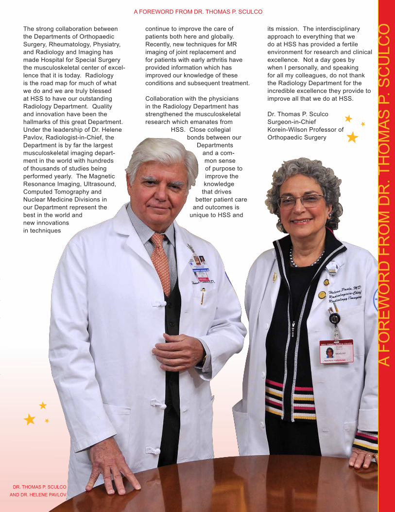
The strong collaboration between the Departments of Orthopaedic Surgery, Rheumatology, Physiatry, and Radiology and Imaging has made Hospital for Special Surgery the musculoskeletal center of excel-lence that it is today. Radiology is the road map for much of what we do and we are truly blessed at HSS to have our outstanding Radiology Department. Quality and innovation have been the hallmarks of this great Department. Under the leadership of Dr. Helene Pavlov, Radiologist-in-Chief, the Department is by far the largest musculoskeletal imaging depart-ment in the world with hundreds of thousands of studies being performed yearly. The Magnetic Resonance Imaging, Ultrasound, Computed Tomography and Nuclear Medicine Divisions in our Department represent the best in the world and new innovations in techniques
continue to improve the care of patients both here and globally. Recently, new techniques for MR imaging of joint replacement and for patients with early arthritis have provided information which has improved our knowledge of these conditions and subsequent treatment. Collaboration with the physicians in the Radiology Department has strengthened the musculoskeletal research which emanates from
HSS. Close collegial bonds between our
Departments and a com-mon sense of purpose to improve the knowledge
that drives better patient care
and outcomes is unique to HSS and
its mission. The interdisciplinary approach to everything that we do at HSS has provided a fertile environment for research and clinical excellence. Not a day goes by when I personally, and speaking for all my colleagues, do not thank the Radiology Department for the incredible excellence they provide to improve all that we do at HSS.
Dr. Thomas P. Sculco Surgeon-in-Chief Korein-Wilson Professor of Orthopaedic Surgery
3
A FO
RE
WO
RD
FR
OM
DR
. TH
OM
AS
P. S
CU
LCO
DR. THOMAS P. SCULCO
AND DR. HELENE PAVLOV
A FOREWORD FROM DR. THOMAS P. SCULCO

4
Our 2009 Annual Report highlights recent information and accomplish-ments of the staff and Faculty of the Department of Radiology and Imaging.
In 2009, the Department of Radiology and Imaging grew in both volume and reputation and continues to be integral to the daily functioning and overall success of Hospital for Special Surgery (HSS) and the referring physician’s private practices at HSS, in the tri-state area and from afar. The Department performed over 235,000 musculoskel-etal imaging examinations in 2009 and currently, the Department represents over 200 employees who individually and collectively strive to provide qual-ity service and meet the exceptional requirements expected at HSS.
Our theme remains constant - “All Images are NOT Created Equal,” and our goal is that our images and reports fulfill the referring physicians’ and our patients’ expectations. Top priority is given to precise image acquisition and clarity along with diagnostic interpretive accuracy. There is direct physician oversight for the majority of our Magnetic Resonance (MR), Computed Tomography (CT), Ultrasound (US), Interventional Radiology (IR) and Nuclear Medicine (NM) examinations, and even conven-tional radiographs often get radiologist validation of optimal positioning and technical factors. The extensive physician involvement in our image acquisition and our skilled musculo-skeletal dedicated radiologic and MR technologists and sonographers along with the interventional team of Nurse Practitioner, Physician Assistants and nurses, help set HSS Imaging apart from other academic sites as well as private imaging facilities. Physician attention provides both “HSS quality” and also assures that the imaging ex-aminations with the highest probability of diagnostic yield to optimally answer the clinical question are performed in appropriate order.
In addition to the quality and expertise behind every image, radiation hy-giene is given paramount importance. Radiation Safety for our patients and our employees is taken very seri-ously by staff and Faculty with strict
A LE
TTE
R F
RO
M T
HE
RA
DIO
LOG
IST-
IN-C
HIE
F
TAB
LE O
F C
ON
TEN
TS01 A LETTER FROM THE RADIOLOGIST-IN-CHIEF
02 THE PATIENT EXPERIENCE
04 DIVISION OF MAGNETIC RESONANCE
05 DIVISION OF ULTRASOUND
06 DIVISION OF INTERVENTIONAL RADIOLOGY AND CT
07 DIVISION OF TELERADIOLOGY
07 DIVISION OF NUCLEAR MEDICINE
08 EDUCATION AND TRAINING
12 RESEARCH AND ACADEMICS
15 VISUALIZING THE PILLARS
16 ECONOMICS
18 HOW WE SEE THE VALUE OF IMAGING
20 COMING AND GOING
23 PUBLICATIONS\PRESENTATIONS\GRANTS
TABLE OF CONTENTS
1
adherence to shielding and using the lowest possible dose without compris-ing image clarity.
This report, with the assistance of Richard Fleury, Executive Director, HSS Radiologists, and Ed White, Assistant Vice President, highlights the overall Department accomplishments and the specific goals achieved within each clinical division. Each Division had specific challenges and the Chiefs of these Divisions and the Assistant Director, Supervisor, and/or Lead Technologists in each area discuss how their challenges were met and expectations exceeded.
Major improvements that benefited both the employees as well as the overall Department efficiency resulted from employee suggestions presented during the “Chats with the Chief” and at Gallup meetings. Over 90% of the 96 issues brought to the atten-tion of leadership, were resolved, satisfied or improved. Ed White, AVP, and Ralph Bianco, VP, facilitated operational changes to bet-ter deal with the increased growth in vol-ume in order to provide optimal service to our patients
and our referring physician’s practices. Additional staff and equipment and organizational innovation and im-provements were initiated for optimiz-ing patient throughput and preventing avoidable patient delays. Patients’ letters and patient satisfaction surveys collected in 2009 acknowledged our success in satisfying patient expecta-tions for both quality service and clinical outcomes.
Education, under the guidance of the Department Director of Education and Fellowship Training, Dr. Carolyn M. Sofka, along with the assistance of the HSS Department of Education secured ACGME accreditation for our MSK Fellowship. This accomplishment further distinguished the uniqueness of the HSS Musculoskeletal Radiology Fellowship from other such programs. Furthermore, this year, every daily radiology conference has achieved CME accreditation. These and other accomplishments are detailed by Dr.
Carolyn Sofka and Amy Lefkovic, Fellowship Coordinator, in the
Education section on page 8.
Research, under the guid-ance of the Department Director of Research, Dr. Hollis G. Potter was distinguished this year by
securing an ROI Challenge Grant; a remarkable achieve-
ment given the vast number of applications. This and the other
Department research accomplishments are
discussed by Dr. Hollis G. Potter
and Kristi Legget, Clinical
Research Coordinator,
in the
Research section on page 12.
The Five Pillars of the HSS strategic plan has had an incredible start within our Department. Each pillar was assigned a color and a champion and every member of the Department has selected a pillar (or two) and is identified with the pillar by a color tab on their ID badge. All are on board and working together even more cohe-sively and even more efficiently than ever to support the Hospital mission.
The 2009 Department successes could not have been accomplished without an incredible staff and Faculty, and the extremely high caliber of our referring physicians and their office staffs with whom we consult daily. Working together, collabora-tively, under the strong administrative leadership provided by Mr. Louis A. Shapiro, Dr. Thomas P. Sculco, Dr. Stephen Paget, Dr. Steven Goldring, Mr. John Cox and Dr. Laura Robbins, has allowed the Department to build on its strong foundation and venture positively into 2010.
Given the current media spotlight on imaging and the battle cry of overuse and the high cost associated with it, it is important to emphasize that the focus of imaging at HSS has ALWAYS been on the appropriate use of diagnostic imaging and the value that it brings to the patient evalua-tion. Imaging provides the objective confirmation and roadmap on which surgery is planned, on which conser-vative treatment can be determined to be sufficient, and on which disease progression and healing can be verified.
I hope you share my opinion that Imaging at HSS is another area on which our patients and referring physicians can rely and of which all of us at HSS, our alumni and the Board of Trustees can take pride. Please take the time to read the 2009 Annual Report and I hope you “see what I see.”
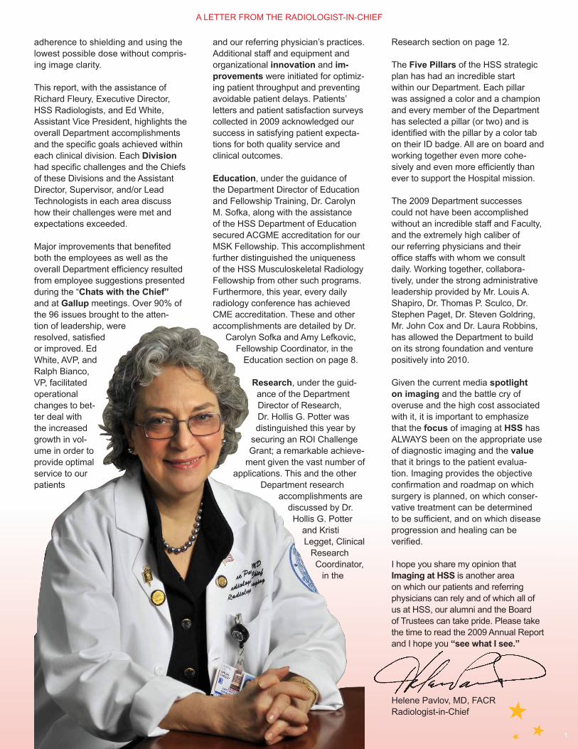
Helene Pavlov, MD, FACRRadiologist-in-Chief
A LETTER FROM THE RADIOLOGIST-IN-CHIEF

THE
PAT
IEN
T E
XP
ER
IEN
CE
2
Improving Patient Experience Many Steps at a TimeOur teams are working in a variety of ways to help improve the overall patient experience.
Pre-Registration Streamlines the Patient Throughput ProcessLed by Mary Giesa, RT, we set out to refine and improve the Department’s already successful pre-registration process. With a goal of increasing the number of pre-registrations, Mary and her team met with the office managers to identify what additional information could be collected prior to a patient arriving in the Department. Twenty-four to 48 hours prior to the patient arriving for their imaging examinations, the patient is contacted to complete required paperwork. This process is helping reduce patient wait time and improve the patient experience when they arrive to the Department. It also makes scheduling easier for the referring physicians’ office staff.
New processes were put in place be-ginning October 2009 and since then between 85% and 95% of patients on the main floor and in pediatrics are preregistered.
“We like to call this express check in. A patient arrives, their paperwork is ready and they move immediately to the waiting area. This has cut down reception wait time and helps us make a great first impression. All in all it makes the experience easier for the patient and they feel like VIPs because they have received special treatment. Thanks to everyone for making this an incredibly successful initiative,” said Mary Giesa, RT.
First Impressions Training In an effort to continually improve patient and referring physician satisfaction, Denise Williams, Training Manager of Organizational Learning and Development led role-playing workshops to help our team under-stand how they interact with others and how they can improve their customer service skills.
“We have seen some incred-ible improvements throughout the Department. From our high satisfac-tion ratings to the numerous letters from patients and the kudos from referring physicians, we are seeing the benefits from our efforts really pay off,” said Robyn Pack, Assistant Director, Clinical Services.
Increased Information for Patients Virtual tours of each modality have been prepared to help ease patient’s minds before their appointment and also educate them in regards to their imaging procedure. Virtual tours of each modality have been created by Maribel Maldonado, Academic Technologies Coordinator, and Tess Leynes, Nurse Practitioner. These virtual tours provide detailed
descriptions of each area and will be posted on the website in early 2010. Additional improvements in the website include patient instructions prior to scheduled appointments and detailed patient discharge instruc-tions for review pre- and post- pro-cedures. There is also online map of the third floor so patients know where they need to be and will feel comfort-able when exiting the elevators in our various service locations.
New Web URL and Website RedesignThe site has been upgraded with more patient information, features and innovative functionality. We provide information for seniors needing X-rays and the HSS paediatric website sec-tion has specific information regarding imaging. Also, the Department website currently offers online MRI appointment scheduling, and similar scheduling options for the other modalities are planned. Patients have the ability to request an appointment online and within one business day they will be contacted by a scheduler to confirm their appointment. The site is easy to use and has been well received by referring physicians and their staff who attended our meet-ings during the summer. Visit us at: http://imaging.hss.edu.
OUR RECEPTION AREA HAS BECOME EVEN MORE WELCOMING WITH NEW SIGNAGE AND A NEW
UNIFORM INITIATIVE HELPS FURTHER ENHANCE OUR FIRST IMPRESSION.
THE PATIENT EXPERIENCE
3
New Process for Collecting Customer Service InsightsThe Department has rolled out new customer service forms that are in line with the Hospital’s questionnaire. Questions on the form ask about patient wait time, the waiting area, registration processes, nursing, technologists and the overall experi-ence. The form is detailed and is specific to the Department’s different areas and the individual modalities. Although we continue to perform well and rank in the high 90th percentile in terms of satisfaction, we are striving to institute other ways of improving patients’ satisfaction.
Department Improvements Continue to Focus on Employee Engagement, Positive Service and Quality Patient CareSignificant changes to the Department were initiated in 2009, many because of employee suggestions at the Department Chats. New equipment, new counter space, new storage areas, new walls and new ceilings are all a part of our ongoing commitment to our patients and employees. Our goal is to make each individual experi-ence the best that it can be. Some of our most recent changes include:
•The Pediatric X-ray Center has been updated and refurbished with beautiful aesthetic details. Positive comments from patients and staff say that the new space looks great and
is a vast improved work and patient environment.
•Much of the Main Department technologists’ time at work is spent in the ‘The Tech Mall.’ This operational hub and center of daily activity has had a physical facelift. New counters and workstations and recessed equipment help give this space a welcoming open ambience.
•In the OR, the dark room was eliminated and transformed into a new high-tech work area including a digitizer, CR reader and technologist digital workstation.
•Five replacement C-Arms have been received. Requests for further upgraded C-Arm capability in the OR, continue. New state of the art C-arms are being reviewed by radiology and a team of sur-geons to evaluate
multiplanar 3D capabilities available on some of the newer models.
•Replacement of ERPB 4th floor X-ray equipment has been upgraded to Digital Radiology (DR) with a new Canon tethered digital receptor for increased image acquisition flexibility.
•Belaire 2 and the East River Professional Building technologist work area on the Ground Floor underwent complete renovations.
•Major renovations in the Sports Medicine suite on Belaire 1 are in progress.
•The completion of two additional radiographic rooms in the Caspary
Building is progressing on schedule in order to accommodate the
Center for Hip Pain and Preservation and new surgeons joining HSS.
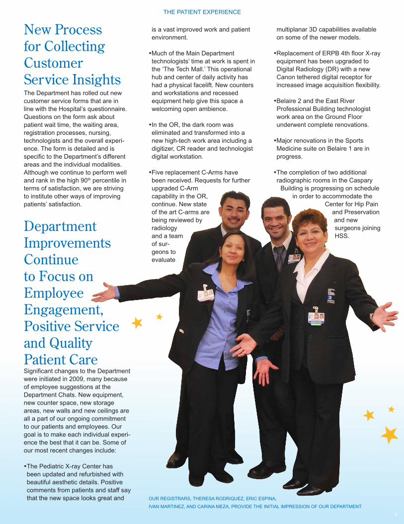
OUR REGISTRARS, THERESA RODRIQUEZ, ERIC ESPINA,
IVAN MARTINEZ, AND CARINA MEZA, PROVIDE THE INITIAL IMPRESSION OF OUR DEPARTMENT
THE PATIENT EXPERIENCE

DIV
ISIO
N O
F M
AG
NE
TIC
RE
SO
NA
NC
E
4
Evolutions in Technology and ServiceMR volume grew by 6.2% in 2009. The MR Division expanded in volume and in technology and is continuously look-ing to the future. Some of the latest changes in the MRI Division include:
•The 8th MRI unit is installed and helps further satisfy the demands for expanded service and easier patent scheduling. We have upgraded to a new GE 750 3T with 15x software, which is the most up to date MRI platform.
•In December 2009, one of our existing units was upgraded to the GE 1.5T 450. The equipment is faster and the gradient abilities have been improved to allow for additional scan-ning of patients with metal implants.
•The MR division was the first division to offer online appointment schedul-ing on the Department website.
Cleverly Looking Beyond the Typical Increases in Consumption Helps Drive Higher Utilization 2009 was a challeng-ing year for everyone, but like the rest of the Department, the MRI Division exceeded expectations with an impressive 6% over what was forecasted. Part of this has to do with the continued growth in MR imaging worldwide, but also be-cause the MR team looked
beyond the routine trends in order to expand our offerings and service.
“We tried to be proactive in how we could increase the use of the magnets and accomplished this by utilizing and leveraging our research efforts. Our research is always clinically relevant and directed at how we can help refer-ring physicians better diagnose patient conditions, which results in better care and faster recovery,” commented Dr. Potter.
The imaging of arthroplasty is a perfect example of how this strategy has proven to be successful for the MRI Division and a value to referring physicians. A few years ago the thought of placing a patient with a knee replacement into an MRI was unheard of because of the metal in an arthroplasty. Dr. Potter and her team worked to develop new MRI pulse sequences that made the imaging of arthroplasty possible. Patients that are suffering from post joint replacement pain that is often not detectable using traditional methods of imaging such as conventional radiographs can often find the answer using these MRI techniques. Scanning patients with total joint replacements has resulted in an increased number of these MR scans (approximately 8-10 per day.)
2010 will be another great year for the Division. With eight magnets
in operation, HSS is now the largest MRI academic imaging center solely dedicated to Musculoskeletal Imaging in the State of New York. The MR team is always looking into how to optimize existing scanners for increased patient throughput.
“Our team is a well-oiled ma-chine and our leadership knows everyone that works here, which has fostered a strong sense of camaraderie. We understand the challenges associated with having an MRI and do everything we can to make patients feel comfort-able. Positive letter after posi-tive letter from patients and referring physicians are proof that the processes we have put into place are actually working. In addition, our QA process always ensures that a patient never leaves our Division un-less they have had the best scan possible. At the end of the day we focus on providing the best service and images possi-ble and the rest just falls right into place.” Ruth Ann Lindner, RT
JUNG JOO, RT,
RUTH ANN LINDER, RT
AND DR. HOLLIS G.
POTTER, CHIEF,
DIVISION OF MR
DIVISION OF MAGNETIC RESONANCE

Ultrasound Continues Its Unprecedented Growth The Division of Ultrasound (US), led by Ronald Adler, PhD, MD, Chief, Division of Ultrasound and Body CT, has been making great strides in terms of growth by offering exquisite diagnostic and targeted non-surgical treatment options for various tendon and bursal conditions and neuromas. Ultrasound grew by 13.9% which Dr. Adler attributes “…to more referring physicians recognizing the value of Ultrasound in orthopaedics. More than ever before, physicians now understand the clinical value of this modality for its diagnostic and treat-ment capabilities.”
The US Division continues to introduce new clinical applications including platelet rich plasma (PRP) injections as well as cryotherapy (cold therapy).
Platelet Rich Plasma (PRP) Injections Gain Traction in 2009Early in 2009, a New York Times reporter was referred to Dr. Adler by Dr. Joseph Feinberg, Associate Attending Physiatrist HSS, to receive a new treat-ment that is gaining a lot of notoriety in the press. PRP injections were talked about frequently in the news throughout 2009, mainly for its healing properties in injured ten-dons. Platelet activation plays a key role in the process of wound and soft tissue healing.
PRP was used as
early as the 1990s in maxillo-facial and plastic surgery. PRP, involves a concentration of platelets above baseline that are obtained from the patients own blood and is used to promote healing of injured tendons, ligaments, muscles and joints. The patient’s blood is centrifuged and the activated platelets are injected directly into the area of abnormal tissue, releasing growth factors that recruit and increase the prolifera-tion of reparative cells. Ultrasound imaging is used to guide the injection and ensure that the PRP is precisely delivered into the affected area. The side effects are very limited as the process utilizes the patients own blood.
Several clinical studies have dem-onstrated improved function and decreased pain for various condi-tions, including - but not limited to - elbow, wrist, shoulder, hip, knee and ankle tendonosis. Early work is also showing promise for osteoarthritis.
It is extremely satisfying to see suc-cessful results as patients leave our Department, improved.
Doctors Begin Using Cryotherapy and Cryoablation to Treat PatientsCryotherapy is one of the simplest and oldest therapeutic modalities used in the treatment of acute soft tissue injuries. Cryotherapy is also used as an adjunct in the rehabilitation process following orthopaedic procedures, such as total knee arthroscopy, anterior cruciate ligament reconstruction of the knee, or total knee replacement.
Dr. Adler has been working closely with Daniel Richman, MD, Attending (Pain Management) Anesthesiologist, to deliver ultrasound-guided cryoablation. Cryoablation is a process that uses extreme cold to destroy affected tissue. The process is most notably used in or-thopaedics to help treat painful neuro-mas. Neuromas can be challenging to treat because they are hard to pinpoint, but ultrasound allows Dr. Adler to target the exact location and Dr. Richman to deliver the cryoablation precisely to the
area.
DR. RONALD S. ADLER, CHIEF, DIVISION OF US,
EXAMINING AN ANKLE WITH ULTRASOUND
DIV
ISIO
N O
F U
LTR
AS
OU
ND
5
DIVISION OF ULTRASOUND
DIV
ISIO
N O
F IN
TER
VE
NTI
ON
AL
RA
DIO
LOG
Y A
ND
CT
6
Ongoing Outreach to Referring Physicians about our Services Helps Inform and Increase Same Day Patient ‘Add-Ons’ for CT Examinations and IR ProceduresThe Division of Interventional Radiology (IR) and CT, led by Gregory Saboeiro, Chief, with the assistance of Joanna Waldman, RT Supervisor, grew by 9.1% and the Division of IR, with the assistance of Tess Leynes, NP, Assistant Director, grew by over 6%. IR procedures, including both diagnostic and treatment procedures performed by Radiologists using fluoroscopy (e.g., epidural injections, facet joint injections, etc.) increased by 17.2% over 2008 as a result of outreach efforts to referring physicians informing them of the Department policy of adding patients who are experiencing pain to the schedule on very short notice.
Interventional Radiology Continues to GrowInterventional Radiology (IR) performs minimally invasive procedures using image guidance. Some of these interventional procedures are per-formed for purely diagnostic purposes, (myelograms), while other procedures are performed for treatment (epidural
injections, facet joint injections, etc). Most interventional procedures are performed under fluoroscopic or ultrasound guidance, and a few are per-formed under CT (biopsies). Image guid-ance directs precise needle or catheter placement to the target zone for biopsy and confirms the injection site of contrast or therapeutic medication. The imaging provides a road map for the interventional radiologist.
Improving the Patient ExperienceBehind the scenes and with the help of Tess Leynes, Nurse Practitioner and Robert Polintan Physician’s Assistant the IR team has accom-plished their goal of ensuring that the Department provides the best service possible to both our patients and our referring physicians.
Meetings were held with HSS referring physicians and their office staff to im-prove communications and streamline patient scheduling and throughput. Robyn Pack, Yuliana Belyayeva, Maribel Maldonado, Tess Leynes, NP, Ruth Ann Lindner, RT, and Ed White, AVP, met with referring physicians and their staff to gain feedback and pro-vide training on how to best schedule their patients for appointments and to learn how we can best meet their office flow and their patients’ needs.
Patient instructions and the informa-tion required to prepare for an inter-ventional procedure was put online as an available reference. Referring physician offices and our staff are now better prepared to answer patients’
questions regardless if the patient calls us directly or if they call the referring physicians’ offices. Improving communications has resulted in a bet-ter and more efficient and satisfactory patient experience.
The team attributes the increase CT and IR volume to a number of positive improvements including the addition of 10 new technologists; moving the pro-cedure rooms to provide more privacy; a more comfortable waiting room for the patients and their escorts; new pa-tient scheduling procedures designed to help office staff better address their patients’ needs; easier accommoda-tion of add-ons; and participaton with all major insurance carriers.
According to Dr. Saboeiro, “More clini-cians and patients are realizing that performing injection procedures using dedicated imaging equipment (CT, US and fluoro) and highly trained physi-cians and technologists to ensure min-imum radiation exposure, as offered in the Department of Radiology and Imaging, is beneficial and in general provides longer-acting pain relief when compared to unguided injec-tions. By collaborating with our refer-ring physicians, the patients receive expert diagnostic and targeted pain relief treatments. Patients’ continuity of care is maintained with their refer-ring physician while their work up and management is enhanced through an expert imaging consultation with the radiologist. This healthcare team effort helps HSS achieve the highest level of overall patient care.”
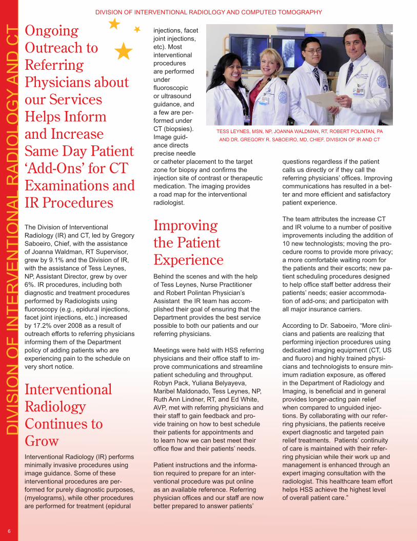
TESS LEYNES, MSN, NP, JOANNA WALDMAN, RT, ROBERT POLINTAN, PA
AND DR. GREGORY R. SABOEIRO, MD, CHIEF, DIVISION OF IR AND CT
DIVISION OF INTERVENTIONAL RADIOLOGY AND COMPUTED TOMOGRAPHY
DIV
ISIO
N O
F N
UC
LEA
R M
ED
ICIN
E
7
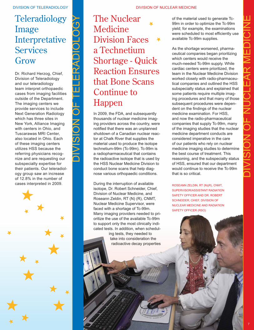
The Nuclear Medicine Division Faces a Technetium Shortage - Quick Reaction Ensures that Bone Scans Continue to HappenIn 2009, the FDA, and subsequently thousands of nuclear medicine imag-ing providers across the country, were notified that there was an unplanned shutdown of a Canadian nuclear reac-tor at Chalk River that supplies the material used to produce the isotope technetium-99m (Tc-99m). Tc-99m is a radiopharmaceutical that contains the radioactive isotope that is used by the HSS Nuclear Medicine Division to conduct bone scans that help diag-nose various orthopaedic conditions.
During the interruption of available isotope, Dr. Robert Schneider, Chief, Division of Nuclear Medicine, and Roseann Zeldin, RT (N) (R), CNMT, Nuclear Medicine Supervisor, were faced with a shortage of Tc-99m. Many imaging providers needed to pri-oritize the use of the available Tc-99m to support only the most clinically indi-cated tests. In addition, when schedul-
ing tests, they needed to take into consideration the radioactive decay properties
of the material used to generate Tc-99m in order to optimize the Tc-99m yield; for example, the examinations were scheduled to most efficiently use available Tc-99m supplies.
As the shortage worsened, pharma-ceutical companies began prioritizing which centers would receive the much-needed Tc-99m supply. While cardiac centers were prioritized, the team in the Nuclear Medicine Division worked closely with radio-pharmaceu-tical companies and outlined the HSS subspecialty status and explained that some patients require multiple imag-ing procedures and that many of those subsequent procedures were depen-dent on the findings of the nuclear medicine examination. For HSS, and now the radio-pharmaceutical companies that supply Tc-99m, many of the imaging studies that the nuclear medicine department conducts are considered imperative in the care of our patients who rely on nuclear medicine imaging studies to determine the best course of treatment. This reasoning, and the subspecialty status of HSS, ensured that our department would continue to receive the Tc-99m that is so critical.
Teleradiology Image Interpretative Services GrowDr. Richard Herzog, Chief, Division of Teleradiology and our teleradiology team interpret orthopaedic cases from imaging facilities outside of the Department. The imaging centers we provide services to include Next Generation Radiology which has three sites in New York, Alliance Imaging with centers in Ohio, and Tuscarawas MRI Center, also located in Ohio. Each of these imaging centers utilizes HSS because the referring physicians recog-nize and are requesting our subspecialty expertise for their patients. Our teleradiol-ogy group saw an increase of 12.8% in the number of cases interpreted in 2009.
DIV
ISIO
N O
F TE
LER
AD
IOLO
GY
ROSEANN ZELDIN, RT (N)(R), CNMT,
SUPERVISOR/ASSISTANT RADIATION
SAFETY OFFICER AND DR. ROBERT
SCHNEIDER, CHIEF, DIVISION OF
NUCLEAR MEDICINE AND RADIATION
SAFETY OFFICER (RSO)
DIVISION OF TELERADIOLOGY DIVISION OF NUCLEAR MEDICINE
ED
UC
ATIO
N A
ND
TR
AIN
ING
8
The Musculoskeletal Radiology Fellowship Program Receives Accreditation from the Accreditation Council for Graduate Medical Education (ACGME)The Department of Radiology and Imaging is pleased to announce that the Musculoskeletal Radiology (MSK) Fellowship Program is now accred-ited by the Accreditation Council for Graduate Medical Education (ACGME).
Subspecialty Radiology fellowship programs are required to acquire accreditation support through their parent Radiology resi-dency programs which for us is through our relationship with NewYork-Presbyterian Hospital. However, the volume and variety of cases at Hospital for Special Surgery, coupled with the exceptional teaching Faculty and facilities, provided the foundation for the ACGME to grant HSS the privilege of being the sole sponsoring institution for our MSK fellow-ship program. It is now the only MSK fellowship program of its kind in the country.
“The goal of our fellowship program is to provide the strongest foundation for training in musculoskeletal imaging in the United States,” said Carolyn M. Sofka, MD, Director of Education
and Fellowship Training for Hospital for Special Surgery, Department of Radiology and Imaging.
“Hospital for Special Surgery is the premier hospital for orthopaedic and rheumatologic diseases and we have a reputation for offering the best in orthopaedic and musculoskeletal education,” said Laura Robbins, DSW, Vice President for Education and Academic Affairs at Hospital for Special Surgery. “This accreditation allows us options on programming and educational decisions indepen-dent of a sponsoring institution. More importantly, the accreditation holds us to an even higher standard and reflects the excellence associated with the Department of Radiology and Imaging’s outstanding musculoskel-etal fellowship program.”
The HSS Department of Radiology and Imaging Faculty are recognized globally in the radiology, orthopaedic and rheumatologic communities for their imaging expertise and innovative research, as well as their commit-ment to teaching. The Department of Radiology and Imaging is also one of the first departments at HSS to receive Continuing Medical Education (CME) accreditation for all of its five regularly scheduled weekly conferences. “Our team is rightfully
proud of this major accomplishment. ACGME helps to highlight the strengths of our educational commit-ment and further distinguishes our MSK Fellowship from all the others,” said Helene Pavlov, MD, FACR, Radiologist-in-Chief, Hospital for Special Surgery.
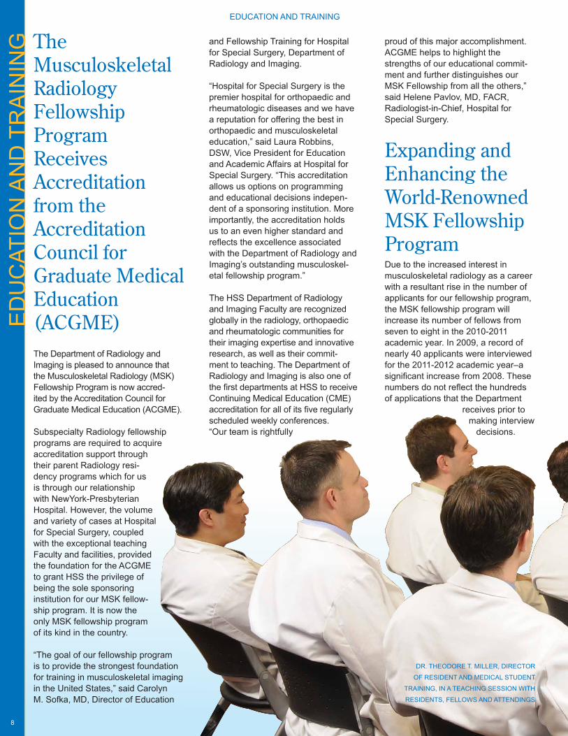
Expanding and Enhancing the World-Renowned MSK Fellowship ProgramDue to the increased interest in musculoskeletal radiology as a career with a resultant rise in the number of applicants for our fellowship program, the MSK fellowship program will increase its number of fellows from seven to eight in the 2010-2011 academic year. In 2009, a record of nearly 40 applicants were interviewed for the 2011-2012 academic year–a significant increase from 2008. These numbers do not reflect the hundreds of applications that the Department
receives prior to making interview
decisions.
DR. THEODORE T. MILLER, DIRECTOR
OF RESIDENT AND MEDICAL STUDENT
TRAINING, IN A TEACHING SESSION WITH
RESIDENTS, FELLOWS AND ATTENDINGS
EDUCATION AND TRAINING

9
2009−10 FellowsIn July 2009 we welcomed seven new Musculoskeletal Radiology fellows into the Department.
Graham Campbell, MDDiagnostic Radiology Residency at Foothills Medical Center, Calgary ABDuring the last five years, Dr. Campbell has worked in a community private general radiology practice group where he was involved in neuroangiography and interventional neuro-procedures.
Yoshimi Endo, MDDiagnostic Radiology Residency at SUNY Downstate Medical CenterDr. Endo was the NYRS Quiz winner in musculoskeletal radiology in 2008 and conducted musculoskeletal
research that was published in the American Journal of Radiology. He has also published four additional radiology papers and had two recent RSNA presentations.
Thomas Hash, MD (MR Fellow)Diagnostic Radiology Residency at Bethesda Naval HospitalDr. Hash has practiced general radiol-ogy for the past four years and is cur-rently Head of Radiology at Pensacola Naval Hospital. He was an exceptional medical student and was elected chief resident during his residency and performed in the top 10th percentile on his in-service and American Board of
Radiology certification examinations.
Akira Murakami, MDDiagnostic Radiology Residency at Boston Medical CenterDr. Murakami has been recognized by his colleagues at Boston Medical Center as one of the most diligent and meticulous radiologists, capable of ac-complishing a disproportionately large amount of work on a regular basis to perfection. He is co-author of the article “Detection of Vascular Injuries in Patients with Blunt Pelvic Trauma by Using 64-Channel Multidetector CT” in Radiographics.
EDUCATION AND TRAINING EDUCATION AND TRAINING
10
Hsiu Su, MDDiagnostic Radiology Residency at Stonybrook University HospitalDr. Su has been honored as one of the brightest and most well-rounded residents in the Stonybrook program. He is co-author of the article “FDG PET Imaging Features of a Perigraft Leak and Thrombus in a Patient with Dissecting Descending Thoracic Aortic Aneurysm” in Clinical Nuclear Medicine.
Harlan Stock, MDDiagnostic Radiology Residency at Lenox Hill Hospital and Nuclear Medicine Fellowship at Memorial Sloan Kettering Cancer CenterDr. Stock is not only an accomplished physician; he was also the Connecticut State singles tennis champion in 1995. Beyond the court, he co-authored the article “Osteoporosis: A Disease in Men” in Clinical Orthopaedics and Related Research.
Gregory Wilde, MDDiagnostic Radiology Residency at Christiana Care Health SystemDr. Wilde is known for his excellent diagnostic skills, which has set him apart from his other classmates. He was awarded the Shaw Trust Award for Research in Radiology in 2005. Dr. Wilde was co-author of the article “Radiological Reasoning: Miliary Disease, Vertebral Osteomyelitis, and Soft-Tissue Abscesses” in American Journal of Roetgenology.
Restructured Training Program for Radiology Residents and Medical StudentsTraining residents from NewYork-
Presbyterian Hospital (NYPH) and St. Vincent’s Hospital, and medical students from Weill Cornell Medical College (WCMC) in musculoskeletal
radiology has been ongoing for many
years. For many of these students this is their very first exposure to musculoskeletal radiology.
Beginning in 2009, Dr. Theodore T. Miller, Attending Radiologist, Hospital for Special Surgery and Director, Resident and Medical Student Training, worked with Drs. Sofka and Pavlov to restructure the musculoskeletal radiology resident rotation at HSS. Residents and medical students spend time reading MRIs and conventional radiographs, observing or participat-ing in Ultrasound and Interventional Radiology procedures, under the supervision of the HSS Attending Radiologists, and working with our MSK Fellows. This revamped HSS rotation has proven to be very popular with the NYPH and St. Vincent’s residents and WCMC medical students.
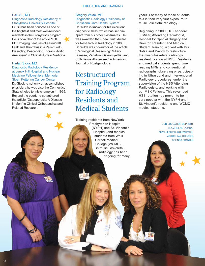
OUR EDUCATION SUPPORT
TEAM: IRENE LAJARA,
AMY LEFKOVIC, ROBYN PACK,
MARIBEL MALDONADO,
BELINDA FRANQUI
EDUCATION AND TRAINING
The RHF Academic Center and Library EvolvesUnder the guidance of Richard Fleury, Maribel Maldonado, Academic Media Coordinator and Irene Lajara, Academic Technologist Assistant, the Library continues to evolve despite major reconstruction. Although space was relinquished, requiring moving many of our journals, connecting to online editions helped to reduce paper and create more space.
The RHF Academic Center and Library recently purchased a new state-of-the-art Teaching File system with a centralized database that is used by the Radiologists and Fellows
to import interesting and educational cases directly from the PACS system. Drs. Bogner, Schneider, Chang and Miller are pivotal in providing oversight for the Teaching File. These selected cases are archived and will be available for teaching and presentations.
In the coming months the Center will have a new projector mounted and installed making the space an additional fully functional presenta-tion venue.
Improved Education Program for New and Seasoned Technologists Training and education for new technologists has been revamped with a formal program established under the direction of Maureen Firth, MS, RT, (R) (M) QM, and Ralph Lopez, RT.
Before they start in our Department, each new hire goes through an extensive orientation where they learn department and hospital organization and leadership, safety and environmental care, as well as mission values and expectations, Joint Commission requirements and more. This orientation helps to prepare them for daily work in the Department and interaction with other departments in the Hospital.
Following the Hospital orientation the new employee starts to work in theDepartment, where the focused and intense Department training begins. Each new technologist is trained for an extended period of time in a specific modality and for specific body parts in order to help better prepare our new technologists to meet the quality expectation of the Department. The program includes hands-on training with skilled experienced technologists for two weeks. After the technologists’ second week, they may begin working on their own, with close oversight from seasoned technolo-gists specifically picked to be part of the Education Team. When “cleared” to work independently, after passing specific “competency expectations,” the new technologists are ready to hit the ground running with confidence. This enhanced and intensive training program has expedited the process and is considered to be a vast improvement to the past, “catch as you can” learning process.
A seasoned technologist competency program was also initiated by Ralph Lopez, Chief Technologist, and Maureen Firth. This program is de-signed to assure that skills are current and maintained at expected levels.
11
In 2009 more than 40 stu-dents including residents, orthopaedic residents, medical students and observers came to HSS Department of Radiol-ogy and Imaging to learn from our expert team of Faculty and staff.
The Robert H. Freiberger, MD Academic Center and Library is the largest and possibly the only library strictly dedicated to Musculoskeletal Radiology. It is used by Faculty, staff and physicians outside of HSS to help expand their knowledge in orthopaedic imaging.
Observers from Far and Wide Come to Learn from Our ExpertsDuring the past several years, the Department opened its doors for practicing radiologists and trainees interested in learning more about musculoskeletal radiology. These have included medical students not only from Cornell University but from all over the country exhibiting an inter-est in musculoskeletal radiology and orthopedics. Practicing radiologists both from the United States and from abroad who wish to hone their skills in musculoskeletal imaging techniques, specifically MRI and ultrasound, have visited the Department.
Drs. Sofka, Schneider and Bogner; Maribel Maldonado, Irene Lajara, and the entire Education Division and Faculty have been busy add-ing new teaching materials to the Robert H. Freiberger Academic Center and Library for both the musculosk-eletal radiology fellows as well as the observers and visitors. Teaching resources include videos from lectures and conferences given by the Faculty at local, national and international meetings, PowerPoint presentations, and real time procedures performed under imaging (CT, US and fluoro) guidance. Images of interesting cases and from teaching conferences are also being archived by Faculty into the new Digital Teaching File.
To learn more about observership opportunities, please visit the website at www.hss.edu/radvisitor.
EDUCATION AND TRAINING EDUCATION AND TRAINING
The Radiology CRP’s goal is to ensure that all Research projects that are imaging based or use imaging as an outcome measure are scientifically valid, with appropriate imaging methodology and outcome criteria. Once approved by the CRP, the proposal is sent to the IRB to ensure patient safety and HIPAA compliance.
In 2010, the Department will con-tinue its groundbreaking research. Dr. Hollis G. Potter and Dr. Li Foong Foo will continue focusing on the use of quantitative MRI in cartilage repair with the HSS Cartilage Registry along with Dr. Riley Williams, and Dr. Matt Koff,
RE
SE
AR
CH
AN
D A
CA
DE
MIC
S
12
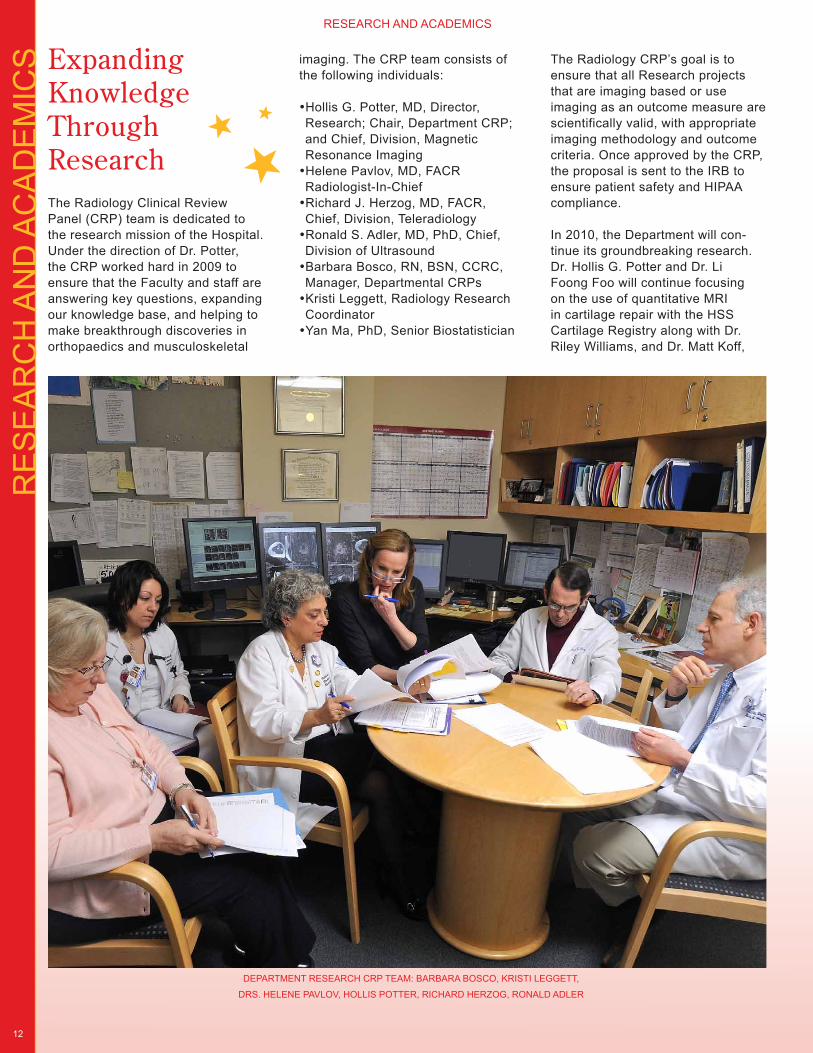
Expanding Knowledge Through ResearchThe Radiology Clinical Review Panel (CRP) team is dedicated to the research mission of the Hospital. Under the direction of Dr. Potter, the CRP worked hard in 2009 to ensure that the Faculty and staff are answering key questions, expanding our knowledge base, and helping to make breakthrough discoveries in orthopaedics and musculoskeletal
RESEARCH AND ACADEMICS
DEPARTMENT RESEARCH CRP TEAM: BARBARA BOSCO, KRISTI LEGGETT,
DRS. HELENE PAVLOV, HOLLIS POTTER, RICHARD HERZOG, RONALD ADLER
imaging. The CRP team consists of the following individuals:
•Hollis G. Potter, MD, Director, Research; Chair, Department CRP; and Chief, Division, Magnetic Resonance Imaging•Helene Pavlov, MD, FACR Radiologist-In-Chief•Richard J. Herzog, MD, FACR, Chief, Division, Teleradiology•Ronald S. Adler, MD, PhD, Chief, Division of Ultrasound•Barbara Bosco, RN, BSN, CCRC, Manager, Departmental CRPs•Kristi Leggett, Radiology Research Coordinator•Yan Ma, PhD, Senior Biostatistician
13
Assistant Scientist, at HSS. Dr. Ronald Adler will continue his ongoing work in the area of rotator cuff repair using ultrasound con-trast agents to explore vascularity following surgery along with Dr. Stephen Fealy. Dr. Pavlov is work-ing with various surgeons including Drs. Jonathan Deland, Scott Ellis and paediatric spine surgeon Dr. Daniel Green, to identify various orthopaedic applications for upright CT image acquisition software.
MRI Division Receives the Department’s First NIH Grant The American Recovery and Reinvestment Act of 2009 is a Federal public law passed by the 111th United States Congress and signed into law by President Barack Obama on February 17, 2009. As part of the Recovery Act, NIH has designated at least $200 million in Fiscal Year 2009 - 2010 for a new initiative called the NIH Challenge Grants in Health and Science Research, to fund 200 or more grants, contingent upon the submission of a sufficient number of scientifically meritorious ap-plications. The NIH has identified a range of Challenge Areas that focus on specific knowledge gaps, scien-tific opportunities, new technologies, data generation, or research meth-ods that would benefit from an influx of funds to quickly advance the area in significant ways.
Dr. Hollis G. Potter, Chief, Division of Magnetic Resonance Imaging and Director of Research, along with Matthew F. Koff, PhD, Assistant Scientist in the MRI Division, sub-mitted a grant proposal to evaluate soft tissue properties of repaired menisci using a special MRI imaging technique. The team of researchers is studying whether repaired menisci can be non-invasively assessed in their ability to withstand load. This MRI technique will allow the team to
actually quan-tify meniscal healing, which could not be objectively done previ-ously.
Approximately 30,000-35,000 applications were received by the NIH and the grant prepared by Dr. Potter and Dr. Koff was accepted. This is the first Federal grant to be received by the Department and helps to further validate our position as musculoskeletal imaging subspe-cialty experts. Congratulations to Dr. Potter and Dr. Koff for helping to win this grant which totaled $850,000.
Drs. Adler and Fealy Publish Ultrasound Rotator Cuff Research Sponsored by Major League Baseball Association GrantIn 2007, the Major League Baseball Association provided a three-year research grant to Dr. Adler working in conjunction with Dr. Stephen Fealy, Assistant Attending Orthopaedic Surgeon to conduct research on vas-cularity associated with rotator cuff injuries. This research focuses on ultrasound contrast agents that are administered by Dr. Adler to targeted areas within the rotator cuff and tracked with ultrasound. He and his team are looking at vascularity within various areas of the cuff, assessing response to therapy regimens and determining whether age plays a
major factor in vascularity. The results of this research have been published in the Journal of Shoulder and Elbow Surgery. The conclu-sion, as well
as future direction of this research, will have major implications in treat-ment recommendations.
PRP Investigations for Validation of Clinical EffectivenessIn 2010, Dr. Adler, in conjunction with Dr. Peter Moley, Assistant Attending Physiatrist, Hospital for Special Surgery, are planning research focused on treating hamstring tendonitis either with PRP or autologous blood injections under ultrasound guidance. The goal of the research will be to see if there is a difference in outcomes between PRP and autogolous blood treatments. Since previously only purely anecdotal evidence regarding the benefits of PRP existed, it is our hope to publish validated clinical results based on quantitative and scientific evidence.
Multiplanar (CT) and 3D Volume Imaging with Patient Weight Bearing Under a Strategic Research Agreement between HSS and Philips Medical Systems, proprietary software is available to capture multiplanar –sagitial, coronal, and axial–images and 3D volume reformations from a
“I have the utmost respect for Dr. Koff. He has been a tremendous asset to the MRI Division, the Department and to HSS. His skills compile the perfect fu-sion between a scientist/researcher and someone who is well versed at competing for grant dollars in a clinical MRI setting. We are very lucky to have him on our team.” Dr. Hollis G. Potter
RESEARCH AND ACADEMICS RESEARCH AND ACADEMICS
14
single 180 degree sweep of the X-ray beam and a flat screen capture. This technology is being employed by Dr. Pavlov and her designees in collaboration with our surgeons. The images can be reformatted into 3D volume images with multiple bone or X-ray surface renderings. Mario Solano, RT, has worked exten-sively with this equipment, and this technology is the only multiplanar imaging capture that can be per-formed while the patient is standing and weight bearing. Dr. Pavlov’s work with Drs. Bedi and R. Williams et al. on tibial tunnel orientation in cadaver knees was presented at the 2009 Annual Meetings of the AAOS and AAOSM. Clinical research on various painful flat foot deformities with Drs. Ellis and Deland et al. was presented at the 2009 AOFAS Annual meeting. This latter work was accepted for publication in Foot & Ankle International.
Additional research on cadaver spines with Drs. Greene and Kepler et al. will be presented as a poster at the ORS in 2010. This investigation has clinical application for identifying spondylosis and spondylolisthesis in children. This technique has equal sensitivity but less ionizing radiation than CT, and because the image can be obtained while the patient is weight bearing, malalignment may be more optimally demonstrated and this one radiographic examination may replace conventional X-rays and CT examinations.
Throughout 2009, Faculty members in the Department along with other physicians and surgeons in the Hospital and in other institutions have
been working on various research studies. The following is a sampling of some of their work:
•Dr. Adler, along with other Hospital for Special Surgery physi-cians including Dr. Russell Warren, Attending Orthopaedic Surgeon, and Dr. Stephen Fealy, Assistant Attending Orthopaedic Surgeon, former Assistant Attending Radiologist, Dr. Maderazo as well as others, researched and published on rotator cuff repair and the vascular-ity of the supraspinatus tendon in the Journal of Shoulder and Elbow Surgery and the American Journal of Sports Medicine.
•Dr. Bogner, Attending Radiologist, published an article in Sports Health: A Multidisciplinary Approach on the “Imaging of Cervical Spine Trauma.”
•Drs. Chang and Miller published a piece on the “Imaging of Tendons” in Sports Health: A Multidisciplinary Approach.
•Drs. Potter, Foo, Boettner and Principal Investigator Dr. Pellicci, Attending Orthopedic Surgeon, Hospital for Special Surgery, published an article “MRI Shows Biologic Restoration of Posterior Soft Tissue Repairs after THA” in Clinical Orthopaedics and Related
Research.
•Dr. Sofka was the Guest Edi-tor of Radiol-ogy Clinics of North America entitled “The History of Clinical Mus-culoskeletal Radiology”.
Collaboration with GE on Cartilage ResearchWorking with General Electric Healthcare, the Division of MRI is a testing ground for software that allows the team to accurately develop 3D models of articular cartilage. These models will ultimately be used as potential digital templates for partial joint replacement and tissue engineering. In addition, quantitative MRI techniques provide a noninvasive “microscope” into repair cartilage, and the initial results from a two-year study were recently published in the American Journal of Sports Medicine.
RESEARCH AND ACADEMICS
A complete list of Department pub-lications, presentations, grants and service commitments of the HSS Radiologists in the Department of Radiology and Imaging begins on page 23 of this report.
Strategic Research Agreement with Philips Med-ical Systems enables 3D US and Fluoroscopic workstation investigations to explore orthopae-dic applications As a luminary site for Philips Medical Systems, the Divisions of Ultrasound and General X-ray are testing 3D Ultrasound and Fluoroscopic workstations to better understand the utility of 3D technology in orthopaedics. Initial 3D weight bearing applications in foot and ankle dynamics has been published in Foot & Ankle, Intl.
VIS
UA
LIZI
NG
TH
E P
ILLA
RS
15
The Hospital’s Five Pillars strategic goals initiative is the organization’s framework for defining the future direction and approach towards the achievement of its goals and objectives to help ensure long-term sustainability. Radiology Department involvement with the Hospital’s Pillars program got off to a great start when Maribel Maldonado, in collaboration with Dr. Pavlov, created a poster illustrating the People, Growth, Service, Quality and Economics pillars. This artfully crafted poster is prominently displayed throughout the department. The pillars were also assigned colors and champi-ons. Each member of the Department is part of a pillar (or two) and their pillar is evident by a color on their ID badge. The poster and the color coding are reminders to everyone that they have a role in positively affecting each of the Pillars and in so doing affects the imperatives for success which include:•Protect and Build Upon the Legacy•Focus on the Fundamentals•Enable Success with New Systems and Processes
PeopleEmployee engagement, key to the feel within the Department, and teamwork is invaluable within the clinical divisions, the research and the education teams. Equally important is the teamwork between the divisions, and between the Department as a whole and the office managers, clinical services and various administrative departments. The environment has a major affect on attitude. The overhaul of the technologist work area was welcome as was the formalization of the training program for new and seasoned technologists.
ServiceService commitment to our patients, their escorts and our referring physicians and their office staff were major initiatives in 2009 and included increased preregistration, enhanced process of patient throughput with an increased number of smaller supervised zones, and the addition of an Associate Director, Christopher
Smith to oversee the operations. First impression enhancements and training programs were also put into place. Information for patients was added to our website and includes pre and post procedure information, discharge instructions, virtual tours of the Department and initial steps to help patients schedule appointments.
QualityQuality requires due diligence every day. Academically, in 2009 we had a significant number of research publications and presentations, and in addition, this year Dr. Potter is the Principal Investigator and recipient along with her colleagues of a coveted Federal Challenge Grant. Education and the training of future technologists, employees and future radiologists was highlighted in 2009 with ACGME accreditation of our MSK Fellowship program and all five (5) weekly CME Accredited imaging conferences.
In 2009, the Department conducted a New York City Department of Health and Office of Radiological Health inspection. This year the NYCDOH had a new lead inspector who was also an X-ray technologist, and was therefore, well versed in imaging procedures and protocol. An educated inspector is our best and most welcomed inspector.
This was the first year that all of the information required for the inspection was provided in electronic format. The QA/QC team under the leadership of Maureen Firth, MS, RT (R) (M) QM, cre-ated digital work lists that were available electronically for the inspectors.
The inspector performed a weeklong comprehensive review of our entire program for QA/QC policies, pro-cedures, radiation safety, physicist reports, equipment calibration and ongoing monitoring. At the end of the inspection, the Department received accolades for its performance.
Regulatory compliance and keeping up on our Gold Standard rating is a top priority for the entire team in 2010. Maureen Firth, Mary Giesa and Ed White get credit for their leadership in
helping maintain and documenting our quality standards.
GrowthGrowth within our Department, a barometer for the overall hospital, mea-sured a 9.1% increase over 2008. This growth is a result of having engaged employees, attention to customer service and first impression, and quality in everything we do. In addition to servicing all potential surgical patients, inpatients and post-op patients, the Department also performs non-surgical treatments that help patients postpone surgery or better identify which surgery will be most effective. New treatment offerings include targeted injections under imaging guidance; 3D US examinations which can give dynamic information of joints and tendons; and conventional multiplanar image and 3D volume reformations obtained while patient is weight bearing are unique HSS Imaging offerings.
EconomicsEconomics is a means for measur-ing successful business practices. Decreases in reimbursement means more work for the same (or less) income. All available forecasting information indicates further reim-bursement cuts can be expected. Additionally, with the focus on exces-sive use of medical imaging and overuse of ionizing radiation grabbing the front pages, increased oversight and demands for additional documen-tation to justify medical necessity can be anticipated. It is proven that self re-ferral and the shift to in-office imaging accounts for the rapid and exponential spending growth in the use of medical imaging. It is this shift which helped to put the costs of medical imaging in the crosshairs of the healthcare problem. At HSS, all of our imaging is performed for medical necessity as determined by our referring physicians and surgeons. There is no self referral. Despite these national monumental challenges, prudent strategic planning, cost reduction and improved practice operations helped make 2009 another successful year.
RESEARCH AND ACADEMICS VISUALIZING THE PILLARS
EC
ON
OM
ICS
ECONOMICS
16
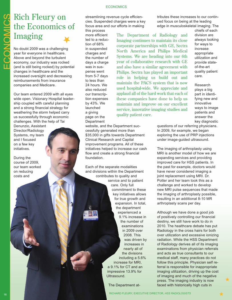
Rich Fleury on the Economics of ImagingNo doubt 2009 was a challenging year for everyone in healthcare. Above and beyond the turbulent economy, our industry was rocked (and is still being rocked) by potential changes in healthcare and the increased oversight and decreasing reimbursements from insurance companies and Medicare.
Our team entered 2009 with all eyes wide open. Visionary Hospital leader-ship coupled with careful planning and a strong financial strategy for weathering the storm helped carry us successfully through economic challenges. With the help of Tai Denunzio, Assistant Director/Radiology Systems, my team and I focused on a few key initiatives.
During the course of 2009, our team worked on reducing costs and
streamlining revenue cycle efficien-cies. Suspended charges were a key focus area and our efforts in making this process more efficient led to a reduc-tion of 68% in suspended charges and the number of days a charge was in sus-pense went from 5-7 days to less than 24 hours. We also reduced our transcrip-tion expenses by 43%. We launched a giving page on the Department website, and the Department suc-cessfully generated more than $35,000 in gifts towards Department Clinical Research and Capital improvement programs. All of these initiatives helped to increase our cash flow and create a strong financial foundation.
Each of the separate modalities and divisions within the Department
contributes to quality and service and to patient
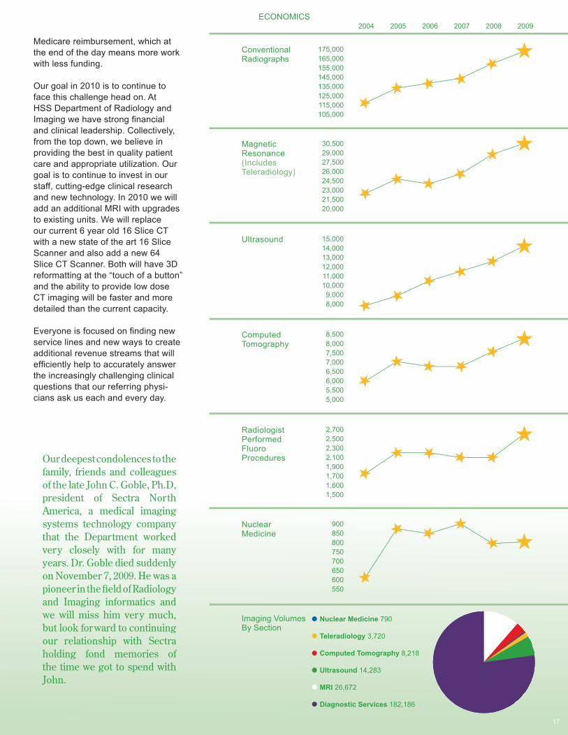
care. Only full commitment to these key initiatives allows for true growth and expansion. In total, the department experienced a 9.1% increase in the number of examinations in 2009 over 2008. This was driven by increases in nearly all of
the divisions including a 5.6%
increase for MRI, a 9.1% for CT and an impressive 13.9% for Ultrasound.
The Department at-
tributes these increases to our contin-ued focus on being at the leading edge in musculoskeletal imaging. The
chiefs of each division are always looking for ways to increase appropriate utilization and provide state-of-the-art quality patient care.
Research plays a big part in identi-fying new and innovative ways to image patients and answer the key diagnostic
questions of our referring physicians. In 2009, for example, we began exploring the use of PRP injections under image-guided ultrasound.
The imaging of arthroplasty using MRI is another model of how we are expanding services and providing improved care for HSS patients. In the past for example, doctors would have never considered imaging a joint replacement using MRI. Dr. Potter and her team took this as a challenge and worked to develop new MRI pulse sequences that made the imaging of arthroplasty possible, resulting in an additional 8-10 MR arthroplasty scans per day.
Although we have done a good job of positively controlling our financial destiny, we still have work to do in 2010. The healthcare debate has put Radiology in the cross hairs for both over utilization and excessive ionizing radiation. While the HSS Department of Radiology derives all of its imaging examinations from physician referrals and acts as true consultants to our medical staff, many practices do not follow this principle. Physician self re-ferral is responsible for inappropriate imaging utilization, driving up the cost of imaging and much of the negative press. The imaging industry is now faced with historically high cuts in
The Department of Radiology and Imaging continues to maintain its close corporate partnerships with GE, Sectra North America and Philips Medical Systems. We are heading into our 6th year of collaborative research with GE and also have a similar agreement with Philips. Sectra has played an important role in helping us build out and maintain the PACS system that is now used hospital-wide. We appreciate and applaud all of the hard work that each of these companies have done to help us maintain and improve on our excellent service, innovative imaging studies and quality patient care.
RICHARD FLEURY, EXECUTIVE DIRECTOR, HSS RADIOLOGISTS
ECONOMICS ECONOMICS
17
Our deepest condolences to the family, friends and colleagues of the late John C. Goble, Ph.D, president of Sectra North America, a medical imaging systems technology company that the Department worked very closely with for many years. Dr. Goble died suddenly on November 7, 2009. He was a pioneer in the field of Radiology and Imaging informatics and we will miss him very much, but look forward to continuing our relationship with Sectra holding fond memories of the time we got to spend with John.
Medicare reimbursement, which at the end of the day means more work with less funding.
Our goal in 2010 is to continue to face this challenge head on. At HSS Department of Radiology and Imaging we have strong financial and clinical leadership. Collectively, from the top down, we believe in providing the best in quality patient care and appropriate utilization. Our goal is to continue to invest in our staff, cutting-edge clinical research and new technology. In 2010 we will add an additional MRI with upgrades to existing units. We will replace our current 6 year old 16 Slice CT with a new state of the art 16 Slice Scanner and also add a new 64 Slice CT Scanner. Both will have 3D reformatting at the “touch of a button” and the ability to provide low dose CT imaging will be faster and more detailed than the current capacity.
Everyone is focused on finding new service lines and new ways to create additional revenue streams that will efficiently help to accurately answer the increasingly challenging clinical questions that our referring physi-cians ask us each and every day.
2004 2005 2006 2007 2008 2009
Imaging VolumesBy Section
Nuclear Medicine 790
Teleradiology 3,720
Computed Tomography 8,218
Ultrasound 14,283
MRI 26,672
Diagnostic Services 182,186
2,7002,5002,3002,1001,9001,7001,6001,500
RadiologistPerformedFluoroProcedures
Ultrasound 15,00014,00013,00012,00011,00010,000
9,0008,000
NuclearMedicine
900850800750700650600550
ComputedTomography
8,5008,0007,5007,0006,5006,0005,5005,000
MagneticResonance(Includes Teleradiology)
30,50029,00027,50026,00024,50023,00021,50020,000
ConventionalRadiographs
175,000165,000155,000145,000135,000125,000115,000105,000
HO
W W
E S
EE
TH
E V
ALU
E O
F IM
AG
ING
HOW WE SEE THE VALUE OF IMAGING
18
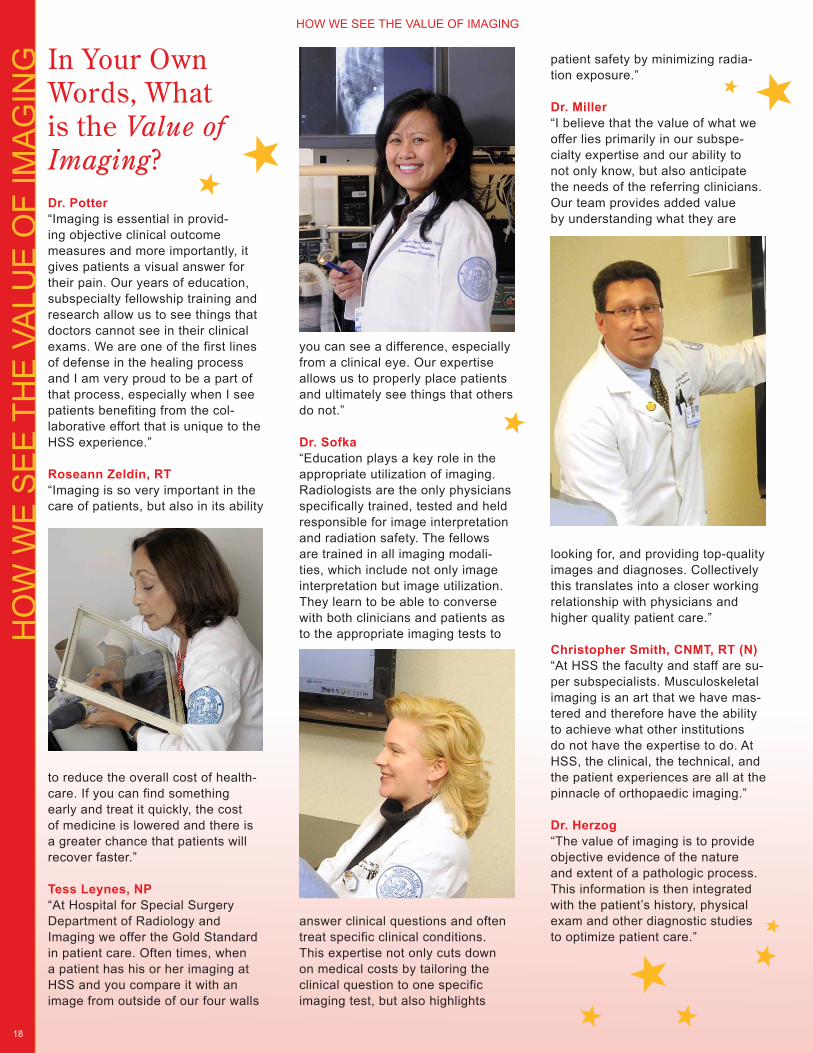
In Your Own Words, What is the Value of Imaging?Dr. Potter“Imaging is essential in provid-ing objective clinical outcome measures and more importantly, it gives patients a visual answer for their pain. Our years of education, subspecialty fellowship training and research allow us to see things that doctors cannot see in their clinical exams. We are one of the first lines of defense in the healing process and I am very proud to be a part of that process, especially when I see patients benefiting from the col-laborative effort that is unique to the HSS experience.”
Roseann Zeldin, RT“Imaging is so very important in the care of patients, but also in its ability
to reduce the overall cost of health-care. If you can find something early and treat it quickly, the cost of medicine is lowered and there is a greater chance that patients will recover faster.”
Tess Leynes, NP “At Hospital for Special Surgery Department of Radiology and Imaging we offer the Gold Standard in patient care. Often times, when a patient has his or her imaging at HSS and you compare it with an image from outside of our four walls
you can see a difference, especially from a clinical eye. Our expertise allows us to properly place patients and ultimately see things that others do not.”
Dr. Sofka “Education plays a key role in the appropriate utilization of imaging. Radiologists are the only physicians specifically trained, tested and held responsible for image interpretation and radiation safety. The fellows are trained in all imaging modali-ties, which include not only image interpretation but image utilization. They learn to be able to converse with both clinicians and patients as to the appropriate imaging tests to
answer clinical questions and often treat specific clinical conditions. This expertise not only cuts down on medical costs by tailoring the clinical question to one specific imaging test, but also highlights
patient safety by minimizing radia-tion exposure.”
Dr. Miller“I believe that the value of what we offer lies primarily in our subspe-cialty expertise and our ability to not only know, but also anticipate the needs of the referring clinicians. Our team provides added value by understanding what they are
looking for, and providing top-quality images and diagnoses. Collectively this translates into a closer working relationship with physicians and higher quality patient care.”
Christopher Smith, CNMT, RT (N)“At HSS the faculty and staff are su-per subspecialists. Musculoskeletal imaging is an art that we have mas-tered and therefore have the ability to achieve what other institutions do not have the expertise to do. At HSS, the clinical, the technical, and the patient experiences are all at the pinnacle of orthopaedic imaging.”
Dr. Herzog“The value of imaging is to provide objective evidence of the nature and extent of a pathologic process. This information is then integrated with the patient’s history, physical exam and other diagnostic studies to optimize patient care.”
partmental conferences that help each other understand each other’s clinical scenarios. That our clinical staff keeps abreast with the latest development and cutting edge of what is possible in imaging and considers assessing their value, and/or including them in our day to day work load. This may generate ongoing research collabora-tions with our referring clinicians in the long term further understanding of disease processes or effectiveness of treatment. That we treat their patients with courtesy and respect.”
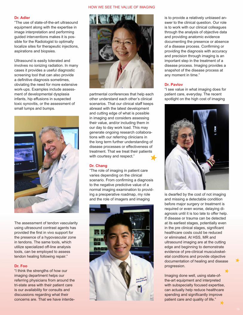
Dr. Chang“The role of imaging in patient care varies depending on the clinical scenario. From confirming a diagnosis to the negative predictive value of a normal imaging examination to provid-ing a preoperative roadmap, my role and the role of imagers and imaging
HOW WE SEE THE VALUE OF IMAGING HOW WE SEE THE VALUE OF IMAGING
19
is to provide a relatively unbiased an-swer to the clinical question. Our role is to work with our clinical colleagues through the analysis of objective data and providing anatomic evidence documenting the presence or absence of a disease process. Confirming or providing the diagnosis with accuracy and precision through imaging is an important step in the treatment of a disease process. Imaging provides a snapshot of the disease process at any moment in time.”
Dr. Pavlov “I see value in what imaging does for patient care, everyday. The recent spotlight on the high cost of imaging
is dwarfed by the cost of not imaging and missing a detectable condition before major surgery or treatment is required or even worse, delaying di-agnosis until it is too late to offer help. If disease or trauma can be detected at its earliest stages, potentially even in the pre clinical stages, significant healthcare costs could be reduced or eliminated. At HSS, MR and ultrasound imaging are at the cutting edge and beginning to demonstrate evidence of pre-clinical musculoskel-etal conditions and provide objective documentation of healing and disease progression.
Imaging done well, using state-of-the-art equipment and interpreted with subspecialty focused expertise, can actually help reduce healthcare spending and significantly improve patient care and quality of life.”
Dr. Adler“The use of state-of-the-art ultrasound equipment along with the expertise in image interpretation and performing guided interventions makes it is pos-sible for the Radiologist to optimally localize sites for therapeutic injections, aspirations and biopsies.
Ultrasound is easily tolerated and involves no ionizing radiation. In many cases it provides a useful diagnostic screening tool that can also provide a definitive diagnosis sometimes, obviating the need for more extensive work-ups. Examples include assess-ment of developmental dysplasia infants, hip effusions in suspected toxic synovitis, or the assessment of small lumps and bumps.
The assessment of tendon vascularity using ultrasound contrast agents has provided the first in vivo support for the presence of a hypovascular zone in tendons. The same tools, which utilize specialized off-line analysis tools, can be employed to assess tendon healing following repair.”
Dr. Foo“I think the strengths of how our imaging department helps our referring physicians from around the tri-state area with their patient care is our availability for consults and discussions regarding what their concerns are. That we have interde-
CO
MIN
G A
ND
GO
ING
COMING AND GOING
20
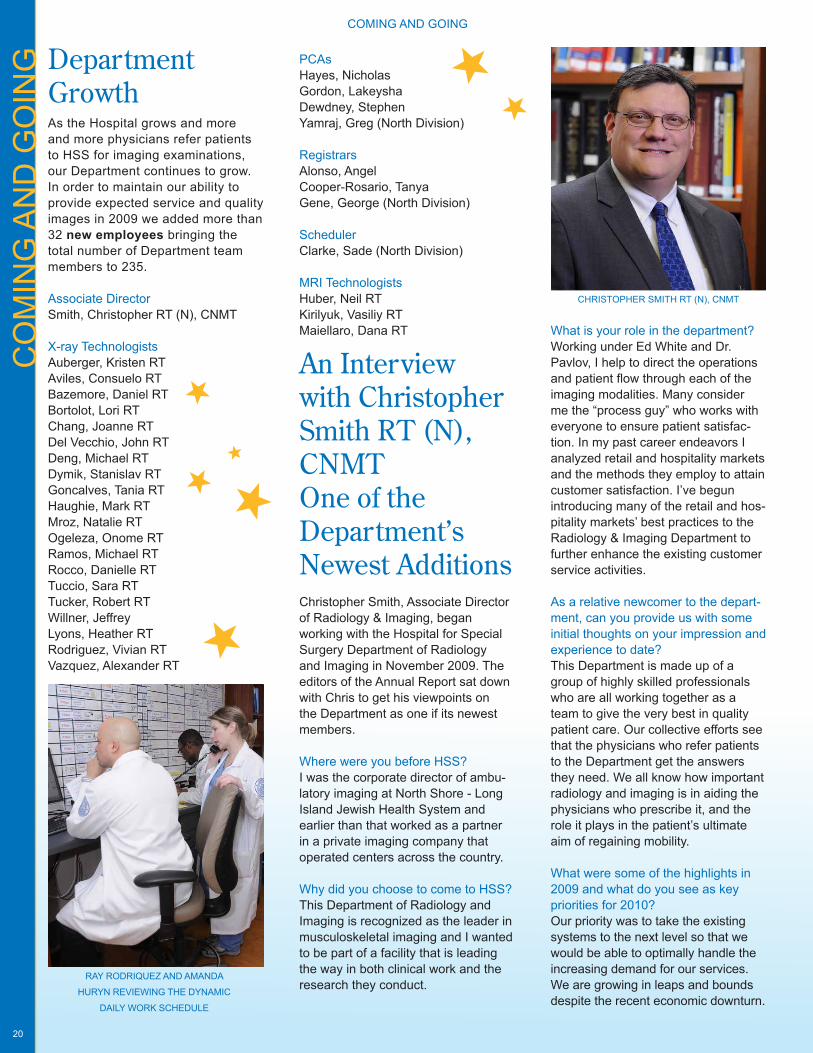
What is your role in the department?Working under Ed White and Dr. Pavlov, I help to direct the operations and patient flow through each of the imaging modalities. Many consider me the “process guy” who works with everyone to ensure patient satisfac-tion. In my past career endeavors I analyzed retail and hospitality markets and the methods they employ to attain customer satisfaction. I’ve begun introducing many of the retail and hos-pitality markets’ best practices to the Radiology & Imaging Department to further enhance the existing customer service activities.
As a relative newcomer to the depart-ment, can you provide us with some initial thoughts on your impression and experience to date?This Department is made up of a group of highly skilled professionals who are all working together as a team to give the very best in quality patient care. Our collective efforts see that the physicians who refer patients to the Department get the answers they need. We all know how important radiology and imaging is in aiding the physicians who prescribe it, and the role it plays in the patient’s ultimate aim of regaining mobility.
What were some of the highlights in 2009 and what do you see as key priorities for 2010?Our priority was to take the existing systems to the next level so that we would be able to optimally handle the increasing demand for our services. We are growing in leaps and bounds despite the recent economic downturn.
Department Growth As the Hospital grows and more and more physicians refer patients to HSS for imaging examinations, our Department continues to grow. In order to maintain our ability to provide expected service and quality images in 2009 we added more than 32 new employees bringing the total number of Department team members to 235.
Associate DirectorSmith, Christopher RT (N), CNMT
X-ray TechnologistsAuberger, Kristen RTAviles, Consuelo RTBazemore, Daniel RTBortolot, Lori RTChang, Joanne RTDel Vecchio, John RTDeng, Michael RTDymik, Stanislav RTGoncalves, Tania RTHaughie, Mark RTMroz, Natalie RTOgeleza, Onome RTRamos, Michael RTRocco, Danielle RTTuccio, Sara RTTucker, Robert RTWillner, JeffreyLyons, Heather RTRodriguez, Vivian RTVazquez, Alexander RT
An Interview with Christopher Smith RT (N), CNMT One of the Department’s Newest AdditionsChristopher Smith, Associate Director of Radiology & Imaging, began working with the Hospital for Special Surgery Department of Radiology and Imaging in November 2009. The editors of the Annual Report sat down with Chris to get his viewpoints on the Department as one if its newest members.
Where were you before HSS?I was the corporate director of ambu-latory imaging at North Shore - Long Island Jewish Health System and earlier than that worked as a partner in a private imaging company that operated centers across the country.
Why did you choose to come to HSS?This Department of Radiology and Imaging is recognized as the leader in musculoskeletal imaging and I wanted to be part of a facility that is leading the way in both clinical work and the research they conduct.
CHRISTOPHER SMITH RT (N), CNMT
PCAsHayes, NicholasGordon, LakeyshaDewdney, StephenYamraj, Greg (North Division)
RegistrarsAlonso, AngelCooper-Rosario, TanyaGene, George (North Division)
SchedulerClarke, Sade (North Division)
MRI TechnologistsHuber, Neil RT Kirilyuk, Vasiliy RT Maiellaro, Dana RT
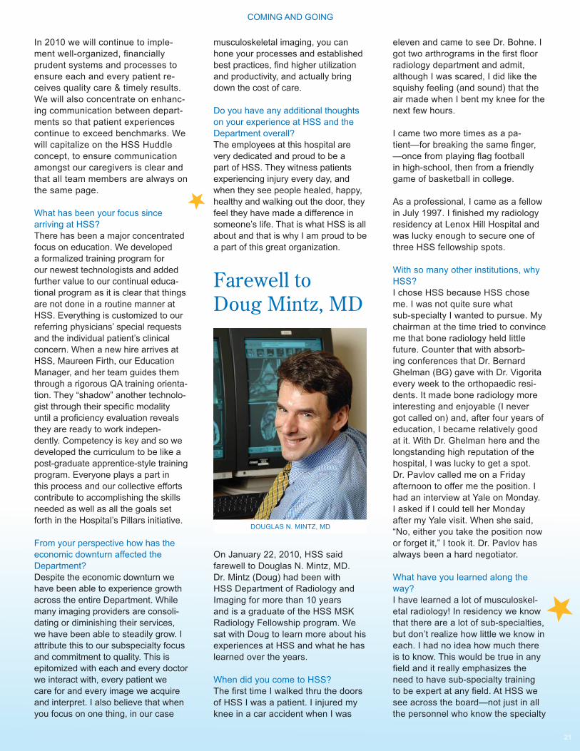
RAY RODRIQUEZ AND AMANDA
HURYN REVIEWING THE DYNAMIC
DAILY WORK SCHEDULE
COMING AND GOING COMING AND GOING
21
In 2010 we will continue to imple-ment well-organized, financially prudent systems and processes to ensure each and every patient re-ceives quality care & timely results. We will also concentrate on enhanc-ing communication between depart-ments so that patient experiences continue to exceed benchmarks. We will capitalize on the HSS Huddle concept, to ensure communication amongst our caregivers is clear and that all team members are always on the same page.
What has been your focus since arriving at HSS?There has been a major concentrated focus on education. We developed a formalized training program for our newest technologists and added further value to our continual educa-tional program as it is clear that things are not done in a routine manner at HSS. Everything is customized to our referring physicians’ special requests and the individual patient’s clinical concern. When a new hire arrives at HSS, Maureen Firth, our Education Manager, and her team guides them through a rigorous QA training orienta-tion. They “shadow” another technolo-gist through their specific modality until a proficiency evaluation reveals they are ready to work indepen-dently. Competency is key and so we developed the curriculum to be like a post-graduate apprentice-style training program. Everyone plays a part in this process and our collective efforts contribute to accomplishing the skills needed as well as all the goals set forth in the Hospital’s Pillars initiative.
From your perspective how has the economic downturn affected the Department?Despite the economic downturn we have been able to experience growth across the entire Department. While many imaging providers are consoli-dating or diminishing their services, we have been able to steadily grow. I attribute this to our subspecialty focus and commitment to quality. This is epitomized with each and every doctor we interact with, every patient we care for and every image we acquire and interpret. I also believe that when you focus on one thing, in our case
Farewell to Doug Mintz, MD
On January 22, 2010, HSS said farewell to Douglas N. Mintz, MD. Dr. Mintz (Doug) had been with HSS Department of Radiology and Imaging for more than 10 years and is a graduate of the HSS MSK Radiology Fellowship program. We sat with Doug to learn more about his experiences at HSS and what he has learned over the years.
When did you come to HSS? The first time I walked thru the doors of HSS I was a patient. I injured my knee in a car accident when I was
eleven and came to see Dr. Bohne. I got two arthrograms in the first floor radiology department and admit, although I was scared, I did like the squishy feeling (and sound) that the air made when I bent my knee for the next few hours.
I came two more times as a pa-tient—for breaking the same finger, —once from playing flag football in high-school, then from a friendly game of basketball in college.
As a professional, I came as a fellow in July 1997. I finished my radiology residency at Lenox Hill Hospital and was lucky enough to secure one of three HSS fellowship spots.
With so many other institutions, why HSS?I chose HSS because HSS chose me. I was not quite sure what sub-specialty I wanted to pursue. My chairman at the time tried to convince me that bone radiology held little future. Counter that with absorb-ing conferences that Dr. Bernard Ghelman (BG) gave with Dr. Vigorita every week to the orthopaedic resi-dents. It made bone radiology more interesting and enjoyable (I never got called on) and, after four years of education, I became relatively good at it. With Dr. Ghelman here and the longstanding high reputation of the hospital, I was lucky to get a spot.Dr. Pavlov called me on a Friday afternoon to offer me the position. I had an interview at Yale on Monday. I asked if I could tell her Monday after my Yale visit. When she said, “No, either you take the position now or forget it,” I took it. Dr. Pavlov has always been a hard negotiator.
What have you learned along the way?I have learned a lot of musculoskel-etal radiology! In residency we know that there are a lot of sub-specialties, but don’t realize how little we know in each. I had no idea how much there is to know. This would be true in any field and it really emphasizes the need to have sub-specialty training to be expert at any field. At HSS we see across the board—not just in all the personnel who know the specialty
DOUGLAS N. MINTZ, MD
musculoskeletal imaging, you can hone your processes and established best practices, find higher utilization and productivity, and actually bring down the cost of care.
Do you have any additional thoughts on your experience at HSS and the Department overall?The employees at this hospital are very dedicated and proud to be a part of HSS. They witness patients experiencing injury every day, and when they see people healed, happy, healthy and walking out the door, they feel they have made a difference in someone’s life. That is what HSS is all about and that is why I am proud to be a part of this great organization.
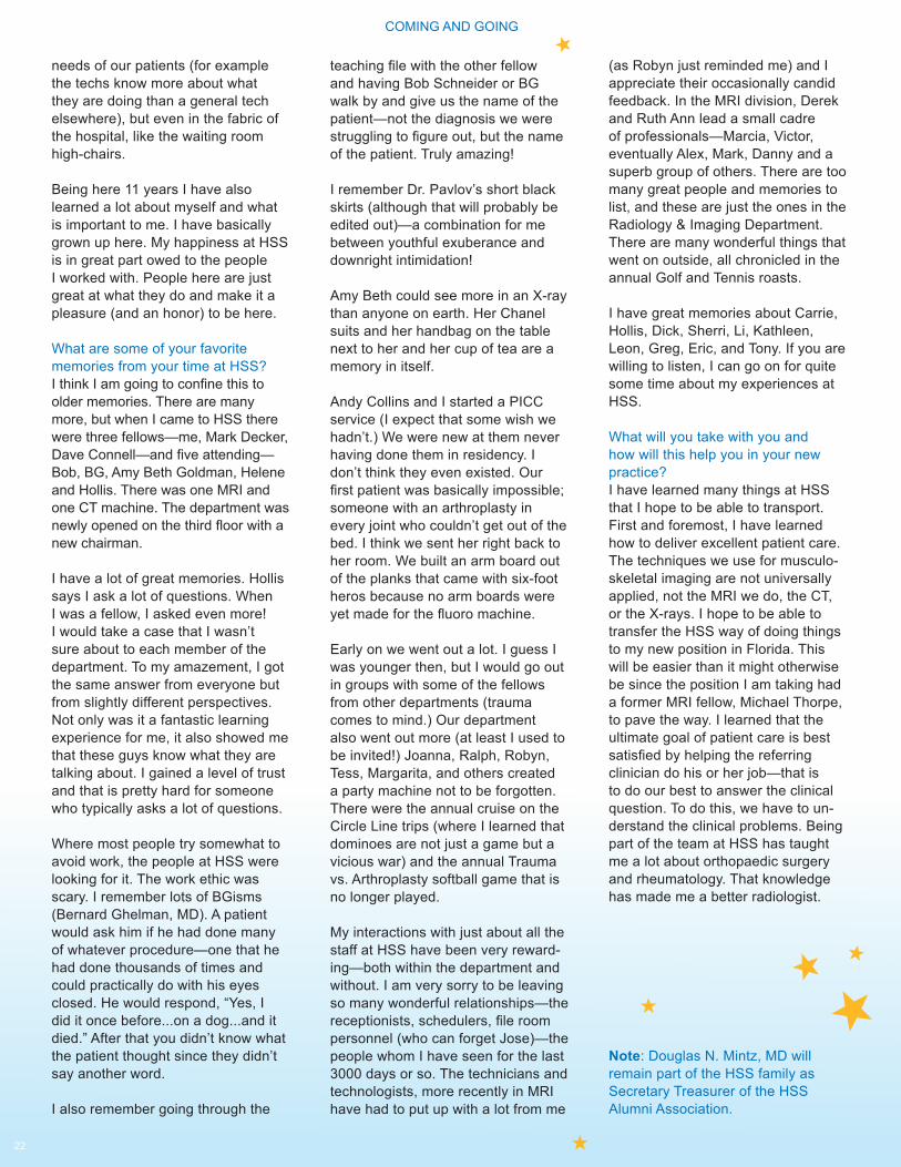
22
teaching file with the other fellow and having Bob Schneider or BG walk by and give us the name of the patient—not the diagnosis we were struggling to figure out, but the name of the patient. Truly amazing!
I remember Dr. Pavlov’s short black skirts (although that will probably be edited out)—a combination for me between youthful exuberance and downright intimidation!
Amy Beth could see more in an X-ray than anyone on earth. Her Chanel suits and her handbag on the table next to her and her cup of tea are a memory in itself.
Andy Collins and I started a PICC service (I expect that some wish we hadn’t.) We were new at them never having done them in residency. I don’t think they even existed. Our first patient was basically impossible; someone with an arthroplasty in every joint who couldn’t get out of the bed. I think we sent her right back to her room. We built an arm board out of the planks that came with six-foot heros because no arm boards were yet made for the fluoro machine.
Early on we went out a lot. I guess I was younger then, but I would go out in groups with some of the fellows from other departments (trauma comes to mind.) Our department also went out more (at least I used to be invited!) Joanna, Ralph, Robyn, Tess, Margarita, and others created a party machine not to be forgotten. There were the annual cruise on the Circle Line trips (where I learned that dominoes are not just a game but a vicious war) and the annual Trauma vs. Arthroplasty softball game that is no longer played.
My interactions with just about all the staff at HSS have been very reward-ing—both within the department and without. I am very sorry to be leaving so many wonderful relationships—the receptionists, schedulers, file room personnel (who can forget Jose)—the people whom I have seen for the last 3000 days or so. The technicians and technologists, more recently in MRI have had to put up with a lot from me
needs of our patients (for example the techs know more about what they are doing than a general tech elsewhere), but even in the fabric of the hospital, like the waiting room high-chairs.
Being here 11 years I have also learned a lot about myself and what is important to me. I have basically grown up here. My happiness at HSS is in great part owed to the people I worked with. People here are just great at what they do and make it a pleasure (and an honor) to be here.
What are some of your favorite memories from your time at HSS?I think I am going to confine this to older memories. There are many more, but when I came to HSS there were three fellows—me, Mark Decker, Dave Connell—and five attending—Bob, BG, Amy Beth Goldman, Helene and Hollis. There was one MRI and one CT machine. The department was newly opened on the third floor with a new chairman.
I have a lot of great memories. Hollis says I ask a lot of questions. When I was a fellow, I asked even more! I would take a case that I wasn’t sure about to each member of the department. To my amazement, I got the same answer from everyone but from slightly different perspectives. Not only was it a fantastic learning experience for me, it also showed me that these guys know what they are talking about. I gained a level of trust and that is pretty hard for someone who typically asks a lot of questions.
Where most people try somewhat to avoid work, the people at HSS were looking for it. The work ethic was scary. I remember lots of BGisms (Bernard Ghelman, MD). A patient would ask him if he had done many of whatever procedure—one that he had done thousands of times and could practically do with his eyes closed. He would respond, “Yes, I did it once before...on a dog...and it died.” After that you didn’t know what the patient thought since they didn’t say another word.
I also remember going through the
(as Robyn just reminded me) and I appreciate their occasionally candid feedback. In the MRI division, Derek and Ruth Ann lead a small cadre of professionals—Marcia, Victor, eventually Alex, Mark, Danny and a superb group of others. There are too many great people and memories to list, and these are just the ones in the Radiology & Imaging Department. There are many wonderful things that went on outside, all chronicled in the annual Golf and Tennis roasts.
I have great memories about Carrie, Hollis, Dick, Sherri, Li, Kathleen, Leon, Greg, Eric, and Tony. If you are willing to listen, I can go on for quite some time about my experiences at HSS.
What will you take with you and how will this help you in your new practice?I have learned many things at HSS that I hope to be able to transport. First and foremost, I have learned how to deliver excellent patient care. The techniques we use for musculo-skeletal imaging are not universally applied, not the MRI we do, the CT, or the X-rays. I hope to be able to transfer the HSS way of doing things to my new position in Florida. This will be easier than it might otherwise be since the position I am taking had a former MRI fellow, Michael Thorpe, to pave the way. I learned that the ultimate goal of patient care is best satisfied by helping the referring clinician do his or her job—that is to do our best to answer the clinical question. To do this, we have to un-derstand the clinical problems. Being part of the team at HSS has taught me a lot about orthopaedic surgery and rheumatology. That knowledge has made me a better radiologist.
Note: Douglas N. Mintz, MD will remain part of the HSS family as Secretary Treasurer of the HSS Alumni Association.
COMING AND GOING
PU
BLI
CAT
ION
S \
PR
ES
EN
TATI
ON
S \
GR
AN
TS
23
RONALD S. ADLER, PHD, MD
Published
1. Acharya SS., Schloss R., Dyke JP., Mintz DN., Christos P., Dimichele DM., Adler RS., Power doppler sonography in the diagnosis of hemophilic synovitis, A promising tool. Thromb Haemost. 2008 Dec; 6 (12):2055-61 23.(2)
2. Gamradt S., Gallo R., Adler RS.. Maderazo A., Altchek D., Warren F., Fealy S., Vascularity of the supraspi-natus tendon three months after repair: characterization using contrast - en-hanced ultrasound. J Shoulder Elbow Surg. 2009 Jun 12. 1 - 8
3. Nho SJ., Adler RS., Tomlinson DP., Allen AA., Cordasco F., Warren R., Altchek D., MacGillivray J., Arthroscopic Rotator Cuff Repair: Prospective Evalu-ation with Sequential ultrasonography. American Journal of Sports Medicine 2009 Jun 16. 37:1938 - 1946
4. Adler RS., Garofalo G., Ultrasound in the Evaluation of the Inflammatory Myopathies Current Rheumatology Reports. 2009 Aug; 11(4):302 - 308
5. Daftary A., Adler RS., Sonographic Evaluation of the Achilles Tendon. Ultrasound Quarterly - September 2009, Volume 25, Issue 3 103 - 110
6. Kim H., Walcott-Sapp S., Leggett K., Bass A., Adler RS., Pavlov H., Westrich G., Detection of Pulmonary Embolism in the Post-Operative Orthopedic Patient using Spiral CT Scans. Hospital for Special Surgery Journal, September 2009, Volume 5, 1 - 4
7. Mandl LA., Hotchkiss RN., Adler RS., Lyman S., Daluiski A., Wolfe SW., Katz JN. Injectable hyaluronan for the treat-ment of carpometacarpal osteoarthritis: open label pilot trail. Curr Med Res Opin. 2009 Sep; 25 (9):2103 - 8
8. Nho SJ., Adler RS., Answorth A., Cordasco F., Warren R., Altchek D., MacGillivray J. Arthroscopic Rotator Cuff Repair: Prospective Evaluation with Sequential ultrasonography. American Journal of Sports Medicine. October 1, 2009. 37:1938 - 1945
9. Nho SJ, Shindle MK, Adler RS., Warren RF, Altchek DW, MacGillvray JD. Prospective analysis of arthroscopic rotator cuff repair: subgroup analysis. Journal of Shoulder Elbow Surg. 2009 Sep-Oct; 18(5):697 - 704
10. Adler RS., Garofalo G., Ultrasound in the Evaluation of the Inflammatory Myopathies Current Medicine Group, Current Rheumatology Reports 2009, 11:302 - 308
11. Kagan LJ., Adler RS., Garofalo G., MD. “Ultrasound in the Evaluation of the Inflammatory Myopathies” Chapter 9, The Inflammatory Myopathies. Humana Press 2009 pg. 147
In Press
1. Dyke JP., Mintz DN., Schloss R W, Adler RS., DiMichele DM., Ballon D., Acharya SS., Assessment of Clinically Active Synovitis in Hemophilic Arthropa-thy Using Dynamic Contrast Enhanced (DCE) Magnetic Resonance Imaging Radiology
2. Adler RS., in “Percutaneous Ultrasound Guided Interventions in the Musculoskeletal System” Chapter 25 in Diagnostic Ultrasound Fourth Edition Eds. Mosby Inc. Rumack C., Wilson S., Charboneau W., Levine D.
3. Adler RS., 2009 ARRS Categorical Course Manuscript - Percutaneious Ultrasound Guided Interventions in the Musculoskeletal System. American Journal of Radiology
4. Pavlov H., Sofka CM., Saboeiro GR., Adler RS., Mintz DN., “Imaging of Foot and Ankle Injuries” in Athletes in Sports Medicine of the Foot and Ankle, Eds. Altchek D, Positano R. Lippincott Williams and Wilkins (Book Chapter)
5. Sofka CM., Adler RS., Saboeiro GR., Pavlov H., “Sonographic Evaluation and Sonographic - Guided Therapeutic Options of Lateral Ankle Pain: Peroneal Tendon Pathology Associated with the Presence of an Os Peroneum,” HSS Journal- Musculoskeletal Journal of Hospital for Special Surgery
Presentations
1. Adler, RS., “An Overview of Muscu-loskeletal (MSK) Ultrasound Imaging” Philips Research Facility, Briarcliff Manor, NY 1/19/2009
2. Adler RS., “Contrast-enhanced ultra-sound characterization of the vascularity of the repaired rotator cuff.” Society of Skeletal Radiology, Isle of Palms, SC., March 9 - 11, 2009
3. Adler RS., “Ultrasound-Guided Musculoskeletal Applications” 2009 Annual Meeting American Roentgen Ray Society, April 27-29 2009
4. Adler RS., “Elastrography Strain Im-age,” Philips Research, Briarcliff Manor N.Y., 8/19/2009
5. Adler RS., “New Techniques in Sonographic Imaging”, Annual Meeting International Skeletal Society, (ISS). Washington DC, September 2009
6. Adler RS., Miller TT., “Sensitivity of Imaging Modalities for Calcium Hydroxy-apatite in a Soft Tissue Phantom”, 95th Radiological Society of North America (RSNA) Scientific Assembly and Annual Meeting 12/1/2009
Resident/Fellow CME Presentations
1. Adler RS., Ultrasound Tutorial: Ankle/foot hands on - Part I 1/13/2009
2. Adler RS., Ultrasound Tutorial: Ankle/foot hands on - Part II 1/27/2009
3. Adler RS., Ultrasound Tutorial: Nerves, 2/10/2009
COMING AND GOING PUBLICATIONS / PRESENTATIONS / GRANTS
24
4. Adler RS., Ultrasound Tutorial: Carpal Tunnel demonstration, 2/24/2009
5. Adler RS., Sahai A., MR/US Correla-tion/Case Presentation, 2/24/2009
6. Adler RS., Ultrasound Tutorial: Arthritis study demonstration, 3/17/2009
7. Adler RS., Ultrasound Tutorial: Arthritis, 3/24/2009
8. Adler RS., Ultrasound Tutorial: Troubleshooting Session II, 4/21/2009
9. Adler RS., Ultrasound Tutorial: Troubleshooting Session III, 5/5/2009
10. Adler RS., Ngo M. Interesting Cases/Case Presentation, 5/5/2009
11. Adler RS., Ultrasound Tutorial: Review and Evaluation, 5/26/2009
12. Adler RS., Ultrasound Tutorial: DVT’s, 7/7/2009
13. Adler RS., Ultrasound Tutorial: Hands on DVT’s, 7/21/2009
14. Adler RS., Ultrasound Tutorial: Shoulder Demonstration, 8/4/2009
15. Adler RS., “MSK Ultrasound”, Grand Rounds: Hospital for Special Surgery, 8/5/2009
16. Adler RS., Ultrasound Tutorial: Shoulder hands on, 8/18/2009
17. Adler RS., Ultrasound Tutorial: Elbow Demonstration, 9/8/2009
18. Adler RS., Ultrasound Tutorial: Elbow hands on, 9/22/2009
19. Adler RS., Ultrasound Tutorial: Wrist Demonstration, 10/6/2009
20. Adler RS., Ultrasound Tutorial: Wrist hands on, 10/20/2009
21. Adler RS., Ultrasound Tutorial: Thigh/Knee Demonstration, 1/17/2009
22. Adler RS., Ultrasound Tutorial: Thigh/Knee Hands on, 12/8/2009
23. Adler RS., Ultrasound Tutorial: Ankle/Foot Demonstration, 12/22/2009
Grants
1. Adler RS., Major League Baseball Grant (COPI) Grant # 3135400 January 1, 2007 - 2010 $150,000
Service Commitments
1. HSS - Infection Control Committee
2. Department CRP Committee
ERIC BOGNER, MD
Published
1. Bogner E., “What’s the Diagnosis” Case 17: 8 year old with right knee pain following minor trauma, http://www.hss.edu/Diagnosis.asp, 1/2009 2. Bogner E., “What’s the Diagnosis” Case 18: 13 year old boy with left ankle pain and limp. Notes mild discomfort of the right ankle, http://www.hss.edu/Diag-nosis.asp, 2/2009 3. Bogner E., “What’s the Diagnosis” Case 19: 25 year old man with 4 years of knee pain and clicking sensation, http://www.hss.edu/Diagnosis.asp, 3/2009 4. Bogner E., “What’s the Diagnosis” Case 20: 30 year old female with long standing history of decreased range of motion in multiple joints, http://www.hss.edu/Diagnosis.asp, 4/2009 5. Bogner E., “What’s the Diagnosis” Case 21: 35 year old woman with increasing left foot pain, http://www.hss.edu/Diagnosis.asp, 5/2009 6. Bogner E., “What’s the Diagnosis” Case 22: 43 year old man with 1 year of knee pain.6/2009 7. Bogner E., “What’s the Diagnosis” Case 23: 50 year old woman with long standing wrist pain, http://www.hss.edu/Diagnosis.asp, 7/2009 8. Bogner E., “What’s the Diagnosis” Case 24: 55 year old man with bilateral knee pain, http://www.hss.edu/Diagno-sis.asp, 8/2009 9. Bogner E., “What’s the Diagnosis” Case 25: 19 year old man with long standing elbow pain, http://www.hss.edu/Diagnosis.asp, 9/2009
10. Bogner E., “What’s the Diagnosis” Case 26: 45 year old woman with long standing hip pain, http://www.hss.edu/Diagnosis.asp, 10/2009
11. Bogner E., Imaging cervical spine trauma, Sports Health A Multidisciplinary Approach 10/09; 1(5) 384 - 391
PUBLICATIONS / PRESENTATIONS / GRANTS
25
12. Bogner E., “What’s the Diagnosis” Case 27: “Rheumatoid Arthritis”, http://www.hss.edu/Diagnosis.asp, 10/2009
13. Bogner E., “What’s the Diagnosis” Case 28: “Peri prosthetic Fractures”, http://www.hss.edu/Diagnosis.asp, 11/2009
14. Bogner E., “What’s the Diagnosis” Case 29 “Pectoralis Tear”, http://www.hss.edu/Diagnosis.asp, 12/2009
In Press
1.Zifchock B., Backus S., Bogner E., Pavlov H., Mandl L., Chen C., Garrison G., Brown A., Cordasco F., Williams R., Hunter D., Bedi A., Hillstrom H., “Effect of knee bracing on OA.” American Society of Biomechanics
Poster
1.Bogner E., Wu A., Su E., Incidence and risk factors of neuropathy after THA. 76th Annual American Academy of Ortho-pedic Surgeons, Las Vegas 02/2009
Presentations
1. Bogner E., “Incidence and risk factors of neuropathy after THA”. 76thAn-nual Meeting of the American Academy of Orthopedic Surgeons, Las Vegas, NV, 2/27/2009
2. Zifchock B., Backus S., Bogner E., Pavlov H., Mandl L., Chen C., Garrison G., Bown A., Cordasco F., Williams R., Hunter D., Bedi A., Hillstrom H., The Immediate Bilateral Effects of Unilateral Knee Bracing for the Treatment of Knee Osteoarthritis: Primary results. American Society of Biomechanics, University Park, PA - August 26 - 29, 2009
Resident/Fellow CME Presentations
1. Bogner E., Deyer T., Shoulder Instability and Interesting Cases, (MSK Radiology Fellow). 1/9/2009
2. Bogner E., Gallo C., Interesting Cases, 1/23/2009
3. Bogner E., Interesting Cases, 1/30/2009
4. Bogner E., Johnson K., Shoulder Impingement - and Interesting Cases, (MSK Radiology Fellow) 2/13/2009
5. Bogner E., Interesting Cases, 2/27/2009
6. Bogner E., Ngo M., Elbow: Ligament Injuries and Interesting Cases, 3/6/2009
7. Bogner E., Interesting Cases, 3/13/2009
8. Bogner E., Sahai A., Hip impinge-ment - Interesting Cases, 3/20/2009
9. Bogner E., Deyer T., Case Confer-ence/Case Presentation, 3/24/2009
10. Bogner E., Endo Y., Tumors/Case Presentation, 3/24/2009
11. Bogner E., Interesting Cases, 3/27/2009
12. Bogner E., Deyer T., Knee ligament injuries - Interesting Cases, 4/3/2009
13. Bogner E., Kheyfits V., Imaging in Rheumatology: pediatrics, 4/10/2009
14. Bogner E., Interesting Cases, 4/24/2009
15. Bogner E., Johnson K., Ankle tendon pathology - Interesting Cases, 5/1/2009
16. Bogner E., Ngo M., Imaging in Rheumatology: Adult and Interesting Cases, 5/8/2009
17. Bogner E., Interesting Cases, 5/22/2009
18. Bogner E., Kheyfits V., Muskulosk-elatal Tumors and Interesting Cases, 5/29/2009
19. Bogner E., Interesting Cases, 6/5/2009
20. Bogner E., Interesting Cases, 6/12/2009
21. Bogner E., Sahai A., Lumbar Disc Disease and Interesting Cases, 6/19/2009
22. Bogner E., “Interesting cases”, Grand Rounds: Hospital for Special Surgery, 6/24/2009
23. Bogner E., Deyer T., Imaging of Arthroplasty and Interesting Cases, 6/26/2009
24. Bogner E., Endo Y., Knee- Liga-ments (normal anatomy) and Interesting Cases, 7/17/2009
25. Bogner E., Murakami A., Hip - (normal anatomy) and Interesting Cases, 7/31/2009
26. Bogner E., Su H., Shoulder - Labrocapsular anatomy and Interesting Cases, 8/14/2009
27. Bogner E., Deyer T., Ankle - Ten-dons and Interesting Cases, 8/21/2009
28. Bogner E., Endo Y., Wrist Liga-ments and TFCC and Interesting Cases, 9/4/2009
29. Bogner E., Hash T., Wrist Tendons and Interesting Cases, 9/11/2009
30. Bogner E., Wilde G., Elbow and Interesting Cases, 9/18/2009
31. Bogner E., Wilde G., Stock H., Elbow and Finger anatomy and Interest-ing Cases, 9/25/2009
32. Bogner E., Teaching File Confer-ence, 10/1/2009
PUBLICATIONS / PRESENTATIONS / GRANTS PUBLICATIONS / PRESENTATIONS / GRANTS
26
33. Bogner E., Murakami A., Nerve anatomy of the lower extremity Part I, 10/9/2009
34. Bogner E., Murakami A., Nerve anatomy of the lower extremity Part II, 10/16/2009
35. Bogner E., Endo Y., Ultrasound anatomy- rotator cuff and Interesting Cases, 10/23/2009
36. Bogner E., Campbell G., Ultrasound anatomy - elbow and Interesting Cases, 10/30/2009
37. Bogner E., Murakami A., Ultrasound anatomy of the wrist and fingers, 11/6/2009
38. Bogner E., Teaching File Confer-ence, 11/12/2009
39. Bogner E., Su H., Ultrasound anatomy - knee and Interesting Cases, 11/13/2009
40. Bogner E., Wilde G., Ultrasound - Ankle, 12/4/2009
41. Bogner E., Campbell G., Ultrasound - Foot, 12/11/2009
42. Bogner E., Su H., Ultrasound - Knee, 12/18/2009
43. Bogner E., “Interesting Cases”, Grand Rounds: Ultrasound anatomy - knee, Hospital for Special Surgery, 12/16/2009
Service Commitments
1. HSS - Pharmacy & Therapeutics Committee
2. HSS - CME Committee (Alternate)
3. HSS - Safety Focus Group 1/30/09
4. Manuscript Reviewer: CORR, Sports Health, RADIOLOGY, Radiographics, AJSM
5. New York Roentgen Society-Alternate Delegate
ANTHONY CHANG, MD
Published
1. Chang A., Imaging Guided Treatment of Meniscal Cysts, Hospital for Special Surgery Journal 2009; 5:58-60
2. Chang A., Miller TT., Imaging of Ten-dons. Sports Health: A Multidisciplinary Approach 2009; 1(4):293-300
3. Chang A., CT guided treatment of lumbar facet joint synovial cysts. Hospital for Special Surgery Journal Volume 5, Number 2/September 2009; 5:165-168
Resident/Fellow CME Presentations
1. Chang A., “Interesting Cases”, NYPH Resident Conference, 3/10/2009
2. Chang A., “MSK Infection”, NYPH Resident Core Curriculum, 7/31/2009
3. Chang A., Rheumatology Clinical Pathology Conference, 9/23/2009
4. Chang A., “Infections”/Case Pre-sentation, NYPH Resident Conference, 10/6/2009
5. Chang A., Perino G., “Surface Lesions”, Radiology/Pathology Teaching Conference, 11/16/2009
6. Chang A., Perino G., “Surface Lesions”, Radiology/Pathology Teaching Conference, 11/30/2009
Service Commitment
1. Utilization Management Committee Meeting, 7/20/2009
LI FOONG FOO, MD
Published
1. Pellicci PM., Potter HG., Foo LF., Boettner F., MRI shows biologic restora-tion of posterior soft tissue repairs after THA. Clin Orthop Relat Res, 2009; 467:940–945
2. Crawford DC., Heveran CM., Cannon WD., Foo LF., Potter HG., Autlogous cartilage tissue transplant (ACTI) NeoCart & [reg] for treatment of Grade III Chondral Injury to the Femur. Prospective clinical safety trial at 2 years. American Journal of Sports Med, 2009; 37:1334 - 43
3. Cooper HJ., Ranawat AS., Potter HG., Foo LF., Jawetz ST., Ranawat CS., Magnetic Resonance Imaging in the Diagnosis and Management of Hip Pain After Total Hip Arthroplasty. Journal of Arthroplasty 2009:24(5); 661-667 August 2009
In Press
1. Kim M., Foo LF., Uggen C., Lyman S., Ryaby JT., Moynihan D., Grande DA., Potter HG., Pleshko N., Evaluation of early osteochondral defect repair in a rabbit model utilizing Fourier transform inafred imaging spectroscopy (FT-IRIS), magnetic resonance imaging (MRI) and quantitative T2 mapping. Tissue Engineering 2009
2. Shindle MK., Foo LF., Kelly BT., Pot-ter HG., “Articular Cartilage.” In Khanna AJ, ed. Musculoskeletal MRI Essentials for the Orthopedic Surgeon. New York Thieme, 2009
3. Bedi A., Foo LF., Williams RJ III., Potter HG., and the Cartilage Study Group., “The Maturation of synthetic Scaffolds for Osteochondral Donor Sites of the Knee: An MRI and T2-Mapping Analysis” Cartilage OnLine First, Dec 2009; vol.0. pp.1947603509355970v1
Presentations
1. Foo LF., “Femoroacetabular Impinge-ment” Sports and Arthroplasty Confer-ence, Hospital for Special Surgery, 1/15/2009
PUBLICATIONS / PRESENTATIONS / GRANTS
27
2. Foo LF., Chong LR., Koff MF., Trun-cale K., Riley T., Semler E., Rodeo S., Potter HG., “Quantitative MR imaging of cartilage repair in a goat model” - 55th Annual Meeting of the Orthopaedic Research Society. Las Vegas, NV. February 22-25, 2009
3. Fortier L., Potter HG., Rickey E., Schnabel L., Ellsworth J., Foo LF., Nixon A., “Concentrated bone marrow aspirate improves full-thickness cartilage repair” - 55th Annual Meeting of the Orthopaedic Research Society. Las Vegas, NV. February 22-25, 2009
4. Davisson T., Zhang R., Foo LF., Juda G., Masini M., Coyle S., Aponte C., Potter HG., Long M., Monica H., “Novel allograft sponge supports fill of osteochondral defects in caprine Model” - 55th Annual Meeting of the Orthopae-dic Research Society. Las Vegas, NV. February 22-25, 2009
5. Foo LF., “Assessment of synthetic scaffolds for osteochondral donor sites of the knee using MRI and T2 mapping: A longitudinal analysis”- Annual Meeting of the Orthopaedic Research Society, 2/27/09
6. Koff M., Chong L.R, Foo LF., Truncale K., Semler E., Rodeo S., Potter HG., “Quantitative MR imaging of cartilage repair in a goat model” - International Cartilage Repair Society, Miami, Florida, May 23-26, 2009
7. Fortier LA., Potter HG., Rickey E.J., Schnabel L.V., Ellsworth J.R., Foo LF., Nixon A. “Concentrated bone marrow aspirate improves full-thickness cartilage repair” - International Cartilage Repair Society, Miami, Florida, May 23-26, 2009,190, 19.2.3
8. Foo LF., “Assessment of synthetic resorbable scaffolds for osteochondral donor sites of the knee using carti-lage-sensitive MRI and T2-mapping: A longitudinal analysis” - American Orthopedic Society for Sports Medicine (AOSSM), Keystone, Colorado.7/9/09
9. Foo LF., “Shoulder Instability” Sports Medicine Shoulder Service, Hospital for Special Surgery, 12/3/09
Resident/Fellow CME Presentations
1. Foo LF., “Interesting Cases”, NYPH Resident Conference, 1/13/2009
2. Foo LF., “Cartilage Imaging”, NYPH Resident Conference, 4/7/2009
3. Foo LF., “MSK, CT and MRI”, NYPH CORE Curriculum, 7/29/2009
4. Foo LF., “MRI-Knee/Shoulder”, NYPH Resident Conference - Case Presenta-tion, 10/20/09
Grants
1. General Electric Health Care Re-search Support for orthopaedic applica-tions of MRI T2 Mapping of articular cartilage; MR imaging of arthroplasty; Segmentation analysis of cartilage. Principle Investigators: Hollis G. Potter, M.D 2005-2010
2. Improving Outcomes for Middle-aged Adults with Medical Meniscal Damage. Principle Investigator: James J. Irrgang Co-Investigators: Fernando Boada, PhD, Constance R. Chu, MD, Li Foong Foo, MBChB, Christopher Harner, MD, Hollis G. Potter, MD, Jon K. Sekiya, MD, James E. Bost, PhD, NIH RO1 Submit-ted September 2006 - Resubmitted 2008 $3,141,384
3. NIH/NIAMS Challenge Grant (RC1AR058255-01) 09/23/09 - 08/31/11 $466,632 (2009-2011)
Service Commitments
1. HSS, Health Information Management Committee
2. Cartilage Study Group
BERNARD GHELMAN, MD
Published
1. Ghelman B., Kepler CK., Lyman S., Della Valle AG CT Outperforms Radiog-raphy for Determination of Acetabular Cup Version after THA Clin Orthop Related Res. 467, (9), Sept. 2009 467:2362-2370
Resident/Fellow CME Presentations
1. Ghelman B., DiCarlo E., “Radiology Pathology”, Grand Rounds: Hospital for Special Surgery, 1/14/2009
2. Ghelman B., DiCarlo E., “Radiology Pathology”, Grand Rounds: Hospital for Special Surgery, 1/21/2009
3. Ghelman B., “Bone Tumors”, Grand Rounds: Hospital for Special Surgery, 2/4/2009
4. Ghelman B., DiCarlo E., “Radiology Pathology”, Grand Rounds: Hospital for Special Surgery 3/18/2009
5. Ghelman B., DiCarlo E., “Radiology Pathology”, Grand Rounds: Hospital for Special Surgery, 3/22/2009
6. Ghelman B., Kheyfits V., Interesting Cases/Case Presentation, 4/21/2009
7. Ghelman B., DiCarlo E., “Radiology Pathology”, Grand Rounds: Hospital for Special Surgery, 6/17/2009
8. Ghelman B., “Imaging of Arthritis”, NYRA Rheumatology Board Review Course: Hospital for Special Surgery, 9/13/2009
PUBLICATIONS / PRESENTATIONS / GRANTS PUBLICATIONS / PRESENTATIONS / GRANTS
28
9. Ghelman B., DiCarlo E., “Radiology Pathology”, Grand Rounds: Hospital for Special Surgery 9/16/2009
10. Ghelman B., Wilde G., Metabolic/Systemic Diseases/AVN / Case Presen-tation, 10/1/2009
11. Ghelman B., DiCarlo E., “Radiology Pathology”, Grand Rounds: Hospital for Special Surgery 10/21/2009
12. Ghelman B., DiCarlo E., “Radiology Pathology”, Grand Rounds: Hospital for Special Surgery, 11/18/2009
13. Ghelman B., “Imaging of Arthritis”, Grand Rounds: Hospital for Special Surgery, 12/8/2009
RICHARD HERZOG, MD, FACR
Published
1. Carrino J., et al, including Herzog R., Reliability of Magnetic Resonance Imaging Findings of the Lumbar Spine, Radiology 2009, 250, 161 - 170
2. Carragee JE, et al, including Herzog R., Does discography cause accelerated progression of degeneration changes in the lumbar disc: A ten-year matched cohort study, Spine 2009 34(21), pp 2338 - 2345
3. Herzog R., Saboeiro GR., Imaging for Lower Back Pain: An Overview, Hospital for Special Surgery, 5/4/2009 http://www.hss.edu/conditions_imaging-back-pain.asp
Presentations
1. Herzog R., “Case Presentations”, Physiatry Grand Rounds: Weill Cornell Medical College, 1/22/2009
2. Herzog R., “Musculoskeletal Radiol-ogy Case Review”, Physiatry Grand Rounds: Weill Cornell Medical College, 2/10/2009
3. Herzog R., “Musculoskeletal Radiol-ogy Case Review”, Physiatry Grand Rounds: Weill Cornell Medical College, 3/10/2009
4. Herzog R., “Musculoskeletal Radiol-ogy Case Review”, Physiatry Grand Rounds: Weill Cornell Medical College, 4/21/2009
5. E. Carragee, A. Don, E. Hurwitz, J. Cuellar, J. Carrino, R. Herzog, “Does discography cause accelerated progres-sion of degeneration changes in the lumbar disc: A ten-year cohort - con-trolled study”. 2009 Annual Congress of the International Society for the Study of the Lumbar Spine, (ISSLS) Miami, Florida May 04 - 08, 2009
6. Herzog R., “Radiology Case Review”, Physiatry Grand Rounds: Weill Cornell Medical College, 5/12/2009
7. Herzog R., “Radiology Case Review”, Physiatry Grand Rounds: Weill Cornell Medical College, 6/9/2009.
8. Herzog R., “Radiology Case Review”, Physiatry Grand Rounds: Weill Cornell Medical College, 7/14/2009
9. Herzog R., “Spine Imaging”, Physiat-ry Grand Rounds: Weill Cornell Medical College, 9/8/2009
10. Herzog R., Spine Service Confer-ence: Case Presentations 10/22/2009
11. Herzog R., “Radiology Case Review”, Physiatry Grand Rounds: Weill Cornell Medical College, 10/27/2009
12. Herzog R., “Spine/MSK Case Review”, Physiatry Grand Rounds: Weill Cornell Medical College, 11/17/2009
Resident/Fellow CME Presentations
1. Herzog R., MRI Conference: Spine, 1/20/2009
2. Herzog R., MRI Conference: Spine, 3/17/2009
3. Herzog R., MRI Conference: Spine, 3/21/2009
4. Herzog R., MRI Conference: Spine, 5/5/2009
5. Herzog R., MRI Conference: Spine, 6/16/2009
6. Herzog R., MRI Conference: Spine, 7/21/2009
7. Herzog R., “Spine Imaging”, Grand Rounds: Hospital for Special Surgery, 8/12/2009
8. Herzog R., MRI Conference: Spine, 8/18/2009
9. Herzog R., MRI Conference: Spine, 9/22/2009
10. Herzog R., MRI Conference: Spine, 10/13/2009
11. Herzog R., MRI Conference: Spine, 10/17/2009
12. Herzog R., MRI Conference: Spine, 12/15/2009
PUBLICATIONS / PRESENTATIONS / GRANTS PUBLICATIONS / PRESENTATIONS / GRANTS
29
Grants
1. NIAMS/DARTHMOUTH -Spine Patient Outcome Research Trial - MRI Evaluations.Multidisciplinary Clinical Research Center in Musculoskeletal Diseases. NIH funded for the interpreta-tion of the MRI exams performed for the SPORT NIH study.
Service Commitments
1. HSS Institutional Review Board
2. Department CRP Committee
THEODORE T. MILLER, MD, FACR
Published
1. Chang A., Miller TT., Imaging of Ten-dons. Sports Health: A Multidisciplinary Approach, July 2009 1 (4):293 - 300
2. Miller TT., MRI of the Medial and Lat-eral Ligaments of the Knee. Seminars in MSK Radiology 4 (13):340 - 352
3. Miller TT., Sofka CM., Zhang P., Khurana JS., Systematic approach to tumors and tumor- like conditions of soft tissue in Diagnostic Imaging of Muscu-loskeletal Diseases, Bonakdarpour A, Reinus WR, Khurana JS, eds. Humana Press, Springer New York. 2009 pp. 313 - 350
Presentations
1. Miller TT., “Journal Club”, Hospital for Special Surgery 1/7/09
2. Miller TT., “MR Imaging and Sonog-raphy of Sports Injuries”, Grand Rounds: Dept of Radiology, SUNY Stony Brook, NY 3/3/2009
3. Miller TT., “MR Imaging and Sonog-raphy of Sports Injuries”, Grand Rounds: Dept of Radiology, University of Wiscon-sin, Madison, WI 3/5/2009
4. Miller TT., “Imaging of Hip Arthro-plasty”, Dept of Radiology, University of Wisconsin, Madison, WI 3/5/2009
5. Miller TT., “Osteomyelitis and Septic Arthritis”, International Diagnostic Course: Davos, Switzerland, 3/29/2009 - 4/3/2009
6. Miller TT., “US of Hip Arthroplasty”, New York Roentgen Society-Annual Meeting, NY Roentgen Society, 5/7/2009
7. Miller TT., “Sonography of Elbow Anatomy and Pathology”, The Leading Edge in Diagnostic Ultrasound Conference, Thomas Jefferson University, Atlantic City, NJ 5/19/2009 - 5/20/2009
8. Miller TT., “Sonography of Abnormali-ties of the Hand and Wrist”, The Leading Edge in Diagnostic Ultrasound Confer-
ence, Thomas Jefferson University, Atlantic City, NJ 5/19/2009-5/20/2009
9. Miller TT., “MRI and Sonography of the Musculoskeletal System: An Evi-dence Based Approach”, The Leading Edge in Diagnostic Ultrasound Confer-ence, Thomas Jefferson University, Atlantic City, NJ 5/19/2009 - 5/20/2009
10. Miller TT., “Ultrasound of the Hip: Lateral and Posterior Aspects”, European Society of Skeletal Radiology, Genoa, Italy 6/11/2009 - 6/13/2009
11. Miller TT., “Introduction to Mus-culoskeletal Imaging”, Department of Orthopedics core curriculum series, Hospital for Special Surgery, 7/20/2009
12. Miller TT., Society of Academic Bone Radiologists; Interesting Cases 7/30/09 - 7/31/2009
13. Miler TT., “Nerve Entrapment in the Elbow”, International Skeletal Society, Washington DC., 9/4/2009
14. Miller TT., “Musculoskeletal Ultrasound”, International Diagnostic Course, Anavyssos, Greece, 10/4/2009 - 10/9/2009
15. Miller TT., “Musculoskeletal Infection”, International Diagnostic Course, Anavyssos, Greece, 10/4/2009 - 10/9/2009
16. Miller TT., “Etiologies of Rotator Cuff Tears”, Department of Radiology Grand Rounds, Vanderbilt Medical Center, Nashville, TN, 10/20/2009 - 10/21/2009
17. Miller TT., “Resident Case Confer-ences”, Visiting Professor, Vanderbilt University October 20 - 21, 2009
18. Miller TT., Adler RS., “Sensitivity of Imaging Modalities for Calcium Hydroxy-apatite in a Soft Tissue Phantom” 95th Radiological Society of North America (RSNA) Scientific Assembly and Annual Meeting, 12/1/2009
Resident/Fellow CME Presentations
1. Miller TT., Sahai A., Board Review Cases/Case Presentation,. 5/19/209
PUBLICATIONS / PRESENTATIONS / GRANTS
30
2. Sofka CM., Miller TT., “Journal Club and Attending Research Presentations”, Grand Rounds, Hospital for Special Surgery, 9/9/2009
3. Miller TT., EndoY., Ultrasound Anatomy: Shoulder and Interesting Cases 10/28/2009
4. Miller TT., “Faculty Research Presen-tations/Journal Club”, Grand Rounds, Hospital for Special Surgery, 11/11/2009
5. Miller TT., Su H., Trauma/Case Presentation, 11/17/2009
6. Miller TT., Su H., Trauma/Case Presentation 12/18/2009
Service Commitments
1. Director, Resident/Medical Student Training
2. HSS-CME Committee
3. HSS-By-Laws Committee
4. HSS-Medical Student Advisory Committee
5. NYPH -Education Committee
6. Editorial Board-RadioGraphics, RADIOLOGY, The Spine Journal
7. Manuscript Reviewer: Pediatric Radiology, The Spine Journal, Radio-Graphics, Arthritis & Rheumatism, Jrnl of Clin US, Skeletal Radiology RADIOL-OGY, CORR
8. New York Roentgen Society - Presi-dent 2008 - 2009
9. New York State Radiological Society - Committee on Education
10. American Institute of Ultrasound in Medicine (AIUM) - Scientific Program Committee for Annual Meeting
11. Radiological Society of North America (RSNA) - Scientific Program Committee Chair, MSK Educational Exhibits Panel
12. Society of Academic Bone Radiolo-gists - Treasurer
13. International Skeletal Society (ISS): Sponsorship Committee, Closed Program Committee, Refresher Course Committee, Chair - Electronic Educa-tional Exhibit
DOUGLAS N. MINTZ, MD
Published
1. Gardner M., Mintz DN., Helfet D., Lorich D., Potter HG., The Incidence of Soft Tissue Injury in Operative Tibial Pla-teau; An MRI Analysis of 103 Patients, Journal of Orthopaedic Trauma 19(2), February 2009, pp 79 - 84
In Press
1. Dyke J P., Mintz DN., Schloss RW., Adler RS., DiMichele DM., Ballon D., Acharya SS., Assessment of Clinically Active Synovitis in Hemophilic Arthropa-thy Using Dynamic Contrast Enhanced (DCE) Magnetic Resonance Imaging” Radiology
2. Nousiainen MT., Sen MK., Mintz DN., Lorich D., Paul O., Helfet DL., The Use Osteochondral Allograft in the Treatment of a Severe Femoral Head Fracture. Journal of Orthopaedic Trauma, JOT
3. Pavlov H., Sofka CM., Saboeiro GR., Adler RS., Mintz DN., “Imaging of Foot and Ankle Injuries” in Athletes in Sports Medicine of the Foot and Ankle, Eds. Altchek D, Positano R. Lippincott Williams and Wilkins (Book Chapter)
4. Mintz DN., Saboeiro GR., Imaging of the Hip, Hospital for Special Surgery Journal
5. Delos D., Shindle MK., Mintz DN., Warren RF., Case Report: “Meniscec-tomy of the Sternoclavicular Joint”: A Report of Two Cases. Journal of Shoulder and Elbow Surgery
6. Shindle MK., Kelly BT., Mintz MN., “Magnetic Resonance Imaging of the Hip: Current Techniques and Spectrum of Disease”, in MRI of the Musculoskel-etal System Essentials for the Orthopae-dic Surgeon/Thieme
Presentations
1. Mintz DN., 11th Annual Sports Medicine the Young Athlete Conference, Weill College of Medicine Cornell Uni-versity, New York, New York, 2/21/2009
2. Mintz DN., “Partial ACL Tears”, 32nd Annual Meeting, Society of Skeletal
PUBLICATIONS / PRESENTATIONS / GRANTS
31
Radiology, (SSR) Wild Dunes Resort Isle of Palms, SC 3/8/2009
3. Mintz DN., Musculoskeletal Radiol-ogy Quiz Bowl, 95th Scientific Assembly and Annual Meeting, Radiological Society of North America, (RSNA), Chicago Illinois, 12/2/2009
Resident/Fellow CME Presentations 1. Mintz DN., MRI Conference: Shoul-der, 1/6/2009
2. Mintz DN., MRI Conference: Elbow, 1/13/2009
3. Mintz DN., MRI Conference: Hand and Wrist, 1/27/2009
4. Mintz DN., MRI Conference: Knee, 2/10/2009
5. Mintz DN., MRI Conference: Foot and Ankle, 2/24/2009
6. Mintz DN., MRI Conference: Hip, 3/31/2009
7. Mintz DN., MRI Conference: Knee, 4/7/2009
8. Mintz DN., MRI Conference: Shoul-der, 4/28/2009
9. Mintz DN., MRI Conference: Hand and Wrist, 5/12/2009
10. Mintz DN., MRI Conference: Elbow, 5/19/2009
11. Mintz DN., MRI Conference: Knee, 6/2/2009
12. Mintz DN., MRI Conference: Foot and Ankle, 6/9/2009
13. Mintz DN., Gallo C., Case Presenta-tion, 6/16/2009
14. Mintz DN., MRI Conference: Shoulder, 6/23/2009
15. Mintz DN., MRI Conference: Hip, 7/7/2009
16. Mintz DN., MRI Conference: Wrist, 7/14/2009
17. Mintz DN., MRI Conference: Ankle, 7/28/2009
18. Mintz DN., MRI Conference: Mid Foot, 8/4/2009
19. Mintz DN., MRI Conference: Fore Foot, 8/11/2009
20. Mintz DN., MRI Conference: Wrist, 8/18/2009
21. Mintz DN., MRI Conference: Hand, 9/1/2009
22. Mintz DN., MRI Conference: Knee, 9/8/2009
23. Mintz DN., Stock H., Foot and Interesting Cases, 10/2/2009
24. Mintz DN., MRI Conference: Foot and Ankle, 10/6/2009
25. Mintz DN., MRI Conference: Shoulder, 10/20/2009
26. Mintz DN., MRI Conference: Elbow, 10/27/2009
27. Mintz DN., Stock H., MRI-elbow/wrist/hand, 11/3/2009
28. Mintz DN., MRI Conference: Hand and Wrist, 11/10/2009
29. Mintz DN., MRI Conference: Ankle, 12/22/2009
Grants
1. Three-dimensional in vivo carpal kinematics of the wrist injuries PI: Scott W. Wolfe, #2 ROI AR 44005-06, National Institutes of Health
Service Commitments
1. HSS-Ethics Committee
2. HSS-Alumni Affairs Committee-Secre-tary Treasurer, Alumni Association
3. Manuscript Reviewer - COOR
4. Radiological Society of North America (RSNA)-Scientific Program Committee
HELENE PAVLOV, MD, FACR
Published
1. Sofka CM., Pavlov H.,: The History of Clinical Musculosketal Radiology, Radiology Clinics of N.A .2009 47:3 pgs. 349-356
2. Jo Kim, H., Walcott-Sapp S., Leggett K., Bass A., Adler RS., Pavlov H.,Westrich G., Detection of Pulmonary Embolism in the Post-Operative Ortho-pedic Patient using Spiral CT Scans. Hospital for Special Surgery Journal. September 2009, Volume 5, 1-4
3. Pavlov H., My Family Doctor - Imag-ing Studies, Radiation and Children - Things to Think About When Your Child is Having an Imaging Study Done http://www.familydoctormag.com/blog/2009/03/imaging-studies-radiation-and-children-guidelines-for-cts-x-rays-and-more/ March11, 2009
4. Pavlov H., My Family Doctor - What Seniors Should Look for When Choos-ing An Imaging Provider, May-June 2009
5. Pavlov H., RT Image - A Conversa-tion with . . . Helene Pavlov, MD, FACR “Not All Images are Created Equal”, http://www.topix.net/med/radiol-ogy/2009/07/a-conversation-with-he lene- pavlov-md-facr July 6, 2009, 30-32
6. Pavlov H., RADIOLOGY TODAY Dan Harvey/ Implementing Digital Radiography, http://www.radiologytoday.net/archive/071309p12.shtml July 13, 2009 13-15
7. Pavlov H., Rising Healthcare Costs and Self-Referral, http://www.huffington-post.com/helene-pavlov/radiology-rising-healthca_b_192219.html April 30, 2009
8. Pavlov H., What to Look for When You Need Orthopedic Imaging, http://www.huffingtonpost.com/helene-pavlov/what-to-look-for-when- you_b_196031.html May 7, 2009
9. Pavlov H., Imaging Studies, Radia-tion and Children, http://www.huffington post.com/helene-pavlov/imaging-studies radiation_b_201741.html May 12, 2009
PUBLICATIONS / PRESENTATIONS / GRANTS PUBLICATIONS / PRESENTATIONS / GRANTS

32
10. Pavlov H., Tennis Season Is Here - All About a Common Noncontact Injuries in the Lower Leg, http://www.huffingtonpost.com/helene-pavlov/ten nis-season-is-here_b_204751.html May 23, 2009
11. Pavlov H., Myth Buster: It is Dangerous to Have an MRI After Knee or Hip Replacement Surgery, http://www.huffingtonpost.com/helene-pavlov/radiol ogy-myth-buster-it_b_209883.html June 02, 2009
12. Pavlov H., Innovative MRI Tech-nique Helps Identify Mystery Knee Pain following Knee Replacement http://www.huffingtonpost.com/helene-pavlov/fol low-up----innovative-m_b_211904.html June 6, 2009
13. Pavlov H., Myth Buster: Medical So-nography (Ultrasound) Is Only Used to Image a Pregnant Woman, http://www.huffingtonpost.com/helene-pavlov/radiol ogy-myth-buster-med_b_214800.html June 12, 2009
14. Pavlov H., Case Study - Ultrasound Guided Injections Offer Immediate and Lasting Relief for New Moms, http://www.huffingtonpost.com/helene-pav lov/follow-up-case-study----u_b_219861.html June 24, 2009
15. Pavlov H., Ultrasound Diagnosing and Treating a Common Condition in Runners, http://www.huffingtonpost.com/helene-pavlov/another-ultrasound exampl_b_225178.html July 02, 2009
16. Pavlov H., RT Image Article A Conversation with . . . Helene Pavlov, MD, FACR http://www.rtimage.com/A_Conversation_with_Helene_Pav-lov_MD_FACR_Not_all_images_are_cre ated_equal/content=9904J05E48BEAA844056987244A860441 July 06, 2009
17. Pavlov H., How MRI is Being Used to Identify Early Signs of Osteoarthritis, http://www.huffingtonpost.com/he lene-pavlov/how-mri-is-being-used-to_b_229255.html July 10, 2009
18. Pavlov H., A Couple Things How MRI is Being Used to Identify Early Signs of Osteoarthritis, http://www.acouplethings.com/blog/2009/07/he lene-pavlov-how-mri-is-being- used-to-
identify-early-signs-of-osteoarthritis/ July 10, 2009
19. Pavlov H., Wopular Blog How MRI Is Being Used To Identify Early Signs Of Osteoarthritis, http://www.wopular.com/helene-pavlov-how-mri-being-used-identify-early-signs-osteoarthritis July 10, 2009
20. Pavlov H., Lockergnome Study Identified Potential Fix For Damaged Knees http://www.lockergnome.com/news/2009/07/13/study-identifies-potential-fix-for-damaged-knees/ July 13, 2009
21. Pavlov H., What Is A Nuclear Medicine Examination? http://www.huffingtonpost.com/helene-pavlov/what-is-a-nuclear-medicin_b_231799.html July 13, 2009
22. Pavlov H., Beyond the X-Ray: Radiology and The Use of Imaging Guidance For Tissue Sampling, http://www.huffingtonpost.com/helene-pavlov/beyond-the-x-ray-radiolog_b_246759.html July 29, 2009
23. Pavlov H., Wopular Blog pick up Helene Pavlov, MD: Beyond The X-ray: Radiology And The Use Of Imaging
Guidance For Tissue Sampling, http://www.wopular.com/helene-pavlov-be yond-x-ray-radiology-and-use-imaging-guidance-tissue-sampling July 29, 2009
24. Pavlov H., A COUPLE THINGS Beyond the X-Ray: Radiology and The Use of Imaging Guidance For Tissue Sampling, http://www.acouplethings.com/blog/2009/07/helene-pavlov-be yond-the-x-ray-radiology-and-the-use-of-imaging-guidance-for-tissue-sampling/ July 29, 2009
25. Pavlov H., 29-July Ultrasound Tech-nician School Beyond the X-Ray: Radi-ology and The Use of Imaging Guidance for … Procedures utilizing ultrasound can be performed safely during preg-nancy, http://ultrasoundtechnicianschool.net/ultrasound-technician-school-news/beyond-the-x-ray-radiology-and-the-use-of-imaging-guidance-for-huffington-post/
In Press
1. Pavlov H., Sofka CM., Saboeiro GR., Adler RS., Mintz DN., “Imaging of Foot and Ankle Injuries” in Athletes in Sports Medicine of the Foot and Ankle, Eds. Altchek D, Positano R. Lippincott Williams and Wilkins (Book Chapter)
PUBLICATIONS / PRESENTATIONS / GRANTS
33
2. Zifchock B., Backus S., Bogner E., Pavlov H., Mandl L., Chen C., Garrison G., Brown A., Cordasco F., Williams R., Hunter D., Bedi A., Hillstrom H., “Effect of knee bracing on OA.” American Society of Biomechanics
3. Sofka CM., Adler RS., Saboeiro GR., Pavlov H., “Sonographic Evaluation and Sonographic-Guided Therapeutic Options of Lateral Ankle Pain: Peroneal Tendon Pathology Associated with the Presence of an Os Peroneum”, HSS Journal- Musculoskeletal Journal of Hospital for Special Surgery
Resident/Fellow CME Presentations
1. Pavlov H., “Quality Assurance Quiz”, Grand Rounds: Hospital for Special Surgery, 2/6/2009
2. Pavlov H., “M&M Conference”, Grand Rounds: Hospital for Special Surgery, 6/3/2009
3. Pavlov H., “Quality Assurance”, Grand Rounds: Hospital for Special Surgery, 10/7/2009
Presentations
1. Bedi A., Raphael B., Maderazo A, Pavlov H., Williams R, “Transtibial versus Anteromedial Portal drilling of the Femoral Tunnel for ACL reconstruc-tion: A comparison of tunnel length and obliquity”, 76th Annual Meeting of the American Academy of Orthopaedic Surgery, Las Vegas, Nevada - 2/25/2009
2. Bedi A., Raphael B., Maderazo A., Pavlov H., Williams R.,“Transtibial versus Anteromedial Portal drilling of the Femoral Tunnel for ACL reconstruc-tion: A comparison of tunnel length and obliquity”. Annual Meeting of American Orthopedic Society of Sports Medicine, Las Vegas, Nevada 2/27/2009
3. Pavlov H., “M&M Conference”, Grand Rounds: Hospital for Special Surgery, 4/1/2009
4. Pavlov H., Orthopaedic Radiology – Present and Future. Pennsylvania Orthopaedic Society, Pittsburgh, PA., 4/24/2009
5. Ellis S., Williams B., Deyer T., Lehto S., Yu J., Maderazo A., Pavlov H., De-land J., “Assessment of Lateral Hindfoot Pain in Flatfoot Deformity Using Weight bearing, Multiplanar CT imaging”. Sum-mer Meeting of the American Orthopae-dic Foot and Ankle Society, Vancouver, BC, Canada, July 2009
6. Zifchock B., Backus S., Bogner E., Pavlov H., Mandl L., Chen C., Garrison G., Bown A., Cordasco F., Williams R., Hunter D., Bedi A., Hillstrom H. The Immediate Bilateral Effects of Unilateral Knee Bracing for the Treatment of Knee Osteoarthritis: Primary results, American
Society of Biomechanics University Park, PA – August 26-29, 2009
7. Pavlov H., “Radiographic Musculosk-eletal Injury in Patients”- Core Lectures, Hospital for Special Surgery, 9/24/2009
8. Pavlov H., “Future of Orthopaedic Imaging” – J. Lachman Dedication, Honorary Lecture, Temple University School of Medicine. Philadelphia PA, 10/31/2009
9. Pavlov H., Sixteenth Annual NYSRS Radiology Resident Career Workshop Rockefeller Research Laboratory, New York NY, 11/5/2009
Service Commitments
1. HSS-Medical Board
2. HSS-Web Specialty Sites Committee
3. HSS-Education Website Steering Committee
4. HSS-Hospital Committee on Quality Assessment & Improvement
5. HSS-Multi Specialties Peer Review Quality Assessment and Performance Improvement Committee
6. HSS-Quality Coordinating Committee
7. HSS-Hospital Quality Council
8. HSS-Credentialing Committee
9. HSS-Professional Safety Committee
10. HSS-Radioisotope & Radiation Safety
11. HSS- Physician’s Hospital Organization,(PHO)
12. NYPH Committee of Review for Promotions and Appointments
13. Manuscript Reviewer- JBJS; Arthritis and Rheumatism, RADIOLOGY, COOR
14. Sports Health, (AAOS), Skeletal Radiology
15. New York Roentgen Society - President
PUBLICATIONS / PRESENTATIONS / GRANTS PUBLICATIONS / PRESENTATIONS / GRANTS
34
HOLLIS G. POTTER, MD
Published
1. Koff MF., Potter HG., Noncontrast MR Techniques and Imaging of Cartilage. Radiol Clin North Am. 2009; 47(3):495-504
2. Potter HG., Black BR., Chong LR., New techniques in articular cartilage imaging. Clinics in Sports Medicine 2009; 28:77-94
3. Potter HG., Chong LR., Magnetic res-onance imaging assessment of chondral lesions and repair, J Bone Joint Surg (A) 2009; 91:126-31
4. Pellicci PM., Potter HG., Foo LL., Boettner F., MRI shows biologic restora-tion of posterior soft tissue repairs after THA. Clin Orthop Relat Res, 2009; 467:940–945
5. Crawford DC., Heveran CM., Cannon WD., Foo LF., Potter HG., Autologous Cartilage Tissue Implant (ACTI) NeoCart&[reg] for Treatment of Grade III Chondral Injury to the Femur. Prospective Clinical Safety Trial at 2 Years. American Journal of Sports Med, 2009,37:1334-43
6. Gardner M., Mintz DN., Helfet D., Lorich D., Potter HG., Title: The Incidence of Soft Tissue Injury in Opera-tive Tibial Plateau; An MRI Analysis of 103 Patients, Journal of Orthopaedic Trauma,19(2), 2005, pp 79-84
7. Scanzello CR., Umoh E., Pessler F., Diaz-Torne C., Miles T., Dicarlo E., Pot-ter HG., Mandl L., Marx R., Rodeo S., Goldring SR., Crow MK., Local cytokine profiles in knee osteoarthritis: elevated synovial fluid interleukin-15 differentiates early from end-stage disease. Osteoar-thritis Cartilage 2009, Volume 17, Issue 8, 2009, Pages 1040-1048
8. Black BR., Chong LR., Potter HG., Cartilage imaging in sports medicine. Sports Medicine and Arthroscopy Review: 2009, Volume 17 - Issue 1 - pp 68-80
9. Cooper HJ., Ranawat AS., Potter HG., Foo LF., Jawetz ST., Ranawat CS., Magnetic Resonance Imaging in
the Diagnosis and Management of Hip Pain After Total Hip Arthroplasty. 2009, 661-667 Journal of Arthroplasty 24,(5)
10. Lonner JH., Fehring TK., Hanssen AD., Pellegrini Jr VD., Padgett DE., Wright TM., Potter HG., Revision total knee arthroplasty: the preoperative evaluation, Journal of Bone and Joint Surgery 2009; 91 (5):64-8
In Press
1. Shindle MK., Foo LF., Kelly BT., Pot-ter HG., “Articular cartilage”. In: Khanna AJ, ed. Musculoskeletal MRI: Essentials for the Orthopaedic Surgeon. New York: Thieme; 2009
2. Kim M., Foo LF., Uggen C., Lyman S., Ryaby JT., Moynihan D., Grande DA., Potter HG., Pleshko N., Evaluation of early osteochondral defect repair in a rabbit model utilizing Fourier transform inafred imaging spectroscopy (FT-IRIS), magnetic resonance imaging (MRI) and quantitative T2 mapping, Tissue Engineering 2009
3. Ballyns J., Cohen D., Malone E., Maher S., Potter HG., Wright T., Lipson H., Bonassar L., An optical method for evaluation of geometric fidelity for anatomically shaped tissue engineered constructs, Tissue Engineering 2009
4. Potter HG., Schachar J., High resolu-tion noncontrast MRI of the hip, Journal of Magnetic Resonance Imaging, 2009
5. Bedi A., Foo LF., Williams RJ III., Potter HG., and the Cartilage Study Group, “The Maturation of synthetic Scaffolds for Osteochondral Donor Sites of the Knee: An MRI and T2-Mapping Analysis” Cartilage OnLine First, Dec 2009;pp.1947603509355970v1
Presentations
1. Potter HG., “State of the art in-vivo and ex-vivo imaging for tendon and ligament repair”, AOSSM Ligament and Tendon Repair and Regeneration Think Tank. Miami, FL. January 24 - 25, 2009
2. Potter HG., “MRI of Articular Carti-lage: trauma, degeneration, and repair”, Grand Rounds: Department of Radiol-
ogy, Yale School of Medicine, New Haven, CT, 2/5/2009
3. Potter HG., “Cartilage Repair”, Hos-pital for Special Surgery/Merck Sorono Collaboration, February, 2009
4. “Quantitative MR imaging of cartilage repair in a goat model”. Foo LF., Chong LR., Koff MF., Truncale K., Riley T., Semler E., Rodeo S., Potter HG., 55th Annual Meeting of the Orthopaedic Research Society. Las Vegas, NV. February 22 - 25, 2009
5. “Concentrated bone marrow aspirate improves full-thickness cartilage repair”. Fortier L., Potter HG., Rickey E., Schna-bel L., Ellsworth J., Foo LF., Nixon A., 55th AnnualMeeting of the Orthopaedic Research Society. Las Vegas, NV. February 22 - 25, 2009
6. “Novel allograft sponge supports fill of osteochondral defects in caprine model”, Davisson T., Zhang R., Foo LF., Juda G., Masini M., Coyle S., Aponte C., Potter HG., Long M., Monica H., 55th Annual Meeting of the Orthopaedic Research Society. Las Vegas, NV. February 22 - 25, 2009
7. Davisson T., Zhang R., Foo LF., Juda G., Masini M., Coyle S., Aponte C., Potter HG., Long M., Monica H., “Novel allograft sponge supports fill of osteo-chondral defects in Caprine model.”, 55th Annual Meeting of the Orthopedic Research Society. Las Vegas, NV. February 22 - 25, 2009
8. Potter HG., Moderator: “Femoroac-etabular impingement imaging”, 55th Annual Meeting of the Orthopaedic Research Society, Las Vegas, NV. February 22 - 25, 2009
9. Potter HG., “Current and future techniques in cartilage imaging: injury, degeneration and repair. MRI evaluation of the rotator cuff. MRI for comparison of particle disease between fixed-bearing and rotating platform in TKA in the same patient.” AAOS 2009 Annual Meeting. Las Vegas, NV. February 25 - 28, 2009
10. Potter HG., MR imaging of cartilage injury and repair. MR imaging of the wrist. Post-operative and arthroplasty imaging with MRI. 18th Singapore Radio-
PUBLICATIONS / PRESENTATIONS / GRANTS

35
logical Society Annual Scientific Meet-ing. Tan Tock Seng Hospital, Singapore, Singapore. March 26 - 29, 2009
11. Potter HG., Magnetic resonance imaging of articular cartilage: trauma, degeneration, and repair, Keynote Speaker: Comparative Orthopaedics Day. The University of Missouri Com-parative Orthopaedic Laboratory and Department of Orthopaedic Surgery. Columbia, Missouri. April 10, 2009
12. Koff MF., Chong LR., Virtue P., Ying L., Foo LF., Potter HG., “Imaging Technologies in RA: Strengths and weakness of plain films, ultrasound and MR with examples Future of imaging for RA” MRI Analysis of Bone & Physeal Cartilage. Hospital for Special Surgery, April 16, 2009
13. “MRI Analysis of Bone & Physeal Cartilage” Potter HG., Koff MF., Chong LR., Virtue P., Ying L., Foo LF., Interna-
tional Society for Magnetic Resonance in Medicine (ISMRM) 17th
Scientific Meeting and Exhibition. Honolulu, HI. April 18 - 24, 2009
14. Potter HG., Moderator: Quantitative MR analysis of articular cartilage: MSK applications. Case-based teaching: MRI of the lower extremity. Clinical science for engineers: Cartilage from form to function, International Society for Mag-netic Resonance in Medicine (ISMRM) 17th Scientific Meeting & Exhibition. Honolulu, HI. April 18 - 24, 2009
15. Potter HG., MRI of Arthroplasty, New York Roentgen Society -Annual Spring Meeting, New York NY, 5/7/2009
16. Potter HG., MRI of the cartilage”, Grand Rounds: “Department of Ortho-paedic Surgery, University of Connecti-cut Health Center. Farmington, CT. May 21 - 22, 2009.
17. Fortier LA., Potter HG., Rickey E.J., Schnabel L.V., Ellsworth J.R., Foo L.F., Nixon A., “Concentrated bone marrow aspirate improves full-thickness cartilage repair”, International Cartilage Repair Society, Miami, Florida, May 23 - 26, 2009, 190, 19.2.3
18. Potter HG., Meniscal Tears: MR diagnosis. Ligaments: MR diagnosis. MR in femoroacetabular impingement. Extra-articular hip disorders: Pubalgia. Ankle ligaments, instability and OCDs. Ankle tendon disorders: MR diagnosis. Bone and cartilage injuries. Sports Medicine 2009: Advances in MRI and Orthopaedic Management. Boston, MA. June 14 - 15, 2009
19. Potter HG., MRI of joint arthroplasty. MR of cruciate and collateral ligaments, and the postoperative knee. MR of articular cartilage. “Pardon the inter-ruption” MR protocol panel discussion. Panel discussion: knee, 18th Annual Current Issues of MRI in Orthopaedics and Sports Medicine. San Francisco, CA. August 30 - September 2, 2009
20. Potter HG., Imaging of the lateral elbow. Imaging medial & anterior elbow (MRI and US). 2009 Combined Meeting of the ASSH/ASHT - Precourse 9. San Francisco, CA. 9/3/2009
21. Potter HG., Koff MF., Juluri V., Su E., “MR imaging of metal on metal surface replacements”. Closed Scientific Meeting, Hip Society, Palo Alto, CA. September 24 - 26, 2009
22. Potter HG., Application of MRI techniques. Post-Traumatic Arthritis of the Knee as a Model for the Study of Osteoarthritis, European Society for Magnetic Resonance in Medicine, Chicago, IL. October 1 - 3, 2009
23. Potter HG., Imaging techniques in the hip joint. International Society of Hip Arthroscopy 2009 Annual Scientific Meeting, New York, NY. 10/9/2009
24. Potter HG.,Osteolysis: advances in MR imaging. HSS Alumni Associa-tion- 91st Annual Meeting, New York, NY. November 12 - 13, 2009
25. Potter HG., Current concepts in MR interpretation of the knee, shoulder, and hip. 28th Fall Course AANA, Palm Dessert, CA. November 20 - 22, 2009
26. Potter HG., Emerging techniques in MSK imaging. 95th Radiologic Society of North America (RSNA) Scientific Assembly and Annual Meeting, Chicago, IL. November 29 - December 4, 2009
Resident/Fellow CME Presentations
1. Potter HG., “MRI of articular carti-lage”, Rheumatology Grand Rounds: Hospital for Special Surgery, 5/27/2009
2. Potter HG., “MRI Physics Part I”, Grand Rounds: Hospital for Special Surgery, 7/8/2009
3. Potter HG., “MRI Physics Part II”, Grand Rounds: Hospital for Special Surgery, 7/15/2009
4. Potter HG., “MRI Physics Part III”, Grand Rounds: Hospital for Special Surgery, 7/22/2009
Grants
1. NIH/NIBIB Training Grant (1T35-EB006732) 2007-2012 $70,496.92/yr
2. General Electric Health Care 2005-2010 $1,000,000
PUBLICATIONS / PRESENTATIONS / GRANTS PUBLICATIONS / PRESENTATIONS / GRANTS
36
3. “MLB grant: Adaptive Changes in Symptomatic and Asymptomatic Throw-ing Shoulders of Elite Baseball Players” 2009-2010 $50,000
4. NIH RCI Challenge grant: Evaluation of an MRI Biomarker For Meniscal Repair (RC1AR058255-01) 09/23/09 - 08/31/11 $466,632.00
Service Commitments
1. Director, Department of Research
2. HSS Department CRP Chairman
3. Consultant, Orthopaedic and Rehabilitation Devices Panel, Medical Devices Advisory Committee and Center for Devices and Radiological Health, Food and Drug Administration, Depart-ment of Health and Human Services, Rockville MD
4. Consultant, Cellular, Tissue and Gene Therapies Advisory Committee, Food and Drug Administration, Rockville/Gaithersburg, MD
5. Consultant, National Institute of Health (NIH); Skeletal Biology Develop-ment and Disease Study Section
6. Consultant, NIAMS BAA, Ancillary and Complementary Research to the Osteoarthritis Initiative: Assessment of Structural Change Working Group, OARSI FDA
7. Special Emphasis Panel, NIH (NIAMS), BRDG-SPAN and Catalyst ARRA
8. Editorial Board, International Cartilage Repair Society Journal, Cartilage
9. Manuscript Reviewer, ACR, Ortho-paedic Research Society (ORS)
10. Associate Editor for Imaging, Sports Health (AOSSM)
11. International Society for Magnetic Resonance in Medicine, (ISMRM) - An-nual Meeting Program Committee
12. AOSSM Research Committee
13. eMRI Task Force
GREGORY R. SABOEIRO, MD
Published
1. Sofka CM., Saboeiro GR., Ultra-sound-Guided Ganglion Cyst Aspiration. Hospital for Special Surgery Journal 2009; 4 (2):161 - 163
2. Herzog R., Saboeiro GR., Imaging for Lower Back Pain: An Overview. Hospital for Special Surgery Journal, 5/4/2009, http://www.hss.edu/condi tions_imaging-back-pain.asp
In Press
1. Pavlov H., Sofka CM., Saboeiro GR., Adler RS., Mintz DN., “Imaging of Foot and Ankle Injuries” in Athletes in Sports Medicine of the Foot and Ankle, Eds. Altchek D. Positano R. Lippincott Williams and Wilkins (Book Chapter)
2. Sofka CM., Adler RS., Saboeiro GR., Pavlov H., “Sonographic Evaluation and Sonographic-Guided Therapeutic Options of Lateral Ankle Pain: Peroneal Tendon Pathology Associated with the Presence of an Os Peroneum”, HSS Journal-Musculoskeletal Journal of Hospital for Special Surgery
3. Mintz DN., Saboeiro GR., Imaging of the Hip, Hospital for Special Surgery Journal
Presentations
1. Saboeiro GR., “Hip Pain-Muscu-loskeletal Medicine for Primary Care Practitioner” Hospital for Special Surgery 2/19/2009
2. Saboeiro GR.,”Ultrasound of Peripheral Nerves” American Institute for Ultrasound in Medicine Meeting, (AIUM), 4/4/2009
3. Saboeiro GR., “Ultrasound Guided Soft Tissue Biopsies” American Institute for Ultrasound in Medicine (AIUM) - Ultrasound Course 4/5/2009
4. Saboeiro GR., Moderator, MSK Section- “Postoperative Spine Imaging” NY Roentgen Society Spring Meeting- 5/7/2009
5. Saboeiro GR., “Ultrasound Guided Procedures of the Hip” American Insti-tute of Ultrasound in Medicine - Mayo Clinic, Ultrasound Course 7/9/09-7/12/2009
6. Saboeiro GR., “Ultrasound Guided Procedures of the Shoulder” American Institute of Ultrasound in Medicine - Mayo Clinic, Ultrasound Course 7/9/09 - 7/12/2009
7. Saboeiro GR., “Ultrasound Guided Procedures of the Knee” American Institute of Ultrasound in Medicine - Mayo Clinic, Ultrasound Course 7/9/09 - 7/12/2009
8. Saboeiro GR., “Ultrasound Guided Procedures of the Wrist” American Institute of Ultrasound in Medicine - Mayo Clinic, Ultrasound Course 7/9/09 - 7/12/2009
Resident/Fellow CME Presentations
1. Saboeiro GR., “Interesting Cases”, NYPH Resident Conference, 1/13/2009
2. Saboeiro GR., “Interesting MSK Cases”, NYPH Resident Conference, 5/19/2009
3. Saboeiro GR., “Post Operative Spine CT”, Grand Rounds: Hospital for Special Surgery, 7/29/2009
PUBLICATIONS / PRESENTATIONS / GRANTS
37
4. Saboeiro GR., “Osteoid Osteoma and Radiofrequency Ablation”, Grand Rounds: Hospital for Special Surgery, 9/23/2009
5. Saboeiro GR., Bansal M., “Sclerosing and Lytic Bone Diseases, Radiol-ogy/Pathology Teaching Conference, 10/19/2009
6. Saboeiro GR., Bansal M., “Sclerosing and Lytic Bone Diseases, Radiology/Pa-thology Teaching Conference, 11/2/2009
7. Saboeiro GR., “Imaging of Arthritis”, NYPH Resident Conference, 12/18/2009
Service Commitments
1. HSS - DEXA Review Meeting
2. HSS - Perioperative Services Com-mittee
3. HSS - Clinical Informatics Committee
4. HSS - Physician Advisory Board Hospital for Special Surgery Culture of Safety Committee
5. HSS - Spine Care Institute Multidisci-plinary Conference
6. NYPH Musculoskeletal Interventional Processing 7/30/2009
7. Manuscript Reviewer- Journal of Vas-cular & Interventional Radiology, COOR, The Journal of Arthropathy, Journal of Rheumatology, Journal of Ultrasound in Medicine
ROBERT SCHNEIDER, MD
Published
1. Vogiatzi, Fung, Cheung, Bichinsky, Olivieri, Kirby, Kwiatkowski, Cunnigan, Holm, Schneider R., et al Bone Disease in Thalassemia: A Frequent and Still Unresolved Problem Journal Bone & Mineral Research 2009, 24, (3),543-557: 543-557
2. Gordon JK., Margo C., Lu T., Schneider R., Chiu A., Furman RR., Solomon G., Bass A., Erkan D., Overlap Between Systemic Lupas Erthematosus and Kiruchi Fujimoto Disease Hospital for Special Surgery Journal 2009
3. Wernecke G., Namdari S., Di-Carlo EF., Schneider R., Lane J., Case Report of Spontaneous, Nonspinal Fractures in Multiple Myeloma Patient on Long term Pamidronate and Zole-dronic Acid Hospital for Special Surgery Journal 2009
Presentations
1. Schneider R., “Total Knee Prosthet-ics with Sarcoid like Implant Reaction”, Clinical Pathological Conference-Rheu-matology, Hospital for Special Surgery, 2/18/2009
2. Schneider R., “Approach to Read-ing Plain Films”, Fellows Introduc-tory Course 2009 for 1st Year Fellows, Hospital for Special Surgery, 7/9/2009
3. Schneider R., “Implant Reaction Simulating Sarcoidosis” 36th Annual International Skeletal Society (ISS) Closed Course, 8/31/2009
Resident/Fellow CME Presentations
1. Schneider R., “Case presentations from the International Skeletal Society”, Grand Rounds: Hospital for Special Surgery, 1/28/2009
2. Schneider R., Teaching File Confer-ence, 1/15/2009
3. Schneider R., Teaching File Confer-ence, 1/22/2009
4. Schneider R., Kheyfits V., Diag-nosis of Hip Pain/Case Presentation, 1/27/2009
5. Schneider R., Teaching File Confer-ence, 1/29/2009
6. Schneider R., Teaching File Confer-ence, 2/5/2009
7. Schneider R., Teaching File Confer-ence, 2/26/2009
8. Schneider R., Teaching File Confer-ence, 3/12/2009
9. Schneider R., Teaching File Confer-ence, 3/19/2009
10. Schneider R., Teaching File Confer-ence, 3/26/2009
11. Schneider R., Teaching File Confer-ence, 4/2/2009
12. Schneider R., Teaching File Confer-ence, 4/9/2009
13. Schneider R., Teaching File Confer-ence, 4/23/2009
14. Schneider R., Teaching File Confer-ence, 4/30/2009
15. Schneider R., Teaching File Confer-ence, 5/7/2009
16. Schneider R Teaching File Confer-ence, 5/142009
17. Schneider R., Gallo C., Cervical Disc Disease and Interesting Cases, 5/15/2009
18. Schneider R., Teaching File Confer-ence, 5/21/2009
19. Schneider R., Teaching File Confer-ence, 5/28/2009
20. Schneider R., Teaching File Confer-ence, 6/11/2009
21. Schneider R., Teaching File Confer-ence, 6/18/2009
22. Schneider R., Teaching File Confer-ence, 6/25/2009
PUBLICATIONS / PRESENTATIONS / GRANTS PUBLICATIONS / PRESENTATIONS / GRANTS
38
23. Schneider R., Teaching File Confer-ence, 7/9/2009
24. Schneider R., Campbell G., Knee, Menisci (normal anatomy), 7/10/2009
25. Schneider R., Teaching File Confer-ence, 7/16/2009
26. Schneider R., Teaching File Confer-ence, 7/23009
27. Schneider R., Hash T., Knee, Extraarticular anatomy and Interesting cases, 7/24/2009
28. Schneider R., Teaching File Confer-ence, 8/6/2009
29. Schneider R., Teaching File Confer-ence, 8/13/2009
30. Schneider R. “Musculoskeletal Nuclear Medicine”, Grand Rounds: Hospital for Special Surgery, 8/19/2009
31. Schneider R., Teaching File Confer-ence, 8/20/2009
32. Schneider R., Teaching File Confer-ence, 9/3/2009
33. Schneider R., Teaching File Confer-ence, 9/17/2009
34. Schneider R., Teaching File Confer-ence, 9/24/2009
35. Schneider R., Teaching File Confer-ence, 10/15/2009
36. Schneider R., Teaching File Confer-ence, 10/22/2009
37. Schneider R, Teaching File Confer-ence, 10/29/2009
38. Schneider R., Teaching File Confer-ence, 11/19/2009
39. Schneider R., Teaching File Confer-ence, 12/3/2009
40. Schneider R., Teaching File Confer-ence, 12/10/2009
41. Schneider R., Teaching File Confer-ence, 12/17/2009
Service Commitments
1. HSS - Radiation Safety Officer
2. HSS - Radiation Safety Committee - Chair
CAROLYN M. SOFKA, MD
Published
1. Sofka CM., guest editor, Musculosk-eletal Radiology: Past, Present Future, Radiology Clinics of N.A. 47:3 2009
2. Sofka CM., Pavlov H.,: The History of Clinical Musculosketal Radiology, Radiology Clinics of N.A. 47:3 2009 Page 349 - 356
3. Sofka CM., O’Loughlin P, Kennedy J. Fracture of the medial tubercle of the posterior process of the talus: magnetic resonance imaging appearance with clinical follow up. Hospital for Special Surgery Journal 5, (2) 2009 161 - 164
4. Sofka CM., “Magnetic Resonance Imaging of Myopathies and Myositis”, L. Kagen, ed The Inflammatory Myopa-thies, Humana Press New York 2009, pp.127 - 146
5. Miller TT., Sofka CM., Zhang P, Khurana JS., “Systematic approach to tumors and tumor - like conditions of soft tissue”, in Diagnostic Imaging of Musculoskeletal Diseases, Bonakdar-pour A, Reinus WR, Khurana JS, eds. Humana Press, Springer New York. 2009 pp.313 - 350
In Press
1. Pavlov H., Sofka CM., Saboeiro GR., Adler RS., Mintz DN., “Imaging of Foot and Ankle Injuries” in Athletes in Sports Medicine of the Foot and Ankle, Eds. Altchek D, Positano R. Lippincott Williams and Wilkins (Book Chapter)
2. Sofka CM., Adler RS., Saboeiro GR., Pavlov H., “Sonographic Evaluation and Sonographic-Guided Therapeutic Options of Lateral Ankle Pain: Peroneal Tendon Pathology Associated with the Presence of an Os Peroneum”, HSS Journal - Musculoskeletal Journal of Hospital for Special Surgery
3. Sofka CM., “Posterior Ankle Impinge-ment: Clarification and Confirmation of the Pathoanatomy”, Hospital for Special Surgery Journal
PUBLICATIONS / PRESENTATIONS / GRANTS PUBLICATIONS / PRESENTATIONS / GRANTS

39
Presentations
1. Sofka CM., “Imaging the Post Opera-tive Knee” Musculoskeletal Imaging: The Essentials, Naples, FL 3/28/2009
2. Sofka CM., “Arthroplasty imaging: MRI and Ultrasound”. Musculoskeletal Imaging: The Essentials, Naples, FL 3/28/2009
3. Sofka CM., “Musculoskeletal Ultra-sound” Musculoskeletal Imaging: The Essentials, Naples, FL 3/28/2009
4. Sofka CM., “Imaging of Shoulder Instability” Musculoskeletal Imaging: The Essentials, Naples, FL 3/28/2009
5. Sofka CM., “Ultrasound of Arthritis” Musculoskeletal Imaging: The Essen-tials. Naples, FL 3/28/2009
6. Sofka CM., “Peripheral nerve ultra-sound” - Hands On Instructor. American Institute of Ultrasound in Medicine Annual Meeting, New York, NY 4/4/2009
7. Sofka CM., “Ultrasound in Rheuma-tology”. Instructional Course. American Roentgen Ray Society Meeting, Boston, MA 5/1/2009
8. Sofka CM., Perino G., Bansal M., Salvati E., “Case Presentation, CPPD pseudogout in extraskeletal chondroma” International Skeletal Society (ISS) 36th Annual Radiological Refresher Course, 9/1/2009
Resident/Fellow CME Presentations
1. Sofka CM., MRI Conference: Hip, 2/3/2009
2. Sofka CM., Sahai A., Board Review / Case Presentation, 2/10/2009
3. Sofka CM., “MR Imaging of Arthro-plasty”, MRI Conference 2/29/09
4. Sofka CM., MRI Conference: Shoulder, 3/3/2009
5. Sofka CM., “Society of Skeletal Radiology Presentations and Journal Club”, Grand Rounds, Hospital for Special Surgery, 3/24/2009
6. Sofka CM., MRI Conference: Elbow, 3/10/2009
7. Sofka CM., MRI Conference: Hand and Wrist, 3/24/2009
8. Sofka CM., Ultrasound Tutorial, Troubleshooting Session I, “Advances in Hip Imaging”, 4/7/2009
9. Sofka CM., “Journal Club”, Hospital for Special Surgery, 5/6/2009
10. Sofka CM., Fellow Research Presentations, 5/13/2009
11. Sofka CM., MRI Conference: Hip, 5/26/2009
12. Sofka CM., NYPH Core Curriculum, 7/28/09
13. Sofka CM., Hip Pain: Current De-velopments in Diagnosis and Treatment, New York, New York 6/19/2009
14. Sofka CM., Miller TT., “Journal Club and Attending Research Presentations”, Grand Rounds, Hospital for Special Surgery, 9/9/2009
15. Sofka CM., “Radiology Pathology”, Grand Rounds: Hospital for Special Surgery, 09/16/09
16. Sofka CM., MRI Conference: Interesting MRI Cases, 9/29/2009
17. Sofka CM., MRI Conference: Interesting MRI Cases, 11/3/2009
18. Sofka CM., “Research Presenta-tions/Journal Club”, Grand Rounds, Hospital for Special Surgery, 11/11/2009
19. Sofka CM., MRI Conference: Interesting MRI Cases, 12/1/2009
20. Sofka CM., MRI Conference: Interesting MRI Cases, 12/8/2009
21. Sofka CM., “Radiology Pathology”, Circulatory Conditions Part I, Grand Rounds, Hospital for Special Surgery, 12/14/2009
22. Sofka CM., “Radiology Pathology”, Circulatory Conditions Part II, Grand Rounds, Hospital for Special Surgery, 12/28/2009
Service Commitments
1. HSS Journal - Deputy Editor, Editorial Board, Advisory Board and manuscript reviewer
2. HSS Professional Education Advisory Committee
3. HSS Graduate Medical Education Committee
4. HSS Fellowship Committee
5. Activity Director Department CME conferences
6. Director, Education and Fellowship Training, Department of Radiology & Imaging
7. Assistant Editor for Literature Review, HSS Ultrasound Quarterly
8. Manuscript Reviewer - Journal of Musculoskeletal Medicine, American Journal Roentgenology, AJR Integrative Imaging (AJR II), COOR, Spine, Foot and Ankle International, European Radiology, Skeletal Radiology, ISMRM, and Arthritis and Rheumatism
9. New York State Radiological Society - Delegate for NYSRS
10. New York Roentgen Society - American College of Radiology Alternate delegate
11. Interorganizational Research Council (IORC) representative from the SRU
12. Society of Radiologists in Ultrasound (SRU) - Research Committee, Constitu-tion
13. American College of Radiology - Councilor to for NYSRS
14. American Association for Women Radiologists-Membership Committee
15. American Institute of Ultrasound in Medicine: Membership Committee, Technical Standards Committee, Chair, Subcommittee for review and update of technical Bulletin on Transducer Manipulation.
PUBLICATIONS / PRESENTATIONS / GRANTS
40
Hospital for Special Surgery is an affiliate of NewYork-Presbyterian Healthcare System and Weill Cornell Medical College
For more information about the Department of Radiology and Imaging at HSS
please contact us.
Email: [email protected]: (800)606-1260
To request an appointment
www.hss.edu/radappt
For insurance informationwww.hss.edu/insurance
Please visit our websiteFor professionals: www.hss.edu/radiology
For patients: www.hss.edu/imaging