Embed Size (px)
Citation preview
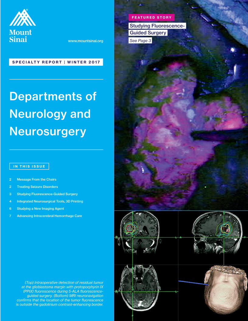
Studying Fluorescence- Guided Surgery
See Page 3
F E A T U R E D S T O R Y
Departments of Neurology and Neurosurgery
www.mountsinai.org
(Top) Intraoperative detection of residual tumor at the glioblastoma margin with protoporphyrin IX (PPIX) fluorescence during 5-ALA fluorescence-
guided surgery. (Bottom) MRI neuronavigation confirms that the location of the tumor fluorescence is outside the gadolinium contrast-enhancing border.
I N T H I S I S S U E
2 Message From the Chairs
2 Treating Seizure Disorders
3 Studying Fluorescence-Guided Surgery
4 Integrated Neurosurgical Tools, 3D Printing
6 Studying a New Imaging Agent
7 Advancing Intracerebral Hemorrhage Care
S P E C I A L T Y R E P O R T | W I N T E R 2 0 1 7
M E S S A G E F R O M T H E C H A I R S
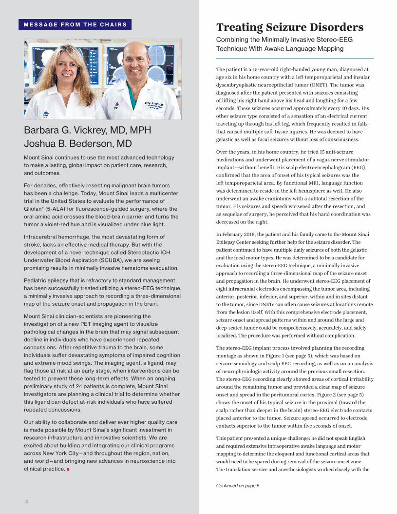
Barbara G. Vickrey, MD, MPH Joshua B. Bederson, MDMount Sinai continues to use the most advanced technology to make a lasting, global impact on patient care, research, and outcomes.
For decades, effectively resecting malignant brain tumors has been a challenge. Today, Mount Sinai leads a multicenter trial in the United States to evaluate the performance of Gliolan® (5-ALA) for fluorescence-guided surgery, where the oral amino acid crosses the blood-brain barrier and turns the tumor a violet-red hue and is visualized under blue light.
Intracerebral hemorrhage, the most devastating form of stroke, lacks an effective medical therapy. But with the development of a novel technique called Stereotactic ICH Underwater Blood Aspiration (SCUBA), we are seeing promising results in minimally invasive hematoma evacuation.
Pediatric epilepsy that is refractory to standard management has been successfully treated utilizing a stereo-EEG technique, a minimally invasive approach to recording a three-dimensional map of the seizure onset and propagation in the brain.
Mount Sinai clinician-scientists are pioneering the investigation of a new PET imaging agent to visualize pathological changes in the brain that may signal subsequent decline in individuals who have experienced repeated concussions. After repetitive trauma to the brain, some individuals suffer devastating symptoms of impaired cognition and extreme mood swings. The imaging agent, a ligand, may flag those at risk at an early stage, when interventions can be tested to prevent these long-term effects. When an ongoing preliminary study of 24 patients is complete, Mount Sinai investigators are planning a clinical trial to determine whether this ligand can detect at-risk individuals who have suffered repeated concussions.
Our ability to collaborate and deliver ever higher quality care is made possible by Mount Sinai’s significant investment in research infrastructure and innovative scientists. We are excited about building and integrating our clinical programs across New York City—and throughout the region, nation, and world—and bringing new advances in neuroscience into clinical practice.
Treating Seizure DisordersCombining the Minimally Invasive Stereo-EEG Technique With Awake Language Mapping
The patient is a 15-year-old right-handed young man, diagnosed at
age six in his home country with a left temporoparietal and insular
dysembryoplastic neuroepithelial tumor (DNET). The tumor was
diagnosed after the patient presented with seizures consisting
of lifting his right hand above his head and laughing for a few
seconds. These seizures occurred approximately every 10 days. His
other seizure type consisted of a sensation of an electrical current
traveling up through his left leg, which frequently resulted in falls
that caused multiple soft-tissue injuries. He was deemed to have
gelastic as well as focal seizures without loss of consciousness.
Over the years, in his home country, he tried 15 anti-seizure
medications and underwent placement of a vagus nerve stimulator
implant—without benefit. His scalp electroencephalogram (EEG)
confirmed that the area of onset of his typical seizures was the
left temporoparietal area. By functional MRI, language function
was determined to reside in the left hemisphere as well. He also
underwent an awake craniotomy with a subtotal resection of the
tumor. His seizures and speech worsened after the resection, and
as sequelae of surgery, he perceived that his hand coordination was
decreased on the right.
In February 2016, the patient and his family came to the Mount Sinai
Epilepsy Center seeking further help for the seizure disorder. The
patient continued to have multiple daily seizures of both the gelastic
and the focal motor types. He was determined to be a candidate for
evaluation using the stereo-EEG technique, a minimally invasive
approach to recording a three-dimensional map of the seizure onset
and propagation in the brain. He underwent stereo-EEG placement of
eight intracranial electrodes encompassing the tumor area, including
anterior, posterior, inferior, and superior, within and in sites distant
to the tumor, since DNETs can often cause seizures at locations remote
from the lesion itself. With this comprehensive electrode placement,
seizure onset and spread patterns within and around the large and
deep-seated tumor could be comprehensively, accurately, and safely
localized. The procedure was performed without complication.
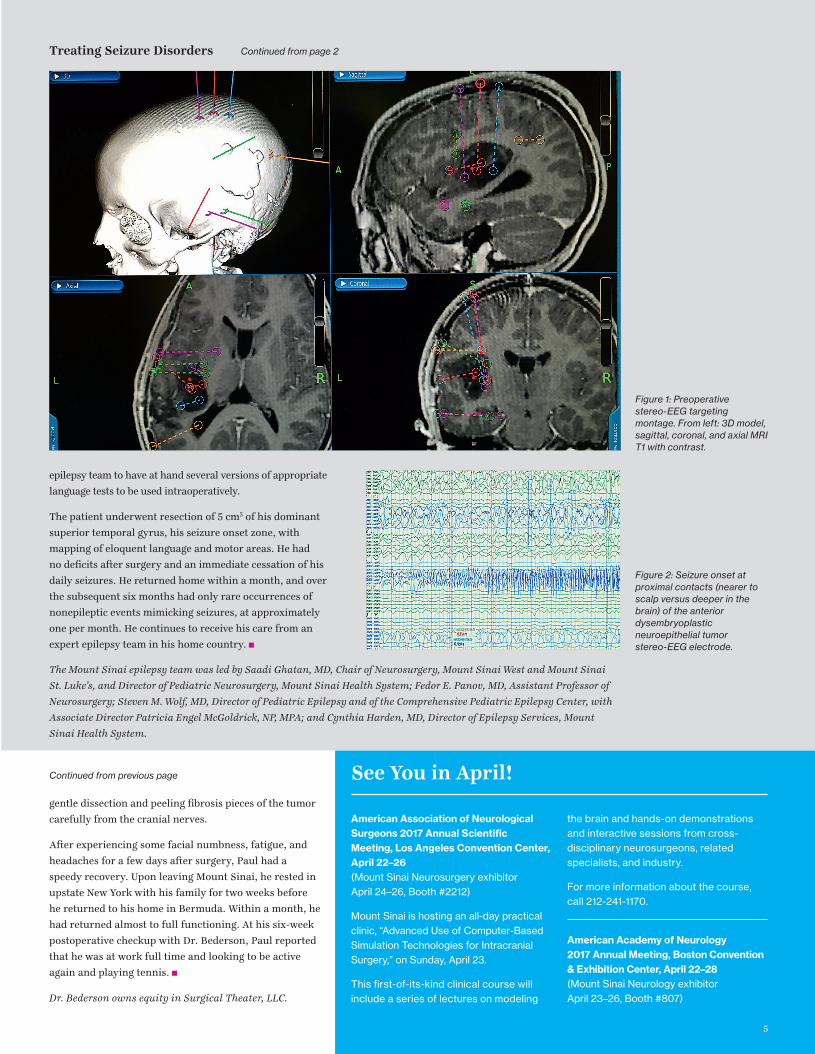
The stereo-EEG implant process involved planning the recording
montage as shown in Figure 1 (see page 5), which was based on
seizure semiology and scalp EEG recording, as well as on an analysis
of neurophysiologic activity around the previous small resection.
The stereo-EEG recording clearly showed areas of cortical irritability
around the remaining tumor and provided a clear map of seizure
onset and spread in the peritumoral cortex. Figure 2 (see page 5)
shows the onset of his typical seizure in the proximal (toward the
scalp rather than deeper in the brain) stereo-EEG electrode contacts
placed anterior to the tumor. Seizure spread occurred to electrode
contacts superior to the tumor within five seconds of onset.
This patient presented a unique challenge: he did not speak English
and required extensive intraoperative awake language and motor
mapping to determine the eloquent and functional cortical areas that
would need to be spared during removal of the seizure onset zone.
The translation service and anesthesiologists worked closely with the
Continued on page 5
2
3
Studying Fluorescence-Guided Surgery A Multicenter Trial Under Way for Malignant Gliomas
In June 2016, a new investigator-initiated trial—titled
A Multicenter Study of 5-Aminolevulinic Acid (5-ALA) to
Enhance Visualization of Malignant Tumor in Patients
with Newly Diagnosed or Recurrent Malignant Gliomas: A
Safety, Histopathology, and Correlative Biomarker Study—
began enrolling patients in the Mount Sinai Health System.
This is the first multicenter study in North America
utilizing 5-ALA, the active substance in Gliolan® for
fluorescence-guided surgery (FGS) of brain tumors. The
study is sponsored by Constantinos G. Hadjipanayis, MD,
PhD, Professor of Neurosurgery, and Oncological Sciences;
Director of Neurosurgical Oncology at the Mount Sinai
Health System; and Chair of Neurosurgery at Mount
Sinai Beth Israel. He was the first in the United States to
perform FGS of a brain tumor after Gliolan administration
and is the U.S. Food and Drug Administration (FDA)
Investigational New Drug holder. Isabelle M. Germano,
MD, Professor of Neurosurgery, Neurology, and
Oncological Sciences, and Director of the Comprehensive
Brain Tumor Program, is the principal investigator.
Approximately 10 to 20 centers will take part in this study,
which will include up to 100 new or recurrent malignant
glioma patients. The Henry Ford Health System in Detroit,
the first site to be added, enrolled its first patient in
January. A correlative serum biomarker study will be
included for participating centers.
Gliolan FGS has emerged as an important technique for
the safe and effective removal of brain tumors by direct
visualization of tumor tissue after using a special blue
light in the operating room (see cover images). Gliolan
is orally administered to patients and is rapidly taken up
into the bloodstream, where it penetrates the blood-brain
barrier and is taken up by brain tumors. Once it enters
brain tumor cells, it is metabolized to its fluorescent form,
known as protoporphyrin IX (PPIX). PPIX is visualized as
violet-red in tumor tissue with use of a special operative
microscope. Better visualization of tumor tissue after 5-ALA
administration permits more complete resection of brain
tumors and overall benefit to the patient (see images above).
This has been shown in a large, randomized study in Europe.
Gliolan FGS is currently approved for brain tumor removal
in Europe and Asia, and safety has been demonstrated in
many patients. The multicenter trial at Mount Sinai will
evaluate the utility of Gliolan, and its metabolite PPIX,
as an intraoperative fluorescent detection agent for the
visualization of malignant tumor during the removal of
malignant brain tumors, either new or recurrent.
Recently, Dr. Hadjipanayis completed a National Institutes
of Health (NIH)-funded R21 study on the use of 5-ALA FGS
in combination with new whole-brain proton spectroscopic
imaging in brain tumor patients. This novel study could
lead the way to better identification of malignant brain
tumor tissue and radical resection of tumors. Currently,
Dr. Hadjipanayis and Daniel J. Brat, MD, PhD, Vice
Chair, Translational Programs, Department of Pathology
and Laboratory Medicine, Emory University School of
Medicine, are studying the expression of proteins in
different regions of brain cancer, known as glioblastoma
(GBM). By utilizing 5-ALA FGS, Dr. Hadjipanayis and
his team can differentiate the tumor margin, where
fluorescence is weaker, from the tumor mass. This NIH-
funded R01 study aims to provide important information
on protein expression at the tumor margin so potential
drug therapies can target this problematic area. Nearly all
GBM tumors in patients relapse at the tumor margin due
to cancer cells that reside away from the tumor mass and
escape treatment.
Dr. Hadjipanayis and his team have been integral to the
development of Gliolan in the United States over the last
six years. Gliolan received orphan drug status from the
FDA in 2013 and is now being considered for fast-track
FDA approval.
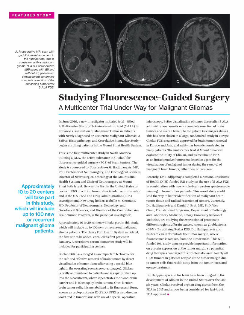
A. Preoperative MRI scan with gadolinium enhancement in
the right parietal lobe is consistent with a malignant
glioma. B. & C. Postoperative MRI scans with (B) and without (C) gadolinium
enhancement confirming complete resection of the
enhancing tumor after 5-ALA FGS.
Approximately 10 to 20 centers
will take part in this study,
which will include up to 100 new
or recurrent malignant glioma
patients.
F E A T U R E D S T O R Y
A B C
4
Integrated Neurosurgical Tools, 3D Printing:An Epidermoid Tumor in Posterior Fossa Is Removed
Paul, a 39-year-old male, had begun noticing some loss of
hearing and trouble with balance. These symptoms were
minor at the time, and with his very active lifestyle, Paul
dismissed them. But when he and his wife were on vacation
in New York City, he noticed a fullness in his right ear.
This prompted him to see Eric A. Munzer, DO, Assistant
Clinical Professor, Otolaryngology, Icahn School of
Medicine at Mount Sinai, who recommended an MRI. The
results showed a large, almost 5 cm diameter epidermoid
tumor in the posterior fossa, and Paul was immediately
referred to Joshua B. Bederson, MD, Professor and Chair
of Neurosurgery for the Mount Sinai System, and Clinical
Director of the Neurosurgery Simulation Core.
In August 2016, the patient came to see Dr. Bederson and
Leslie Schlachter, PA-C. The team confirmed Dr. Munzer’s
findings that the tumor was compressing the patient’s
brain stem, and they also learned that it surrounded five
different cranial nerves (V7, 8, 9, 10, and 11). The effect
on the cranial nerves was very evident in the patient’s
symptoms, including intermittent choking when drinking
liquids. Paul was scheduled for surgery in a month.
The Mount Sinai team mobilized a specific combination
of integrated neurosurgical tools for preoperative
simulation, planning, intraoperative navigation, and
microscope integration to help Paul. Mount Sinai is
one of only a few sites in North America that has this
capability. By utilizing Surgical Theater’s patient-specific
3D simulation platform, linked to Brainlab’s surgical
navigation hardware and software platforms, which were
integrated into the Leica microscope, the surgeon was
able to see, in real time, a “heads-up” display of outlined
critical structures projected into the eyepieces of the
microscope during surgery.
A 3D print of the patient’s anatomy also was made to
increase the surgeon’s awareness and provide a lifelike
model for preoperative planning and orientation. First,
the patient’s MRI and CT data were taken to segment
the structures of the skull, tumor, and other important
anatomy. The cranial nerves also were segmented and
printed, in addition to important vasculature—information
that was translated into a multicolor, multistructural
gypsum powder print. Under the direction of Anthony
B. Costa, PhD, Assistant Professor of Neurosurgery, and
Scientific Director of the Neurosurgery Simulation Core,
the Medical Modeling Core completed the task of building
digital representations of the patient’s anatomy through
numerical analysis of preoperative MRI and CT scans.
Through the Medical Modeling Core’s collaboration with
Mount Sinai’s Rapid Prototyping Center, the final 3D print
was completed in about six hours using the in-house 3D
Systems’ ProJet® CJP 660Pro full-color 3D printer. This
technology allows for vision where traditional imaging
methods are not highly contrasting.
The surgical approach was a far-lateral transcondylar
craniotomy, allowing for a direct approach to the tumor,
avoiding the brain stem. The epidermoid was revealed to
be cystic, containing pearls. Removal took several hours of
The team used a specific
combination of integrated tools for preoperative
simulation, planning,
intraoperative navigation, and
microscope integration.
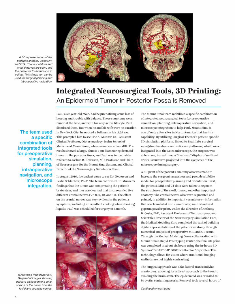
A 3D representation of the patient’s anatomy using MRI
and CTA. The vasculature and cranial nerves are seen, and
the posterior fossa tumor is in yellow. This simulation can be used for surgical planning and
intraoperative navigation.
(Clockwise from upper left) Sequential images showing
delicate dissection of a small portion of the tumor from the
facial and acoustic nerves. Continued on next page
epilepsy team to have at hand several versions of appropriate
language tests to be used intraoperatively.
The patient underwent resection of 5 cm3 of his dominant
superior temporal gyrus, his seizure onset zone, with
mapping of eloquent language and motor areas. He had
no deficits after surgery and an immediate cessation of his
daily seizures. He returned home within a month, and over
the subsequent six months had only rare occurrences of
nonepileptic events mimicking seizures, at approximately
one per month. He continues to receive his care from an
expert epilepsy team in his home country.
The Mount Sinai epilepsy team was led by Saadi Ghatan, MD, Chair of Neurosurgery, Mount Sinai West and Mount Sinai
St. Luke’s, and Director of Pediatric Neurosurgery, Mount Sinai Health System; Fedor E. Panov, MD, Assistant Professor of
Neurosurgery; Steven M. Wolf, MD, Director of Pediatric Epilepsy and of the Comprehensive Pediatric Epilepsy Center, with
Associate Director Patricia Engel McGoldrick, NP, MPA; and Cynthia Harden, MD, Director of Epilepsy Services, Mount
Sinai Health System.
Figure 1: Preoperative stereo-EEG targeting montage. From left: 3D model, sagittal, coronal, and axial MRI T1 with contrast.
Figure 2: Seizure onset at proximal contacts (nearer to scalp versus deeper in the brain) of the anterior dysembryoplastic neuroepithelial tumor stereo-EEG electrode.
American Association of Neurological Surgeons 2017 Annual Scientific Meeting, Los Angeles Convention Center, April 22–26 (Mount Sinai Neurosurgery exhibitor April 24–26, Booth #2212)
Mount Sinai is hosting an all-day practical clinic, “Advanced Use of Computer-Based Simulation Technologies for Intracranial Surgery,” on Sunday, April 23.
This first-of-its-kind clinical course will include a series of lectures on modeling
the brain and hands-on demonstrations and interactive sessions from cross-disciplinary neurosurgeons, related specialists, and industry.
For more information about the course, call 212-241-1170.
American Academy of Neurology 2017 Annual Meeting, Boston Convention & Exhibition Center, April 22–28 (Mount Sinai Neurology exhibitor April 23–26, Booth #807)
Treating Seizure Disorders Continued from page 2
gentle dissection and peeling fibrosis pieces of the tumor
carefully from the cranial nerves.
After experiencing some facial numbness, fatigue, and
headaches for a few days after surgery, Paul had a
speedy recovery. Upon leaving Mount Sinai, he rested in
upstate New York with his family for two weeks before
he returned to his home in Bermuda. Within a month, he
had returned almost to full functioning. At his six-week
postoperative checkup with Dr. Bederson, Paul reported
that he was at work full time and looking to be active
again and playing tennis.
Dr. Bederson owns equity in Surgical Theater, LLC.
Continued from previous page See You in April!
5
6
Studying a New Imaging Agent A Window Into Traumatic Brain Injury in Living Patients
Researchers at the Icahn School of Medicine at Mount
Sinai are pioneering the use of a new imaging agent used
with positron emission tomography (PET) to detect and
monitor the progression of repetitive traumatic brain
injury in patients with a history of concussions.
Sam Gandy, MD, PhD, Director of the Center for
Cognitive Health and of the National Football League
(NFL) Neurological Care Program at the Icahn School
of Medicine at Mount Sinai, and Dara L. Dickstein,
PhD, Adjunct Assistant Professor of Neuroscience, and
Geriatrics and Palliative Medicine, coauthored a proof-
of-concept study in Translational Psychiatry in 2016. The
new PET agent—a ligand known as T807/AV1451—was
used to examine a 39-year-old retired NFL player who had
experienced 22 concussions, four of which resulted in loss
of consciousness, during an 11-year NFL career.
The patient exhibited agitation, impulsivity, sensitivity
to light, periods of severe rage, and a decline in executive
functioning, processing speed, and fine motor skills, as
well as difficulty with working memory—all progressive
neuropsychiatric symptoms associated with chronic
traumatic encephalopathy (CTE). The disease involves
widespread axonal disruption and the eventual degeneration
of the neocortex, hippocampus and other limbic structures,
and basal forebrain, and is also believed to be a precursor to
various neurodegenerative diseases.
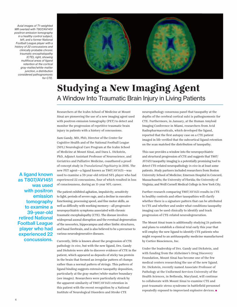
Currently, little is known about the progression of CTE
pathology in vivo, but with the new ligand, Drs. Gandy
and Dickstein were able to discover evidence of CTE in the
patient, which appeared as deposits of sticky tau protein
in the brain that formed an irregular pattern of clumps
rather than a normal pattern of strings. This pattern of
ligand binding suggests extensive tauopathy deposition,
particularly at the gray-matter/white-matter boundary
(see images). Researchers were particularly struck by
the apparent similarity of T807/AV1451 retention in
this patient with the recent recognition by a National
Institute of Neurological Disorders and Stroke CTE
neuropathology consensus panel that tauopathy at the
depths of the cerebral cortical sulci is pathognomonic for
CTE. Furthermore, in January, at the Human Amyloid
Imaging Conference in Miami, researchers from Avid
Radiopharmaceuticals, which developed the ligand,
reported that the first autopsy case on a CTE patient
imaged in life verified that the subcortical ligand retention
on the scan matched the distribution of tauopathy.
This case provides a window into the neuropsychiatric
and structural progression of CTE and suggests that T807/
AV1451 tauopathy imaging is a potentially promising tool to
detect CTE-related neuropathology in vivo in at least some
patients. Study partners included researchers from Boston
University School of Medicine; Emerson Hospital in Concord,
Massachusetts; the University of Florida; the University of
Virginia; and Weill Cornell Medical College in New York City.
Further research comparing T807/AV1451 results in CTE
to healthy controls and other tauopathies will clarify
whether there is a signature pattern that can be attributed
to CTE and whether and under what conditions tauopathy
imaging can be used clinically to identify and track
progression of CTE-related neurodegeneration.
The Mount Sinai team is additionally studying 24 patients
and plans to establish a clinical trial early this year that
will employ the new ligand to identify CTE patients who
might respond to an antitauopathy medicine manufactured
by Cortice Biosciences, Inc.
Under the leadership of Drs. Gandy and Dickstein, and
with funding from the Alzheimer’s Drug Discovery
Foundation, Mount Sinai has become one of the few
medical centers researching the use of the new ligand.
Dr. Dickstein, recently named Associate Professor of
Pathology at the Uniformed Services University of the
Health Sciences, in Bethesda, Maryland, will continue
to collaborate with Mount Sinai to examine CTE and
post-traumatic stress syndrome in battlefield personnel
repeatedly exposed to improvised explosive devices.
Axial images of T1-weighted MR overlaid with T807/AV1451 positron emission tomography
in a healthy control subject, left, and a former National
Football League player with a history of 22 concussions and
clinically probable chronic traumatic encephalopathy
(CTE), right, showing multifocal areas of ligand
retention at the cortical gray-matter/white-matter
junction, a distribution considered pathognomonic
for CTE.
A ligand known as T807/AV1451
was used with positron
emission tomography
to examine a 39-year-old
retired National Football League player who had experienced 22
concussions.
7
Advancing Intracerebral Hemorrhage CareMaking Gains in Minimally Invasive Clot Evacuation
The least treatable and most devastating form of stroke,
spontaneous intracerebral hemorrhage (ICH), is a leading
cause of mortality, morbidity, and disability worldwide.
Currently, there are very few treatment options for
patients with this condition, but recently, minimally
invasive clot evacuation has gained popularity as a result
of multiple small but successful prospective trials and
iterative advances in technology.
J Mocco, MD, MS, Director of the Cerebrovascular Center
at the Mount Sinai Health System, and Professor and Vice
Chair for Education in the Department of Neurosurgery
at the Icahn School of Medicine at Mount Sinai, and
Christopher P. Kellner, MD, Director of the Health
System’s Intracerebral Hemorrhage program and Assistant
Professor of Neurosurgery, have developed an original
technique to perform minimally invasive clot evacuation in
patients with this condition.
The treatment—Stereotactic ICH Underwater Blood
Aspiration (SCUBA) technique—brings together a
combination of stereotactic guidance, a low-profile
endoscopic sheath, the Apollo™ System device by Penumbra,
Inc., for suction with an ultrasonicator, combined air and
fluid strategies, and electrocautery. Dr. Mocco, who holds
the Investigational Device Exemption for Apollo—approved
in 2014 by the U.S. Food and Drug Administration for use
in the evacuation of intraventricular hemorrhage and in
2016 for the evacuation of intracerebral hemorrhage—is
the principal investigator of a National Institutes of Health
(NIH) multicenter clinical trial known as INVEST: Minimally
Invasive Endoscopic Surgery vs. Medical Management in
Supratentorial Intraparenchymal Hemorrhage.
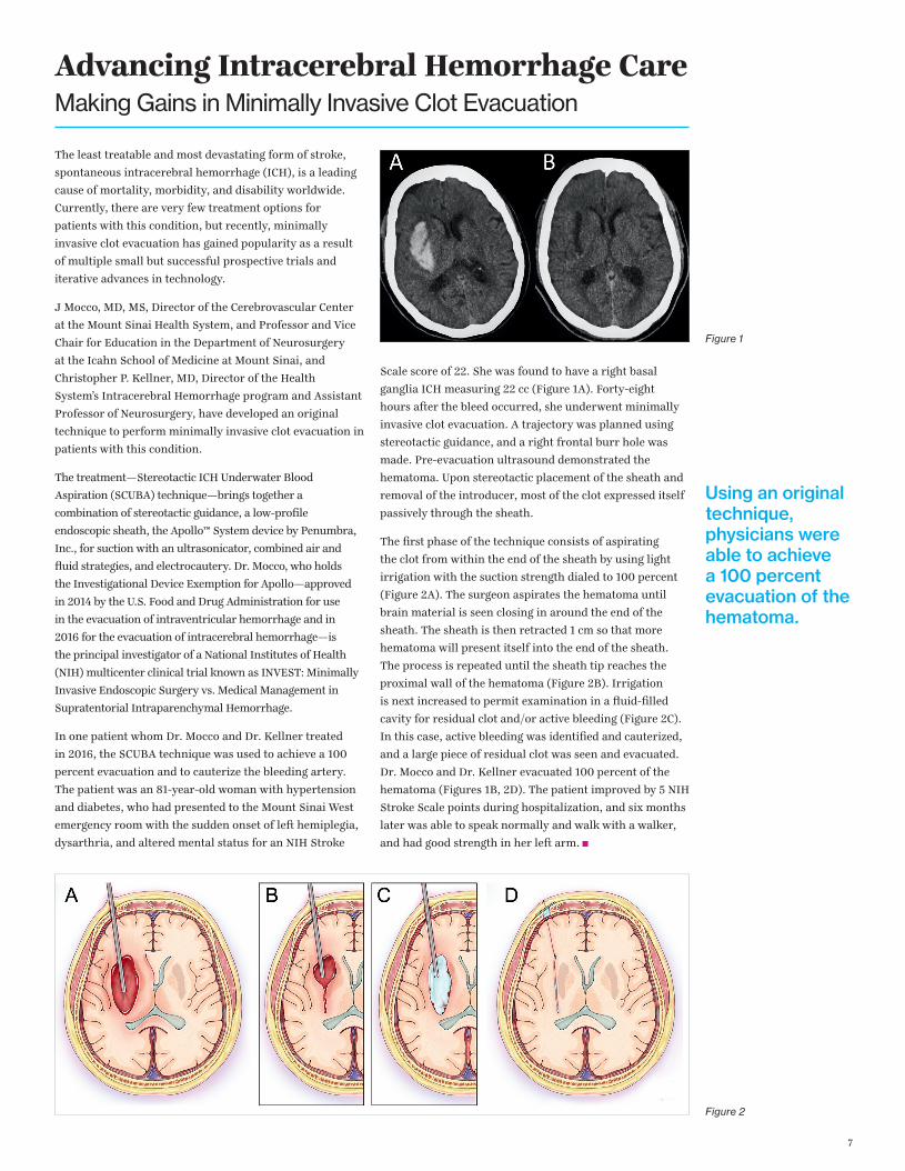
In one patient whom Dr. Mocco and Dr. Kellner treated
in 2016, the SCUBA technique was used to achieve a 100
percent evacuation and to cauterize the bleeding artery.
The patient was an 81-year-old woman with hypertension
and diabetes, who had presented to the Mount Sinai West
emergency room with the sudden onset of left hemiplegia,
dysarthria, and altered mental status for an NIH Stroke
Scale score of 22. She was found to have a right basal
ganglia ICH measuring 22 cc (Figure 1A). Forty-eight
hours after the bleed occurred, she underwent minimally
invasive clot evacuation. A trajectory was planned using
stereotactic guidance, and a right frontal burr hole was
made. Pre-evacuation ultrasound demonstrated the
hematoma. Upon stereotactic placement of the sheath and
removal of the introducer, most of the clot expressed itself
passively through the sheath.
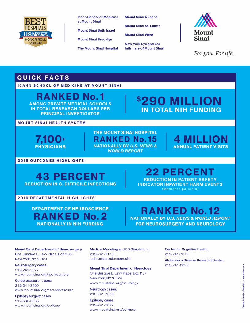
The first phase of the technique consists of aspirating
the clot from within the end of the sheath by using light
irrigation with the suction strength dialed to 100 percent
(Figure 2A). The surgeon aspirates the hematoma until
brain material is seen closing in around the end of the
sheath. The sheath is then retracted 1 cm so that more
hematoma will present itself into the end of the sheath.
The process is repeated until the sheath tip reaches the
proximal wall of the hematoma (Figure 2B). Irrigation
is next increased to permit examination in a fluid-filled
cavity for residual clot and/or active bleeding (Figure 2C).
In this case, active bleeding was identified and cauterized,
and a large piece of residual clot was seen and evacuated.
Dr. Mocco and Dr. Kellner evacuated 100 percent of the
hematoma (Figures 1B, 2D). The patient improved by 5 NIH
Stroke Scale points during hospitalization, and six months
later was able to speak normally and walk with a walker,
and had good strength in her left arm.
Figure 2
Figure 1
Using an original technique, physicians were able to achieve a 100 percent evacuation of the hematoma.
22 PERCENTREDUCTION IN PATIENT SAFETY
INDICATOR INPATIENT HARM EVENTS( M e d i c a r e p a t i e n t s )
43 PERCENTREDUCTION IN C. DIFFICILE INFECTIONS
4 MILLION ANNUAL PATIENT VISITS
$ 290 MILLIONIN TOTAL NIH FUNDING
THE MOUNT SINAI HOSPITAL
R ANKED No. 15 NATIONALLY BY U.S. NEWS &
WORLD REPORT
RANKED No. 1AMONG PRIVATE MEDICAL SCHOOLS IN TOTAL RESEARCH DOLLARS PER
PRINCIPAL INVESTIGATOR
Icahn School of Medicineat Mount Sinai
Mount Sinai Beth Israel
Mount Sinai Brooklyn
The Mount Sinai Hospital
Mount Sinai Queens
Mount Sinai St. Luke’s
Mount Sinai West
New York Eye and EarInfi rmary of Mount Sinai
Con
cept
/Des
ign:
Suk
a N
Y/s
ukac
reat
ive.
com
For you. For life.
Q U I C K F A C T S
M O U N T S I N A I H E A LT H S Y S T E M
2 0 1 6 O U T C O M E S H I G H L I G H T S
2 0 1 6 D E P A R T M E N T A L H I G H L I G H T S
I C A H N S C H O O L O F M E D I C I N E A T M O U N T S I N A I
7,100+PHYSICIANS
Mount Sinai Department of NeurosurgeryOne Gustave L. Levy Place, Box 1136 New York, NY 10029
Neurosurgery cases:212-241-2377www.mountsinai.org/neurosurgery
Cerebrovascular cases:212-241-3400www.mountsinai.org/cerebrovascular
Epilepsy surgery cases: 212-636-3666www.mountsinai.org/epilepsy
Medical Modeling and 3D Simulation:212-241-1170icahn.mssm.edu/neurosim
Mount Sinai Department of NeurologyOne Gustave L. Levy Place, Box 1137 New York, NY 10029www.mountsinai.org/neurology
Neurology cases: 212-241-7076
Epilepsy cases: 212-241-2627www.mountsinai.org/epilepsy
Center for Cognitive Health: 212-241-7076
Alzheimer’s Disease Research Center:212-241-8329
DEPARTMENT OF NEUROSCIENCE
RANKED No. 2NATIONALLY IN NIH FUNDING
RANKED No. 12NATIONALLY BY U.S. NEWS & WORLD REPORT
FOR NEUROSURGERY AND NEUROLOGY