Embed Size (px)
Citation preview

JOURNAL OF BACTERIOLOGYVol. 88, No. 1, P. 200-209 July, 1964Copyright © 1964 American Society for Microbiology
Printed in U.S.A.
DEPENDENCY OF TREPONEMA MICRODENTIUM ON OTHERORAL ORGANISMS FOR ISOBUTYRATE, POLYAMINES, AND A
CONTROLLED OXIDATION-REDUCTION POTENTIAL
S. S. SOCRANSKY, W. J. LOESCHE, C. HUBERSAK, AND J. B. MACDONALD
Forsyth Dental Center and Harvard School of Dental Medicine, Boston, Massachusetts
Received for publication 29 January 1964
ABSTI.ACT
SOCRANSKY, S. S. (Forsyth I)ental Center, Bos-ton, Mass.), W. J. LOESCHE, C. HUBERS.K,AND J. B. MACDONALD. Dependency of Treponeinamticrodentium on other oral organisms for isobutyr-atte, polyamines, and a controlled oxidation-reduc-tion potential. J. Bacteriol. 88:200-209. 1964.-Strains of Treponema microdentiiunt can be culti-vated on a variety of autoclaved commerciallyavailable media in the presence of other oral or-ganisms. Organisms supporting growth in thesecircumstances include a facultative diphtheroidaccompanied by either a strain of Fusobacteriuimor a motile gram-negative anaerobic rod. Culturefiltrates and lysates of these "supporting or-ganisms" failed to substitute for growing or-ganisms. Measurement of the oxidation-reductionpotential of the test system demonstrated that thespirochetes grew in a narrow range of Eh (opti-miiutm, -190 mv). The supporting organisms couldbe replaced by their filtrates when the Eh of themediunm was poised in this range by a combinationof reducing agents. Both filtrates contained aheat-labile factor required by the spirochete,which could be replaced by 5 /Ag/ml of cocarboxyl-ase. Isobutyric acid, which could be detected inthe fusiform filtrate, and putrescine which couldbe detected in the diphtheroid filtrate, replacedt he spirochete's remaining filtrate requirement.Maximal growth occurred when any of the follow-ing were incorporated into the medium: 2 ,ug/mlof sodiuml isobutyrate; 250 ,ug/ml of putrescinedihydrochloride; 200 ug/ml of spermidine phos-phosphate, or 150 ug/ml of spermine tetrahydro-chloride.
Oral sl)ilochetes are usually cultivated on acoml)lex Tyndallized veal heart infusion mediumcontaining ascitic fluid (Hampp, 1943; Roseburyet al., 1951) or rabbit blood (B3erger, 1958;Socranskv, Macdonald, and Sawyer, 1959).Attempts to simplify and define this mediumhave met with partial success. Hampp and Nevin(1959) cultivated Borrelia vincentii in a veal
heart infusion medium in which five coenzymesand glucose-i-phosphate substituted for asciticfluid enrichment. Later, the same investigators(Nevin and Hampp, 1959), using a simplifiedbasal medium, demonstrated a requirement of B.vincentii for five coenzymes, oleic acid, carbondioxide, and L-asparagine. In addition, smallquantities of ascitic fluid were essential forgrowth.
Since enrichment substances such as asciticfluid are difficult to obtain and variable in abilityto support sp)irochetal growth, potential sourcesof the spirochetes' growth requirements werelooked for in their normal environment. Onelikely source of these factors appeared to beother organisms present in the oral cavity. Nevin,Hampp, and Duey (1960) demonstrated stimula-tion of growth of B. vincentii by an oral micro-aerophilic diphtheroid. The stimulation could beduplicated by the addition of cocarboxylase oracetyl phosp)hate to the medium. Neither of thesecompounds could be demonstrated in the diph-theroid or its culture filtrate. Neither the diph-theroid nor cocarboxylase replaced the require-ment for ascitic fluid of B. vincentii.
Preliminary experiments in our laboratoryrevealed that oral spirochetes would growabundantly on autoclaved commercially avail-able media in the presence of other oral organ-isms. The media used did not require blood,ascitic fluid, or any other enrichment additive.The purpose of the studies reported here was toisolate and identify the organisms "supporting-'spirochetal growth, and to investigate thenature of the growth-promoting substancesinvolved.
MATERIALS AND METHODSPreparation of mnedium and inoculation of
spirochetes. PPLO broth (13BL), without crystalviolet or serum, was autoclaved at 15 psi for 15min with 1.2% agar (Difco), and poured into
200
on May 3, 2018 by guest
http://jb.asm.org/
Dow
nloaded from

GROWTH REQUIREMENTS OF T. MICRODENTIUM
petri plates (6 by 1.5 cm); the plates werefilled to within 2 mm of the rim. After the mediumhad cooled, wells were cut into the center of theagar medium by means of sterile glass tubing(8 mm in diameter), creating a "spirochetewell plate" (Rosebury and Foley, 1941). Mixedspirochetes and oral bacteria, obtained fromthe gingival crevice area of man, were inoculateddirectly into the central well of the "spirochetewell plates." After 7 days of anaerobic incubation,the spirochetes grew out as a haze, away fromother forms of bacteria which were retained inthe central well of the well plate. Spirochetesfree from other bacteria could then be transferredby Pasteur pipette from the periphery of thespirochetal haze to the central wells of fresh wellplates. Stock cultures of spirochetes were main-tained in early experiments by the serial transferof spirochetes with the other oral organismsgrowing in the central well. A pure strain ofspirochetes was obtained for later experiments byserial transfer of single surface colonies in ananaerobic environment as previously described(Socransky et al., 1959).The spirochetes investigated in this study were
considered to be Treponema microdentium on thebasis of size and morphology as revealed byelectron microscopy (Listgarten, Loesche, andSocransky, 1963).
Cultivation of organisms other than spirochetes.Mixed oral organisms were maintained by serialtransfer as mixed cultures in well plates or asconfluent drops on heart infusion-blood agarplates. Pure cultures of organisms other thanspirochetes were isolated on PPLO Agar andmaintained on PPLO Agar or in PPLO broth.When two or more organisms were placed in thewell, they were first combined by means of a"wheel plate" (Macdonald, Sutton, and Knoll,1954) in which each organism was streakedfrom the periphery of the plate to a commoncentral hub. A loopful of the organisms in thecentral hub was then placed in the well alongwith the spirochetal inoculum. In later experi-ments, broth cultures of the "supporting organ-isms" were combined and diluted to an opticaldensity of 1.0 at 560 m,u; 0.05 ml of this dilutionwas placed in the central well, along with thespirochetal inoculum.
Incubation. All plates and broth culturesexcepting those involved in measuring oxidation-reduction potential were incubated anaerobicallyin Brewer jars in 95% H2 and 5% CO2 for 3 to
, AGAR MEDIUM
SPIROCHETAL HAZE
CENTRAL WELL
EPOXY RESIN
SATURATED KCL 2% AGAR BRIDGE
- SArURATED KCL
PLATINUM
CALOMELMERCURYPLATINUM CONTACT
FIG. 1. Apparatus used to measure Eh of growingorganisms. The entire apparatus was placed in adesiccator with the leads from the platinum andcalomel electrodes passing through a plasticine seal.
7 days at 35 to 37 C. The atmosphere used inmeasuring oxidation-reduction potential is de-scribed below.Measurement of oxidation-reduction potential.
An apparatus was constructed as shown in Fig.1. Three platinum electrodes, consisting of 24-gauge platinum wire, were embedded in the baseof a petri plate (6 by 1.5 cm) by an epoxy resin(Araldite; Ciba Pharmaceutical Products, Inc.,Summit, N.J.). The leads soldered to theseelectrodes were Teflon-insulated 18-gauge braidedlead in wires which could be autoclaved in situ.A saturated potassium chloride 2% agar bridgejoined the medium in the plate to a saturatedcalomel electrode. The entire apparatus wasplaced in a Brewer jar or a desiccator with theleads from the platinum and calomel electrodespassing through a plasticine seal to a Beckmanmodel G pH meter. The atmosphere in the jarswas exchanged by evacuating the jar to 30 mmof mercury and filling it with nitrogen (highpurity; Linde Co., New York, N.Y.) seven times.On the final exchange, 50k CO2 was introducedwith the nitrogen. In certain experiments, alkalinepyrogallol or glucose oxidase was activated in thejar after the atmosphere had been exchanged, toremove residual oxygen.The electrodes were tested in the 95% N2
and 5% CO2 atmosphere by measuring thepotential of known oxidation-reduction systemsincluding 0.1 N and saturated calomel solutions,as well as potassium ferricyanide-ferrocyanidesystems and ferrous-ferric chloride systems.The last two systems were measured in buffersolutions in the presence and absence of 1.2%agar. All test measurements were comparedwith those of a Beckman platinum electrodekept as a standard.
Finally, all experiments were repeated, in-
VOL. 88, 1964 201
on May 3, 2018 by guest
http://jb.asm.org/
Dow
nloaded from

SOCRANSKY ET AL.
corporating 1 to 20 ppm of the following oxida-tion-reduction indicators in the medium: methyl-ene blue, indigo carmine, phenosafranine, andneutral red.
Preparation of filtrates and lysates of supportingorganisms. The organism to be tested wasinoculated into PPL.O broth with or without0.1 % glucose and incubated anaerobically for2, 4, 6, and 9 days at 35 C. The cultures werecentrifuged at 10,000 X g for 30 min, and thesupernatant fluid was removed and sterilizedby passage through an ultrafine glass filter(Corning Glass Works, Corning, N.Y.). Thepacked organisms were suspended in fresh PPLObroth with and without 200 pg/ml of L-eysteineand sonically disrupted in a 9-ke sonic oscillatorfor 1 hr. The resultant suspension was resus-pended to the orginal volume of the culture inPPLO broth and sterilized by filtration or byheating to 100 C for 20 min. Packed, nonviableorganisms were prepared by heating brothcultures of the organisms for 30 min at 80 Cand centrifuging to concentrate the organisms.
In later experiments, the fusiform and diph-theroid cultures were grown anaerobically for 4days in PPLO broth supplemented with 0.1%glucose. The cultures were centrifuged at10,000 X g, and the supernatant fluids werefilter-sterilized through 0.45-, Millipore filtersor autoclaved at 10 psi for 10 min.A ssay of filtrate activity. The basal medium
for the assay of filtrate activity consisted ofPPLO broth supplemented with 1,000 ,g/ml ofglucose, 800 ,ug/ml of L-eysteine, 400 Mg/ml ofnicotinamide, and 5 ,ug/ml of cocarboxylase.The eysteine and cocarboxylase were filter-sterilized and added aseptically to the remainderof the medium which had been previously auto-claved at 10 psi for 10 min. For growth androutine maintenance of spirochetes, the basalmedium was supplemented with 5% diphtheroidculture filtrate and 5% fusiform culture filtrate.
T'wo assay systems were used. In the first,the basal medium supplemented with 1.2%agar (Difco) and the substances being assayedwas poured into petri plates (6 by 1.5 cm) andthe spirochetes were inoculated into the central"well." Results were considered positive when atypical spirochetal haze was observed afterincubation, and negative when there was novisible indication of growth.
In the second assay system, the spirochetes
were inoculated into the basal medium supple-mented with the substances being assayed andchanges in culture optical density at 470 m,uwere determined by use of a lBeckman model Bslpectrophotometer. It was recognized in earlyexperiments that spirochetes exhibit a markedcarry-over of nutrients, even after washingwith basal media. To overcome this difficulty,the spirochetes were serially transferred forfive passages in duplicate tubes of the test me-dium before being assayed.
Preliminary characterization of the active factorsin the filtrates. l'o partially characterize the activefactors in the culture filtrates, samples of thefiltrates weere subjected to the following pro-cedures: (i) heating to 56, 80, and 100 C for 30miD at pH 7.0; (ii) autoclaving at 121 C for 15min; (iii) extraction three times with 3 volumesof ether at pH 2.5, 7.0, and 10.5; (iv) steamdistillation at pH 2.5, 7.0, and 10.5; and (v)dialysis against 0.067 M phosphate buffer (pH7.0).
Determination of isobutyrate and putrescineuptake. T. mnicrodentium was grown in the basalmedium supplemented with 0.5 ,ug/ml of sodiumisobutyrate for two successive passages and thentransferred to a medium containing 2 mg/ml ofsodium isobutyrate-1-C'4. The organisms weregrown anaerobically for 3 days and harvestedby centrifugation. Radioactivity was determinedin the culture supernatant, and also in a sampleof the cells after collection on a Millipore filter.The filter was cemented to a planchet for count-ing. The remaining cells were then washed threetimes with 0.067 M phosphate buffer (pH 7.0)and suspended in a 1% solution of unlabeledsodium isobutyrate for 24 hr at 4 C. Radioac-tivity was again determined in the cells and thesuspending medium. The cells were reharvestedby centrifugation, suspended in distilled water,and sonically disrupted for 1 hr in a 9-ke Ray-theon sonic oscillator. A sample of this suspen-sion was steam-distilled at pH 2.0, and anothersample was subjected to column chromatography(Wiseman and Irvin, 1957).To test for the incorporation of putrescine, T.
microdentium was grown in the basal mediumsupplemented with 50 ,g/ml of putrescinedihydrochloride for three successive passagesand then inoculated into a medium containing100 ,ug/ml of putrescine-1 ,4-C14 dihydrochloride.After 3 days of anaerobic incubation, the cells
202 J. BACTERIOL.
on May 3, 2018 by guest
http://jb.asm.org/
Dow
nloaded from

GROWTH REQUIREMENTS OF T. MICRODEAYIUT2U0
were harvested and washed three times with0.067 M phosphate buffer (pH 7.0); activity wasdetermined in the cells and in the supernatantwashing fluid.
RESULTS
Demonstration of the required role of other oralorganisms. Gingival debris from eight individuals,containing spirochetes and other bacteria, wasinoculated into PPLO Agar well plates as de-scribed above and incubated anaerobicallv.Spirochetes from all eight individuals grew outas large hazes and were serially transferred tofresh plates with and without other oral organ-isms. In all instances, spirochetes grew wheninoculated with the other oral organisms butdid not grow in the absence of these organisms.Spirochetes from seven of the eight sources weremaintained for eight passages before beingdiscarded; spirochetes from the other individualwere maintained for 72 passages until a knowncombination of organisms replaced the mixedoral flora. In every passage, the ability of thespirochetes to grow on the basal medium withoutother organisms was tested with uniformlynegative results.
In the presence of other oral organisms, thespirochetes grew equally well in autoclaved orfilter-sterilized basal media, or in different basalmedia, such as Trypticase Soy Agar (BBL),Trvpticase with 5% yeast extract (Difco), orBrain Heart Infusion Agar (Difco) with andwithout 10% horse blood. Spirochetes isolatedfrom any of the eight individuals could bemaintained by the mixed bacteria from the otherseveii individuals.
Identification of organisms capable of supportingspirochetal growth. A group of 16 organisms whichhad been isolated and characterized by Mac-donald et a]. (1954) during studies of mixedanaerobic infections was demonstrated to supportspirochetal growth. Although all 16 strainstogether supported spirochetal growth, none ofthe 16 strains individually was effective. Strainswere systematically deleted from the mixtureuntil two minimal effective combinations wereidentified. The first consisted of a facultativediphtheroid (JB3B) with a fusiform (JF5), thesecond consisted of the diphtheroid (JB3B)with an anaerobic, motile, gram-negative rod(K80). Since the fusiform was easier to growthan the motile gram-negative rod, the combina-
tion of the diphtheroid and fusiform was used insucceeding experiments. These two organismscould support spirochetes freshly isolated fromthe oral cavity.Attempts to replace the diphtheroid or the
fusiform, or both, by strains of facultativestreptococci, lactobacilli, other "diphtheroids"(both facultative and anaerobic), staphylococci,Veillonella, and other fusiforms, failed. How-ever, certain strains of peptostreptococci couldreplace the fusiform. No organism tested couldreplace the diphtheroid.
Attempts to replace the fus(form and diphtheroidby their filtrates, sonic lysates, and heat-killedcells. Culture "filtrates" and culture "lysates"of the fusiform and diphtheroid were added tothe basal medium in concentrations varyingfrom 0.001 to 100%° of the final medium in anattempt to substitute for one or both of therequired viable organisms. In addition, thefusiform filtrate was prepared from culturesgrown in PPLO broth containing up to 50%of the diphtheroid filtrate and vice versa. Fil-trates and lysates were also prepared from cul-tures in which the two organisms were growntogether in broth or in agar.
Finally, heat-killed fusiforms and diphtheroidswere placed in the wells of some of the testmedia in concentrations approximately equalto the final concentrations the viable organismsattained in the central well.
In no instance could either organism bereplaced by its culture supernatant, culturelysate, heat-killed cells, or any combination ofthe above prepared in varying concentrationsor with varied periods of incubation.
Separation of living supporting organisms fromspirochetes. Since the lysates, heat-killed organ-isms, and culture supernatants of the supportingorganisms failed to support growth, it wasnecessary to determine whether the supportingorganisms could be physically separated from thespirochetes and still support growth.
In one experiment, three recessed wells wereplaced 1.5 cm apart in the agar medium containedin a petri plate (15 by 1.5 cm). Spirochetes wereinoculated into the central well, the diphtheroidwas inoculated into one outside well, and thefusiform was inoculated into the opposite well.Typical spirochetal hazes resulted in this ex-periment, but only after a 7-day incubationperiod. The ability of the spiroehetes to grow in
VoiJ. 88, 1964 203
on May 3, 2018 by guest
http://jb.asm.org/
Dow
nloaded from

SOCRANSKY ET AL.
ADo *A000A-100
AL 0 000
~AL-200 A
AAt-300
0 10 20TIl
FIG. 2. Effect of groxidation-reduction potePPLO "well plate."diphtheroid alone, 0;Joid and fusiform + sp
this exl)eriment demoring organisms would mwhile separated fromfrom each other.
In a second exper
diphtheroid were inocda 100 m,u Millipore filof a PI'LO Agar plateallowed to grow anae
and then removed alcchetes were inoculatedmedium, and the plate,bically for 4 to 6 additithe plates showed thathese plates without tling organisms, but no
had been preincubatedsupporting organisms.
Measurement of pHfailure of filtrates andorganisms to support
vet the ability of theseparated from the su
certain conditions, s
that the supporting or
physicochemical envirin addition to, or inste"growth factors."To investigate chan
of spirochetes with t
pH indicators (bromowere incorporated in ttions of 5 to 25 ppm. I
of the above cultures were periodically read bymeans of a glass electrode. These measurementsdemonstrated that pH remained consistentlybetween 7.2 and 7.6.
0(i)(0 (0 (D 0) Measurement of Eh in growing cultures. All@0.* - v v electrodes selected for this experiment had a0 range within 45 mv of the arbitrarily selected
° ° o o o standard electrode when tested against the fouri As A A A A A different oxidation-reduction systems. A range
of 10 mv is not excessive when one considers thevariations inherent in a system consisting of a
30 40 50 60 70 cOmplex medium on which are growing threeIME IN HOURS distinct groups of microorganisms.rowing organisms on the It was recognized that residual oxygen mightential in the central well of a markedly affect the oxidation-reduction measure-
Uninoculated medium, (D; ments in our system. However, it was shown thatfusiformn alone, A; diphthe- the same measurements were attained after theirochete, 0. simple nitrogen-exchange technique as when
traces of residual oxygen were removed bynstrated that the support- activating glucose oxidase or alkaline pyrogallolaintain spirochetal growth in the jar.the spirochetes and also Figure 2 shows the Eh attained by the elec-
trode in the central well of the test plate, plottediment, the fusiform and against the time of incubation in hours. Each ofulated onto the surface of the curves represents the mean of four separateIter, placed on the surface experiments. The spirochetes grew only whenThe two organisms were inoculated with the diphtheroid and fusiform. In
*robically for 2 to 4 days all experiments where there was spirochetalrng with the filter. Spiro- growth, the Eh readings in the central wellI into the underlying agar were between -185 and -220 mv. In twos were reincubated anaero- additional experiments, the spirochetes failedional days. Examination of to grow. In one instance the Eh was above thisLt the spirochetes grew on range, and in the other instance the Eh wasie presence of the support- below this range. The diphtheroid growing alonet on control plates which produced an Eh of -90 to -110 mv. The fusi-anaerobically without the form growing alone produced an Eh of -240
to -280 mv. The Eh of uninoculated media wasin growing cultures. The -50 to -70 mv. The readings of the secondlysates of the supporting and third electrodes are not plotted. The Ehspirochetal growth, and of the second electrode placed 1 cm from thespirochetes to grow when well remained at -50 to -70 mv until theipporting organisms under spirochetal haze approached and then gradually,uggested the possibility dropped to about -185 mv. The third electroderganisms were altering the was placed at the periphery of the plate as aronment of the medium control, and its Eh reading remained consistentlyad of, producing chemical between -50 and -70 mv. Eh indicators in-
corporated into the medium served as visualLges in pH during growth confirmation of the above results.he supporting organisms, Replacement of the viable supporting organisms.tthymol blue, phenol red) Since the uninoculated medium had an Eh 120the medium in concentra- to 150 mv higher than the range in which spiro-In addition, the pH values chetal growth occurred, attempts were made to
J. BACTERIOL.204
on May 3, 2018 by guest
http://jb.asm.org/
Dow
nloaded from
GROWTH REQUIREMENTS OF T. MICRODENTIUM
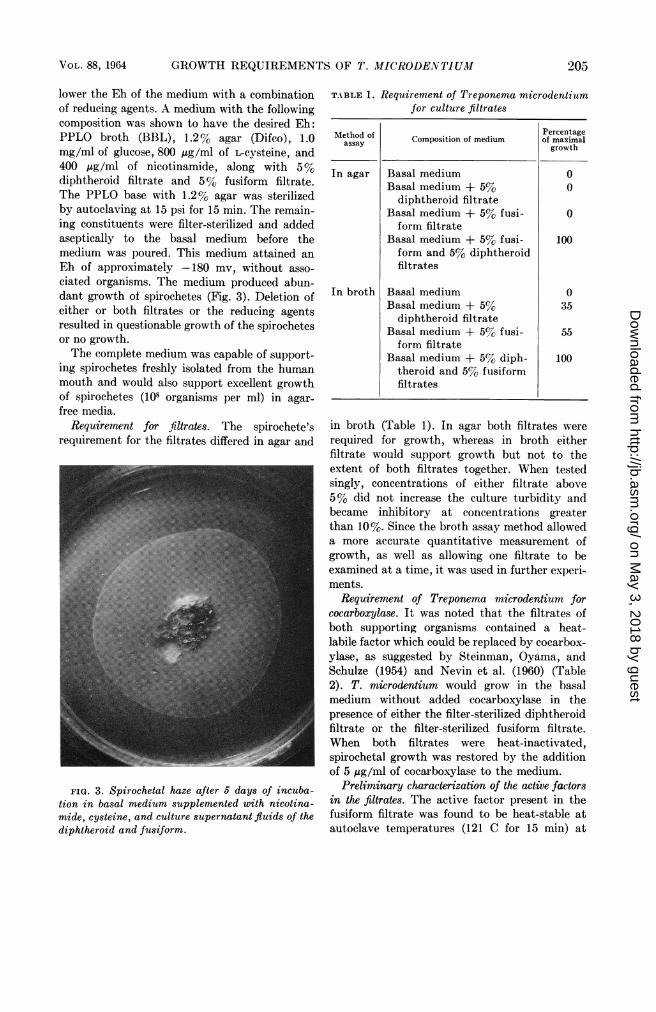
lower the Eh of the medium with a combinationof reducing agents. A medium with the followingcomposition was shown to have the desired Eh:PPLO broth (BBL), 1.2% agar (Difco), 1.0mg/ml of glucose, 800 Aug/ml of L-cysteine, and400 ,ug/ml of nicotinamide, along with 5%diphtheroid filtrate and 5% fusiform filtrate.The PPLO base with 1.2% agar was sterilizedby autoclaving at 15 psi for 15 min. The remain-ing constituents were filter-sterilized and addedaseptically to the basal medium before themedium was poured. This medium attained anEh of approximately -180 mv, without asso-ciated organisms. The medium produced abun-dant growth of spirochetes (Fig. 3). Deletion ofeither or both filtrates or the reducing agentsresulted in questionable growth of the spirochetesor no growth.The complete medium was capable of support-
ing spirochetes freshly isolated from the humanmouth and would also support excellent growthof spirochetes (101 organisms per ml) in agar-free media.
Requirement for filtrates. The spirochete'srequirement for the filtrates differed in agar and
FIG. 3. Spirochetal haze after 5 days of incuba-tion in basal medium supplemented with nicotina-mide, cysteine, and culture supernatant fluids of thediphtheroid and fusiform.
TABLE 1. Requirement of Treponema microdentiumfor culture filtrates
Method of Percentageassay Composition of medium of maximalassay ~~~~~~~~~growth
In agar Basal medium 0Basal medium + 5% 0
diphtheroid filtrateBasal medium + 5% fusi- 0form filtrate
Basal medium + 5% fusi- 100form and 5% diphtheroidfiltrates
In broth Basal medium 0Basal medium + 5% 35
diphtheroid filtrateBasal medium + 5% fusi- 55form filtrate
Basal medium + 5% diph- 100theroid and 5% fusiformfiltrates
in broth (Table 1). In agar both filtrates wererequired for growth, whereas in broth eitherfiltrate would support growth but not to theextent of both filtrates together. When testedsingly, concentrations of either filtrate above5% did not increase the culture turbidity andbecame inhibitory at concentrations greaterthan 10%. Since the broth assay method alloweda more accurate quantitative measurement ofgrowth, as well as allowing one filtrate to beexamined at a time, it was used in further experi-ments.
Requirement of Treponema microdentium forcocarboxylase. It was noted that the filtrates ofboth supporting organisms contained a heat-labile factor which could be replaced by cocarbox-ylase, as suggested by Steinman, Oyama, andSchulze (1954) and Nevin et al. (1960) (Table2). T. microdentium would grow in the basalmedium without added cocarboxylase in thepresence of either the filter-sterilized diphtheroidfiltrate or the filter-sterilized fusiform filtrate.When both filtrates were heat-inactivated,spirochetal growth was restored by the additionof 5 ,ug/ml of cocarboxylase to the medium.
Preliminary characterization of the active factorsin the filtrates. The active factor present in thefusiform filtrate was found to be heat-stable atautoclave temperatures (121 C for 15 min) at
VOL. 88, 1964 205
on May 3, 2018 by guest
http://jb.asm.org/
Dow
nloaded from

SOCRANSKY ET AL.
TABLE 2. Requirement of Treponema microdentiumfor cocarboxylase
Composition of medium Growth
Basal medium* ............................ 0Basal medium* + filter-sterilized diph-
theroid filtrate and autoclaved fusiformfiltrate ................................. +
Basal medium* + filter-sterilized fusiformfiltrate and autoclaved diphtheroid fil-trate ................................. +
Basal medium* + autoclaved diphtheroidand fusiform filtrates .................... 0
Basal medium + autoclaved diphtheroidand fusiform filtrates + 5 IAg/ml of cocar-boxylase ................................ +
* Without cocarboxylase.
E
0-
a
0
0 20 40 6-0 8 0 10 0
p4g/mi SODIUM ISOBUTYRATE
FIG. 4. Effect of concentration of sodium iso-butyrate on the growth of Treponema microdentium.
0.301
E
d
0o20
0*10
0 100 200 300 400
mg/ml POLYAMINE
FIG. 5. Effect of concentration of polyamine on
the growth of Treponema microdentium. Basalmedium supplemented with E3, spermine tetrahydro-chloride; A, spermidine diphosphate; *, putrescinedihydrochloride.
pH 7.0, dialyzable, soluble in ether at pH 2.5,and somewhat soluble in ether at pH 7.0, butnot at pH 10.5. It could be steam-distilled at pH
2.5 but remained in the residue at pH 7.0 and10.5.The active factor present in the diphtheroid
filtrate was also heat-stable at pH 7.0 and wasdialyzable, but was not ether-soluble at pH 2.5and 7.0 and was only slightly soluble at pH10.5. It could not be steam-distilled at any pH.Replacement of the filtrates with known com-
pounds. A variety of acids, including formic,acetic, propionic, butyric, isobutyric, valeric,isovaleric, hexanoic, lactic, pyruvic, succinic,and oleic, were converted to their sodium saltsand added to the basal medium in concentrationsof 0.1, 0.5, 1.0, 3.0, and 10.0 ,ug/ml in attemptsto support spirochetal growth. Only sodiumisobutyrate was capable of supporting spirochetalgrowth; it was required at a level of 2 ,Ag/ml formaximal growth (Fig. 4).
In another series of experiments, the poly-amines, spermine tetrahydrochloride, spermidinephosphate, and putrescine dihydrochloride, wereassayed in attempts to support spirochetalgrowth. All three polyamines were able to supportspirochetal growth, with spermine being mosteffective and putrescine least effective (Fig. 5).The optimal concentrations or spermine tetra-hydrochloride, spermidine phosphate, and putres-cine dihydrochloride were 150, 200, and 250,ug/ml, respectively. The combination of anyone of the polyamines with sodium isobutyratedid not enhance spirochetal growth.Although sodium isobutyrate or one of the
polyamines could support spirochetal growthfor extended passages in broth, these compoundsalone or in combination failed to support growthin Difco agar (Table 3).
Since it was possible that the agar was bindingone or both of the required factors, sodiumisobutyrate and spermine tetrahydrochloridewere added in concentrations up to 1,000 timesthose required for growth in broth. In otherexperiments, the agar was presoaked in 1%concentrations of the growth factors before useor purified by the method of Grabar (1959).The results of these experiments were uniformlynegative. Hardy, Lee, and Nell (1963) demon-strated that anaerobic spirochetes would growas surface or subsurface colonies in the presenceof lowered concentrations of lonagar No. 2.(Consolidated Laboratories Inc., Chicago Heights,Ill.). Replacement of the 1.2% Difco agar in thetest medium with 0.7% Ionagar No. 2 was
206 J. BACTERIOL.
n
.1-
on May 3, 2018 by guest
http://jb.asm.org/
Dow
nloaded from

GROWTH REQUIREMENTS OF T. MICRODENTIUM
successful in overcoming the apparent agarinhibition.
Determinatiorn of the presence of isobutyrate andputrescine in culture filtrates. The filtrates of thetwo supporting organisms were acidified, placedon Celite-sucrose columns, and eluted as de-scribed by Wiseman and Irvin (1957). All frac-tions were collected and assayed for ability tosupport spirochetal growth. Only a single frac-tion, from the fusiform filtrate, was capable ofsupporting spirochetal growth. This fractionmigrated with authentic isobutyric acid and wasassumed to be isobutyric acid.
Polyamines in the filtrates of the supportingorganisms were extracted from alkaline solutioninto t-butanol by the method of Rosenthal andTabor (1956). Samples of the extracts werespotted on Whatman no. 4 chromatographicpaper and developed in three solvent systems:(i) phenol, (ii) n-butanol-acetic acid-water(40:10:50), and (iii) m-cresol-acetic acid-water(50:2:48) (Bremner and Kenten, 1951). Putres-cine was identified in the diphtheroid filtrate onthe basis of having the same RF as authenticputrescine in the three solvent systems employed.No attempt was made to elute the "putrescine"spots to test growth-supporting ability.
Incorporation of isobutyrate and putrescine. TheC14-labeled sodium isobutyrate was taken up bythe spirochetes and incorporated in the cells(Table 4). Approximately two-thirds of theradioactivity of the medium was picked up bythe organisms. This activity was not removedby washing or by exchange with unlabeledsodium isobutyrate. No radioactivity was de-tected in the distillate after steam distillation orin the isobutyric acid band after column chroma-tography. These results suggest that the labeledisobutyrate was taken up by the cells and con-verted to some form other than isobutyrate.The C14 label of putrescine either was not
taken up by the cells or was easily removed bywashing (Table 5). No attempt was made todetermine whether the putrescine was alteredduring growth of the organisms.
DISCUSSION
Several investigators have noted the stimuila-tory or supportive effect of oral organisms on thegrowth of oral spirochetes (Wichelhausen andWichelhausen, 1942; Nevin et al., 1960). Withthe exception of Nevin et al. (1960), no attempt
TABLE 3. Requirement of Treponema microdentiumsfor filtrates in an agar medium
Composition of medium Growth
Basal medium ............................. 0Basal medium + both filtrates............. +Basal medium + sodium isobutyrate.......0Basal medium + polyamine................ 0Basal medium + sodium isobutyrate +polyamine............................... 0
TABLE 4. Incorporation of sodium isobutyrate-1-C14by Treponema microdentium
Counts permin per ml
Fraction of originalculture-
background
Whole culture.......................... 17,955Culture supernatant ................... 5,480Cells................................ 12,300Cells after washing with phosphate
buffer ................ ................ 11,995Cells after 24-hr exchange with unla-
beled isobutyrate.................... 11,920Residue after steam distillation at pH
2.0................................ 12,010Distillate after steam distillation atpH 2.0............................... 0
TABLE 5. Failure of Treponema microdentium totake up putrescine-1, 4-C"4 dihydrochloride
Counts permin per ml
Fraction of originalculture-
background
Whole culture .......................... 18,400Culture supernatant ......... ........... 17,280Cells................................ 609Cells after washing with phosphate
buffer................................ 50
has been made to study this relationship. Theseinvestigators demonstrated that an oral micro-aerophilic diphtheroid was stimulatory to growthof B. vincentii and that the stimulatory effect ofthe diphtheroid could be duplicated by the use ofcocarboxylase or acetyl phosphate. In thepresent investigation, neither acetyl phosphatenor cocarboxylase alone would replace either ofthe filtrates required for spirochetal growth.The two "supporting organisms" in this
investigation appear to control the oxidation-
207VOL. 88, 1964
on May 3, 2018 by guest
http://jb.asm.org/
Dow
nloaded from

SOCRANSKY ET AL.
reduction potential of the medium as well asproviding the spirochetes with "growth factors"consisting of putrescine, isobutyrate, and aheat-labile component which could be replacedby cocarboxylase. The addition of these "growthfactors" as well as reducing agents to PP'LOb1)oth )rovides a medium for the cultivation of T.miicrodentium which is less complex and morereproducible than the ascitic fluid- or rabbitserum-containing media previously employed.'T'he failure of T. miticrodentium to grow in theabove medium in the presence of Difco agar canbe exp)lained by the inhibitory effect of agar asdescribed by Hardy et al. (1963). The fact thatsp)irochetes will grow in the basal medium sup-plemented with both the fusiform and diphtheroidfiltrates in the presence of Difco agar suggests the)ossibility that another factor is lpresent in thesefiltrates which overcomes the inhibitory effect ofthe agar.
Several investigators have recognized therequirement of anaerobic spirochetes for alowered oxidation-reduction potential. For ex-ample, Hardy et al. (1963) stressed the importanceof adding reducing agents to the medium, tocultivate anaerobic spirochetes on solid media.'T'he observations reported in this investigationsuggest that the spirochetes may require a narrowrange of Eh for the initiation of growth. In twoof six Eh experiments, T. nmicrodentium failed togrow in the presence of the supporting organisms.In one instance, the Eh in the central well was70 mv higher than the range in which spirochetalgrowth took place, and in the other instance theEh was 60 mv lower than this range. In addition,T. microdentium would not grow in the presenceof the living diphtheroid and a filtrate of thefusiform, or in the presence of the living fusiformand a filtrate of the diphtheroid. In these experi-ments, the spirochetes had a source of both"chemical growth factors"; however, the Eh ofthe medium was p)robably outside the -185 to-220 mv range in which spirochetal growth wasshown to take place, since the Eh l)roduced bythe diphtheroid growing alone was -90 to -110Mn, whereas the Eh plroduced by the fusiformgrowing alone was -240 to -280 my. Theseobservations contrast with findings. with otheranaerobes which have been found to grow overa fairly wide range of Eh.A requirement for isobutyric acid is not unique
to T. nmicrodentiumi, since several other organisms,
particularly rumen bacteria, have been shown tohave a similar requirement (13ryant and Doetsch1955; Allison, Bryant, and Doetsch, 1958;B3ryant and Robinson, 1961). Wegner and Foster(1960) isolated a Borrelia strain from the rumen,which required both a branched-chain and astraight-chain fatty acid. MIany of the organismsisolated from the rumen could utilize eitherisobutyric or isovaleric acid. However, isovalericacid will not substitute for isobutvric acid as agrowth factor for T. microdentium.The role of isobutyric acid in the metabolism
of T. microdentium has not been elucidated.Allison et al. (1962a, b) demonstrated that, inrumen bacteria, fatty acids function at least inlpart to provide a carbon skeleton for the bio-svnthesis of branched-chain amino acids andlipids. The lipid component included 14-carbonto 17-carbon branched-chain fatty acids andaldehydes.An unusual finding in this study was the
ability of polyamines to replace isobutyric acidas a growth factor for T. microdentium. Bacteriahave been shown to require polyamines forgrowth (Herbst and Snell, 1949; 1\Iartin, Pelezar,and Hansen, 1952; Sneath 1955). In addition,polyamines have been shown to have stabilizingeffects on bacteria (Mager, 1955), lprotoplasts(,Mager, 1959), and mitochondria (Tabor, 1960).The high concentration of the polyamines rie-quired for the growth of T. microdentium sug-gests that they act by stabilizing the organismsrather than by providing some essential metabo-lite. Some substantiation for this hypothesis isprovided by the failure of labeled l)utrescine tobe incorl)orated into the organisms. The avail-able data suggest the possibility that isobutyricacid is incorporated into a structural site re-sponsible for cell integrity. In the presence of asuboptimal concentration of isobutyrate, thepolyamines are necessary to maintain cellularstructure.
ACKNOWLEDGMENTS
This investigation was supported by PublicHealth Service grants D-1305 and D-1471 fromthe National Institute of Dental Research, andby a grant from the Colgate-Palmolive Co.
LITERATURE CITED
ALLISON, M. J., M. P. BRYANT, AND R. N. DOETSCH.1958. Volatile fatty acid growth factor for
2d08 J. BACTERIOL.
on May 3, 2018 by guest
http://jb.asm.org/
Dow
nloaded from

GROWTH REQUIREMENTS OF T. MICRODEATJTIU2I
cellulolytic cocci of bovine rumen. Science128:474-475.
ALLISON, M. J., M. P. BRYANT, AND R. N. DOETSCH.1962a. Studies on the metabolic function ofbranched-chain volatile fatty acids, growthfactors for ruminocci. I. Incorporation ofisovalerate into leucine. J. Bacteriol. 83:523-532.
ALLISON, M. J., M. P. BRYANT, I. KATZ, ANDM. KEENEY. 1962b. Metabolic function ofbranched-chain volatile fatty acids, growthfactors for ruminococci. II. Biosynthesis ofhigher branched-chain fatty acids and alde-hydes. J. Bacteriol. 83:1084-1093.
BERGER, U. 1958. I)ie Treponemen der Mund-hohle und ihre Bedeutung fur die pathogenesder oralen Fusospirochatosen. Beitrage ZurHygiene und Epidemiologie. Barth, Leipzig.
BREMNER, J. M., AND R. H. KENTEN. 1951. Paperchromatography of amines. Biochem. J.49 :651-655.
BRYANT, M. P., AND R. N. 1)OETSCH. 1955. Factorsnecessary for the growth of Bacteroides suc-cinogenes in the volatile acid fraction of rumenfluid. J. Dairy Sci. 38:340-350.
BRYANT, M. P., AND I. M. RoBINSON. 1961. Somenutritional requirements of the genus Rumino-coccus. Appl. Microbiol. 9:91-95.
GRABAR, P. 1959. Immunoelectrophoretic analysis.Methods Biochem. Analy. 7:1-38.
HAMPP, E. G. 1943. Method for routine isolationand cultivation of the smaller oral trepo-nemes. J. Am. Dental Assoc. 30:1066-1075.
HAMPP, E. G., AND T. A. NEVIN. 1959. Substitu-tion of known compounds for ascitic fluid inthe cultivation of Borrelia vincentii. J. Bac-teriol. 77:800-803.
HARDY, P. H., Y. C. LEE, AND E. E. NELL. 1963.Colonial growth of anaerobic spirochetes onsolid media. J. Bacteriol. 86:616-626.
HERBST, E. J., AND E. E. SNELL. 1949. Putrescineand related compounds as growth factors forHemophilus parainfluenzae 7901. J. Biol.Chem. 181:47-54.
LISTGARTEN, M. A., W. J. LOESCHE, AND S. S.SOCRANSKY. 1963. Morphology of Treponemamicrodentium as revealed by electron micros-copy of ultrathin sections. J. Bacteriol.85:932-939.
MACDONALD, J. B., R. M. SUTTON, AND M. L.KNOLL. 1954. The production of fusospirochetalinfections in guinea pigs with recombinedpure cultures. J. Infect. Diseases 95:275-284.
MAGER, J. 1955. Influence of osmotic pressure on
the polyamine requirement of Neisseriaperfiava and Pasteur ella tularensis for growthin defined media. Nature 176:933-934.
MAGER, J. 1959. The stabilizing effect of spermineand related polyamines and bacterial proto-plasts. Biochim. Biophys. Acta 36:529-531.
MARTIN, W. H., JR., M. J. PELCZAR, JR., ANDP. A. HANSEN. 1952. Putrescine as a growthrequirement for Neisseria. Science 116:483-484.
NEVIN, T. A., AND E. G. HAMPP. 1959. Partiallydefined medium for the cultivation of Borreliavincentii. J. Bacteriol. 78:263-266.
NEVIN, T. A., E. G. HAMPP, AND B. V'. DUEY.1960. Interaction between Borrelia vincenthiand an oral diphtheroid. J. Bacteriol. 80:783-786.
ROSEBURY, T., AND G. FOLEY. 1941. Isolation andpure cultivation of the smaller mouth spiro-chetes by an improved method. Proc. Soc.Exptl. Biol. Med. 47:368-374.
ROSEBURY, T., J. B. MACDONALD, S. A. ELLISON,AND S. G. ENGEL. 1951. Media and methodsfor the separation and cultivation of oralspirochetes. Oral Sug. Oral Med. Oral Pathol.4:68-85.
ROSENTHAL, S. M., AND C. W. TABOR. 1956. Thepharmacology of spermine and spermidine.Distribution and excretion. J. Pharmacol.Exptl. Therap. 116:131-138.
SNEATH, P. H. A. 1955. Putrescine as an essentialgrowth factor for a mutant of Aspergillusnidulans. Nature 175:818.
SOCRANSKY, S. S., J. B. MACDONALD, AND S. SAWYER,1959. The cultivation of Treponema micro-dentium as surface colonies. Arch. Oral Biol.1:171-172.
STEINMAN, H. G., U. I. OYAMA, H. 0. SCHULZE.1954. Carbon dioxide, cocarboxylase citro-vorum factor, and coenzyme A as essentialgrowth factors for a saprophytic treponeme(S-69). J. Biol. Chem. 211:327-335.
TABOR, C. W. 1960. Effects of spermine, putres-cine, and cadaverine on mitochondria. Federa-tion Proc. 19:139.
WEGNER, G. H., AND E. M. FOSTER. 1960. Fattyacid requirements of certain rumen bacteria.J. Dairy Sci. 43:566-568.
WICHELHAUSEN, 0. W., AND R. H. WICHELHAUSEN.1942. Cultivation and isolation of mouthspirochetes. J. Dental Res. 21:543-559.
WISEMAN, H. G., AND H. M. IRVIN. 1957. Deter-mination of organic acids in silage. J. Agr.Food Chem. 5:213-215.
N'OL. 88, 1964 209
on May 3, 2018 by guest
http://jb.asm.org/
Dow
nloaded from