Embed Size (px)
Citation preview

Dept of Periodontology-clinical hand-out JSSDCH JSS UNIVERSITY Mysore
1 Clinical hand-out- BY -Dr. Anitha. S
Topic: CLINICAL FEATURES-OF DIFFERENT GINGIVAL CONDISITONS/DISEASES
Further reading: Text book of periodontology- Carranza
Color atlas Periodontology
SL.NO
CLINICAL FEATURES NORMAL CHANGES-underlying tissue change alters the color
DISEASE/CONDITION
1 COLOUR- determined by-
• Vascularity
• Thickness of epithelium
• Degree of keratinization
• Presence/absence of pigmentation (MELANIN)
• PINK/CORAL PINK-
• WITH MELANIN PIGMENTATION
• CHANGES CAN BE MARGINAL, PAPILLARY, DIFFUSE, PATCHY
• Changes vary with intensity of inflammation differ in both nature & distribution, changes start in IDP
• Red/REDDISH BLUE-> vascular proliferation & reduction of keratinization
• BLUE-venous stasis
• PALE-< VASCULARIZATION/EPI
• Associated with systemic diseases-abnormal pigmentation are non specific
• Should stimulate further diagnostic efforts or referral to the appropriate specialist
• ENDOGENOUS ORAL PIGMENTATIONS- caused by melanin, melanin, bilirubin, iron
• Melanin oral pigmentations & are often found found in highly pigmented ethnic groups
• >> melanin pigmentation- 1. Addison’s disease-

Dept of Periodontology-clinical hand-out JSSDCH JSS UNIVERSITY Mysore
2 Clinical hand-out- BY -Dr. Anitha. S
THELIAL KERATINIZATION>
• Red color gradually changes become dull, whitish gray-acute inflammation, gray color-produced by tissue necrosis & is demarcated from adjacent gingival by a thin, sharply defined erythematous zone
• Metallic pigmentation-heavy metals such as arsenic, bismuth, mercury, lead; silver absorbed systemically from therapeutic use/occupational/household environments may discolor the gingival & other areas of the oral mucosa. These changes are rare but still be ruled out in suspected cases
adrenal dysfunction & produce isolated patches of discoloration varying bluish black to brown
2. Peutz-Jehger’s syndrome-produces intestinal polyposis & melanin pigmentation in the oral mucosa & lips
3 Albright’s syndrome(polyostotic fibrous dysplasia & Von Recklinghausen’s disease-neurofibromatosis)-produce melanin pigmentation
• Jaundice-yellowish discoloration-oral mucosa
• Iron-hemochromatosis may produce blue-gray pigmentation of oral mucosa
• Pale gingival-anemia
• Reddish-polycythemia vera, leukemia
• Coal & metal dust, coloring agents- in food or lozenges
• Tobacco-hyperkeratosis of gingival, causes significant > in melanin pigmentation of oral mucosa
• Localized bluish black areas
Dept of Periodontology-clinical hand-out JSSDCH JSS UNIVERSITY Mysore
3 Clinical hand-out- BY -Dr. Anitha. S
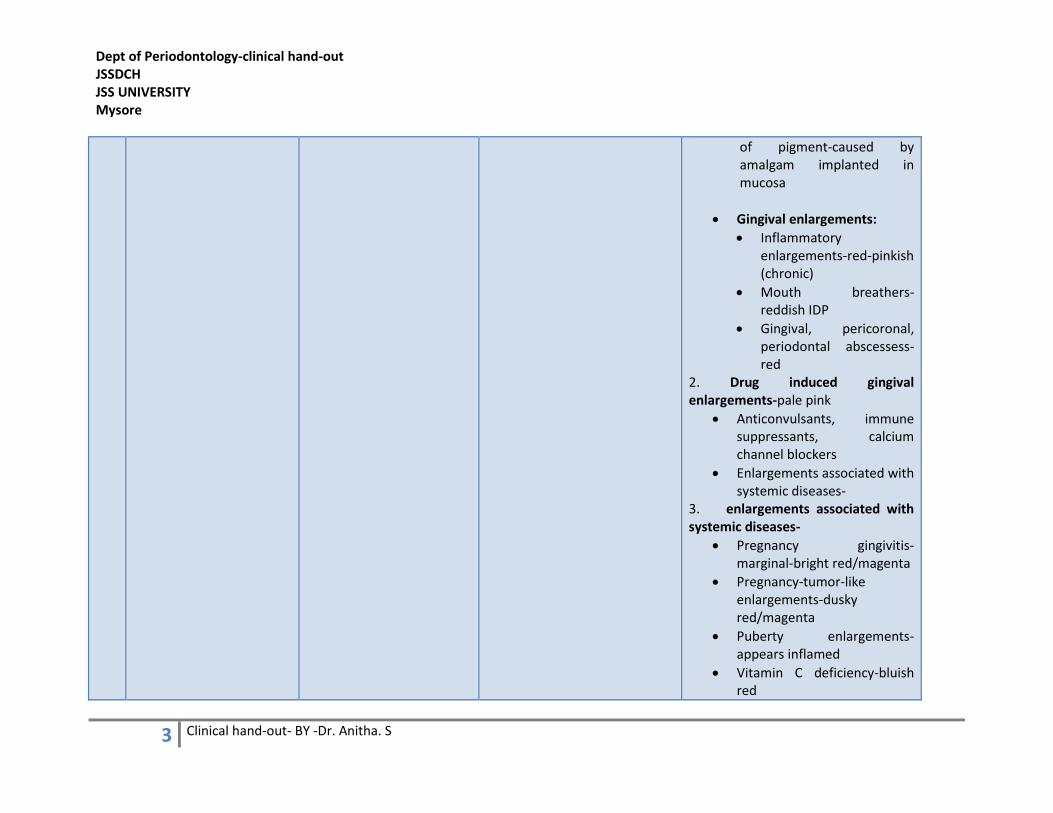
of pigment-caused by amalgam implanted in mucosa
• Gingival enlargements:
• Inflammatory enlargements-red-pinkish (chronic)
• Mouth breathers- reddish IDP
• Gingival, pericoronal, periodontal abscessess-red
2. Drug induced gingival enlargements-pale pink
• Anticonvulsants, immune suppressants, calcium channel blockers
• Enlargements associated with systemic diseases-
3. enlargements associated with systemic diseases-
• Pregnancy gingivitis-marginal-bright red/magenta
• Pregnancy-tumor-like enlargements-dusky red/magenta
• Puberty enlargements- appears inflamed
• Vitamin C deficiency-bluish red

Dept of Periodontology-clinical hand-out JSSDCH JSS UNIVERSITY Mysore
4 Clinical hand-out- BY -Dr. Anitha. S
• Plasma cell gingivitis-red (Solitary plasma cell gingivitis-pink)
• Non specific conditioned enlargement (granuloma pyogenicum)- -similar to pregnancy gingival enlargement
4. Systemic diseases that cause gingival enlargement-
• Granulomatous diseases-
• Wegner’s granulomatosis-reddish purple papillary enlargement
• Sarcoidosis-red 5.neoplastic enlargements- a. Benign -
• Fibroma-pinkish-reddish (ulcerated)
• Peripheral giant cell granuloma-pink-deep red-purplish blue
• Central giant cell granuloma-
• leukoplakia-whitish patch/plaque, does not rub off
• Gingival cyst-pink
• Hemangioma-reddish

Dept of Periodontology-clinical hand-out JSSDCH JSS UNIVERSITY Mysore
5 Clinical hand-out- BY -Dr. Anitha. S
b. Malignant-
• Squamous cell carcinoma-reddish
• Malignant melanoma-darkly pigmented-
c. False enlargements-pinkish ULCERS-HIV/NON HIV infected-depressed gray center surrounded by elevated red border
• PRIMARY HERPETIC GINGIVOSTOMATITIS-RED, ELEVATED VESICLES HALO LIKE MARGINS-greyish vesicles, depressed, yellowish or grayish white central portion; shiny discoloration & edematous enlargement of gingivae
• Necrotising ulcerative gingivitis (NUG)-red, shiny, hemorrhagic, covered with grayish pseudo membranous slough
• Necrotising ulcerative periodontitis (NUP)- red, shiny, hemorrhagic, covered with grayish pseudo membranous slough
Dept of Periodontology-clinical hand-out JSSDCH JSS UNIVERSITY Mysore
6 Clinical hand-out- BY -Dr. Anitha. S
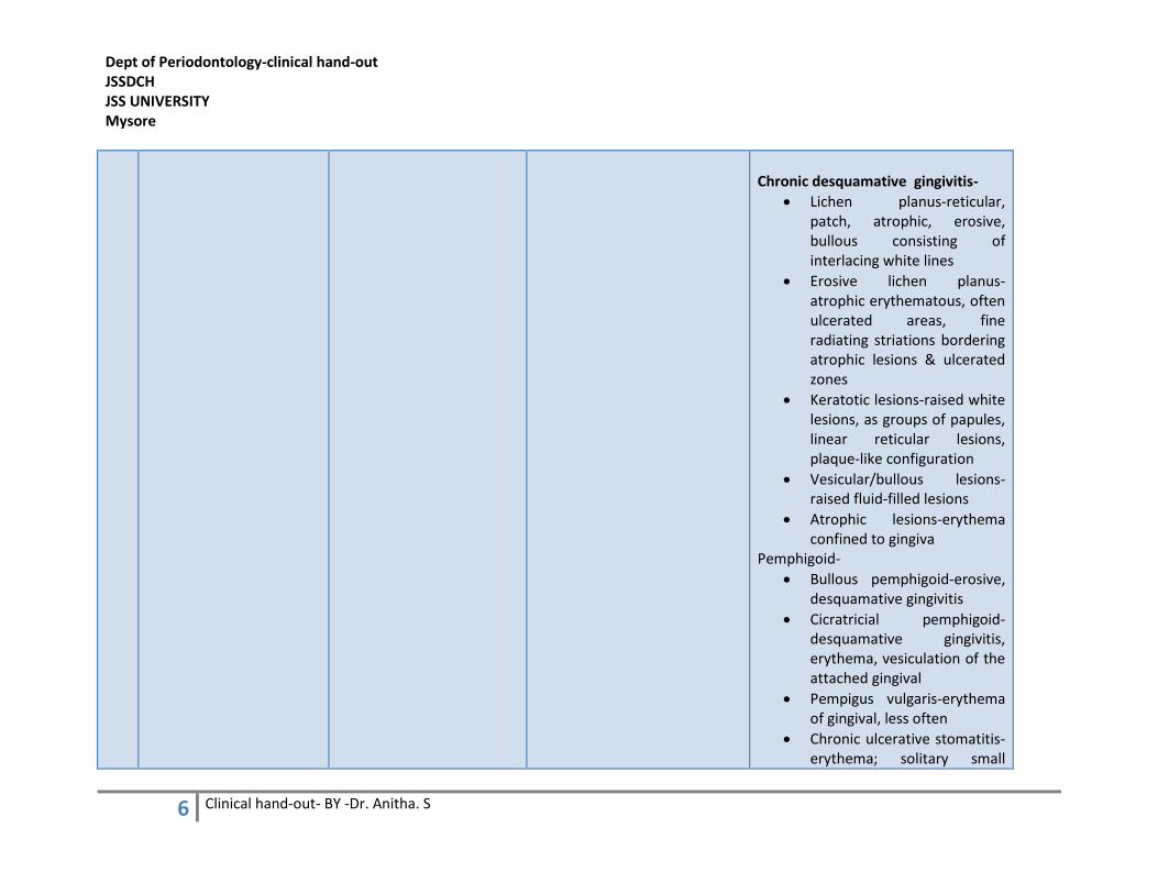
Chronic desquamative gingivitis-
• Lichen planus-reticular, patch, atrophic, erosive, bullous consisting of interlacing white lines
• Erosive lichen planus-atrophic erythematous, often ulcerated areas, fine radiating striations bordering atrophic lesions & ulcerated zones
• Keratotic lesions-raised white lesions, as groups of papules, linear reticular lesions, plaque-like configuration
• Vesicular/bullous lesions-raised fluid-filled lesions
• Atrophic lesions-erythema confined to gingiva
Pemphigoid-
• Bullous pemphigoid-erosive, desquamative gingivitis
• Cicratricial pemphigoid-desquamative gingivitis, erythema, vesiculation of the attached gingival
• Pempigus vulgaris-erythema of gingival, less often
• Chronic ulcerative stomatitis-erythema; solitary small

Dept of Periodontology-clinical hand-out JSSDCH JSS UNIVERSITY Mysore
7 Clinical hand-out- BY -Dr. Anitha. S
blisters, erosions
• Linear immunoglobulin A disease (linear immunoglobulin A dermatosis)-erosive gingivitis, ulcerations
• Dermatitis herpetiformis-clusters of vesicles, papules
• Systemic lupus erythematous-oral ulcerations
• Chronic cutaneous lupus erythematosus-desquamtive lesions
• Erythema multiforme-multiple, large, shallow painful ulcers with an erythematous border-in oral mucosa, including gingival
• Drug eruptions-deep ulcerations with purpuric lesions with gingival often affected
Periodontal pocket-
• Red, bluish red vertical zone extending margin to the alveolar mucosa

Dept of Periodontology-clinical hand-out JSSDCH JSS UNIVERSITY Mysore
8 Clinical hand-out- BY -Dr. Anitha. S
CONTOUR-depends on shape of teeth & their alignment in arc, the location & size of the facial & lingual embrasures
• MG Envelopes the tooth in a collar like fashion & follows a scalloped outline on facial & lingual surfaces
• Forms a straight line along teeth with relatively flat surfaces
• Teeth with pronounced mesio-distal convexity, normal contour is attenuated, & the gingival is located farther apically
• Lingual version-gingiva is horizontal & thickened
• Above mentioned diseases would have loss of contouring
• Mc Call festoon-life-preservator shaped enlargement of the gingival
• Stillman’s cleft- apostrophe shaped, narrow, triangular gingival recession indentation of the gingiva
• Loss of tooth- “saddle-shaped” gingival, loss of contour
•
3 CONSISTENCY-
• collagenous nature of lamina propria & its contiguity with mucoperiosteum of alveolar bone determine the firmness of attached gingiva
• firm & resilient with exception of MG, tightly bound to underlying periosteum of alveolar bone
• DESTRUCTIVE(EDEMATOUS)
• FIBROTIC CHANGES BOTH CHANGES COEXIST
• CALCIFIED CHANGES- ROOT REMNANTS, CALCULUS DEPOSITS REMOVED forcefully gingival during scaling, root remnants,
•

Dept of Periodontology-clinical hand-out JSSDCH JSS UNIVERSITY Mysore
9 Clinical hand-out- BY -Dr. Anitha. S
• gingival fibers contribute to the firmness of the gingival margin
cementum fragments, or cementicles
• Chronic inflammation & fibrosis, occasionally foreign body, giant cell activity, occur in relation to these masses
• Enclosed in an osteoid-like matrix
• Crystalline foreign bodies
• Tooth brushing promotes keratinization of the oral epithelium, enhancing capillary gingival circulation, thickeneing alveolar bone
• Mechanical stimulation by tooth brushing was found to increase proliferative activity of the junctional basal cells in dog gingival by 2.5 times compared with using a scaler
• chronic inflammation-smooth, shiny or firm & nodular-depending on whether dominant
Dept of Periodontology-clinical hand-out JSSDCH JSS UNIVERSITY Mysore
10 Clinical hand-out- BY -Dr. Anitha. S
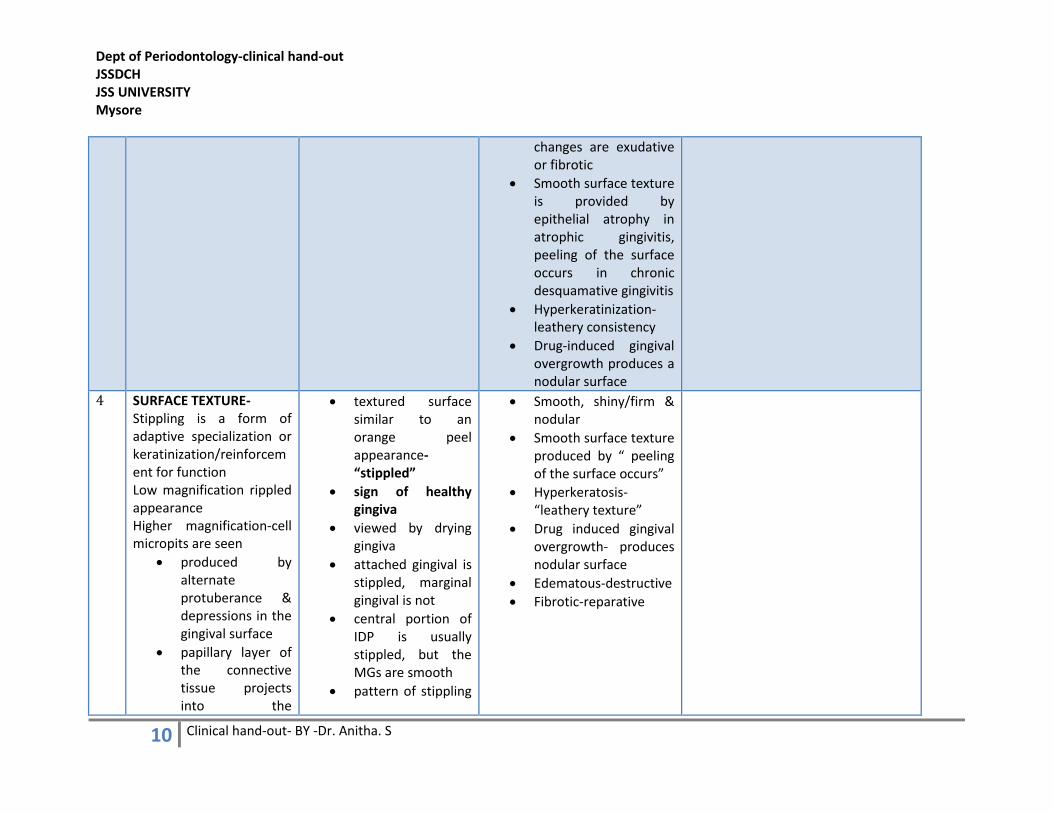
changes are exudative or fibrotic
• Smooth surface texture is provided by epithelial atrophy in atrophic gingivitis, peeling of the surface occurs in chronic desquamative gingivitis
• Hyperkeratinization-leathery consistency
• Drug-induced gingival overgrowth produces a nodular surface
4 SURFACE TEXTURE- Stippling is a form of adaptive specialization or keratinization/reinforcement for function Low magnification rippled appearance Higher magnification-cell micropits are seen
• produced by alternate protuberance & depressions in the gingival surface
• papillary layer of the connective tissue projects into the
• textured surface similar to an orange peel appearance-“stippled”
• sign of healthy gingiva
• viewed by drying gingiva
• attached gingival is stippled, marginal gingival is not
• central portion of IDP is usually stippled, but the MGs are smooth
• pattern of stippling
• Smooth, shiny/firm & nodular
• Smooth surface texture produced by “ peeling of the surface occurs”
• Hyperkeratosis-“leathery texture”
• Drug induced gingival overgrowth- produces nodular surface
• Edematous-destructive
• Fibrotic-reparative

Dept of Periodontology-clinical hand-out JSSDCH JSS UNIVERSITY Mysore
11 Clinical hand-out- BY -Dr. Anitha. S
elevations & the elevated & depressed areas are covered by stratified squamous epithelium
• degree of keratinization & prominence of stippling appear to be related
might vary among individuals & different areas of the same mouth
• less prominent on lingual than facial surfaces & may be absent in some persons
• stippling varies with age
• absent in infancy-
• > stippling gingiva is stimulated with tooth brushing
Stippled
appearance

Dept of Periodontology-clinical hand-out JSSDCH JSS UNIVERSITY Mysore
12 Clinical hand-out- BY -Dr. Anitha. S
5 SIZE
6 POSITION- Refers to the level at which the gingival margin is attached to tooth
• Susceptibility is influenced by position of teeth in arch
• Root-bone angle
• M-D curvature of tooth surface
• Rotated, tilted, or facially displaced teeth, bony plate is thinned out
• The distance b/w apical end of junctional epithelium & crest of alveolus remains constant throughout continuous tooth eruption (1.07mm)
•
Apical migration of gingival is called recession
• physiologic recession-due to aging-not accepted @ present
• pathologic recession-excessive exposure
1. traumatic lesions-chemical, thermal, physical-most common lesions
2. chemical injuries-aspirin, hydrogen peroxide, silver nitrate, phenol, endodontic materials
3. in acute cases- appearance of slough, erosion, or ulceration, & accompanying erythema are common features
Gingival recession due to-
• tooth malposition
• friction from soft tissue-soft tissue ablation
• faulty tooth brushing- gingival abrasion
• abnormal frenum attachment
• TFO
• Inspite of minimal plaque

Dept of Periodontology-clinical hand-out JSSDCH JSS UNIVERSITY Mysore
13 Clinical hand-out- BY -Dr. Anitha. S
7 BLEEDING ON PROBING -----------------
8
EXUDATION --------------------
9 ABSCESS-localised collection of pus
• Gingival abscess- impingement of foreign particle (tooth brush bristle, fish bone, etc) in the gingiva; no involvement of supporting structures other than gingiva
• Periodontal abscess-involvement of supporting structures
• Periapical abscess-associated with a decayed tooth
-----------------------

Dept of Periodontology-clinical hand-out JSSDCH JSS UNIVERSITY Mysore
14 Clinical hand-out- BY -Dr. Anitha. S
Dept of Periodontology-clinical hand-out JSSDCH, JSS university
Topic: CLINICAL FEATURES OF DIFFERENT GINGIVAL CONDITIONS/DISEASES
Wegner’s granulomatosis Plasma cell gingivitis Gingivitis
ANUG copper ingestion- heavy metal pigmentation