Embed Size (px)
Citation preview
Der Einfluss von Clozapin, N-Desmethylclozapin und Chlorpromazin auf die
in-vitro-Produktion von Thromboxan
Dissertation
zur Erlangung des akademischen Grades
Dr. med.
an der Medizinischen Fakultät
der Universität Leipzig
eingereicht von:
Renate Luise Schmidt
geboren am 22.11.1987 in Borna
angefertigt an der:
Claussen-Simon-Stiftungsprofessur für Neurobiologie affektiver Störungen innerhalb der
Klinik und Poliklinik für Psychiatrie und Psychotherapie der Universität Leipzig
Betreuer:
Prof. Dr. med. Hubertus Himmerich
PD Dr. rer. nat. Uta Ceglarek
Beschluss über die Verleihung des Doktorgrades vom
22.07.2014

Meinen Eltern, Sabine und Stephan Schmidt.
Inhaltsverzeichnis
Seite 1. Einführung 2
1.1 Symptomatik und Ätiopathogenese der Schizophrenie 1.2 Eikosanoide und mehrfach ungesättigte Fettsäuren 1.3 Eikosanoide und monoaminerge Veränderungen bei Schizophrenie 1.4 Eikosanoide und immunologische Veränderungen bei Schizophrenie 1.5 Eikosanoide und hormonelle Veränderungen bei Schizophrenie 1.6 Membranlipide und Eikosanoide in schizophrenen Patienten 1.7 Therapeutische Optionen für die Behandlung der Schizophrenie 1.8 Der Vollbluttest als in-vitro-Verfahren für psychopharmakologische
Untersuchungen 2. Aufgabenstellung 26 3. Materialien und Methoden 27
3.1 Versuchspersonen 3.2 Versuchsdurchführung 3.3 TxB2-Messung 3.4 Statistische Analyse 3.5 Ethik
4. Ergebnisse 30 5. Diskussion 34
5.1 Zusammenfassung der Ergebnisse 5.2 Bedeutung der Ergebnisse 5.3 Limitationen der Untersuchung 5.4 Besonderheiten des Vollblutverfahrens in der
psychopharmakologischen Forschung 5.5 Zusammenfassung der Ergebnisinterpretation basierend auf der
Literatur 5.6 Anmerkung
6. Zusammenfassung 37 7. Literaturverzeichnis 39 8. Anlagen 52
8.1 Publikation 1 - Mechanisms of Involvement of Eicosanoids and their Precursors in the Pathophysiology and Treatment of Schizophrenia
8.2 Publikation 2 - Impact of Clozapine, N-Desmethylclozapine and Chlorpromazine on Thromboxane Production in Vitro
9. Erklärung über die eigenständige Abfassung der Arbeit 71 10. Lebenslauf und wissenschaftlicher Werdegang 72
10.1 Lebenslauf 10.2 Publikationen 10.3 Posterpräsentationen
11. Danksagung 74
Bibliographische Zusammenfassung
Schmidt, Renate Luise
Der Einfluss von Clozapin, N-Desmethylclozapin und Chlorpromazin auf die in-vitro-
Produktion von Thromboxan
Universität Leipzig, Monographische Dissertation
74 Seiten, 152 Literaturstellen, 12 Abbildungen, 5 Tabellen
Referat:
Thromboxan A2 (TxA2) und die Aktivierung seines Rezeptors modulieren Vasokonstriktion
und Thrombozytenaggregation ebenso wie dopaminerge und serotonerge Signalwege.
Letztere spielen eine bedeutende Rolle in der Pathophysiologie der Schizophrenie und stellen
somit Zielstrukturen für Antipsychotika (APs) dar.
Da man bereits dem ersten Antipsychotikum (AP) Chlorpromazin (CPZ) eine reduzierende
Wirkung auf die TxA2-Produktion nachweisen konnte, stellten wir die Hypothese auf, dass
auch das AP Clozapin sowie dessen Metabolit N-Desmethylclozapin (NDMC) einen Einfluss
auf die TxA2-Produktion haben könnten.
Wir bestimmten die Konzentration von Thromboxan B2 (TxB2), dem Metaboliten des
instabilen Moleküls TxA2, in stimulierten und unstimulierten Blutproben 10 gesunder
Probandinnen. Hierfür verwendeten wir ein Vollblutverfahren. Als Stimulanz verwendeten
wir das Toxic Shock Syndrome Toxin-1 (TSST-1) oder den monoklonalen Antikörper OKT3
(Muromonab-CD3), der gegen das Oberflächenantigen CD3 gerichtet ist, kombiniert mit dem
monoklonalen Antikörper 5C3, der mit dem Protein CD40 interagiert und es stimuliert. Das
Blut wurde mit den APs CPZ, Clozapin oder NDMC in einer von vier verschiedenen
Konzentrationen versetzt. Zusätzlich wurden die Thromboxanspiegel im Blut ohne Zusatz von
APs unter verschiedenen Stimulationskonditionen gemessen. Unter TSST-1-Stimulation
genauso wie unter OKT3/5C3-Stimulation konnten wir eine signifikante (p<0.05)
Verringerung der TxB2-Produktion durch den Zusatz von Clozapin in den verschiedenen
Konzentrationen feststellen. NDMC führte sowohl ohne Stimulation als auch unter TSST-1-
Stimulation zu verringerten TxB2-Konzentrationen. In sehr niedriger Konzentration reduzierte
CPZ die TxB2-Spiegel in dem unstimulierten ebenso wie in dem mit TSST-1 stimulierten
Blut. Clozapin, NDMC und CPZ könnten also auch über die Modulation der TxA2- und TxB2-
Prduktion das Neurotransmittersystem beeinflussen. Außerdem könnten typische
Nebenwirkungen der AP, wie zum Beispiel die orthostatische Hypotension, aus den
Veränderungen der TxA2- und TxB2-Konzentrationen resultieren.
Abkürzungsverzeichnis
Abkürzungsverzeichnis
15d-PGJ2 15d-Prostaglandin J2
5-HT 5-Hydroxytryptamin
5-HTT 5-Hydroxytryptamintransporter
AA Arachidonsäure
Abb. Abbildung
AGNP Arbeitsgemeinschaft für Neuropsychopharmakologie und
Pharmakopsychiatrie
AK Alzheimer-Krankheit
AP(s) Antipsychotikum / Antipsychtika
BDNF Brain derived neurotrophic factor
cAMP zyklisches Adenosinmonophosphat
CD Cluster of Differentiation
COX Cyclooxgygenase
cps Zählungen pro Sekunde
CPZ Chlorpromazin
DAG Diacylglycerol
DHA Docosahexaensäure
DHET Dihydroxyeicosatriensäure
DSM-IV Diagnostic and Statistical Manual of Mental Disorders, 4. Ausgabe
DTI Diffusions-Tensor-Bildgebung
E-EPA Ethyl-Eicosapentaensäure
EET Epoxyeicosatriensäure
EPA Eicosapentaensäure
ESBA S-Ethylsulfonylbenzoylalanin
GABA γ-Aminobuttersäure
GPCR G-Protein-gekoppelter Rezeptor
HETE Hydroxyeicosatetraensäure
IFN-γ Interferon-gamma
IL Interleukin
IP3 Inositoltriphosphat
IQB Interquartilabstand
KMO Kynurenin-3-Monooxygenase
Abkürzungsverzeichnis
KYNA Kynurensäure
LCMS Flüssigkeits-Chromatographie mit Tandem-Massenspektrometrie
LT Leukotrien
MHC-II Haupthistokompatibilitätskomplex Klasse II
min Minute
MRT Magnet-Resonanz-Tomographie
NDMC N-desmethylclozapin
NMDA N-Methyl-D-Asparaginsäure
OKT3 Anti-CD3-Antikörper
PC Prostazyklin
PET Positronen-Emissions-Tomographie
PG Prostaglandin
PGE1 Prostaglandin E1
PGH2 Prostaglandin H2
PIP2 Phosphatidylinositol-(4,5)-bisphosphat
PPAR-γ Peroxisom-Proliferator-aktivierter Rezeptor Gamma
PUFA(s) mehrfach ungesättigte Fettsäure/Fettsäuren
SKID-I Strukturiertes Klinisches Interview nach DSM-IV
Tab. Tabelle
TDM therapeutic drug monitoring
TP Thromboxan-Rezeptor
TSST-1 Toxic-Schock-Syndrom-Toxin-1
Tx Thromboxan
TxA2 Thromboxan A2
TxB2 Thromboxan B2
ZNS Zentralnervensystem
1. Einführung
2
1. Einführung
1.1 Symptomatik und Ätiopathogenese der Schizophrenie
Mehr als ein Jahrhundert ist vergangen, seit Eugen Bleuler (1857-1939) die durch Emil
Kraepelin (1857-1929) vormals beschriebene „dementia praecox“ überarbeitet und in sein
Konzept der Schizophrenien übernommen hat (Bleuler 1911; Kraepelin 1910).
Trotzdem sind Ätiologie, Neuropathologie und Pathophysiologie der Schizophrenien bis
heute nur schwer fassbar. Die vierte Auflage des Diagnostic and Statistical Manual of Mental
Disorders (DSM-IV) (American Psychiatric Association 2000) und die zehnte Überarbeitung
der International Classification of Diseases and Related Health Problems (ICD-10) (World
Health Organization 1994) definieren zahlreiche spezifische Kriterien für die Diagnose der
Schizophrenie. Nichtsdestotrotz bleibt diese Erkrankungsbezeichnung ein klinisches
Konstrukt, welches durch subjektive Symptome, verschiedene Verhaltensweisen und den
jeweiligen Verlauf der Erkrankung bestimmt wird. Hierdurch stellt die Diagnose der
Schizophrenie noch immer eine klinische Herausforderung dar.
Im klinischen Alltag werden die Symptome der Schizophrenie in „Positiv-“ und
Negativsymptome sowie in „assoziierte“ Symptome hinsichtlich Affektivität und Kognition
unterteilt (Lindenmayer et al. 1995; Kay et al. 1990). Laut DMS-IV (American Psychiatric
Association 2000) stellen die Positivsymptome Wahn und Halluzination und die
Negativsymptome - wie ungeordnete Sprache, desorganisiertes Verhalten, Alogie, affektive
Verflachung und Antriebslosigkeit - charakteristische klinische Symptome dar. Aber auch
kognitive Symptome sind keine Seltenheit. So treten häufig ungeordnetes oder langsames
Denken, Verständnisschwierigkeiten, verminderte Konzentrationsfähigkeit, verringertes
Erinnerungsvermögen, sowie Schwierigkeiten mit dem Ausdrücken und Einordnen von
Gedanken, Gefühlen und Verhalten auf. Auch katatone Symptome - charakterisiert durch
Störungen der Intentionsbewegungen und Körperhaltung - werden beschrieben. Um eine
Schizophrenie zu diagnostizieren, müssen andere Ursachen für die beschriebenen Symptome
ausgeschlossen werden. Hierzu zählen beispielsweise durch einen Substanzmissbrauch
bedingte Störungen, aber auch Gehirnerkrankungen, wie zum Beispiel ein Hirntumor, die
diese Symptome erklären könnten.
In den vergangenen Jahren entwickelten Forscher verschiedene pathophysiologische Theorien
für die Schizophrenie, beruhend auf Biomarkern, die statistisch mit der Krankheit in
Zusammenhang stehen. Dies sind genetische und epigenetische Faktoren, strukturelle
Veränderungen des Gehirns, sowohl auf makroskopischer als auch auf zellarchitektonischer
1. Einführung
3
Ebene, Störungen der neuronalen Konnektivität sowie neurokognitive Dysfunktion und
neurochemische Veränderungen (Jablensky 2006; Bray et al. 2010; Balu et al. 2011).
Nicht zuletzt aufgrund dieser heterogenen biologischen und neuropsychologischen
Forschungsergebnisse vermutet man eine multifaktorielle Ätiopathogenese der Schizophrenie.
Es wird vermutet, dass die Kombination von Umweltfaktoren und biologischen Faktoren zu
einer erhöhten Vulnerabilität für die Erkrankung führt (Lwin et al. 2011). Interessanterweise
besteht bei einigen dieser neurobiologischen Entdeckungen eine Verbindung zu den
Eikosanoiden und in deren Stoffwechsel involvierten Enzymen.
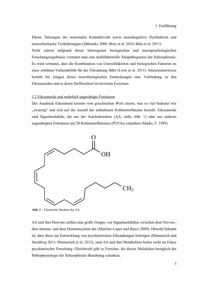
1.2 Eikosanoide und mehrfach ungesättigte Fettsäuren
Der Ausdruck Eikosanoid kommt vom griechischen Wort είκοσι, was so viel bedeutet wie
„zwanzig“ und sich auf die Anzahl der enthaltenen Kohlenstoffatome bezieht. Eikosanoide
sind Signalmoleküle, die aus der Arachidonsäure (AA; siehe Abb. 1) oder aus anderen
ungesättigten Fettsäuren mit 20 Kohlenstoffatomen (PUFAs) entstehen (Marks, F. 1999).
Abb. 1 – Chemische Struktur der AA
AA und ihre Derivate stellen eine große Gruppe von Signalmolekülen zwischen dem Nerven-,
dem Immun- und dem Hormonsystem dar (Malcher-Lopes und Buzzi 2009). Obwohl bekannt
ist, dass diese zur Entwicklung von psychiatrischen Erkrankungen beitragen (Himmerich und
Steinberg 2011; Himmerich et al. 2012), sind AA und ihre Metaboliten bisher nicht im Fokus
psychiatrischer Forschung. Gleichwohl gibt es Forscher, die diesen Molekülen bezüglich der
Pathophysiologie der Schizophrenie Beachtung schenken.
1. Einführung
4
Eikosanoide modulieren Entzündung und Immunität in der Körperperipherie und diverse
Prozesse innerhalb des Zentralen Nervensystems (ZNS). Man geht davon aus, dass das ZNS
AA über den Blutstrom aus der Leber erhält. Die zerebralen Endothelien und die Astrozyten
sind in der Lage, zirkulierende Linoleate zu akkumulieren, um daraus AA zu synthetisieren,
die sie den Neuronen zur Verfügung stellen (Moore et al. 1990, Moore et al. 1991). Neben der
AA zählen Leinölsäure (Acidum linolicum), Eicosapentaensäure (EPA), Docosahexaensäure
(DHA) und α-Linolensäure zu den wichtigen Ausgangsstoffen der Eikosanoide.
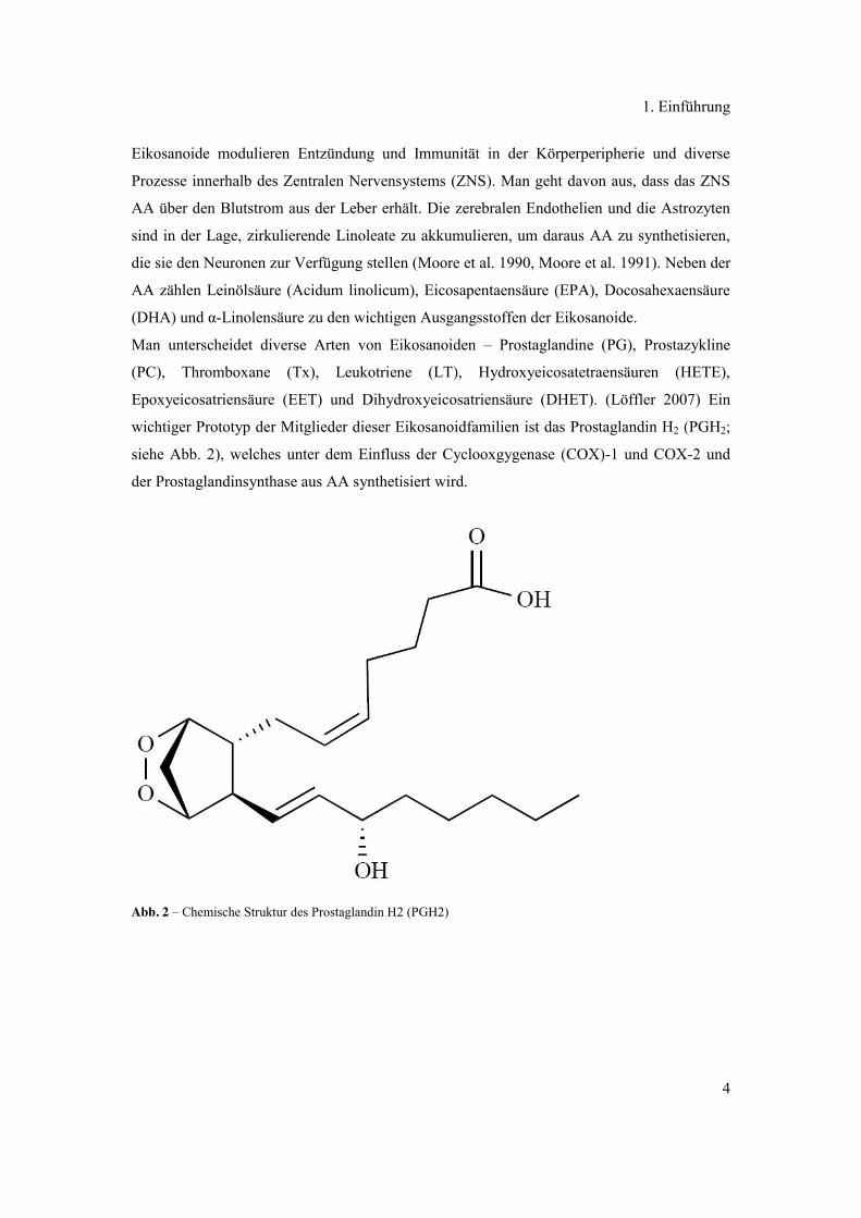
Man unterscheidet diverse Arten von Eikosanoiden – Prostaglandine (PG), Prostazykline
(PC), Thromboxane (Tx), Leukotriene (LT), Hydroxyeicosatetraensäuren (HETE),
Epoxyeicosatriensäure (EET) und Dihydroxyeicosatriensäure (DHET). (Löffler 2007) Ein
wichtiger Prototyp der Mitglieder dieser Eikosanoidfamilien ist das Prostaglandin H2 (PGH2;
siehe Abb. 2), welches unter dem Einfluss der Cyclooxgygenase (COX)-1 und COX-2 und
der Prostaglandinsynthase aus AA synthetisiert wird.
Abb. 2 – Chemische Struktur des Prostaglandin H2 (PGH2)
1. Einführung
5
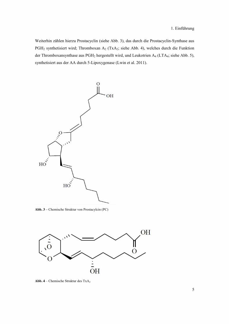
Weiterhin zählen hierzu Prostacyclin (siehe Abb. 3), das durch die Prostacyclin-Synthase aus
PGH2 synthetisiert wird; Thromboxan A2 (TxA2; siehe Abb. 4), welches durch die Funktion
der Thromboxansynthase aus PGH2 hergestellt wird, und Leukotrien A4 (LTA4; siehe Abb. 5),
synthetisiert aus der AA durch 5-Lipoxygenase (Lwin et al. 2011).
Abb. 3 – Chemische Struktur von Prostacylcin (PC)
Abb. 4 – Chemische Struktur des TxA2
1. Einführung
6
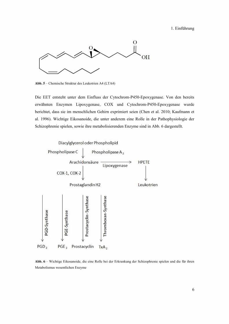
Abb. 5 – Chemische Struktur des Leukotrien A4 (LTA4)
Die EET entsteht unter dem Einfluss der Cytochrom-P450-Epoxygenase. Von den bereits
erwähnten Enzymen Lipoxygenase, COX und Cytochrom-P450-Epoxygenase wurde
berichtet, dass sie im menschlichen Gehirn exprimiert seien (Chen et al. 2010; Kaufmann et
al. 1996). Wichtige Eikosanoide, die unter anderem eine Rolle in der Pathophysiologie der
Schizophrenie spielen, sowie ihre metabolisierenden Enzyme sind in Abb. 6 dargestellt.
Abb. 6 – Wichtige Eikosanoide, die eine Rolle bei der Erkrankung der Schizophrenie spielen und die für ihren
Metabolismus wesentlichen Enzyme
1. Einführung
7
Eikosanoidrezeptoren sind in die Zellmembran integrierte Proteine. Die meisten von ihnen
gehören zur Gruppe der G-Protein-gekoppelten Rezeptoren. Diese Rezeptoren werden im
gesamten Gehirn gefunden und zeigen eine Kopplung an Second Messengers, wie
beispielsweise an zyklisches Adenosinmonophosphat (cAMP) (Coleman et al. 1994). Nicht
nur durch Eikosanoide, sondern auch durch Aktivierung mittels Dopamin- und
Noradrenalinrezeptoren kommt es zur Modulation dieser Second Messengers. Weiterhin
konnte gezeigt werden, dass Eikosanoide auch Einfluss auf die dopaminergen und
serotonergen Signalwege nehmen (Mitsumori et al. 2011; Wacker et al. 2003). Diese spielen
eine entscheidende Rolle in der Pathophysiologie der Schizophrenie. Umgekehrt soll
Dopamin stimulierend auf die Eikosanoidsynthese, in diesem Fall speziell auf die
Thromboxansynthese wirken (Alanko et al. 1991). In der Literatur können einige Beispiele
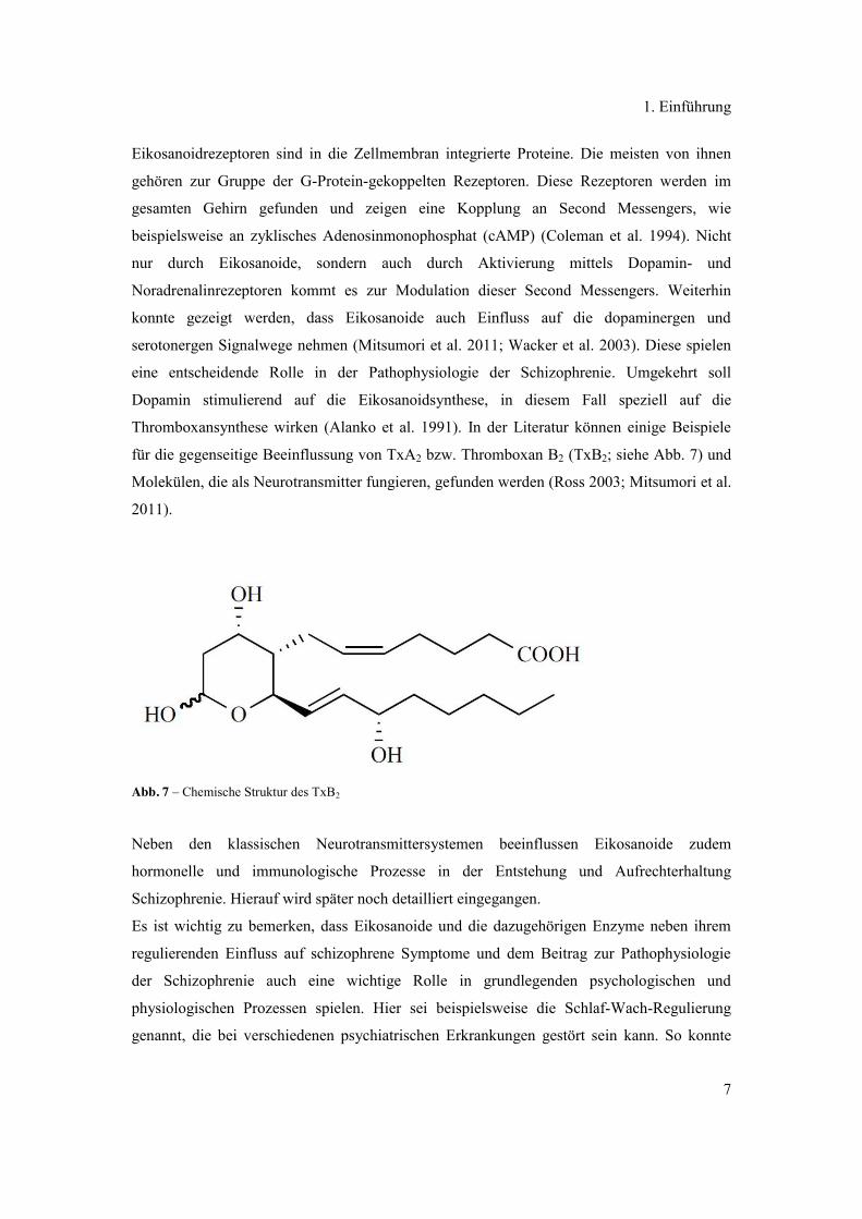
für die gegenseitige Beeinflussung von TxA2 bzw. Thromboxan B2 (TxB2; siehe Abb. 7) und
Molekülen, die als Neurotransmitter fungieren, gefunden werden (Ross 2003; Mitsumori et al.
2011).
Abb. 7 – Chemische Struktur des TxB2
Neben den klassischen Neurotransmittersystemen beeinflussen Eikosanoide zudem
hormonelle und immunologische Prozesse in der Entstehung und Aufrechterhaltung
Schizophrenie. Hierauf wird später noch detailliert eingegangen.
Es ist wichtig zu bemerken, dass Eikosanoide und die dazugehörigen Enzyme neben ihrem
regulierenden Einfluss auf schizophrene Symptome und dem Beitrag zur Pathophysiologie
der Schizophrenie auch eine wichtige Rolle in grundlegenden psychologischen und
physiologischen Prozessen spielen. Hier sei beispielsweise die Schlaf-Wach-Regulierung
genannt, die bei verschiedenen psychiatrischen Erkrankungen gestört sein kann. So konnte
1. Einführung
8
gezeigt werden, dass die COX-2-Aktivität und das Vorhandensein von PGD2 Müdigkeit und
Schlaf induzieren (Weschenfelder et al. 2012).
1.3 Eikosanoide und monoaminerge Veränderungen bei Schizophrenie
In den sechziger Jahren des vergangenen Jahrhunderts postulierte van Rossum (van Rossum
1966) einen Zusammenhang zwischen Schizophrenie und dem dopaminergen System des
Gehirns. Im Laufe der Zeit wurde seine Theorie mehrfach durch Befunde verschiedenster
Forscher unterstützt (Horn und Snyder 1971; Bürki et al. 1975; Lieberman et al. 1987; Davis
et al. 1991) und ist bis heute Ausgangpunkt von Studien (Pogarell et al. 2012; Bauer et al.
2012).
Der vielversprechendste Hinweis auf die Richtigkeit der Dopamin-Hypothese ist, dass
Antipsychotika (APs), welche eine effektive Behandlungsmöglichkeit der Schizophrenie
darstellen, als Blocker der Dopamin-D2-Rezeptoren wirken. Darauf basierend wurde die
Schizophrenie als hyperdopaminerge Störung aufgefasst (Remington et al. 2011). Allerdings
konnte die effektive Wirkung typischer anti-dopaminerger APs hauptsächlich auf
Positivsymptome, jedoch weniger auf Negativsymptome nachgewiesen werden. Hingegen
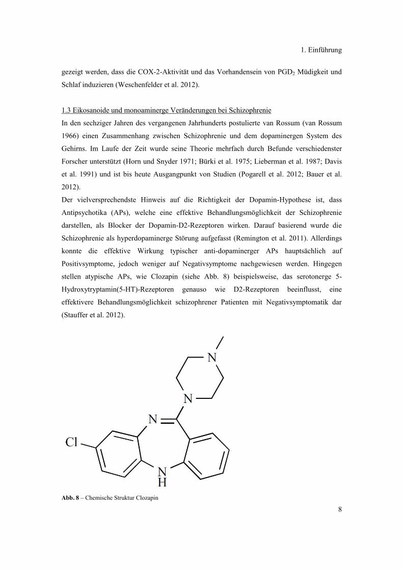
stellen atypische APs, wie Clozapin (siehe Abb. 8) beispielsweise, das serotonerge 5-
Hydroxytryptamin(5-HT)-Rezeptoren genauso wie D2-Rezeptoren beeinflusst, eine
effektivere Behandlungsmöglichkeit schizophrener Patienten mit Negativsymptomatik dar
(Stauffer et al. 2012).
Abb. 8 – Chemische Struktur Clozapin
1. Einführung
9
Neben der Modulation des Dopamin- und 5-HT-Systems zeigt sich bei der Schizophrenie
ebenso eine Veränderung der glutamatergen und der γ-Aminobuttersäure (GABA)-
Signaltransduktion (Di Pietro und Seamans 2007; Lisman et al. 2008). In Studien wurde
mehrfach berichtet, dass Thromboxane Einfluss auf in der Schizophrenie veränderte
Neurotransmittersysteme nehmen (Ross 2003; Mitsumori et al. 2011). Dieser wechselseitige
Einfluss zeigt sich in der Körperperipherie, beispielsweise im kardiovaskulären System,
genauso wie im ZNS. Bezüglich des Serotonins befinden sich auf Thrombozyten
Thromboxanrezeptoren (TP), deren Stimulation durch TxA2 oder TxA2-Rezeptoragonisten zur
Aktivierung der Blättchen und somit zur Freisetzung von Serotonin führt (Moncada und Vane
1978). Veränderungen in der Funktion des 5-HT-Reuptake-Transporters (5-HTT) führen zur
Modulation der TxA2- und TxB2-Produktion innerhalb des Gehirns (Di Pietro und Seamans
2007). Im Gehirn selbst befinden sich TP auf verschiedenen Zelltypen, so auf Astrozyten
(Inagaki und Wada 1994; Gao et al. 1997) und Zellen des zerebrovaskulären Systems (Ansar
et al. 2010). Diese Rezeptoren fanden sich weiterhin in Hirnarealen, wie zum Beispiel dem
Hippokampus, welche eine entscheidende Rolle in der Pathophysiologie der Schizophrenie
spielen (Nishihara et al. 2000). Nicht nur in der Schizophrenie scheinen die Thromboxane von
Bedeutung zu sein. Auch in anderen psychiatrischen Erkrankungen, bei denen es zu gestörter
Funktion der Kognition und des Gedächtnisses kommt, wie beispielsweise bei der Alzheimer-
Krankheit (AK) wird über den Einfluss der Thromboxane diskutiert. Im Zusammenhang
damit werden derzeit TP-Antagonisten als neue Therapieoption gegen AK erforscht (Praticò
2010). Über die TP erleichtert TxA2 die Dopaminfreisetzung im Striatum und ist an der
Saccharose- Aufnahme bei Mäusen beteiligt, wobei es sich um ein Dopamin-abhängiges
motivationales Verhalten handelt (Mitsumori et al. 2011). Weiterhin moduliert TxA2 die
Neurotransmission via glutamaterger N-methyl-d-asparaginsäure (NMDA)- und GABA-
Rezeptoren (Mitsumori et al. 2011). Die klinische Relevanz dieser Forschungsergebnisse ist
zum jetzigen Zeitpunkt allerdings nicht ausreichend wissenschaftlich belegt. Die derzeit
einzige zugängliche Studie hierzu konnte keinen signifikanten Unterschied des TxB2-
Plasmaspiegels bei Schizophrenen im Vergleich zu gesunden Probanden nachweisen
(Dietrich-Muszalska und Olas 2009). Jedoch muss erwähnt werden, dass alle an dieser Studie
beteiligten Patienten mit APs therapiert wurden, sodass ein Unterschied zwischen Patienten
und gesunden Kontrollen möglicherweise durch pharmakologische Wirkungen verschleiert
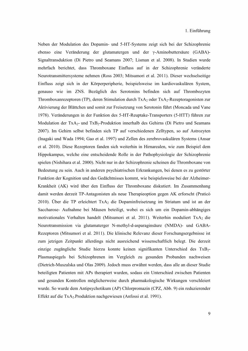
wurde. So wurde dem Antipsychotikum (AP) Chlorpromazin (CPZ, Abb. 9) ein reduzierender
Effekt auf die TxA2-Produktion nachgewiesen (Anfossi et al. 1991).
1. Einführung
10
Abb. 9 – Chemische Struktur CPZ
Weiterhin kommt es durch dieses Medikament zu einer Erhöhung der hemmenden Wirkung
von Phentolamin auf die Thrombozytenaggregation und TxB2-Synthese (Anfossi et al. 1990)
und zu einer Inhibierung der durch Palmitaldehydacetal-Phosphatidsäure induzierten
Plättchenaggregation (Brammer und Maguire 1984). Somit ist vorstellbar, dass auch die
anderen bei dieser Studie als Medikation zugelassenen APs einen Einfluss auf die TxA2-
Eigenschaften genommen haben könnten.
Es ist davon auszugehen, dass CPZ ein Blocker der AA-Kaskade ist (Takayasu et al. 1989)
bzw. die Freisetzung von AA aus Phospholipiden in Thrombozytenmembranen hemmt.
Möglicherweise geschieht dies durch eine Hemmung von Phospholipasen (Ishigooka et al.
1985), wie beispielsweise der Phospholipase A2 (Nikolov 1984). Allerdings muss gesagt
werden, dass die hemmende Wirkung von CPZ auf die TxB2-Produktion nur in bestimmten
Konzentrationen nachgewiesen werden konnte (Chang et al. 1983).
Im Jahr 1983 wiesen Kaiya et al. nach, dass sich der inhibitorische Effekt des Prostaglandin
E1 (PGE1) auf die Thrombozytenaggregation durch APs normalisieren lies. In dieser Studie
untersuchten sie die Plättchenaggregation als Antwort auf verschiedene Stimuli, wie zum
Beispiel Adrenalin, AA, Dopamin und 5-HT, bei 18 unmedizierten Patienten, 13 medizierten
Patienten und 13 Kontrollpersonen (Kaiya et al. 1983).
Die klinische Besserung der Patienten nach APs-Medikation korrelierte signifikant mit einer
erhöhten Aggregation der Thrombozyten als Antwort auf einen AA-Stimulus (Kaiya et al.
1983). Ishigooka et al. vermuteten 1985, dass verschiedene APs, wie Mepyramin,
Promethazin, Phentolamin, und Clozapin die Synthese von Prostaglandinen oder
1. Einführung
11
Thromboxanen hemmen könnten. Deshalb untersuchten sie diese Medikamente im Hinblick
auf ihre Auswirkungen auf die Aggregation von Kaninchenthrombozyten und stellten fest,
dass die durch Kollagen und AA evozierte Plättchenaggregation durch die APs gehemmt
werden konnte (Ishigooka et al. 1985). In einer daran anschließenden Studie konnte eine
dosisabhängige, negative Korrelation zwischen den Konzentrationen von CPZ und
Trifluoperazin einerseits und der Thromboxankonzentration auf der anderen Seite gezeigt
werden (Anfossi et al. 1991).
1.4 Eikosanoide und immunologische Veränderungen bei Schizophrenie
Emil Kraepelin und Julius Wagner Jauregg (1857-1940) wiesen gegen Ende des 19. und
Anfang des 20. Jahrhunderts darauf hin, dass akute Infektionen und Fieber die Symptome der
Schizophrenie verschärfen bzw. verringern könnten (Steinberg und Himmerich 2011;
Himmerich, Kirkby et al. 2010). Heutige immunologische Hypothesen können sich auf
genetische Daten stützen (Debnath et al. 2013). Darüber hinaus konnte gezeigt werden, dass
Komplikationen während der Schwangerschaft und perinatale Infektionen zu einem Anstieg
der Vulnerabilität für ein späteres Auftreten von Schizophrenie führen. Weiterhin weisen
Mütter, deren Kinder in der Adoleszenz an Schizophrenie leiden, einen signifikanten Anstieg
des Interleukin (IL)-8-Plasmaspiegels während des zweiten Trimesters auf (Berthold-
Losleben et al. 2009; Himmerich et al. 2009).
Ferner führt eine infektiöse Erkrankung, wie Toxoplasmose, während der Schwangerschaft zu
einem erhöhten Risiko für das Kind, später eine Schizophrenie zu entwickeln (Berthold-
Losleben et al. 2009).
Möglicherweise aufgrund dieser Infektionskrankheiten zeigen Patienten mit Schizophrenie
eine reduzierte in-vitro-Produktion von IL-2 und verringerte Plasmaspiegel von Interferon
(IFN)-γ, was auf eine reduzierte Typ 1- und eine erhöhte Typ 2- Immunantwort hinweist
(Himmerich et al. 2009). Diese erhöhte Typ 2-Immunantwort geht einher mit erhöhten IL-4-
Spiegeln (Müller et al. 2000), und einer Korrelation zwischen IL-10-Spiegeln im Liquor mit
der Schwere der psychotischen Symptome bei schizophrenen Patienten (van Kammen et al.
1997).
Die immunologischen Veränderungen könnten zur dopaminergen Dysfunktion beitragen, was
zur Dysbalance zwischen glutamaterger und dopaminerger Neurotransmission aufgrund der
erhöhten Produktion von Kynurensäure (KYNA) im Gehirn bei Schizophrenieerkrankten
führen kann (Müller et al. 2011; Müller et al. 2012; Himmerich., Sorge et al. 2012). Außer
durch Infektionen können diese Veränderungen auch durch eine Umstellung des
1. Einführung
12
Eikosanoidstoffwechsel verursacht sein. So zeigen Patienten mit Schizophrenie aufgrund
höherer COX-2-Expression und -Aktivität (Das und Khan 1998) eine erhöhte PGE2-
Produktion (Kaiya et al. 1989; Martínez-Gras et al. 2011). Andere Faktoren, die
möglicherweise zur immunologischen Dysbalance und zu erhöhten PGE2-Produktion bei
schizophrenen Patienten führen, sind der Peroxisom-Proliferator-aktivierte Rezeptor-γ
(PPAR-γ) und das 15d-Prostaglandin J2 (15d-PGJ2) (Kaiya et al. 1989).
PGE2 inhibiert die Produktion von Typ 1-Zytokinen, wie IFN-γ, IL-2 (Simmons et al. 2004)
und stimuliert die Produktion von Typ 2-Zytokinen, wie IL-4, IL-5 und IL-10 (Stolina et al.
2000). Typ-2-Zytokine hemmen die Kynurenin-3-Monooxygenase (KMO), das
geschwindigkeitslimitierende Enzym des Kynureninstoffwechselweges. Wegen der
Hemmung der KMO wird Kynurenin nicht zu 3-Hydroxykynurenin sondern zu KYNA
metabolisiert. Tatsächlich finden sich bei Schizophrenie-Patienten erhöhte Konzentrationen
von KYNA im Gehirn und im Liquor (Erhardt 2001, Nilsson 2005; Linderholm 2012).
Bei einer Studie, die das postmortale Gewebe schizophrener Patienten untersuchte, fand sich
eine signifikante Reduktion der KMO-Genexpression und der KMO-Enzymaktivität im
frontalen Augenfeld (Brodmanareal 8) dieser Patienten (Wonodi et al. 2011). Bei KYNA
handelt es sich um einen Glutamat-NMDA-Rezeptor-Antagonisten, dessen Signalisierung das
glutamaterge/dopaminerge Gleichgewicht im Gehirn stören kann. Somit entsteht eine
dopaminerge Überstimulation, die typisch für die Schizophrenieerkrankung ist (Müller et al.
2011; Müller et al. 2012).
Die Erhöhung der KYNA-Spiegel kann unter anderem zur Verschlechterung der kognitiven
Funktionen führen, während eine Verringerung diese verbessern kann. In den methodisch
wegweisenden Studien von Pocivavsek et al. wurde die Wirkung einer akuten Up-bzw.
Downregulierung der endogenen KYNA auf extrazellulär vorkommendes Glutamat im
Hippokampus untersucht und die Leistung von Sprague-Dawley-Ratten im Morris-
Wasserlabyrinth beobachtet. In dieser Studie reduzierte KYNA den extrazellulären
Glutamatspiegel, während der spezifische Kynurenin-Aminotransferase-II-Inhibitor S-
Ethylsulfonylbenzoylalanin (ESBA) selbigen ansteigen ließ. Vergleichbare Effekte auf den
hippokampalen Glutamat-Spiegel wurden nach intraventrikulärer Applikation des KYNA-
Vorläufers Kynurenin gemessen. Während die Behandlung mit ESBA die Leistung im
Morris-Wasserlabyrinth verbesserte, wurde bei zusätzlicher intraventrikuläre Anwendung von
Kynurenin eine Leistungsminderung beobachtet. Somit konnten diese Studien zeigen, dass
KYNA als endogener Modulator des extrazellulären Glutamats im Hippokampus dient und
folglich für die Reglung hippokampaler kognitiver Funktionen verantwortlich ist (Pocivavsek
1. Einführung
13
et al. 2011). Man vermutet, dass der immunologische Effekt vieler aktueller APs darauf
beruht, dass die immunologische Dysbalance und die Überproduktion der KYNA zum Teil
wieder ausgeglichen werden. Auf dieser Grundlage basierende klinische Studien, die mit
COX-2-Inhibitoren durchgeführt wurden, deuten darauf hin, dass es eine günstige Wirkung
dieser entzündungshemmenden Therapie bei Schizophrenie gibt, insbesondere in frühen
Stadien der Erkrankung (Müller, Krause et al. 2010). Dies deutet auf eine zentrale oder
zumindest zusätzliche Rolle der Eikosanoide, wie PGE2, in der Pathophysiologie der
Schizophrenie hin, und stellt möglicherweise ein neues Ziel für die Behandlung der
Erkrankung dar, worauf im weiteren Verlauf näher eingegangen wird.
Zusammenfassend zeigen schizophrenen Patienten also eine erhöhte PGE2-Produktion
aufgrund erhöhter COX-2-Expression und -aktivität. PGE2 wiederum stimuliert die
Produktion der Typ 2-Zytokine, welche KMO hemmen. Wegen dieser Hemmung wird
Kynurenin nicht zu 3-Hydroxykynurenin metabolisiert, sondern zu KYNA, einem
glutamatergen NMDA-Rezeptor-Antagonisten. Im Gegenzug stört KYNA das glutamaterge/
dopaminerge Gleichgewicht im Gehirn und führt so zur dopaminergen Überstimulation bei
schizophrenen Patienten.
1.5 Eikosanoide und hormonelle Veränderungen bei Schizophrenie
Seit Anfang des 20. Jahrhunderts wurden endokrinologische Aspekte der Pathophysiologie
psychiatrischer Störungen zunehmend interessanter. Hierzu trugen nicht zuletzt die Psychiater
Paul Julius Möbius (1853-1907) und Manfred Bleuler (1903-1994), der Begründer der
Disziplin „endokrine Psychiatrie“ bei (Himmerich, Steinberg 2011). Im speziellen Fall der
Schizophrenie, könnten Sexualhormone, wie Östrogen, aber auch Schilddrüsenhormone eine
zentrale Rolle spielen, wie ich nachfolgend erläutern werde.
Epidemiologische Erkenntnisse und Life-Cycle-Daten weisen auf signifikante Unterschiede in
der Häufigkeit und dem Verlauf der Schizophrenie zwischen Männern und Frauen hin.
Hierdurch schließt man auf eine protektive Rolle des Östrogens. Präklinische in-vitro und in-
vivo-Untersuchungen haben bestätigt, dass Östradiol durch die Interaktion mit zentralen
Neurotransmitter-Systemen an der Pathogenese der Schizophrenie beteiligt ist. Obwohl die
Ergebnisse aus randomisierten, kontrollierten Studien zum antipsychotischen Potential von
Östrogen vielversprechend waren, gibt es bisher noch keine praxiswirksame Umsetzung der
Östrogen-Hypothese (Kulkarni et al. 2012).
Schilddrüsenhormone sind sowohl während der Entwicklung als auch im adulten Gehirn von
entscheidender Bedeutung. Schwankungen der Schilddrüsenhormonspiegel zu verschiedenen
1. Einführung
14
Zeitpunkten während der Entwicklung, aber auch während des späteren Lebens haben
Auswirkungen auf eine eventuelle Manifestationen von psychiatrischen Erkrankungen und
das Ansprechen auf deren Behandlung. Es wird beschrieben, dass Wechselbeziehungen
zwischen der thyreotropen Achse und den wichtigen Neurotransmittersystemen,
einschließlich serotonerger, dopaminergen, glutamaterger und GABAerger Netzwerke, an der
Pathophysiologie der Schizophrenie beteiligt sind. Dies deutet auf eine Deregulierung der
Schilddrüsenhormone als häufiges Merkmal der Schizophrenie hin (Santos et al. 2012).
Interessant ist, dass Östradiol dafür bekannt ist, die Aktivität der COX-1 und -2 zu erhöhen
und somit den Prostaglandin-Stoffwechsel zu modulieren (Amateau und McCarthy 2004).
Weiterhin konnte gezeigt werden, dass die Östradiol-induzierte Prostaglandinsynthese
Einfluss auf die glutamaterge Neurotransmission nimmt (McCarthy 2010). Aus Tierversuchen
gibt es Hinweise, dass Schilddrüsenhormone außerdem die COX-2-Aktivität und den
Eikosanoidstoffwechsel beeinflussen (Carey et al. 2008). Allerdings sind derzeit nicht
genügend Daten vorhanden, um eine Hypothese über die die Verknüpfung von Eikosanoiden,
ihres modulierenden Einflusses auf bestimmte Hormone und der Pathophysiologie der
Schizophrenie zu postulieren. Eine sinnvolle Interpretation dieser Ergebnisse ist, dass
Östrogene und Schilddrüsenhormone über die Induktion von COX-1 und -2 und der damit
verbundenen Erhöhung der PGE2-Synthese zur Manifestation von Schizophrenie führen
können.
1.6 Membranlipide und Eikosanoide bei schizophrenen Patienten
Wie bereits oben erwähnt, spielen Fettsäuren und im Speziellen der PUFA-Stoffwechsel bei
der Entwicklung von Schizophrenie eine Schlüsselrolle, was auch in vielen
wissenschaftlichen Arbeiten bereits mehrfach gezeigt wurde (Ross 2003). PUFAs sind nicht
nur ein wesentlicher Bestandteil der Zellen im Gehirn, sondern auch in allen anderen
Körperzellen. Wesentliche Bestandteile der Zellmembranen sind Phospholipide, die eine
Lipid-Doppelschicht bilden. Phospholipide geben der Membran ihre Struktur und Integrität,
und stellen die Lipid-Umgebung für zahlreiche Membran-gebundene und integrale Proteine,
einschließlich Neurotransmitter-Rezeptoren, Ionenkanäle und Transporter bereit (Ross 2003).
Die meisten Phospholipide enthalten Diglycerid, eine Phosphat-Gruppe und ein einfaches
organisches Molekül, wie beispielsweise Cholin. Membranphospholipide, wie beispielsweise
Phosphatidylinositol-(4,5)-bisphosphat (PIP2), haben üblicherweise eine PUFA in Position 2
(siehe Abb. 10).
1. Einführung
15
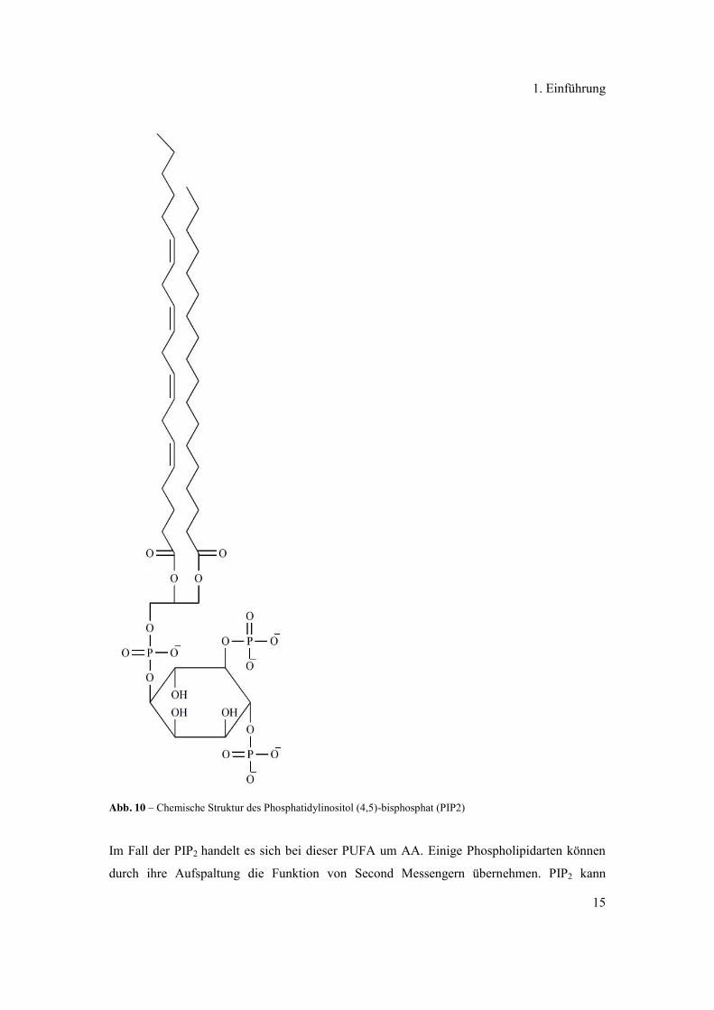
Abb. 10 – Chemische Struktur des Phosphatidylinositol (4,5)-bisphosphat (PIP2)
Im Fall der PIP2 handelt es sich bei dieser PUFA um AA. Einige Phospholipidarten können
durch ihre Aufspaltung die Funktion von Second Messengern übernehmen. PIP2 kann
1. Einführung
16
beispielsweise durch das Enzym Phospholipase C in Inositoltriphosphat (IP3) und
Diacylglycerol (DAG) gespalten werden, welche auf Reize durch verschiedene Stimuli die
Funktion von G-Proteinen ausführen (Choi et al. 2005). Beispielsweise sind 5-HT-Rezeptoren
und der metabotrope Glutamat-Rezeptor G-Protein-gekoppelte Rezeptoren (GPCR)
(Fredriksson et al. 2003). Beide Rezeptorarten spielen eine Rolle in der Pathophysiologie der
Schizophrenie (Ohuoha et al. 1993; Goff und Coyle 2001). PIP2 leitet afferente Signale weiter,
die es über einen GPCR, wie den 5-HT-Rezeptor erhält. Durch die Spaltung von PIP2 durch
eine Lipase kann es zusätzlich zur Freisetzung von AA kommen. In diesem Fall wirkt AA
PIP2 entgegen. Dabei verringert AA zwar die Verfügbarkeit spannungsabhängiger
Kalziumkanäle, erhöht aber deren Sensitivität (Roberts-Crowley et al. 2009). Aufgrund der
Funktion der AA als Ausgangsmolekül für die Prostaglandinsynthese liegt nahe, dass der
Phospholipidmetabolismus die Prostaglandinsignalübertragung beeinflusst.
Auf der Grundlage klinischer und biochemischer Beobachtungen stellte Horrobin die
sogenannte „Phospholipid-Membran-Hypothese der Schizophrenie“ auf, in der er postulierte,
die Schizophrenie sei eine Erkrankung, die durch einen abnormalen Metabolismus und
gestörte Phospholipidmembran-Strukturen im Gehirn und in anderen Geweben charakterisiert
sei (Horrobin et al. 1994; Horrobin 1998). Tatsächlich gibt es Hinweise darauf, dass
Schizophrenie mit einem abnormalen Stoffwechsel der Membranphospholipide und der
mehrfach ungesättigten Fettsäuren, insbesondere der AA und ihrer Derivate verbunden sein
kann (Fenton et al. 2000; Ross 2003). Horrobin und seine Mitarbeiter, ebenso wie andere
Forschungsgruppen, fanden heraus, dass bei Schizophreniepatienten im Vergleich zu
gesunden Probanden verringerte PUFA-Konzentrationen im Serum, im Plasma, in peripherem
Gewebe und post mortem im Gehirn zu messen sind (Horrobin et al. 1994; Yao et al. 2000;
Landén et al. 2002; McNamara et al. 2007; Freeman et al. 2006).
Darüber hinaus wurden immer wieder Anomalien der weißen Substanz bei
Schizophrenieerkrankten nachgewiesen (Di et al. 2009; Connor et al. 2011). Dieser Befund ist
besonders für die Rolle der PUFAs in der Pathophysiologie von Schizophrenie interessant, da
PUFAs für die Myelinisierungsaktivität bzw. die Aufrechterhaltung der Myelinisierung durch
Oligodendrozyten notwendig zu sein scheinen.
Tatsächlich konnten Peters et al. (Peters et al. 2009; Peters et al. 2013) mit Hilfe von
Diffusions-Tensor-Bildgebung (DTI) in einer Magnet-Resonanz-Tomographie(MRT)-Studie
eine signifikante Korrelation zwischen Gesamt-PUFA-Konzentration und Myelinisierung
eines frontotemporalen Trakts weißer Substanz in neuerkrankten Schizophrenie-Patienten
nachweisen und später diese Ergebnisse auch replizieren. Aus diesem Grund könnten PUFAs
1. Einführung
17
ein wichtiges therapeutisches Ziel in der Behandlung der Schizophrenie darstellen. In der
Literatur wurde erkennbar, dass die meisten Studien EPA und seinen Ester Ethyl-
Eicosapentaensäure (E-EPA) zur Behandlung von Schizophrenie, meist zusätzlich zu einem
AP, einsetzten. Jedoch sind die Ergebnisse systematischer Übersichtsarbeiten nicht schlüssig
(Berger et al. 2006). Eine ausführliche Zusammenfassung der Studien über die therapeutische
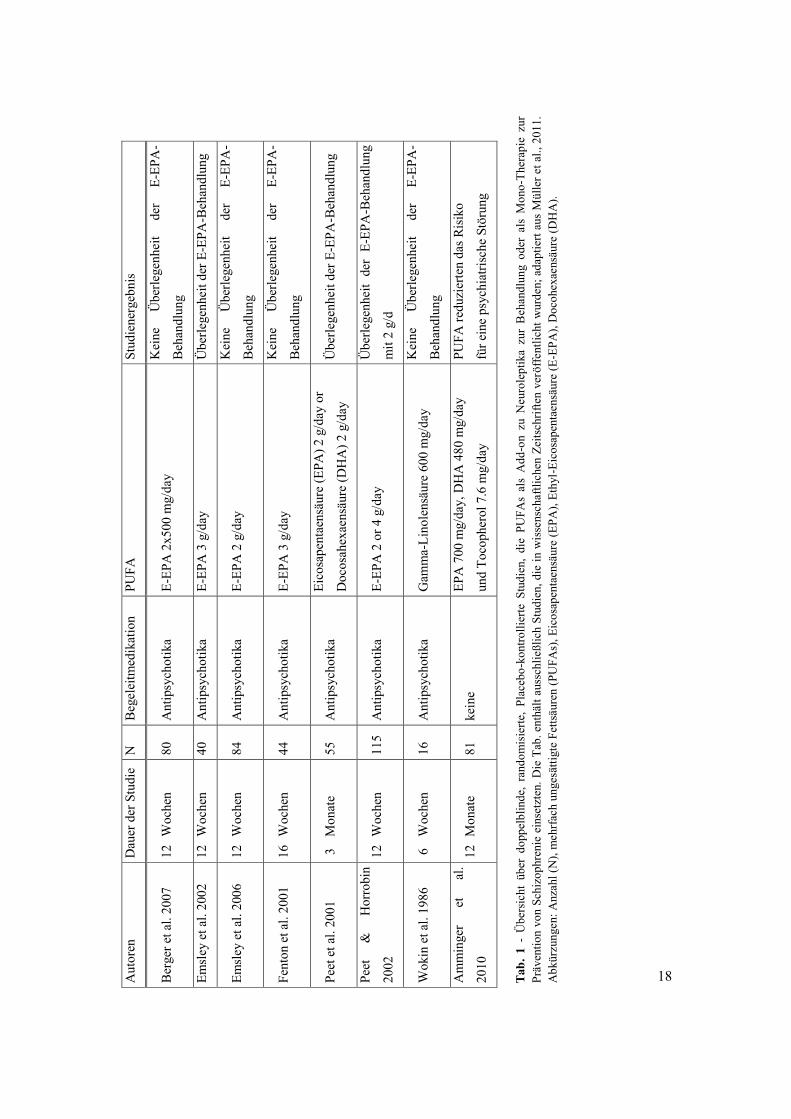
Wirksamkeit von PUFAs, speziell von E-EPA ist in Tabelle (Tab.) 1 dargestellt.
.
Aut
oren
D
auer
der
Stu
die
N
Beg
elei
tmed
ikat
ion
PUFA
St
udie
nerg
ebni
s
Ber
ger e
t al.
2007
12
Woc
hen
80
Ant
ipsy
chot
ika
E-EP
A 2
x500
mg/
day
Kei
ne
Übe
rlege
nhei
t de
r E-
EPA
-
Beh
andl
ung
Emsl
ey e
t al.
2002
12
Woc
hen
40
Ant
ipsy
chot
ika
E-EP
A 3
g/d
ay
Übe
rlege
nhei
t der
E-E
PA-B
ehan
dlun
g
Emsl
ey e
t al.
2006
12
Woc
hen
84
Ant
ipsy
chot
ika
E-EP
A 2
g/d
ay
Kei
ne
Übe
rlege
nhei
t de
r E-
EPA
-
Beh
andl
ung
Fent
on e
t al.
2001
16
Woc
hen
44
Ant
ipsy
chot
ika
E-EP
A 3
g/d
ay
Kei
ne
Übe
rlege
nhei
t de
r E-
EPA
-
Beh
andl
ung
Peet
et a
l. 20
01
3
Mon
ate
55
Ant
ipsy
chot
ika
Eico
sape
ntae
nsäu
re (E
PA) 2
g/d
ay o
r D
ocos
ahex
aens
äure
(DH
A) 2
g/d
ay
Übe
rlege
nhei
t der
E-E
PA-B
ehan
dlun
g
Peet
&
H
orro
bin
2002
12
Woc
hen
115
Ant
ipsy
chot
ika
E-EP
A 2
or 4
g/d
ay
Übe
rlege
nhei
t de
r E-
EPA
-Beh
andl
ung
mit
2 g/
d
Wok
in e
t al.
1986
6
W
oche
n 16
A
ntip
sych
otik
a G
amm
a-Li
nole
nsäu
re 6
00 m
g/da
y K
eine
Ü
berle
genh
eit
der
E-EP
A-
Beh
andl
ung
Am
min
ger
et
al.
2010
12
Mon
ate
81
kein
e EP
A 7
00 m
g/da
y, D
HA
480
mg/
day
un
d To
coph
erol
7.6
mg/
day
PUFA
redu
zier
ten
das R
isik
o
für e
ine
psyc
hiat
risch
e St
örun
g Ta
b. 1
- Ü
bers
icht
übe
r do
ppel
blin
de,
rand
omis
ierte
, Pl
aceb
o-ko
ntro
llier
te S
tudi
en,
die
PUFA
s al
s A
dd-o
n zu
Neu
role
ptik
a zu
r B
ehan
dlun
g od
er a
ls M
ono-
Ther
apie
zur
Pr
även
tion
von
Schi
zoph
reni
e ei
nset
zten
. Die
Tab
. ent
hält
auss
chlie
ßlic
h St
udie
n, d
ie in
wis
sens
chaf
tlich
en Z
eits
chrif
ten
verö
ffen
tlich
t wur
den;
ada
ptie
rt au
s M
ülle
r et a
l., 2
011.
A
bkür
zung
en: A
nzah
l (N
), m
ehrf
ach
unge
sätti
gte
Fetts
äure
n (P
UFA
s), E
icos
apen
taen
säur
e (E
PA),
Ethy
l-Ei
cosa
pent
aens
äure
(E-E
PA),
Doc
ohex
aens
äure
(DH
A).
18
1. Einführung
19
In einer Placebo-kontrollierten Studie konnte gezeigt werden, dass PUFAs zudem eine
präventive Wirkung auf psychotische Störungen haben (Amminger et al. 2010).
Wie bereits im Abschnitt über Eikosanoide und monoaminerge Veränderungen bei
Schizophrenie erwähnt, spielt die Dopamin-Signalübertragung eine entscheidende Rolle bei
dieser Erkrankung. Obwohl die Ergebnisse vorläufig sind, ist es interessant zu wissen, dass
die dopaminerge Funktion auch in Tierstudien durch PUFAs beeinflusst wurde (Zimmer et al.
1998; Zimmer et al. 2002). Der Wachstumsfaktor Brain derived neurotrophic factor (BDNF)
ist ein weiteres Molekül mit einem potenziell wichtigen Einfluss auf die Entwicklung von
Schizophrenie (Kordi-Tamandani et al. 2012; Martinotti et al. 2012). BDNF ist für die
neuronale Plastizität und die Entwicklung des ZNS essentiell und ist weithin an der
Entstehung psychiatrischer Erkrankungen beteiligt (Autry und Monteggia 2012). Es konnte
gezeigt werden, dass diätetisch zugeführte PUFAs zu einer Erhöhung der BDNF-
Konzentration im Hippokampus von Ratten führten (Wu et al. 2004; Wu et al. 2008). Es
wurde berichtet, dass der bei unmedizierten schizophrenen Menschen erhöhte AA-
Metabolismus in Thrombozyten durch die Behandlung mit APs normalisiert werden konnte.
So beobachteten beispielsweise Das und Khan eine AA-induzierte Chemilumineszenz in
Blutproben von acht neu diagnostizierten unmedizierten schizophrenen Patienten. Nach der
Behandlung mit Sulpirid oder Haloperidol verringerte sich die Chemilumineszenz (Das und
Khan 1998). Die Autoren folgerten, dass die Neuroleptika den AA-Stoffwechsel beeinflussten
(Martínez-Gras et al. 2011). Sethom et al. untersuchten ebenfalls periphere Blutzellen. Sie
untersuchten mittels Gaschromatographie das Profil der Membran-PUFAs schizophrener
Patienten vor und nach der Behandlung mit APs. Allerdings fanden sie deutlich geringere
Gesamt-PUFA-Konzentrationen in den Erythrozytenmembranen schizophrener Patienten
(Sethom et al. 2010).
Zusammenfassend lässt sich sagen, dass es einige Hinweise darauf gibt, dass PUFAs eine
wichtige Vermittlerrolle in der Pathophysiologie der Schizophrenie spielen. Jedoch sind
eindeutige Schlussfolgerungen aufgrund der widersprüchlichen Ergebnisse in der aktuellen
Literatur nur begrenzt möglich (siehe Tab. 1).
1.7 Therapeutische Optionen für die Behandlung der Schizophrenie
Aufgrund der hier diskutierten Studien scheinen PUFA- und Eikosanoidstoffwechsel eine
entscheidende Rolle in der Pathophysiologie sowie in der Behandlung der Schizophrenie zu
spielen. Tab. 2 zeigt die verschiedenen Erkenntnisse über PUFAs, Eikosanoide und die
Enzyme des Eikosanoidstoffwechsels, die in dieser Monographie beschrieben sind.

20
M
olek
üle
Patie
nten
mit
Schi
zoph
reni
e W
irkun
g de
r Ant
ipsy
chot
ika
Ref
eren
zen
PUFAs PU
FAs
Ver
ringe
rte G
esam
t- PU
FA-K
onze
ntra
tion
im
Seru
m, P
lasm
a, p
erip
here
n G
eweb
e,
Eryt
hroz
yten
mem
bran
en u
nd G
ehirn
sc
hizo
phre
ner P
atie
nten
gef
unde
n, a
ber
erhö
hte
AA
-Akt
ivitä
t.
Beh
andl
ung
mit
Sulp
irid
oder
Hal
oper
idol
ve
rrin
gert
AA
-Akt
ivitä
t. M
artín
ez-G
ras e
t al.
2011
; Hor
robi
n et
al.
1994
; Yao
et a
l. 20
00; L
andé
n et
al.
2002
; McN
amar
a et
al.
2007
; Fre
eman
et a
l. 20
06; S
etho
m
et a
l. 20
10
Eikosanoide
TxA
2 un
d Tx
B2
Kei
n si
gnifi
kant
er U
nter
schi
ed z
wis
chen
TxB
2-Pl
asm
aspi
egel
bei
med
izie
rten
Schi
zoph
reni
e-Pa
tient
en u
nd g
esun
den
Kon
trollp
erso
nen.
Beh
andl
ung
mit
Chl
orpr
omaz
in o
der
Trifl
uope
razi
n ve
rrin
gert
TxA
2-Kon
zent
ratio
n,
Beh
andl
ung
mit
Clo
zapi
n, N
DM
C,
Chl
orpr
omaz
in re
duzi
ert T
xB2-S
ynth
ese
Die
trich
-Mus
zals
ka e
t al.
2009
; A
nfos
si e
t al.
1991
PGE 1
V
erm
inde
rte H
emm
wirk
ung
auf
Thro
mbo
zyte
nagg
rega
tion.
K
aiya
et a
l. 19
83
PGE 2
Er
höht
e Pr
oduk
tion
bei s
chiz
ophr
enen
Pa
tient
en.
Que
tiapi
n re
duzi
ert P
GE 2
-Spi
egel
in M
äuse
n,
Ola
nzap
in v
errin
gert
PGE 2
-Kon
zent
ratio
nen
im
Rat
tenh
irn,
PGE 2
wird
als
neu
er V
erm
ittle
r der
an
tipsy
chot
isch
en B
ehan
dlun
g vo
rges
chla
gen
Am
min
ger e
t al.
2010
; Kim
et a
l. 20
12; C
heon
et a
l. 20
11; A
dkin
s et
al. 2
012
Enzyme
CO
X-1
un
d C
OX
-2
Erhö
hte
Expr
essi
on u
nd A
ktiv
ität v
on C
OX
-1.
Hal
oper
idol
ver
ringe
rt st
riata
le C
OX
-2-
Expr
essi
on b
ei R
atte
n.
van
Kam
men
et a
l. 19
97, M
ülle
r, N
., M
yint
, A.-M
. et a
l. 20
12; M
artín
ez-
Gra
s et a
l. 20
11; M
arin
et a
l. 20
07
Tab.
2 –
Nac
hwei
se f
ür d
ie B
edeu
tung
der
Eik
osan
oide
bei
der
Sch
izop
hren
ie. D
ie T
ab. z
eigt
wis
sens
chaf
tlich
e Er
kenn
tnis
se z
ur B
etei
ligun
g vo
n PU
FAs,
Eiko
sano
iden
und
En
zym
en i
m E
ikos
anoi
dsto
ffw
echs
el b
ei P
atie
nten
mit
Schi
zoph
reni
e un
d di
e W
irkun
g vo
n N
euro
lept
ika
auf
dies
e M
olek
üle.
Für
Ein
zelh
eite
n si
ehe
Text
. A
bkür
zung
en:
Meh
rfac
h un
gesä
ttigt
e Fe
ttsäu
ren
(PU
FAs)
, A
rach
idon
säur
e (A
A),
Thro
mbo
xan
A2
(TxA
2),
Thro
mbo
xan
B2
(TxB
2),
Pros
tagl
andi
n E 1
(PG
E 1),
Pros
tagl
andi
n E 2
(PG
E 2),
Cyc
loox
ygen
ase
1 (C
OX
-1),
Cyc
loox
ygen
ase
2 (C
OX
-2),
N-D
esm
ethy
lclo
zapi
n (N
DM
C).
1. Einführung
21
Zusammengefasst ergeben diese Daten einen Hinweis für die wesentliche Bedeutung der
PUFAs und Eikosanoide in der Pathophysiologie der Schizophrenie. Allerdings wäre der
letzte und schwierigste Beweis für die Richtigkeit dieser Hypothese ihre Anwendbarkeit im
klinischen Einsatz. Diesbezüglich sind noch viele Fragen offen.
Auf der Grundlage der Erkenntnisse, dass im Serum, im Plasma, in peripheren Geweben und
post mortem im Gehirn von Schizophreniepatienten niedrigere PUFA-Konzentrationen als in
den Kontrollen gesunder Menschen gefunden wurden (Horrobin et al. 1994; Yao et al. 2000;
Landén et al. 2002; McNamara et al. 2007; Freeman et al. 2006), testeten mehrere Studien die
therapeutische Wirkung von PUFAs bei Schizophrenie. Die meisten von ihnen verwendeten
EPA oder E-EPA (siehe Tab. 1). Allerdings zeigte eine Metaanalyse von randomisierten,
kontrollierten Studien zum symptomatischen Outcome keine positive Wirkung von EPA-
Augmentation bei Schizophrenie (Fusar-Poli und Berger 2012). Weitere wichtige
Erkenntnisse zum Eikosanoidstoffwechsel bei schizophrenen Patienten beschreiben eine
erhöhte COX-2-Aktivierung, mit folgend erhöhten PGE2-Spiegeln (siehe Tab. 2) (Das und
Khan 1998; Martínez-Gras et al. 2011). PGE2 stimuliert die Produktion von Typ-2-Zytokinen
bei schizophrenen Patienten, was wiederum über die Hemmung der KMO zu einem Überfluss
an KYNA führt (Müller 2011; Müller 2012). Es ist möglich, dass dieser NMDA-Rezeptor-
Antagonist dann zum für die Schizophrenie typischen glutamatergen/dopaminergen
Ungleichgewicht beiträgt. Daher testete man COX-2-Inhibitoren, die zur Reduktion von PGE2
führen, als Augmentation für den therapeutischen Effekt von APs. Tab. 3 bietet einen
Überblick zu doppelt verblindeten, randomisierten, Placebo-kontrollierten Studien mit COX-
2-Inhibitoren als Add-on-Medikament zur antipsychotischen Therapie bei Schizophrenie.
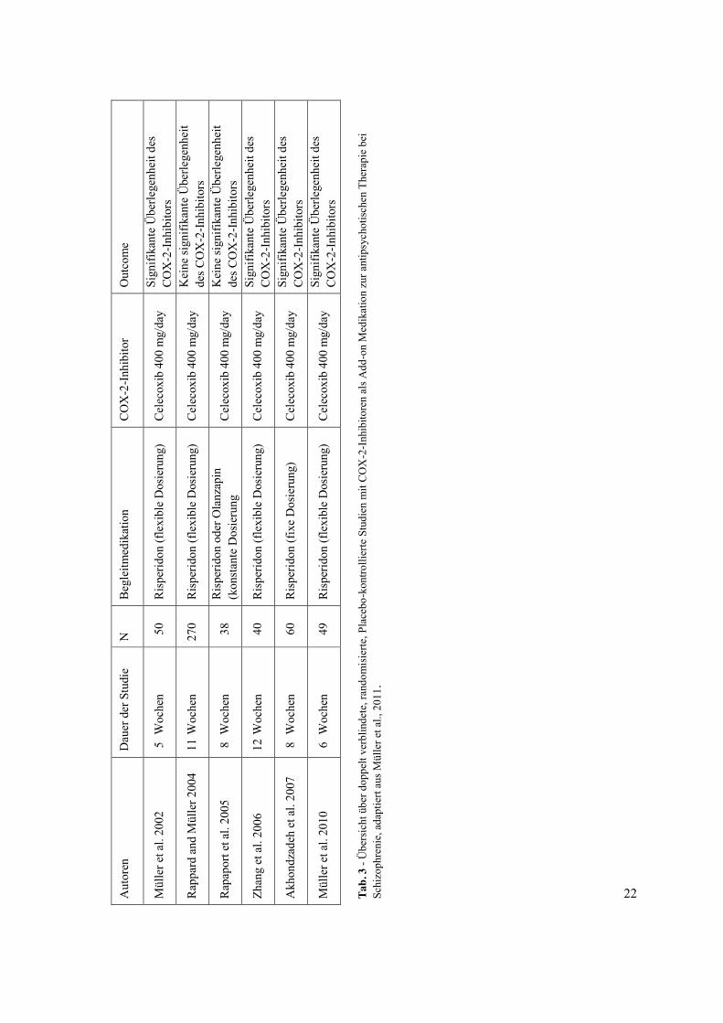
22 Aut
oren
D
auer
der
Stu
die
N
Beg
leitm
edik
atio
n C
OX
-2-In
hibi
tor
Out
com
e
Mül
ler e
t al.
2002
5
Woc
hen
50
Ris
perid
on (f
lexi
ble
Dos
ieru
ng)
Cel
ecox
ib 4
00 m
g/da
y Si
gnifi
kant
e Ü
berle
genh
eit d
es
CO
X-2
-Inhi
bito
rs
Rap
pard
and
Mül
ler 2
004
11 W
oche
n 27
0 R
ispe
ridon
(fle
xibl
e D
osie
rung
) C
elec
oxib
400
mg/
day
Kei
ne si
gnifi
kant
e Ü
berle
genh
eit
des C
OX
-2-In
hibi
tors
Rap
apor
t et a
l. 20
05
8 W
oche
n 3
8 R
ispe
ridon
ode
r Ola
nzap
in
(kon
stan
te D
osie
rung
C
elec
oxib
400
mg/
day
Kei
ne si
gnifi
kant
e Ü
berle
genh
eit
des C
OX
-2-In
hibi
tors
Zhan
g et
al.
2006
12
Woc
hen
40
Ris
perid
on (f
lexi
ble
Dos
ieru
ng)
Cel
ecox
ib 4
00 m
g/da
y Si
gnifi
kant
e Ü
berle
genh
eit d
es
CO
X-2
-Inhi
bito
rs
Akh
ondz
adeh
et a
l. 20
07
8 W
oche
n 6
0 R
ispe
ridon
(fix
e D
osie
rung
) C
elec
oxib
400
mg/
day
Sign
ifika
nte
Übe
rlege
nhei
t des
C
OX
-2-In
hibi
tors
Mül
ler e
t al.
2010
6
Woc
hen
49
Ris
perid
on (f
lexi
ble
Dos
ieru
ng)
Cel
ecox
ib 4
00 m
g/da
y Si
gnifi
kant
e Ü
berle
genh
eit d
es
CO
X-2
-Inhi
bito
rs
Tab.
3 -
Übe
rsic
ht ü
ber d
oppe
lt ve
rblin
dete
, ran
dom
isie
rte, P
lace
bo-k
ontro
llier
te S
tudi
en m
it C
OX
-2-I
nhib
itore
n al
s Add
-on
Med
ikat
ion
zur a
ntip
sych
otis
chen
The
rapi
e be
i Sc
hizo
phre
nie,
ada
ptie
rt au
s Mül
ler e
t al.,
201
1.
1. Einführung
23
Diese Studien deuten auf einen Verbesserung der Behandlung von Schizophrenie mit APs
durch eine Augmentation mit COX-2-Inhibitoren. Allerdings ist der therapeutische Nutzen
von COX-2-Hemmern bei der Schizophrenie noch umstritten. Der klinische Nutzen der COX-
2-Hemmung, der in einer ersten prospektiven, randomisierten, doppelblinden Studie mit dem
COX-2-Inhibitor Celecoxib neben der Behandlung mit Risperidon im akuten Schub der
Schizophrenie gezeigt werden konnte (Müller et al. 2002), ließ sich in einem zweiten Versuch
durch Müller et al. nicht replizieren. Jedoch ergaben weitere Analyse der Daten dieser Studie,
dass das Ergebnis der Therapie mit Celecoxib mit der Dauer der Erkrankung zusammenhing
(Müller 2010). Die Wirksamkeit von Celecoxib scheint in den ersten Jahren der Erkrankung
am erfolgreichsten zu sein (Müller 2010). In einer anschließenden doppelt verblindeten,
placebo-kontrollierten, randomisierten Studie mit Amisulprid plus Celecoxib versus
Amisulprid plus Placebo bei Patienten im frühen Stadium der Schizophrenie berichteten
Müller et al. über eine signifikant überlegene therapeutische Wirkung in der Celecoxib-
Gruppe gegenüber der Placebo-Gruppe (Müller, Krause et al. 2010). Daher scheinen COX-2-
Inhibitoren, eine spezifische therapeutische Option im frühen Stadium der Schizophrenie zu
sein. Ob sie auch in der Behandlung von Prodromalzuständen der Schizophrenie eingesetzt
werden könnten, muss in weiteren klinischen Studien evaluiert werden. EPA hingegen scheint
eine gangbare Behandlungsoption für prodromale Psychosen zu sein, da man nachweisen
konnte, dass EPA in Kombination mit Docosahexaensäure und Tocopherol das Risiko der
Entwicklung einer psychotischen Störung reduziert (Amminger et al. 2010). Bisher konnte
noch nicht nachgewiesen werden, ob COX-2-Hemmer oder EPA durch Modulation der
Dopamin-Signalisierung das Auftreten von schizophrenen Symptomen verbessern oder
verhindern. Diese Moleküle üben ihre vorteilhafte Wirkung über einen unbekannten
Mechanismus aus, wobei unklar bleibt, ob ein direkter oder indirekter Zusammenhang mit der
Dopamin-Signalisierung besteht. Die Erkenntnisse über die Bedeutung von PUFAs,
Eikosanoiden und COX-2 bei der Behandlung von schizophrenen Patienten, auch im Hinblick
auf ihre Auswirkungen auf die Antipsychotikawirkung, beantworten nicht die Frage, ob
Eikosanoide eine von dopaminerger Signalisierung unabhängige Rolle bei der Entwicklung
von Schizophrenie spielen. Daher sollten in weiteren klinischen Studien und Untersuchungen
Daten über den Eikosanoidstoffwechsel und über die Dopamin-Signalübertragung gesammelt
werden. Beispielsweise könnte ein Versuch mit COX-2-Inhibitoren eine Positronen-
Emissions-Tomographie (PET) des dopaminergen Systems zu Beginn und während der
Therapie mit selbigen beinhalten.
1. Einführung
24
Es hat sich gezeigt, dass TxA2 die Freisetzung von Dopamin erleichtert (Mitsumori et al.
2011). Daher könnte die Überaktivität des dopaminergen Systems bei schizophrenen
Patienten durch die Einschränkung der TxA2- Synthese reduziert werden.
Da bereits gezeigt werden konnte, dass PGE2, COX-2, TxA2 und TxB2 in die
Pathophysiologie der Schizophrenie bzw. deren Behandlung involviert sind und auch, dass es
eine Verringerung der Gesamt-PUFA-Konzentration im Serum, Plasma, peripheren Gewebe,
Erythrozytenmembranen und im Gehirn schizophrener Patienten gibt (siehe Tab. 2), stellt sich
die Frage, ob es von Vorteil wäre, die Funktion oder die Einnahme von AA als Vorstufe der
Eikosanoide zu beeinflussen. Um die Spiegel von PGE2 und TxA2 und TxB2 herab zu setzen,
könnte man theoretisch versuchen, die Verfügbarkeit von AA zu reduzieren. Allerdings
könnte die verringerte Verfügbarkeit von AA zu erheblichen Störungen der Integrität und der
Funktion der Zellmembranen führen. Zusätzlich ist zu bedenken, dass AA nicht nur für die
Synthese von PGE2, COX-2, TxA2 und TxB2 verwendet wird, sondern auch über den
Lipoxygenase-Weg zu Leukotrienen metabolisiert wird (Marks und Fürstenberger 1999), die
eine wichtige Rolle als Signalmoleküle in verschiedenen Körperfunktionen spielen. Hier sei
beispielsweise die Immunabwehr, einschließlich der Verteidigung gegen Bakterien, Pilze und
Parasiten genannt. (Rogerio und Anibal 2012). Daher kann die Verringerung der AA zu
verschiedenen unerwünschten Nebenwirkungen führen. Die derzeitig verfügbaren Studien zu
Eikosanoiden und Schizophrenie sind vorläufiger Natur und die Ergebnisse zum Teil
widersprüchlich. Es ist bisher nicht klar, welche Untergruppe der schizophrenen Patienten
besonders von einer Behandlung, die auf den Eikosanoidstoffwechsel zielt, wie zum Beispiel
die mit COX-2-Inhibitoren, profitieren können. Darüber hinaus werden derzeit die
Mechanismen des Zusammenspiels zwischen Eikosanoiden, Zytokinen, Neurotransmittern
und Hormonen noch nicht vollständig verstanden. Allerdings scheinen Eikosanoide, die mit
Zytokinen, Neurotransmittern und Hormonen interagieren ein Glied der pathophysiologischen
Kette zu sein, die zur Entwicklung von Schizophrenie führen kann. In-vivo-Untersuchungen
bezüglich der Wirkung von Neuroleptika auf den Eikosanoidstoffwechsel von Patienten mit
Schizophrenie können tiefere Einblicke in die Wechselwirkungen zwischen dopaminergen
und serotonergen Systemen einerseits und Eikosanoidstoffwechsel und Signalisierung
andererseits geben, und somit auf deren Bedeutung für die Pathophysiologie und Behandlung
von Schizophrenie hinweisen.
1. Einführung
25
1.8 Der Vollbluttest als in-vitro-Verfahren für psychopharmakologische Untersuchungen
Wir untersuchten in der vorliegenden Studie den Effekt von CPZ, Clozapin und NDMC auf
die Thromboxanproduktion in einem Vollbluttest. Dies ist ein etabliertes in-vitro-Verfahren
für die Untersuchung psychopharmakologischer Medikamente (Kirchner et al. 1982; Seidel et
al. 1996). Wir verwendeten unterschiedliche Stimulantien, um das Vollblut zu aktivieren,
nämlich das TSST-1 oder eine Kombination aus den monoklonalen Antikörpern OKT3 und
5C3 (OKT3/5C3).
TSST-1 ist ein Exotoxin, welches von Staphylococcen produziert wird. Es ist verantwortlich
für das Toxic Shock Syndrome. TSST-1 führt zu einer unspezifischen Bindung des
Haupthistokompatibilitätskomplexes Klasse II (MHC-II) mit T-Zell-Rezeptoren, was zu einer
polyklonalen T-Zell-Aktivierung und einer immunologischen Reaktion führt (Dinges et al.
2000).
Der murine monoklonale Antikörper OKT3 (Muromonab-CD3) bindet an den T-Zell
Rezeptor CD3 und ist ein etablierter T-Zell-Aktivator (Adair et al. 1994). Der monoklonale
Antikörper 5C3 stimuliert menschliches CD40 und aktiviert auf diese Weise B-Zellen (Pound
et al. 1999). Mit der Kombination dieser beiden Stimulanzien können B- und T-Zellen
gleichzeitig aktiviert werden.
Zu dem mit Hilfe von TSST-1 oder OKT3/5C3 stimulierten Vollblut gaben wir CPZ,
Clozapin und NDMC in verschiedenen Konzentrationen hinzu, um die Effekte dieser
Medikamente auf die Thromboxanproduktion zu untersuchen.
2. Aufgabenstellung
26
2. Aufgabenstellung
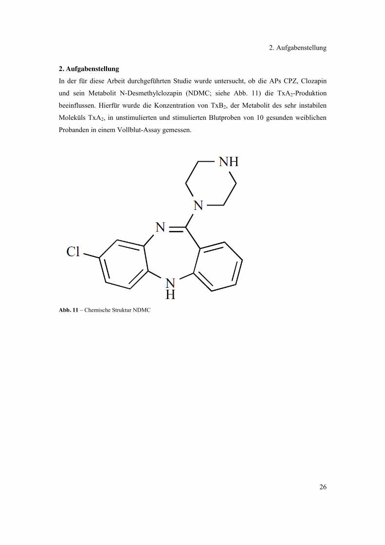
In der für diese Arbeit durchgeführten Studie wurde untersucht, ob die APs CPZ, Clozapin
und sein Metabolit N-Desmethylclozapin (NDMC; siehe Abb. 11) die TxA2-Produktion
beeinflussen. Hierfür wurde die Konzentration von TxB2, der Metabolit des sehr instabilen
Moleküls TxA2, in unstimulierten und stimulierten Blutproben von 10 gesunden weiblichen
Probanden in einem Vollblut-Assay gemessen.
Abb. 11 – Chemische Struktur NDMC
3. Materialien und Methoden
27
3. Materialien und Methoden
3.1 Versuchspersonen
Es wurden zehn gesunde weibliche Probanden ohne psychiatrische Störungen im Alter von 22
bis 45 Jahren (Mittelwert: 29,9 ± 8,67 (SD) Jahre) in die Studie eingeschlossen. Die
Einnahme von Immunmodulatoren, illegalen Drogen oder regelmäßiger Alkoholkonsum
führte zum Ausschluss der Versuchsperson. Die Probanden wurden durch einen erfahrenen
Psychiater mit dem Structured Clinical Interview für DSM-IV (SKID-I) auf psychiatrische
Erkrankungen, gescreent. Das Vorhandensein einer psychiatrischen Störung führte ebenfalls
zum Ausschluss aus der Studie.
3.2 Versuchsdurchführung
Das Vollblut-Assay wurde durchgeführt, wie es zuvor von Kirchner et al. und Seiderl et al
beschrieben wurde (Kirchner et al. 1982; Seidel et al. 1996). Allen Probanden wurde einmalig
Blut mit einer Heparin-Monovette (Sarstedt, Nürtingen, Deutschland) entnommen und
innerhalb von 1-2 h nach der Blutentnahme in einem Vollblut-Assay untersucht. Die Zell-
Konzentration wurde mit RPMI 1640 Medium (Biochrom, Berlin, Deutschland) auf 3-4 x 10⁹
Zellen/l eingestellt. Anschließend wurden 100 µL dieses Blutes sowie RPMI-Lösung in ein
Reagenzglas eingefüllt und mit 100 µL reinem Psychopharmakon sowie RPMI gemischt, was
zu einer endgültigen Zellkonzentration von 1,5-2 x 10⁹ Zellen/l führte. Richtwerte für die
beabsichtigten Endkonzentrationen der Psychopharmaka in dieser Mischung wurden den
„Therapeutic drug monitoring (TDM) expert group of the Association of
Neuropsychopharmacology and Pharmacopsychiatry (AGNP) consensus guidelines:
Therapeutic drug monitoring in psychiatry“ (Baumann et al. 2004) entnommen. Wir
verwendeten die maximalen therapeutischen Konzentrationen von 300 ng/ml für CPZ, 600
ng/ml für Clozapin und 600 ng/ml für NDMC. Zusätzlich verwendeten wir das 0,25-, 0,5-und
2-Fache der maximalen therapeutische Konzentration. Zur Kontrolle diente Blut ohne
psychopharmakologische Substanzen. Das Design dieser Studie ist in Tab. 4 dargestellt.
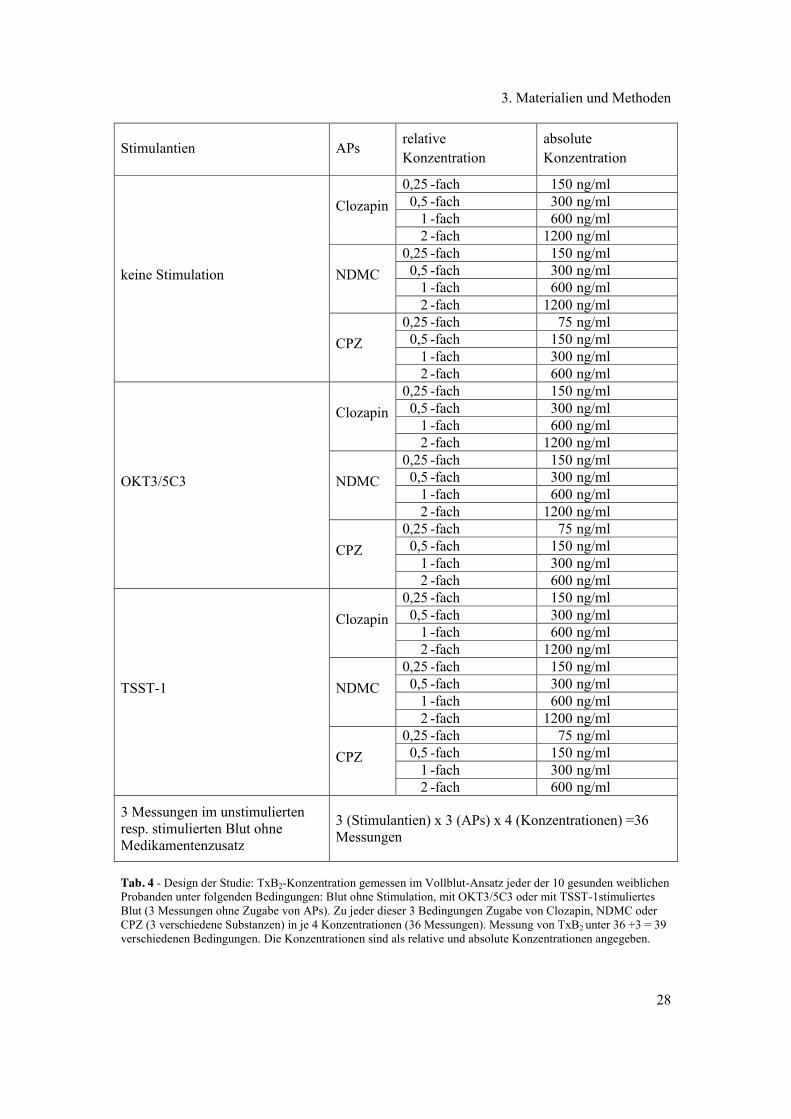
3. Materialien und Methoden
28
Stimulantien APs relative Konzentration
absolute Konzentration
keine Stimulation
Clozapin 0,25 -fach 150 ng/ml 0,5 -fach 300 ng/ml 1 -fach 600 ng/ml 2 -fach 1200 ng/ml
NDMC 0,25 -fach 150 ng/ml 0,5 -fach 300 ng/ml 1 -fach 600 ng/ml 2 -fach 1200 ng/ml
CPZ 0,25 -fach 75 ng/ml 0,5 -fach 150 ng/ml 1 -fach 300 ng/ml 2 -fach 600 ng/ml
OKT3/5C3
Clozapin 0,25 -fach 150 ng/ml 0,5 -fach 300 ng/ml 1 -fach 600 ng/ml 2 -fach 1200 ng/ml
NDMC 0,25 -fach 150 ng/ml 0,5 -fach 300 ng/ml 1 -fach 600 ng/ml 2 -fach 1200 ng/ml
CPZ 0,25 -fach 75 ng/ml 0,5 -fach 150 ng/ml 1 -fach 300 ng/ml 2 -fach 600 ng/ml
TSST-1
Clozapin 0,25 -fach 150 ng/ml 0,5 -fach 300 ng/ml 1 -fach 600 ng/ml 2 -fach 1200 ng/ml
NDMC 0,25 -fach 150 ng/ml 0,5 -fach 300 ng/ml 1 -fach 600 ng/ml 2 -fach 1200 ng/ml
CPZ 0,25 -fach 75 ng/ml 0,5 -fach 150 ng/ml 1 -fach 300 ng/ml 2 -fach 600 ng/ml
3 Messungen im unstimulierten resp. stimulierten Blut ohne Medikamentenzusatz
3 (Stimulantien) x 3 (APs) x 4 (Konzentrationen) =36 Messungen
Tab. 4 - Design der Studie: TxB2-Konzentration gemessen im Vollblut-Ansatz jeder der 10 gesunden weiblichen Probanden unter folgenden Bedingungen: Blut ohne Stimulation, mit OKT3/5C3 oder mit TSST-1stimuliertes Blut (3 Messungen ohne Zugabe von APs). Zu jeder dieser 3 Bedingungen Zugabe von Clozapin, NDMC oder CPZ (3 verschiedene Substanzen) in je 4 Konzentrationen (36 Messungen). Messung von TxB2 unter 36 +3 = 39 verschiedenen Bedingungen. Die Konzentrationen sind als relative und absolute Konzentrationen angegeben.
3. Materialien und Methoden
29
CPZ erhielten wir von Sigma-Aldrich Laborchemikalien GmbH (Seelze, Deutschland).
Clozapin und NDMC wurden freundlicherweise von Novartis Pharma zur Verfügung gestellt.
Zur Stimulation der Blutzellen verwendeten wir 5 g/ml rekombinantes TSST-1 oder 100
ng/ml OKT3/5C3. Alle Reagenzgläser wurden abgedeckt und die Proben in einer Atmosphäre
von 5% CO2 und 37°C für 48h inkubiert. Nach der Inkubation wurden die zellfreien
Überstände entnommen und bei -70°C gelagert.
3.3 Bestimmung von TxB2
Da es sich bei TxA2 um ein sehr instabiles Molekül handelt, wurden die Spiegel seines
wesentlich stabileren Metaboliten TxB2 bestimmt. Die Analyse erfolgte mittels
Flüssigchromatographie in Kombination mit Tandem-Massenspektrometrie (LC-MS/MS).
Entsprechend der von Kortz et al. (Kortz et al. 2009) für Eikosanoid-Messungen
beschriebenen LC-MS/MS-Methode wurden die Überstände aus dem Vollbluttest durch
Proteinfällung und nachfolgender off-line Festphasenextraktion aufgereinigt. Die
chromatographische Trennung erfolgte auf einer Kinetex C-18 Core-shell-Säule
(2,1 × 100 mm, 2,6 µm; Phenomenex, Aschaffenburg, Deutschland) mit einem Shimadzu
Prominence UFLC System (Shimadzu Europa, Duisburg, Deutschland). Für die
massenspektrometrische Analyse wurde ein 5500 QTrap Massenspektrometer (AB Sciex,
Framingham, Massachusetts, USA) mit negativer Elektrosprayionisierung verwendet.
3.4 Statistische Analyse
Aufgrund der geringen Anzahl von Datenpunkten, beschreiben wir die deskriptive Statistik
unter Zuhilfenahme nichtparametrischen Maßnahmen. Zum Vergleich der TxB2-
Konzentrationen in unstimulierten und stimulierten Blut ohne und mit verschiedenen
Konzentrationen von APs wurden der nicht-parametrisch gepaarte Wilcoxon-Tests verwendet.
Aufgrund des explorativenn Charakters dieser Studie wurde ein unkorrigierter p-Wert kleiner
0,05 als signifikant angesehen.
3.5 Ethik
Die Studie wurde von der lokalen Ethikkommission der Medizinischen Fakultät der
Universität Leipzig (# 351-10-13122010) genehmigt.
4. Ergebnisse
30
4. Ergebnisse
Das konsistenteste Ergebnis dieser Studie ist, dass unter OKT3/5C3 sowie TSST-1-
Stimulation Clozapin zu einer signifikanten Abnahme der TxB2-Konzentrationen über alle
Anwendungskonzentrationen führte (p <0,01). Zusätzlich reduzierte sein Metabolit NDMC
TxB2 signifikant unter der unstimulierten Bedingung (p <0,05) und unter TSST-1-Stimulation
(p <0,01 für das 0,5-, 1- und 2-Fache der maximalen therapeutischen Konzentration, p <0,05
für die 0,25 -fache Konzentration). CPZ verringerte ebenfalls die TxB2-Produktion bei
niedrigen Wirkstoffkonzentrationen im unstimulierten und TSST-1-stimulierten Blut. Für
detaillierte Erläuterungen zu den deskriptiven Statistiken sowie Vergleiche zu den TxB2-
Spiegeln, siehe Tab. 5.
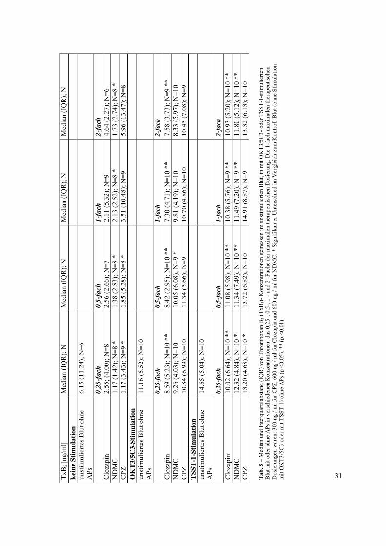
31 TxB
2 [n
g/m
l] M
edia
n (I
QR
); N
M
edia
n (I
QR
); N
M
edia
n (I
QR
); N
M
edia
n (I
QR
); N
ke
ine S
timul
atio
n
un
stim
ulie
rtes B
lut o
hne
APs
6.
15 (1
1.24
); N
=6
0,
25-fa
ch
0,5-
fach
1-
fach
2-
fach
C
loza
pin
2.55
; (4.
00);
N=8
2.
56 (2
.66)
; N=7
2.
11 (5
.32)
; N=9
4.
64 (2
.27)
; N=6
N
DM
C
1.17
(1.4
2); N
=8 *
1.
38 (2
.83)
; N=8
*
2.13
(2.5
2); N
=8 *
1.
73 (2
.74)
; N=8
*
CPZ
1.
17 (3
.43)
; N=9
*
1.85
(5.2
8); N
=8 *
3.
51 (1
0.48
); N
=9
5.96
(13.
47);
N=8
O
KT3
/5C
3-St
imul
atio
n
un
stim
ulie
rtes B
lut o
hne
APs
11
.16
(5.5
2); N
=10
0,
25-fa
ch
0,5-
fach
1-
fach
2-
fach
C
loza
pin
8.59
(5.2
3); N
=10
**
8.42
(2.9
5); N
=10
**
7.30
(4.7
1); N
=10
**
7.58
(3.7
3); N
=9 *
* N
DM
C
9.26
(4.0
3); N
=10
10.0
5 (6
.08)
; N=9
*
9.81
(4.1
9); N
=10
8.33
(5.9
7); N
=10
CPZ
10
.84
(6.9
9); N
=10
11.3
4 (5
.66)
; N=9
10
.70
(4.8
6); N
=10
10.4
5 (7
.08)
; N=9
TS
ST-1
-Stim
ulat
ion
unst
imul
ierte
s Blu
t ohn
e A
Ps
14.6
5 (5
.04)
; N=1
0
0,
25-fa
ch
0,5-
fach
1-
fach
2-
fach
C
loza
pin
10.0
2 (6
.64)
; N=1
0 **
11
.08
(5.9
8); N
=10
**
10.3
8 (5
.76)
; N=9
**
10.9
3 (5
.20)
; N=1
0 **
N
DM
C
12.3
2 (4
.84)
; N=1
0 *
11.3
4 (7
.49)
; N=1
0 **
11
.49
(7.2
0); N
=9 *
* 11
.80
(5.1
2); N
=10
**
CPZ
13
.20
(4.6
8); N
=10
* 13
.72
(6.8
2); N
=10
14.9
1 (8
.87)
; N=9
13
.32
(6.1
3); N
=10
Tab.
5 –
Med
ian
und
Inte
rqua
rtila
bsta
nd (I
QR
) von
Thr
ombo
xan
B2 (
TxB
2)- K
onze
ntra
tione
n ge
mes
sen
im u
nstim
ulie
rten
Blu
t, in
mit
OK
T3/5
C3-
ode
r TSS
T-1-
stim
ulie
rten
Blu
t mit
oder
ohn
e A
Ps in
ver
schi
eden
en K
onze
ntra
tione
n: d
as 0
,25-
, 0.5
-, 1
- und
2 -F
ache
der
max
imal
en th
erap
eutis
chen
Dos
ieru
ng. D
ie 1
-fach
max
imal
en th
erap
eutis
chen
D
osie
rung
en w
aren
: 300
ng
/ ml f
ür C
PZ, 6
00 n
g / m
l für
Clo
zapi
n un
d 60
0 ng
/ m
l für
ND
MC
. * S
igni
fikan
ter U
nter
schi
ed im
Ver
glei
ch z
um K
ontro
ll-B
lut (
ohne
Stim
ulat
ion
mit
OK
T3/5
C3
oder
mit
TSST
-1) o
hne
APs
(p <
0,05
), **
(p <
0,01
).
4. Ergebnisse
32
Abb. 12 zeigt ein LC-MS-Chromatogramm von TxB2-Konzentrationen in TSST-1-
stimuliertem Blut mit und ohne Zusatz von Clozapin in dem 0,25-Fachen der maximalen
therapeutischen Konzentration. Aufgrund von Schwierigkeiten beim Messen von TxB2 in
unstimuliertem Blut mit der angewandten Methode fehlen mehrere TxB2-Werte, weil diese
unterhalb der Nachweisgrenze liegen. In 4 von 10 Proben konnte kein TxB2 nachgewiesen
werden. Unter stimulierten Bedingungen jedoch konnten TxB2-Werte in den meisten Fällen
gemessen werden. In allen stimulierten Proben konnte die TxB2-Konzentration in mindestens
N = 9 von 10 Proben unter verschiedenen Bedingungen bestimmt werden.
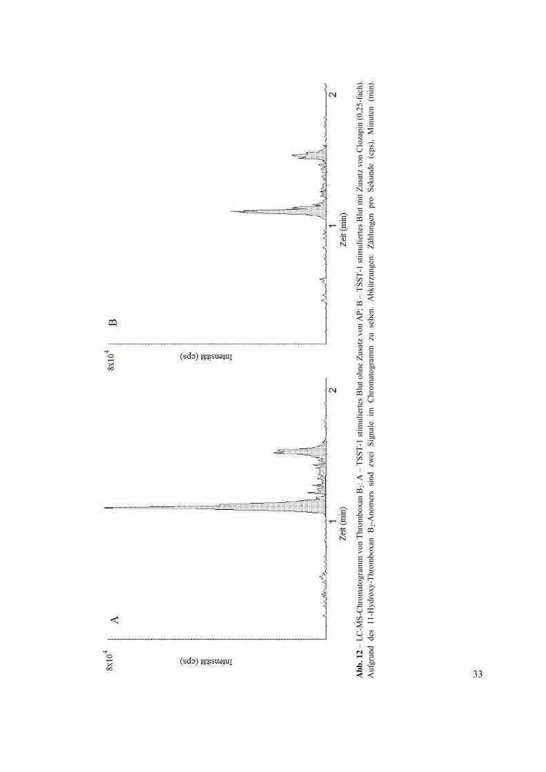
33
A
bb. 1
2 –
LC-M
S-C
hrom
atog
ram
m v
on T
hrom
boxa
n B
2: A
– T
SST-
1 st
imul
ierte
s B
lut o
hne
Zusa
tz v
on A
P; B
– T
SST-
1 st
imul
ierte
s B
lut m
it Zu
satz
von
Clo
zapi
n (0
,25-
fach
). A
ufgr
und
des
11-H
ydro
xy-T
hrom
boxa
n B
2-A
nom
ers
sind
zw
ei S
igna
le i
m C
hrom
atog
ram
m z
u se
hen.
Abk
ürzu
ngen
: Zä
hlun
gen
pro
Seku
nde
(cps
), M
inut
en (
min
).
5. Diskussion
34
5. Diskussion
Da man für Clozapin und CPZ nachweisen konnte, dass sie die TxA2-Produktion verringern
(Himmerich, Schmidt, L. et al. 2012), könnten diese Medikamente - komplementär zu ihrer
direkten Wirkung auf Dopamin- und 5-HT-Rezeptoren – auch dadurch antipsychotisch
wirken, dass sie die Dopaminsignalübertragung über die TxA2- oder TxB2-Synthese
modulieren. Allerdings beeinflussen die Thromboxane ebenfalls die
Thrombozytenaggregation und Vasokonstriktion (Wacker et al. 2003) sowohl im Körper
(Narumiya et al. 1999) als auch im Gehirn (Toth et al. 2011). Daher könnten bekannte
Nebenwirkungen von Clozapin und ähnlichen Neuroleptika, wie orthostatische Hypotonie,
auch oder zumindest teilweise durch den Einfluss der Thromboxane auf den Muskeltonus der
Blutgefäße erklärt werden (Leung et al. 1996).
Weiterhin könnten die zahlreichen immunologischen Eigenschaften und verschiedene
Nebenwirkungen von Clozapin (Pollmächer et al. 2000) mit den immunologischen
Eigenschaften des TxA2 (Ricciotti und FitzGerald 2011) in Verbindung stehen.
5.1 Zusammenfassung der Ergebnisse
Wir testeten den Einfluss von CPZ, Clozapin und seinem Metaboliten NDMC auf die TxB2-
Synthese in-vitro mit einem Vollblut-Assay. Wie aufgrund der Literaturrecherche zu
erwarten, reduzierte CPZ die TxB2-Synthese unter bestimmten Bedingungen: Es zeigten sich
in unserer Untersuchung allerdings nur in niedrigen Dosen von CPZ, ohne zusätzliche
Stimulation und unter TSST-1-Stimulation, signifikante Effekte. Das beständigste und
überzeugendste Ergebnis dieser Studie ist, dass unter OKT3/5C3 sowie TSST-1-Stimulation
Clozapin zu einer signifikanten Abnahme der TxB2-Spigel über alle
Anwendungskonzentrationen führte (p <0,01). Zusätzlich reduziert der Metabolit des
Clozapins NDMC TxB2 signifikant unter unstimulierten Bedingungen und unter TSST-1-
Stimulation. Deshalb ist anzunehmen, dass Clozapin und sein Metabolit zur Verringerung der
Produktion von TxA2 und seines Metaboliten TxB2 führen.
5.2 Bedeutung der Ergebnisse
Die erhaltenen Ergebnisse können bezüglich des antipsychotischen Wirkmechanismus von
Clozapin sowie bezüglich der Pathophysiologie der Nebenwirkungen von Clozapin
interpretiert werden. Da TxA2 die 5-HT- (Moncada und Vane 1978) sowie die Dopamin-
Signalübertragung (Mitsumori et al. 2011) erhöht, könnte sich die antipsychotische Wirkung
von Clozapin und NDMC neben der direkten antagonistischen Wirkung auf Dopamin- und 5-
5. Diskussion
35
HT-Rezeptor-Ebene auch über die Modulation des Dopamin- und 5-HT-Systems mittels
Blockierung der TxA2 -Produktion entfalten. Bekannte Nebenwirkungen von Clozapin, wie
orthostatische Hypotonie (Muench und Hamer 2010), venöse Thromboembolie (Paciullo
2008) und zerebrale Sinusvenenthrombose (Werring et al. 2009) könnten auch, zumindest
teilweise, durch den Einfluss der Thromboxane auf den Muskeltonus der Blutgefäße
vermittelt sein (Leung et al. 1996). Zusätzlich könnten die zahlreichen immunologischen
Eigenschaften und verschiedene Nebenwirkungen von Clozapin (Himmerich und Schmidt
2012; Pollmächer et al. 2000) mit den immunologischen Eigenschaften des TxA2 (Ricciotti,
E., FitzGerald, G.A. 2011) verknüpft sein.
5.3 Limitationen der Untersuchung
Wie bereits erwähnt, handelt es sich hierbei um eine kleine Pilotstudie und das
Signifikanzniveau wurde nicht für multiples Testen kontrolliert. Daher müssten alle Effekte
unabhängig voneinander repliziert werden. Die beschriebenen Daten über die TxB2-
Veränderungen sind von einer in-vitro-Studie mit menschlichem Blut von gesunden
Probanden abgeleitet. Diese Einschränkungen müssen berücksichtigt werden, wenn man
Rückschlüsse auf die die tatsächliche Situation im lebenden Patienten aus den Ergebnissen
ziehen will. Die beschriebenen Ergebnisse sind also vorläufiger Natur. Unserem Wissen nach
ist dies die erste Studie, die die Wirkung von Clozapin und NDMC auf die Thromboxan-
Produktion untersucht. Allerdings liefern diese Daten eine alternative und komplementäre
Sicht zu bestehenden Neurotransmitter-Hypothesen bezüglich der Schizophrenie und zu
aktuellen Meinungen über die Wirkungsmechanismen von APs.
5.4 Besonderheiten des Vollblutverfahrens in der psychopharmakologischen Forschung
Aus methodischen Gründen sowie aus Gründen der chemischen Metrologie nutzten wir den
beschriebenen stimulierten in-vitro-Ansatz. Diese methodischen Gründe beinhalten u.a. die
Vergleichbarkeit mit anderen Studien (Himmerich, Fulda, et al. 2010; Potvin et al. 2008;
Drzyzga et al. 2006; Hinze-Selch, D., Pollmächer, T. 2001; Arolt et al. 2000) und die
Angleichung des in-vitro-Zustandes an die natürlichen Gegebenheiten im menschlichen
Körper, in dem dynamische Prozesse, wie Immunstimulation, regelmäßig stattfinden. Aus der
Sicht der chemischen Metrologie ist es ratsam, die Produktion von Molekülen wie
Thromboxan zu stimulieren, um sie in einen Bereich zu bringen, der sicherer zu messen ist,
sodass sich Fehlmessungen reduzieren lassen. Wie man in Tab. 5 sehen kann, war im
stimulierten Blut in mindestens 9 von 10 Proben eine TxB2 - Konzentration nachweisbar,
5. Diskussion
36
während in den unstimulierten Ansätzen die Konzentrationen zum Teil so gering waren, dass
es durch Probleme bei den Messungen zu fehlenden Werten kam. Die Praktikabilität dieses
Ansatzes wurde zuvor in mehreren Studien (Himmerich, Schönherr et al. 2011; Himmerich,
Fulda et al. 2010), welche nahelegten, dass diese Experimente relativ einfach durchzuführen
sind, nachgewiesen. Auf der anderen Seite kann man nicht ausschließen, dass Tests mit APs
im Rahmen eines Stimulationsparadigmas zu Artefakten führen können, die auf spezifischen
Wechselwirkungen der APs mit den Stimulantien beruhen. Dennoch dienen in-vitro-
Stimulations-Paradigmen als Simulation der dynamischen Natur der normalen Lebensdauer
von Blutzellen. Weiterhin haben in-vitro-Paradigmen mit immunologischen
Stimulationsmethoden in den letzten zwei Jahrzehnten in der psychopharmakologischen
Forschung eine Reihe von vergleichbaren in-vitro-Studien mit relevanten wissenschaftlichen
Auswirkungen etabliert (Himmerich, Schönherr et al. 2011; Himmerich, Fulda et al. 2010;
Kirchner et al. 1982; Seidel et al. 1996; Potvin et al. 2008; Drzyzga et al. 2006; Hinze-Selch,
D., Pollmächer, T. 2001; Arolt et al. 2000).
5.5 Zusammenfassung der Ergebnisinterpretation basierend auf der Literatur
Es ist bekannt, dass Thromboxane die 5-HT- (Moncada,und Vane 1978) sowie die Dopamin-
Signalitransduktion (Mitsumori et al. 2011) modulieren. Weiterhin konnte gezeigt werden,
dass sie in die Regelung verschiedener Systeme, in denen typische Nebenwirkungen von
Clozapin und seinem Metaboliten NDMC stattfinden, involviert sind. Aus der Erkenntnis,
dass es unter Clozapin und seinem Metaboliten NDMC zu einer Abnahme der
Thromboxansynthese kommt, schließen wir, dass Thromboxane an der antipsychotischen
Wirkung sowie den Nebenwirkungen dieser Arzneimittel beteiligt sind.
5.6 Anmerkung
Auf der Grundlage dieser Dissertationsschrift wurden mittlerweile eine Übersichtsarbeit zur
Rolle der Eikosanoide für die Pathophysiologie und Therapie der Schizophrenie (Schmidt et
al., 2013) und eine Originalarbeit (Himmerich, Schmidt et al., 2012) publiziert. Die
Publikationen befinden sich im Anhang.
6. Zusammenfassung
37
6. Zusammenfassung
Dissertation zur Erlangung des akademischen Grades Dr.med.
Der Einfluss von Clozapin, N-Desmethylclozapin und Chlorpromazin auf die
in-vitro-Produktion von Thromboxan
eingereicht von: Renate Luise Schmidt
geboren am 22.11.1987
in Borna
angefertigt an: der Universität Leipzig,
Claussen-Simon-Stiftungsprofessur für Neurobiologie affektiver
Störungen innerhalb der Klinik und Poliklinik für Psychiatrie
und Psychotherapie
Betreuer: Prof. Dr. med. Hubertus Himmerich
PD Dr. rer. nat. Uta Ceglarek
Monat/Jahr Einreichung: Januar 2014
Die Hypothese, dass das AP Clozapin, ebenso wie dessen Metabolit NDMC die Produktion
von TxA2 beeinfluss könnten, stellten wir nach ausführlicher Literaturrecherche auf. Letztere
zeigte, dass bereits beim ersten AP CPZ eine reduzierende Wirkung auf die TxA2-Produktion
nachgewiesen werden konnte. TxA2 und die Aktivierung seines Rezeptors modulieren
Vasokonstriktion und Thrombozytenaggregation. Weiterhin nehmen sie Einfluss auf
dopaminerge und serotonerge Signalwege. In der Pathophysiologie der Schizophrenie spielen
eben diese eine bedeutende Rolle und stellen somit Zielstrukturen für APs dar.
6. Zusammenfassung
38
Um die Konzentration von TxB2, dem Metaboliten des instabilen Moleküls TxA2 in
stimulierten und unstimulierten Blutproben 10 gesunder Probandinnen zu messen,
verwendeten wir ein Vollblutverfahren. Um signifikante Ergebnisse zu erhalten, stimulierten
wir die Proben mit TSST-1 oder dem monoklonalen Antikörper OKT3 (Muromonab-CD3),
der gegen das Oberflächenantigen CD3 gerichtet ist, kombiniert mit dem monoklonalen
Antikörper 5C3, der mit dem Protein CD40 interagiert und es stimuliert.
Weiterhin versetzten wir das Blut mit den APs CPZ, Clozapin oder NDMC in einer von vier
verschiedenen Konzentrationen. Außerdem wurden die Thromboxanspiegel im Blut ohne
Zusatz von APs unter verschiedenen Stimulationskonditionen gemessen.
Durch den Zusatz von Clozapin in den verschiedenen Konzentrationen kam es zu einer
signifikanten (p<0.05) Verringerung der TxB2-Produktion in den mit TSST-1 und ebenso in
den mit OKT3/5C3 versetzen Proben, was wir im Rahmen unserer Studie feststellen konnten.
Weiterhin konnten wir zeigen, dass CPZ in sehr niedriger Konzentration die TxB2-Spiegel im
unstimulierten und im mit TSST-1 stimulierten Blut reduziert.
Daraus lässt sich schlussfolgern, dass Clozapin, NDMC und CPZ auch über eine Modulation
der TxA2- und TxB2-Produktion das Neurotransmittersystem beeinflussen könnten. Auch
typische Nebenwirkungen der AP, wie zum Beispiel die orthostatische Hypotension, könnten
aus den Veränderungen der TxA2- und TxB2-Konzentrationen resultieren.
7. Literaturverzeichnis
7. Literaturverzeichnis Adair, J.R.; Athwal, D.S.; Bodmer, M.W.; Bright, S.M.; Collins, A.M.; Pulito, V.L.; Rao,
P.E.; Reedman, R.; Rothermel, A.L.; Xu, D.; Zivin, R.A.; Jolliffe, L.K. Humanization of the murine anti-human CD3 monoclonal antibody OKT3. Hum. Antibodies Hybridomas, 1994, 5, 41-47.
Adkins, D.E.; Khachane, A.N.; McClay, J.L.; Aberg, K.; Bukszár, J.; Sullivan, P.F.; van den
Oord, E.J. SNP-based analysis of neuroactive ligand-receptor interaction pathways implicates PGE2 as a novel mediator of antipsychotic treatment response: data from the CATIE study. Schizophr. Res., 2012, 135, 200-1.
Akhondzadeh, S.; Tabatabaee, M.; Amini, H.; Ahmadi Abhari, S.A.; Abbasi, S.H.; Behnam,
B. Celecoxib as adjunctive therapy in schizophrenia: a double-blind, randomized and placebo-controlled trial. Schizophr. Res., 2007, 90, 179-85.
Alanko, J.; Riutta, A.; Vapaatalo, H.; Mucha, I. Catecholamines decrease leukotriene B3 and
increase thromboxane B2 synthesis in A23 187-stimulated human whole blood. Prostaglandins, 1991, 42, 279-287.
Amateau, S.K.; McCarthy, M.M. Induction of PGE2 by estradiol mediates developmental
masculinization of sex behavior. Nat. Neurosci., 2004, 7, 643-50. American Psychiatric Association. Diagnostic and statistical manual of mental disorders:
DSM-IV-TR; Washington, DC, 2000. Amminger, G.P.; Schäfer, M.R.; Papageorgiou, K.; Klier, C.M.; Cotton, S.M.; Harrigan,
S.M.; Mackinnon, A.; McGorry, P.D.; Berger, G.E. Long-chain omega-3 fatty acids for indicated prevention of psychotic disorders: a randomized, placebo-controlled trial. Arch. Gen. Psychiatry, 2010, 67, 146-54.
Andreasen, N.C.; Olsen, S. Negative v positive schizophrenia. Definition and validation.
Arch. Gen. Psychiatry., 1982, 39, 789-94. Anfossi, G.; Massucco, P.; Mularoni, E.; Cavalot, F.; Burzacca, S.; Mattiello, L.; Trovati, M.
Phenothiazines inhibit collagen-induced thromboxane B2 synthesis and increase forskolin anti-aggregating effects in human platelets. Gen. Pharmacol., 1991, 22, 773-778.
Anfossi, G.; Trovati, M.; Mularoni, E.; Massucco, P.; Cavalot. F.; Mattiello, L.; Emanuelli, G.
Phenothiazine compounds enhance phentolamine effects on platelet aggregation and thromboxane B2 production. Arch. Int. Pharmacodyn. Ther., 1990, 307, 142-61.
7. Literaturverzeichnis
Ansar, S.; Larsen, C.; Maddahi, A.; Edvinsson, L. Subarachnoid hemorrhage induces enhanced expression of thromboxane A2 receptors in rat cerebral arteries. Brain Res., 2010, 1316, 163-72.
Arolt, V.; Rothermundt, M.; Wandinger, K.P.; Kirchner, H. Decreased in vitro production of
interferon-gamma and interleukin-2 in whole blood of patients with schizophrenia during treatment. Mol. Psychiatry, 2000, 5, 150-158.
Autry, A.E.; Monteggia, L.M. Brain-derived neurotrophic factor and neuropsychiatric
disorders. Pharmacol. Rev., 2012, 64, 238-58. Balu, D.T., Coyle, J.T. Neuroplasticity signaling pathways linked to the pathophysiology of
schizophrenia. Neurosci. Biobehav. Rev., 2011, 35, 848-70. Bauer, M.; Praschak-Rieder, N.; Kasper, S.; Willeit, M. Is dopamine neurotransmission
altered in prodromal schizophrenia? A review of the evidence. Curr. Pharm. Des., 2012, 18, 1568-79.
Baumann, P.; Hiemke, C.; Ulrich, S.; Eckermann, G.; Gaertner, I.; Gerlach, M.; Kuss, H.J.;
Laux, G.; Müller-Oerlinghausen, B.; Rao, M.L.; Riederer, P.; Zernig, G. Arbeitsgemeinschaft für Neuropsychopharmakologie und Pharmakopsychiatrie. The AGNP-TDM expert group consensus guidelines: therapeutic drug monitoring in psychiatry. Pharmacopsychiatry, 2004, 37, 243-265.
Becker, S.; Kortz, L.; Helmschrodt, C.; Thiery, J.; Ceglarek, U. LC-MS-based metabolomics
in the clinical laboratory. J. Chromatogr. B Analyt. Technol. Biomed. Life Sci., 2012, 883-884, 68-75.
Berger, G.E.; Proffitt, T.M.; McConchie, M.; Yuen, H.; Wood, S.J.; Amminger, G.P.; Brewer,
W.; McGorry, P.D. Ethyleicosapentaenoic acid in first-episode psychosis: a randomized, placebo- controlled trial. J. Clin. Psychiatry., 2007, 68, 1867-75.
Berger, G.E.; Smesny, S.; Amminger, G.P. Bioactive lipids in schizophrenia. Int. Rev.
Psychiatry, 2006, 18, 85-98. Berthold-Losleben, M.; Heitmann, S.; Himmerich, H. Antiinflammatory drugs in psychiatry.
Inflamm. Allergy Drug Targets, 2009, 8, 266-76. Bleuler, E. Dementia praecox oder Gruppe der Schizophrenien; Deuticke: Leipzig, 1911. Brammer, J.P.; Maguire, M.H. Arachidonate metabolism, 5-hydroxytryptamine release and
aggregation in human platelets activated by palmitaldehyde acetal phosphatidic acid. Br. J. Pharmacol., 1984, 82, 61-72.
7. Literaturverzeichnis
Bray, N.J.; Leweke, F.M.; Kapur, S.; Meyer-Lindenberg, A. The neurobiology of schizophrenia: new leads and avenues for treatment. Curr. Opin. Neurobiol., 2010, 20, 810-5.
Brown, A.S.; Hooton, J.; Schaefer, C.A.; Zhang, H.; Petkova, E.; Babulas, V.; Perrin, M.;
Gorman, J.M.; Susser, E.S. Elevated maternal interleukin-8 levels and risk of schizophrenia in adult offspring. Am. J. Psychiatry, 2004, 161, 889-95.
Bürki, H.R.; Eichenberger, E.; Sayers, A.C.; White, T.G. Clozapine and the dopamine
hypothesis of schizophrenia, a critical appraisal. Pharmakopsychiatr. Neuropsychopharmakol., 1975, 8, 115-21.
Carey, L.C.; Valego, N.K.; Chen, K.; Rose, J.C. Thyroid hormone regulates renocortical
COX-2 and PGE2 expression in the late gestation fetal sheep. Reprod. Sci., 2008, 15, 598-603.
Chang, J.; Piperata, S.; Skowronek, M.; Lewis, A.J. Effects of thiazinamium chloride,
promethazine and chlorpromazine on thromboxane B2 synthesis, phagocytosis and respiratory burst by rat alveolar macrophages. Biochem. Pharmacol., 1983, 32, 2671-2677.
Chen, H.; Dzitoyeva, S.; Manev, H. 5-Lipoxygenase in mouse cerebellar Purkinje cells.
Neuroscience., 2010, 171, 383-9. Cheon Y, Park JY, Modi HR, Kim HW, Lee HJ, Chang L, Rao JS, Rapoport SI. Chronic
olanzapine treatment decreases arachidonic acid turnover and prostaglandin E2 concentration in rat brain. J. Neurochem., 2011, 119, 364-76.
Choi, S.-Y.; Chang, J.; Jiang, B.; Seol, G.-H.; Min, S.-S.; Han, J.- S.; Shin, H.-S.; Gallagher,
M.; Kirkwood, A. Multiple receptors coupled to phospholipase C gate long-term depression in visual cortex. J. Neurosci., 2005, 25, 11433-43.
Coleman, R.A.; Smith, W.L.; Narumiya, S. International Union of Pharmacology
classification of prostanoid receptors: properties, distribution, and structure of the receptors and their subtypes. Pharmacol. Rev., 1994, 46, 205-29.
Connor, C.M.; Crawford, B.C.; Akbarian, S. White matter neuron alterations in schizophrenia
and related disorders. Int. J. Dev. Neurosci., 2011, 29, 325-34. Das, I.; Khan, N.S. Increased arachidonic acid induced platelet chemiluminescence indicates
cyclooxygenase overactivity in schizophrenic subjects. Prostaglandins Leukot. Essent. Fatty Acids, 1998, 58, 165-8.
7. Literaturverzeichnis
Davis, K.L.; Kahn, R.S.; Ko, G.; Davidson, M. Dopamine in schizophrenia: a review and reconceptualization. Am. J. Psychiatry, 1991, 148, 1474-86.
Debnath, M.; Cannon, D.M.; Venkatasubramanian, G. Variation in the major
histocompatibility complex [MHC] gene family in schizophrenia: Associations and functional implications. Prog. Neuropsychopharmacol. Biol. Psychiatry, 2013, 42, 49-62.
Di Pietro, N.C.; Seamans, J.K. Dopamine and serotonin interactions in the prefrontal cortex:
insights on antipsychotic drugs and their mechanism of action. Pharmacopsychiatry, 2007, 40 Suppl 1, 27- 33.
Di, X.; Chan, R. C.K.; Gong, Q.-Y. White matter reduction in patients with schizophrenia as
revealed by voxel-based morphometry: an activation likelihood estimation meta-analysis. Prog. Neuropsychopharmacol. Biol. Psychiatry, 2009, 33, 1390-4.
Dietrich-Muszalska, A.; Olas, B. Isoprostenes as indicators of oxidative stress in
schizophrenia. World J. Biol. Psychiatry, 2009, 10, 27-33. Dinges, M.M.; Orwin, P.M.; Schlievert, P.M. Exotoxins of Staphylococcus aureus. Clin.
Microbiol. Rev., 2000, 13, 16-34. Drzyzga, L.; Obuchowicz, E.; Marcinowska, A.; Herman, Z.S. Cytokines in schizophrenia
and the effects of antipsychotic drugs. Brain Behav. Immun., 2006, 20, 532-545. Emsley, R.; Myburgh, C.; Oosthuizen, P.; van Rensburg, S.J. Randomized, placebo-controlled
study of ethyl-eicosapentaenoic acid as supplemental treatment in schizophrenia. Am. J. Psychiatry, 2002, 159, 1596-8.
Emsley, R.; Niehaus, D.J.; Koen, L.; Oosthuizen, P.P.; Turner, H.J.; Carey, P.; van Rensburg,
S.J.; Maritz, J.S.; Murck, H. The effects of eicosapentaenoic acid in tardive dyskinesia: a randomized, placebocontrolled trial. Schizophr. Res., 2006, 84, 112-20.
Erhardt, S.; Blennow, K.; Nordin, C.; Skogh, E.; Lindström, L. H.; Engberg, G. Kynurenic
acid levels are elevated in the cerebrospinal fluid of patients with schizophrenia. Neurosci. Lett., 2001, 313, 96-8.
Fenton, W.S.; Dickerson, F.; Boronow, J.; Hibbeln, J.R.; Knable, M. A placebo-controlled
trial of omega-3 fatty acid (ethyl eicosapentaenoic acid) supplementation for residual symptoms and cognitive impairment in schizophrenia. Am. J. Psychiatry, 2001, 158, 2071-4.
Fenton, W.S.; Hibbeln, J.; Knable, M. Essential fatty acids, lipid membrane abnormalities,
and the diagnosis and treatment of schizophrenia. Biol. Psychiatr., 2000, 47, 8-21.
7. Literaturverzeichnis
Fredriksson, R.; Lagerström, M.C.; Lundin, L.-G.; Schiöth, H.B. The G-protein-coupled receptors in the human genome form five main families. Phylogenetic analysis, paralogon groups, and fingerprints. Mol. Pharmacol., 2003, 63, 1256-72.
Freeman, M.P.; Hibbeln, J.R.; Wisner, K.L.; Davis, J.M.; Mischoulon, D.; Peet, M.; Keck,
P.E.; Marangell, L.B.; Richardson, A.J.; Lake, J.; Stoll, A.L. Omega-3 fatty acids: evidence basis for treatment and future research in psychiatry. J. Clin. Psychiatry, 2006, 67, 1954-67.
Fusar-Poli, P.; Berger, G. Eicosapentaenoic acid interventions in schizophrenia: meta-analysis
of randomized, placebo-controlled studies. J. Clin. Psychopharmacol., 2012, 32, 179-85. Gao, H.; Peng, B.; Welch, W.J., Wilcox, C.S. Central thromboxane receptors: mRNA
expression and mediation of pressor responses. Am. J. Physiol., 1997, 272, 1493-1500. Goff, D.C.; Coyle, J.T. The emerging role of glutamate in the pathophysiology and treatment
of schizophrenia. Am. J. Psychiatry, 2001, 158, 1367-77. Himmerich, H.; Berthold-Losleben, M.; Pollmächer, T. The relevance of the TNF-alpha
system in psychiatric disorders. Fortschr. Neurol. Psychiatr., 2009, 77, 334-45. Himmerich, H.; Fulda, S.; Sheldrick, A.J.; Blümäkers, B.; Rink, L. Interferon-reduction by
tricyclic antidepressants. Int. J. Psychiatry Med., 2010, 40, 413-424. Himmerich, H.; Kirkby, K.C.; Steinberg, H. From Julius Wagner- Jauregg to biomarkers and
immunological therapies. World J. Biol. Psychiatry, 2010, 11, 154-5. Himmerich, H.; Schmidt, L.; Becker, S.; Kortz, L.; Schonherr, J.; Mergl, R.; Bauer, K.; Sack,
U.; Sheldrick, A. J.; Thiery, J.; Ceglarek, U. Impact of clozapine, N desmethylclozapine and chlorpromazine on thromboxane production in vitro. Med. Chem., 2012, 8, 1032-8.
Himmerich, H.; Schönherr, J.; Fulda, S.; Sheldrick, A.J.; Bauer, K.; Sack, U. Impact of
antipsychotics on cytokine production in-vitro. J. Psychiatr. Res., 2011, 45, 1358-1365. Himmerich, H.; Sorge, S.; Kirkby, K.C.; Steinberg, H. Schizophrene Störungen. Die
Entwicklung immunologischer Krankheitsund Therapiekonzepte. Nervenarzt, 2012, 83, 7-8, 10-2, 14-5.
Himmerich, H.; Sorge, S.; Kirkby, K.C.; Steinberg, H. Schizophrenic disorders: The
development of immunological concepts and therapy in psychiatry. Nervenarzt, 2012, 83, 7-15.
Himmerich, H.; Steinberg, H. Hormone-based classification and therapy concepts in
psychiatry. Fortschr. Neurol. Psychiatr., 2011, 79, 384-394.
7. Literaturverzeichnis
Himmerich, H.; Steinberg, H. Hormonelle Krankheits- und Therapiekonzepte in der
Psychiatrie. Fortschr. Neurol. Psychiatr., 2011, 79, 384-94. Hinze-Selch, D.; Pollmächer, T. In vitro cytokine secretion in individuals with schizophrenia:
results, confounding factors, and implications for further research. Brain Behav. Immun., 2001, 15, 282- 318.
Horn, A. S.; Snyder, S. H. Chlorpromazine and dopamine: conformational similarities that
correlate with the antischizophrenic activity of phenothiazine drugs. Proc. Natl. Acad. Sci. U.S.A., 1971, 68, 2325-8.
Horrobin, D.F. Schizophrenia as a prostaglandin deficiency disease. Lancet, 1977, 1, 936-937. Horrobin, D.F. The membrane phospholipid hypothesis as a biochemical basis for the
neurodevelopmental concept of schizophrenia. Schizophr. Res., 1998, 30, 193-208. Horrobin, D.F.; Glen, A.I.; Vaddadi, K. The membrane hypothesis of schizophrenia.
Schizophr. Res., 1994, 13, 195-207. Inagaki, N.; Wada, H. Histamine and prostanoid receptors on glial cells. Glia, 1994, 11, 102-
9. Ishigooka, J.; Shizu, Y.; Wakatabe, H.; Tanaka, K.; Miura, S. Different effects of centrally
acting drugs on rabbit platelet aggregation: with special reference to selective inhibitory effects of antipsychotics and antidepressants. Biol. Psychiatry, 1985, 20, 866-873.
Jablensky, A. Subtyping schizophrenia: implications for genetic research. Mol. Psychiatry.,
2006, 11, 815-36. Kaiya, H.; Imai, H.; Muramatsu, Y.; Nozaki, M.; Fujimura, H.; Adachi, S.; Namba, M.
Platelet aggregation response in schizophrenia and prostaglandin E1. Psychiatry Res., 1983, 9, 309-18.
Kaiya, H.; Uematsu, M.; Ofuji, M.; Nishida, A.; Takeuchi, K.; Nozaki, M.; Idaka, E. Elevated
plasma prostaglandin E2 levels in schizophrenia. J. Neural Transm., 1989, 77, 39-46. Kaufmann, W.E.; Worley, P.F.; Pegg, J.; Bremer, M.; Isakson, P. COX-2, a synaptically
induced enzyme, is expressed by excitatory neurons at postsynaptic sites in rat cerebral cortex. Proc. Natl. Acad. Sci. U.S.A., 1996, 93, 2317-21.
Kay, S.R.; Sevy, S. Pyramidical model of schizophrenia. Schizophr. Bull., 1990, 16, 537-545.
7. Literaturverzeichnis
Kim, H.; Bang, J.; Chang, H.W.; Kim, J.Y.; Park, K.U.; Kim. S.H.; Lee, K.J.; Cho, C.H., Hwang, I.; Park, S.D.; Ha, E.; Jung, S.W. Anti-inflammatory effect of quetiapine on collagen-induced arthritis of mouse. Eur. J. Pharmacol., 2012, 678, 55-60.
Kirchner, H.; Kleinicke, C.; Digel, W. A whole-blood technique for testing production of
human interferon by leukocytes. J. Immunol. Methods, 1982, 48, 213-219. Kordi-Tamandani, D.M.; Sahranavard, R.; Torkamanzehi, A. DNA methylation and
expression profiles of the brain-derived neurotrophic factor (BDNF) and dopamine transporter (DAT1) genes in patients with schizophrenia. Mol. Biol. Rep., 2012, 39, 10889-93.
Kortz, L.; Geyer, R.; Ludwig, U.; Planert, M.; Bruegel, M.; Leichtle, A.; Fiedler, G.M.;
Thiery, J.; Ceglarek, U. Simultaneous eicosanoid profiling and identification by liquid chromatography and hybrid triple quadrupole-linear ion trap mass spectrometry for metabolomic studies in human plasma. J. Lab. Med., 2009, 33, 341-348.
Kraepelin, E. Psychiatrie: Ein Lehrbuch f. Studierende u. Aerzte, 1910. Kulkarni, J.; Hayes, E.; Gavrilidis, E. Hormones and schizophrenia. Curr. Opin. Psychiatry,
2012, 25, 89-95. Landén, M.; Davidsson, P.; Gottfries, C.-G.; Månsson, J.-E.; Blennow, K. Reduction of the
synaptophysin level but normal levels of glycerophospholipids in the gyrus cinguli in schizophrenia. Schizophr. Res., 2002, 55, 83-8.
Leung, E.; Walsh, L.K.; Pulido-Rios, M.T.; Eglen, R.M. Characterization of putative 5-ht7
receptors mediating direct relaxation in Cynomolgus monkey isolated jugular vein. Br. J. Pharmacol.,1996, 117, 926-930.
Lieberman, J.A.; Kane, J.M.; Alvir, J. Provocative tests with psychostimulant drugs in
schizophrenia. Psychopharmacology (Berl.), 1987, 91, 415-33. Lindenmayer, J.P.; Grochowski, S.; Hyman, R.B. Five factor model of schizophrenia:
replication across samples. Schizophr. Res., 1995, 14, 229-34. Linderholm, K.R.; Skogh, E.; Olsson, S.K.; Dahl, M.-L.; Holtze, M.; Engberg, G.;
Samuelsson, M.; Erhardt, S. Increased levels of kynurenine and kynurenic acid in the CSF of patients with schizophrenia. Schizophr. Bull., 2012, 38, 426-32.
Lisman, J.E.; Coyle, J.T.; Green, R.W.; Javitt, D.C.; Benes, F.M.; Heckers, S.; Grace, A.A.
Circuit-based framework for understanding neurotransmitter and risk gene interactions in schizophrenia. Trends Neurosci., 2008, 31, 234-42.
7. Literaturverzeichnis
Löffler, G. Biochemie und Pathobiochemie. Springer, Heidelberg, 2007, 418-424. Lwin, A.; Symeon, C.; Jan, F.; Sule. A. Morbidity and mortality in schizophrenia. Br. J. Hosp.
Med., 2011, 72, 628-30. Malcher-Lopes, R.; Buzzi, M. Glucocorticoid-regulated crosstalk between arachidonic acid
and endocannabinoid biochemical pathways coordinates cognitive-, neuroimmune-, and energy homeostasis-related adaptations to stress. Vitam. Horm., 2009, 81, 263-313.
Marin, C.; Saldaña, M.; Roca-Ferrer, J.; Bonastre, M.; Aguilar, E.; Mullol, J. Striatal and
nigral COX-2 expression after chronic typical and atypical neuroleptic administration in rats. Prog. Neuropsychopharmacol. Biol. Psychiatry, 2007, 31, 678-82.
Marks, F.; Fürstenberger, G. Prostaglandins, leukotrienes, and other eicosanoids: From
biogenesis to clinical application; Wiley-VCH: Weinheim, New York, 1999. Martínez-Gras, I.; Pérez-Nievas, B.G.; García-Bueno, B.; Madrigal, J.L.; Andrés-Esteban, E.;
Rodríguez-Jiménez, R.; Hoenicka, J.; Palomo, T.; Rubio, G.; Leza, J.C. The anti-inflammatory prostaglandin 15d-PGJ2 and its nuclear receptor PPARgamma are decreased in schizophrenia. Schizophr. Res., 2011, 128, 15-22.
Martinotti, G.; Di Iorio, G.; Marini, S.; Ricci, V.; Berardis, D. de; Di Giannantonio, M. Nerve
growth factor and brain-derived neurotrophic factor concentrations in schizophrenia: a review. J. Biol. Regul. Homeost. Agents, 2012, 26, 347-56.
McCarthy, M.M. How it's made: organisational effects of hormones on the developing brain.
J. Neuroendocrinol., 2010, 22, 736-42. McNamara, R.K.; Jandacek, R.; Rider, T.; Tso, P.; Hahn, C.-G.; Richtand, N.M.; Stanford,
K.E. Abnormalities in the fatty acid composition of the postmortem orbitofrontal cortex of schizophrenic patients: gender differences and partial normalization with antipsychotic medications. Schizophr. Res., 2007, 91, 37-50.
Midtvedt, K.; Fauchald, P.; Lien, B.; Hartmann, A.; Albrechtsen, D.; Bjerkely, B.L.;
Leivestad, T.; Brekke, I.B. Individualized T cell monitored administration of ATG versus OKT3 in steroid-resistant kidney graft rejection. Clin. Transplant., 2003, 17, 69-74.
Mitsumori, T.; Furuyashiki, T.; Momiyama, T.; Nishi, A.; Shuto, T.; Hayakawa, T.; Ushikubi,
F.; Kitaoka, S.; Aoki, T.; Inoue, H.; Matsuoka, T.; Narumiya, S. Thromboxane receptor activation enhances striatal dopamine release, leading to suppression of GABAergic transmission and enhanced sugar intake. Eur. J. Neurosci., 2011, 34, 594-604.
Moncada, S.; Vane, J.R. Pharmacology and endogenous roles of prostaglandin endoperoxides,
thromboxane A2, and prostacyclin. Pharmacol. Rev., 1978, 30, 293-331. 4
7. Literaturverzeichnis
Moore, S.A.; Yoder, E.; Murphy, S.; Dutton, G.R.; Spector, A.A. Astrocytes, not neurons,
produce docosahexaenoic acid (22:6 omega-3) and arachidonic acid (20:4 omega-6). J. Neurochem., 1991, 56, 518-24.
Moore, S.A.; Yoder, E.; Spector, A.A. Role of the blood-brain barrier in the formation of
long-chain omega-3 and omega-6 fatty acids from essential fatty acid precursors. J. Neurochem., 1990, 55, 391-402.
Muench, J.; Hamer, A.M. Adverse effects of antipsychotic medications. Am. Fam. Physician,
2010, 81, 617-622. Müller, N. COX-2 inhibitors as antidepressants and antipsychotics: clinical evidence. Curr.
Opin. Investig. Drugs, 2010, 11, 31-42. Müller, N.; Krause, D.; Dehning, S.; Musil, R.; Schennach-Wolff, R.; Obermeier, M.; Möller,
H.J.; Klauss, V.; Schwarz, M.J.; Riedel, M. Celecoxib treatment in an early stage of schizophrenia: results of a randomized, double-blind, placebo-controlled trial of celecoxib augmentation of amisulpride treatment. Schizophr Res., 2010, 121, 118-24.
Müller, N.; Myint, A.-M.; Schwarz, M.J. Immunological treatment options for schizophrenia.
Curr. Pharm. Biotechnol., 2012, 13, 1606-13. Müller, N.; Myint, A.-M.; Schwarz, M.J. Kynurenine pathway in schizophrenia:
pathophysiological and therapeutic aspects. Curr. Pharm. Des., 2011, 17, 130-136. Müller, N.; Riedel, M.; Ackenheil, M.; Schwarz, M.J. Cellular and humoral immune system
in schizophrenia: a conceptual reevaluation. World J. Biol. Psychiatry, 2000, 1, 173-9. Müller, N.; Riedel, M.; Scheppach, C.; Brandstätter, B.; Sokullu, S.; Krampe, K.;
Ulmschneider, M.; Engel, R.R., Möller, H.J.; Schwarz, M.J. Beneficial antipsychotic effects of celecoxib add-on therapy compared to risperidone alone in schizophrenia. Am. J. Psychiatry, 2002, 159, 1029-34.
Müller, N.; Schwarz, M.J. Immune system and schizophrenia. Curr. Immunol. Rev., 2010, 6,
213-20. Narumiya, S.; Sugimoto, Y.; Ushikubi, F. Prostanoid receptors: Structures, properties, and
functions. Physiol. Rev., 1999, 79, 1193-1226. Nikolov, R. Arachidonic acid cascade and anti-hypoxic drugs. Methods Find. Exp. Clin.
Pharmacol., 1984, 6, 231-234.
7. Literaturverzeichnis
Nilsson, L. K.; Linderholm, K. R.; Engberg, G.; Paulson, L.; Blennow, K.; Lindström, L.H.; Nordin, C.; Karanti, A.; Persson, P.; Erhardt, S. Elevated levels of kynurenic acid in the cerebrospinal fluid of male patients with schizophrenia. Schizophr. Res., 2005, 80, 315-22.
Nishihara, M.; Yokotani, K.; Inoue, S.; Osumi, Y. U-46619, a selective thromboxane A2
mimetic, inhibits the release of endogenous noradrenaline from the rat hippocampus in vitro. Jpn. J. Pharmacol., 2000, 82, 226-31.
Ohuoha, D.C.; Hyde, T.M.; Kleinman, J.E. The role of serotonin in schizophrenia: an
overview of the nomenclature, distribution and alterations of serotonin receptors in the central nervous system. Psychopharmacology (Berl.), 1993, 112 (Suppl 1), 5-15.
Paciullo, C.A. Evaluating the association between clozapine and venous thromboembolism.
Am. J. Health Syst. Pharm., 2008, 65, 1825-1829. Peet, M.; Brind, J.; Ramchand, C.N.; Shah, S.; Vankar, G.K. Two double-blind placebo-
controlled pilot studies of eicosapentaenoic acid in the treatment of schizophrenia. Schizophr Res., 2001, 49, 243-51.
Peet, M.; Horrobin, D.F.; E-E Multicentre Study Group. A doseranging exploratory study of
the effects of ethyl-eicosapentaenoate in patients with persistent schizophrenic symptoms. J. Psychiatr. Res., 2002, 36, 7-18.
Peters, B.D.; Duran, M.; Vlieger, E.J.; Majoie, C.B.; den Heeten, G. J.; Linszen, D.H.; Haan,
L. de. Polyunsaturated fatty acids and brain white matter anisotropy in recent-onset schizophrenia: a preliminary study. Prostaglandins Leukot. Essent. Fatty Acids, 2009, 81, 61-3.
Peters, B.D.; Machielsen, M.W. J.; Hoen, W.P.; Caan, M.W.A.; Malhotra, A.K.; Szeszko, P.
R.; Duran, M.; Olabarriaga, S. D.; Haan, L. de. Polyunsaturated Fatty Acid Concentration Predicts Myelin Integrity in Early-Phase Psychosis. Schizophr. Bull., 2013, Epub.
Pocivavsek, A.; Wu, H.Q.; Potter, M.C.; Elmer, G.I.; Pellicciari, R.; Schwarcz, R.
Fluctuations in endogenous kynurenic acid control hippocampal glutamate and memory. Neuropsychopharmacology 2011, 36, 2357-67.
Pogarell, O.; Koch, W.; Karch, S.; Dehning, S.; Müller, N.; Tatsch, K.; Poepperl, G.; Möller,
H.-J. Dopaminergic neurotransmission in patients with schizophrenia in relation to positive and negative symptoms. Pharmacopsychiatry, 2012, 45 Suppl 1, 36-41.
Pollmächer, T.; Haack, M.; Schuld, A.; Kraus, T.; Hinze-Selch, D. Effects of antipsychotic
drugs on cytokine networks. J. Psychiatr. Res., 2000, 34, 369-382.
7. Literaturverzeichnis
Potvin, S.; Stip, E.; Sepehry, A.A.; Gendron, A.; Bah, R.; Kouassi, E. Inflammatory cytokine alterations in schizophrenia: a systematic quantitative review. Biol. Psychiatry, 2008, 63, 801-808.
Pound, J.D.; Challa, A.; Holder, M.J.; Armitage, R.J.; Dower, S.K.; Fanslow, W.C.; Kikutani,
H.; Paulie, S.; Gregory, C.D.; Gordon, J. Minimal crosslinking and epitope requirements for CD40-dependent suppression of apoptosis contrast with those for promotion of the cell cycle and homotypic adhesions in human B cells. Int. Immunol, 1999, 11, 11-20.
Praticò, D. The neurobiology of isoprostanes and Alzheimer's disease. Biochim. Biophys.
Acta. 2010, 1801, 930-3. Rapaport, M.H.; Delrahim, K.K.; Bresee, C.J.; Maddux, R.E.; Ahmadpour, O.; Dolnak, D.;
Celecoxib augmentation of continuously ill patients with schizophrenia. Biol. Psychiatry, 2005, 57, 1594-6.
Rappard, F.; Müller, N. Celecoxib Add-on Therapy Does Not Have Beneficial Antipsychotic
Effects Over Risperidone Alone in Schizophrenia. Neuropsychopharmacology, 2004, 29 (Suppl. 1), 222.
Remington, G.; Agid, O.; Foussias, G. Schizophrenia as a disorder of too little dopamine:
implications for symptoms and treatment. Expert Rev. Neurother., 2011, 11, 589-607. Ricciotti, E.; FitzGerald, G.A. Prostaglandins and inflammation. Arterioscler. Thromb. Vasc.
Biol., 2011, 31, 986-1000. Roberts-Crowley, M.L.; Mitra-Ganguli, T.; Liu, L.; Rittenhouse, A.R. Regulation of voltage-
gated Ca2+ channels by lipids. Cell Calcium, 2009, 45, 589-601. Rogerio, A.P.; Anibal, F.F. Role of leukotrienes on protozoan and helminth infections.
Mediators Inflamm. 2012, 2012, 595694. Ross, B.M. Phospholipid and eicosanoid signaling disturbances in schizophrenia.
Prostaglandins Leukotr. Essent. Fatty Acids, 2003, 69, 407-412. Santos, N.C.; Costa, P.; Ruano, D.; Macedo, A.; Soares, M.J.; Valente, J.; Pereira, A.T.;
Azevedo, M. H.; Palha, J. A. Revisiting thyroid hormones in schizophrenia. J. Thyroid Res., 2012, 2012, 569147.
Schönherr, J.; Himmerich, H.; Fulda, S.; Bauer, K.; Sack, U. Zytokinproduktion in vitro unter
Einfluss von Antidepressiva. Nervenheilkunde, 2011, 30, 884-891.
7. Literaturverzeichnis
Seidel, A.; Arolt, V.; Hunstiger, M.; Rink, L.; Behnisch, A.; Kirchner, H. Increased CD56+ natural killer cells and related cytokinesin major depression. Clin. Immunol. Immunopathol., 1996, 78, 83-85.
Sethom, M.M.; Fares, S.; Bouaziz, N.; Melki, W.; Jemaa, R.; Feki, M.; Hechmi, Z.; Kaabachi,
N. Polyunsaturated fatty acids deficits are associated with psychotic state and negative symptoms in patients with schizophrenia. Prostaglandins Leukot. Essent. Fatty Acids, 2010, 83, 131-6.
Simmons, D.L.; Botting, R.M.; Hla, T. Cyclooxygenase isozymes: the biology of
prostaglandin synthesis and inhibition. Pharmacol Rev., 2004, 56, 387-437. Stauffer, V. L.; Song, G.; Kinon, B. J.; Ascher-Svanum, H.; Chen, L.; Feldman, P.D.; Conley,
R.R. Responses to antipsychotic therapy among patients with schizophrenia or schizoaffective disorder and either predominant or prominent negative symptoms. Schizophr. Res., 2012, 134, 195-201.
Steinberg, H.; Himmerich, H. Emil Kraepelin's habilitation and his thesis: A pioneer work for
modern systematic reviews, psychoimmunological research and categories of psychiatric diseases. World J. Biol. Psychiatry, 2011, Epub.
Stolina, M.; Sharma, S.; Lin, Y.; Dohadwala, M.; Gardner, B.; Luo, J.; Zhu, L.; Kronenberg,
M.; Miller, P. W.; Portanova, J.; Lee, J. C.; Dubinett, S. M. Specific inhibition of cyclooxygenase 2 restores antitumor reactivity by altering the balance of IL-10 and IL-12 synthesis. J. Immunol., 2000, 164, 361-70.
Takayasu, M.; Kondo, K.; Terao, S. Endothelin-induced mobilization of Ca2+ and the
possible involvement of platelet activating factor and thromboxane A2. Biochem. Biophys. Res. Commun., 1989, 160, 751-757.
Toth, P.; Rozsa, B.; Springo, Z.; Doczi, T.; Koller, A. Isolated human and rat cerebral arteries
constrict to increases in flow: role of 20-HETE and TP receptors. J. Cereb. Blood Flow Metab., 2011, 31, 2096-2105.
van Kammen, D.P.; McAllister-Sistilli, C.G.; Kelley, M.E. In Current update in
psychoimmunology; Wieselmann, Ed.; Springer: Berlin, Heidelberg, New York, 1997, pp. 51-55.
van Rossum, J. M. The significance of dopamine-receptor blockade for the mechanism of
action of neuroleptic drugs. Arch. Int. Pharmacodyn. Ther., 1966, 160, 492-4. Wacker, M.J.; Wilhelm, H.L.; Gomez, S.E.; Floor, E.; Orr, J.A. Role of serotonin in
thromboxane A2-induced coronary chemoreflex. Am. J. Physiol. Heart Circ. Physiol., 2003, 284, 867-875.
7. Literaturverzeichnis
Werring, D.; Hacking, D.; Losseff, N.; Jäger, H.R.; Walsh, M.; Foong, J. Cerebral venous sinus thrombosis may be associated with clozapine. J. Neuropsychiatry Clin. Neurosci., 2009, 21, 343-345.
Weschenfelder, J.; Sander, C.; Kluge, M.; Kirkby, K.C.; Himmerich, H. The influence of
cytokines on wakefulness regulation: clinical relevance, mechanisms and methodological problems. Psychiatr. Danub., 2012, 24, 112-26.
Wolkin, A.; Jordan, B.; Peselow, E.; Rubinstein, M.; Rotrosen, J. Essential fatty acid
supplementation in tardive dyskinesia. Am. J. Psychiatry, 1986, 143, 912-4. Wonodi, I.; Stine, O.C.; Sathyasaikumar, K.V.; Roberts, R.C.; Mitchell, B.D.; Hong, L.E.;
Kajii, Y.; Thaker, G.K.; Schwarcz, R. Downregulated kynurenine 3-monooxygenase gene expression and enzyme activity in schizophrenia and genetic association with schizophrenia endophenotypes. Arch. Gen. Psychiatry, 2011, 68, 665-74.
World Health Organization. ICD-10: International Statistical Classification of Diseases and
Related Health Problems; World Health Organization: Geneva, 1994. Wu, A.; Ying, Z.; Gomez-Pinilla, F. Docosahexaenoic acid dietary supplementation enhances
the effects of exercise on synaptic plasticity and cognition. Neuroscience, 2008, 155, 751-9.
Wu, A.; Ying, Z.; Gomez-Pinilla, F. The interplay between oxidative stress and brain-derived
neurotrophic factor modulates the outcome of a saturated fat diet on synaptic plasticity and cognition. Eur. J. Neurosci., 2004, 19, 1699-707.
Yao, J.K.; Leonard, S.; Reddy, R.D. Membrane phospholipid abnormalities in postmortem
brains from schizophrenic patients. Schizophr. Res., 2000, 42, 7-17. Zhang, Y.; Chun Chen, D.; Long Tan, Y.; Zhou, D.F. A doubleblind, placebo-controlled trial
of celecoxib addes to risperidone in first-episode and drug-naive patients with schizophrenia. Eur. Arch. Psychiatry Clin. Neurosci., 2006, 256 (Suppl. 2), II/50.
Zimmer, L.; Hembert, S.; Durand, G.; Breton, P.; Guilloteau, D.; Besnard, J.C.; Chalon, S.
Chronic n-3 polyunsaturated fatty acid diet-deficiency acts on dopamine metabolism in the rat frontal cortex: a microdialysis study. Neurosci. Lett., 1998, 240, 177-81.
Zimmer, L.; Vancassel, S.; Cantagrel, S.; Breton, P.; Delamanche, S.; Guilloteau, D.; Durand,
G.; Chalon, S. The dopamine mesocorticolimbic pathway is affected by deficiency in n-3 polyunsaturated fatty acids. Am. J. Clin. Nutr., 2002, 75, 662-7.

8. Anlagen
8. Anlagen
8.1 Publikation 1 - Mechanisms of Involvement of Eicosanoids and their Precursors in the
Pathophysiology and Treatment of Schizophrenia
Send Orders for Reprints to [email protected] Medicinal Chemistry, 2013, 9, 763-773 763
Mechanisms of Involvement of Eicosanoids and their Precursors in the Pathophysiology and Treatment of Schizophrenia
Luise Schmidt1, Uta Ceglarek2,3, Linda Kortz2,3, Marcus Hoop4, Kenneth Clifford Kirkby5, Joachim Thiery2 and Hubertus Himmerich1,*
1Department of Psychiatry and Psychotherapy, University of Leipzig, Leipzig, Germany; 2Institute of Laboratory Medicine, Clinical Chemistry and Molecular Diagnostics, University Hospital Leipzig, Leipzig, Germany; 3LIFE Leipzig Research Center for Civilization Diseases, University of Leipzig, Leipzig, Germany; 4Institute for Chemical and Bio-Engineering, ETH Zurich, Zurich, Switzerland; 5Department of Psychiatry, University of Tasmania, Hobart, Tasmania, Australia
Abstract: The pathophysiology of schizophrenia has not been fully elucidated but there are converging leads to under-standing this complex psychiatric disorder. One family of molecules that may play a crucial role in the development of schizophrenia is the eicosanoids. Review of the literature on eicosanoids in patients with schizophrenia points to findings in three areas: precursor molecules such as polyunsaturated fatty acids (PUFAs) and specifically arachidonic acid (AA), the actions of specific eicosanoids such as thromboxane A2 (TxA2), thromboxane B2 (TxB2) and prostaglandin E2 (PGE2), and enzymes with important functions in eicosanoid metabolism such as cyclooxygenase 2 (COX-2).
It has also been found that classical as well as second generation antipsychotics, drugs used to treat schizophrenia, influ-ence eicosanoid metabolism. For example, clozapine and its metabolite N-desmethylclozapine (NDMC) decreased TxB2 production in vitro.
Eicosanoids and the enzymes involved in their metabolism may provide novel future drug targets. Therapeutic response to COX-2 inhibitors has already been demonstrated in patients at an early stage of schizophrenia. COX-2 inhibitors may ex-ert this therapeutic action through their effects in reducing PGE2, type-2 cytokine and kynurenic acid production and strengthening glutamatergic neurotransmission.
Keywords: Schizophrenia, antipsychotics, thromboxane A2, thromboxane B2.
INTRODUCTION
More than a hundred years have passed since Emil Kra-epelin (1856-1926) described “dementia praecox” [1], sub-sequently elaborated in Eugen Bleuler’s (1857-1939) con-cept of the schizophrenias [2]. However, the etiology, neuro-pathology and pathophysiology of schizophrenia remain elu-sive. The 4th edition of the Diagnostic and Statistical Manual of Mental Disorders (DSM-IV) [3] and the 10th revision of the International Classification of Diseases and Related Health Problems (ICD-10) [4] provide specific criteria for diagnosis. However, schizophrenia remains a broad clinical entity defined by subjective symptoms, behavioral signs, and course of illness. Diagnosing schizophrenia remains a diag-nostic challenge.
In clinical practice, schizophrenic symptoms are usually divided into “positive” and “negative” symptoms [5] and associated symptoms regarding affectivity and cognition [6, 7]. According to DMS-IV [3], characteristic clinical symp-toms include the positive symptoms delusions and hallucina-tions, and negative symptoms such as disorganized speech, *Address correspondence to this author at the Department of Psychiatry, University of Leipzig; Semmelweisstr. 10, 04103 Leipzig, Germany; Tel: +49-(0)341-97-24490; Fax: +49-(0)341-97-24559; E-mail: [email protected]
disorganized behavior, alogia, affective flattening, and avoli-tion. Cognitive symptoms are also common, such as disor-ganized thinking, slow thinking, difficulty understanding, poor concentration, poor memory, difficulty expressing thoughts, and difficulties integrating thoughts, feelings and behavior. Catatonic symptoms characterized by disturbances in intentional movement and posture can also appear. For the diagnosis of schizophrenia, other causes of the above men-tioned symptoms have to be excluded, for example a sub-stance-related disorder or a general medical condition such as a brain tumor that could explain these symptoms.
Over the years, researchers have developed pathophysi-ological theories for schizophrenia based on biomarkers sta-tistically associated with the disorder. These include genetic or epigenetic factors, structural brain abnormalities on both macroscopic and cytoarchitectural levels, perturbation in synaptic connectivity, neurocognitive dysfunction and neu-rochemical changes [8-10]. Due to these heterogeneous bio-logical and neuropsychological findings, schizophrenia is assumed to have a multifactorial etiopathogenesis. This combines biological and environmental factors leading to increased vulnerability to the disorder [11]. Interestingly, several of these neurobiological findings have associations with eicosanoids and the enzymes involved in their metabo-lism.
1875-6638/13 $58.00+.00 © 2013 Bentham Science Publishers
764 Medicinal Chemistry, 2013, Vol. 9, No. 6 Schmidt et al.
POLYUNSATURATED FATTY ACIDS AND EICOSANOIDS
The term eicosanoids is derived from the Greek word ������, meaning twenty, and refers to the number of their carbon atoms. Eicosanoids are signaling molecules derived from arachidonic acid (AA; see Fig. 1) or other 20 carbon polyunsaturated fatty acids (PUFAs) [12]. Eicosanoids modulate inflammation and immunity in bodily systems, and various processes within the central nervous system. The central nervous system is thought to receive AA from the liver through the bloodstream but additionally, the cerebral endothelium and astrocytes accumulate circulating linoleate, and are able to synthesize AA from linoleate making it avail-able to neurons [13, 14]. Besides AA, important PUFA pre-cursors of eicosanoids include linoleic acid, eicosapentaenoic acid (EPA), docosahexaenoic acid and alpha-linolenic acid.
O
OH
CH3
Fig. (1). Chemical structure of arachidonic acid (AA).
There are various types of eicosanoids – (1) the prosta-glandins (PG), (2) the prostacyclins (PC), (3) the throm-boxanes (Tx), (4) the leukotrienes (LT), (5) the hydroxyei-cosatetraenoic acids (HETE), (6) the epoxyeicosatrienoic acids (EET) and (7) the dihydroxyeicosatrienoic acids (DHET). Important prototypes of members of these eicosa-noid families are prostaglandin H2 (PGH2; see Fig. 2) which is produced under the influence of the cyclooxygenases (COX)-1 and COX-2 and prostaglandin synthase from ara-chidonic acid, prostacyclin (see Fig. 3) produced from PGH2 by the action of prostacyclin synthase, thromboxane A2 (TxA2; see Fig. 4) produced from PGH2 by the action of thromboxane synthase, and leukotriene A4 (LTA4; see Fig. 5) which is synthesized from AA by 5-lipoxygenase [11]. EETs are formed by the action of cytochrome P450 epoxy-genase. The above mentioned enzymes lipoxygenase, cy-clooxygenase and cytochrome P450 epoxygenase have been reported expressed in the brain [15, 16]. Important eicosa-noids which play a role in the pathophysiology of schizo-phrenia and their metabolizing enzymes are depicted in (Fig. 6).
O
O
O
OH
OH Fig. (2). Chemical structure of prostaglandin H2 (PGH2).
HO
O
O
OH
HO
Fig. (3). Chemical structure of prostacyclin (PC).
O
OH
O
OH
O
Fig. (4). Chemical structure of thromboxane A2 (TxA2).
OH
OO
Fig. (5). Chemical structure of leukotriene A4 (LTA4).
Fig. (6). Important eicosanoids which play a role in the pathophysi-ology of schizophrenia and their metabolizing enzymes. Abbrevia-tions: Cyclooxygenase 1 (COX-1), cyclooxygenase 2 (COX-2), prostaglandin D2 (PGD2), prostaglandin E2 (PGE2), thromboxane A2 (TxA2), hydroperoxyeicosatetraenoic acid (HPETE).
Eicosanoid receptors are integral membrane proteins, most are G protein-coupled receptors. Eicosanoid receptors
!"#$%&'&%$()*&+*)+,-*./-*&"/"0
1)#$-"0*2"$+#$"0!"#$%"#&'%($)*+ !"#$%"#&'%($)*,-
3,454.'%#/01)2($)
+34567*+345-
,)*.6#'"2+37 8(9:*6)"(2(
,;!7 ,;47 5<17,)*.6#$%$&"2
,)*.
6#$%
$&"2
+=%2
6-#.
(
,;4+
=%26
-#.(
,;!+
=%26
-#.(
5-)*
>?*
<#2(
+=%2
6-#.
(
Eicosanoids and Schizophrenia Medicinal Chemistry, 2013, Vol. 9, No. 6 765
have been found throughout the brain and shown to be linked to second messengers, such as cyclic adenosine monophos-phate (cAMP) [17]. These second messengers are also modulated by the activation of dopamine or norepinephrine receptors. Additionally, eicosanoid receptors have been shown to modulate dopaminergic and serotonergic signaling [18, 19] which are important signaling pathways in the pathophysiology of schizophrenia. Besides classical neuro-transmitter signaling, eicosanoids also influence hormonal and immunological processes in schizophrenia as detailed below.
As well as their regulating influences on schizophrenia symptoms and contribution to the pathophysiology of schizophrenia, it is important to note that eicosanoids and the enzymes involved in their metabolism are important for ba-sic psychological and physiological functions such as sleep-wake-regulation, which is disturbed in several psychiatric disorders. For example, COX-2 activity and PGD2 have been shown to induce tiredness and induce sleep [20].
EICOSANOIDS AND MONOAMINERGIC CHANGES IN SCHIZOPHRENIA
In the 1960’s van Rossum [21] postulated a correlation between schizophrenia and the brain dopaminergic system. Over the years, his theory was supported by further research [22-25] and remains a target of present studies [26, 27].
The most telling proof of the dopamine concept is that antipsychotics, effective treatment for schizophrenia, act as dopamine D2 blockers. On this basis, schizophrenia was construed as a hyperdopaminergic disorder [28]. However, typical anti-dopaminergic antipsychotics have been proven to be effective in controlling positive symptoms, but less effective in treating negative symptoms. Atypical antipsy-chotics such as clozapine, that act on serotonin (5-HT) recep-tors as well as D2 receptors are more effective treatment for negative symptoms in patients with schizophrenia [29]. In addition to dopamine and 5-HT findings, both glutamatergic and �-aminobutyric acid (GABA) signal transduction have been reported to be altered in schizophrenia [30].
Thromboxanes have also repeatedly been described to mutually influence neurotransmitters altered in schizophre-nia [31, 18]. This mutual influence is found in the body’s periphery such as in the cardiovascular system as well as in the central nervous system. Regarding 5-HT, thromboxane receptors (TPs) are present on platelets, and stimulation of these receptors by TxA2 or by TxA2 receptor agonists acti-vates these platelets leading to the release of 5-HT [32]. Changes in 5-HT reuptake transporter (5-HTT) function lead to alterations in TxA2 and TxB2 production within the brain [33]. The brain contains thromboxane receptors on different cell types such as astrocytes [34, 35] and cells of the cere-brovascular system [36]. These receptors are found in brain areas which are important for the pathophysiology of schizo-phrenia such as the hippocampus [37]. Thromboxane recep-tors seem not only to play a role in schizophrenia, but also in other psychiatric diseases with impaired cognitive functions and impaired memory such as Alzheimer's disease (AD). Therefore, thromboxane receptor antagonists are currently under investigation as novel therapies for AD [38].
Via the thromboxane receptors, TxA2 facilitates dopa-mine release in the striatum and is involved in sucrose intake in mice, which is a dopamine-dependent motivational behav-ior [18]. Additionally, TxA2 signaling modulates neuro-transmission via glutamatergic N-methyl-d-aspartic acid (NMDA) and GABA receptors [18]. The relevance of these findings in the clinical setting is unclear. A single study by Dietrich-Muszalska and Olas reported no significant differ-ence between TxB2 plasma levels in patients with schizo-phrenia and healthy controls [39]. However, all patients in this study were treated with antipsychotics (APs), which may have concealed a difference between patients and healthy controls. The antipsychotic (AP) chlorpromazine (CPZ) has been shown to reduce TxA2 production [40], and other antip-sychotics, allowed as medication in this study may possibly have TxA2-reducing properties.
In 1983, Kaiya et al. showed that the reduced inhibitory effect of prostaglandin E1 (PGE1) on platelet aggregation in schizophrenia was normalized after antipsychotic medica-tion. In this study, platelet aggregation response to various stimulants, e.g. epinephrine, AA, dopamine and 5-HT, was examined in 18 unmedicated patients, 13 medicated patients, and 13 control subjects. Clinical improvement in patients after antipsychotic medication was significantly correlated with an increase in the platelet aggregation response to AA [41]. Ishigooka et al. hypothesized in 1985 that different antipsychotics like mepyramine, promethazine, phentola-mine, and clozapine might inhibit the synthesis of prosta-glandins or thromboxanes. Therefore they studied these drugs with regard to their effects on aggregation of rabbit platelets and found that these drugs inhibited the platelet aggregation evoked by collagen and AA [42]. In a subse-quent study, a dose-dependent negative correlation between the concentrations of chlorpromazine and trifluoperazine on the one hand and thromboxane on the other was demon-strated [40].
In a study by the authors, we investigated whether the an-tipsychotic clozapine and its metabolite N-desmethylclo-zapine (NDMC) influence TxA2 production. We measured levels of thromboxane B2 (TxB2), the metabolite of the very unstable molecule TxA2, in unstimulated and stimulated blood samples of 10 healthy female subjects in a whole blood assay. We found that under certain conditions, TxB2 concentrations were significantly decreased by clozapine, NDMC and chlorpromazine. Knowing that TxA2 influences neurotransmitter signaling, we concluded that clozapine, NDMC and chlorpromazine possibly act on neurotransmitter systems via modulation of TxA2 or TxB2 production [43] in addition to their direct effect on dopamine and 5-HT recep-tors. The finding that TxA2 facilitates dopamine release, further suggests that the overactivity of the dopaminergic system in schizophrenic patients could be reduced by re-stricting TxA2 production.
EICOSANOIDS AND IMMUNOLOGICAL CHANGES IN SCHIZOPHRENIA
At the end of the 19th and the beginning of the 20th cen-tury, Emil Kraepelin and Julius Wagner Jauregg (1857–1940) noted that acute infections and fever could exacerbate or diminish symptoms of schizophrenia [44, 45].
766 Medicinal Chemistry, 2013, Vol. 9, No. 6 Schmidt et al.
Present day immunological hypotheses can include refer-ence to genetic data [46]. Moreover, it has been shown that complications during pregnancy and perinatal infections in-crease the vulnerability for later onset of schizophrenia; and mothers, whose children suffer from schizophrenia in ado-lescence, show a significant increase of IL-8 plasma levels during the second trimester [47, 48]. Additionally, infectious diseases like toxoplasmosis during pregnancy bear a higher risk for the child to develop schizophrenia later on [47]. Pos-sibly due to those infectious diseases, patients suffering from schizophrenia have a reduced in-vitro production of IL-2 and decreased plasma levels of interferon (IFN)-� which means a reduced type 1 and an increased type 2 immune response [49]. This increased type 2 immune response goes along with findings of elevated IL-4 levels [50], and an association of IL-10 cerebral spinal fluid levels with the severity of the psychotic symptoms in schizophrenic patients [51].
The immunological changes may contribute to dopa-minergic dysfunction, leading to the dysbalance in glu-taminergic versus dopaminergic neurotransmission seen in schizophrenia due to increased production of kynurenic acid (KYNA) in the brain [52-54]. Besides resulting from infec-tions, these changes may also be caused by an alteration of eicosanoid metabolism, as patients with schizophrenia are reported to have elevated PGE2 production [55, 56] due to increased COX-2 expression and activity [57]. Other factors contributing to the immunological dysbalance and elevated PGE2 production in schizophrenic patients could include peroxisome proliferator-activated receptor gamma (PPAR�) and 15d-prostaglandin J2 (15d-PGJ2) [56]. PGE2 stimulates the production of type-2 cytokines such as interleukin (IL)-4, IL-5, IL-6 and IL-10 and inhibits the production of type-1 cytokines such as IFN-�, IL-2 and IL-12 [58]. Type-2 cyto-kines inhibit kynurenine 3-monooxygenase (KMO), the rate-limiting enzyme of the kynurenine pathway. Due to KMO inhibition, kynurenine is not metabolized to 3-hydroxy-kynurenine via KMO but to KYNA. Indeed, concentrations of KYNA are elevated in the brain and cerebrospinal fluid of schizophrenia patients [59-61]. A study investigating the postmortem tissue of schizophrenic patients found a signifi-cant reduction in KMO gene expression and KMO enzyme activity in the frontal eye field (FEF; Brodmann area 6) of these patients [62]. KYNA is a glutamatergic NMDA recep-tor antagonist and its signaling can therefore disturb the glu-tamatergic/dopaminergic balance within the brain leading to dopaminergic over-signaling which is typical for schizo-phrenia [52, 53]. The KYNA elevations may – among other things – aggravate cognitive functions, whereas reductions may improve it. In the methodologically groundbreaking studies of Pocivavsek et al., the effects of acute up- or down-regulation of endogenous KYNA on extracellular glutamate in the hippocampus and on performance in the Morris water maze (MWM) was tested in Sprague–Dawley rats. In this study, KYNA reduced, whereas the specific kynurenine aminotransferase-II inhibitor S-ethylsulfonylbenzoylalanine (ESBA) raised extracellular glutamate levels in the hippo-campus. Comparable effects on hippocampal glutamate lev-els were seen after intra-cerebroventricular application of the KYNA precursor kynurenine. Additionally, intra-cerebro-ventricular treatment with kynurenine impaired, whereas ESBA improved performance in the MWM. These studies
show that KYNA serves as an endogenous modulator of ex-tracellular glutamate in the hippocampus and regulates hip-pocampus-related cognitive function [63].
The immunological effects of many current antipsychot-ics, however, have been proposed to restore in part this im-mune imbalance and the overproduction of KYNA. On this basis, clinical studies with COX-2 inhibitors have been con-ducted suggesting favourable effects of this anti-inflammatory therapy in schizophrenia, in particular in an early stage of the disorder [64]. This suggests a central or at least additional role of eicosanoids such as PGE2 in the pathophysiology of schizophrenia and possibly as a drug target for the treatment of the disorder as we will explain below in more detail.
In summary, schizophrenic patients show an elevated PGE2 production due to increased COX-2 expression and activity. PGE2 in turn, stimulates the production of type-2 cytokines which inhibit KMO. Due to KMO inhibition, kynurenine is not metabolized to 3-hydroxykynurenine but to KYNA, a glutamatergic NMDA receptor antagonist. In turn, KYNA disturbs the glutamatergic/dopaminergic bal-ance within the brain leading to dopaminergic over-signaling in schizophrenic patients.
EICOSANOIDS AND HORMONAL CHANGES IN SCHIZOPHRENIA
From the beginning of the 20th century, endocrinological aspects became of increasing interest in the pathophysiology of psychiatric disorders, for example through the psychia-trists Paul Julius Möbius (1853-1907) and Manfred Bleuler (1903-1994), the founder of the new discipline "endocrine psychiatry" [65]. In the case of schizophrenia, sexual hor-mones like estrogens and thyroid hormones may play a piv-otal role as explained below.
Epidemiological and life-cycle data point to significant differences in the incidence and course of schizophrenia be-tween men and women, suggesting a protective role of oes-trogen. In-vitro and in-vivo preclinical researches have con-firmed oestradiol's interactions with central neurotransmitter systems implicated in the pathogenesis of schizophrenia. Although results from randomized controlled trials investi-gating the antipsychotic potential of oestrogen have been promising, the oestrogen hypothesis has not yet been trans-lated into effective clinical practice [66].
Thyroid hormones are crucial during development and in the adult brain. Fluctuations in the levels of thyroid hor-mones at various times during development and throughout life can impact on manifestations of psychiatric disease and response to treatment. Interrelations between the pituitary-thyroid axis and major neurosignaling systems involved in the pathophysiology of schizophrenia, including the seroton-ergic, dopaminergic, glutamatergic, and GABAergic net-works are described suggesting that deregulation of thyroid hormones is a common feature in schizophrenia [67].
Interestingly, oestradiol is known to increase COX-1 and -2 activity and thereby modulates prostaglandin metabolism [68]. Additionally, oestradiol-induced prostaglandin synthe-sis has been found to alter glutamatergic neurotransmission [69]. There is also evidence from animal studies that thyroid
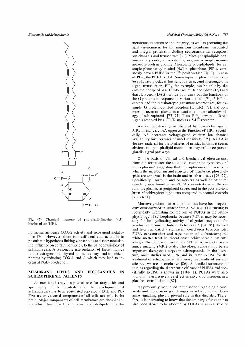
Eicosanoids and Schizophrenia Medicinal Chemistry, 2013, Vol. 9, No. 6 767
hormones influence COX-2 activity and eicosanoid metabo-lism [70]. However, there is insufficient data available to postulate a hypothesis linking eicosanoids and their modulat-ing influence on certain hormones, to the pathophysiology of schizophrenia. A reasonable interpretation of these findings is that estrogens and thyroid hormones may lead to schizo-phrenia by inducing COX-1 and -2 which may lead to in-creased PGE2 production.
MEMBRANE LIPIDS AND EICOSANOIDS IN SCHIZOPHRENIC PATIENTS
As mentioned above, a pivotal role for fatty acids and specifically PUFA metabolism in the development of schizophrenia has been postulated repeatedly [31], and PU-FAs are an essential component of all cells not only in the brain. Major components of cell membranes are phospholip-ids which form the lipid bilayer. Phospholipids give the
membrane its structure and integrity, as well as providing the lipid environment for the numerous membrane associated and integral proteins, including neurotransmitter receptors, ion channels and transporters [31]. Most phospholipids con-tain a diglyceride, a phosphate group, and a simple organic molecule such as choline. Membrane phospholipids, for ex-ample phosphatidylinositol (4,5)-bisphosphate (PIP2), com-monly have a PUFA in the 2nd position (see Fig. 7). In case of PIP2, the PUFA is AA. Some types of phospholipids can be split into products that function as second messengers in signal transduction. PIP2, for example, can be split by the enzyme phospholipase C into inositol triphosphate (IP3) and diacylglycerol (DAG), which both carry out the functions of the G proteins in response to various stimuli [71]. 5-HT re-ceptors and the metabotropic glutamate receptor are, for ex-ample, G protein-coupled receptors (GPCR) [72]; and both types of receptors play a significant role in the pathophysiol-ogy of schizophrenia [73, 74]. Thus, PIP2 forwards afferent signals received by a GPCR such as a 5-HT receptor.
AA can additionally be liberated by lipase cleavage of PIP2. In that case, AA opposes the function of PIP2. Specifi-cally, AA decreases voltage-gated calcium ion channel availability but increases channel sensitivity [75]. As AA is the raw material for the synthesis of prostaglandins, it seems obvious that phospholipid metabolism may influence prosta-glandin signal pathways.
On the basis of clinical and biochemical observations, Horrobin formulated the so-called ‘membrane hypothesis of schizophrenia’ suggesting that schizophrenia is a disorder in which the metabolism and structure of membrane phosphol-ipids are abnormal in the brain and in other tissues [76, 77]. Specifically, Horrobin and co-workers as well as other re-search groups found lower PUFA concentrations in the se-rum, the plasma, in peripheral tissues and in the post-mortem brain of schizophrenia patients compared to normal controls [76, 78-81].
Moreover, white matter abnormalities have been repeat-edly demonstrated in schizophrenia [82, 83]. This finding is specifically interesting for the role of PUFAs in the patho-physiology of schizophrenia, because PUFAs may be neces-sary for the myelinating activity of oligodendrocytes or for myelin maintenance. Indeed, Peters et al. [84, 85] showed and later replicated a significant correlation between total PUFA concentration and myelination of a frontotemporal white matter tract in recent-onset schizophrenia patients, using diffusion tensor imaging (DTI) in a magnetic reso-nance imaging (MRI) study. Therefore, PUFAs may be an important therapeutic target in schizophrenia. In the litera-ture, most studies used EPA and its ester E-EPA for the treatment of schizophrenia. However, the results of system-atic reviews are inconclusive [86]. A detailed summery of studies regarding the therapeutic efficacy of PUFAs and spe-cifically E-EPA is shown in (Table 1). PUFAs were also found to have a preventive effect on psychotic disorders in a placebo-controlled trial [87].
As previously mentioned in the section regarding eicosa-noids and monoaminergic changes in schizophrenia, dopa-mine signalling plays a pivotal role in this disorder. There-fore, it is interesting to know that dopaminergic function has also been shown to be affected by PUFAs in animal studies
OH OHO
O
OH
P O
O
O
P O
O
O
O
P OO
O
O O
O O
Fig. (7). Chemical structure of phosphatidylinositol (4,5)-bisphosphate (PIP2).
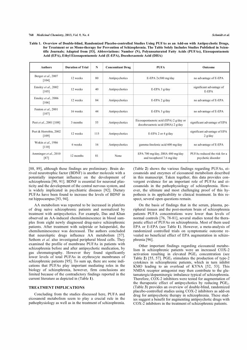
768 Medicinal Chemistry, 2013, Vol. 9, No. 6 Schmidt et al.
[88, 89], although these findings are preliminary. Brain de-rived neurotrophic factor (BDNF) is another molecule with a potentially important influence on the development of schizophrenia [90, 91]. BDNF is essential for neuronal plas-ticity and the development of the central nervous system, and is widely implicated in psychiatric diseases [92]. Dietary PUFAs have been found to increase the levels of BDNF in rat hippocampus [93, 94].
AA metabolism was reported to be increased in platelets of drug naive schizophrenic patients and normalized by treatment with antipsychotics. For example, Das and Khan observed an AA-induced chemiluminescence in blood sam-ples from eight newly diagnosed drug-naive schizophrenic patients. After treatment with sulpiride or haloperidol, the chemiluminescence was decreased. The authors concluded that neuroleptic drugs influence AA metabolism [57]. Sethom et al. also investigated peripheral blood cells. They examined the profile of membrane PUFAs in patients with schizophrenia before and after antipsychotic medication, by gas chromatography. However they found significantly lower levels of total PUFAs in erythrocyte membranes of schizophrenic patients [95]. To sum up, there are some indi-cations that PUFAs play important mediating roles in the biology of schizophrenia, however, firm conclusions are limited because of the contradictory findings reported in the current literature as depicted in (Table 1).
TREATMENT IMPLICATIONS
Concluding from the studies discussed here, PUFA and eicosanoid metabolism seem to play a crucial rule in the pathophysiology as well as in the treatment of schizophrenia.
(Table 2) shows the various findings regarding PUFAs, ei-cosanoids and enzymes of eicosanoid metabolism described in this manuscript. Taken together, this data provides con-vergent evidence for an important role of PUFAs and ei-cosanoids in the pathophysiology of schizophrenia. How-ever, the ultimate and most challenging proof of this hy-pothesis is its applicability to clinical treatment. In this re-spect, several open questions remain.
On the basis of findings that in the serum, plasma, pe-ripheral tissues and the post-mortem brain of schizophrenia patients PUFA concentrations were lower than levels of normal controls [76, 78-81], several studies tested the thera-peutic effect of PUFAs on schizophrenia. Most of them used EPA or E-EPA (see Table 1). However, a meta-analysis of randomized controlled trials on symptomatic outcome re-vealed no beneficial effect of EPA augmentation in schizo-phrenia [96].
Other important findings regarding eicosanoid metabo-lism in schizophrenic patients were an increased COX-2 activation resulting in elevated PGE2 concentration (see Table 2) [55, 57]. PGE2 stimulates the production of type-2 cytokines in schizophrenic patients, which in turn inhibit KMO leading to an overload of KYNA [52, 53]. This NMDA receptor antagonist may then contribute to the glu-tamatergic/dopaminergic imbalance typical of schizophrenia. Therefore, COX-2 inhibitors were tested for augmentation of the therapeutic effect of antipsychotics by reducing PGE2. (Table 3) provides an overview of double-blind, randomized placebo-controlled studies using COX-2 inhibitors as add-on drug for antipsychotic therapy in schizophrenia. These stud-ies suggest a benefit for augmenting antipsychotic drugs with COX-2 inhibitors in the treatment of schizophrenic patients.
Table 1. Overview of Double-blind, Randomized Placebo-controlled Studies Using PUFAs as an Add-on with Antipsychotic Drugs, for Treatment or as Mono-therapy for Prevention of Schizophrenia. The Table Solely Includes Studies Published in Scien-tific Journals; Adapted from [53]. Abbreviations: Number (N), Polyunsaturated Fatty Acids (PUFAs), Eicosapentaenoic Acid (EPA), Ethyl Eicosapentaenoic Acid (E-EPA), Docohexaenoic Acid (DHA)
Authors Duration of Trial N Concomitant Drug PUFA Outcome
Berger et al., 2007 [104]
12 weeks 80 Antipsychotics E-EPA 2x500 mg/day no advantage of E-EPA
Emsley et al., 2002 [105]
12 weeks 40 Antipsychotics E-EPA 3 g/day significant advantage of
E-EPA
Emsley et al., 2006 [106]
12 weeks 84 Antipsychotics E-EPA 2 g/day no advantage of E-EPA
Fenton et al., 2001 [107]
16 weeks 44 Antipsychotics E-EPA 3 g/day no advantage of E-EPA
Peet et al., 2001 [108] 3 months 55 Antipsychotics Eicosapentaenoic acid (EPA) 2 g/day or
docohexaenoic acid (DHA) 2 g/day significant advantage of EPA
Peet & Horrobin, 2002 [109]
12 weeks 115 Antipsychotics E-EPA 2 or 4 g/day significant advantage of EPA
2 g/day
Wokin et al., 1986 [110]
6 weeks 16 Antipsychotics gamma-linolenic acid 600 mg/day no advantage of E-EPA
Amminger et al., 2010 [87]
12 months 81 None EPA 700 mg/day, DHA 480 mg/day
and tocopherol 7.6 mg/day PUFAs reduced the risk for a
psychotic disorder
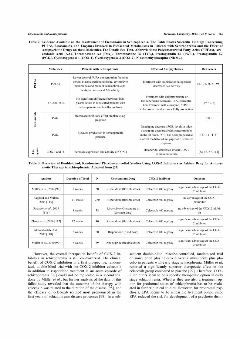
Eicosanoids and Schizophrenia Medicinal Chemistry, 2013, Vol. 9, No. 6 769
However, the overall therapeutic benefit of COX-2 in-hibitors in schizophrenia is still controversial. The clinical benefit of COX-2 inhibition in a first prospective, random-ized, double-blind trial with the COX-2 inhibitor celecoxib in addition to risperidone treatment in an acute episode of schizophrenia [97] could not be replicated in a second trial done by Müller et al., but further analysis of the data of this failed study revealed that the outcome of the therapy with celecoxib was related to the duration of the disease [98], and the efficacy of celecoxib seemed most pronounced in the first years of schizophrenic disease processes [98]. In a sub-
sequent double-blind, placebo-controlled, randomized trial of amisulpride plus celecoxib versus amisulpride plus pla-cebo in patients with early stage schizophrenia, Müller et al. reported a significantly superior therapeutic effect in the celecoxib group compared to placebo [99]. Therefore, COX-2 inhibitors seem to be a specific therapeutic option in early stage schizophrenia. Whether they are also a treatment op-tion for prodromal states of schizophrenia has to be evalu-ated in further clinical studies. However, for prodromal psy-chosis, EPA seems to be a feasible treatment option since EPA reduced the risk for development of a psychotic disor-
Table 2. Evidence Available on the Involvement of Eicosanoids in Schizophrenia. The Table Shows Scientific Findings Concerning PUFAs, Eicosanoids, and Enzymes Involved in Eicosanoid Metabolisms in Patients with Schizophrenia and the Effect of Antipsychotic Drugs on these Molecules. For Details See Text. Abbreviations: Polyunsaturated Fatty Acids (PUFAs), Ara-chidonic Acid (AA), Thromboxane A2 (TxA2), Thromboxane B2 (TxB2), Prostaglandin E1 (PGE1), Prostaglandin E2 (PGE2), Cyclooxygenase 1 (COX-1), Cyclooxygenase 2 (COX-2), N-desmethylclozapine (NDMC)
Molecules Patients with Schizophrenia Effects of Antipsychotics References
PUFA
s
PUFAs
Lower general PUFA concentration found in serum, plasma, peripheral tissue, erythrocyte membranes and brain of schizophrenic pa-
tients, but increased AA activity.
Treatment with sulpiride or haloperidol decreases AA activity
[57, 76, 78-81, 95]
TxA2 and TxB2 No significant difference between TxB2 plasma levels in medicated patients with
schizophrenia and healthy controls
Treatment with chlorpromazine or trifluoperazine decreases TxA2 concentra-
tion, treatment with clozapine, NDMC, chlorpromazine decreases TxB2 production
[39, 40, 3]
PGE1 Decreased inhibitory effect on platelet ag-
gregation [41]
Eic
osan
oids
PGE2 Elevated production in schizophrenic
patients.
Quetiapine decreases PGE2 levels in mice, olanzapine decreases PGE2 concentrations in the rat brain. PGE2 has been proposed as a novel mediator of antipsychotic treatment
response.
[87, 111-113]
En-
zym
es
COX-1 and -2 Increased expression and activity of COX-1 Haloperidol decreases striatal COX-2
expression in rats. [52, 53, 57, 114]
Table 3. Overview of Double-blind, Randomized Placebo-controlled Studies Using COX-2 Inhibitors as Add-on Drug for Antipsy-chotic Therapy in Schizophrenia, Adapted from [53]
Authors Duration of Trial N Concomitant Drug COX-2 Inhibitor Outcome
Müller et al., 2002 [97] 5 weeks 50 Risperidone (flexible dose) Celecoxib 400 mg/day significant advantage of the COX-
2 inhibitor
Rappard and Müller, 2004 [115]
11 weeks 270 Risperidone (flexible dose) Celecoxib 400 mg/day no advantage of the COX-
2inhibitor
Rapaport et al., 2005 [116]
8 weeks 38 Risperidone Olanzapine or
(constant dose) Celecoxib 400 mg/day
no advantage of the COX-2 inhibi-tor
Zhang et al., 2006 [117] 12 weeks 40 Risperidone (flexible dose) Celecoxib 400 mg/day significant advantage of the COX-
2 inhibitor
Akhondzadeh et al., 2007 [118]
8 weeks 60 Risperidone (fixed dose) Celecoxib 400 mg/day significant advantage of the COX-
2 inhibitor
Müller et al., 2010 [99] 6 weeks 49 Amisulpride (flexible dose) Celecoxib 400 mg/day significant advantage of the COX-
2 inhibitor
770 Medicinal Chemistry, 2013, Vol. 9, No. 6 Schmidt et al.
der in combination with docosahexaenoic acid and toco-pherol [87].
It has not yet been shown whether COX-2 inhibitors or EPA improve or prevent the appearance of schizophrenic symptoms by modulating dopamine signaling. These mole-cules may exert their beneficial effects via an unknown mechanism, not or not directly related to dopamine signal-ing. The findings regarding PUFAs, eicosanoids and COX-2 in treatment of schizophrenic patients or with regard to their effects on antipsychotic agents do not answer whether ei-cosanoids play a role in the development of schizophrenia independently of dopaminergic signaling. Therefore, further clinical studies and trials should collect data regarding ei-cosanoid metabolism and regarding dopamine signaling. For example, a trial with COX-2 inhibitors could include posi-tron emission tomography (PET) of the dopaminergic system at baseline and during therapy.
TxA2 had been shown to facilitate dopamine release [18]. Therefore, the overactivitiy of the dopaminergic system in schizophrenic patients could be reduced by restricting TxA2 production. As clozapine and chlorpromazine have been shown to decrease TxA2 production [43], these drugs might – complementary to their direct effect on dopamine and 5-HT receptors – act as antipsychotics by reducing do-pamine signaling via the modulation of TxA2 or TxB2 pro-duction. However, thromboxanes also modulate vasocon-striction and platelet aggregation [19]. Therefore, known side effects of clozapine and similar antipsychotics such as orthostatic hypotension might also be, at least in part medi-ated by the influence of thromboxanes on the muscle tone of the blood vessels [100]. Additionally, the numerous immu-nological properties and various side effects of clozapine [101] may be linked to the immunological properties of TxA2 [102].
As PGE2, COX-2, TxA2 and TxB2 have been shown to be involved in the pathophysiology of schizophrenia or its treatment, and as lower general PUFA concentration has repeatedly been found in the serum, the plasma, the periph-eral tissue, the erythrocyte membranes and the brain of schizophrenic patients (see Table 2), the question arises, whether it would be beneficial to modulate the action or the intake of AA as a precursor molecule of eicosanoids. To reduce PGE2 and TxA2 and TxB2 one could theoretically try to reduce AA availability. However, reduced AA availability may have far-reaching effects on the integrity and function of cell membranes. Additionally, AA is not only used for the production of PGE2, COX-2, TxA2 and TxB2, it is also me-tabolized to leukotrienes via the lipoxygenase pathway [12] which play an important role as signaling molecules in vari-ous bodily functions such as immune defense, including de-fense against bacteria, fungi, and parasites [103]. Therefore, reducing the availability of AA may risk various adverse events and side effects.
The available studies on eicosanoids and schizophrenia are preliminary in nature and the results are partly inconsis-tent. It is not clear which subgroup of schizophrenic patients might particularly benefit from a treatment targeting eicosa-noid metabolism such as COX-2 inhibitors. Additionally, the mechanisms of the interplay between eicosanoids, cytokines, neurotransmitters and hormones are not yet fully understood.
However, eicosanoids which interact with cytokines, neuro-transmitters and hormones seem to be one link of a patho-physiological chain that can lead to the development of schizophrenia. In-vivo investigations of the action of antip-sychotics on eicosanoid metabolism of patients with schizo-phrenia may provide deeper insights into the mutual interac-tions of dopaminergic and serotonergic systems on the one hand and eicosanoid metabolism and signaling on the other hand, and its relevance for the pathophysiology and treat-ment of schizophrenia.
CONFLICT OF INTEREST
Prof. Himmerich received speaker honoraria from Astra-Zeneca, Lilly and Servier, consulting fees from Bristol-Myers Squibb, and chemical substances for study support from AstraZeneca, Novartis and Wyeth. All other authors reported no biomedical financial interests or potential con-flicts of interest.
ACKNOWLEDGEMENTS
The study was supported by the Claussen-Simon-Foundation and by LIFE – Leipzig Research Center for Civi-lization Diseases, University Leipzig. LIFE is funded by means of the European Union, by the European Regional Development Fund (ERDF) and by means of the Free State of Saxony within the framework of the excellence initiative.
The mentioned sponsors did not have any influence on the review process, the writing of the manuscript and in the decision to submit the paper for publication.
REFERENCES [1] Kraepelin, E. Psychiatrie: Ein Lehrbuch f. Studierende u. Aerzte,
1910. [2] Bleuler, E. Dementia praecox oder Gruppe der Schizophrenien;
Deuticke: Leipzig, 1911. [3] American Psychiatric Association. Diagnostic and statistical man-
ual of mental disorders: DSM-IV-TR; Washington, DC, 2000. [4] World Health Organization. ICD-10: International Statistical Clas-
sification of Diseases and Related Health Problems; World Health Organization: Geneva, 1994.
[5] Andreasen, N.C.; Olsen, S. Negative v positive schizophrenia. Definition and validation. Arch. Gen. Psychiatry., 1982, 39, 789-94.
[6] Lindenmayer, J.P.; Grochowski, S.; Hyman, R.B. Five factor model of schizophrenia: replication across samples. Schizophr. Res., 1995, 14, 229-34.
[7] Kay, S.R.; Sevy, S. Pyramidical model of schizophrenia. Schizophr. Bull., 1990, 16, 537-545.
[8] Jablensky, A. Subtyping schizophrenia: implications for genetic research. Mol. Psychiatry., 2006, 11, 815-36.
[9] Bray, N.J.; Leweke, F.M.; Kapur, S.; Meyer-Lindenberg, A. The neurobiology of schizophrenia: new leads and avenues for treat-ment. Curr. Opin. Neurobiol., 2010, 20, 810-5.
[10] Balu, D.T., Coyle, J.T. Neuroplasticity signaling pathways linked to the pathophysiology of schizophrenia. Neurosci. Biobehav. Rev., 2011, 35, 848-70.
[11] Lwin, A.; Symeon, C.; Jan, F.; Sule. A.. Morbidity and mortality in schizophrenia. Br. J. Hosp. Med., 2011, 72, 628-30.
[12] Marks, F.; Fürstenberger, G. Prostaglandins, leukotrienes, and other eicosanoids: From biogenesis to clinical application; Wiley-VCH: Weinheim, New York, 1999.
[13] Moore, S.A.; Yoder, E.; Spector, A.A. Role of the blood-brain barrier in the formation of long-chain omega-3 and omega-6 fatty acids from essential fatty acid precursors. J. Neurochem., 1990, 55, 391-402.
[14] Moore, S.A.; Yoder, E.; Murphy, S.; Dutton, G.R.; Spector, A.A. Astrocytes, not neurons, produce docosahexaenoic acid (22:6
Eicosanoids and Schizophrenia Medicinal Chemistry, 2013, Vol. 9, No. 6 771
omega-3) and arachidonic acid (20:4 omega-6). J. Neurochem., 1991, 56, 518-24.
[15] Chen, H.; Dzitoyeva, S.; Manev, H. 5-Lipoxygenase in mouse cerebellar Purkinje cells. Neuroscience., 2010, 171, 383-9.
[16] Kaufmann, W.E.; Worley, P.F.; Pegg, J.; Bremer, M.; Isakson, P. COX-2, a synaptically induced enzyme, is expressed by excitatory neurons at postsynaptic sites in rat cerebral cortex. Proc. Natl. Acad. Sci. U.S.A., 1996, 93, 2317-21.
[17] Coleman, R.A.; Smith, W.L.; Narumiya, S. International Union of Pharmacology classification of prostanoid receptors: properties, dis-tribution, and structure of the receptors and their subtypes. Pharma-col. Rev., 1994, 46, 205-29.
[18] Mitsumori, T.; Furuyashiki, T.; Momiyama, T.; Nishi, A.; Shuto, T.; Hayakawa, T.; Ushikubi, F.; Kitaoka, S.; Aoki, T.; Inoue, H.; Matsuoka, T.; Narumiya, S. Thromboxane receptor activation en-hances striatal dopamine release, leading to suppression of GABAergic transmission and enhanced sugar intake. Eur. J. Neuro-sci., 2011, 34, 594-604.
[19] Wacker, M.J.; Wilhelm, H.L.; Gomez, S.E.; Floor, E.; Orr, J.A. Role of serotonin in thromboxane A2-induced coronary chemore-flex. Am. J. Physiol. Heart Circ. Physiol., 2003, 284, 867-75.
[20] Weschenfelder, J.; Sander, C.; Kluge, M.; Kirkby, K.C.; Him-merich, H. The influence of cytokines on wakefulness regulation: clinical relevance, mechanisms and methodological problems. Psy-chiatr. Danub., 2012, 24, 112-26.
[21] van Rossum, J. M. The significance of dopamine-receptor blockade for the mechanism of action of neuroleptic drugs. Arch. Int. Phar-macodyn. Ther., 1966, 160, 492-4.
[22] Horn, A. S.; Snyder, S. H. Chlorpromazine and dopamine: confor-mational similarities that correlate with the antischizophrenic activ-ity of phenothiazine drugs. Proc. Natl. Acad. Sci. U.S.A., 1971, 68, 2325-8.
[23] Bürki, H.R.; Eichenberger, E.; Sayers, A.C.; White, T.G. Clozapine and the dopamine hypothesis of schizophrenia, a critical appraisal. Pharmakopsychiatr. Neuropsychopharmakol., 1975, 8, 115-21.
[24] Lieberman, J.A.; Kane, J.M.; Alvir, J. Provocative tests with psy-chostimulant drugs in schizophrenia. Psychopharmacology (Berl.), 1987, 91, 415-33.
[25] Davis, K.L.; Kahn, R.S.; Ko, G.; Davidson, M. Dopamine in schizophrenia: a review and reconceptualization. Am. J. Psychiatry, 1991, 148, 1474-86.
[26] Pogarell, O.; Koch, W.; Karch, S.; Dehning, S.; Müller, N.; Tatsch, K.; Poepperl, G.; Möller, H.-J. Dopaminergic neurotransmission in patients with schizophrenia in relation to positive and negative symptoms. Pharmacopsychiatry, 2012, 45 (Suppl 1), 36-41.
[27] Bauer, M.; Praschak-Rieder, N.; Kasper, S.; Willeit, M. Is dopa-mine neurotransmission altered in prodromal schizophrenia? A re-view of the evidence. Curr. Pharm. Des., 2012, 18, 1568-79.
[28] Remington, G.; Agid, O.; Foussias, G. Schizophrenia as a disorder of too little dopamine: implications for symptoms and treatment. Expert Rev. Neurother., 2011, 11, 589-607.
[29] Stauffer, V. L.; Song, G.; Kinon, B. J.; Ascher-Svanum, H.; Chen, L.; Feldman, P.D.; Conley, R.R. Responses to antipsychotic therapy among patients with schizophrenia or schizoaffective disorder and either predominant or prominent negative symptoms. Schizophr. Res., 2012, 134, 195-201.
[30] Lisman, J.E.; Coyle, J.T.; Green, R.W.; Javitt, D.C.; Benes, F.M.; Heckers, S.; Grace, A.A. Circuit-based framework for understand-ing neurotransmitter and risk gene interactions in schizophrenia. Trends Neurosci., 2008, 31, 234-42.
[31] Ross, B.M. Phospholipid and eicosanoid signaling disturbances in schizophrenia. Prostaglandins Leukot. Essent. Fatty Acids, 2003, 69, 407-12.
[32] Moncada, S.; Vane, J.R. Pharmacology and endogenous roles of prostaglandin endoperoxides, thromboxane A2, and prostacyclin. Pharmacol. Rev., 1978, 30, 293-331.
[33] Di Pietro, N.C.; Seamans, J.K. Dopamine and serotonin interactions in the prefrontal cortex: insights on antipsychotic drugs and their mechanism of action. Pharmacopsychiatry, 2007, 40 Suppl 1, 27-33.
[34] Inagaki, N.; Wada, H. Histamine and prostanoid receptors on glial cells. Glia, 1994, 11, 102-9.
[35] Gao, H.; Peng, B.; Welch, W.J., Wilcox, C.S. Central thromboxane receptors: mRNA expression and mediation of pressor responses. Am. J. Physiol., 1997, 272, 1493-1500.
[36] Ansar, S.; Larsen, C.; Maddahi, A.; Edvinsson, L. Subarachnoid hemorrhage induces enhanced expression of thromboxane A2 re-ceptors in rat cerebral arteries. Brain Res., 2010, 1316, 163-72.
[37] Nishihara, M.; Yokotani, K.; Inoue, S.; Osumi, Y. U-46619, a se-lective thromboxane A2 mimetic, inhibits the release of endogenous noradrenaline from the rat hippocampus in vitro. Jpn. J. Pharma-col., 2000, 82, 226-31.
[38] Praticò, D. The neurobiology of isoprostanes and Alzheimer's dis-ease. Biochim. Biophys. Acta. 2010, 1801, 930-3.
[39] Dietrich-Muszalska, A.; Olas, B. Isoprostenes as indicators of oxi-dative stress in schizophrenia. World J. Biol. Psychiatry, 2009, 10, 27-33.
[40] Anfossi, G.; Massucco, P.; Mularoni, E.; Cavalot, F.; Burzacca, S.; Mattiello, L.; Trovati, M. Phenothiazines inhibit collagen-induced thromboxane B2 synthesis and increase forskolin anti-aggregating effects in human platelets. Gen. Pharmacol., 1991, 22, 773-8.
[41] Kaiya, H.; Imai, H.; Muramatsu, Y.; Nozaki, M.; Fujimura, H.; Adachi, S.; Namba, M. Platelet aggregation response in schizophre-nia and prostaglandin E1. Psychiatry Res., 1983, 9, 309-18.
[42] Ishigooka, J.; Shizu, Y.; Wakatabe, H.; Tanaka, K.; Miura, S. Dif-ferent effects of centrally acting drugs on rabbit platelet aggrega-tion: with special reference to selective inhibitory effects of antip-sychotics and antidepressants. Biol. Psychiatry, 1985, 20, 866-73.
[43] Himmerich, H.; Schmidt, L.; Becker, S.; Kortz, L.; Schonherr, J.; Mergl, R.; Bauer, K.; Sack, U.; Sheldrick, A. J.; Thiery, J.; Ceglarek, U. Impact of clozapine, N-desmethylclozapine and chlor-promazine on thromboxane production in vitro. Med. Chem., 2012, 8, 1032-8.
[44] Steinberg, H.; Himmerich, H. Emil Kraepelin's habilitation and his thesis: A pioneer work for modern systematic reviews, psychoim-munological research and categories of psychiatric diseases. World J. Biol. Psychiatry, 2011, Epub.
[45] Himmerich, H.; Kirkby, K.C.; Steinberg, H. From Julius Wagner-Jauregg to biomarkers and immunological therapies. World J. Biol. Psychiatry, 2010, 11, 154-5.
[46] Debnath, M.; Cannon, D.M.; Venkatasubramanian, G. Variation in the major histocompatibility complex [MHC] gene family in schizophrenia: Associations and functional implications. Prog. Neuropsychopharmacol. Biol. Psychiatry, 2013, 42, 49-62.
[47] Berthold-Losleben, M.; Heitmann, S.; Himmerich, H. Anti-inflammatory drugs in psychiatry. Inflamm. Allergy Drug Targets, 2009, 8, 266-76.
[48] Brown, A.S.; Hooton, J.; Schaefer, C.A.; Zhang, H.; Petkova, E.; Babulas, V.; Perrin, M.; Gorman, J.M.; Susser, E.S. Elevated ma-ternal interleukin-8 levels and risk of schizophrenia in adult off-spring. Am. J. Psychiatry, 2004, 161, 889-95.
[49] Himmerich, H.; Berthold-Losleben, M.; Pollmächer, T. The rele-vance of the TNF-alpha system in psychiatric disorders. Fortschr. Neurol. Psychiatr., 2009, 77, 334-45.
[50] Müller, N.; Riedel, M.; Ackenheil, M.; Schwarz, M.J. Cellular and humoral immune system in schizophrenia: a conceptual re-evaluation. World J. Biol. Psychiatry, 2000, 1, 173-9.
[51] van Kammen, D.P.; McAllister-Sistilli, C.G.; Kelley, M.E. In Cur-rent update in psychoimmunology; Wieselmann, Ed.; Springer: Berlin, Heidelberg, New York, 1997, pp. 51-55.
[52] Müller, N.; Myint, A.-M.; Schwarz, M.J. Kynurenine pathway in schizophrenia: pathophysiological and therapeutic aspects. Curr. Pharm. Des., 2011, 17, 130-136.
[53] Müller, N.; Myint, A.-M.; Schwarz, M.J. Immunological treatment options for schizophrenia. Curr. Pharm. Biotechnol., 2012, 13, 1606-13.
[54] Himmerich, H.; Sorge, S.; Kirkby, K.C.; Steinberg, H. Schizo-phrene Störungen. Die Entwicklung immunologischer Krankheits- und Therapiekonzepte. Nervenarzt, 2012, 83, 7-8, 10-2, 14-5.
[55] Kaiya, H.; Uematsu, M.; Ofuji, M.; Nishida, A.; Takeuchi, K.; Nozaki, M.; Idaka, E. Elevated plasma prostaglandin E2 levels in schizophrenia. J. Neural Transm., 1989, 77, 39-46.
[56] Martínez-Gras, I.; Pérez-Nievas, B.G.; García-Bueno, B.; Madrigal, J.L.; Andrés-Esteban, E.; Rodríguez-Jiménez, R.; Hoenicka, J.; Palomo, T.; Rubio, G.; Leza, J.C. The anti-inflammatory prosta-glandin 15d-PGJ2 and its nuclear receptor PPARgamma are de-creased in schizophrenia. Schizophr. Res., 2011, 128, 15-22.
[57] Das, I.; Khan, N.S. Increased arachidonic acid induced platelet chemiluminescence indicates cyclooxygenase overactivity in schizophrenic subjects. Prostaglandins Leukot. Essent. Fatty Acids, 1998, 58, 165-8.
772 Medicinal Chemistry, 2013, Vol. 9, No. 6 Schmidt et al.
[58] Stolina, M.; Sharma, S.; Lin, Y.; Dohadwala, M.; Gardner, B.; Luo, J.; Zhu, L.; Kronenberg, M.; Miller, P. W.; Portanova, J.; Lee, J. C.; Dubinett, S. M. Specific inhibition of cyclooxygenase 2 restores an-titumor reactivity by altering the balance of IL-10 and IL-12 syn-thesis. J. Immunol., 2000, 164, 361-70.
[59] Erhardt, S.; Blennow, K.; Nordin, C.; Skogh, E.; Lindström, L. H.; Engberg, G. Kynurenic acid levels are elevated in the cerebrospinal fluid of patients with schizophrenia. Neurosci. Lett., 2001, 313, 96-8.
[60] Nilsson, L. K.; Linderholm, K. R.; Engberg, G.; Paulson, L.; Blen-now, K.; Lindström, L.H.; Nordin, C.; Karanti, A.; Persson, P.; Erhardt, S. Elevated levels of kynurenic acid in the cerebrospinal fluid of male patients with schizophrenia. Schizophr. Res., 2005, 80, 315-22.
[61] Linderholm, K.R.; Skogh, E.; Olsson, S.K.; Dahl, M.-L.; Holtze, M.; Engberg, G.; Samuelsson, M.; Erhardt, S. Increased levels of kynurenine and kynurenic acid in the CSF of patients with schizo-phrenia. Schizophr. Bull., 2012, 38, 426-32.
[62] Wonodi, I.; Stine, O.C.; Sathyasaikumar, K.V.; Roberts, R.C.; Mitchell, B.D.; Hong, L.E.; Kajii, Y.; Thaker, G.K.; Schwarcz, R. Downregulated kynurenine 3-monooxygenase gene expression and enzyme activity in schizophrenia and genetic association with schizophrenia endophenotypes. Arch. Gen. Psychiatry, 2011, 68, 665-74.
[63] Pocivavsek, A.; Wu, H.Q.; Potter, M.C.; Elmer, G.I.; Pellicciari, R.; Schwarcz, R. Fluctuations in endogenous kynurenic acid control hippocampal glutamate and memory. Neuropsychopharmacology 2011, 36, 2357-67.
[64] Müller, N. COX-2 inhibitors as antidepressants and antipsychotics: clinical evidence. Curr. Opin. Investig. Drugs, 2010, 11, 31-42.
[65] Himmerich, H.; Steinberg, H. Hormonelle Krankheits- und Thera-piekonzepte in der Psychiatrie. Fortschr. Neurol. Psychiatr., 2011, 79, 384-94.
[66] Kulkarni, J.; Hayes, E.; Gavrilidis, E. Hormones and schizophrenia. Curr. Opin. Psychiatry, 2012, 25, 89-95.
[67] Santos, N.C.; Costa, P.; Ruano, D.; Macedo, A.; Soares, M.J.; Va-lente, J.; Pereira, A.T.; Azevedo, M. H.; Palha, J. A. Revisiting thy-roid hormones in schizophrenia. J. Thyroid Res., 2012, 2012, 569147.
[68] Amateau, S.K.; McCarthy, M.M. Induction of PGE2 by estradiol mediates developmental masculinization of sex behavior. Nat. Neu-rosci., 2004, 7, 643-50.
[69] McCarthy, M.M. How it's made: organisational effects of hormones on the developing brain. J. Neuroendocrinol., 2010, 22, 736-42.
[70] Carey, L.C.; Valego, N.K.; Chen, K.; Rose, J.C. Thyroid hormone regulates renocortical COX-2 and PGE2 expression in the late ges-tation fetal sheep. Reprod. Sci., 2008, 15, 598-603.
[71] Choi, S.-Y.; Chang, J.; Jiang, B.; Seol, G.-H.; Min, S.-S.; Han, J.-S.; Shin, H.-S.; Gallagher, M.; Kirkwood, A. Multiple receptors coupled to phospholipase C gate long-term depression in visual cor-tex. J. Neurosci., 2005, 25, 11433-43.
[72] Fredriksson, R.; Lagerström, M.C.; Lundin, L.-G.; Schiöth, H.B. The G-protein-coupled receptors in the human genome form five main families. Phylogenetic analysis, paralogon groups, and finger-prints. Mol. Pharmacol., 2003, 63, 1256-72.
[73] Ohuoha, D.C.; Hyde, T.M.; Kleinman, J.E. The role of serotonin in schizophrenia: an overview of the nomenclature, distribution and alterations of serotonin receptors in the central nervous system. Psychopharmacology (Berl.), 1993, 112 (Suppl 1), 5-15.
[74] Goff, D.C.; Coyle, J.T. The emerging role of glutamate in the pathophysiology and treatment of schizophrenia. Am. J. Psychiatry, 2001, 158, 1367-77.
[75] Roberts-Crowley, M.L.; Mitra-Ganguli, T.; Liu, L.; Rittenhouse, A.R. Regulation of voltage-gated Ca2+ channels by lipids. Cell Calcium, 2009, 45, 589-601.
[76] Horrobin, D.F.; Glen, A.I.; Vaddadi, K. The membrane hypothesis of schizophrenia. Schizophr. Res., 1994, 13, 195-207.
[77] Horrobin, D.F. The membrane phospholipid hypothesis as a bio-chemical basis for the neurodevelopmental concept of schizophre-nia. Schizophr. Res., 1998, 30, 193-208.
[78] Yao, J.K.; Leonard, S.; Reddy, R.D. Membrane phospholipid ab-normalities in postmortem brains from schizophrenic patients. Schizophr. Res., 2000, 42, 7-17.
[79] Landén, M.; Davidsson, P.; Gottfries, C.-G.; Månsson, J.-E.; Blen-now, K. Reduction of the synaptophysin level but normal levels of
glycerophospholipids in the gyrus cinguli in schizophrenia. Schizophr. Res., 2002, 55, 83-8.
[80] McNamara, R.K.; Jandacek, R.; Rider, T.; Tso, P.; Hahn, C.-G.; Richtand, N.M.; Stanford, K.E. Abnormalities in the fatty acid composition of the postmortem orbitofrontal cortex of schizo-phrenic patients: gender differences and partial normalization with antipsychotic medications. Schizophr. Res., 2007, 91, 37-50.
[81] Freeman, M.P.; Hibbeln, J.R.; Wisner, K.L.; Davis, J.M.; Mischou-lon, D.; Peet, M.; Keck, P.E.; Marangell, L.B.; Richardson, A.J.; Lake, J.; Stoll, A.L. Omega-3 fatty acids: evidence basis for treat-ment and future research in psychiatry. J. Clin. Psychiatry, 2006, 67, 1954-67.
[82] Di, X.; Chan, R. C.K.; Gong, Q.-Y. White matter reduction in pa-tients with schizophrenia as revealed by voxel-based morphometry: an activation likelihood estimation meta-analysis. Prog. Neuropsy-chopharmacol. Biol. Psychiatry, 2009, 33, 1390-4.
[83] Connor, C.M.; Crawford, B.C.; Akbarian, S. White matter neuron alterations in schizophrenia and related disorders. Int. J. Dev. Neu-rosci., 2011, 29, 325-34.
[84] Peters, B.D.; Duran, M.; Vlieger, E.J.; Majoie, C.B.; den Heeten, G. J.; Linszen, D.H.; Haan, L. de. Polyunsaturated fatty acids and brain white matter anisotropy in recent-onset schizophrenia: a prelimi-nary study. Prostaglandins Leukot. Essent. Fatty Acids, 2009, 81, 61-3.
[85] Peters, B.D.; Machielsen, M.W. J.; Hoen, W.P.; Caan, M.W.A.; Malhotra, A.K.; Szeszko, P. R.; Duran, M.; Olabarriaga, S. D.; Haan, L. de. Polyunsaturated Fatty Acid Concentration Predicts Myelin Integrity in Early-Phase Psychosis. Schizophr. Bull., 2013, Epub.
[86] Berger, G.E.; Smesny, S.; Amminger, G.P. Bioactive lipids in schizophrenia. Int. Rev. Psychiatry, 2006, 18, 85-98.
[87] Amminger, G.P.; Schäfer, M.R.; Papageorgiou, K.; Klier, C.M.; Cotton, S.M.; Harrigan, S.M.; Mackinnon, A.; McGorry, P.D.; Berger, G.E. Long-chain omega-3 fatty acids for indicated preven-tion of psychotic disorders: a randomized, placebo-controlled trial. Arch. Gen. Psychiatry, 2010, 67, 146-54.
[88] Zimmer, L.; Hembert, S.; Durand, G.; Breton, P.; Guilloteau, D.; Besnard, J.C.; Chalon, S. Chronic n-3 polyunsaturated fatty acid diet-deficiency acts on dopamine metabolism in the rat frontal cor-tex: a microdialysis study. Neurosci. Lett., 1998, 240, 177-81.
[89] Zimmer, L.; Vancassel, S.; Cantagrel, S.; Breton, P.; Delamanche, S.; Guilloteau, D.; Durand, G.; Chalon, S. The dopamine mesocor-ticolimbic pathway is affected by deficiency in n-3 polyunsaturated fatty acids. Am. J. Clin. Nutr., 2002, 75, 662-7.
[90] Kordi-Tamandani, D.M.; Sahranavard, R.; Torkamanzehi, A. DNA methylation and expression profiles of the brain-derived neurotro-phic factor (BDNF) and dopamine transporter (DAT1) genes in pa-tients with schizophrenia. Mol. Biol. Rep., 2012, 39, 10889-93.
[91] Martinotti, G.; Di Iorio, G.; Marini, S.; Ricci, V.; Berardis, D. de; Di Giannantonio, M. Nerve growth factor and brain-derived neu-rotrophic factor concentrations in schizophrenia: a review. J. Biol. Regul. Homeost. Agents, 2012, 26, 347-56.
[92] Autry, A.E.; Monteggia, L.M. Brain-derived neurotrophic factor and neuropsychiatric disorders. Pharmacol. Rev., 2012, 64, 238-58.
[93] Wu, A.; Ying, Z.; Gomez-Pinilla, F. The interplay between oxida-tive stress and brain-derived neurotrophic factor modulates the out-come of a saturated fat diet on synaptic plasticity and cognition. Eur. J. Neurosci., 2004, 19, 1699-707.
[94] Wu, A.; Ying, Z.; Gomez-Pinilla, F. Docosahexaenoic acid dietary supplementation enhances the effects of exercise on synaptic plas-ticity and cognition. Neuroscience, 2008, 155, 751-9.
[95] Sethom, M.M.; Fares, S.; Bouaziz, N.; Melki, W.; Jemaa, R.; Feki, M.; Hechmi, Z.; Kaabachi, N. Polyunsaturated fatty acids deficits are associated with psychotic state and negative symptoms in pa-tients with schizophrenia. Prostaglandins Leukot. Essent. Fatty Ac-ids, 2010, 83, 131-6.
[96] Fusar-Poli, P.; Berger, G. Eicosapentaenoic acid interventions in schizophrenia: meta-analysis of randomized, placebo-controlled studies. J. Clin. Psychopharmacol., 2012, 32, 179-85.
[97] Müller, N.; Riedel, M.; Scheppach, C.; Brandstätter, B.; Sokullu, S.; Krampe, K.; Ulmschneider, M.; Engel, R.R.; Möller, H.J.; Schwarz, M.J. Beneficial antipsychotic effects of celecoxib add-on therapy compared to risperidone alone in schizophrenia. Am. J. Psychiatry, 2002, 159, 1029-34.
[98] Müller, N.; Schwarz, M.J. Immune system and schizophrenia. Curr. Immunol. Rev., 2010, 6, 213-20.
Eicosanoids and Schizophrenia Medicinal Chemistry, 2013, Vol. 9, No. 6 773
[99] Müller, N.; Krause, D.; Dehning, S.; Musil, R.; Schennach-Wolff, R.; Obermeier, M.; Möller, H.J.; Klauss, V.; Schwarz, M.J.; Riedel, M. Celecoxib treatment in an early stage of schizophrenia: results of a randomized, double-blind, placebo-controlled trial of celecoxib augmentation of amisulpride treatment. Schizophr Res., 2010, 121, 118-24.
[100] Leung, E.; Walsh, L.K.; Pulido-Rios, M.T.; Eglen, R. M. Charac-terization of putative 5-ht7 receptors mediating direct relaxation in Cynomolgus monkey isolated jugular vein. Br. J. Pharmacol., 1996, 117, 926-30.
[101] Pollmächer, T.; Haack, M.; Schuld, A.; Kraus, T.; Hinze-Selch, D. Effects of antipsychotic drugs on cytokine networks. J. Psychiatr. Res., 2000, 34, 369-82.
[102] Ricciotti, E.; FitzGerald, G.A. Prostaglandins and inflammation. Arterioscler. Thromb. Vasc. Biol., 2011, 31, 986-1000.
[103] Rogerio, A.P.; Anibal, F.F. Role of leukotrienes on protozoan and helminth infections. Mediators Inflamm. 2012, 2012, 595694.
[104] Berger, G.E.; Proffitt, T.M.; McConchie, M.; Yuen, H.; Wood, S.J.; Amminger, G.P.; Brewer, W.; McGorry, P.D. Ethyl-eicosapentaenoic acid in first-episode psychosis: a randomized, pla-cebo-controlled trial. J. Clin. Psychiatry, 2007, 68, 1867-75.
[105] Emsley, R.; Myburgh, C.; Oosthuizen, P.; van Rensburg, S.J. Ran-domized, placebo-controlled study of ethyl-eicosapentaenoic acid as supplemental treatment in schizophrenia. Am. J. Psychiatry, 2002, 159, 1596-8.
[106] Emsley, R.; Niehaus, D.J.; Koen, L.; Oosthuizen, P.P.; Turner, H.J.; Carey, P.; van Rensburg, S.J.; Maritz, J.S.; Murck, H. The effects of eicosapentaenoic acid in tardive dyskinesia: a randomized, placebo-controlled trial. Schizophr. Res., 2006, 84, 112-20.
[107] Fenton, W.S.; Dickerson, F.; Boronow, J.; Hibbeln, J.R.; Knable, M. A placebo-controlled trial of omega-3 fatty acid (ethyl eicosap-entaenoic acid) supplementation for residual symptoms and cogni-tive impairment in schizophrenia. Am. J. Psychiatry, 2001, 158, 2071-4.
[108] Peet, M.; Brind, J.; Ramchand, C.N.; Shah, S.; Vankar, G.K. Two double-blind placebo-controlled pilot studies of eicosapentaenoic acid in the treatment of schizophrenia. Schizophr Res., 2001, 49, 243-51.
[109] Peet, M.; Horrobin, D.F.; E-E Multicentre Study Group. A dose-ranging exploratory study of the effects of ethyl-eicosapentaenoate in patients with persistent schizophrenic symptoms. J. Psychiatr. Res., 2002, 36, 7-18.
[110] Wolkin, A.; Jordan, B.; Peselow, E.; Rubinstein, M.; Rotrosen, J. Essential fatty acid supplementation in tardive dyskinesia. Am. J. Psychiatry, 1986, 143, 912-4.
[111] Kim, H.; Bang, J.; Chang, H.W.; Kim, J.Y.; Park, K.U.; Kim. S.H.; Lee, K.J.; Cho, C.H., Hwang, I.; Park, S.D.; Ha, E.; Jung, S.W. Anti-inflammatory effect of quetiapine on collagen-induced arthritis of mouse. Eur. J. Pharmacol., 2012, 678, 55-60.
[112] Cheon Y, Park JY, Modi HR, Kim HW, Lee HJ, Chang L, Rao JS, Rapoport SI. Chronic olanzapine treatment decreases arachidonic acid turnover and prostaglandin E2 concentration in rat brain. J. Neurochem., 2011, 119, 364-76.
[113] Adkins, D.E.; Khachane, A.N.; McClay, J.L.; Aberg, K.; Bukszár, J.; Sullivan, P.F.; van den Oord, E.J. SNP-based analysis of neuro-active ligand-receptor interaction pathways implicates PGE2 as a novel mediator of antipsychotic treatment response: data from the CATIE study. Schizophr. Res., 2012, 135, 200-1.
[114] Marin, C.; Saldaña, M.; Roca-Ferrer, J.; Bonastre, M.; Aguilar, E.; Mullol, J. Striatal and nigral COX-2 expression after chronic typical and atypical neuroleptic administration in rats. Prog. Neuropsycho-pharmacol. Biol. Psychiatry, 2007, 31, 678-82.
[115] Rappard, F.; Müller, N. Celecoxib Add-on Therapy Does Not Have Beneficial Antipsychotic Effects Over Risperidone Alone in Schizophrenia. Neuropsychopharmacology, 2004, 29 (Suppl. 1), 222.
[116] Rapaport, M.H.; Delrahim, K.K.; Bresee, C.J.; Maddux, R.E.; Ah-madpour, O.; Dolnak, D.; Celecoxib augmentation of continuously ill patients with schizophrenia. Biol. Psychiatry, 2005, 57, 1594-6.
[117] Zhang, Y.; Chun Chen, D.; Long Tan, Y.; Zhou, D.F. A double-blind, placebo-controlled trial of celecoxib addes to risperidone in first-episode and drug-naive patients with schizophrenia. Eur. Arch. Psychiatry Clin. Neurosci., 2006, 256 (Suppl. 2), II/50.
[118] Akhondzadeh, S.; Tabatabaee, M.; Amini, H.; Ahmadi Abhari, S.A.; Abbasi, S.H.; Behnam, B. Celecoxib as adjunctive therapy in schizophrenia: a double-blind, randomized and placebo-controlled trial. Schizophr. Res., 2007, 90, 179-85.
Received: January 22, 2013 Revised: March 09, 2013 Accepted: April 09, 2013

8. Anlagen
8.2 Publikation 2 - Impact of clozapine, N desmethylclozapine and chlorpromazine on
thromboxane production in vitro
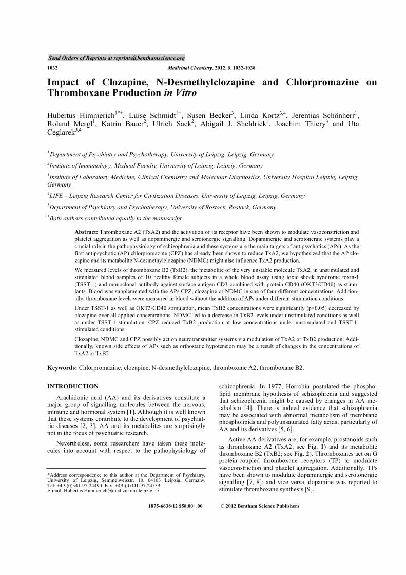
Send Orders of Reprints at [email protected]
1032 Medicinal Chemistry, 2012, 8, 1032-1038
1875-6638/12 $58.00+.00 © 2012 Bentham Science Publishers
Impact of Clozapine, N-Desmethylclozapine and Chlorpromazine on Thromboxane Production in Vitro
Hubertus Himmerich1*+, Luise Schmidt1+, Susen Becker3, Linda Kortz3,4, Jeremias Schönherr1,Roland Mergl1, Katrin Bauer2, Ulrich Sack2, Abigail J. Sheldrick5, Joachim Thiery3 and Uta Ceglarek3,4
1Department of Psychiatry and Psychotherapy, University of Leipzig, Leipzig, Germany
2Institute of Immunology, Medical Faculty, University of Leipzig, Leipzig, Germany
3Institute of Laboratory Medicine, Clinical Chemistry and Molecular Diagnostics, University Hospital Leipzig, Leipzig,
Germany
4LIFE – Leipzig Research Center for Civilization Diseases, University of Leipzig, Leipzig, Germany
5Department of Psychiatry and Psychotherapy, University of Rostock, Rostock, Germany
+Both authors contributed equally to the manuscript.
Abstract: Thromboxane A2 (TxA2) and the activation of its receptor have been shown to modulate vasoconstriction and platelet aggregation as well as dopaminergic and serotonergic signalling. Dopaminergic and serotonergic systems play a crucial role in the pathophysiology of schizophrenia and these systems are the main targets of antipsychotics (APs). As the first antipsychotic (AP) chlorpromazine (CPZ) has already been shown to reduce TxA2, we hypothesized that the AP clo-zapine and its metabolite N-desmethylclozapine (NDMC) might also influence TxA2 production.
We measured levels of thromboxane B2 (TxB2), the metabolite of the very unstable molecule TxA2, in unstimulated and stimulated blood samples of 10 healthy female subjects in a whole blood assay using toxic shock syndrome toxin-1 (TSST-1) and monoclonal antibody against surface antigen CD3 combined with protein CD40 (OKT3/CD40) as stimu-lants. Blood was supplemented with the APs CPZ, clozapine or NDMC in one of four different concentrations. Addition-ally, thromboxane levels were measured in blood without the addition of APs under different stimulation conditions.
Under TSST-1 as well as OKT3/CD40 stimulation, mean TxB2 concentrations were significantly (p<0.05) decreased by clozapine over all applied concentrations. NDMC led to a decrease in TxB2 levels under unstimulated conditions as well as under TSST-1 stimulation. CPZ reduced TxB2 production at low concentrations under unstimulated and TSST-1-stimulated conditions.
Clozapine, NDMC and CPZ possibly act on neurotransmitter systems via modulation of TxA2 or TxB2 production. Addi-tionally, known side effects of APs such as orthostatic hypotension may be a result of changes in the concentrations of TxA2 or TxB2.
Keywords: Chlorpromazine, clozapine, N-desmethylclozapine, thromboxane A2, thromboxane B2.
INTRODUCTION
Arachidonic acid (AA) and its derivatives constitute a major group of signalling molecules between the nervous, immune and hormonal system [1]. Although it is well known that these systems contribute to the development of psychiat-ric diseases [2, 3], AA and its metabolites are surprisingly not in the focus of psychiatric research.
Nevertheless, some researchers have taken these mole-cules into account with respect to the pathophysiology of
*Address correspondence to this author at the Department of Psychiatry, University of Leipzig, Semmelweisstr. 10, 04103 Leipzig, Germany, Tel: +49-(0)341-97-24490; Fax: +49-(0)341-97-24559; E-mail: [email protected]
schizophrenia. In 1977, Horrobin postulated the phospho-lipid membrane hypothesis of schizophrenia and suggested that schizophrenia might be caused by changes in AA me-tabolism [4]. There is indeed evidence that schizophrenia may be associated with abnormal metabolism of membrane phospholipids and polyunsaturated fatty acids, particularly of AA and its derivatives [5, 6].
Active AA derivatives are, for example, prostanoids such as thromboxane A2 (TxA2; see Fig. 1) and its metabolite thromboxane B2 (TxB2; see Fig. 2). Thromboxanes act on G protein-coupled thromboxane receptors (TP) to modulate vasoconstriction and platelet aggregation. Additionally, TPs have been shown to modulate dopaminergic and serotonergic signalling [7, 8]; and vice versa, dopamine was reported to stimulate thromboxane synthesis [9].
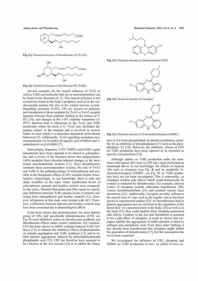
Antipsychotics and Thromboxane Medicinal Chemistry, 2012, Vol. 8, No. 6 1033
COOH
O
OH
O
Fig. (1). Chemical structure of thromboxane A2 (TxA2).
COOH
O
OH
OH
HO
Fig. (2). Chemical structure of thromboxane B2 (TxB2).
Several examples for the mutual influence of TxA2 as well as TxB2 and molecules that act as neurotransmitters can be found in the literature [6, 7]. This mutual influence is not exclusively found in the body’s periphery such as in the car-diovascular system, but also in the central nervous system. Regarding serotonin (5-HT), TPs are present on platelets, and stimulation of these receptors by TxA2 or TxA2 receptor agonists activates these platelets leading to the release of 5-HT [10]; and changes in the 5-HT reuptake transporter (5-HTT) function lead to alterations in the TxA2 and TxB2 production within the brain [11]. TxA2 also facilitates do-pamine release in the striatum and is involved in sucrose intake in mice which is a dopamine-dependent motivational behaviour [7]. Additionally, TxA2 signalling modulates neu-rotransmission via N-methyl-D-aspartic acid (NMDA) and !-aminobutyric acid (GABA) [7].
Interestingly, dopamine, 5-HT, NMDA and GABA signal transduction have been reported to be altered in schizophre-nia, and a review of the literature shows that antipsychotics (APs) modulate these disorder-induced changes in the men-tioned neurotransmitter systems [11]. Since thromboxanes modulate these neurotransmitter systems, the role of TxA2 and TxB2 in the pathophysiology of schizophrenia and pos-sibly in the therapeutic effects of APs warrants further inves-tigation. Surprisingly, to our knowledge, there is only one study available on the topic where tromboxane levels of schizophrenic patients and healthy controls were compared. In this study, Dietrich-Muszalska and Olas report no signifi-cant difference between TxB2 plasma levels of patients suf-fering from schizophrenia and healthy controls [12]. How-ever, all patients in this study were treated with APs. There-fore, a difference between patients and healthy controls may have been concealed due to pharmalogical effects.
It has been shown that phenothiazines, the most famous group of APs, and specifically chlorpromazine (CPZ; see Fig. 3) exert inhibitory action on thromboxane synthesis and thromboxane effects under certain conditions. For example, CPZ has been shown to inhibit collagen-induced TxB2 syn-thesis [13], to enhance the inhibitory effects of phentolamine on platelet aggregation and TxB2 synthesis [14] and to in-hibit platelet aggregation induced by palmitaldehydeacetal phosphatidic acid [15]. CPZ has therefore been assumed to be a blocker of the AA cascade [16] or to inhibit the libera-
tion of AA from phospholipids in platelet membranes, possi-bly by an inhibition of phospholipases [17] such as the phos-pholipase A2 [18]. However, the inhibitory effects of CPZ on TxB2 production have been reported to be restricted to specific concentrations [19].
Although studies on TxB2 production under the treat-ment with typical APs such as CPZ have been performed as mentioned above, to our knowledge, the effects of atypical APs such as clozapine (see Fig. 4) and its metabolite N-desmethylclozapine (NDMC; see Fig. 5) on TxB2 produc-tion have not yet been investigated. This is noteworthy, as clozapine exhibits side effects which could theoretically be evoked or mediated by thromboxanes. For example, adverse events of clozapine include orthostatic hypotension [20], venous thromboembolism [21] and cerebral venous sinus thrombosis [22]. Additionally, clozapine possibly influences the muscle tone of veins such as the jugular vein as has been shown in experimental studies [23]. As thromboxanes lead to platelet aggregation and are involved in the regulation of the blood flow via vasoconstriction in the body [24] as well as in the brain [25], they could mediate these clozapine-associated side effects. Contrary to the fact that thrombosis is assumed to be a side effect of clozapine, it could be shown that clo-zapine inhibits the aggregation of rabbit platelets evoked by collagen and arachidonic acid. From these latter findings it has already been hypothesised that clozapine might inhibit the generation of thromboxanes [17], but this assumption has never been examined.
We investigated the influence of CPZ, clozapine and NDMC on TxB2 production in-vitro. In earlier in-vitro ex-
S
N
N
Cl
Fig. (3). Chemical structure of chlorpromazine (CPZ).
N
NH
Cl
N
N
Fig. (4). Chemical structure of clozapine.
NH
N
Cl
N
NH
Fig. (5). Chemical structure of N-desmethylclozapine (NDMC).
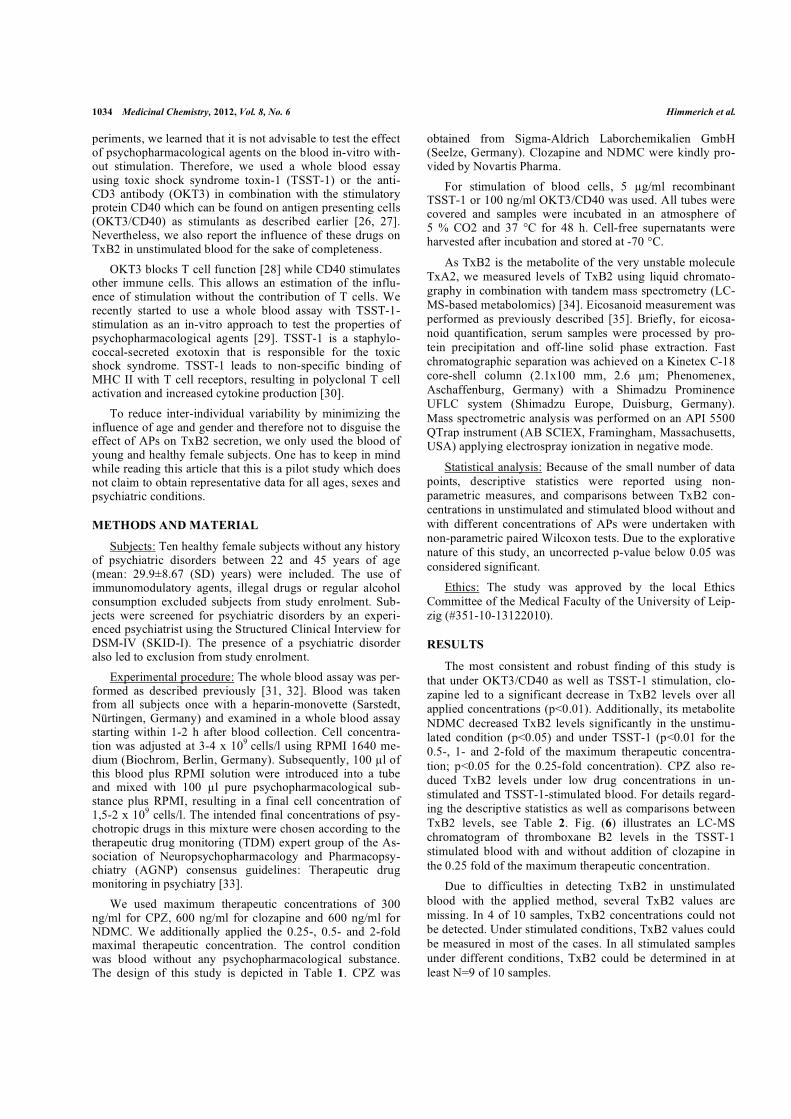
1034 Medicinal Chemistry, 2012, Vol. 8, No. 6 Himmerich et al.
periments, we learned that it is not advisable to test the effect of psychopharmacological agents on the blood in-vitro with-out stimulation. Therefore, we used a whole blood essay using toxic shock syndrome toxin-1 (TSST-1) or the anti-CD3 antibody (OKT3) in combination with the stimulatory protein CD40 which can be found on antigen presenting cells (OKT3/CD40) as stimulants as described earlier [26, 27]. Nevertheless, we also report the influence of these drugs on TxB2 in unstimulated blood for the sake of completeness.
OKT3 blocks T cell function [28] while CD40 stimulates other immune cells. This allows an estimation of the influ-ence of stimulation without the contribution of T cells. We recently started to use a whole blood assay with TSST-1-stimulation as an in-vitro approach to test the properties of psychopharmacological agents [29]. TSST-1 is a staphylo-coccal-secreted exotoxin that is responsible for the toxic shock syndrome. TSST-1 leads to non-specific binding of MHC II with T cell receptors, resulting in polyclonal T cell activation and increased cytokine production [30].
To reduce inter-individual variability by minimizing the influence of age and gender and therefore not to disguise the effect of APs on TxB2 secretion, we only used the blood of young and healthy female subjects. One has to keep in mind while reading this article that this is a pilot study which does not claim to obtain representative data for all ages, sexes and psychiatric conditions.
METHODS AND MATERIAL
Subjects: Ten healthy female subjects without any history of psychiatric disorders between 22 and 45 years of age (mean: 29.9±8.67 (SD) years) were included. The use of immunomodulatory agents, illegal drugs or regular alcohol consumption excluded subjects from study enrolment. Sub-jects were screened for psychiatric disorders by an experi-enced psychiatrist using the Structured Clinical Interview for DSM-IV (SKID-I). The presence of a psychiatric disorder also led to exclusion from study enrolment.
Experimental procedure: The whole blood assay was per-formed as described previously [31, 32]. Blood was taken from all subjects once with a heparin-monovette (Sarstedt, Nürtingen, Germany) and examined in a whole blood assay starting within 1-2 h after blood collection. Cell concentra-tion was adjusted at 3-4 x 10⁹ cells/l using RPMI 1640 me-dium (Biochrom, Berlin, Germany). Subsequently, 100 !l of this blood plus RPMI solution were introduced into a tube and mixed with 100 !l pure psychopharmacological sub-stance plus RPMI, resulting in a final cell concentration of 1,5-2 x 10⁹ cells/l. The intended final concentrations of psy-chotropic drugs in this mixture were chosen according to the therapeutic drug monitoring (TDM) expert group of the As-sociation of Neuropsychopharmacology and Pharmacopsy-chiatry (AGNP) consensus guidelines: Therapeutic drug monitoring in psychiatry [33].
We used maximum therapeutic concentrations of 300 ng/ml for CPZ, 600 ng/ml for clozapine and 600 ng/ml for NDMC. We additionally applied the 0.25-, 0.5- and 2-fold maximal therapeutic concentration. The control condition was blood without any psychopharmacological substance. The design of this study is depicted in Table 1. CPZ was
obtained from Sigma-Aldrich Laborchemikalien GmbH (Seelze, Germany). Clozapine and NDMC were kindly pro-vided by Novartis Pharma.
For stimulation of blood cells, 5 !g/ml recombinant TSST-1 or 100 ng/ml OKT3/CD40 was used. All tubes were covered and samples were incubated in an atmosphere of 5 % CO2 and 37 °C for 48 h. Cell-free supernatants were harvested after incubation and stored at -70 °C.
As TxB2 is the metabolite of the very unstable molecule TxA2, we measured levels of TxB2 using liquid chromato-graphy in combination with tandem mass spectrometry (LC-MS-based metabolomics) [34]. Eicosanoid measurement was performed as previously described [35]. Briefly, for eicosa-noid quantification, serum samples were processed by pro-tein precipitation and off-line solid phase extraction. Fast chromatographic separation was achieved on a Kinetex C-18 core-shell column (2.1x100 mm, 2.6 !m; Phenomenex, Aschaffenburg, Germany) with a Shimadzu Prominence UFLC system (Shimadzu Europe, Duisburg, Germany). Mass spectrometric analysis was performed on an API 5500 QTrap instrument (AB SCIEX, Framingham, Massachusetts, USA) applying electrospray ionization in negative mode.
Statistical analysis: Because of the small number of data points, descriptive statistics were reported using non-parametric measures, and comparisons between TxB2 con-centrations in unstimulated and stimulated blood without and with different concentrations of APs were undertaken with non-parametric paired Wilcoxon tests. Due to the explorative nature of this study, an uncorrected p-value below 0.05 was considered significant.
Ethics: The study was approved by the local Ethics Committee of the Medical Faculty of the University of Leip-zig (#351-10-13122010).
RESULTS
The most consistent and robust finding of this study is that under OKT3/CD40 as well as TSST-1 stimulation, clo-zapine led to a significant decrease in TxB2 levels over all applied concentrations (p<0.01). Additionally, its metabolite NDMC decreased TxB2 levels significantly in the unstimu-lated condition (p<0.05) and under TSST-1 (p<0.01 for the 0.5-, 1- and 2-fold of the maximum therapeutic concentra-tion; p<0.05 for the 0.25-fold concentration). CPZ also re-duced TxB2 levels under low drug concentrations in un-stimulated and TSST-1-stimulated blood. For details regard-ing the descriptive statistics as well as comparisons between TxB2 levels, see Table 2. Fig. (6) illustrates an LC-MS chromatogram of thromboxane B2 levels in the TSST-1 stimulated blood with and without addition of clozapine in the 0.25 fold of the maximum therapeutic concentration.
Due to difficulties in detecting TxB2 in unstimulated blood with the applied method, several TxB2 values are missing. In 4 of 10 samples, TxB2 concentrations could not be detected. Under stimulated conditions, TxB2 values could be measured in most of the cases. In all stimulated samples under different conditions, TxB2 could be determined in at least N=9 of 10 samples.
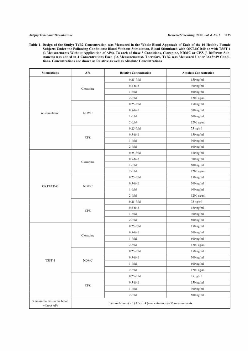
Antipsychotics and Thromboxane Medicinal Chemistry, 2012, Vol. 8, No. 6 1035
Table 1. Design of the Study: TxB2 Concentration was Measured in the Whole Blood Approach of Each of the 10 Healthy Female Subjects Under the Following Conditions: Blood Without Stimulation, Blood Stimulated with OKT3/CD40 or with TSST-1 (3 Measurements Without Application of APs). To each of these 3 Conditions, Clozapine, NDMC or CPZ (3 Different Sub-stances) was added in 4 Concentrations Each (36 Measurements). Therefore, TxB2 was Measured Under 36+3=39 Condi-tions. Concentrations are shown as Relative as well as Absolute Concentrations
Stimulations APs Relative Concentration Absolute Concentration
0.25-fold 150 ng/ml
0.5-fold 300 ng/ml
1-fold 600 ng/ml Clozapine
2-fold 1200 ng/ml
0.25-fold 150 ng/ml
0.5-fold 300 ng/ml
1-fold 600 ng/ml NDMC
2-fold 1200 ng/ml
0.25-fold 75 ng/ml
0.5-fold 150 ng/ml
1-fold 300 ng/ml
no stimulation
CPZ
2-fold 600 ng/ml
0.25-fold 150 ng/ml
0.5-fold 300 ng/ml
1-fold 600 ng/ml Clozapine
2-fold 1200 ng/ml
0.25-fold 150 ng/ml
0.5-fold 300 ng/ml
1-fold 600 ng/ml NDMC
2-fold 1200 ng/ml
0.25-fold 75 ng/ml
0.5-fold 150 ng/ml
1-fold 300 ng/ml
OKT3/CD40
CPZ
2-fold 600 ng/ml
0.25-fold 150 ng/ml
0.5-fold 300 ng/ml
1-fold 600 ng/ml Clozapine
2-fold 1200 ng/ml
0.25-fold 150 ng/ml
0.5-fold 300 ng/ml
1-fold 600 ng/ml NDMC
2-fold 1200 ng/ml
0.25-fold 75 ng/ml
0.5-fold 150 ng/ml
1-fold 300 ng/ml
TSST-1
CPZ
2-fold 600 ng/ml
3 measurements in the blood
without APs 3 (stimulations) x 3 (APs) x 4 (concentrations) =36 measurements
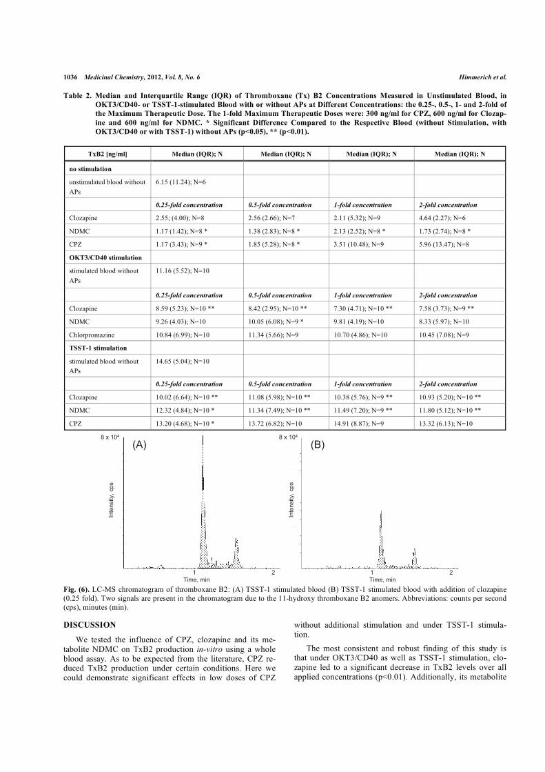
1036 Medicinal Chemistry, 2012, Vol. 8, No. 6 Himmerich et al.
Table 2. Median and Interquartile Range (IQR) of Thromboxane (Tx) B2 Concentrations Measured in Unstimulated Blood, in OKT3/CD40- or TSST-1-stimulated Blood with or without APs at Different Concentrations: the 0.25-, 0.5-, 1- and 2-fold of the Maximum Therapeutic Dose. The 1-fold Maximum Therapeutic Doses were: 300 ng/ml for CPZ, 600 ng/ml for Clozap-ine and 600 ng/ml for NDMC. * Significant Difference Compared to the Respective Blood (without Stimulation, with OKT3/CD40 or with TSST-1) without APs (p<0.05), ** (p<0.01).
TxB2 [ng/ml] Median (IQR); N Median (IQR); N Median (IQR); N Median (IQR); N
no stimulation
unstimulated blood without
APs
6.15 (11.24); N=6
0.25-fold concentration 0.5-fold concentration 1-fold concentration 2-fold concentration
Clozapine 2.55; (4.00); N=8 2.56 (2.66); N=7 2.11 (5.32); N=9 4.64 (2.27); N=6
NDMC 1.17 (1.42); N=8 * 1.38 (2.83); N=8 * 2.13 (2.52); N=8 * 1.73 (2.74); N=8 *
CPZ 1.17 (3.43); N=9 * 1.85 (5.28); N=8 * 3.51 (10.48); N=9 5.96 (13.47); N=8
OKT3/CD40 stimulation
stimulated blood without
APs
11.16 (5.52); N=10
0.25-fold concentration 0.5-fold concentration 1-fold concentration 2-fold concentration
Clozapine 8.59 (5.23); N=10 ** 8.42 (2.95); N=10 ** 7.30 (4.71); N=10 ** 7.58 (3.73); N=9 **
NDMC 9.26 (4.03); N=10 10.05 (6.08); N=9 * 9.81 (4.19); N=10 8.33 (5.97); N=10
Chlorpromazine 10.84 (6.99); N=10 11.34 (5.66); N=9 10.70 (4.86); N=10 10.45 (7.08); N=9
TSST-1 stimulation
stimulated blood without
APs
14.65 (5.04); N=10
0.25-fold concentration 0.5-fold concentration 1-fold concentration 2-fold concentration
Clozapine 10.02 (6.64); N=10 ** 11.08 (5.98); N=10 ** 10.38 (5.76); N=9 ** 10.93 (5.20); N=10 **
NDMC 12.32 (4.84); N=10 * 11.34 (7.49); N=10 ** 11.49 (7.20); N=9 ** 11.80 (5.12); N=10 **
CPZ 13.20 (4.68); N=10 * 13.72 (6.82); N=10 14.91 (8.87); N=9 13.32 (6.13); N=10
Fig. (6). LC-MS chromatogram of thromboxane B2: (A) TSST-1 stimulated blood (B) TSST-1 stimulated blood with addition of clozapine (0.25 fold). Two signals are present in the chromatogram due to the 11-hydroxy thromboxane B2 anomers. Abbreviations: counts per second (cps), minutes (min).
DISCUSSION
We tested the influence of CPZ, clozapine and its me-tabolite NDMC on TxB2 production in-vitro using a whole blood assay. As to be expected from the literature, CPZ re-duced TxB2 production under certain conditions. Here we could demonstrate significant effects in low doses of CPZ
without additional stimulation and under TSST-1 stimula-tion.
The most consistent and robust finding of this study is that under OKT3/CD40 as well as TSST-1 stimulation, clo-zapine led to a significant decrease in TxB2 levels over all applied concentrations (p<0.01). Additionally, its metabolite
8 x 10⁴ 8 x 10⁴
(A) (B)
Inte
nsity
, cps
Inte
nsity
, cps
Time, min Time, min2121
Antipsychotics and Thromboxane Medicinal Chemistry, 2012, Vol. 8, No. 6 1037
NDMC reduced TxB2 production under TSST-1 stimulation and in unstimulated blood. Therefore, one has to assume that clozapine and its metabolite decrease the synthesis of TxA2 and its metabolite TxB2.
The obtained results may have implications regarding clozapine’s mechanism of antipsychotic action as well as the pathophysiology of clozapine’s side effects. As TxA2 in-creases 5-HT [10] as well as dopamine signalling [7], the antipsychotic action of clozapine and NDMC might unfold via modulation of the dopamine and 5-HT system via block-ing TxA2 production in addition to direct antagonistic effects at dopamine and 5-HT receptor level. Known side effects of clozapine such as orthostatic hypotension might also be, at least in part mediated by the influence of thromboxanes on the muscle tone of the blood vessels [23]. Additionally, the numerous immunological properties and various side effects of clozapine [3, 36] may be linked to the immunological properties of TxA2 [37].
As stated above, this is a small pilot study and the level of significance was not controlled for multiple testing. Therefore, all effects need to be independently replicated. The reported data on TxB2 alterations are derived from an in-vitro assay using human blood of healthy subjects. Hence, these limitations must be taken into account when drawing conclusions. The reported findings are of preliminary nature. To our knowledge, this is the first study investigating the effect of clozapine and NDMC on thromboxane production. However, these data provide an alternative and complemen-tary viewpoint to established neurotransmitter hypotheses of schizophrenia and to the current opinion regarding the mechanisms of how antipsychotics work.
We used this stimulated in-vitro approach for methodo-logical reasons as well as for reasons of chemical metrology. Methodological reasons include the comparability with other studies [29, 38-41] and the approximation of the in-vitro
condition to the natural conditions in the human body where dynamic processes such as immune stimulation regularly take place. From the viewpoint of chemical metrology, it is advisable to stimulate the production of molecules such as thromboxanes into a range which is safer to measure to avoid false measurements. As one can see in Table 2, in the stimu-lated blood, a minimum of 9 of 10 TxB2 concentrations was detectable, whereas in the unstimulated blood, we had more missing values due to measurement problems because of too low TxB2 concentrations. The practicability of this approach has been demonstrated in several former studies [26, 29] in which we made the experience that these experiments are relatively easy to perform.
On the other hand, we can not exclude that testing APs in the framework of a stimulation paradigm may cause artifacts due to a specific interaction of the APs with the stimulant. Nevertheless, in-vitro stimulation paradigms serve as a simu-lation of the dynamic nature of the normal life of blood cells and in-vitro paradigms using immunological stimulation methods have been established during the last two decades in psychopharmacological research and a number of compara-ble in-vitro studies with relevant scientific impact are avail-able [26, 29, 31, 32, 38-41].
CONCLUSION
Thromboxanes are known to modulate 5-HT [10] as well as dopamine signalling [7], and thromboxanes have been shown to be involved in the regulation of systems in which typical side effects of clozapine and its metabolite NDMC take place. Therefore, we conclude from the finding of a decrease in thromboxane synthesis under clozapine and its metabolite NDMC that thromboxanes seem to be involved in the antipsychotic effects as well as the side effects of these drugs.
CONFLICT OF INTEREST
Prof. Himmerich received speaker honoraria from Astra-Zeneca, Lilly and Servier, consulting fees from Bristol-Myers Squibb, and chemical substances for study support from AstraZeneca, Novartis and Wyeth. All other authors reported no biomedical financial interests or potential con-flicts of interest.
ACKNOWLEDGEMENTS
The study was supported by the Claussen-Simon-Foundation and by LIFE – Leipzig Research Center for Civi-lization Diseases, University Leipzig. LIFE is funded by means of the European Union, by the European Regional Development Fund (ERDF) and by means of the Free State of Saxony within the framework of the excellence initiative. Novartis Pharma supported the study by providing the pure substances of clozapine and N-desmethylclozapine. The mentioned sponsors did not have any influence on the study design, the collection, analysis and interpretation of data, the writing of the report and in the decision to submit the paper for publication.
REFERENCES
[1] Malcher-Lopes, R.; Buzzi, M. Glucocorticoid-regulated crosstalk between arachidonic acid and endocannabinoid biochemical path-ways coordinates cognitive-, neuroimmune-, and energy homeosta-sis-related adaptations to stress. Vitam. Horm., 2009, 81, 263-313.
[2] Himmerich, H.; Steinberg, H. Hormone-based classification and therapy concepts in psychiatry. Fortschr. Neurol. Psychiatr., 2011,79, 384-394.
[3] Himmerich, H.; Sorge, S.; Kirkby, K.C.; Steinberg, H. Schizo-phrenic disorders: The development of immunological concepts and therapy in psychiatry. Nervenarzt, 2012, 83, 7-15.
[4] Horrobin, D.F. Schizophrenia as a prostaglandin deficiency dis-ease. Lancet, 1977, 1, 936-937.
[5] Fenton, W.S.; Hibbeln, J.; Knable, M. Essential fatty acids, lipid membrane abnormalities, and the diagnosis and treatment of schizophrenia. Biol. Psychiatr., 2000, 47, 8-21.
[6] Ross, B.M. Phospholipid and eicosanoid signaling disturbances in schizophrenia. Prostaglandins Leukotr. Essent. Fatty Acids, 2003,69, 407-412.
[7] Mitsumori, T.; Furuyashiki, T.; Momiyama, T.; Nishi, A.; Shuto, T.; Hayakawa, T.; Ushikubi, F.; Kitaoka, S.; Aoki, T.; Inoue, H.; Matsuoka, T.; Narumiya, S. Thromboxane receptor activation en-hances striatal dopamine release, leading to suppression of GABAergic transmission and enhanced sugar intake. Eur. J. Neu-
rosci., 2011, 34, 594-604. [8] Wacker, M.J.; Wilhelm, H.L.; Gomez, S.E.; Floor, E.; Orr, J.A.
Role of serotonin in thromboxane A2-induced coronary chemore-flex. Am. J. Physiol. Heart Circ. Physiol., 2003, 284, 867-875.
[9] Alanko, J.; Riutta, A.; Vapaatalo, H.; Mucha, I. Catecholamines decrease leukotriene B3 and increase thromboxane B2 synthesis in A23 187-stimulated human whole blood. Prostaglandins, 1991, 42,279-287.
1038 Medicinal Chemistry, 2012, Vol. 8, No. 6 Himmerich et al.
[10] Moncada, S.; Vane, J.R. Pharmacology and endogenous roles of prostaglandin endoperoxides, thromboxane A2, and prostacyclin. Pharmacol. Rev., 1975, 30, 293-331.
[11] Di Pietro, N.C.; Seamans, J.K. Dopamine and serotonin interac-tions in the prefrontal cortex: insights on antipsychotic drugs and their mechanism of action. Pharmacopsychiatry, 2007, 40(Suppl 1), 27-33.
[12] Dietrich-Muszalska, A.; Olas, B. Isoprostenes as indicators of oxidative stress in schizophrenia. World J. Biol. Psychiatry, 2009,10, 27-33.
[13] Anfossi, G.; Massucco, P.; Mularoni, E.; Cavalot, F.; Burzacca, S.; Mattiello, L.; Trovati, M. Phenothiazines inhibit collagen-induced thromboxane B2 synthesis and increase forskolin anti-aggregating effects in human platelets. Gen. Pharmacol., 1991, 22, 773-778.
[14] Anfossi, G.; Trovati, M.; Mularoni, E.; Massucco, P.; Cavalot. F.; Mattiello, L.; Emanuelli, G. Phenothiazine compounds enhance phentolamine effects on platelet aggregation and thromboxane B2 production. Arch. Int. Pharmacodyn. Ther., 1990, 307, 142-61.
[15] Brammer, J.P.; Maguire, M.H. Arachidonate metabolism, 5-hydroxytryptamine release and aggregation in human platelets acti-vated by palmitaldehyde acetal phosphatidic acid. Br. J. Pharma-
col., 1984, 82, 61-72. [16] Takayasu, M.; Kondo, K.; Terao, S. Endothelin-induced mobiliza-
tion of Ca2+ and the possible involvement of platelet activating factor and thromboxane A2. Biochem. Biophys. Res. Commun.,1989, 160, 751-757.
[17] Ishigooka, J.; Shizu, Y.; Wakatabe, H.; Tanaka, K.; Miura, S. Dif-ferent effects of centrally acting drugs on rabbit platelet aggre-gation: with special reference to selective inhibitory effects of an-tipsychotics and antidepressants. Biol. Psychiatry, 1985, 20, 866-873.
[18] Nikolov, R. Arachidonic acid cascade and anti-hypoxic drugs. Methods Find. Exp. Clin. Pharmacol., 1984, 6, 231-234.
[19] Chang, J.; Piperata, S.; Skowronek, M.; Lewis, A.J. Effects of thiazinamium chloride, promethazine and chlorpromazine on thromboxane B2 synthesis, phagocytosis and respiratory burst by rat alveolar macrophages. Biochem. Pharmacol., 1983, 32, 2671-2677.
[20] Muench, J.; Hamer, A.M. Adverse effects of antipsychotic medica-tions. Am. Fam. Physician, 2010, 81, 617-622.
[21] Paciullo, C.A. Evaluating the association between clozapine and venous thromboembolism. Am. J. Health Syst. Pharm., 2008, 65,1825-1829.
[22] Werring, D.; Hacking, D.; Losseff, N.; Jäger, H.R.; Walsh, M.; Foong, J. Cerebral venous sinus thrombosis may be associated with clozapine. J. Neuropsychiatry Clin. Neurosci., 2009, 21, 343-345.
[23] Leung, E.; Walsh, L.K.; Pulido-Rios, M.T.; Eglen, R.M. Charac-terization of putative 5-ht7 receptors mediating direct relaxation in Cynomolgus monkey isolated jugular vein. Br. J. Pharmacol.,1996, 117, 926-930.
[24] Narumiya, S.; Sugimoto, Y.; Ushikubi, F. Prostanoid receptors: Structures, properties, and functions. Physiol. Rev., 1999, 79, 1193-1226.
[25] Toth, P.; Rozsa, B.; Springo, Z.; Doczi, T.; Koller, A. Isolated human and rat cerebral arteries constrict to increases in flow: role of 20-HETE and TP receptors. J. Cereb. Blood Flow Metab., 2011,31, 2096-2105.
[26] Himmerich, H.; Schönherr, J.; Fulda, S.; Sheldrick, A.J.; Bauer, K.; Sack, U. Impact of antipsychotics on cytokine production in-vitro.J. Psychiatr. Res., 2011, 45, 1358-1365.
[27] Schönherr, J.; Himmerich, H.; Fulda, S.; Bauer, K.; Sack, U. Zy-tokinproduktion in vitro unter Einfluss von Antidepressiva. Ner-
venheilkunde, 2011, 30, 884-891. [28] Midtvedt, K.; Fauchald, P.; Lien, B.; Hartmann, A.; Albrechtsen,
D.; Bjerkely, B.L.; Leivestad, T.; Brekke, I.B. Individualized T cell monitored administration of ATG versus OKT3 in steroid-resistant kidney graft rejection. Clin. Transplant., 2003, 17, 69-74.
[29] Himmerich, H.; Fulda, S.; Sheldrick, A.J.; Blümäkers, B.; Rink, L. Interferon-! reduction by tricyclic antidepressants. Int. J. Psychia-
try Med., 2010, 40, 413-424. [30] Dinges, M.M.; Orwin, P.M.; Schlievert, P.M. Exotoxins of Staphy-
lococcus aureus. Clin. Microbiol. Rev., 2000, 13, 16-34. [31] Kirchner, H.; Kleinicke, C.; Digel, W. A whole-blood technique for
testing production of human interferon by leukocytes. J. Immunol.
Methods, 1982, 48, 213-219. [32] Seidel, A.; Arolt, V.; Hunstiger, M.; Rink, L.; Behnisch, A.; Kirch-
ner, H. Increased CD56+ natural killer cells and related cytokines in major depression. Clin. Immunol. Immunopathol., 1996, 78, 83-85.
[33] Baumann, P.; Hiemke, C.; Ulrich, S.; Eckermann, G.; Gaertner, I.; Gerlach, M.; Kuss, H.J.; Laux, G.; Müller-Oerlinghausen, B.; Rao, M.L.; Riederer, P.; Zernig, G. Arbeitsgemeinschaft für Neuropsy-chopharmakologie und Pharmakopsychiatrie. The AGNP-TDM ex-pert group consensus guidelines: therapeutic drug monitoring in psychiatry. Pharmacopsychiatry, 2004, 37, 243-265.
[34] Becker, S.; Kortz, L.; Helmschrodt, C.; Thiery, J.; Ceglarek, U. LC-MS-based metabolomics in the clinical laboratory. J. Chroma-
togr. B Analyt. Technol. Biomed. Life Sci., 2012, 883-884, 68-75. [35] Kortz, L.; Geyer, R.; Ludwig, U.; Planert, M.; Bruegel, M.;
Leichtle, A.; Fiedler, G.M.; Thiery, J.; Ceglarek, U. Simultaneous eicosanoid profiling and identification by liquid chromatography and hybrid triple quadrupole-linear ion trap mass spectrometry for metabolomic studies in human plasma. J. Lab. Med., 2009, 33,341-348.
[36] Pollmächer, T.; Haack, M.; Schuld, A.; Kraus, T.; Hinze-Selch, D. Effects of antipsychotic drugs on cytokine networks. J. Psychiatr.
Res., 2000, 34, 369-382. [37] Ricciotti, E.; FitzGerald, G.A. Prostaglandins and inflammation.
Arterioscler. Thromb. Vasc. Biol., 2011, 31, 986-1000. [38] Potvin, S.; Stip, E.; Sepehry, A.A.; Gendron, A.; Bah, R.; Kouassi,
E. Inflammatory cytokine alterations in schizophrenia: a systematic quantitative review. Biol. Psychiatry, 2008, 63, 801-808.
[39] Drzyzga, L.; Obuchowicz, E.; Marcinowska, A.; Herman, Z.S. Cytokines in schizophrenia and the effects of antipsychotic drugs. Brain Behav. Immun., 2006, 20, 532-545.
[40] Hinze-Selch, D.; Pollmächer, T. In vitro cytokine secretion in indi-viduals with schizophrenia: results, confounding factors, and impli-cations for further research. Brain Behav. Immun., 2001, 15, 282-318.
[41] Arolt, V.; Rothermundt, M.; Wandinger, K.P.; Kirchner, H. De-creased in vitro production of interferon-gamma and interleukin-2 in whole blood of patients with schizophrenia during treatment. Mol. Psychiatry, 2000, 5, 150-158.
Received: May 11, 2012 Revised: June 26, 2012 Accepted: July 03, 2012

9. Erklärung über die eigenständige Abfassung der Arbeit
71
9. Erklärung über die eigenständige Abfassung der Arbeit
Hiermit erkläre ich, dass ich die vorliegende Arbeit selbständig und ohne unzulässige
Hilfe oder Benutzung anderer als der angegebenen Hilfsmittel angefertigt habe. Ich
versichere, dass Dritte von mir weder unmittelbar noch mittelbar geldwerte
Leistungen für Arbeiten erhalten haben, die im Zusammenhang mit dem Inhalt der
vorgelegten Dissertation stehen, und dass die vorgelegte Arbeit weder im Inland
noch im Ausland in gleicher oder ähnlicher Form einer anderen Prüfungsbehörde
zum Zweck einer Promotion oder eines anderen Prüfungsverfahrens vorgelegt
wurde. Alles aus anderen Quellen und von anderen Personen übernommene
Material, das in der Arbeit verwendet wurde oder auf das direkt Bezug genommen
wird, wurde als solches kenntlich gemacht. Insbesondere wurden alle Personen
genannt, die direkt an der Entstehung der vorliegenden Arbeit beteiligt waren.
…............................…. Datum
…............................…. Unterschrift
10. Lebenslauf und wissenschaftlicher Werdegang
72
10. Lebenslauf und wissenschaftlicher Werdegang
10.1 Lebenslauf
Name und Vorname: Schmidt, Renate Luise Geburtsdatum: 22.11.1987 Geburtsort: Borna
Derzeitige Position: Assistenzärztin für Orthopädische
Chirurgie, Unfall- und Handchirurgie des HELIOS
Klinikums Borna
Ausbildung: Seit 07/2013 Assistenzärztin für Orthopädische
Chirurgie, Unfall- und Handchirurgie, des HELIOS
Klinikums Borna Seit 02/2011 Promotion an der Klinik und Poliklinik für Psychiatrie,
Universität Leipzig 11/2012 Erfolgreicher Abschluss der Ärztlichen Prüfung 2008 – 2012 Klinisches Studium an der Universität Leipzig 09/2008 Erfolgreicher Abschluss des ersten Abschnitts der
ärztlichen Prüfung 2006 – 2008 Vorklinisches Studium an der Universität Leipzig 06/2006 Abschluss: Allgemeine Hochschulreife (Abitur) 1998 – 2006 Wiprecht-Gymnasium-Groitzsch 1994 – 1998 Grundschule Groitzsch
10. Lebenslauf und wissenschaftlicher Werdegang
73
10.2 Publikationen
x Schmidt, L.; Ceglarek, U.; Kortz, L.; Hoop, M.; Kirkby, K.C.; Thiery, J.; Himmerich,
H. Mechanisms of Involvement of Eicosanoids and their Precursors in the
Pathophysiology and Treatment of Schizophrenia. Med Chem., 2013, 9, 763-767.
x Himmerich, H.; Schmidt, L.; Becker, S.; Kortz, L.; Schonherr, J.; Mergl, R.; Bauer,
K.; Sack, U.; Sheldrick, A. J.; Thiery, J.; Ceglarek, U. Impact of clozapine, N
desmethylclozapine and chlorpromazine on thromboxane production in vitro. Med.
Chem., 2012, 8, 1032-8.
10.3 Posterpräsentationen
x 11. Research Festival Leipzig 2012, Leipzig, 2012: „Impact of clozapine, N-
desmethylclozapine and chlorpromazine on thromboxane production in-vitro”
x Deutsche Gesellschaft für Psychiatrie, Psychotherapie und Nervenheilkunde
(DGPPN), Berlin, 2012: „Impact of clozapine, N-desmethylclozapine and
chlorpromazine on thromboxane production in-vitro.”
10. Lebenslauf und wissenschaftlicher Werdegang
74
11. Danksagung
Leider lässt sich eine wahrhafte Dankbarkeit mit Worten nicht ausdrücken. Johann Wolfgang von Goethe
Dennoch möchte ich an dieser Stelle meinen Dank an all diejenigen richten, die mich bei
dieser Arbeit unterstützten.
Allen voran möchte ich mich bei meinem Doktorvater Herrn Prof. Dr. Hubertus Himmerich
bedanken. Nicht nur für die Überlassung des Themas, sondern auch für die zahllosen
Gespräche, in denen er mir mit Rat und Tat zur Seite stand und mir wertvolle Hinweise zur
Anfertigung meiner Arbeit gab.
Weiterhin möchte ich meiner Zweitbetreuerin Frau PD Dr. Uta Ceglarek und ihren
Mitarbeiterinnen Dr. Susen Becker, Juliane Dorow, Linda Kortz, Babette Niescher und
Christin Helmschrodt danken, die mir während der nervenaufreibenden Zeit im Labor immer
wieder hilfsbereit unter die Arme griffen und so den experimentellen Teil meiner Arbeit
entscheidend voran brachten.
In diesem Zusammenhang gilt mein Dank auch Herrn Prof. Dr. Ulrich Sack und Frau Katrin
Bauer sowie Herrn Jeremias Schönherr, in deren Untersuchungen meine Arbeit ihre
Grundlagen findet.
Der Claussen-Simon-Stiftung danke ich für die Finanzierung der Studie und der Fa. Novartis,
für die Bereitstellung der Reinsubstanz von Clozapin und NDMC.
Herrn Prof. Dr. Joachim Thiery danke ich für wichtige Ratschläge zur Literaturrecherche und
konstruktive Hinweise zur Ausformulierung der Arbeitshypothese.
Mein ganz persönlicher Dank gilt meiner Familie und meinen Freunden, die mir in all den
Jahren immer den Rücken stärkten, mich motivierten und mit viel Geduld und Liebe dafür
sorgten, dass ich heute meinen beruflichen Traum leben kann.