Embed Size (px)
Citation preview
Dermatology Trivia
Asfa Akhtar D.O., FAOCD, FAADCleveland Clinic Florida
Associate Professor of Internal Medicine and Dermatology
Nova Southeastern University
Assistant Professor of Dermatology
Charles E. Schmidt College of Medicine
Florida Atlantic University
Hidradenitis Suppurativa
▪ Chronic recurrent, debilitating disease
predominantly affecting a young active
population .
▪ Dysfunction of the folliculoinfundibular
unit
affecting the apocrine-bearing skin.
Hidradenitis Suppurativa
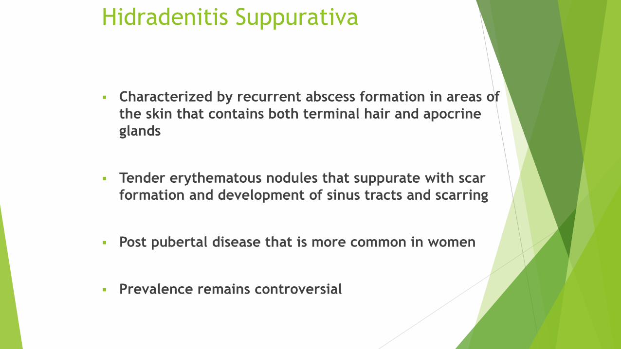
▪ Characterized by recurrent abscess formation in areas of
the skin that contains both terminal hair and apocrine
glands
▪ Tender erythematous nodules that suppurate with scar
formation and development of sinus tracts and scarring
▪ Post pubertal disease that is more common in women
▪ Prevalence remains controversial
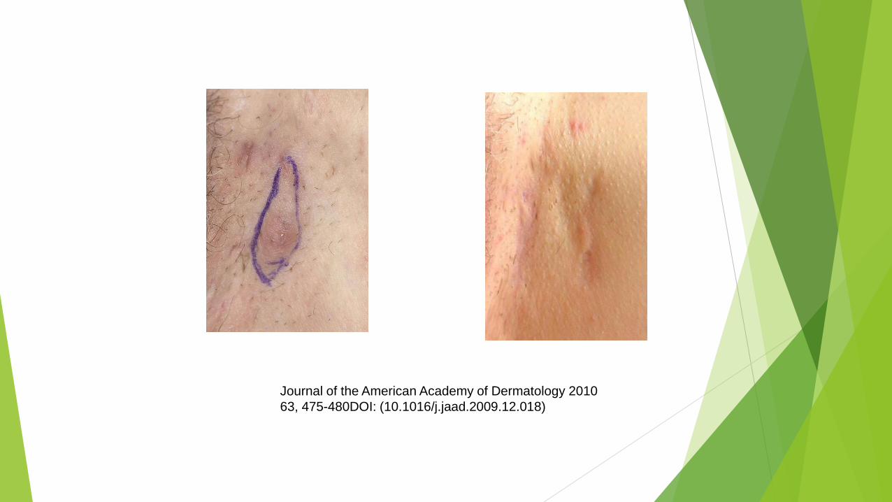
Journal of the American Academy of Dermatology 2010
63, 475-480DOI: (10.1016/j.jaad.2009.12.018)
Hidradenitis Suppurativa
▪ Etiology and pathophysiology of HS have yet to
be fully elucidated
▪ Phenotype of the disease has different
morphologic features covering a wide
spectrum
▪ Has a negative impact physically, emotionally
and psychologically on the patient
Hidradenitis Suppurativa
▪ Dysfunctional cutaneous immune
response to commensal bacteria
▪ Painful inflammatory chronic
condition
▪ Under-reported and misdiagnosed
▪ About a 1/3rd of patients have their
first symptoms before the age of 18
Hidradenitis Suppurativa
▪ Deep painful abscesses and chronic draining sinus tracts are present in these regions:
▪ Axillae
▪ Anogenital
▪ Inframmary
▪ Inguinal
Hidradenitis Suppurativa
▪ Hallmark
▪ Double comedone
▪Black head with 2 or more
openings that communicate with
the subdermis
Hidradenitis Suppurativa
▪ Diagnosis: Three criteria
1. Typical lesions
Deep seated cystic lesions, abscesses, scarring, pseudo comedones
2. Location
▪ ≥1 of the areas HS:
Axillae, groin perineal region, buttocks, Inframammary, Intermammary
3. Chronic nature
Hidradenitis Suppurativa
Systemic associations
Obesity (12%—18%)
Metabolic syndrome
Diabetes Mellitus (DM)
Smoking
Arthritis
Spondyloarthropathy
Hormone related disorders
Hidradenitis Suppurativa
Systemic associations
Substance dependence
Depression
Inflammatory Bowel Disease—9
times more likely to develop HS
than the general population
Hidradenitis Suppurativa
Systemic associations
Follicular occlusion tetrad(HS,acne
congloblata,dissecting cellulitis of the
scalp and pilonidal cyst)
Hidradenitis Suppurativa
Systemic associations
Pyoderma gangrenosum
PASH (pyoderma gangrenosum, acne, suppurative hidradenitis)
PAPASH (pyogenic arthritis, pyoderma gangrenosum, acne and suppurative hidradenitis
PsAPASH (psoriatic arthritis, pyoderma gangrenosum,acne and suppurative hidradenitis)
Hidradenitis Suppurativa
Scoring of Disease Severity
Hurley Staging
Stage I: Abscess formation, single or multiple without sinus tracts and cicatrization
Stage II: Recurrent abscesses with tract formation and cicatrization, single or multiple, and widely separated lesions
Stage III: Diffuse or near-diffuse involvement or multiple interconnected tracts and abscesses across the entire area
Hidradenitis Suppurativa
HS is worsened by hormones
particularly androgens
83% improvement was noted in 47
patients on a dairy free diet
Avoidance of foods with high glycemic
index and dairy might be beneficial.
Hidradenitis Suppurativa
There is increasing evidence that the
immune system plays a role
Increased levels of cytokines such as
IL1-β and TNF-α have been noted in
tissue cultures
Other cytokines thought to be
upregulated include IL-10, IL-12, IL-17
and IL-23
Hidradenitis Suppurativa
Systemic therapy is the mainstay of treatment
Antibiotics
Tetracycline, doxycycline, rifampin, clindamycin, dapsone
Retinoids
Acitretin
Isotretinoin
Hidradenitis Suppurativa
Systemic therapy is the mainstay
of treatment
Antiandrogens
Immunosuppresants
Tacrolimus
Cyclosporine
Hidradenitis Suppurativa
Biologic agents for HS
Effective for moderate to severe HS
Adalimumab, anakinra, etanercept,
infliximab, and ustekinumab
Adalimumab is a fully humanized
monoclonal antibody that corresponds to
the human immunoglobulin G1 and has
heavy and light chain variable regions
exhibiting specificity for human TNFα
Hidradenitis Suppurativa
Biologic agents for HS Infliximab
Chimeric antibody composed of both human and mouse proteins targeting TNFα
Ustekinumab
Human anti-p40 monoclonal antibody
P40 is a shared subunit of human interleukins (ILs )-12 and -23
Anakinra is a recombinant IL-1α receptor antagonist
Hidradenitis Suppurativa
Biologic agents for HS
Strongest evidence with
adalimumab and infliximab
May 2015 US FDA approved
adalimumab for management of
moderate to severe HS
Hidradenitis Suppurativa
Topical therapy such as antibiotics
and keratolytics combined with
proper wound dressings are used as
adjunctive treatment
Hidradenitis Suppurativa
Laser and light therapy for HS
Laser hair reduction
Photodynamic therapy
CO2 Excision and vaporization
For advanced HS, surgical excision is the mainstay of
treatment
HS remains a widely unrecognized and difficult to
treat condition resulting in diagnostic delay and
patient dissatisfaction
There is a renewed interest in understanding this
devastating disease and current research is
encouraging
Hidradenitis Suppurativa
Summary
Hidradenitis suppurativa (HS) is an uncommon, but not rare inflammatory skin disease, it affects 98 per 100,000 people in the United States
HS affects females more than males. African Americans and other ethnic populations are at higher risks
One study showed highest incidence among 30-39 year old adults
Clinical presentations include erythema followed by the presentation of deep-acne like cysts, folliculitis, boils mucopurulent discharge
BMI of 40 and smoking are corelated with HS
DermatoloyTimes, Volume 38, Number 9
Hidradenitis Suppurativa
Patients with HS have increased risk of death from myocardial infarction,
ischemic stroke, heart disease and other cardiovascular events
Current treatment options include antimicrobials, immunosuppressants, anti-
inflammatories and biologic therapy in severe cases
In chronic cases, surgical excisions may be necessary, but recurrence is
possible
Intense pulsed light, neodymium-doped yttrium aluminum garnet laser and
photodynamic therapy may benefit patients, but the clinical evidence for this
practice is rated low.
DermatologyTimes, Volume 38, Number 9
Discoid lupus erythematosus
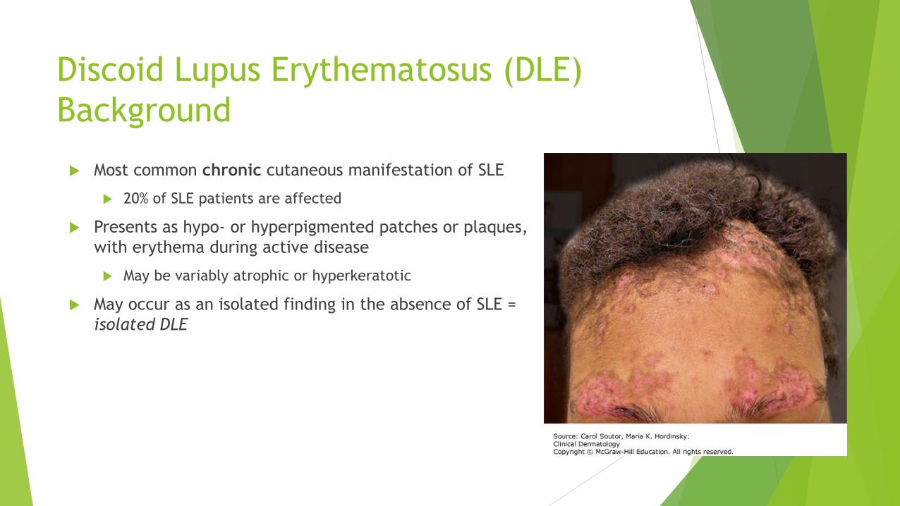
Discoid Lupus Erythematosus (DLE)
Background
Most common chronic cutaneous manifestation of SLE
20% of SLE patients are affected
Presents as hypo- or hyperpigmented patches or plaques,
with erythema during active disease
May be variably atrophic or hyperkeratotic
May occur as an isolated finding in the absence of SLE =
isolated DLE
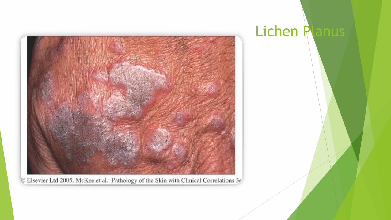
Lichen planus (LP)
Acute or chronic inflammatory dermatosis
Four P’s – purple, polygonal, pruritic, papule
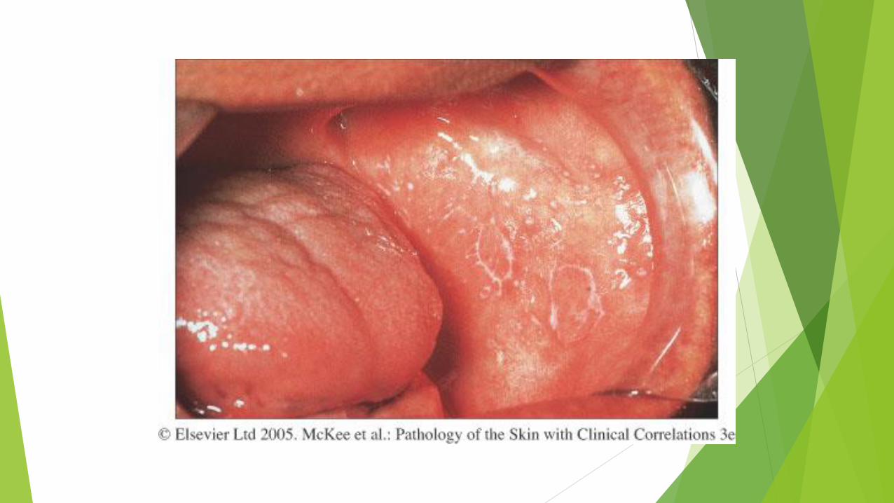
Milky white reticulated patches in the mouth
Idiopathic
Drugs (gold, mercury)
Infection (HCV)
Flat topped sharply defined papules with white lines
(Wickham’s striae)
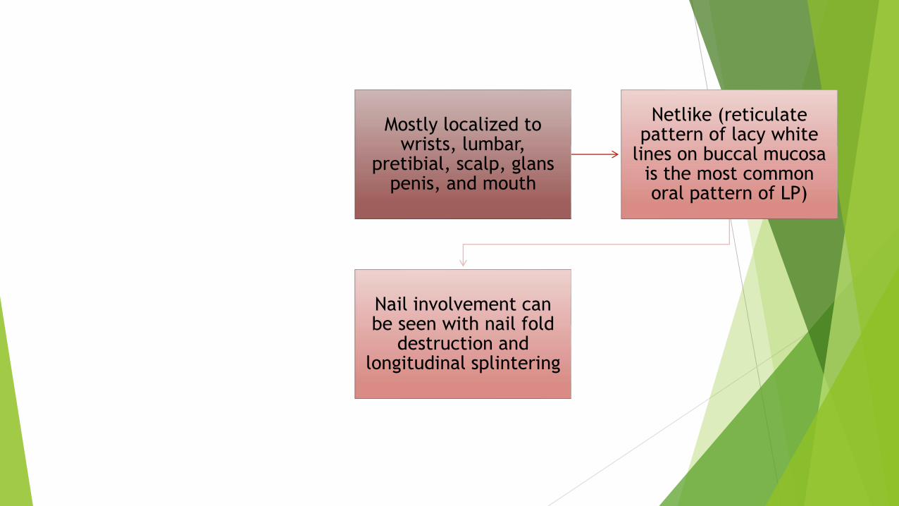
Lichen
planus (LP) Mostly localized to wrists, lumbar,
pretibial, scalp, glans penis, and mouth
Netlike (reticulate pattern of lacy white
lines on buccal mucosa is the most common oral pattern of LP)
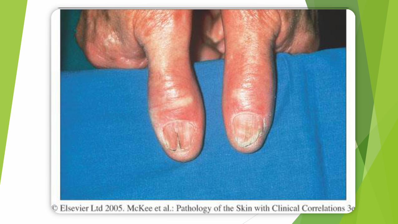
Nail involvement can be seen with nail fold
destruction and longitudinal splintering
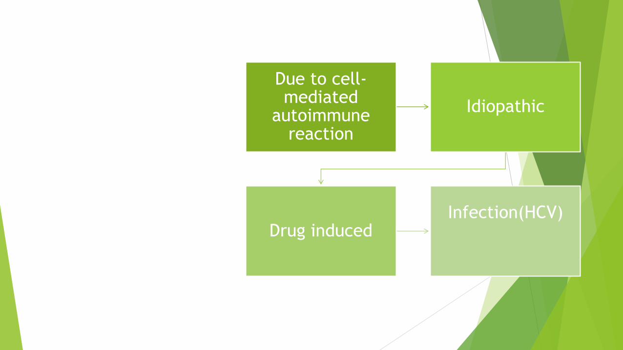
Lichen
planus (LP) Due to cell-mediated
autoimmune reaction
Idiopathic
Drug inducedInfection(HCV)
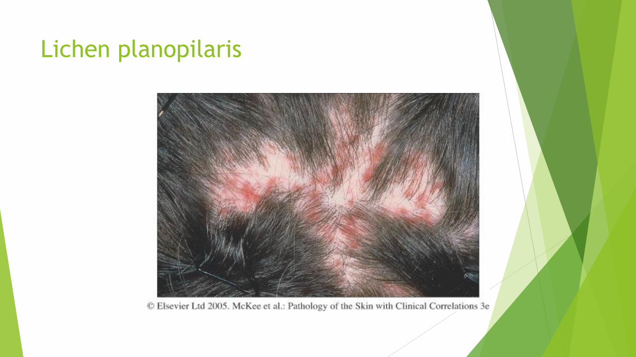
Lichen planopilaris
Lichen planus
Treatment
Superpotent topical corticosteroids topical calcineurin inhibitors,
intralesional and or systemic
corticosteroids,phototherapy,methotrexate,acitretin,metronidazole
Lichen Planus
Sweet Syndrome
(Acute febrile neutrophilic
dermatosis)
An inflammatory process expressed clinically by
markedly edematous acuminate papules and
edematous plaques situated mostly on the face,
upper part of the trunk, and arms, especially the
hands, and often accompanied by fever and
leukocytosis.
Described in 1964 by Sweet
Four features
Fever
Leukocytosis
Acute tender lesions
Papillary dermal infiltrate of neutrophils
Primarily affects adults
86% of patients are women
Four subtypes
Classic type (71%)
Associated with neoplasia (11%)
Associated with inflammatory disease
Associated with pregnancy (2%)
SS is a reactive phenomenon and considered a cutaneous marker of systemic disease
AML
Streptococcal infection
Inflammatory bowel disease, solid tumors, pregnancy and other hematologic malignancy, infections
Medications such as granulocyte colony-stimulating factor, oral contraceptives, minocycline
Clinical manifestations
Acute, tender, erythematous, plaques, occasionally blisters with annular or arciform pattern occur on the head, neck, arms and legs
The trunk is rarely involved
Fever (50%)
Arthritis, arthralgia or myalgias can be seen in up to 2/3rd of cases
Conjunctivitis or episcleritis (30%)
Oral apthae (13%)
Cardiac, renal, hepatic and pulmonary involvement is rare
Laboratory studies
Moderate neutrophilia (<50%)
Elevated ESR (>30mm/hr.)
Increased alkaline phosphatase
The hallmark of Sweet syndrome is a nodular and diffuse dermal
infiltrate of neutrophils with karyorrhexis and massive papillary
dermal edema
Treatment
Systemic corticosteroids –Gold standard
Oral potassium iodide
Colchicine
Indomethacin
Dapsone
Doxycycline
Cyclosporine
Diagnosis
Contact dermatitis to Para-
phenylenediamine (PPD) in black
henna tattoo
Allergic Contact Dermatitis to Henna
Tattoo
Allergic Contact Dermatitis (ACD) affects
>14.5 million people in the US every year
High economic burden
Identification of the allergen combined
with education can prevent progression
of condition
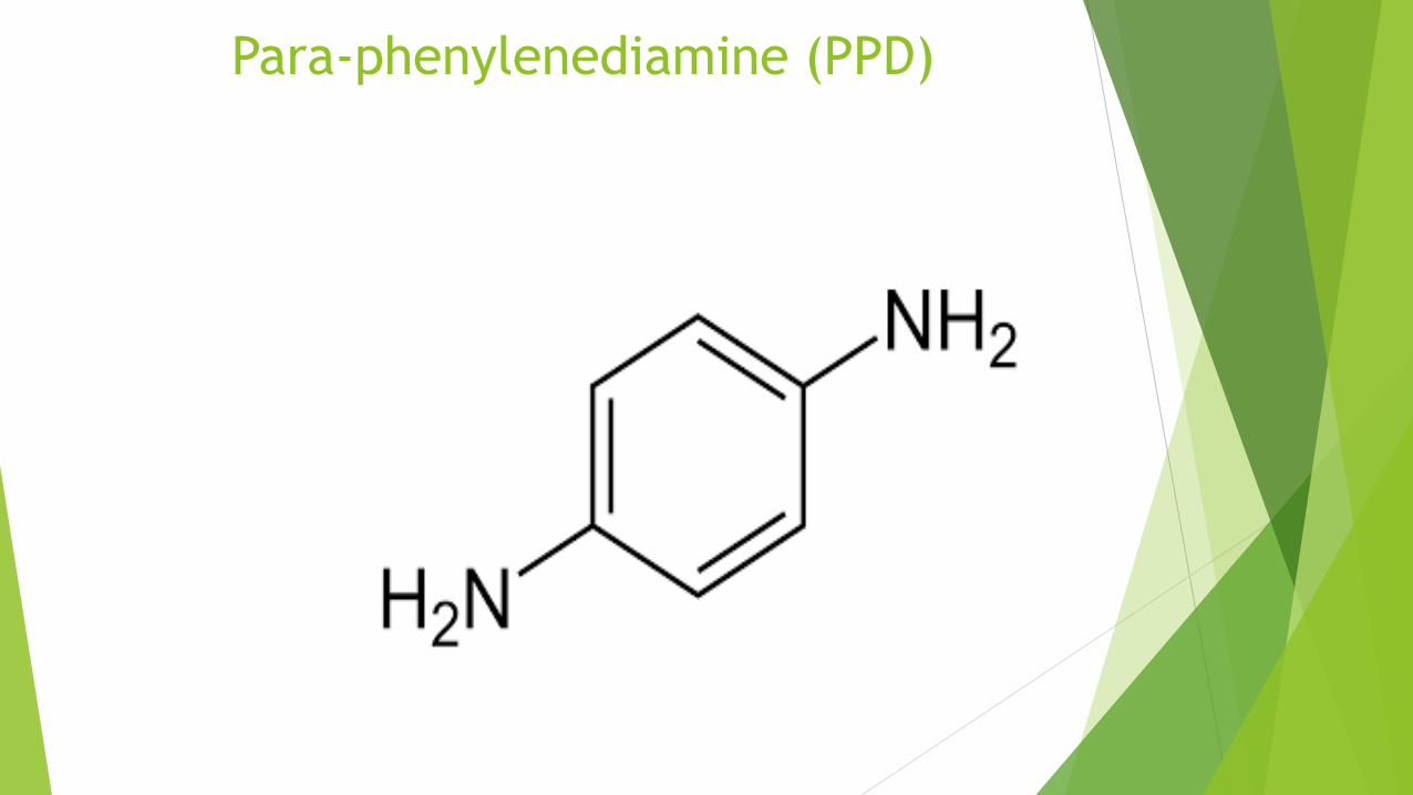
Para-phenylenediamine (PPD)
▪ Contact allergen of the year 2006 by the
American Contact Dermatitis Society
(ACDS)
▪ PPD is a chemical substance commonly
used in permanent hair dye
▪ Initially formulated for use in hair dye at
the end of the 19th century
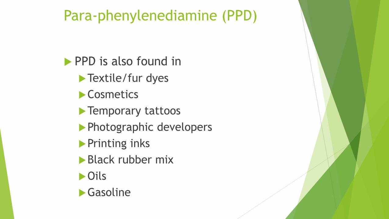
Para-phenylenediamine (PPD)
PPD is also found in
Textile/fur dyes
Cosmetics
Temporary tattoos
Photographic developers
Printing inks
Black rubber mix
Oils
Gasoline
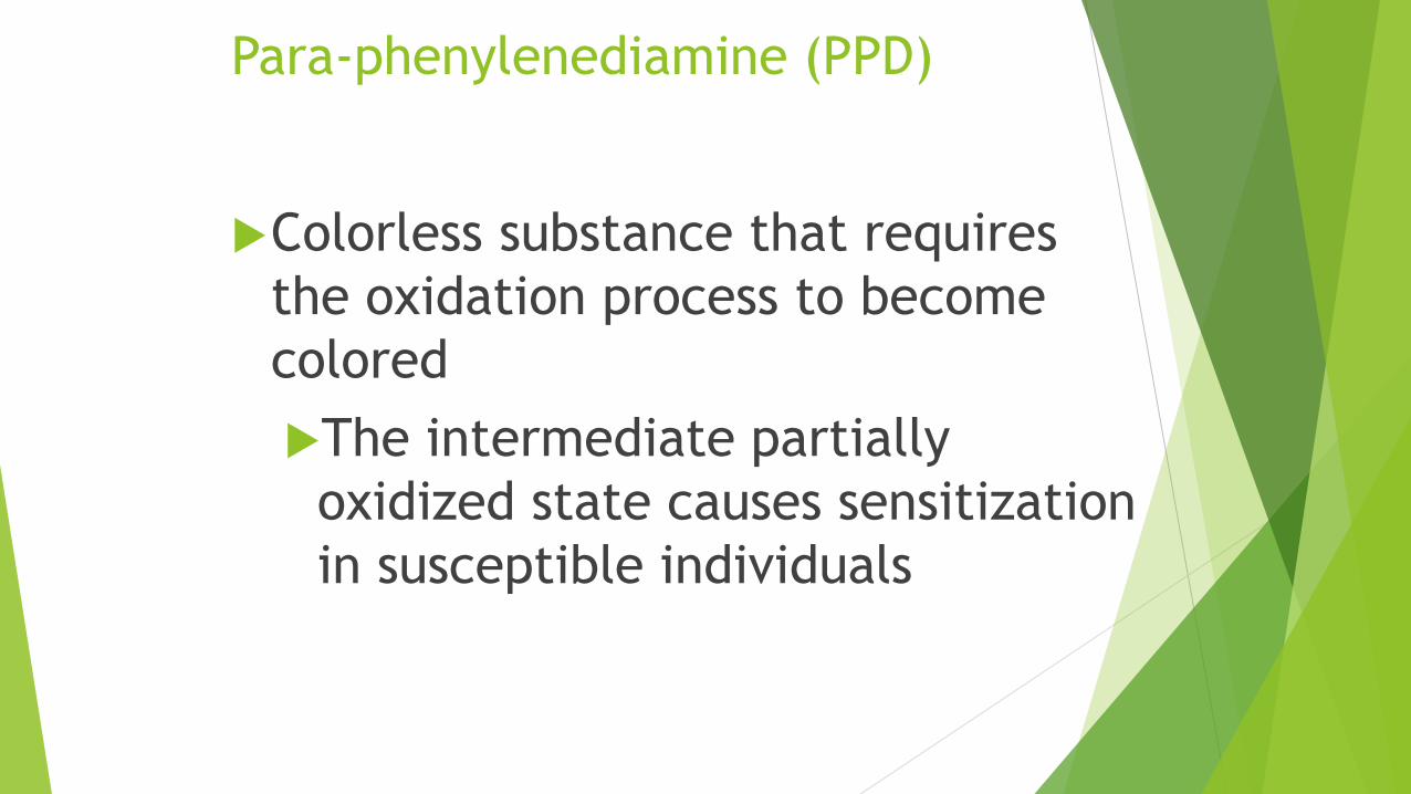
Para-phenylenediamine (PPD)
Colorless substance that requires
the oxidation process to become
colored
The intermediate partially
oxidized state causes sensitization
in susceptible individuals
Para-phenylenediamine (PPD)
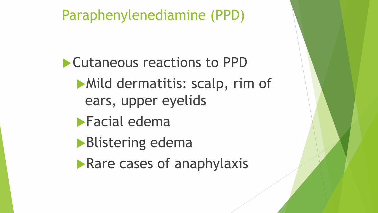
Paraphenylenediamine (PPD)
Cutaneous reactions to PPD
Mild dermatitis: scalp, rim of
ears, upper eyelids
Facial edema
Blistering edema
Rare cases of anaphylaxis
Para-phenylenediamine (PPD)
Cross reactions
Azo and aniline dyes
Benzocaine
Procaine
Para-aminobenzoic acid (PABA)
Sulfonamides
Hydrochlorothiazide
Para-phenylenediamine (PPD)
FDA prohibits the use of PPD on the skin
Black henna tattoo is natural henna mixed with PPD
Can result in severe dermatitis, scarring and post-inflammatory pigment alteration
Maximum permitted concentration in hair dye is 6%
Levels of PPD in henna tattoos can be as high as 29.5%
Para-phenylenediamine (PPD)
Patch testing
Critical in identifying the allergen
Metallic and vegetable based hair dyes
Para-toluenediamine sulfate (PTDS)
Tolerated by 50% of people allergic to PPD
The Contact Allergen Management Program
Assists with identifying allergen free
products
Erythema Elevatum Diutinum
Erythema elevatum diutinum (EED) is a rare chronic
leukocytoclastic vasculitis of unknown etiology.
First described by Hutchinson (1888) and Bury (1889).
The disease may occur in any age group but is more
common in adults, particularly in the third, fourth,
and fifth decades.
Equal incidence in men and women.
Cause is unknown but hypothesized to be an
immune complex disease.
Can be associated with inflammatory bowel
disease, RA, SLE, IgA gammopathy, strep
infection, multiple myeloma, myelodysplasia,
celiac disease,HBV and HIV infection.
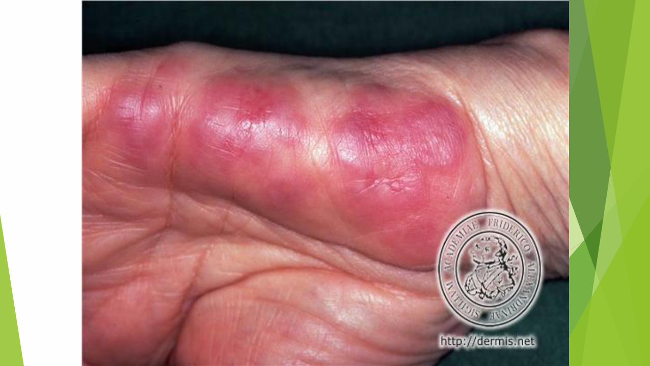
Clinical features include symmetric, persistent,
red-brown, red-purple or yellowish papules,
nodules and plaques that favor the extensor
surfaces of joints particularly the hands and
knees.
The mucous membranes and trunk are generally
spared but the ears and face may be affected.
Initially the lesions are soft but become
fibrotic over time.
Pain, aching, burning and hypo or
hyperpigmentation are associated symptoms.
EED is chronic condition lasting upto 35 years
with periods of remission and exacerbation.
EED may be present for many years before
the diagnosis of hematological abnormalities
becoming apparent in a patient.

Histologically, acute lesions resemble Sweet's
syndrome, showing papillary dermal edema,
neutrophilic vasculitis with leukocytoclasis and
fibrinoid change. Older lesions, on the other hand,
reveal perivascular fibrosis and granulation tissue
that clinically present as firm, dome-shaped
nodules.
In addition, these chronic lesions may show
xanthomatization (extracellular cholesterolosis) that
clinically may give a yellowish tinge to the nodules.
Depending on the degree of dermal edema and
infiltrate, there may be a zone that is unaffected in
the papillary dermis.
Dapsone is considered the drug of choice in treating EED, primarily due to its rapid onset of action. However, lesions promptly recur following dapsone withdrawal.
Other oral medications proven effective include niacinamide, colchicines, chloroquine, phenformin, clofazimine, and cyclophosphamide.
Systemic corticosteroids are generally ineffective. One report of a patient with EED with IgA paraproteinemia and refractory to other modalities responded to intermittent plasma exchange.

Pityriasis alba

Melasma
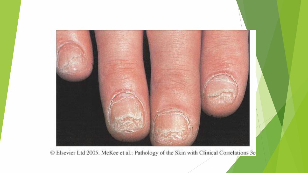

Alopecia areata
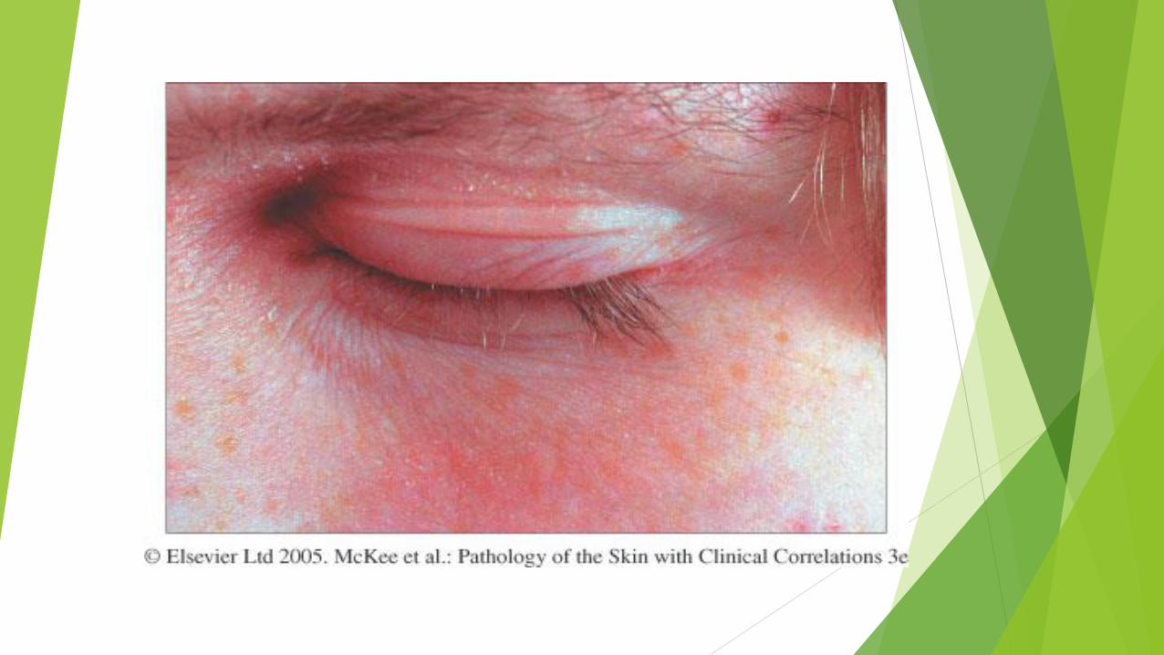
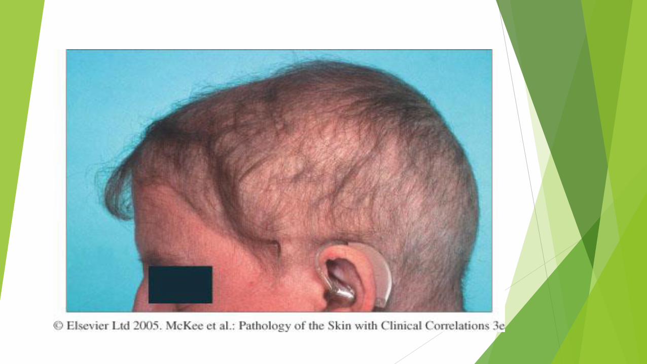
Trichotillomania
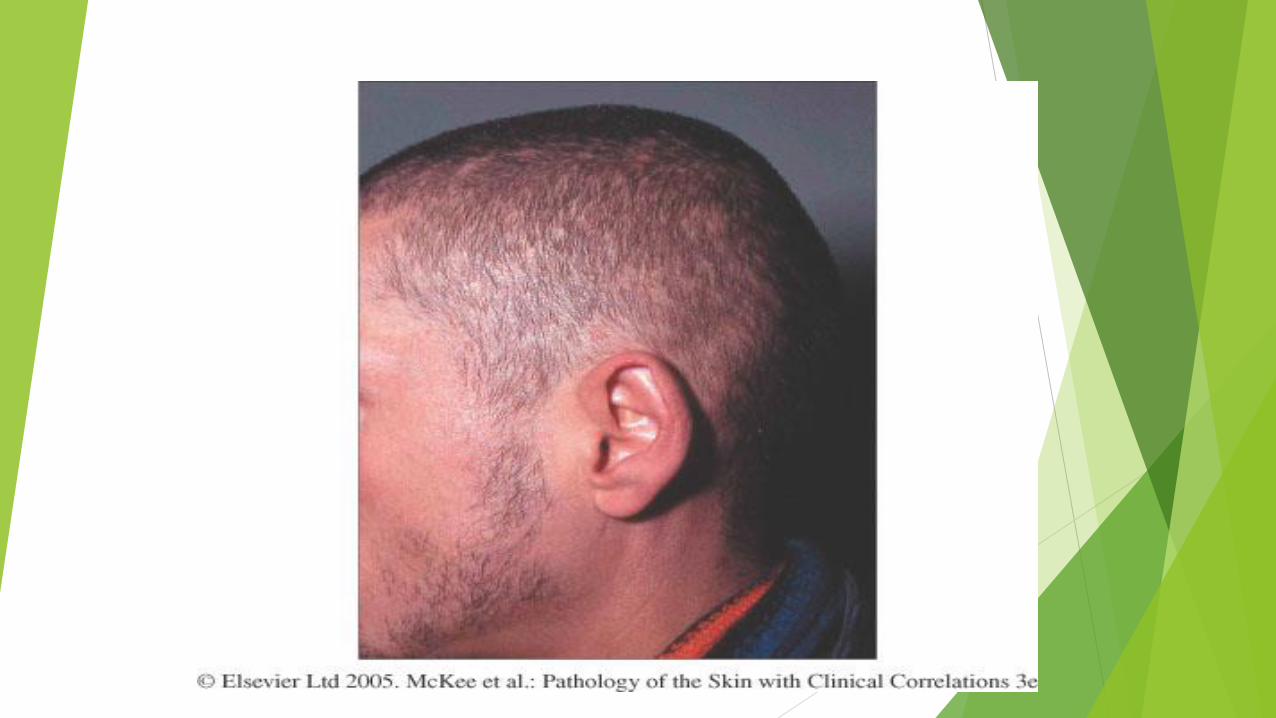

Syphilitic alopecia

Thank you