Embed Size (px)
Citation preview
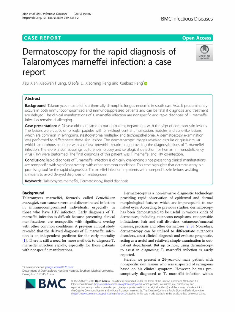
CASE REPORT Open Access
Dermatoscopy for the rapid diagnosis ofTalaromyces marneffei infection: a casereportJiayi Xian, Xiaowen Huang, Qiaofei Li, Xiaoming Peng and Xuebiao Peng*
Abstract
Background: Talaromyces marneffei is a thermally dimorphic fungus endemic in south-east Asia. It predominantlyoccurs in both immunocompromised and immunosuppressed patients and can be fatal if diagnosis and treatmentare delayed. The clinical manifestations of T. marneffei infection are nonspecific and rapid diagnosis of T. marneffeiinfection remains challenging.
Case presentation: A 24-year-old man came to our outpatient department with the sign of common skin lesions.The lesions were cuticolor follicular papules with or without central umbilication, nodules and acne-like lesions,which are common in syringoma, steatocystoma multiplex and trichoepithelioma. A dermatoscopy examinationwas performed to differentiate these skin lesions. The dermatoscopic images revealed circular or quasi-circularwhitish amorphous structure with a central brownish keratin plug, providing the diagnostic clues of T. marneffeiinfection. Therefore, a skin scrapings culture, skin biopsy and serological detection for human immunodeficiencyvirus (HIV) were performed. The final diagnosis of this patient was T. marneffei and HIV co-infection.
Conclusion: Rapid diagnosis of T. marneffei infection is clinically challenging since presenting clinical manifestationsare nonspecific with significant overlap with other common conditions. This case highlights that dermatoscopy is apromising tool for the rapid diagnosis of T. marneffei infection in patients with nonspecific skin lesions, assistingclinicians to avoid delayed diagnosis or misdiagnosis.
Keywords: Talaromyces marneffei, Dermatoscopy, Rapid diagnosis
BackgroundTalaromyces marneffei, formerly called Penicilliummarneffei, can cause severe and disseminated infectionin immunocompromised individuals, especially inthose who have HIV infection. Early diagnosis of T.marneffei infection is difficult because presenting clinicalmanifestations are nonspecific with significant overlapwith other common conditions. A previous clinical studyrevealed that the delayed diagnosis of T. marneffei infec-tion is an independent predictor for the early mortality[1]. There is still a need for more methods to diagnose T.marneffei infection rapidly, especially for those patientswith nonspecific manifestations.
Dermatoscopy is a non-invasive diagnostic technologyproviding rapid observation of epidermal and dermalmorphological features which are imperceptible to ournaked eyes. According to previous studies, dermatoscopyhas been demonstrated to be useful in various kinds ofdermatoses, including cutaneous neoplasms, ectoparasiticinfestations, hair and nail disorders, cutaneous/mucosaldiseases, psoriasis and other dermatoses [2, 3]. Nowadays,dermatoscopy can be utilized to differentiate cutaneousdisorders, assist clinical diagnosis and evaluate prognostic,acting as a useful and relatively simple examination in out-patient department. But up to now, using dermatoscopyto assist in diagnosing T. marneffei infection is rarelyreported.Herein, we present a 24-year-old male patient with
nonspecific skin lesions who was suspected of syringomabased on his clinical symptom. However, he was pre-sumptively diagnosed as T. marneffei infection within
© The Author(s). 2019 Open Access This article is distributed under the terms of the Creative Commons Attribution 4.0International License (http://creativecommons.org/licenses/by/4.0/), which permits unrestricted use, distribution, andreproduction in any medium, provided you give appropriate credit to the original author(s) and the source, provide a link tothe Creative Commons license, and indicate if changes were made. The Creative Commons Public Domain Dedication waiver(http://creativecommons.org/publicdomain/zero/1.0/) applies to the data made available in this article, unless otherwise stated.
* Correspondence: [email protected] of Dermatology, Nanfang Hospital, Southern Medical University,Guangzhou 510515, China
Xian et al. BMC Infectious Diseases (2019) 19:707 https://doi.org/10.1186/s12879-019-4351-2
minutes after dermatoscopy examination. The diagnosisof T. marneffei infection was confirmed by fungal cul-ture and histopathological examination a few days later.
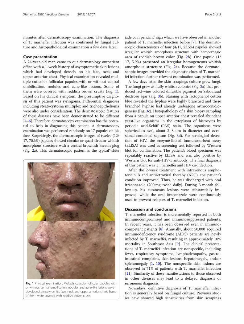
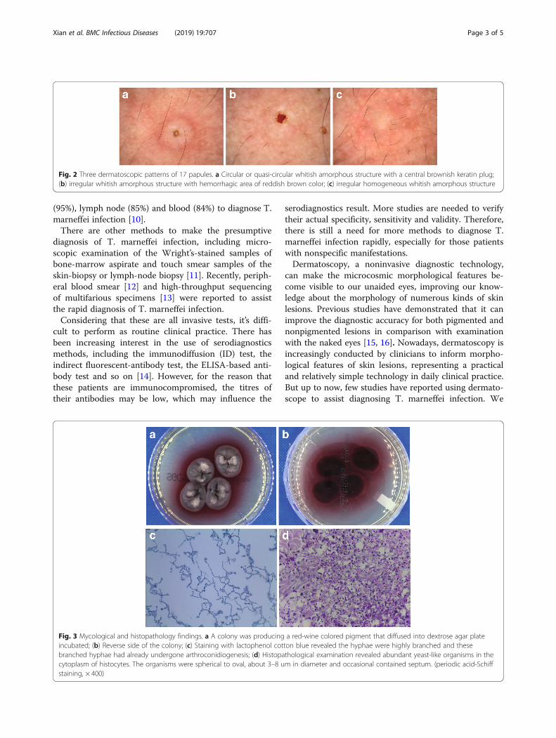
Case presentationA 24-year-old man came to our dermatology outpatientoffice with a 1-week history of asymptomatic skin lesionswhich had developed densely on his face, neck andupper anterior chest. Physical examination revealed mul-tiple cuticolor follicular papules with or without centralumbilication, nodules and acne-like lesions. Some ofthem were covered with reddish brown crusts (Fig. 1).Based on his clinical symptom, the presumptive diagno-sis of this patient was syringoma. Differential diagnosesincluding steatocystoma multiplex and trichoepitheliomawere also under consideration. The dermatoscopic featuresof these diseases have been demonstrated to be different[4–6]. Therefore, dermatoscopy examination has the poten-tial to help in diagnosing this patient. A dermatoscopyexamination was performed randomly on 17 papules on hisface. Surprisingly, the dermatoscopic images of twelve (12/17, 70.6%) papules showed circular or quasi-circular whitishamorphous structure with a central brownish keratin plug(Fig. 2a). This dermatoscopic pattern is the typical“white
jade coin pendant” sign which we have observed in anotherpatient of T. marneffei infection before [7]. The dermato-scopic characteristics of four (4/17, 23.5%) papules showedirregular whitish amorphous structure with hemorrhagicarea of reddish brown color (Fig. 2b). One papule (1/17, 5.9%) presented an irregular homogeneous whitishamorphous structure (Fig. 2c). Because the dermato-scopic images provided the diagnostic clues of T. marnef-fei infection, further relevant examination was performed.A few days later, the skin scrapings culture grew fungi.
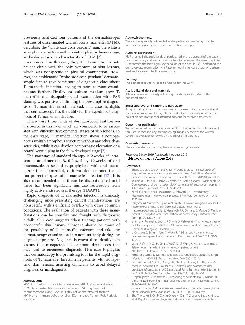
The fungi grew as fluffy whitish colonies (Fig. 3a) that pro-duced red-wine colored diffusible pigment on Sabourauddextrose agar (Fig. 3b). Staining with lactophenol cottonblue revealed the hyphae were highly branched and thesebranched hyphae had already undergone arthroconidio-genesis (Fig. 3c). Histopathology of a skin biopsy samplingfrom a papule on upper anterior chest revealed abundantyeast-like organisms in the cytoplasm of histocytes byperiodic acid-Schiff (PAS) stain. The organisms werespherical to oval, about 3–8 um in diameter and occa-sional contained septum (Fig. 3d). For serological detec-tion of HIV, the enzyme-linked immunosorbent assay(ELISA) was used as screening test followed by Westernblot for confirmation. The patient’s blood specimen wasrepeatably reactive by ELISA and was also positive byWestern blot for anti-HIV-1 antibody. The final diagnosisof this patient was T. marneffei and HIV co-infection.After the 2-week treatment with intravenous ampho-
tericin B and antiretroviral therapy (ART), the patient’scondition improved. Thus, he was discharged with oralitraconazole (200 mg twice daily). During 3-month fol-low-up, his cutaneous lesions were substantially im-proved, while the oral itraconazole were continuouslyused to prevent relapses of T. marneffei infection.
Discussion and conclusionsT. marneffei infection is incrementally reported in bothimmunocompromised and immunosuppressed patients.In recent years, it has been observed even in immuno-competent patients [8]. Annually, about 50,000 acquiredimmunodeficiency syndrome (AIDS) patients are newlyinfected by T. marneffei, resulting in approximately 10%mortality in Southeast Asia [9]. The clinical presenta-tions of T. marneffei infection are nonspecific, includingfever, respiratory symptoms, lymphadenopathy, gastro-intestinal complains, skin lesions, hepatomegaly, and/orsplenomegaly [1, 10]. The nonspecific skin lesions areobserved in 71% of patients with T. marneffei infection[11]. Similarity of these manifestations to those observedin other diseases may lead to a delayed diagnosis orerroneous diagnosis.Nowadays, definitive diagnosis of T. marneffei infec-
tion is generally based on fungal culture. Previous stud-ies have showed high sensitivities from skin scrapings
Fig. 1 Physical examination. Multiple cuticolor follicular papules withor without central umbilication, nodules and acne-like lesions weredeveloped densely on his face, neck and upper anterior chest. Someof them were covered with reddish brown crusts
Xian et al. BMC Infectious Diseases (2019) 19:707 Page 2 of 5
(95%), lymph node (85%) and blood (84%) to diagnose T.marneffei infection [10].There are other methods to make the presumptive
diagnosis of T. marneffei infection, including micro-scopic examination of the Wright’s-stained samples ofbone-marrow aspirate and touch smear samples of theskin-biopsy or lymph-node biopsy [11]. Recently, periph-eral blood smear [12] and high-throughput sequencingof multifarious specimens [13] were reported to assistthe rapid diagnosis of T. marneffei infection.Considering that these are all invasive tests, it’s diffi-
cult to perform as routine clinical practice. There hasbeen increasing interest in the use of serodiagnosticsmethods, including the immunodiffusion (ID) test, theindirect fluorescent-antibody test, the ELISA-based anti-body test and so on [14]. However, for the reason thatthese patients are immunocompromised, the titres oftheir antibodies may be low, which may influence the
serodiagnostics result. More studies are needed to verifytheir actual specificity, sensitivity and validity. Therefore,there is still a need for more methods to diagnose T.marneffei infection rapidly, especially for those patientswith nonspecific manifestations.Dermatoscopy, a noninvasive diagnostic technology,
can make the microcosmic morphological features be-come visible to our unaided eyes, improving our know-ledge about the morphology of numerous kinds of skinlesions. Previous studies have demonstrated that it canimprove the diagnostic accuracy for both pigmented andnonpigmented lesions in comparison with examinationwith the naked eyes [15, 16]. Nowadays, dermatoscopy isincreasingly conducted by clinicians to inform morpho-logical features of skin lesions, representing a practicaland relatively simple technology in daily clinical practice.But up to now, few studies have reported using dermato-scope to assist diagnosing T. marneffei infection. We
Fig. 2 Three dermatoscopic patterns of 17 papules. a Circular or quasi-circular whitish amorphous structure with a central brownish keratin plug;(b) irregular whitish amorphous structure with hemorrhagic area of reddish brown color; (c) irregular homogeneous whitish amorphous structure
Fig. 3 Mycological and histopathology findings. a A colony was producing a red-wine colored pigment that diffused into dextrose agar plateincubated; (b) Reverse side of the colony; (c) Staining with lactophenol cotton blue revealed the hyphae were highly branched and thesebranched hyphae had already undergone arthroconidiogenesis; (d) Histopathological examination revealed abundant yeast-like organisms in thecytoplasm of histocytes. The organisms were spherical to oval, about 3–8 um in diameter and occasional contained septum. (periodic acid-Schiffstaining, × 400)
Xian et al. BMC Infectious Diseases (2019) 19:707 Page 3 of 5
previously analyzed four patterns of the dermatoscopicfeatures of disseminated talaromycosis marneffei (DTM),describing the “white jade coin pendant” sign, the whitishamorphous structure with a central plug or hemorrhage,as the dermatoscopic characteristic of DTM [7].As observed in this case, the patient came to our out-
patient clinic with the only symptom of skin lesions,which was nonspecific in physical examination. How-ever, the emblematic “white jade coin pendant” dermato-scopic feature gave some sort of diagnostic clues aboutT. marneffei infection, leading to more relevant exami-nations further. Finally, the culture medium grew T.marneffei and histopathological examination with PASstaining was positive, confirming the presumptive diagno-sis of T. marneffei infection ahead. This case highlightsthat dermatoscopy has the utility for the expeditious diag-nosis of T. marneffei infection.There were three kinds of dermatoscopic features we
discovered in this case, which are considered to be associ-ated with different developmental stages of skin lesions. Inthe early stage, T. marneffei infection shows a homoge-neous whitish amorphous structure without any other char-acteristics, while it can develop hemorrhagic ulceration or acentral keratin plug in the fully-developed stage [7].The mainstay of standard therapy is 2 weeks of intra-
venous amphoteracin B, followed by 10 weeks of oralitraconazole. A secondary prophylaxis with oral itraco-nazole is recommended, as it was demonstrated that itcan prevent relapses of T. marneffei infection [17]. It isalso recommended that it can be discontinuated untilthere has been significant immune restoration fromhighly active antiretroviral therapy (HAART).Rapid diagnosis of T. marneffei infection is clinically
challenging since presenting clinical manifestations arenonspecific with significant overlap with other commonconditions. The evaluation of patients with these mani-festations can be complex and fraught with diagnosticpitfalls. Our case suggests when treating patients withnonspecific skin lesions, clinicians should be aware ofthe possibility of T. marneffei infection and take thedermatoscopy examination into account early during thediagnostic process. Vigilance is essential to identify skinlesions that masquerade as common dermatoses thatmay lead to erroneous diagnosis. This case highlightsthat dermatoscopy is a promising tool for the rapid diag-nosis of T. marneffei infection in patients with nonspe-cific skin lesions, assisting clinicians to avoid delayeddiagnosis or misdiagnosis.
AbbreviationsAIDS: Acquired immunodeficiency syndrome; ART: Antiretroviral therapy;DTM: Disseminated talaromycosis marneffei; ELISA: Enzyme-linkedimmunosorbent assay; HAART: Highly active antiretroviral therapy;HIV: Human immunodeficiency virus; ID: Immunodiffusion; PAS: Periodicacid-Schiff
AcknowledgementsThe authors gratefully acknowledge the patient for permitting us to learnfrom his medical condition and to write this case report.
Authors’ contributionsXb P analyzed the patient’s data, participated in the diagnosis of the patient.Jy X took history and was a major contributor in writing the manuscript. XwH performed the histological examination of the papule. Qf L performed thedermatoscopy examination. Xm P performed the fungal culture. All authorsread and approved the final manuscript.
FundingThe authors received no specific funding for this work.
Availability of data and materialsAll data generated or analyzed during this study are included in thispublished article.
Ethics approval and consent to participateAn approval by ethics committee was not necessary for the reason that allthe data was acquired through tests conducted for clinical purposes. Thepatient signed institutional informed consent for receiving treatments.
Consent for publicationWritten informed consent was obtained from the patient for publication ofthis Case Report and any accompanying images. A copy of the writtenconsent is available for review by the Editor of this journal.
Competing interestsThe authors declare that they have no competing interests.
Received: 2 May 2019 Accepted: 1 August 2019
References1. Zheng J, Gui X, Cao Q, Yang R, Yan Y, Deng L, Lio J. A clinical study of
acquired immunodeficiency syndrome associated Penicillium Marneffeiinfection from a non-endemic area in China. PLoS One. 2015;10(6):e130376.
2. Yelamos O, Braun RP, Liopyris K, Wolner ZJ, Kerl K, Gerami P, Marghoob AA.Dermoscopy and dermatopathology correlates of cutaneous neoplasms.J Am Acad Dermatol. 2019;80(2):341–63.
3. Micali G, Lacarrubba F, Massimino D, Schwartz RA. Dermatoscopy.Alternative uses in daily clinical practice. J Am Acad Dermatol. 2011;64(6):1135–46.
4. Sakiyama M, Maeda M, Fujimoto N, Satoh T. Eruptive syringoma localized inintertriginous areas. J Dtsch Dermatol Ges. 2014;12(1):72–3.
5. Navarrete-Dechent C, Bajaj S, Marghoob AA, Gonzalez S, Munoz D. Multiplefamilial trichoepithelioma: confirmation via dermoscopy. Dermatol PractConcept. 2016;6(3):51–4.
6. Sharma A, Agrawal S, Dhurat R, Shukla D, Vishwanath T. An unusual case offacial Steatocystoma multiplex: a Clinicopathologic and Dermoscopic report.Dermatopathology. 2018;5(2):58–63.
7. Li Q, Wang C, Zeng K, Peng X, Wang F. AIDS-associated disseminatedtalaromycosis (penicilliosis) marneffei. J Dtsch Dermatol Ges. 2018;16(10):1256–9.
8. Wang P, Chen Y, Xu H, Ding L, Wu Z, Xu Z, Wang K. Acute disseminatedTalaromyces marneffei in an immunocompetent patient.MYCOPATHOLOGIA. 2017;182(7–8):751–4.
9. Armstrong-James D, Meintjes G, Brown GD. A neglected epidemic: fungalinfections in HIV/AIDS. Trends Microbiol. 2014;22(3):120–7.
10. Le T, Wolbers M, Chi NH, Quang VM, Chinh NT, Huong Lan NP, Lam PS,Kozal MJ, Shikuma CM, Day JN, et al. Epidemiology, seasonality, andpredictors of outcome of AIDS-associated Penicillium marneffei infection inHo Chi Minh City, Viet Nam. Clin Infect Dis. 2011;52(7):945–52.
11. Supparatpinyo K, Khamwan C, Baosoung V, Sirisanthana T, Nelson KE.Disseminated Penicillium marneffei infection in Southeast Asia. Lancet.1994;344(8915):110–3.
12. Othman J, Brown CM. Talaromyces marneffei and dysplastic neutrophils onblood smear in newly diagnosed HIV. BLOOD. 2018;131(2):269.
13. Zhu Y, Ai J, Xu B, Cui P, Cheng Q, Wu H, Qian Y, Zhang H, Zhou X, Xing L,et al. Rapid and precise diagnosis of disseminated T.marneffei infection
Xian et al. BMC Infectious Diseases (2019) 19:707 Page 4 of 5
assisted by high-throughput sequencing of multifarious specimens in a HIV-negative patient: a case report. BMC Infect Dis. 2018;18(1).
14. Vanittanakom N, Cooper CJ, Fisher MC, Sirisanthana T. Penicillium marneffeiinfection and recent advances in the epidemiology and molecular biologyaspects. Clin Microbiol Rev. 2006;19(1):95–110.
15. Sinz C, Tschandl P, Rosendahl C, Akay BN, Argenziano G, Blum A, Braun RP,Cabo H, Gourhant J, Kreusch J, et al. Accuracy of dermatoscopy for thediagnosis of nonpigmented cancers of the skin. J Am Acad Dermatol.2017;77(6):1100–9.
16. Kittler H, Pehamberger H, Wolff K, Binder M. Diagnostic accuracy ofdermoscopy. LANCET ONCOL. 2002;3(3):159–65.
17. Supparatpinyo K, Perriens J, Nelson KE, Sirisanthana T. A controlled trial ofitraconazole to prevent relapse of Penicillium marneffei infection in patientsinfected with the human immunodeficiency virus. N Engl J Med. 1998;339(24):1739–43.
Publisher’s NoteSpringer Nature remains neutral with regard to jurisdictional claims inpublished maps and institutional affiliations.
Xian et al. BMC Infectious Diseases (2019) 19:707 Page 5 of 5