Embed Size (px)
Citation preview
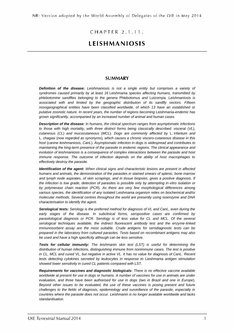
Definition of the disease: Leishmaniosis is not a single entity but comprises a variety of
syndromes caused primarily by at least 16 Leishmania species affecting humans, transmitted by
phlebotomine sandflies belonging to the genera Phlebotomus and Lutzomyia. Leishmaniosis is
associated with and limited by the geographic distribution of its sandfly vectors. Fifteen
nosogeographical entities have been classified worldwide, of which 13 have an established or
putative zoonotic nature. In recent years, the number of regions becoming Leishmania-endemic has
grown significantly, accompanied by an increased number of animal and human cases.
Description of the disease: In humans, the clinical spectrum ranges from asymptomatic infections
to those with high mortality, with three distinct forms being classically described: visceral (VL),
cutaneous (CL) and mucocutaneous (MCL). Dogs are commonly affected by L. infantum and
L. chagasi (now regarded as synonyms), which causes a chronic viscero-cutaneous disease in this
host (canine leishmaniosis, CanL). Asymptomatic infection in dogs is widespread and contributes to
maintaining the long-term presence of the parasite in endemic regions. The clinical appearance and
evolution of leishmaniosis is a consequence of complex interactions between the parasite and host
immune response. The outcome of infection depends on the ability of host macrophages to
effectively destroy the parasite.
Identification of the agent: When clinical signs and characteristic lesions are present in affected
humans and animals, the demonstration of the parasites in stained smears of splenic, bone marrow
and lymph node aspirates, of skin scrapings, and in tissue biopsies, gives a positive diagnosis. If
the infection is low grade, detection of parasites is possible only by attempting in-vitro isolation or
by polymerase chain reaction (PCR). As there are very few morphological differences among
various species, the identification of any isolated Leishmania organism relies on biochemical and/or
molecular methods. Several centres throughout the world are presently using isoenzyme and DNA
characterisation to identify the agent.
Serological tests: Serology is the preferred method for diagnosis of VL and CanL, even during the
early stages of the disease. In subclinical forms, seropositive cases are confirmed by
parasitological diagnosis or PCR. Serology is of less value for CL and MCL. Of the several
serological techniques available, the indirect fluorescent antibody test and the enzyme-linked
immunosorbent assay are the most suitable. Crude antigens for serodiagnostic tests can be
prepared in the laboratory from cultured parasites. Tests based on recombinant antigens may also
be used and have a high specificity although can be less sensitive.
Tests for cellular immunity: The leishmanin skin test (LST) is useful for determining the
distribution of human infections, distinguishing immune from nonimmune cases. The test is positive
in CL, MCL and cured VL, but negative in active VL. It has no value for diagnosis of CanL. Recent
tests detecting cytokines secreted by leukocytes in response to Leishmania antigen stimulation
showed lower sensitivity in cured CL patients compared with LST.
Requirements for vaccines and diagnostic biologicals: There is no effective vaccine available
worldwide at present for use in dogs or humans. A number of vaccines for use in animals are under
evaluation, and three have been authorised for use in dogs (two in Brazil and one in Europe).
Beyond other issues to be evaluated, the use of these vaccines is posing present and future
challenges to the fields of diagnosis, epidemiology and surveillance of the parasite, especially in
countries where the parasite does not occur. Leishmanin is no longer available worldwide and lacks
standardisation.

Definition of the disease: Leishmaniosis comprises a variety of syndromes caused by members of the
protozoan parasite Leishmania (Kinetoplastida: Trypanosomatidae), which are transmitted to mammal hosts by
the bite of infected phlebotomine sandflies belonging to the genera Phlebotomus (Old World)1 and Lutzomyia (New World). The disease results from the multiplication of amastigote forms in macrophages of the reticuloendothelial system. T lymphocytes and cytokines, play a crucial role in determining whether infection evolves toward a protective immune status or a progressive and manifest disease. In inbred mouse models, two different T helper (Th) cell subsets are involved, termed Th1 and Th2, which differ in their profile of secreted cytokines by a polarised activation; the Th1 response confers protection, whereas the Th2 response renders the host susceptible to infection. However, in human patients and dogs with clinically apparent infections, Th1 and Th2 type responses are not characteristically polarised, as both activating (e.g. interferon gamma, interleukin 12) and suppressive cytokines (e.g. interleukin 10, interleukin 13, interleukin 4, TGF beta) are detected (Murray et al., 2005).
Causal pathogen: Sixteen well recognised Leishmania species are agents of human leishmanioses. In the New World, tegumentary forms are caused by L. braziliensis, L. guyanensis, L. panamenisis, L. shawi, L. naϊffi, L. lainsoni, L. lindenbergi, L. peruviana, L. mexicana, L. venezuelensis and L. amazonensis; visceral and, more rarely, cutaneous forms, are caused by L. infantum. In the Old World, cutaneous forms are caused by L. tropica, L. major and L. aethiopica; visceral and, more rarely, cutaneous forms, are caused by L. infantum and L. donovani. Leishmania infantum and L. chagasi have been found to be identical by genotyping and should be regarded as synonyms (Kuhls et al., 2011). In addition, the taxonomic position of other Leishmania agents of tegumentary forms (L. killicki in the Old World; L. pifanoi, L. garnhami and L. colombiensis in the New World) is still under discussion. Other Leishmania species not pathogenic to humans include New and Old World rodent parasites and an agent of cutaneous leishmaniosis in the red kangaroo in Australia (Dougall et al., 2011). Dogs are mainly affected by L. infantum, but in some instances parasites belonging to L. braziliensis, L. peruviana, and L. tropica have been isolated from this host (Mohebali et al., 2005; Reithinger et al., 2002).
Description of the disease: Various forms of clinical manifestations of human leishmaniosis have been
described and divided into three major clinical entities: visceral leishmaniosis (VL, kala azar), cutaneous leishmaniosis (localised, diffuse or disseminated CL, oriental sore, uta, pian bois, chiclero’s ulcer) and mucocutaneous leishmaniosis (MCL, espundia) (World Health Organization [WHO], 2010). The diseases are mainly zoonoses with two exceptions, that of CL due to L. tropica in urban areas of the Middle East, and that of VL due to L. donovani in the Indian sub-continent (northern India, Nepal and Bangladesh) and in some parts of Eastern Africa (e.g. Sudan and Ethiopia). Canine leishmaniosis (CanL) is a chronic viscero-cutaneous disease caused by L. infantum, for which the dog acts as the source reservoir. Resistant asymptomatic dogs may exceed >50% of the infected canine population. Typical external signs recorded in susceptible dogs that evolve towards full-blown disease are lymph node enlargement, weight loss, exfoliative dermatitis, alopecia, ulcers and ocular alterations. Recently, leishmaniosis in cats has emerged in some areas with clinical features resembling tegumentary forms, more rarely systemic (Gramiccia, 2011). Sporadic tegumentary cases have been reported in equids and cattle.
Zoonotic risk and biosafety requirements: Direct contact with infected hosts or handling of biological samples
and parasite cultures from these hosts do not require special precautions because of the sandfly-borne nature of the infections and the lack of resistant forms in the environment. Leishmania spp. are classed in Risk Group 2 for human infection and should be handled with appropriate measures as described in Chapter 1.1.4 Biosafety and biosecurity: Standard for managing biological risk in the veterinary laboratory and animal facilities. Biocontainment measures should be determined by risk analysis as described in Chapter 1.1.4.
Differential diagnosis: In humans, the differential diagnosis of VL depends on the local disease pattern
associated with endemic areas. In many of them it includes chronic malaria, disseminated histoplasmosis, hepatosplenic schistosomiasis, typhoid fever, brucellosis, tuberculosis, endocarditis, relapsing fever, and African trypanosomosis. Other cosmopolitan diseases include syphilis, lymphomas, chronic myeloid leukaemia, sarcoidosis, malignant histiocytosis, and liver cirrhosis. For CL, differential diagnosis includes insect bites, furuncular myiasis, bacterial tropical ulcers, keloid, lupus vulgaris, discoid lupus erythematosus, and sarcoidosis. In dogs, the most common leishmaniosis signs may be confounded with ehrlichiosis, babesiosis and vector-borne or intestinal helminthiases.
1 In this chapter, the term ‘New World’ refers to the Americas, and the term ‘Old World’ refers to Europe, Africa and Asia
(WHO, 2010).

Techniques for diagnosis of leishmaniosis are detailed below. The list and fitness of each test for different purposes are given In Table 1.
Method
Purpose
Population freedom from
infection
Individual animal freedom from
infection prior to movement
Contribution to
eradication policies
Confirmation of clinical
cases
Prevalence of infection – surveillance
Immune status in individual animals
or populations post-vaccination
Agent identification2
Microscopy – –
n/a
++ –
n/a In-vitro culture – + ++ –
PCR ++ +++ ++ ++
Detection of immune response
IFAT ++ ++
n/a
++ +++
n/a
ELISA +++ ++ ++ +++
Direct agglutination test
++ ++ ++ ++
Rapid immuno-chromatographic
assay – – ++ +
Tests for cellular immunity
+ – – ++
Key: +++ = recommended method; ++ = suitable method; + = may be used in some situations, but cost, reliability, or other factors severely limits its application; – = not appropriate for this purpose;
n/a = not applicable because the relevant purposes are not relevant to animal leishmaniosis at present. Although not all of the tests listed as category +++ or ++ have undergone formal validation, their routine nature and the fact that
they have been used widely without dubious results, makes them acceptable. PCR = polymerase chain reaction; IFAT = Indirect fluorescent antibody test; ELISA = enzyme-linked immunosorbent assay.
Clinical examination of suspected cases, parasitological diagnosis and immunodiagnosis are the routine methods available for the diagnosis of leishmaniosis. However, the demonstration of the parasite is the only way to confirm the disease conclusively. In VL and CanL, isolation and identification of the parasite from biopsies (lymph node, bone marrow, and spleen aspirate) coupled with molecular and immunodiagnostic tests are recommended. Parasitological diagnosis is necessary for confirmation of CL (through lesion scraping or needle aspiration from the edge of the lesions) as neither clinical examination nor serology is adequate. Smears of biopsy material are stained with Giemsa stain and examined microscopically at ×600–1000 magnification to observe the presence of amastigote forms. Material should also be cultured in appropriate media at 22–26°C for the microscopic observation of promastigote forms, which are morphologically identical to development stages observed in infected phlebotomine sandflies.
2 A combination of agent identification methods applied on the same clinical sample is recommended.

Amastigote are small intracellular rounded or oval body, 1.5–3 × 2.5–6.5 µm in size, found in vacuoles within the cytoplasm of the macrophages. There is no free flagellum. The organism has a relatively large nucleus and a kinetoplast consisting of a rod-like body and a dot-like basal body.
Promastigote are elongated extracellular organism, body size 15–20 × 1.5–3.5 µm with a single flagellum 15–28 µm long, arising close to the kinetoplast at the anterior. The nucleus is situated centrally.
The choice of the isolation and culture methods will depend on the immediate circumstances and on the technical capability and experience of the laboratory staff (WHO, 2010). In-vitro isolation offers certain advantages over the in-vivo methods: cultures become positive more rapidly (5–30 days compared with months for lesions to appear on an animal) and the materials are less expensive. However, for in-vitro isolation, the techniques used should be
carried out under strictly sterile conditions, which is rarely feasible in the field. Unfortunately, there is still no ‘universal’ culture medium in which all the different leishmanias will grow easily, and it is almost impossible to predict which medium will be best suited to the growth of a particular isolate of Leishmania. Individual laboratories have to find the most suitable medium among biphasic blood agar media and tissue culture media supplemented with fetal calf serum (Evans, 1987). When attempting primary isolation of unknown organisms, a blood agar-based medium should be used – preferably NNN medium (Novy, McNeil and Nicolle), otherwise brain–heart infusion (BHI) agar medium or EMTM (Evans’ modified Tobie’s medium) should be used. For bulk cultivation of established isolates, suitable media are reported in Section B.1.3 below (see Evans, 1987 for media composition). The organisms from patients with chronic CL and MCL can be very difficult to cultivate. The parasites sometimes die when subcultured, even when the initial isolation is successful. This seems especially common when the initial isolation has been into a rich medium. Often this can be overcome if subcultures are made into less nutritionally rich media, such as NNN, or one of the semisolid media such as ‘sloppy Evans’ or semisolid Locke blood agar.
As several alternative methods have been developed during the past decades, in-vivo isolation of Leishmania in susceptible animals (e.g. the Syrian hamster Mesocricetus auratus or BALB/c mice) is no longer recommended for routine diagnosis.
Morphological identification enables identification of Leishmania at genus level, but not at species or subspecies level. Several techniques may be used to identify the different Leishmania species, subspecies or strains. Fifteen recognised Leishmania Identification Centres were listed by WHO in 2010.
Also known as multi-locus enzyme electrophoresis (MLEE), isoenzyme characterisation is the reference method for species identification (Rioux et al., 1990; WHO, 2010), although this technique
requires cultivation of a large number of parasites (5 × 10⁹–1 × 10¹⁰). The principles of enzyme electrophoresis are as follows: soluble enzymes are extracted from the organisms grown in media for bulk cultivation (BHI medium, MEM/FCS/EBLB [minimal essential medium/fetal calf serum/Evans’ blood lysate broth] medium, Schneider’s Drosophila medium). A small amount of the extract is then
placed in an inert supporting substance, the matrix, containing a buffer at a fixed pH. The matrix is usually starch gel, but it could equally well be absorbent cellulose acetate, acrylamide or agarose. The pH of the buffer in the matrix is usually chosen so that the isoenzymes are negatively charged. A direct current is passed through the matrix carried by the ions in the buffer. When electrophoresis is completed, most proteins will have moved in the matrix towards the anode, depending on the amount of negative charge. If stained at this stage with a general protein stain, many bands will be seen. However, the high substrate and cofactor specificity of enzymes make it possible to stain only these proteins. Hence, the electrophoretic mobility of one particular enzyme can be compared among several organisms. The stained matrix with its collection of stained isoenzyme bands is known as a zymogram. Normally one or more extracts from reference organisms, in which the enzyme banding patterns are well documented, are included in the gel to aid the interpretation of results. Most enzymes used for characterisation purposes are stained by methods incorporating a dehydrogenase reaction. At least 12 enzymes should be examined; organisms showing identical zymograms are classified into zymodemes of a given species.

PCR methods are available for diagnosis and/or identification of Leishmania from different types of human and animal samples. Essentially, techniques developed either to identify established isolates of Leishmania or to detect organisms from fresh or frozen, formalin-fixed and paraffin-embedded tissue
biopsies, include: (a) digestion of material with proteinase K and DNA extraction. These steps can be either performed using in-house protocols and reagents, or by commercial kits that are widely available; (b) standard PCR amplification using oligonucleotide sequences (primers) selected from the small-subunit rRNA gene (Mathis & Deplazes, 1995), kinetoplast DNA minicircles (Maarten et al., 1992), or other highly repetitive genomic DNA sequences (Bulle et al., 2002; Piarroux et al., 1993); (c) analysis of amplification products by 1–2% agarose gel.
For diagnostic purposes, a nested or semi-nested PCR using internal primers from the above sequences can be performed to increase sensitivity (Cruz et al., 2002). In acute human VL, PCR has a
sensitivity comparable with that of culture-based methods, but gives results much faster. On the other hand, in subclinical or mild human VL forms, and in immunosuppressed patients, PCR results more sensitive than any other direct parasitological and serological techniques. In CanL, the diagnostic efficacy of PCR as compared with serology depends on the natural course of the disease, the sensitivity being highest shortly after infection (Oliva et al., 2006; Quinnell et al., 2001). Less invasive samples, such as conjunctival swabs, have been assayed in dogs with successful results (Di Muccio et al., 2012). In American human CL and MCL, PCR appears to be consistently more sensitive than any previously recommended method of diagnosis (De Brujin et al., 1993). Real-time PCR methods, which
allow the continuous monitoring of the accumulation of PCR products during amplification, have been described and the specific equipment is available commercially. They can be more sensitive than conventional PCR, and are mainly aimed at studying the kinetics of infection and monitoring therapeutic response (Bell & Ranford-Cartwright, 2002; Bossolasco et al., 2003; Reithinger & Dujardin, 2007). In addition, real-time PCR has been reported to be useful for evaluating infections in less invasive samples such as peripheral blood (Francino et al., 2006).
With regard to parasite identification, different techniques have been developed that allow Leishmania characterisation at the species or strain level such as (a) PCR-restriction fragment length polymorphism (RFLP) analysis, in which the PCR products are digested by appropriate restrictions enzymes and the resulting restriction fragment patterns are analysed (Marfurt et al., 2003; Minodier et al., 1997; Montalvo et al., 2012; Volpini et al., 2004); (b) multilocus microsatellite typing (MLMT) (Kuhls et al., 2011) and (c) multilocus sequence typing (MLST) (Mauricio et al., 2006). In these cases, repeated and polymorphic DNA sequences are targeted, such as ribosomal internal transcribed spacer 1 (ITS1), cysteine protease B, kinetoplast DNA minicircles, surface glycoprotein 63, heat-shock protein 70, mini-exons and microsatellites (Reithinger & Dujardin, 2007; Schönian et al., 2008).
Sensitivity and specificity of most diagnostic protocols can be significantly increased by hybridisation to genus- or species-specific DNA probes. Originally, these probes were labelled with radioactive isotopes but currently they are commonly labelled with fluorescent dyes.
Several serological tests are used for detecting anti-leishmanial antibodies. Sensitivity values in humans reported below for each test apply only to individuals who are not immunocompromised. A high percentage of patients with VL co-infected with human immunodeficiency virus (HIV) have been reported to be seronegative for anti-leishmanial antibodies (Gradoni et al., 1993).
The indirect fluorescent antibody (IFA) test is widely used because it is easy to perform. The test is genus specific, although significant cross-reactions have been reported in individuals infected with Trypanosoma cruzi. For these subjects, serological tests based on specific recombinant Leishmania
antigens would be more appropriate (see Sections B.2.2.2 and B.2.2.4 below). In Chagas’ disease-free areas, the IFA test for the diagnosis of clinical VL or CanL has a sensitivity of 96% and specificity of 98%, which is similar to the ELISA (enzyme-linked immunosorbent assay). Although amastigotes from frozen sections or smears of infected organs can be used as antigen, cultured promastigotes represent the commonest antigen source.
i) Harvest 3–4 ml of the liquid media of a 3-day-old culture showing flourishing promastigote growth (see Section B.1 for culture media).

ii) Wash the organisms three times with phosphate-buffered saline (PBS), pH 7.2–7.4, by centrifugation at 350 g for 15 minutes at room temperature.
iii) Resuspend the final cell pellet in PBS and adjust the promastigote concentration to
approximately 4 × 10⁶/ml with the aid of a haemocytometer.
iv) Distribute 30 µl of the promastigote suspension on to each circle of a multispot slide and allow to dry at room temperature.
v) Fix the promastigotes in cold acetone for 10 minutes, then put the slides into a plastic box and keep in a deep freezer (–35°C) for no longer than 2–3 months.
i) Wash the frozen antigen-coated slides in PBS and allow to dry at room temperature.
ii) Inactivate the sera for 30 minutes in a water bath at 56°C.
iii) Make doubling dilutions of test sera from 1/80 to 1/10,240 for human VL, and from 1/40 to 1/5120 for CanL. Positive and negative control sera, at dilutions of 1/80 and 1/160 for human VL, and of 1/40 and 1/80 for CanL, are also included in the test. No standard sera are available, but internal standards should be prepared and titrated.
iv) Distribute 30 µl of diluted serum samples on to each slide circle and incubate for 30 minutes at 37°C.
v) Remove the serum samples by vigorous washing in PBS, followed by immersion of the slides in PBS for 10 minutes. Allow the slides to dry.
vi) Distribute 30 µl of diluted fluorescein isothiocyanate (FITC)-conjugated anti-immunoglobulin on to each slide circle and incubate for 30 minutes at 37°C. FITC-conjugated anti-human and anti-dog immunoglobulins are commercially available. Follow the instructions for the appropriate dilution.
vii) Repeat step v and mount with a cover-slip in a few drops of PBS/glycerol (50% [v/v] of each).
viii) Read the slides under a fluorescent microscope. The highest dilution showing fluorescent promastigotes is taken to be the antibody titre.
In acute human VL, the threshold titre usually ranges from 1/80 to 1/160. Asymptomatic or subclinical human disease usually results in titres below 1/80. In CanL the threshold titre ranges from 1/40 (indicative of exposure but not necessarily of established infection) to 1/160 (indicative of established infection), whereas a titre of 1/320 or above can be indicative of the disease in clinically suspected dogs (Paltrinieri et al., 2010). As regards other domestic mammals (e.g. cats) no standardised IFA threshold limits are available. As IFA test performance may vary in different laboratories, it is better for each laboratory to define its own threshold titre using defined positive and negative reference sera.
The ELISA can be carried out on serum or on a measured volume of blood. The blood is collected by needle-prick on to suitable absorbent paper strips and allowed to dry. The sample is eluted and tested at a single dilution previously determined to give an acceptable sensitivity and specificity. This test can be used for seroepidemiological surveys under field conditions.
In the classical method, the antigen is prepared as follows: promastigotes harvested from cultures are washed four times with PBS, pH 7.2, at 1000 g for 15 minutes. The packed promastigotes are
resuspended in twice their volume of distilled water, and then sonicated at medium amplitude in an ice bath. The suspension is left at 4°C overnight to allow the proteins to come into solution. After a final centrifugation at 4000 g for 10 minutes to eliminate the cellular debris, the overlay, representing the concentrated soluble antigen, is dispensed into vials and stored at –20°C until required. For use in the test, it is reconstituted with PBS to the predetermined optimal protein concentration (around 20 µg/ml) as measured by Lowry’s method. Enzyme (usually horseradish peroxidase)-conjugated reagents consist of anti-dog goat immunoglobulins or Protein A (Hamarsheh et al., 2012).
The ELISA is useful for the diagnosis of Old and New World leishmanioses. There is little or no cross-reaction with other diseases and, according to the Leishmania strain used, sensitivity can range from 86% to 99%.

A detergent-soluble promastigote antigen has been used in ELISA instead of the crude lysate, for the diagnosis of CanL. The detergent was Triton X-100 and the proteic extract was protected with protease inhibitors. Using this method, ELISA sensitivity increased to 99.5%, while its specificity was comparable with that of the IFA test (97%) (Mancianti et al., 1995).
The ELISA methods described above are based on crude antigenic preparations. A recombinant antigen from a cloned protein of L. infantum, called rK39, has been reported to be highly reactive to sera from human and canine visceral leishmaniosis cases when run in an ELISA format. Using 25–50 ng of the antigen, 99% specificity and sensitivity was consistently found for dogs with parasitologically proven disease (Scalone et al., 2002). In HIV-positive patients, K39-ELISA showed higher sensitivity (82%) than the IFA test (54%) (Houghton et al., 1998). The K39 antigen, which shows remarkable stability and reproducibility, is commercially available. More recently, a K9-K39-K26 recombinant chimeric antigen has been evaluated as a single ELISA protocol for serological diagnosis of both human and canine Leishmania infections (Daprà et al., 2008). In dogs, test specificity and sensitivity were reported to be 99.5% and 98.5%, respectively, with high concordance (K value: 0.98) with standard IFA test.
The direct agglutination test (DAT) has been described for the diagnosis of VL and CanL. After test improvement, DAT has been validated as a specific and sensitive assay for field investigations (Boelaert et al., 1999; Cardoso et al., 2004; Ozbel et al., 2000). The antigen consists of promastigotes harvested from cultures, washed in PBS, pH 7.2, treated with 0.4% trypsin (for 45 minutes at 37°C and then washed again), and stained with 0.02% Coomassie brilliant blue. Twofold serial dilutions of serum in PBS are made in V-bottomed microtitre-plate wells; 50 µl of antigen preparation is added to each well, and the plate is then carefully shaken by hand and left for 18 hours at room temperature. The test is read visually against a white background. Positive reactions are indicated by typical light-blue aggregates, while negative samples give a clear sharp-edged blue spot.
A modified DAT for detection of specific anti-leishmanial antibodies in canine reservoir hosts is considered to be highly suitable for wide-scale epidemiological and ecological field work and diagnosis of CanL, having 100% sensitivity and 98.9% specificity (Harith et al., 1988; 1989). The reliability of the test was improved by treating the test sera with 0.2 M 2-mercaptoethanol and incubating them at 37°C.
A rapid immunochromatographic assay using rK39 as antigen (K39 dipstick or strip-test, commercially available) has been evaluated in different endemic settings of VL. The nitrocellulose membrane of the test kit holds an absorbent pad at one end, a band of immobilised anti-protein A antibody (used to detect IgG) at the other (control region), and a band of rK39 antigen in the middle (test region). A protein-A-colloidal gold conjugate is used as the immunochromatographic detection reagent. One small drop (20 µl) of the serum to be examined is placed on the absorbent pad before two large drops (100 µl) of test buffer are added to the pad, and the mixture is allowed to migrate up the strip by capillary action. After 2–10 minutes, the result is positive if two distinct red lines appear (one in the test region and another in the control region), it is negative when no red line appears in the test region, and it is invalid if the control line fails to appear.
In clinical cases of human VL, two commercial brands of K39 dipstick showed 99–100% sensitivity and 95–100% specificity in India (Sundar et al., 2006), 90% sensitivity and 100% specificity in Brazil (Carvalho et al., 2003), and 100% sensitivity and specificity in the Mediterranean basin (Brandonisio et al., 2002). In parasitologically proven CanL, in both asymptomatic and symptomatic cases, the sensitivity of the K39 dipstick was 97% and the specificity 100% (Otranto et al., 2005).
Delayed hypersensitivity is an important feature of all forms of human leishmaniosis and can be measured by the leishmanin skin test (LST), also known as the Montenegro reaction (Manson-Bahr, 1987). LST has no value for
the diagnosis of CanL. Leishmanin is a killed suspension of whole (0.5–1 × 10⁷/ml) or disrupted (250 µg protein/ml) promastigotes in pyrogen-free saline containing phenol. A delayed reaction develops and is read at 48–72 hours.
The false-positive reaction rate in otherwise healthy people is approximately 1%, but this can be higher in areas where there is a background of leishmaniosis, as many of the healthy population may show quite high rates of leishmanin sensitivity. Although there is complete cross-reactivity among all strains of Leishmania, heterologous antigens often give smaller reactions, which may be caused by difficulty in standardisation. LST is used in the

clinical diagnosis of CL and MCL. In VL it will only measure past infections because during active disease, a complete anergy is found. Leishmanins are not available commercially worldwide.
More recently, the measurement of gamma interferon (IFN-γ) as a surrogate marker of cellular immune responses has been assayed in human leishmaniosis, using a format similar to that used for tuberculosis, using ELISA to measure the cytokine secreted by leukocytes in response to Leishmania antigen stimulation (Alimohammadian et al., 2012; Turgay et al., 2010).
There is no effective vaccine available worldwide for prophylactic immunisation against leishmaniosis. In the past, vaccination against Leishmania was limited to the protection of humans from both L. tropica and L. major by prior syringe-induced infection with L. major organisms. Living promastigotes from cultures were injected into the arm and the resulting infection was allowed to run a natural course; after recovery, the individual was firmly immune to subsequent infection with both Leishmania species. This type of immunisation was practised on a limited scale in hyperendemic areas of CL (due to L. major) in Israel, Iran and the former Union of Soviet Socialist Republics (Shuikina et al., 1968).
Currently there are no available laboratory-made antigens for Leishmania vaccine production. There are three licensed inactivated vaccines against CanL, which are patent-protected. The first vaccine was developed in Brazil. Outlines and requirements for production were approved by the Brazilian Ministry of Agriculture, Livestock and Food Supply. The vaccine consists of the glycoprotein-enriched fraction of L. donovani known as ‘fucose-mannose ligand’ (FML). Field studies showed about 80% clinical protection conferred by the antigen administered with QuilA saponin as adjuvant, and also good immunotherapeutic efficacy when used in sick dogs (Palatnik-de-Sousa et al., 2008). A second vaccine was also developed in Brazil, which uses the recombinant A2 antigen of L. donovani in association with a saponin adjuvant. It demonstrated 43% protection against a culture positive state in an artificial challenge model. The third vaccine is licensed in Europe. Outlines and requirements for production have been approved by the European Medicines Agency. The antigen consists of excreted/secreted proteins purified from an aproteinic medium used to grow L. infantum promastigotes, adjuvanted with QA-21 saponin (Moreno et al., 2012). Field efficacy evaluation of this commercial vaccine is still at a preliminary stage. Findings from some 80 beagle dogs exposed to natural infection during 2 years indicated that the vaccine decreased the risk of developing progressive disease by about four fold, but did not prevent infection (Oliva et al., 2012). Surveillance programmes, especially those based solely on serology in dogs, should be revised to take into account the impact of vaccination and to ensure epidemiological data is interpreted appropriately.
At present, a number of promising anti-leishmanial vaccines are under experimental evaluation (Coler & Reed, 2005; Das & Ali, 2012). A chimeric antigen generated from three recombinant Leishmania antigens screened for their ability to elicit cellular immune responses (known as Leish-111f, patent-protected) and adjuvanted with monophosphoryl lipid A – stable emulsion (MPL-SE), represents the first defined vaccine for leishmaniosis in humans, having completed phase 1 and 2 safety and immunogenicity testing in healthy subjects (Coler et al., 2007). The same polyproteinic antigen and adjuvant failed to protect dogs from L. infantum infection in a phase 3 trial (Gradoni et al., 2005), while conferred some protection from disease when used as an immunotherapeutic agent in dogs with mild CanL (Trigo et al., 2010). Recently, a novel chimeric antigen generated from four recombinant antigens (KSAC, patent-protected) has been found promising for vaccine protection in both human VL and CanL (Goto et al., 2011).
As the skin test is not used for diagnosis of animal infections, and the leishmanin antigen has not been internationally standardised, no recommendations can be made by the OIE.

ALIMOHAMMADIAN M.H., JONES S.L., DARABI H., RIAZIRAD F., AJDARY S., SHABANI A., REZAEE M.A., MOHEBALI M., HOSSEINI Z. & MODABBER F. (2012). Assessment of interferon-γ levels and leishmanin skin test results in persons recovered for leishmaniasis. Am. J. Trop. Med. Hyg., 87 (1), 70–75.
BELL A.S. & RANFORD-CARTWRIGHT L.C. (2002). Real-time quantitative PCR in parasitology. Trends Parasitol., 18
(8), 337–342.
BOELAERT M., EL SAFI S., JACQUET D., DE MUYNCK A., VAN DER STUYFT P. & LE RAY D. (1999). Operational validation of the direct agglutination test for diagnosis of visceral leishmaniasis. Am. J. Trop. Med. Hyg., 60, 129–134.
BOSSOLASCO S., GAIERA G., OLCHINI D., GULLETTA M., MARTELLO L., BESTETTI A., BOSSI L., GERMAGNOLI L., LAZZARIN
A., UBERTI-FOPPA C. & CINQUE P. (2003). Real-time PCR assay for clinical management of human immunodeficiency virus-infected patients with visceral leishmaniasis. J. Clin. Microbiol., 41 (11), 5080–5084.
BRANDONISIO O., FUMAROLA L., MAGGI P., CAVALIERE R., SPINELLI R. & PASTORE G. (2002). Evaluation of a rapid immunochromatographic test for serodiagnosis of visceral leishmaniasis. Eur. J. Clin. Microbiol. Infect. Dis., 21,
461–464.
BULLE B., MILLON L., Bart J.M., GALLEGO M., GAMBARELLI F., PORTUS M., SCHNUR L., JAFFE C.L., FERNANDEZ-BARREDO S., ALUNDA J.M. & PIARROUX R. (2002). Practical approach for typing strains of Leishmania infantum by microsatellite analysis. J. Clin. Microbiol., 40, 3391–3397.
CARDOSO L., SCHALLIG H.D., NETO F., KROON N. & RODRIGUES M. (2004). Serological survey of Leishmania infection in dogs from the municipality of Peso da Regua (Alto Douro, Portugal) using the direct agglutination test (DAT) and fast agglutination screening test (FAST). Acta Trop., 91, 95–100.
CARVALHO S.F., LEMOS E.M., COREY R. & DIETZE R. (2003). Performance of recombinant K39 antigen in the diagnosis of Brazilian visceral leishmaniasis. Am. J. Trop. Med. Hyg., 68, 321–324.
COLER R.N. & REED S.G. (2005). Second-generation vaccines against leishmaniasis. Trends Parasitol., 21, 244–
249.
COLER R.N., GOTO Y., BOGATZKI L., RAMAN V. & REED S.G. (2007). Leish-111f, a recombinant polyprotein vaccine that protects against visceral Leishmaniasis by elicitation of CD4+ T cells. Infect. Immun., 75, 4648–4654.
CRUZ I., CAÑAVATE C., RUBIO J. M., MORALES M.A., CHICHARRO C., LAGUNA F., M. JIMÉNEZ-MEJÍAS, SIRERA G., VIDELA
S., ALVAR J. & THE SPANISH HIV-LEISHMANIA STUDY GROUP. (2002). A nested polymerase chain reaction (Ln-PCR) for diagnosing and monitoring Leishmania infantum infection in patients co-infected with human immunodeficiency virus. Trans. R. Soc. Trop. Med. Hyg., 96 (Suppl 1), S185–S189.
DAPRÀ F., SCALONE A., MIGNONE W., FERROGLIO E., MANNELLI A., BIGLINO A., ZANATTA R., GRADONI L. & ROSATI S. (2008). Validation of a recombinant based antibody ELISA for diagnosis of human and canine leishmaniasis. J. Immunoassay Immunochem., 29, 244–256.
DAS A. & ALI N. (2012). Vaccine development against Leishmania donovani. Front. Immunol., 3, 99.
DE BRUJIN M.H.L., LABRADA L.A., SMYTH A.J., SANTRIC C. & BARKER D.C. (1993). A comparative study of diagnosis by the polymerase chain reaction and by current clinical methods using biopsies from Colombian patients with suspected leishmaniasis. Trop. Med. Parasitol., 44, 201–207.
DI MUCCIO T., VERONESI F., ANTOGNONI M.T., ONOFRI A., PIERGILI FIORETTI D. & GRAMICCIA M. (2012). Diagnostic value of conjunctival swab nested-PCR in different categories of dogs naturally exposed to Leishmania infantum infection. J. Clin. Microbiol., 50 (8), 2651–2659.
DOUGALL A.M., ALEXANDER B., HOLT D.C., HARRIS T., SULTAN A.H., BATES P.A., ROSE K. & WALTON S.F. (2011). Evidence incriminating midges (Diptera: Ceratopogonidae) as potential vectors of Leishmania in Australia. Int. J. Parasitol., 41 (5), 571–579.
EVANS D.A. (1987). Leishmania. In: In-Vitro Methods for Parasite Cultivation, Taylor A.E. & Baker J.R., eds.
Academic Press, London, UK, 52–75.

FRANCINO O., ALTET L., SANCHEZ-ROBERT E., RODRIGUEZ A., SOLANO-GALLEGO L., ALBEROLA J., FERRER L., SANCHEZ
A. & ROURA X. (2006). Advantages of real-time PCR assay for diagnosis and monitoring of canine leishmaniosis. Vet. Parasitol., 137 (3-4), 214–221.
GOTO Y., BHATIA A., RAMAN V.S., LIANG H., MOHAMATH R., PICONE A.F., VIDAL S.E., VEDVICK T.S., HOWARD R.F. &
REED S.G. (2011). KSAC, the first defined polyprotein vaccine candidate for visceral leishmaniasis. Clin. Vaccine. Immunol., 18, 1118–1124.
GRADONI L., FOGLIA MANZILLO V., PAGANO A., PIANTEDOSI D., DE LUNA R., GRAMICCIA M., SCALONE A., DI MUCCIO T. & OLIVA G. (2005). Failure of a multi-subunit recombinant leishmanial vaccine (MML) to protect dogs from Leishmania infantum infection and to prevent disease progression in infected animals. Vaccine, 23 (45), 5245–
5251.
GRADONI L., SCALONE A. & GRAMICCIA M. (1993). HIV-Leishmania co-infections in Italy: serological data as an indication of the sequence of acquisition of the two infections. Trans. R. Soc. Trop. Med. Hyg., 87, 94–96.
GRAMICCIA M. (2011). Recent advances in leishmaniosis in pet animals: Epidemiology, diagnostics and anti-vectorial prophylaxis. Vet. Parasitol., 181, 23–30.
HAMARSHEH O., NASEREDDIN A., DAMAJ S., SAWALHA S., AL-JAWABREH H., AZMI K., AMRO A., AREKAT S., ABDEEN Z. &
AL-JAWABREH A. (2012). Serological and molecular survey of Leishmania parasites in apparently healthy dogs in the West Bank, Palestine. Parasit. Vectors, 5, 183.
HARITH A.E., KOLK A.H.J., LEEUWENBURGH J., MUIGAI R., HUIGEN E., JELSMA T. & KAGER P.A. (1988). Improvement of a direct agglutination test for field studies of visceral leishmaniasis. J. Clin. Microbiol., 26, 1321–1325.
HARITH A.E., SLAPPENDEL R.J., REITER I., VAN KNAPEN F., KORTE P.D., HUIGEN E. & KOLK A.H.J. (1989). Application of a direct agglutination test for detection of specific anti-Leshmania antibodies in the canine reservoir. J. Clin. Microbiol., 27, 2252–2257.
HOUGHTON R.L., PETRESCU M., BENSON D.R., SKEIKY Y.A.W., SCALONE A., BADARO R., REED S.G. & GRADONI L. (1998). A cloned antigen (recombinant K39) of Leishmania chagasi diagnostic for visceral leishmaniasis in human
immunodeficiency virus type 1 patients and a prognostic indicator for monitoring patients undergoing drug therapy. J. Infect. Dis., 177, 1339–1344.
KUHLS K., ALAM M.Z., CUPOLILLO E., FERREIRA G.E., MAURICIO I.L., ODDONE R., FELICIANGELI M.D., WIRTH T., MILES
M.A. & SCHÖNIAN G. (2011). Comparative microsatellite typing of new world Leishmania infantum reveals low heterogeneity among populations and its recent old world origin. PLoS Negl. Trop. Dis., 5 (6), e1155.
MAARTEN H.L., DE BRUJIN M.H.L. & BARKER D.C. (1992). Diagnosis of New World leishmaniasis: specific detection of species of the Leishmania braziliensis complex by amplification of kinetoplast DNA. Acta Trop., 52, 45–58.
MANCIANTI F., FALCONE M.L., GIANNELLI C. & POLI A. (1995). Comparison between and enzyme-linked immunosorbent assay using a detergent-soluble Leishmania infantum antigen and indirect immunofluorescence for the diagnosis of canine leishmaniosis. Vet. Parasitol., 59, 13–21.
MANSON-BAHR P.C. (1987). Diagnosis. In: The Leishmaniases in Biology and Medicine. Vol. II. Clinical Aspects an Control, Peters W. & Killick-Kendrick R., eds. Academic Press, London, UK, 703–729.
MARFURT J., NASEREDDIN A., NIEDERWIESER I., JAFFE C.L., BECK H.P. & FELGER I. (2003). Identification and differentiation of Leishmania species in clinical samples by PCR amplification of the miniexon sequence and subsequent restriction fragment length polymorphism analysis. J. Clin. Microbiol., 41, 3147–3153.
MATHIS A. & DEPLAZES P. (1995). PCR and in vitro cultivation for detection of Leishmania spp. in diagnostic samples from humans and dogs. J. Clin. Microbiol., 33, 1145–1149.
MAURICIO I.L., YEO M., BAGHAEI M., DOTO D., PRATLONG F., ZEMANOVA E., DEDET J.P., LUKES J. & MILES M.A. (2006).Towards multilocus sequence typing of the Leishmania donovani complex: resolving genotypes and haplotypes for five polymorphic metabolic enzymes (ASAT, GPI, NH1, NH2, PGD). Int. J. Parasitol., 36, 757–769.
MINODIER P., PIARROUX R., GAMBARELLI F., JOBLET C. & DUMON H. (1997). Rapid identification of causative species in patients with Old World leishmaniasis. J. Clin. Microbiol., 35, 2551–2555.

MOHEBALI M., HAJJARAN H., HAMZAVI Y., MOBEDI I., ARSHI S., ZAREI Z., AKHOUNDI B., NAEINI K.M., AVIZEH R. & FAKHAR M. (2005). Epidemiological aspects of canine visceral leishmaniosis in the Islamic Republic of Iran. Vet. Parasitol., 129, 243–251.
MONTALVO A.M., FRAGA J., MAES L., DUJARDIN J-C. & VAN DER AUWERA G. (2012). Three new sensitive and specific heat-shock protein 70 PCRs for global Leishmania species identification. Eur. J. Clin. Microbiol. Infect. Dis., 31,
1453–1461.
MORENO J., VOULDOUKIS I., MARTIN V., MCGAHIE D., CUISINIER A.M. & GUEGUEN S. (2012). Use of a LiESP/QA-21 vaccine (CaniLeish) stimulates an appropriate Th1-dominated cell-mediated immune response in dogs. PLoS Negl. Trop. Dis., 6 (6), e1683.
MURRAY H.W., BERMAN J.D., MARTIN V., DAVIES C.R. & SARAVIA N.G. (2005). Advances in leishmaniasis. Lancet, 366, 1561-1577.
OLIVA G., NIETO J., FOGLIA MANZILLO V., CAPPIELLO S., FIORENTINO E., DI MUCCIO T., SCALONE A., MORENO J., CHICHARRO C., BUTAUD T., GUEGAND L., MARTIN V., CUISINIER A-C., GUEGUEN S., CAÑAVATE C. & GRADONI L. (2012). Evidence for protection against active infection and disease progression in naïve dogs vaccinated with LiESP/QA-21 (CaniLeish®) exposed to two consecutive Leishmania infantum transmission seasons. Proceedings of the WSAVA / FECAVA / BSAVA (World Small Animal Veterinary Association / Federation of European Companion Animal Veterinary Associations / British Small Animal Veterinary Association) World Congress, Birmingham, UK, 11–15 April 2012, p. 529–530.
OLIVA G., SCALONE A., FOGLIA MANZILLO V., GRAMICCIA M., PAGANO A., DI MUCCIO T. & GRADONII L. (2006). Incidence and time corse of Leishmania infantum infections examined by parasitological, serologic, and Nested-PCR techniques in a cohort of naïve dogs exposed to three consecutive transmission seasons. J. Clin. Microbiol., 44
(4), 1318–1322.
OTRANTO D., PARADIES P., SASANELLI M., LEONE N, de CAPRARIIS D., CHIRICO J., SPINELLI R., CAPELLI G. & BRANDONISIO O. (2005). Recombinant K39 dipstick immunochromatographic test: a new tool for the serodiagnosis of canine leishmaniasis. J. Vet. Diagn. Invest., 17, 32–37.
OZBEL Y., OSKAM L., OZENSOY S., TURGAY N., ALKAN M.Z., JAFFE C.L. & OZCEL M.A. (2000). A survey on canine leishmaniasis in western Turkey by parasite, DNA and antibody detection assays. Acta Trop., 74, 1–6.
PALATNIK-DE-SOUSA C.B., BARBOSA ADE F., OLIVEIRA S.M., NICO D., BERNARDO R.R., SANTOS W.R., RODRIGUES M.M., SOARES I. & BORJA-CABRERA G.P. (2008). FML vaccine against canine visceral leishmaniasis: from second-generation to synthetic vaccine. Expert Rev. Vaccines, 7, 833–851.
PALTRINIERI S., SOLANO-GALLEGO L., FONDATI A., LUBAS G., GRADONI L., CASTAGNARO M., CROTTI A., MAROLI M., OLIVA
G., ROURA X., ZATELLI A. & ZINI E. (2010). Canine Leishmaniasis Working Group, Italian Society of Veterinarians of Companion Animals. Guidelines for diagnosis and clinical classification of leishmaniasis in dogs. J. Am. Vet. Med. Assoc., 236 (11), 1184–1191.
PIARROUX R., AZAIEZ R., LOSSI A.M., REYNIER P., MUSCATELLI F., GAMBARELLI F., FONTES M., DUMON H. & QUILICI M. (1993). Isolation and characterization of a repetitive DNA sequence for Leishmania infantum: development of a visceral leishmaniosis polymerase chain reaction. Am. J. Trop. Med. Hyg., 49, 364–369.
QUINNELL R.J., COURTENAY O., DAVIDSON S., GARCEZ L., LAMBSON B., RAMOS P., SHAW J.J., SHAW M.A. & DYE C. (2001). Detection of Leishmania infantum by PCR, serology and cellular immune response in a cohort study of Brazilian dogs. Parasitology, 122, 253–261.
REITHINGER R. & DAVIES C.R. (2002). American cutaneous leishmaniasis in domestic dogs: an example of the use of the polymerase chain reaction for mass screening in epidemiological studies. Trans. R. Soc. Trop. Med. Hyg., 96 (Suppl 1), S123–126.
REITHINGER R. & DUJARDIN J-C. (2007). Molecular diagnosis of leishmaniasis: current status and future applications. J. Clin. Microbiol., 45, 21–25.
RIOUX J.A. LANOTTE G., SERRES E., PRATLONG F., BASTIEN P. & PERIERES J. (1990). Taxonomy of Leishmania, use of isoenzymes. Suggestions for a new classification. Ann. Parasitol. Hum. Comp. 65, 111–125.
SCALONE A., DE LUNA R., OLIVA G., BALDI L., SATTA G., VESCO G., MIGNONE W., TURILLI C., MONDESIRE R.R., SIMPSON
D., DONOGHUE A.R., FRANK G.R. & GRADONI L. (2002). Evaluation of the Leishmania recombinant K39 antigen as a

diagnostic marker for canine leishmaniasis and validation of a standardized enzyme-linked immunosorbent assay. Vet. Parasitol., 104, 275–285.
SCHÖNIAN G., MAURICIO I., GRAMICCIA M., CAÑAVATE C., BOELAERT M. & DUJARDIN J-C. (2008). Leishmaniases in the Mediterranean in the era of molecular epidemiology. Trends Parasitol. 24, 135–142.
SHUIKINA E.E., SERGIEV V.P., TRIERS I.I., SHCHERBAKOV V.A. & DIVEEV S.KH. (1968). Experience of antileishmaniasis vaccination with cultures of Leishmania tropica major grown in various types of media. Med. Parazitol. (Mosk.), 37, 648–651 (in Russian).
SUNDAR S., MAURYA R., SINGH R.K., BHARTI K., CHAKRAVARTY J., PAREKH A., RAI M., KUMAR K. & MURRAY H.W. (2006). Rapid, noninvasive diagnosis of visceral leishmaniasis in India: comparison of two immunochromatographic strip tests for detection of anti-K39 antibody. J. Clin. Microbiol., 44, 251–253.
TRIGO J., ABBEHUSEN M., NETTO E.M., NAKATANI M., PEDRAL-SAMPAIO G., DE JESUS R.S., GOTO Y., GUDERIAN J., HOWARD R.F. & REED S.G. (2010). Treatment of canine visceral leishmaniasis by the vaccine Leish-111f+MPL-SE. Vaccine, 28, 3333–3340.
TURGAY N., BALCIOGLU I.C., TOZ S.O., OZBEL Y. & JONES S.L. (2010). Quantiferon-Leishmania as an epidemiological tool for evaluating the exposure to Leishmania infection. Am. J. Trop. Med. Hyg., 83, 822–824.
VOLPINI A.C., PASSOS V.M., OLIVEIRA G.C. & ROMANHA A.J. (2004). PCR-RFLP to identify Leishmania (Viannia) braziliensis and L. (Leishmania) amazonensis causing American cutaneous leishmaniasis. Acta Trop., 90, 31–37.
WORLD HEALTH ORGANIZATION (WHO) (2010). Control of the leishmaniases, WHO Technical Report Series 949, WHO, Geneva, Switzerland, 1–186.
*
* *
NB: There is an OIE Reference Laboratory for Leishmaniosis
(see Table in Part 4 of this Terrestrial Manual or consult the OIE Web site for the most up-to-date list: http://www.oie.int/en/our-scientific-expertise/reference-laboratories/list-of-laboratories/ http://www.oie.int/).
Please contact the OIE Reference Laboratory for any further information on diagnostic tests, reagents and vaccines for leishmaniosis


![Ecology of phlebotomine sandflies and putative reservoir ... · San Carlos, state of Tamaulipas. Several years later, Velasco-Castrejón et al. [88] reported six additional cases](https://img.pdfslide.net/doc/110x75/5f9669209c795f3a6a6c7618/ecology-of-phlebotomine-sandflies-and-putative-reservoir-san-carlos-state-of.jpg)