Embed Size (px)
Citation preview
Comptes Rendus
Chimie
Raphaël Lamare, Romain Ruppert, Mourad Elhabiri, Gilles Ulrich,Laurent Ruhlmann and Jean Weiss
Design and synthesis of charged porphyrin dimers for polyoxometalaterecognition
Online first, 17th August 2021
<https://doi.org/10.5802/crchim.105>
Part of the Special Issue:MAPYRO: the French Fellowship of the PyrrolicMacrocyclic Ring
Guest editors: Bernard Boitrel (Institut des Sciences Chimiques de Rennes,CNRS-Université de Rennes 1, France) and Jean Weiss (Institut de Chimie deStrasbourg, CNRS-Université de Strasbourg, France)
© Académie des sciences, Paris and the authors, 2021.Some rights reserved.
This article is licensed under theCreative Commons Attribution 4.0 International License.http://creativecommons.org/licenses/by/4.0/
Les Comptes Rendus. Chimie sont membres duCentre Mersenne pour l’édition scientifique ouverte
www.centre-mersenne.org
Comptes RendusChimieOnline first, 17th August 2021https://doi.org/10.5802/crchim.105
MAPYRO: the French Fellowship of the Pyrrolic Macrocyclic Ring / MAPYRO: lacommunauté française des macrocycles pyrroliques
Design and synthesis of charged porphyrin dimers
for polyoxometalate recognition
Raphaël Lamare a, Romain Ruppert a, Mourad Elhabiri b, Gilles Ulrich c,Laurent Ruhlmann ∗, a and JeanWeiss ∗, a
a Institut de Chimie de Strasbourg, UMR 7177 CNRS-Université de Strasbourg,4 rue Blaise Pascal, 67000 Strasbourg, Franceb Equipe Chimie Bioorganique et Médicinale, Laboratoire d’Innovation Moléculaire etApplications (LIMA), UMR7042, CNRS-Unistra-UHA, European School of Chemistry,Polymers and Materials (ECPM), 25, rue Becquerel, 67087 Strasbourg, France
c ICPEES - Institut de Chimie et Procédés pour l’Énergie, l’Environnement et la Santé,ECPM, 25 rue Becquerel, 67087 Strasbourg, France
E-mails: [email protected] (R. Lamare), [email protected] (R. Ruppert),[email protected] (M. Elhabiri), [email protected] (G. Ulrich), [email protected](L. Ruhlmann), [email protected] (J. Weiss)
Abstract. A series of porphyrin dimers have been prepared and characterized in order to form inclu-sion complexes with Lindqvist-type polyoxometallates. The synthesis of the porphyrin dimer has beenoptimized and can serve general purposes. The formation of inclusion complexes has been monitoredusing spectroscopic methods and moderate affinities with log Kassoc varying from 2.6 to 4.2 have beendetermined and the parameters governing the formation of the complexes have been examined.
Keywords. Porphyrins, Polyoxometalates, Molecular recognition, Fluorescence quenching, Host–guest chemistry.
Online first, 17th August 2021
1. Introduction
Polyoxometalates (POMs) have been widely studiedin photovoltaic applications and in the design of elec-troactive materials despite their absorption in the200–400 nm region that is a significant drawback re-garding their efficiency. This inconvenience has beenmostly circumvented by the use of sensitizers, that
∗Corresponding authors.
absorbs in the visible region, able to transfer elec-trons to POMs. Among these sensitizers, porphyrinsthat offer the advantages of strong absorption coef-ficients in the visible domain have been used in ef-ficient, but rather undefined, assemblies generatedby polymerization at the surface of electrodes [1,2].Discrete species can be prepared by several cova-lent methods. In polyoxomolybdates [3–6] and poly-oxovanadates [7], replacing one or several periph-eral hydroxyl groups by alkoxy groups of a linkeralready connected to a porphyrin unit has proven to
ISSN (electronic) : 1878-1543 https://comptes-rendus.academie-sciences.fr/chimie/
2 Raphaël Lamare et al.
be efficient on several occasions, leading to scaffoldswith photocatalytic activity. In the case of polyoxo-tungstates, it is also possible to substitute an oxo-group by a transition metal ion that will coordinatea peripheral ligand introduced on a porphyrin [8].Coordination chemistry has been employed takingadvantage of the Lewis acidity of metal ions incorpo-rated in the porphyrin core [9,10], however, due to thehighly negative charge of the POMs, electrostatic in-teractions have emerged as an efficient self-assemblydriving force in the field of porphyrin/POM hy-brids [11–13]. Despite their performances in photo-induced processes, materials built on electrostaticinteractions are structurally poorly defined [14–16]which limits the scope of optimization based onmolecular properties. Well-defined species provideinformation on interactions at the molecular scaleand a reliable insight on the performance of the re-sulting molecular materials.
2. Results and discussion
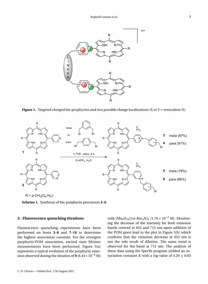
Supramolecular chemistry and its toolbox of non-covalent interactions combined with host–guestprinciples of molecular recognition provide toolsfor the design of hosts adapted for the binding ofdiversely shaped polyoxomolybdates. In an attemptto provide a rational approach to the formation ofwell-defined porphyrin/POM hybrids, charged bis-porphyrinic receptors have been synthesized andused in binding studies of Lindqvist-type POMs. Thedesign of pre-organized porphyrin dimers able to de-velop controlled interactions with polyoxometalatesis described hereafter. In order to introduce chargeson the porphyrin dimers, two options were possi-ble and are summarized in Figure 1. Introduction ofcharges at the position marked in green (X = N+) vianucleophilic addition of a pyridine group on a por-phyrin radical cation, obtained by chemical or elec-trochemical oxidation of a triarylporphyrin, failed formechanistic reasons [17], and thus the introductionof charges in the positions marked in red (Y = N+)has been developed (Figure 1).
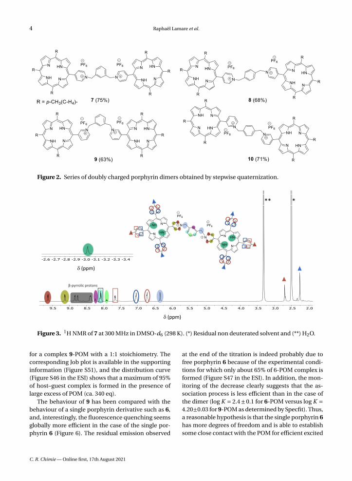
Two main synthetic methods have been devel-oped. The first dealt with the stepwise functionaliza-tion of the linker and the formation of the series ofcompounds represented in Scheme 1.
The yields of porphyrins 3, 4, 5 and 6 have beenoptimized through the testing of a variety of solvents
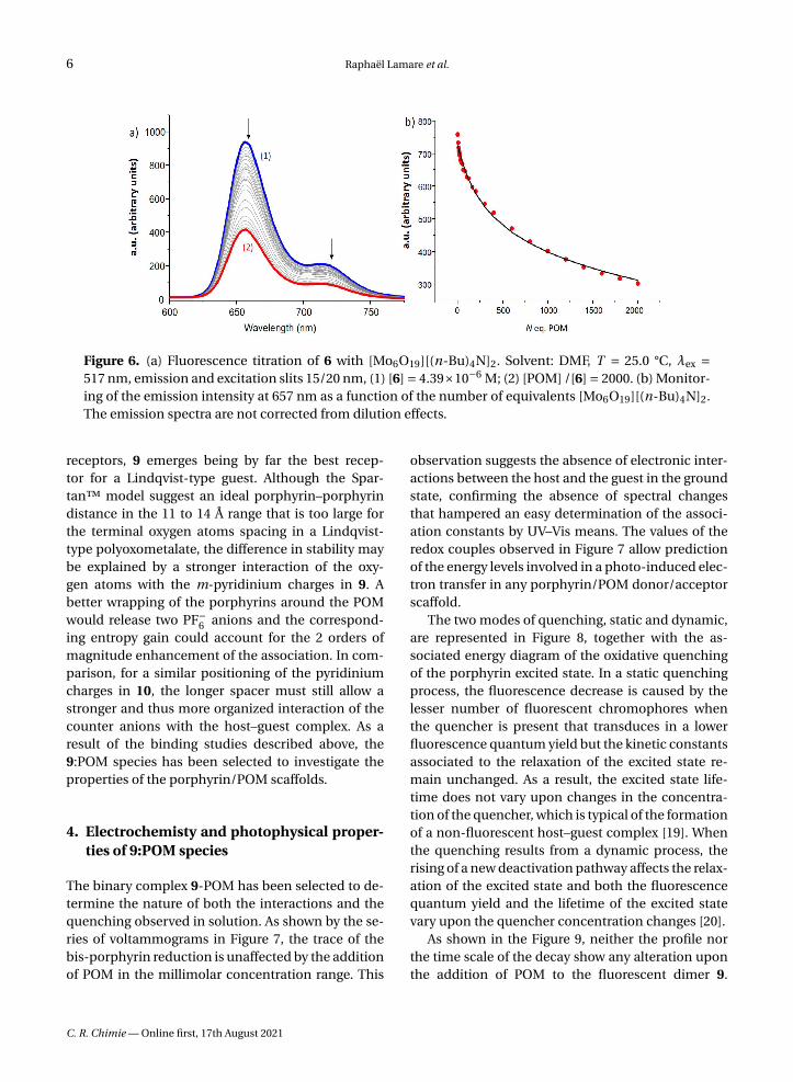
at room temperature and reflux. It must be noted thatsome loss of compound occurs during the anion ex-change that follows the quaternization of the pyri-dine group. The porphyrins 3, 4, 5 and 6 can be in-volved as alkylating agent for a second quaterniza-tion of either 1 or 2 in refluxing THF over 30 h tolead to the bis-porphyrins 7, 8, 9, 10 represented inFigure 2.
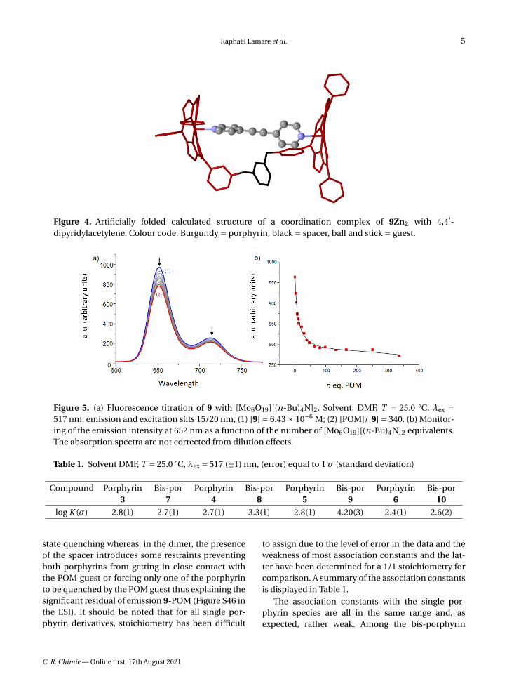
The same series of compounds can be obtainedby the second approach that consists in reacting anexcess of the porphyrins 1 or 2 with the appropri-ate α,α′-dibromoxylene in refluxing THF. This directmethod afforded the doubly charged bis-porphyrins7, 8, 9 and 10 in 46%, 38%, 46% and 32% yield, respec-tively. Although all the bis-porphyrins were intendedto form tweezers in which the two porphyrins arefacing each other, all characterization methods sug-gested that the compounds adopt an extended con-formation in solution, as shown, for example, by the1H NMR spectrum of 7 in DMSO (Figure 3). In thisspectrum, no anisotropic shielding of the porphyrinprotons due to stacking is observed and all sig-nals appear at chemical shifts typical of independentporphyrins.
Primary modelling has been performed usingSpartan (MM2) for 9Zn2 and three different tem-plate guests, namely 4,4′-bipyridine, 4,4′-dipyridy-lacetylene, and 4-(p-phenyl-4-pyridyl)-pyridine, toevaluate the most favourable size for a guest in thehypothetical zinc(II) complexes. The structure cal-culated for 4,4′-dipyridylacetylene as a guest is de-picted in Figure 4. The series of calculated struc-tures suggested that in a cofacial conformation ofthe two porphyrins, an energy minimum should bereached for a distance somewhere between 11 and14 Å for 9Zn2 although all structures have proven tobe quite flexible. For this reason, the choice of POMguest for this study rested on a Lindqvist-type en-tity in which the distance between terminal oxygenatoms is 8 Å (X-ray diffraction) [18]. The inclusion ofLindqvist POM ([Mo6O19]2−) in bis-porphyrins hasbeen investigated by various methods. UV–visibleabsorption titrations were judged not suitable due tothe absence of noticeable spectral variations uponaddition of guest. However, fluorescence quench-ing experiment and excited state lifetime measure-ments on the porphyrin dimers and electrochemistryprovided insightful information on the recognitionprocess.
C. R. Chimie — Online first, 17th August 2021
Raphaël Lamare et al. 3
Figure 1. Targeted charged bis-porphyrins and two possible charge localizations (X or Y = tetravalent N).
Scheme 1. Synthesis of bis-porphyrin precursors 3–6.
3. Fluorescence quenching titrations
Fluorescence quenching experiments have beenperformed on hosts 3–6 and 7–10 to determinethe highest association constant. For the strongestporphyrin/POM association, excited state lifetimemeasurements have been performed. Figure 5(a)represents a typical evolution of the porphyrin emis-sion observed during the titration of 9 (6.43×10⁻⁶ M)
with [Mo6O19][(n-Bu)4N]2 (1.76× 10⁻² M). Monitor-ing the decrease of the intensity for both emissionbands centred at 652 and 712 nm upon addition ofthe POM guest lead to the plot in Figure 5(b) whichconfirms that the emission decrease at 652 nm isnot the sole result of dilution. The same trend isobserved for the band at 712 nm. The analysis ofthese data using the Specfit program yielded an as-sociation constant K with a log value of 4.20 ± 0.03
C. R. Chimie — Online first, 17th August 2021
4 Raphaël Lamare et al.
Figure 2. Series of doubly charged porphyrin dimers obtained by stepwise quaternization.
Figure 3. 1H NMR of 7 at 300 MHz in DMSO-d6 (298 K). (*) Residual non deuterated solvent and (**) H2O.
for a complex 9-POM with a 1:1 stoichiometry. Thecorresponding Job plot is available in the supportinginformation (Figure S51), and the distribution curve(Figure S46 in the ESI) shows that a maximum of 95%of host–guest complex is formed in the presence oflarge excess of POM (ca. 340 eq).
The behaviour of 9 has been compared with thebehaviour of a single porphyrin derivative such as 6,and, interestingly, the fluorescence quenching seemsglobally more efficient in the case of the single por-phyrin 6 (Figure 6). The residual emission observed
at the end of the titration is indeed probably due tofree porphyrin 6 because of the experimental condi-tions for which only about 65% of 6-POM complex isformed (Figure S47 in the ESI). In addition, the mon-itoring of the decrease clearly suggests that the as-sociation process is less efficient than in the case ofthe dimer (log K = 2.4±0.1 for 6-POM versus log K =4.20±0.03 for 9-POM as determined by Specfit). Thus,a reasonable hypothesis is that the single porphyrin 6has more degrees of freedom and is able to establishsome close contact with the POM for efficient excited
C. R. Chimie — Online first, 17th August 2021
Raphaël Lamare et al. 5
Figure 4. Artificially folded calculated structure of a coordination complex of 9Zn2 with 4,4′-dipyridylacetylene. Colour code: Burgundy = porphyrin, black = spacer, ball and stick = guest.
Figure 5. (a) Fluorescence titration of 9 with [Mo6O19][(n-Bu)4N]2. Solvent: DMF, T = 25.0 °C, λex =517 nm, emission and excitation slits 15/20 nm, (1) [9] = 6.43 × 10⁻⁶ M; (2) [POM]/[9] = 340. (b) Monitor-ing of the emission intensity at 652 nm as a function of the number of [Mo6O19][(n-Bu)4N]2 equivalents.The absorption spectra are not corrected from dilution effects.
Table 1. Solvent DMF, T = 25.0 °C, λex = 517 (±1) nm, (error) equal to 1 σ (standard deviation)
Compound Porphyrin3
Bis-por7
Porphyrin4
Bis-por8
Porphyrin5
Bis-por9
Porphyrin6
Bis-por10
log K (σ) 2.8(1) 2.7(1) 2.7(1) 3.3(1) 2.8(1) 4.20(3) 2.4(1) 2.6(2)
state quenching whereas, in the dimer, the presenceof the spacer introduces some restraints preventingboth porphyrins from getting in close contact withthe POM guest or forcing only one of the porphyrinto be quenched by the POM guest thus explaining thesignificant residual of emission 9-POM (Figure S46 inthe ESI). It should be noted that for all single por-phyrin derivatives, stoichiometry has been difficult
to assign due to the level of error in the data and theweakness of most association constants and the lat-ter have been determined for a 1/1 stoichiometry forcomparison. A summary of the association constantsis displayed in Table 1.
The association constants with the single por-phyrin species are all in the same range and, asexpected, rather weak. Among the bis-porphyrin
C. R. Chimie — Online first, 17th August 2021
6 Raphaël Lamare et al.
Figure 6. (a) Fluorescence titration of 6 with [Mo6O19][(n-Bu)4N]2. Solvent: DMF, T = 25.0 °C, λex =517 nm, emission and excitation slits 15/20 nm, (1) [6] = 4.39×10⁻⁶ M; (2) [POM] /[6] = 2000. (b) Monitor-ing of the emission intensity at 657 nm as a function of the number of equivalents [Mo6O19][(n-Bu)4N]2.The emission spectra are not corrected from dilution effects.
receptors, 9 emerges being by far the best recep-tor for a Lindqvist-type guest. Although the Spar-tan™ model suggest an ideal porphyrin–porphyrindistance in the 11 to 14 Å range that is too large forthe terminal oxygen atoms spacing in a Lindqvist-type polyoxometalate, the difference in stability maybe explained by a stronger interaction of the oxy-gen atoms with the m-pyridinium charges in 9. Abetter wrapping of the porphyrins around the POMwould release two PF−
6 anions and the correspond-ing entropy gain could account for the 2 orders ofmagnitude enhancement of the association. In com-parison, for a similar positioning of the pyridiniumcharges in 10, the longer spacer must still allow astronger and thus more organized interaction of thecounter anions with the host–guest complex. As aresult of the binding studies described above, the9:POM species has been selected to investigate theproperties of the porphyrin/POM scaffolds.
4. Electrochemisty and photophysical proper-ties of 9:POM species
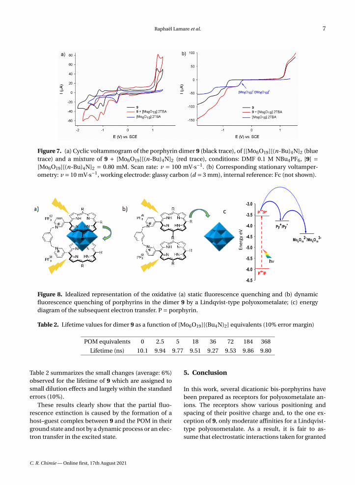
The binary complex 9-POM has been selected to de-termine the nature of both the interactions and thequenching observed in solution. As shown by the se-ries of voltammograms in Figure 7, the trace of thebis-porphyrin reduction is unaffected by the additionof POM in the millimolar concentration range. This
observation suggests the absence of electronic inter-actions between the host and the guest in the groundstate, confirming the absence of spectral changesthat hampered an easy determination of the associ-ation constants by UV–Vis means. The values of theredox couples observed in Figure 7 allow predictionof the energy levels involved in a photo-induced elec-tron transfer in any porphyrin/POM donor/acceptorscaffold.
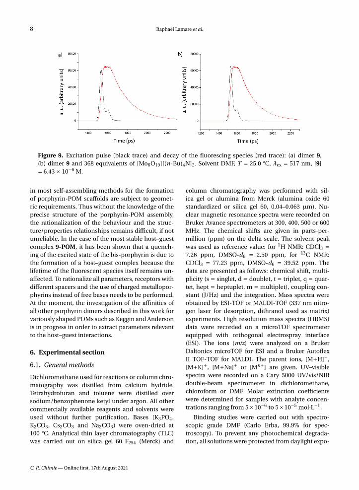
The two modes of quenching, static and dynamic,are represented in Figure 8, together with the as-sociated energy diagram of the oxidative quenchingof the porphyrin excited state. In a static quenchingprocess, the fluorescence decrease is caused by thelesser number of fluorescent chromophores whenthe quencher is present that transduces in a lowerfluorescence quantum yield but the kinetic constantsassociated to the relaxation of the excited state re-main unchanged. As a result, the excited state life-time does not vary upon changes in the concentra-tion of the quencher, which is typical of the formationof a non-fluorescent host–guest complex [19]. Whenthe quenching results from a dynamic process, therising of a new deactivation pathway affects the relax-ation of the excited state and both the fluorescencequantum yield and the lifetime of the excited statevary upon the quencher concentration changes [20].
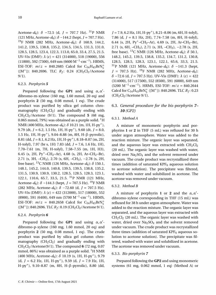
As shown in the Figure 9, neither the profile northe time scale of the decay show any alteration uponthe addition of POM to the fluorescent dimer 9.
C. R. Chimie — Online first, 17th August 2021
Raphaël Lamare et al. 7
Figure 7. (a) Cyclic voltammogram of the porphyrin dimer 9 (black trace), of [[Mo6O19][(n-Bu)4N]2 (bluetrace) and a mixture of 9 + [Mo6O19][(n-Bu)4N]2 (red trace), conditions: DMF 0.1 M NBu4PF6, [9] =[Mo6O19][(n-Bu)4N]2 = 0.80 mM. Scan rate: v = 100 mV·s−1. (b) Corresponding stationary voltamper-ometry: v = 10 mV·s−1, working electrode: glassy carbon (d = 3 mm), internal reference: Fc (not shown).
Figure 8. Idealized representation of the oxidative (a) static fluorescence quenching and (b) dynamicfluorescence quenching of porphyrins in the dimer 9 by a Lindqvist-type polyoxometalate; (c) energydiagram of the subsequent electron transfer. P = porphyrin.
Table 2. Lifetime values for dimer 9 as a function of [Mo6O19][(Bu4N)2] equivalents (10% error margin)
POM equivalents 0 2.5 5 18 36 72 184 368
Lifetime (ns) 10.1 9.94 9.77 9.51 9.27 9.53 9.86 9.80
Table 2 summarizes the small changes (average: 6%)observed for the lifetime of 9 which are assigned tosmall dilution effects and largely within the standarderrors (10%).
These results clearly show that the partial fluo-rescence extinction is caused by the formation of ahost–guest complex between 9 and the POM in theirground state and not by a dynamic process or an elec-tron transfer in the excited state.
5. Conclusion
In this work, several dicationic bis-porphyrins havebeen prepared as receptors for polyoxometalate an-ions. The receptors show various positioning andspacing of their positive charge and, to the one ex-ception of 9, only moderate affinities for a Lindqvist-type polyoxometalate. As a result, it is fair to as-sume that electrostatic interactions taken for granted
C. R. Chimie — Online first, 17th August 2021
8 Raphaël Lamare et al.
Figure 9. Excitation pulse (black trace) and decay of the fluorescing species (red trace): (a) dimer 9,(b) dimer 9 and 368 equivalents of [Mo6O19][(n-Bu)4N]2. Solvent DMF, T = 25.0 °C, λex = 517 nm, [9]= 6.43 × 10⁻⁶ M.
in most self-assembling methods for the formationof porphyrin-POM scaffolds are subject to geomet-ric requirements. Thus without the knowledge of theprecise structure of the porphyrin-POM assembly,the rationalization of the behaviour and the struc-ture/properties relationships remains difficult, if notunreliable. In the case of the most stable host–guestcomplex 9-POM, it has been shown that a quench-ing of the excited state of the bis-porphyrin is due tothe formation of a host–guest complex because thelifetime of the fluorescent species itself remains un-affected. To rationalize all parameters, receptors withdifferent spacers and the use of charged metallopor-phyrins instead of free bases needs to be performed.At the moment, the investigation of the affinities ofall other porphyrin dimers described in this work forvariously shaped POMs such as Keggin and Andersonis in progress in order to extract parameters relevantto the host–guest interactions.
6. Experimental section
6.1. General methods
Dichloromethane used for reactions or column chro-matography was distilled from calcium hydride.Tetrahydrofuran and toluene were distilled oversodium/benzophenone ketyl under argon. All othercommercially available reagents and solvents wereused without further purification. Bases (K3PO4,K2CO3, Cs2CO3 and Na2CO3) were oven-dried at100 °C. Analytical thin layer chromatography (TLC)was carried out on silica gel 60 F254 (Merck) and
column chromatography was performed with sil-ica gel or alumina from Merck (alumina oxide 60standardized or silica gel 60, 0.04–0.063 µm). Nu-clear magnetic resonance spectra were recorded onBruker Avance spectrometers at 300, 400, 500 or 600MHz. The chemical shifts are given in parts-per-million (ppm) on the delta scale. The solvent peakwas used as reference value: for 1H NMR: CDCl3 =7.26 ppm, DMSO-d6 = 2.50 ppm, for 13C NMR:CDCl3 = 77.23 ppm, DMSO-d6 = 39.52 ppm. Thedata are presented as follows: chemical shift, multi-plicity (s = singlet, d = doublet, t = triplet, q = quar-tet, hept = heptuplet, m = multiplet), coupling con-stant (J/Hz) and the integration. Mass spectra wereobtained by ESI-TOF or MALDI-TOF (337 nm nitro-gen laser for desorption, dithranol used as matrix)experiments. High resolution mass spectra (HRMS)data were recorded on a microTOF spectrometerequipped with orthogonal electrospray interface(ESI). The ions (m/z) were analyzed on a BrukerDaltonics microTOF for ESI and a Bruker AutoflexII TOF-TOF for MALDI. The parent ions, [M+H]+,[M+K]+, [M+Na]+ or [Mn+] are given. UV–visiblespectra were recorded on a Cary 5000 UV/vis/NIRdouble-beam spectrometer in dichloromethane,chloroform or DMF. Molar extinction coefficientswere determined for samples with analyte concen-trations ranging from 5×10⁻⁶ to 5×10⁻⁵ mol·L−1.
Binding studies were carried out with spectro-scopic grade DMF (Carlo Erba, 99.9% for spec-troscopy). To prevent any photochemical degrada-tion, all solutions were protected from daylight expo-
C. R. Chimie — Online first, 17th August 2021
Raphaël Lamare et al. 9
sure. All stock solutions were prepared using a Met-tler Toledo XA105 Dual Range (0.01/0.1 mg–41/120 g)to weigh samples, and complete dissolution in DMFwas achieved using an ultrasonic bath. The con-centrations of stock solutions of the receptors andsubstrates were calculated by the quantitative disso-lution of solid samples in DMF.
Luminescence titrations were carried out on so-lutions of dimers and monomers with absorbanceslower than 0.1. The titrations of 2 mL of dimer ormonomer with [Mo6O19][(Bu4N)2] ([Dimers] = 6.43×10⁻⁶ M and [Monomers] = 4.39× 10⁻⁶ M) were car-ried out in a 1 cm Hellma quartz optical cell bythe addition of known microvolumes of solutions of[Mo6O19][(Bu4N)2] with an Eppendorf Research plus.The excitation wavelengths were set at 517 or 559 nm.The luminescence spectra were recorded from 550 to800 nm on a PerkinElmer LS-50B instrument main-tained at 25 °C. The slit widths were set between 15and 20 nm for the emission. Luminescence titrationswere conducted under precise and identical experi-mental conditions.
The spectrophotometric titration of 9 with[Mo6O19][(Bu4N)2] ([9] = 1.76 × 10⁻² M) was car-ried out in a 1 cm Hellma quartz optical cell bythe addition of known microvolumes of solutionsof [Mo6O19][(Bu4N)2] with an Eppendorf Researchplus. Special care was taken to ensure that completeequilibration was attained. The corresponding UV–Vis spectra were recorded from 300 to 800 nm on aCary 5000 (Agilent) spectrophotometer maintainedat 25 °C.
The spectrophotometric data were analyzed withSpecfit [21] program that adjusts the absorptivitiesand the stability constants of the species formed atequilibrium. Specfit uses factor analysis to reducethe absorbance matrix and to extract the eigenvaluesprior to the multi-wavelength fit of the reduced dataset according to the Marquardt algorithm [22,23].
6.2. General procedure for the single porphyrinderivatives 3–6 (GP1)
A mixture of porphyrin 1 or 2 and the α,α′-dibromo-xylene corresponding in THF (15 mL) was refluxedfor 6 h under argon atmosphere. Water was addedto the reaction mixture. The organic layer was sep-arated, and the aqueous layer was extracted withCH2Cl2 (20 mL). The organic layer was washed with
water, dried over Na2SO4 and the solvent removedunder vacuum. The crude product was recrystallizedthree times (addition of saturated KPF6 aqueous so-lution to acetone solution). The precipitate was fil-tered, washed with water and solubilized in acetone.The acetone was then removed under vacuum.
6.2.1. Porphyrin 3
Prepared following the GP1 and using α,α′-dibromo-m-xylene(160 mg, 1.60 mmol, 20 eq) andporphyrin 1 (50 mg, 0.08 mmol, 1 eq). The crudeproduct was purified by silica gel column chro-matography (CH2Cl2) and gradually ending withCH2Cl2/Acetone (9/1). The compound 3 (73 mg,0.071 mmol, 87%) was obtained as a purple solid. 1HNMR (500 MHz, Acetone-d6): δ 9.73 (d, J = 6.6 Hz,2H, H-ortho-py+), 9.16 (d, J = 6.6 Hz, 2H, H-meta-py+), 9.03–8.84 (m, 8H, H-β-pyrrolic), 8.23–8.06 (m,6H, H-tolyl), 7.93 (s, 1H), 7.81 (dt, J = 7.0, 2.0 Hz,1H), 7.68 (d, J = 7.7 Hz, 6H, H-tolyl), 7.65–7.59 (m,2H), 6.43 (s, 2H, Py+–CH2–Ar), 4.81 (d, J = 5.5 Hz,2H, Ar–CH2–Br), 2.72 (s, 9H, −CH3), −2.72 (s, 2H, freebase). 13C NMR (11 MHz, Acetone-d6) δ 161.1, 145.4,144.3, 139.2, 139.1, 139.0, 135.4, 134.8, 134.4, 130.6,129.2, 129.1, 128.7, 128.7, 128.7, 123.7, 123.7, 122.5,122.5, 113.1, 64.3, 27.6, 21.7. 31P NMR (203 MHz,Acetone-d6) δ −144.0 (hept, J = 711.7 Hz). 19F NMR(471 MHz, Acetone-d6) δ −72.6 (d, J = 711.7 Hz).UV–Vis (DMF): λ (ε) = 419 (223000), 516 (12700),552 (7700), 591 (5700), 647 nm (4100 M−1·cm−1).ESI-TOF: m/z = 840.27 Calcd for C54H43N5Br ([M+]):840.27. TLC (silica) R f : 0.25 (CH2Cl2/Acetone 9/1).
6.2.2. Porphyrin 4
Prepared following the GP1 and using α,α′-dibromo-m-xylene (160 mg, 1.60 mmol, 20 eq)and porphyrin 1 (50 mg, 0.08 mmol, 1 eq). Thecrude product was purified by silica gel columnchromatography (CH2Cl2 and gradually ending withCH2Cl2/Acetone 9/1). The compound 4 (76 mg, 0.073mmol, 91%) was obtained as a purple solid. 1H NMR(400 MHz, Acetone-d6): δ 9.72 (d, J = 6.8 Hz, 2H,H-ortho-py+), 9.16 (d, J = 6.8 Hz, 2H, H-meta-py+),9.03–8.95 (m, 4H, H-β-pyrrolic), 8.91 (q, J = 4.9 Hz,4H, H-β-pyrrolic), 8.17–8.06 (m, 6H, H-tolyl), 7.94(d, J = 8.0 Hz, 2H), 7.75 (d, J = 8.0 Hz, 2H), 7.69–7.59 (m, 6H, H-tolyl), 6.43 (s, 2H, Py+–CH2–Ar), 4.78(s, 2H, Ar–CH2–Br), 2.70 (s, 6H, –CH3), 2.69 (s, 3H,–CH3), −2.73 (s, 2H, free base). 13C NMR (126 MHz,
C. R. Chimie — Online first, 17th August 2021
10 Raphaël Lamare et al.
Acetone-d6): δ −72.5 (d, J = 707.7 Hz). 31P NMR(121 MHz, Acetone-d6): δ−144.2 (hept, J = 707.7 Hz).19F NMR (282 MHz, Acetone-d6): δ 160.9, 144.2,141.2, 139.5, 138.8, 135.2, 134.5, 134.5, 131.3, 131.0,128.5, 128.5, 123.4, 122.3, 113.0, 65.0, 33.4, 27.5, 21.5.UV–Vis (DMF): λ (ε) = 421 (314000), 518 (19000), 556(11800), 592 (7300), 649 nm (6600 M−1·cm−1). HRMS,ESI-TOF: m/z = 840.2685 Calcd for C54H43BrN+
5([M+]): 840.2696. TLC R f : 0.24 (CH2Cl2/Acetone9/1).
6.2.3. Porphyrin 5
Prepared following the GP1 and using α,α′-dibromo-m-xylene (160 mg, 1.60 mmol, 20 eq) andporphyrin 2 (50 mg, 0.08 mmol, 1 eq). The crudeproduct was purified by silica gel column chro-matography (CH2Cl2) and gradually ending withCH2Cl2/Acetone (9/1). The compound 5 (68 mg,0.065 mmol, 79%) was obtained as a purple solid. 1HNMR (400 MHz, Acetone-d6): δ 10.21 (m, 1H, H-py+),9.79 (dt, J = 6.2, 1.5 Hz, 1H, H-py+), 9.60 (dt, J = 8.0,1.5 Hz, 1H, H-py+), 9.04–8.88 (m, 8H, H-β-pyrrolic),8.81 (dd, J = 8.1, 6.2 Hz, 1H, H-py+), 8.18–8.07 (m, 2H,H-tolyl), 7.97 (br s, 1H) 7.83 (dd, J = 7.6, 1.6 Hz, 1H),7.70–7.61 (m, 7H, H-tolyl), 7.58–7.55 (m, 1H, H3),6.45 (s, 2H, Py+–CH2–Ar), 4.68 (s, 2H, Ar–CH2–Br),2.71 (s, 3H, –CH3), 2.70 (s, 6H, –CH3), −2.78 (s, 2H,free base). 13C NMR (126 MHz, Acetone-d6): δ 150.1,148.1, 145.2, 144.0, 140.8, 139.5, 139.5, 138.8, 135.2,131.5, 130.9, 130.9, 130.2, 128.5, 128.5, 128.3, 123.1,122.1, 110.6, 65.7, 33.5, 21.5. 31P NMR (121 MHz,Acetone-d6): δ −144.3 (hept, J = 707.5 Hz). 19F NMR(282 MHz, Acetone-d6): δ −72.60 (d, J = 707.5 Hz).UV–Vis (DMF): λ (ε) = 422 (312000), 517 (18000), 552(8900), 591 (6400), 649 nm (5700 M−1·cm−1). HRMS,ESI-TOF: m/z = 840.2658 Calcd for C54H43BrN+
5([M+]): 840.2696. TLC R f : 0.19 (CH2Cl2/Acetone 9/1).
6.2.4. Porphyrin 6
Prepared following the GP1 and using α,α′-dibromo-p-xylene (160 mg, 1.60 mmol, 20 eq) andporphyrin 2 (50 mg, 0.08 mmol, 1 eq). The crudeproduct was purified by silica gel column chro-matography (CH2Cl2) and gradually ending withCH2Cl2/Acetone(9/1). The compound 6 (72 mg, 0.07mmol, 86%) was obtained as a purple solid. 1H NMR(400 MHz, Acetone-d6): δ 10.19 (s, 1H, H-py+), 9.79(d, J = 6.2 Hz, 1H, H-py+), 9.59 (d, J = 7.9 Hz, 1H,H-py+), 9.10–8.87 (m, 8H, H-β-pyrrolic), 8.80 (dd,
J = 7.9, 6.2 Hz, 1H, H-py+), 8.21–8.06 (m, 6H, H-tolyl),7.86 (d, J = 8.1 Hz, 2H), 7.74–7.58 (m, 8H, H-tolyl),6.44 (s, 2H, Py+–CH2–Ar), 4.69 (s, 2H, Ar–CH2–Br),2.71 (s, 6H, –CH3), 2.71 (s, 3H, –CH3), −2.78 (s, 2H,free base). 13C NMR (126 MHz, Acetone-d6): δ 50.1,148.2, 145.2, 139.5, 138.8, 135.2, 134.7, 131.2, 130.8,128.5, 128.5, 128.3, 123.1, 122.1, 65.6, 33.3, 21.5.31P NMR (121 MHz, Acetone-d6): δ −141.3 (hept,J = 707.5 Hz). 19F NMR (282 MHz, Acetone-d6):δ −72.6 (d, J = 707.5 Hz). UV–Vis (DMF): λ (ε) = 422(314000), 517 (17500), 552 (8500), 591 (6000), 649 nm(5200 M−1·cm−1). HRMS, ESI-TOF: m/z = 840.2644Calcd for C54H43BrN+
5 ([M+]): 840.2696. TLC R f : 0.23(CH2Cl2/Acetone 9/1).
6.3. General procedure for the bis-porphyrin 7–10 (GP2)
6.3.1. Method A
A mixture of monomeric porphyrin and por-phyrins 1 or 2 in THF (5 mL) was refluxed for 30 hunder argon atmosphere. Water was added to thereaction mixture. The organic layer was separated,and the aqueous layer was extracted with CH2Cl2
(20 mL). The organic layer was washed with water,dried over Na2SO4 and the solvent removed undervacuum. The crude product was recrystallized threetimes (addition of saturated KPF6 aqueous solutionto acetone solution). The precipitate was filtered,washed with water and solubilized in acetone. Theacetone was removed under vacuum.
6.3.2. Method B
A mixture of porphyrin 1 or 2 and the α,α′-dibromo-xylene corresponding in THF (15 mL) wasrefluxed for 38 h under argon atmosphere. Water wasadded to the reaction mixture. The organic layer wasseparated, and the aqueous layer was extracted withCH2Cl2 (20 mL). The organic layer was washed withwater, dried over Na2SO4 and the solvent removedunder vacuum. The crude product was recrystallizedthree times (addition of saturated KPF6 aqueous so-lution to acetone solution). The precipitate was fil-tered, washed with water and solubilized in acetone.The acetone was removed under vacuum.
6.3.3. Bis-porphyrin 7
Prepared following the GP2 and using monomericsystems (61 mg, 0.062 mmol, 1 eq) (Method A) or
C. R. Chimie — Online first, 17th August 2021
Raphaël Lamare et al. 11
α,α′-Dibromo-m-xylene (18 mg, 0.062 mmol, 1 eq)(Method B) and porphyrin 1 (204 mg, 0.31 mmol,5 eq). The crude product was purified by silica gel col-umn chromatography (CH2Cl2) and gradually endingwith a solution of KPF6 (27 mM) in acetone. The com-pound 7 (80 mg, 0.047 mmol, 75%, Method A) or (49mg, 0.029 mmol, 46%, Method B) was obtained as apurple solid. 1H NMR (500 MHz, DMSO-d6,): δ 9.62(d, J = 6.0 Hz, 4H, H-ortho-py+), 9.12 (d, J = 6.0 Hz,4H, H-meta-py+), 8.83 (d, J = 4.5 Hz, 4H, H-β-pyrrolic), 8.74 (d, J = 4.5 Hz, 4H, H-β-pyrrolic), 8.38(d, J = 4.7 Hz, 4H, H-β-pyrrolic), 8.35 (d, J = 4.7 Hz,4H, H-β-pyrrolic), 8.24 (s, 1H), 8.12–8.02 (m, 6H, H-ortho-tolyl), 7.89 (d, J = 7.7 Hz, 1H), 7.65 (d, J = 7.4Hz, 4H, H-meta-tolyl), 7.01 (d, J = 7.3 Hz, 8H, H-ortho-tolyl), 6.69 (d, J = 7.3 Hz, 8H, H-meta-tolyl),6.33 (s, 4H, –CH2–), 2.69 (s, 6H, –CH3), 2.24 (s, 12H,–CH3), −3.06 (s, 4H, free base). 13C NMR (126 MHz,DMSO-d6): δ 158.5, 143.4, 138.1, 137.6, 137.3, 136.7,135.5, 134.2, 133.4, 132.8, 131.0, 130.4, 129.7, 127.7,126.9, 121.7, 120.3, 112.4, 63.1, 21.1, 20.6. 31P NMR(121 MHz, DMSO-d6): δ −144.2 (hept, J = 711.3 Hz).19F NMR (282 MHz, DMSO-d6): δ −70.1 (d, J = 711.3Hz). UV–Vis (DMF): λ (ε) = 419 (198000), 516 (11300),552 (6300), 591 (4500), 648 nm (3700 M−1·cm−1). ESI-TOF: m/z = 709.83 Calcd for C100H78N2+
10 ([M2+]):709.32. TLC R f : 0.26 (solution of KPF6 (27 mM) inAcetone).
6.3.4. Bis-porphyrin 8
Prepared following the GP2 and using monomericsystems (61 mg, 0.062 mmol, 1 eq) (Method A) orα,α′-Dibromo-p-xylene (18 mg, 0.062 mmol, 1 eq)(Method B) and porphyrin 1 (204 mg, 0.31 mmol,5 eq). The crude product was purified by silica gel col-umn chromatography (CH2Cl2) and gradually endingwith a solution of KPF6 (27 mM in acetone). The com-pound 8 (72 mg, 0.042 mmol, 68%, Method A) or (40mg, 0.024 mmol, 38%, Method B) was obtained as apurple solid. 1H NMR (400 MHz, DMSO-d6): δ 9.66(d, J = 6.2 Hz, 4H, H-ortho-py+), 9.12 (d, J = 6.2 Hz,4H, H-meta-py+), 9.06–8.99 (m, 4H, H-β-pyrrolic),8.97–8.92 (m, 4H, H-β-pyrrolic), 8.90–8.81 (m, 8H, H-β-pyrrolic), 8.11 (s, 4H, H-aryl), 8.10–8.04 (m, 12H,H-ortho-tolyl), 7.65 (d, J = 7.8 Hz, 8H, H-meta-tolyl),7.61 (d, J = 7.8 Hz, 4H, H-meta-tolyl), 6.28 (s, 4H, –CH2−), 2.68 (s, 6H, –CH3), 2.64 (s, 12H, –CH3), −2.89(s, 4H, free base). 13C NMR (126 MHz, DMSO-d6): δ158.8, 143.8, 138.5, 138.2, 138.2, 138.1, 138.0, 136.5,
135.9, 134.6, 133.7, 130.8, 128.2, 122.5, 121.4, 113.1,111.1, 69.0, 30.1, 21.6, 21.5. 31P NMR (121 MHz,DMSO-d6): δ −141.27 (hept, J = 711.3 Hz). 19F NMR(282 MHz, DMSO-d6): δ −70.14 (d, J = 711.3 Hz).UV–Vis (DMF): λ (ε) = 422 (354000), 518 (37600),556 (29200), 592 (22400), 650 nm (19300 M−1·cm−1).HR-ESI-TOF: m/z = 709.8236 Calcd for C100H78N2+
10([M2+]): 709.8216. TLC R f : 0.31 (solution of KPF6
(5 mM) in Acetone).
6.3.5. Bis-porphyrin 9
Prepared following the GP2 and using monomericsystems (61 mg, 0.062 mmol, 1 eq) (Method A) orα,α′-Dibromo-m-xylene (18 mg, 0.062 mmol, 1 eq)(Method B) and porphyrin 2 (204 mg, 0.31 mmol, 5eq). The crude product was purified by silica gel col-umn chromatography (CH2Cl2) and gradually end-ing with a solution of KPF6 (27 mM in acetone). Thecompound 9 (67 mg, 0.039 mmol, 63%, Method A)or (51 mg, 0.030 mmol, 48%, Method B) was ob-tained as a purple solid. 1H NMR (400 MHz, DMSO-d6): δ 10.19 (s, 2H, H-py+), 9.62 (d, J = 6.5 Hz, 2H,H-py+), 9.38 (d, J = 7.9 Hz, 2H, 2H, H-py+), 8.89–8.79 (m, 8H, H-β-pyrrolic), 8.75–8.65 (m, 8H, H-β-pyrrolic), 8.54 (dd, J = 7.9, 6.5 Hz, 2H, H-py+), 8.13–8.05 (m, 4H, H-tolyl), 8.01 (s, 1H), 7.86 (d, J = 7.7Hz, 2H), 7.80 (d, J = 7.3 Hz, 4H, H-tolyl), 7.71 (t,J = 7.7 Hz, 1H), 7.69–7.59 (m, 8H, H-tolyl), 7.40 (d,J = 7.3 Hz, 4H, H-tolyl), 7.22 (d, J = 7.8 Hz, 4H, H-tolyl), 6.21 (s, 4H, –CH2−), 2.68 (s, 6H, –CH3), 2.49(s, 12H, –CH3), −3.01 (s, 4H, free base). 13C NMR(126 MHz, DMSO-d6): δ 1148.9, 147.0, 144.4, 141.6,138.1, 137.8, 137.7, 137.6, 137.4, 135.4, 134.2, 134.0,133.9, 133.8, 130.5, 130.2, 130.2, 130.2, 129.3, 127.8,127.7, 127.6, 127.6, 127.4, 127.0, 121.7, 120.6, 110.2,104.9, 55.9, 32.2, 29.6, 21.1, 20.9. 31P NMR (121 MHz,DMSO-d6): δ −144.2 (hept, J = 711.3 Hz). 19F NMR(282 MHz, DMSO-d6): δ −70.1 (d, J = 711.3 Hz).UV–Vis (DMF): λ (ε) = 422 (154000), 516 (9800), 552(4700), 590 (3600), 647 nm (2500 M−1·cm−1). HR-ESI-TOF: m/z = 709.8220 Calcd for C100H78N2+
10 ([M2+]):709.8216. TLC R f : 0.21 (solution of KPF6 (5 mM) inAcetone).
6.3.6. Bis-porphyrin 10
Prepared following the GP10 and usingmonomeric systems (61 mg, 0.062 mmol, 1 eq)(Method A) or α,α′-Dibromo-p-xylene (18 mg, 0.062mmol, 1 eq) (Method B) and porphyrin 2 (204 mg,
C. R. Chimie — Online first, 17th August 2021
12 Raphaël Lamare et al.
0.31 mmol, 5 eq). The crude product was purifiedby silica gel column chromatography (CH2Cl2) andgradually ending with a solution of KPF6 (27 mM inacetone). The compound 10 (76 mg, 0.044 mmol,71%, Method A) or (34 mg, 0.020 mmol, 32%, MethodB) was obtained as a purple solid. 1H NMR (500 MHz,DMSO-d6): δ 10.26 (s, 2H, H-py+), 9.71 (d, J = 6.3 Hz,2H, H-py+), 9.39 (d, J = 7.9 Hz, 2H, H-py+), 8.88–8.82 (m, 8H, H-β-pyrrolic), 8.77–8.69 (m, 8H, H-β-pyrrolic), 8.64 (dd, J = 7.9, 6.3 Hz, 2H, H-py+), 8.15–8.04 (m, 4H, H-tolyl), 7.91 (s, 4H, H-aryl), 7.83 (d,J = 7.7 Hz, 4H, H-tolyl), 7.70–7.58 (m, 8H, H-tolyl),7.34 (d, J = 7.7 Hz, 4H, H-tolyl), 7.15 (d, J = 7.7 Hz,4H, H-tolyl), 6.17 (s, 4H, –CH2−), 2.68 (s, 6H, –CH3),2.41 (s, 12H, –CH3), −2.97 (s, 4H, free base). 13C NMR(126 MHz, DMSO-d6): δ 162.3, 148.7, 147.0, 144.4,141.6, 138.1, 137.8, 137.6, 137.3, 135.7, 134.2, 134.0,133.8, 133.8, 129.8, 127.7, 127.5, 127.4, 127.2, 121.7,120.7, 110.3, 63.3, 55.8, 35.8, 30.8, 21.1, 20.8. 31P NMR(121 MHz, DMSO-d6): δ −144.2 (hept, J = 711.3 Hz).19F NMR (282 MHz, DMSO-d6): δ −70.1 (d, J = 711.3Hz). UV–Vis (DMF): λ (ε) = 420 (101000), 516 (8500),552 (5900), 590 (4800), 649 nm (4200 M−1·cm−1).HR-ESI-TOF: m/z = 709.3229 Calcd for C100H78N2+
10([M2+]): 709.3200. TLC R f : 0.28 (solution of KPF6
(5 mM) in acetone).
Acknowledgments
The authors are grateful to the University of Stras-bourg and the CNRS for constant financial sup-port. RL gratefully acknowledges the Région Grand-Est and the Fondation Recherche en Chimie for aPhD fellowship. The authors declare no conflicts ofinterest.
Supplementary data
Supporting information for this article is available onthe journal’s website under https://doi.org/10.5802/crchim.105 or from the author.
References
[1] I. Azcarate, Z. Huo, R. Farha, M. Goldmann, H. Xu, B. Hasen-knopf, E. Lacôte, L. Ruhlmann, Chem. Eur. J., 2015, 21, 8271-8280.
[2] J. Hao, A. Giraudeau, Z. Ping, L. Ruhlmann, Langmuir, 2008,24, 1600-1603.
[3] Q. Chen, D. P. Goshorn, C. P. Scholes, X. L. Tan, J. Zubieta,J. Am. Chem. Soc., 1992, 114, 4667-4681.
[4] W. H. Knoth, R. L. Harlow, J. Am. Chem. Soc., 1981, 103, 4265-4266.
[5] K. J. Elliott, A. Harriman, L. Le Pleux, Y. Pellegrin, E. Blart, C. R.Mayer, F. Odobel, Phys. Chem. Chem. Phys., 2009, 11, 8767-8773.
[6] F. Odobel, M. Séverac, Y. Pellegrin, E. Blart, C. Fosse, C. Can-nizzo, C. R. Mayer, K. J. Elliott, A. Harriman, Chem.–Eur. J.,2009, 15, 3130-3138.
[7] Y. Zhu, Y. Huang, Q. Li, D. Zang, J. Gu, Y. Tang, Y. Wei, Inorg.Chem., 2020, 59, 2575-2583.
[8] D. Schaming, C. Costa-Coquelard, I. Lampre, S. Sorgues,M. Erard, X. Liu, J. Liu, L. Sun, J. Canny, R. Thouvenot,L. Ruhlmann, Inorg. Chim. Acta, 2010, 363, 2185-2192.
[9] A. Yokoyama, K. Ohkubo, T. Ishizuka, T. Kojima, S. Fukuzumi,Dalton Trans., 2012, 41, 10006-10013.
[10] C. Li, N. Mizuno, K. Yamaguchi, K. Suzuki, J. Am. Chem. Soc.,2019, 141, 7687-7692.
[11] C. Costa-Coquelard, S. Sorgues, L. Ruhlmann, J. Phys. Chem.A, 2010, 114, 6394-6400.
[12] S.-Q. Liu, J.-Q. Xu, H.-R. Sun, D.-M. Li, Inorg. Chim. Acta, 2000,306, 87-93.
[13] I. Ahmed, R. Farha, M. Goldmann, L. Ruhlmann, Chem. Com-mun., 2013, 49, 496-498.
[14] C. Li, K.-M. Park, H.-J. Kim, Inorg. Chem. Commun., 2015, 60,8-11.
[15] Z. Shi, Y. Zhou, L. Zhang, C. Mu, H. Ren, D. ul Hassan, D. Yang,H. M. Asif, RSC Adv., 2014, 4, 50277-50284.
[16] G. Bazzan, W. Smith, L. C. Francesconi, C. M. Drain, Lang-muir, 2008, 24, 3244-3249.
[17] R. Lamare, L. Ruhlmann, R. Ruppert, J. Weiss, J. Porphyr. Ph-thalocyanines, 2019, 23, 860-868.
[18] A. L. Rheingold, C. B. White, B. S. Haggerty, E. A. Maatta, ActaCrystallogr. C, 1993, 49, 756-758.
[19] A. C. Vaiana, H. Neuweiler, A. Schulz, J. Wolfrum, M. Sauer,J. C. Smith, J. Am. Chem. Soc., 2003, 125, 14564-14572.
[20] L. K. Fraiji, D. M. Hayes, T. C. Werner, J. Chem. Educ., 1992, 69,424-428.
[21] H. Gampp, M. Maeder, C. J. Meyer, A. D. Zuberbühler, Talanta,1985, 32, 95-101.
[22] D. W. Marquardt, J. Soc. Ind. Appl. Math., 1963, 11, 431-441.[23] M. Maeder, A. D. Zuberbuehler, Anal. Chem., 1990, 62, 2220-
2224.
C. R. Chimie — Online first, 17th August 2021