Embed Size (px)
Citation preview
Design of a small molecule against an oncogenicnoncoding RNASai Pradeep Velagapudia, Michael D. Cameronb, Christopher L. Hagab, Laura H. Rosenbergb, Marie Lafitteb,Derek R. Duckettb, Donald G. Phinneyb, and Matthew D. Disneya,c,1
aDepartment of Chemistry, The Scripps Research Institute, Jupiter, FL 33458; bDepartment of Molecular Therapeutics, The Scripps Research Institute, Jupiter,FL 33458; and cDepartment of Neuroscience, The Scripps Research Institute, Jupiter, FL 33458
Edited by Larry Gold, SomaLogic, Inc., Boulder, CO, and approved April 5, 2016 (received for review December 11, 2015)
The design of precision, preclinical therapeutics from sequence isdifficult, but advances in this area, particularly those focused onrational design, could quickly transform the sequence of disease-causing gene products into lead modalities. Herein, we describethe use of Inforna, a computational approach that enables therational design of small molecules targeting RNA to quickly providea potent modulator of oncogenic microRNA-96 (miR-96). We minedthe secondary structure of primary microRNA-96 (pri-miR-96) hairpinprecursor against a database of RNA motif–small molecule interac-tions, which identified modules that bound RNAmotifs nearby and inthe Drosha processing site. Precise linking of these modules togetherprovided Targaprimir-96 (3), which selectively modulates miR-96 pro-duction in cancer cells and triggers apoptosis. Importantly, the com-pound is ineffective on healthy breast cells, and exogenousoverexpression of pri-miR-96 reduced compound potency in breastcancer cells. Chemical Cross-Linking and Isolation by Pull-Down(Chem-CLIP), a small-molecule RNA target validation approach,shows that 3 directly engages pri-miR-96 in breast cancer cells. Invivo, 3 has a favorable pharmacokinetic profile and decreases tu-mor burden in a mouse model of triple-negative breast cancer.Thus, rational design can quickly produce precision, in vivo bio-active lead small molecules against hard-to-treat cancers by target-ing oncogenic noncoding RNAs, advancing a disease-to-gene-to-drug paradigm.
RNA | noncoding RNA | drug design | chemistry | nucleic acids
Many disease association studies have identified biomoleculesthat, if targeted selectively, could be exploited to provide
precision lead therapeutics. Generally, drug-like compounds arescreened to discover modulators of the protein products of genes,particularly enzymes (1). However, evidence is emerging thatRNA is an attractive and unexploited target for precise medicinestrategies. One major implication of the Encyclopedia Of DNAElements (ENCODE) project was that noncoding RNAs playmajor roles in human biology, including disease biology (2). Fur-thermore, sequencing techniques that directly report the identityand abundance of RNAs (3, 4) have revealed many potentiallydruggable RNAs that are differentially expressed in healthy anddisease-affected tissue, for which a precision medicine approachcould be ideal.Outside of the bacterial ribosome, however, RNA is considered
undruggable with small molecules because of the difficulty inidentifying high-affinity, selective compounds. Despite this chal-lenge, there are many advantages of targeting RNA with smallmolecules. For example, RNA secondary structure can be rapidlyannotated from sequence (5–9), thereby identifying functional re-gions that could be inactivated by small molecule binding. Wereasoned that gene expression data could be quickly converted intoprecise, lead therapeutic modalities by combining these methodswith rational design strategies.As part of a program to rationally drug RNAs from sequence,
we established a database of RNA motif–small molecule bindingpartners (10). The database is comprised of privileged interactionsidentified by a library vs. library screen named 2D combinatorial
screening (2DCS) (11, 12). In 2DCS, a library of array-immobilizedsmall molecules is probed for binding to a library of small RNAmotifs that are likely to be present in biological RNAs (11, 12).These interactions are then mined against genomic RNAs toprovide lead therapeutics in a target agnostic manner by Inforna(10), a computational approach developed in our laboratory. Thatis, Inforna parses RNA secondary structures, regardless of whetherthey are determined by phylogeny, structural methods, or compu-tation, and compares them with the RNA motif–small moleculedatabase. Overlap provides lead compounds. Previously, Infornaidentified the lead benzimidazole 1, which avidly bound to theRNA motif present in a Drosha nuclease-processing site in themicroRNA-96 (miR-96) hairpin precursor (10), an oncogenicmicroRNA (miRNA) implicated in breast cancer (13). ThismiRNA suppresses the production of proapoptotic forkhead boxO1 (FOXO1) protein in cancer cells. Targeting the miRNA withan oligonucleotide (an antagomir) or compound 1 selectively in-hibits production of mature miR-96 and triggers apoptosis in breastcancer cells at micromolar concentrations (10, 13).Herein, we used Inforna for the facile lead optimization of 1,
providing highly potent compounds that impede tumorigenesisin vivo. Inforna identified that a bis-benzimidazole (2) bound to a1 nt × 1 nt internal loop adjacent to the Drosha site bound by1 (Fig. 1). The optimal dimeric compound that targets both sites,Targaprimir-96 (3), binds primary microRNA-96 (pri-miR-96)with low nanomolar affinity (>40-fold more avidly than 1) and is>400-fold more potent than 1 for inhibiting miR-96 biogenesis inbreast cancer cells. The effect of 3 is specific to breast cancer cells,because nontumorigenic mammary epithelial cells are unaffected.Furthermore, target validation studies using Chemical Cross-Linking and Isolation by Pull-Down (Chem-CLIP) (14, 15)
Significance
The goal of precision medicine is to identify selective drugs thatmodulate disease-causing biomolecules. This slow process ofteninvolves developing a high-throughput screen to test millions ofpotential drugs to find a few that affect the biomolecule. Here,we describe a facile approach using a disease-causing biomole-cule’s sequence to enable design of specific drugs, eliminatingarduous and time-consuming screens. By using the sequence of anon–protein-coding, oncogenic RNA, we designed a drug spe-cifically targeting the RNA’s folded structure. In cells and ani-mals, the drug inhibits its target, killing cancer cells while leavinghealthy cells unaffected. Thus, a preclinical anticancer drugcandidate can be quickly designed from sequence.
Author contributions: S.P.V. and M.D.D. designed research; S.P.V., C.L.H., and M.D.D. per-formed research; M.D.C., C.L.H., L.H.R., M.L., D.R.D., and D.G.P. contributed new reagents/analytic tools; S.P.V. and M.D.D. analyzed data; and S.P.V. and M.D.D. wrote the paper.
The authors declare no conflict of interest.
This article is a PNAS Direct Submission.1To whom correspondence should be addressed. Email: [email protected].
This article contains supporting information online at www.pnas.org/lookup/suppl/doi:10.1073/pnas.1523975113/-/DCSupplemental.
5898–5903 | PNAS | May 24, 2016 | vol. 113 | no. 21 www.pnas.org/cgi/doi/10.1073/pnas.1523975113
demonstrate that 3 engages pri-miR-96 in cells, and rescue ex-periments are consistent with the proposed mechanism of actionof 3. Compound 3 has a favorable pharmacokinetic profile inmice, and thus we studied the effect of the compound on tumorgrowth in an in vivo preclinical model of triple-negative breastcancer (TNBC). Indeed, 3 inhibited tumor growth, showing that itis possible to target oncogenic noncoding RNAs through smallmolecules as a potential treatment for breast cancer.
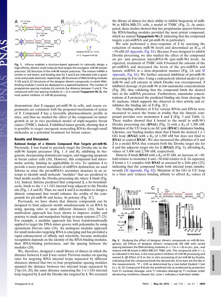
Results and DiscussionRational Design of a Dimeric Compound That Targets pri-miR-96.Previously, 1 was found to precisely target the Drosha site in themiR-96 hairpin precursor (Fig. 1 A and B), inhibiting its bio-genesis, derepressing downstream targets, and triggering apoptosisin breast cancer cells (10). However, this compound had micro-molar activity, limiting its applicability in vivo. To optimize 1 toprovide a more potent modulator of miRNA dysfunction, we usedInforna to mine the pri-miRNA’s secondary structure in an at-tempt to identify small molecule “modules” that are predicted tobind motifs nearby the Drosha processing site where 1 binds (Fig.1A). Indeed, Inforna predicted that compound 2, a bis-benzimid-azole, binds to the 1 × 1 GG internal loop adjacent to the Droshasite (Fig. 1 A and B). Thus, we used 1 and 2 as modules to design adimeric compound that would enhance the avidity of the com-pounds for pri-miR-96 and hence, its potency (Fig. 1C).Previously, we have shown that dimeric compounds can be
designed to bind adjacent motifs simultaneously in an RNA byusing spacing rules to span different distances (16). Such amultivalent approach has been shown to improve avidity andpotency to study and manipulate biology in many systems (17–23).For example, a modular approach has been transformationallyapplied to target the DNA minor groove with polyamides by usingeponymous Dervan rules (24). An analogous modular approachfor small molecules targeting RNA is emerging and has provided arapid enhancement of affinity and selectivity, because molecularrecognition depends on the identity of the RNA-binding modules,their RNA-binding preferences, and the spacing between themodules (20).We, therefore, designed a small library of dimers in which the
distance between 1 and 2 was varied. Previous studies on spacingrules for targeting RNA internal loops separated by differentdistances showed that two to four propylamine spacing modulesspanned the distance between two internal loops separated by2 bp (16, 20), the same distance separating the 1 × 1 GG internalloop targeted by 2 and the Drosha site targeted by 1. We screened
the library of dimers for their ability to inhibit biogenesis of miR-96 in MDA-MB-231 cells, a model of TNBC (Fig. 2). As antici-pated, these studies showed that two propylamine spacers betweenthe RNA-binding modules provided the most potent compound,which we named Targaprimir-96 (3; indicating that the compoundtargets a pri-miRNA and pri-miR-96 in particular).We next performed a dose–response of 3 by assessing the
reduction of mature miR-96 levels and determined an IC50 of∼50 nM (SI Appendix, Fig. S1). Because 3 was designed to inhibitDrosha processing, we also studied the effect of the compoundon pri- and precursor microRNA-96 (pre-miR-96) levels. Asexpected, treatment of TNBC with 3 boosted the amount of thepri-miRNA and decreased the levels of the pre-miRNA andmature miRNA in a dose-dependent manner (Fig. 2A and SIAppendix, Fig. S1). We further assessed inhibition of pri-miR-96processing by 3 in vitro. Using a radioactively labeled model of pri-miR-96 and cell extracts in which Drosha was overexpressed, 3inhibited cleavage of pri-miR-96 at low-nanomolar concentrations(Fig. 2B), thus validating that the compound binds the desiredsites in the miRNA precursor. Furthermore, nanomolar concen-trations of 3 protected the predicted binding site from cleavage byS1 nuclease, which supports the observed in vitro activity and es-tablishes the binding site of 3 (Fig. 2C).The binding affinities of 3 for various RNAs and DNAs were
measured to assess the bonus in avidity that the dimeric com-pound provides over monomers 1 and 2 (Fig. 3 and Table 1).These studies showed that 1 bound to the motif in miR-96’sDrosha processing site (RNA1) (Fig. 3) with a Kd of 1,300 nM.Mutation of the UU loop to an AU pair (RNAC) eliminates binding.Likewise, our binding studies show that 2 binds the desired 1 × 1GG loop (RNA2) with a Kd of 1,500 nM but does not bind toRNA1 or control RNAC. We also measured the affinities of 1 and2 to a model RNA that contains both the Drosha target site for1 and the adjacent target site for 2 (RNA3) (Fig. 3), affording Kdvalues of 3,400 and 2,700 nM, respectively.Dimeric 3 binds RNA3 with a Kd of 85 nM, an increase of ∼40-
fold relative to monomer 1 and ∼30-fold relative to 2. As expected,3 forms a 1:1 complex with RNA3 as assessed by a Jobs plot (25),indicating that the compound binds both internal loops simulta-neously (SI Appendix, Fig. S2). Mutation of the GG or UU loopto a base pair reduces binding affinity to afford Kd values of
Fig. 1. Inforna enabled a structure-based approach to rationally design ahigh-affinity, dimeric small molecule that targets the oncogenic miR-96 hairpinprecursor. (A) Structure of the miR-96 hairpin precursor. The mature miRNA iswritten in red letters, and binding sites for 1 and 2 are indicated with a greencircle and purple diamond, respectively. (B) Structures of RNA-binding modules1 (10) and 2. (C) Structures of the designer dimeric compounds, in which RNA-binding modules 1 and 2 are displayed on a peptoid backbone. The number ofpropylamine spacing modules (n) controls the distance between 1 and 2. Thecompound with two spacing modules (n = 2) is named Targaprimir-96 (3), themost potent inhibitor of miR-96 processing.
Fig. 2. Studying the effect of designer dimeric compounds on miR-96 bio-genesis. (A) Effects of designer dimeric compounds (50 nM) with variedspacing between the RNA-binding modules (n = 1 to n = 4) on pri-, pre-, andmature miR-96 levels in MDA-MB-231 TNBC cells. The compound with n = 2,as indicated in the box, is the most potent inhibitor of miR-96 processing andnamed 3. (B) Effect of 3 on the in vitro processing of pri-miR-96 by Drosha,indicating that the compound binds the desired site. Error bars are the SDs inthe measurements. *P < 0.05 as determined by a two-tailed Student t test(n ≥ 3). (C) Compound 3 binds the predicted site as evidenced by protectionfrom S1 nuclease cleavage. Lane T1 indicates cleavage by T1 nuclease underdenaturing conditions (cleaves Gs). Lane L indicates a hydrolysis ladder.
Velagapudi et al. PNAS | May 24, 2016 | vol. 113 | no. 21 | 5899
BIOCH
EMISTR
YCH
EMISTR
Y
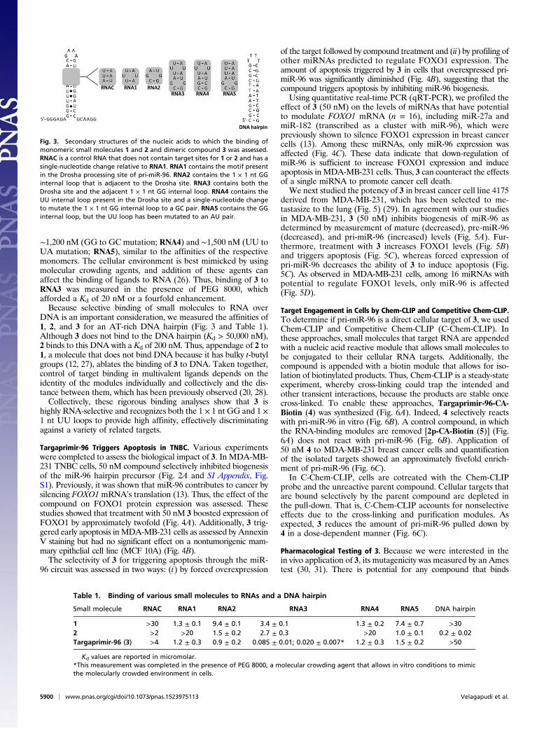
∼1,200 nM (GG to GC mutation; RNA4) and ∼1,500 nM (UU toUA mutation; RNA5), similar to the affinities of the respectivemonomers. The cellular environment is best mimicked by usingmolecular crowding agents, and addition of these agents canaffect the binding of ligands to RNA (26). Thus, binding of 3 toRNA3 was measured in the presence of PEG 8000, whichafforded a Kd of 20 nM or a fourfold enhancement.Because selective binding of small molecules to RNA over
DNA is an important consideration, we measured the affinities of1, 2, and 3 for an AT-rich DNA hairpin (Fig. 3 and Table 1).Although 3 does not bind to the DNA hairpin (Kd > 50,000 nM),2 binds to this DNA with a Kd of 200 nM. Thus, appendage of 2 to1, a molecule that does not bind DNA because it has bulky t-butylgroups (12, 27), ablates the binding of 3 to DNA. Taken together,control of target binding in multivalent ligands depends on theidentity of the modules individually and collectively and the dis-tance between them, which has been previously observed (20, 28).Collectively, these rigorous binding analyses show that 3 is
highly RNA-selective and recognizes both the 1 × 1 nt GG and 1 ×1 nt UU loops to provide high affinity, effectively discriminatingagainst a variety of related targets.
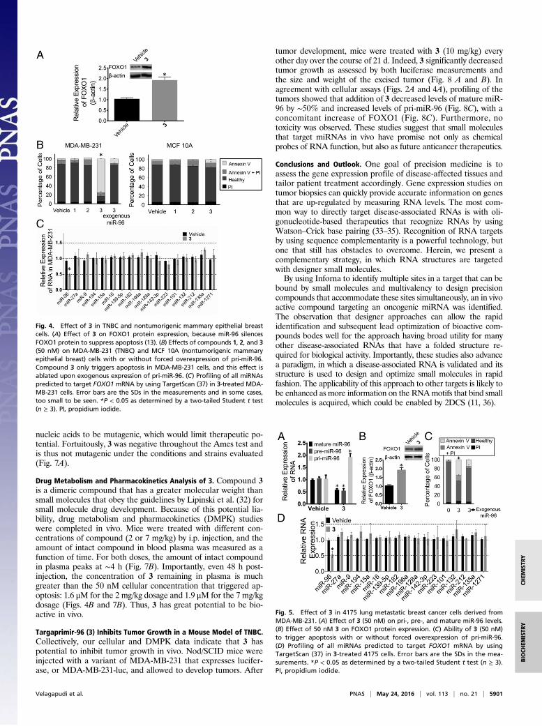
Targaprimir-96 Triggers Apoptosis in TNBC. Various experimentswere completed to assess the biological impact of 3. In MDA-MB-231 TNBC cells, 50 nM compound selectively inhibited biogenesisof the miR-96 hairpin precursor (Fig. 2A and SI Appendix, Fig.S1). Previously, it was shown that miR-96 contributes to cancer bysilencing FOXO1 mRNA’s translation (13). Thus, the effect of thecompound on FOXO1 protein expression was assessed. Thesestudies showed that treatment with 50 nM 3 boosted expression ofFOXO1 by approximately twofold (Fig. 4A). Additionally, 3 trig-gered early apoptosis in MDA-MB-231 cells as assessed by AnnexinV staining but had no significant effect on a nontumorigenic mam-mary epithelial cell line (MCF 10A) (Fig. 4B).The selectivity of 3 for triggering apoptosis through the miR-
96 circuit was assessed in two ways: (i) by forced overexpression
of the target followed by compound treatment and (ii) by profiling ofother miRNAs predicted to regulate FOXO1 expression. Theamount of apoptosis triggered by 3 in cells that overexpressed pri-miR-96 was significantly diminished (Fig. 4B), suggesting that thecompound triggers apoptosis by inhibiting miR-96 biogenesis.Using quantitative real-time PCR (qRT-PCR), we profiled the
effect of 3 (50 nM) on the levels of miRNAs that have potentialto modulate FOXO1 mRNA (n = 16), including miR-27a andmiR-182 (transcribed as a cluster with miR-96), which werepreviously shown to silence FOXO1 expression in breast cancercells (13). Among these miRNAs, only miR-96 expression wasaffected (Fig. 4C). These data indicate that down-regulation ofmiR-96 is sufficient to increase FOXO1 expression and induceapoptosis in MDA-MB-231 cells. Thus, 3 can counteract the effectsof a single miRNA to promote cancer cell death.We next studied the potency of 3 in breast cancer cell line 4175
derived from MDA-MB-231, which has been selected to me-tastasize to the lung (Fig. 5) (29). In agreement with our studiesin MDA-MB-231, 3 (50 nM) inhibits biogenesis of miR-96 asdetermined by measurement of mature (decreased), pre-miR-96(decreased), and pri-miR-96 (increased) levels (Fig. 5A). Fur-thermore, treatment with 3 increases FOXO1 levels (Fig. 5B)and triggers apoptosis (Fig. 5C), whereas forced expression ofpri-miR-96 decreases the ability of 3 to induce apoptosis (Fig.5C). As observed in MDA-MB-231 cells, among 16 miRNAs withpotential to regulate FOXO1 levels, only miR-96 is affected(Fig. 5D).
Target Engagement in Cells by Chem-CLIP and Competitive Chem-CLIP.To determine if pri-miR-96 is a direct cellular target of 3, we usedChem-CLIP and Competitive Chem-CLIP (C-Chem-CLIP). Inthese approaches, small molecules that target RNA are appendedwith a nucleic acid reactive module that allows small molecules tobe conjugated to their cellular RNA targets. Additionally, thecompound is appended with a biotin module that allows for iso-lation of biotinylated products. Thus, Chem-CLIP is a steady-stateexperiment, whereby cross-linking could trap the intended andother transient interactions, because the products are stable oncecross-linked. To enable these approaches, Targaprimir-96-CA-Biotin (4) was synthesized (Fig. 6A). Indeed, 4 selectively reactswith pri-miR-96 in vitro (Fig. 6B). A control compound, in whichthe RNA-binding modules are removed [2p-CA-Biotin (5)] (Fig.6A) does not react with pri-miR-96 (Fig. 6B). Application of50 nM 4 to MDA-MB-231 breast cancer cells and quantificationof the isolated targets showed an approximately fivefold enrich-ment of pri-miR-96 (Fig. 6C).In C-Chem-CLIP, cells are cotreated with the Chem-CLIP
probe and the unreactive parent compound. Cellular targets thatare bound selectively by the parent compound are depleted inthe pull-down. That is, C-Chem-CLIP accounts for nonselectiveeffects due to the cross-linking and purification modules. Asexpected, 3 reduces the amount of pri-miR-96 pulled down by4 in a dose-dependent manner (Fig. 6C).
Pharmacological Testing of 3. Because we were interested in thein vivo application of 3, its mutagenicity was measured by an Amestest (30, 31). There is potential for any compound that binds
Fig. 3. Secondary structures of the nucleic acids to which the binding ofmonomeric small molecules 1 and 2 and dimeric compound 3 was assessed.RNAC is a control RNA that does not contain target sites for 1 or 2 and has asingle-nucleotide change relative to RNA1. RNA1 contains the motif presentin the Drosha processing site of pri-miR-96. RNA2 contains the 1 × 1 nt GGinternal loop that is adjacent to the Drosha site. RNA3 contains both theDrosha site and the adjacent 1 × 1 nt GG internal loop. RNA4 contains theUU internal loop present in the Drosha site and a single-nucleotide changeto mutate the 1 × 1 nt GG internal loop to a GC pair. RNA5 contains the GGinternal loop, but the UU loop has been mutated to an AU pair.
Table 1. Binding of various small molecules to RNAs and a DNA hairpin
Small molecule RNAC RNA1 RNA2 RNA3 RNA4 RNA5 DNA hairpin
1 >30 1.3 ± 0.1 9.4 ± 0.1 3.4 ± 0.1 1.3 ± 0.2 7.4 ± 0.7 >302 >2 >20 1.5 ± 0.2 2.7 ± 0.3 >20 1.0 ± 0.1 0.2 ± 0.02Targaprimir-96 (3) >4 1.2 ± 0.3 0.9 ± 0.2 0.085 ± 0.01; 0.020 ± 0.007* 1.2 ± 0.3 1.5 ± 0.2 >50
Kd values are reported in micromolar.*This measurement was completed in the presence of PEG 8000, a molecular crowding agent that allows in vitro conditions to mimicthe molecularly crowded environment in cells.
5900 | www.pnas.org/cgi/doi/10.1073/pnas.1523975113 Velagapudi et al.
nucleic acids to be mutagenic, which would limit therapeutic po-tential. Fortuitously, 3 was negative throughout the Ames test andis thus not mutagenic under the conditions and strains evaluated(Fig. 7A).
Drug Metabolism and Pharmacokinetics Analysis of 3. Compound 3is a dimeric compound that has a greater molecular weight thansmall molecules that obey the guidelines by Lipinski et al. (32) forsmall molecule drug development. Because of this potential lia-bility, drug metabolism and pharmacokinetics (DMPK) studieswere completed in vivo. Mice were treated with different con-centrations of compound (2 or 7 mg/kg) by i.p. injection, and theamount of intact compound in blood plasma was measured as afunction of time. For both doses, the amount of intact compoundin plasma peaks at ∼4 h (Fig. 7B). Importantly, even 48 h post-injection, the concentration of 3 remaining in plasma is muchgreater than the 50 nM cellular concentration that triggered ap-optosis: 1.6 μM for the 2 mg/kg dosage and 1.9 μM for the 7 mg/kgdosage (Figs. 4B and 7B). Thus, 3 has great potential to be bio-active in vivo.
Targaprimir-96 (3) Inhibits Tumor Growth in a Mouse Model of TNBC.Collectively, our cellular and DMPK data indicate that 3 haspotential to inhibit tumor growth in vivo. Nod/SCID mice wereinjected with a variant of MDA-MB-231 that expresses lucifer-ase, or MDA-MB-231-luc, and allowed to develop tumors. After
tumor development, mice were treated with 3 (10 mg/kg) everyother day over the course of 21 d. Indeed, 3 significantly decreasedtumor growth as assessed by both luciferase measurements andthe size and weight of the excised tumor (Fig. 8 A and B). Inagreement with cellular assays (Figs. 2A and 4A), profiling of thetumors showed that addition of 3 decreased levels of mature miR-96 by ∼50% and increased levels of pri-miR-96 (Fig. 8C), with aconcomitant increase of FOXO1 (Fig. 8C). Furthermore, notoxicity was observed. These studies suggest that small moleculesthat target miRNAs in vivo have promise not only as chemicalprobes of RNA function, but also as future anticancer therapeutics.
Conclusions and Outlook. One goal of precision medicine is toassess the gene expression profile of disease-affected tissues andtailor patient treatment accordingly. Gene expression studies ontumor biopsies can quickly provide accurate information on genesthat are up-regulated by measuring RNA levels. The most com-mon way to directly target disease-associated RNAs is with oli-gonucleotide-based therapeutics that recognize RNAs by usingWatson–Crick base pairing (33–35). Recognition of RNA targetsby using sequence complementarity is a powerful technology, butone that still has obstacles to overcome. Herein, we present acomplementary strategy, in which RNA structures are targetedwith designer small molecules.By using Inforna to identify multiple sites in a target that can be
bound by small molecules and multivalency to design precisioncompounds that accommodate these sites simultaneously, an in vivoactive compound targeting an oncogenic miRNA was identified.The observation that designer approaches can allow the rapididentification and subsequent lead optimization of bioactive com-pounds bodes well for the approach having broad utility for manyother disease-associated RNAs that have a folded structure re-quired for biological activity. Importantly, these studies also advancea paradigm, in which a disease-associated RNA is validated and itsstructure is used to design and optimize small molecules in rapidfashion. The applicability of this approach to other targets is likely tobe enhanced as more information on the RNAmotifs that bind smallmolecules is acquired, which could be enabled by 2DCS (11, 36).
Fig. 5. Effect of 3 in 4175 lung metastatic breast cancer cells derived fromMDA-MB-231. (A) Effect of 3 (50 nM) on pri-, pre-, and mature miR-96 levels.(B) Effect of 50 nM 3 on FOXO1 protein expression. (C) Ability of 3 (50 nM)to trigger apoptosis with or without forced overexpression of pri-miR-96.(D) Profiling of all miRNAs predicted to target FOXO1 mRNA by usingTargetScan (37) in 3-treated 4175 cells. Error bars are the SDs in the mea-surements. *P < 0.05 as determined by a two-tailed Student t test (n ≥ 3).PI, propidium iodide.
Fig. 4. Effect of 3 in TNBC and nontumorigenic mammary epithelial breastcells. (A) Effect of 3 on FOXO1 protein expression, because miR-96 silencesFOXO1 protein to suppress apoptosis (13). (B) Effects of compounds 1, 2, and 3(50 nM) on MDA-MB-231 (TNBC) and MCF 10A (nontumorigenic mammaryepithelial breast) cells with or without forced overexpression of pri-miR-96.Compound 3 only triggers apoptosis in MDA-MB-231 cells, and this effect isablated upon exogenous expression of pri-miR-96. (C) Profiling of all miRNAspredicted to target FOXO1 mRNA by using TargetScan (37) in 3-treated MDA-MB-231 cells. Error bars are the SDs in the measurements and in some cases,too small to be seen. *P < 0.05 as determined by a two-tailed Student t test(n ≥ 3). PI, propidium iodide.
Velagapudi et al. PNAS | May 24, 2016 | vol. 113 | no. 21 | 5901
BIOCH
EMISTR
YCH
EMISTR
Y

Materials and MethodsGeneral Methods. General experimental procedures are given in theSI Appendix.
RNA Isolation and qRT-PCR of miRs. Total RNA was extracted from cells thatwere cultured in either 6- or 12-well plates using a Quick-RNA Miniprep Kit(ZymoResearch) per themanufacturer’s protocol. Approximately 200 ng of totalRNA was used in reverse transcription (RT) reactions, which were completedusing a miScript II RT Kit (Qiagen) per the manufacturer’s protocol. qRT-PCR wasperformed on a 7900HT Fast Real Time PCR System (Applied Biosystem) usingPower SYBR Green Master Mix (Applied Biosystems). All primer sets were pur-chased from Integrated DNA Technologies (IDT) or Eurofins (SI Appendix, TableS1). The expression levels of mature miRNAs, pre-miR-96, and pri-miR-96 werenormalized to U6 small nuclear RNA.
Chem-CLIP and C-Chem-CLIP. Cells were cultured as described above in a100-mm dish and treated with either 50 nM 4 (Chem-CLIP) or 50 nM 4 and 1 or5 μM 3 (C-Chem-CLIP) for 24 h. Total RNA was extracted using TRIzol reagent(Ambion) per the manufacturer’s protocol. Approximately 30 μg of totalRNA was used for pull-down by incubating with 50 μL of high-capacitystreptavidin agarose beads (Thermo Scientific) in 1× phosphate-bufferedsaline (PBS) (10 mM Na2HPO4, 1.8 mM KH2PO4, 137 mM NaCl, 2.7 mM KCl,pH 7.4) for 30 min at room temperature. After centrifugation, the su-pernatant was removed, and the resin was washed with 1× PBS twice. RNAwas eluted from the streptavidin beads by incubation with 100 μL of 1×Elution Buffer [10 mM EDTA, and 95% (vol/vol) formamide] at 65 °C for20 min. The eluted RNA was cleaned up using the Quick-RNA Miniprep Kitper the manufacturer’s protocol. qRT-PCR was completed simultaneously forpri-miR-96 and 18S rRNA as described above using 50 ng of total RNA in theRT reaction. Expression levels of pri-miR-96 were normalized to 18S rRNAbefore and after pull-down by using the ΔΔCt (cycle threshold) method, andthe ratio of their expression levels afforded enrichment.
Reaction of 4 and 5. To determine if 4 or 5 reacts with the miR-96 hairpinprecursor in vitro, 5 μL of 32P-labeled miR-96 hairpin precursor (∼50,000 cpm)was diluted in a total volume of 300 μL 1× PBS. The RNA was folded byheating at 60 °C for 5 min and slowly cooling to room temperature. Com-pound was then added at various concentrations, and the solutions wereincubated for at least 4 h at room temperature. Next, 10 μL of streptavidinresin was added to the samples, which were incubated for an additional30 min at room temperature. After centrifugation, the supernatant wasremoved, and the resin was washed with 1× PBS supplemented with 0.1%(vol/vol) Tween 20. The amount of radioactivity in the supernatant and washand associated with the beads was measured using a Beckman CoulterLS6500 Liquid Scintillation Counter.
Ames Test. Mutagenicity was evaluated using the MUTA-ChromoPlate Kit(Environmental Bio-Detection Products Inc.) according to manufacturer’ssuggested protocol. Two strains of Salmonella typhimurium were used todetect frameshift mutations (TA98) and base pair substitutions (TA100). Mu-tagenic potential was studied in the presence (+S9) and absence (−S9) of ratliver extract S9. Nitrofluorene, amino anthracene, and sodium azide were used
Fig. 7. Pharmacological profiling of 3 for mutational liability and stability.(A) Results of the Ames test, which measures the mutagenicity of a compoundand thus, its potential to cause cancer. TA98 and TA100 are two strains ofS. typhimurium; +S9 and −S9 indicate the presence and absence of rat liverextract S9, respectively. Nitrofluorene, amino anthracene, and sodium azidewere used as positive controls. (B) Amount of 3 in mouse plasma as a functionof time after i.p. injection of 2 and 7 mg/kg compound.
Fig. 6. Chem-CLIP and C-Chem-CLIP (14, 15) to study the engagement of pri-miR-96 by 3. (A) Structure of the Targaprimir-96-CA-Biotin (4) Chem-CLIP probeand the control compound that lacks RNA-binding modules [2p-CA-Biotin (5)].(B) In vitro assessment of the compounds in A for reacting with pri-miR-96.(C) Chem-CLIP and C-Chem-CLIP profiling in MDA-MB-231 cells and quantifi-cation by qRT-PCR showing that 4 enriches the pri-miR-96 target in the pull-down. Coaddition of 3 (1,000 or 5,000 nM) and 4 (50 nM) depleted the amountof pri-miR-96 in the pull-down. Error bars are the SDs in the measurements.*P < 0.05 as determined by a two-tailed Student t test (n ≥ 3).
Fig. 8. In vivo studies of 3 on tumor growth in MDA-MB-231 TNBC.(A) Treatment of Nod/Scid mice xenografted with MDA-MB-231-luc cellswith 3 inhibits tumor growth. (B) Images of the excised tumors showingdecreased tumor size and mass. (C) Effect of 3 on miR-96 biogenesis (Left)and FOXO1 protein levels (Right) in tumors excised from untreated andtreated mice. Error bars are the SDs in the measurements. *P < 0.05 as de-termined by a two-tailed Student t test (n = 5).
5902 | www.pnas.org/cgi/doi/10.1073/pnas.1523975113 Velagapudi et al.
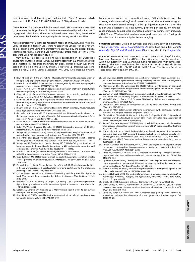
as positive controls. Mutagenicity was evaluated after 5 d of 3 exposure, whichwas tested at 10, 2, 0.4, 0.08, 0.02, 0.003, and 0.006 μM (n = 6 each).
DMPK Studies. A six-mouse mini-pharmacokinetics (PK) assessment was usedto determine the PK profile of 3. FVB/n mice were dosed i.p. with 3 at 2 or 7mg/kg with 25-μL blood draws at indicated time points. Drug levels weredetermined by liquid chromotography/MS-MS using an ABSciex 5500.
Assessing Potency of 3 in Mouse Xenograft Model of TNBC. NOD/Scid mice (B6.CB17-Prkdcscid/Sz; Jackson Labs) were housed in the Scripps Florida vivarium,and all experiments using live animals were approved by the Scripps FloridaInstitutional Animal Care and Use Committee. Female mice (n = 10; 5–7 wkold) were used for xenograft studies.
MDA-MB-231-luc cells (2 million) were suspended in 1× Dulbecco’sphosphate-buffered saline (DPBS) supplemented with 0.9 mg/mL matrigeland injected s.c. into mice mammary fat pads. Tumor growth was moni-tored by injecting 100 μL of 30 mg/mL D-luciferin i.p. Imaging was com-pleted over 2–5 min using a Xenogen IVIS 2000 Live Imaging System.
Luminescence signals were quantified using IVIS analysis software bydrawing a circular/oval region of interest around the luminescent signal.Mice were administered 10 mg/kg 3 by i.p. injection every 48 h after thetumor was detectable (at least 100,000 photons per second) by lumines-cence imaging. Tumors were monitored weekly by luminescent imaging.qRT-PCR and Western blot analyses were performed on mice 3 wk post-visualization of the tumor by luciferase imaging.
Compound Synthesis. Details on synthesis and characterization of dimers (Fig.1 and SI Appendix, Figs. S3–S6 and Scheme S1) as well as 4 and 5 (Fig. 6 and SIAppendix, Figs. S7 and S8 and Scheme S2) are provided in the SI Appendix.
ACKNOWLEDGMENTS. We thank Prof. John L. Cleveland for helpful advice,Prof. Joan Massagué for the 4175 cell line, Kimberley Lowe for assistancewith flow cytometry, and Xiangming Kong for assistance with NMR spec-troscopy. This work was funded by National Institutes of Health GrantsR01-GM9455 and R01-GM9455-S1 (to M.D.D.) and The Nelson Fund forTherapeutic Development (M.D.D.).
1. Dews M, et al. (2010) The myc-miR-17∼92 axis blunts TGFβ signaling and production ofmultiple TGFβ-dependent antiangiogenic factors. Cancer Res 70(20):8233–8246.
2. Brown EJ, et al. (1994) A mammalian protein targeted by G1-arresting rapamycin-receptor complex. Nature 369(6483):756–758.
3. Farazi TA, et al. (2011) MicroRNA sequence and expression analysis in breast tumorsby deep sequencing. Cancer Res 71(13):4443–4453.
4. Qiang XF, et al. (2014) miR-20a promotes prostate cancer invasion and migrationthrough targeting ABL2. J Cell Biochem 115(7):1269–1276.
5. Mathews DH, et al. (2004) Incorporating chemical modification constraints into adynamic programming algorithm for prediction of RNA secondary structure. Proc NatlAcad Sci USA 101(19):7287–7292.
6. Ding Y, et al. (2014) In vivo genome-wide profiling of RNA secondary structure revealsnovel regulatory features. Nature 505(7485):696–700.
7. García-Sacristán A, et al. (2015) A magnesium-induced RNA conformational switch atthe internal ribosome entry site of hepatitis C virus genome visualized by atomic forcemicroscopy. Nucleic Acids Res 43(1):565–580.
8. Watts JM, et al. (2009) Architecture and secondary structure of an entire HIV-1 RNAgenome. Nature 460(7256):711–716.
9. Gutell RR, Weiser B, Woese CR, Noller HF (1985) Comparative anatomy of 16-S-likeribosomal RNA. Prog Nucleic Acid Res Mol Biol 32:155–216.
10. Velagapudi SP, Gallo SM, Disney MD (2014) Sequence-based design of bioactive smallmolecules that target precursor microRNAs. Nat Chem Biol 10(4):291–297.
11. Disney MD, et al. (2008) Two-dimensional combinatorial screening identifies specificaminoglycoside-RNA internal loop partners. J Am Chem Soc 130(33):11185–11194.
12. Velagapudi SP, Seedhouse SJ, French J, Disney MD (2011) Defining the RNA internalloops preferred by benzimidazole derivatives via 2D combinatorial screening andcomputational analysis. J Am Chem Soc 133(26):10111–10118.
13. Guttilla IK, White BA (2009) Coordinate regulation of FOXO1 by miR-27a, miR-96, andmiR-182 in breast cancer cells. J Biol Chem 284(35):23204–23216.
14. Guan L, Disney MD (2013) Covalent small-molecule-RNA complex formation enablescellular profiling of small-molecule-RNA interactions. Angew Chem Int Ed 52(38):10010–10013.
15. Connolly E, et al. (2008) Elevated expression of the miR-17-92 polycistron and miR-21in hepadnavirus-associated hepatocellular carcinoma contributes to the malignantphenotype. Am J Pathol 173(3):856–864.
16. Childs-Disney JL, Tsitovich PB, Disney MD (2011) Using modularly assembled ligands tobind RNA internal loops separated by different distances. ChemBioChem 12(14):2143–2146.
17. Gestwicki JE, Cairo CW, Strong LE, Oetjen KA, Kiessling LL (2002) Influencing receptor-ligand binding mechanisms with multivalent ligand architecture. J Am Chem Soc124(50):14922–14933.
18. Gordon EJ, Sanders WJ, Kiessling LL (1998) Synthetic ligands point to cell surfacestrategies. Nature 392(6671):30–31.
19. Kitov PI, et al. (2000) Shiga-like toxins are neutralized by tailored multivalent car-bohydrate ligands. Nature 403(6770):669–672.
20. Lee MM, et al. (2009) Controlling the specificity of modularly assembled small mol-ecules for RNA via ligand module spacing: Targeting the RNAs that cause myotonicmuscular dystrophy. J Am Chem Soc 131(47):17464–17472.
21. Mammen M, Choi SK, Whitesides GM (1998) Polyvalent interactions in biologicalsystems: Implications for design and use of multivalent ligands and inhibitors. AngewChem Int Ed 37(20):2754–2794.
22. Sucheck SJ, et al. (2000) Design of bifunctional antibiotics that target bacterial rRNAand inhibit resistance-causing enzymes. J Am Chem Soc 122(21):5230–5231.
23. Michael K, Wang H, Tor Y (1999) Enhanced RNA binding of dimerized aminoglycosides.Bioorg Med Chem 7(7):1361–1371.
24. Dervan PB (2001) Molecular recognition of DNA by small molecules. Bioorg MedChem 9(9):2215–2235.
25. Huang CY (1982) Determination of binding stoichiometry by the continuous variationmethod: The Job plot. Methods Enzymol 87:509–525.
26. Ohyashiki M, Ohyashiki JH, Hirota A, Kobayashi C, Ohyashiki K (2011) Age-relateddecrease of miRNA-92a levels in human CD8+ T-cells correlates with a reduction ofnaïve T lymphocytes. Immun Ageing 8(1):11.
27. Sando S, Narita A, Aoyama Y (2007) Light-up Hoechst-DNA aptamer pair: Generationof an aptamer-selective fluorophore from a conventional DNA-staining dye. ChemBioChem8(15):1795–1803.
28. Pushechnikov A, et al. (2009) Rational design of ligands targeting triplet repeatingtranscripts that cause RNA dominant disease: Application to myotonic muscular dys-trophy type 1 and spinocerebellar ataxia type 3. J Am Chem Soc 131(28):9767–9779.
29. Minn AJ, et al. (2005) Genes that mediate breast cancer metastasis to lung. Nature436(7050):518–524.
30. Ames BN, DurstonWE, Yamasaki E, Lee FD (1973) Carcinogens are mutagens: A simpletest system combining liver homogenates for activation and bacteria for detection.Proc Natl Acad Sci USA 70(8):2281–2285.
31. Ames BN, Mccann J, Yamasaki E (1975) Methods for detecting carcinogens and mu-tagens with the Salmonella/mammalian-microsome mutagenicity test. Mutat Res31(6):347–364.
32. Lipinski CA, Lombardo F, Dominy BW, Feeney PJ (2001) Experimental and computa-tional approaches to estimate solubility and permeability in drug discovery and de-velopment settings. Adv Drug Deliv Rev 46(1-3):3–26.
33. Stein CA, Cheng YC (1993) Antisense oligonucleotides as therapeutic agents–is thebullet really magical? Science 261(5124):1004–1012.
34. Swayze EE, Bhat B (2008) The medicinal chemistry of oligonucleotides. Antisense DrugTechnology: Principles, Strategies, and Applications, ed Crooke ST (CRC, Boca Raton,FL), 2nd Ed, pp 143–182.
35. Crooke ST (2004) Progress in antisense technology. Annu Rev Med 55:61–95.36. Childs-Disney JL, Wu M, Pushechnikov A, Aminova O, Disney MD (2007) A small
molecule microarray platform to select RNA internal loop-ligand interactions. ACSChem Biol 2(11):745–754.
37. Lewis BP, Burge CB, Bartel DP (2005) Conserved seed pairing, often flanked byadenosines, indicates that thousands of human genes are microRNA targets. Cell120(1):15–20.
Velagapudi et al. PNAS | May 24, 2016 | vol. 113 | no. 21 | 5903
BIOCH
EMISTR
YCH
EMISTR
Y