Embed Size (px)
Citation preview

Developmental Cell
Review
Design Principles of ProteinBiosynthesis-Coupled Quality Control
Monica C. Rodrigo-Brenni1 and Ramanujan S. Hegde1,*1MRC Laboratory of Molecular Biology, Hills Road, Cambridge CB2 0QH, UK*Correspondence: [email protected]://dx.doi.org/10.1016/j.devcel.2012.10.012
The protein biosynthetic machinery, composed of ribosomes, chaperones, and localization factors, isincreasingly found to interact directly with factors dedicated to protein degradation. The coupling of thesetwo opposing processes facilitates quality control of nascent polypeptides at each stage of their maturation.Sequential checkpoints maximize the overall fidelity of protein maturation, minimize the exposure of defec-tive products to the bulk cellular environment, and protect organisms from protein misfolding diseases.
Cells have extensive surveillance systems to detect errors during
the biosynthesis of essentially all of its major macromolecules.
This includes DNA replication (Reha-Krantz, 2010), transcription
(Sydow and Cramer, 2009), translation (Zaher and Green, 2009),
and maturation of mRNAs (van Hoof and Wagner, 2011), tRNAs
(Yadavalli and Ibba, 2012), and proteins (Buchberger et al.,
2010). Each of these biosynthetic processes has intrinsic limits
on overall fidelity, resulting in a low but tangible rate of errors.
In addition to biosynthetic errors, environmental insults such as
ionizing radiation, reactive oxygen species, and temperature
fluctuations result in damage to cellular macromolecules. Cells
therefore face a constant barrage of defective or damaged
macromolecules that, if left unresolved, have the potential to
disrupt cellular homeostasis, reduce fitness, cause disease,
and contribute to aging. Thus, there is a strong selective pres-
sure to detect defective macromolecules and either correct or
dispose of them.
The potentially disruptive nature of defective macromolecules
places a premium on early detection and rapid resolution. This
explains why many quality control processes have evolved to
act at the site of biosynthesis, before an erroneous product is
released and can engage downstream cellular pathways. The
most obvious examples include internal proofreading by DNA
polymerases (Reha-Krantz, 2010) and kinetic proofreading
during translation (Zaher and Green, 2009). Less obvious is the
exploitation of compartments, such as the nucleus and endo-
plasmic reticulum (ER), for maturation of nascent RNAs and
proteins in a protected environment before their regulated traf-
ficking to their site of function.
These early-acting quality control systems coexist with
partially redundant downstream mechanisms. For example,
proofreading mechanisms during replication are complementary
to DNA mismatch repair that deal with errors after they have
occurred. Similarly, fidelity of decoding during translation is
combined with postsynthesis quality control of misfolded pro-
teins to avoid defects. Thus, numerous quality control mecha-
nisms leading from DNA to functional protein each make unique,
overlapping contributions to minimize the error rate of this com-
plex process. The physiologic relevance of each contribution is
evidenced by the numerous protein misfolding and neurodegen-
erative diseases that result when these quality control processes
fail (Balch et al., 2008).
896 Developmental Cell 23, November 13, 2012 ª2012 Elsevier Inc.
In this Review, we examine the concept of biosynthesis-
coupled quality control during protein maturation. After a brief
historical perspective on the initial development of this field,
we discuss several examples of interactions between various
biosynthetic machinery and degradation factors. This includes
interactions between ribosomes, targeting factors, and chaper-
ones with the ubiquitin-proteasome system. Although the rele-
vance of some of these interactions for quality control is not fully
established, their consideration at this juncture is neverthe-
less worthwhile for providing a conceptual framework for
this emerging area and highlighting key questions for future
research.
Evolving Concepts of Protein Quality ControlNascent proteinsmust fold into their final three-dimensional form
and in many cases must be modified, assembled with partners,
and localized to specific locations in order to function. If these
processes fail, the affected protein must be recognized and
degraded. How this critical triage decision is made has been
a central question in the field of quality control for over 20 years.
One of the earliest mechanisms of triage, kinetic partitioning,
emerged from studies in bacterial systems (Wickner et al.,
1999). In this view, newly synthesized polypeptides released
from the ribosome partition between chaperones and proteases,
both capable of recognizing nonnative proteins. Cycles of chap-
erone binding and release would provide an opportunity to fold,
while partitioning to proteases leads to the irreversible fate of
degradation (Figure 1A). Given that the major classes of chaper-
ones and proteases in bacteria do not interact with each other,
partitioning seems to be the primary strategy for quality control
in bacteria.
Although partitioning was initially postulated to apply to
eukaryotes as well, two sets of observations led to a qualitatively
different concept for quality control. The first concerned the
nature of the degradation system in eukaryotes (Hershko and
Ciechanover, 1998). Quality control in both the cytosol and ER
were found to typically culminate at the proteasome (Buchberger
et al., 2010). Commitment to proteasomal degradation normally
relies on ubiquitin ligases to tag clients with a polyubiquitin chain.
However, the ligases often do not recognize their clients per se,
but rely on adaptors that bring a subset of cellular proteins in
proximity to the ligase. Specificity of degradation is therefore

Figure 1. Passive versus CoupledMechanisms of Quality Control(A) The passive partitioning strategy. Newlysynthesized polypeptides released from the ribo-some partition reversibly between chaperones(blue) and proteases (orange). Upon release fromchaperones, the polypeptide can either fold or re-enter another round of partitioning. Degradationor aggregation are alternative fates for folding-incompetent polypeptides.(B) The coupled strategy. Chaperones engagepolypeptides during synthesis and continue to aidfolding posttranslationally by cycles of binding andrelease. Ubiquitin ligases (orange) can be revers-ibly recruited to the biosynthetic machinery (suchas ribosomes or chaperones) to target nascentpolypeptides for degradation via attachment ofpolyubiquitin (red triangles).
Developmental Cell
Review
imparted by the ligase in conjunction with any associated
adaptor(s) (Deshaies and Joazeiro, 2009).
The second key observation was the discovery that the major
cytosolic chaperone Hsp70 can interact directly with a ubiquitin
ligase (Ballinger et al., 1999). This immediately suggested that
chaperonesmight serve as adaptors that permit certain ubiquitin
ligases to recognize nonnative proteins (Connell et al., 2001;
Hohfeld et al., 2001; Meacham et al., 2001). Some time later,
chaperones in the ER such as BiP, PDI family members, and
GRP94 were also found to interact with components of ubiq-
uitin ligase complexes involved in ER-associated degradation
(Bernardi et al., 2008; Christianson et al., 2008; Denic et al.,
2006; Hosokawa et al., 2008). These observations implied that
nascent eukaryotic proteins did not passively partition between
the folding and degradation machinery as seen in bacteria.
Instead, the two pathways seemed to be more intimately linked,
with quality control relying on an active role for chaperones in
delivering nonnative proteins to degradation factors (Figure 1B).
Chaperone-Associated Quality ControlThe best-studied class of chaperones that links protein folding to
degradation is the Hsp70 family of ATPases (Mayer and Bukau,
2005). The ATP-bound state of Hsp70s has a low affinity for
substrate, favoring dynamic binding and release. By contrast,
the ADP-bound state has a high affinity for substrates, thereby
binding and shielding the client. A wide range of interacting part-
ners (often termed cochaperones) have been described that
regulate the activity of Hsp70s (Kampinga and Craig, 2010).
These include J-domain family members that typically stimu-
late ATPase activity, nucleotide exchange factors that drive
ADP replacement by ATP, factors that recruit Hsp70 to specific
cellular locations, and ubiquitin ligases. Furthermore, the Hsp70
system can function together with other chaperones via their
linking by organizing factors. For example, Hop is a two TPR
domain protein that juxtaposes Hsp70 and Hsp90 via their
C-terminal tails to facilitate folding of certain substrates. Thus,
based on the associated factors, the basic Hsp70 module can
be co-opted for a wide range of functions ranging from protein
folding, protein complex assembly and dissociation, protein tar-
geting, protein translocation, and protein degradation.
A key advance in understanding the role of Hsp70 in degrada-
tion came with the discovery that its C terminus associates with
the ubiquitin ligase CHIP (Ballinger et al., 1999). This suggested
that clients with prolonged Hsp70 interaction would eventually
D
be ubiquitinated, thereby effecting quality control of folding-
defective proteins (Connell et al., 2001; Meacham et al., 2001).
Subsequent studies of Hsp70 interaction partners identified
additional links to degradation pathways including other ubiqui-
tin ligases, the proteasome, and autophagy factors (Esser et al.,
2004; Arndt et al., 2007; Gamerdinger et al., 2011).
Two illustrative examples are the J-domain protein HSJ1
and the nucleotide exchange factor Bag1. HSJ1, like many
J-proteins, stimulates the ATPase activity of Hsp70 to favor
substrate binding (Cheetham et al., 1994). Importantly, however,
it also contains ubiquitin interacting motifs (UIM) that bind to
mono- and polyubiquitin (Chapple et al., 2004; Howarth et al.,
2007; Westhoff et al., 2005). This suggests that ubiquitinated
clients on Hsp70 would preferentially recruit HSJ1 via a bipartite
interaction with both the chaperone (via the J-domain) and
ubiquitin (via the UIM domain). ATP hydrolysis stimulated by
the J-domain would then stabilize this complex.
Bag1, on the other hand, is an exchange factor (Briknarova
et al., 2001; Hohfeld and Jentsch, 1997; Takayama et al., 1997)
that can associate with the proteasome via a ubiquitin-like
(Ubl) domain (Luders et al., 2000). It is therefore plausible that
proteasome-bound Bag1 would recruit Hsp70 complexed with
a ubiquitinated client. Because proteasomes also contain
ubiquitin receptors (Finley, 2009), recruitment could involve
a bipartite interaction, with both Hsp70 and ubiquitin con-
tributing to the avidity. Once recruited, the Bag domain would
stimulate nucleotide exchange to induce release of the sub-
strate, which could then be captured in an unfolded state by
the proteasome. Thus, via the sequential actions of CHIP,
HSJ1, Bag1, and the proteasome, an Hsp70-bound client could
be routed for degradation in a highly regulated manner without
release from the chaperone (Figure 2).
Although this is an attractive scheme and the individual activ-
ities of the factors have been documented, their concerted
sequential action as described above remains to be firmly estab-
lished. Furthermore, the situation is considerably more complex
because of the concurrent presence of dozens of competing
factors (Arndt et al., 2007; Esser et al., 2004; Gamerdinger
et al., 2011) including other J-proteins and Bag proteins (Kam-
pinga and Craig, 2010; Takayama et al., 1999), factors like
HOP that would compete with CHIP for Hsp70 binding (Muller
et al., 2012), and the fact that CHIP can ubiquitinate not only
clients (Murata et al., 2001; Younger et al., 2004), but also
Hsp70 and Bag proteins (Alberti et al., 2002; Jiang et al., 2001).
evelopmental Cell 23, November 13, 2012 ª2012 Elsevier Inc. 897
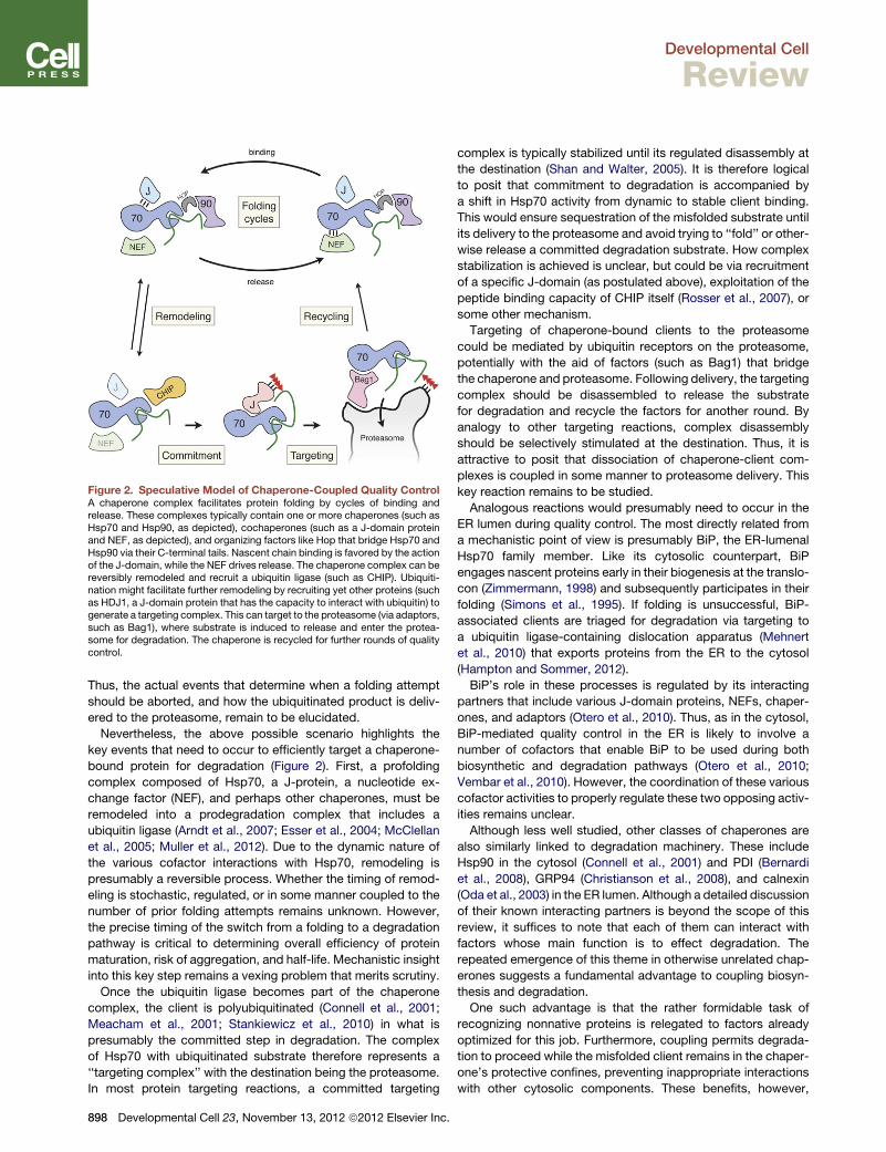
Figure 2. Speculative Model of Chaperone-Coupled Quality ControlA chaperone complex facilitates protein folding by cycles of binding andrelease. These complexes typically contain one or more chaperones (such asHsp70 and Hsp90, as depicted), cochaperones (such as a J-domain proteinand NEF, as depicted), and organizing factors like Hop that bridge Hsp70 andHsp90 via their C-terminal tails. Nascent chain binding is favored by the actionof the J-domain, while the NEF drives release. The chaperone complex can bereversibly remodeled and recruit a ubiquitin ligase (such as CHIP). Ubiquiti-nation might facilitate further remodeling by recruiting yet other proteins (suchas HDJ1, a J-domain protein that has the capacity to interact with ubiquitin) togenerate a targeting complex. This can target to the proteasome (via adaptors,such as Bag1), where substrate is induced to release and enter the protea-some for degradation. The chaperone is recycled for further rounds of qualitycontrol.
Developmental Cell
Review
Thus, the actual events that determine when a folding attempt
should be aborted, and how the ubiquitinated product is deliv-
ered to the proteasome, remain to be elucidated.
Nevertheless, the above possible scenario highlights the
key events that need to occur to efficiently target a chaperone-
bound protein for degradation (Figure 2). First, a profolding
complex composed of Hsp70, a J-protein, a nucleotide ex-
change factor (NEF), and perhaps other chaperones, must be
remodeled into a prodegradation complex that includes a
ubiquitin ligase (Arndt et al., 2007; Esser et al., 2004; McClellan
et al., 2005; Muller et al., 2012). Due to the dynamic nature of
the various cofactor interactions with Hsp70, remodeling is
presumably a reversible process. Whether the timing of remod-
eling is stochastic, regulated, or in some manner coupled to the
number of prior folding attempts remains unknown. However,
the precise timing of the switch from a folding to a degradation
pathway is critical to determining overall efficiency of protein
maturation, risk of aggregation, and half-life. Mechanistic insight
into this key step remains a vexing problem that merits scrutiny.
Once the ubiquitin ligase becomes part of the chaperone
complex, the client is polyubiquitinated (Connell et al., 2001;
Meacham et al., 2001; Stankiewicz et al., 2010) in what is
presumably the committed step in degradation. The complex
of Hsp70 with ubiquitinated substrate therefore represents a
‘‘targeting complex’’ with the destination being the proteasome.
In most protein targeting reactions, a committed targeting
898 Developmental Cell 23, November 13, 2012 ª2012 Elsevier Inc.
complex is typically stabilized until its regulated disassembly at
the destination (Shan and Walter, 2005). It is therefore logical
to posit that commitment to degradation is accompanied by
a shift in Hsp70 activity from dynamic to stable client binding.
This would ensure sequestration of the misfolded substrate until
its delivery to the proteasome and avoid trying to ‘‘fold’’ or other-
wise release a committed degradation substrate. How complex
stabilization is achieved is unclear, but could be via recruitment
of a specific J-domain (as postulated above), exploitation of the
peptide binding capacity of CHIP itself (Rosser et al., 2007), or
some other mechanism.
Targeting of chaperone-bound clients to the proteasome
could be mediated by ubiquitin receptors on the proteasome,
potentially with the aid of factors (such as Bag1) that bridge
the chaperone and proteasome. Following delivery, the targeting
complex should be disassembled to release the substrate
for degradation and recycle the factors for another round. By
analogy to other targeting reactions, complex disassembly
should be selectively stimulated at the destination. Thus, it is
attractive to posit that dissociation of chaperone-client com-
plexes is coupled in some manner to proteasome delivery. This
key reaction remains to be studied.
Analogous reactions would presumably need to occur in the
ER lumen during quality control. The most directly related from
a mechanistic point of view is presumably BiP, the ER-lumenal
Hsp70 family member. Like its cytosolic counterpart, BiP
engages nascent proteins early in their biogenesis at the translo-
con (Zimmermann, 1998) and subsequently participates in their
folding (Simons et al., 1995). If folding is unsuccessful, BiP-
associated clients are triaged for degradation via targeting to
a ubiquitin ligase-containing dislocation apparatus (Mehnert
et al., 2010) that exports proteins from the ER to the cytosol
(Hampton and Sommer, 2012).
BiP’s role in these processes is regulated by its interacting
partners that include various J-domain proteins, NEFs, chaper-
ones, and adaptors (Otero et al., 2010). Thus, as in the cytosol,
BiP-mediated quality control in the ER is likely to involve a
number of cofactors that enable BiP to be used during both
biosynthetic and degradation pathways (Otero et al., 2010;
Vembar et al., 2010). However, the coordination of these various
cofactor activities to properly regulate these two opposing activ-
ities remains unclear.
Although less well studied, other classes of chaperones are
also similarly linked to degradation machinery. These include
Hsp90 in the cytosol (Connell et al., 2001) and PDI (Bernardi
et al., 2008), GRP94 (Christianson et al., 2008), and calnexin
(Oda et al., 2003) in the ER lumen. Although a detailed discussion
of their known interacting partners is beyond the scope of this
review, it suffices to note that each of them can interact with
factors whose main function is to effect degradation. The
repeated emergence of this theme in otherwise unrelated chap-
erones suggests a fundamental advantage to coupling biosyn-
thesis and degradation.
One such advantage is that the rather formidable task of
recognizing nonnative proteins is relegated to factors already
optimized for this job. Furthermore, coupling permits degrada-
tion to proceed while the misfolded client remains in the chaper-
one’s protective confines, preventing inappropriate interactions
with other cytosolic components. These benefits, however,

Developmental Cell
Review
come with the need to now regulate chaperone activity toward
two opposing fates via modulation of its interaction partners. It
is therefore not surprising that the interaction partners, such as
the J-domain family (Kampinga and Craig, 2010), have diversi-
fied considerably more than the chaperones themselves during
evolution.
Achieving the correct balance between biosynthesis and
degradation is critical to maintaining protein homeostasis.
Excessive folding attempts would increase the risk of misfolded
protein aggregation with each cycle that fails. By contrast, over-
aggressive degradation may allow insufficient time for many
proteins to mature. Indeed, relaxing the activity of the chap-
erone-associated ligase CHIP allows a greater proportion of
the difficult-to-fold CFTR to mature (Grove et al., 2009). This
presumably comes at a cost, and the optimal setpoint is a fine
balance between many competing factors (Balch et al., 2008).
For this reason, it is likely that different cells whose priorities
vary widely will have different setpoints as dictated by the
expression patterns and abundances of the various chaperones
and cofactors. For example, postmitotic cells such as neurons
may bemore aggressive in their quality control given their partic-
ular sensitivity to protein misfolding and disease. The relevance
of these cell-type-specific differences will only be revealed after
the roles of each component is clearly defined and placed in
context with its partners. This remains a major challenge in
understanding chaperone-associated quality control.
Although our discussion has focused on chaperone-mediated
recognition of potential degradation substrates, it is worth noting
that some quality control ubiquitin ligases such as San1 and
Hrd1 can directly recognize nonnative features of their clients
(Rosenbaum et al., 2011; Fredrickson et al., 2011; Sato et al.,
2009). Thus, chaperone-associated quality control operates
within a broader framework that includes ubiquitin ligases that
may use different principles of recognition. For a more general
discussion of these other quality control pathways, the reader
is directed to several other papers (Buchberger et al., 2010;
Mehnert et al., 2010; Taylor and Rutter, 2011; Theodoraki
et al., 2012; Varshavsky, 2011).
Quality Control during Protein LocalizationA prerequisite for initiating the folding process for 30%–50%
of all newly synthesized proteins is proper localization into
a compartment such as the ER, mitochondria, peroxisomes, or
plastids (Inaba and Schnell, 2008; Ma et al., 2011; Chacinska
et al., 2009; Shao and Hegde, 2011). Failure of localization
results in an immature precursor in the wrong compartment
that can be detrimental for several reasons. The mislocalized
protein (MLP) would typically lack appropriate cofactors and
chaperones, thereby engaging folding machinery in futile cycles.
MLPs are often precursors that contain hydrophobic domains,
such as signal peptides or transmembrane domains (TMDs),
that make them particularly aggregation prone. MLPs that
contain partial activity could disrupt cellular homeostasis (e.g.,
a mislocalized protease or nuclease). Thus, prompt degradation
of MLPs is crucial for avoiding these adverse fates.
A priori, one might assume that MLPs are handled no differ-
ently than a cytosolic protein that fails to fold. Although this
would seem the simplest solution, it comes at the expense of
occupying the folding machinery for prolonged periods given
D
that the nascent chain is unlikely to ever acquire a stable con-
formation. Furthermore, most chaperones involved in folding
are typically designed to bind short (approximately three- to
five-residue) hydrophobic stretches (Rudiger et al., 1997), and
may not be especially suitable for highly hydrophobic elements
like TMDs and signal peptides. Given that mislocalization seems
to be a relatively frequent event (Kang et al., 2006; Levine et al.,
2005; Rane et al., 2004), particularly under certain conditions like
ER or mitochondrial stress (Kang et al., 2006; Nargund et al.,
2012), relying solely on cytosolic quality control may not be
a particularly suitable solution.
An alternative remedy to this problem is if the targeting
machinery dedicated to localizing clients were linked to degra-
dation such that any delay or failure in targeting results in imme-
diate destruction. Such a system has the advantage that the
localization factors are customized to recognize and shield their
respective clients. Furthermore, the clients are presegregated
from the cytosolic folding machinery, avoiding its futile engage-
ment. Recent studies suggest that, indeed, targeting machinery
to different organelles might interact with ubiquitin ligases for the
purposes of MLP degradation.
This principle is best highlighted with the targeting system for
mammalian tail-anchored (TA) membrane proteins (Hegde and
Keenan, 2011). These single-spanning membrane proteins are
posttranslationally targeted to the ER by an ATPase termed
TRC40 (Favaloro et al., 2008; Stefanovic and Hegde, 2007) or
Get3 in yeast (Schuldiner et al., 2008). This targeting factor
recognizes and shields the TMD of TA proteins in the cytosol
and releases it for insertion upon encountering an ER-localized
receptor. The initial loading of TA proteins onto TRC40 in-
volves several additional cytosolic factors (Jonikas et al., 2009;
Mariappan et al., 2010; Wang et al., 2010).
Biochemical studies in mammalian and yeast systems have
shown that TRC40 is transiently part of a larger TMD-recognition
complex (TRC) that captures, sorts, and loads TA proteins onto
TRC40. The TRC in mammals is composed of a Bag6 subcom-
plex (containing Bag6, TRC35, andUbl4A), TRC40, and probably
SGTA (Mariappan et al., 2010; Winnefeld et al., 2006). Of these
proteins, Bag6, TRC40, and SGTA each are capable of directly
interacting with and shielding the hydrophobic TMD of TA
proteins (Leznicki et al., 2010; Mariappan et al., 2010; Mateja
et al., 2009; Stefanovic and Hegde, 2007; Wang et al., 2010).
Potential clients that engage the TRC are sorted among these
binding proteins, each of which may confer a different fate (Fig-
ure 3A).
Initial engagement with this complex is via Bag6 (Mariappan
et al., 2010) or SGTA (Wang et al., 2010) and may be facilitated
by the ability of certain TRC component(s) to interact with the
ribosome (Mariappan et al., 2010). Once substrates engage the
TRC, targeting to the ER can only be achieved if they are loaded
onto TRC40 (Figure 3A). This loading appears to be highly
specific for the TMDs of ER-destined TA proteins (Hessa et al.,
2011; Mariappan et al., 2010; Wang et al., 2010), although the
basis of this specificity remains unclear. Clients that do not
load onto TRC40 remain associated with TRC via direct binding
to Bag6 (Hessa et al., 2011; Mariappan et al., 2010). This
outcome results in substrate ubiquitination via a yet-unidentified
ubiquitin ligase recruited by a ubiquitin-like (Ubl) domain in Bag6
(Hessa et al., 2011) (Figure 3A).
evelopmental Cell 23, November 13, 2012 ª2012 Elsevier Inc. 899

Figure 3. Quality Control during ProteinLocalization(A) Tail-anchored protein targeting and qualitycontrol. Newly synthesized proteins containinga TMD (red) can engage a TRC after release fromthe ribosome. Initial capture by the TRC might befacilitated by its transient interaction with theribosome. The TRC is a dynamic complex com-posed of a core Bag6-subcomplex (pink), SGTA(green), TRC40 (blue), and a yet-unidentified E3ubiquitin ligase (orange). Bag6, SGTA, and TRC40all have the capacity to bind the TMD, with thelatter strongly favoring only substrates with asingle TMD close to the C terminus (i.e., tail-anchored proteins). Loading of substrate ontoTRC40 results in targeting and insertion into theER membrane, while prolonged engagement withthe TRC results in substrate ubiquitination andtargeting to the proteasome.(B) Quality control during targeting to chloroplastsand mitochondria. Proteins containing a targetingsequence (purple) contain as part of their targetingcomplex the Hsp70 and/or Hsp90 chaperones.These chaperones have receptors at the destina-tion membrane that bind to the same regionas the ubiquitin ligase CHIP. Thus, any failure oftargeting would result in recruitment of CHIP,substrate ubiquitination, and degradation.
Developmental Cell
Review
Thus, TA protein targeting in mammals involves an obligate
engagement with factors that directly link to ubiquitination
machinery. This means that if TRC40 is not immediately avail-
able, then the prolonged interaction with Bag6 would favor
degradation. Indeed, in vitro studies show that depletion or satu-
ration of TRC40 results in increased TA protein ubiquitination
(Hessa et al., 2011). Even if substrates are successfully loaded
onto TRC40, it is conceivable that delayed targetingmight permit
re-engagement with the TRC and eventual ubiquitination via
Bag6. In this manner, any failures in targeting would permit rapid
degradation without release from the machinery designed
specifically to bind and shield highly hydrophobic TMDs.
Avoiding the release of TMD-containing proteins free into the
cytosol is probably important for avoiding aggregation and fruit-
lessly engaging general chaperones such as Hsp70. Indeed, in
yeast (which do not have an obvious Bag6 homolog), deletion
of TA targeting machinery leads to substantial TA protein aggre-
gation (Jonikas et al., 2009; Schuldiner et al., 2008). Aggregation
in yeast, while probably undesirable, can nevertheless be
resolved by either disaggregases such as Hsp104 (Winkler
et al., 2012) or selective partitioning to mother cells during cell
division (Zhou et al., 2011). Thus, the evolution of direct links
between the targeting and degradation machinery in complex
organisms may have been favored by their lower tolerance for
protein misfolding stress and absence of robust disaggregation
systems.
How are failures in other targeting pathways handled by the
cell? In the case of cotranslational targeting of secretory and
membrane proteins to the ER, the answer appears to also involve
900 Developmental Cell 23, November 13, 2012 ª2012 Elsevier Inc.
the Bag6 protein. Analysis of failed
translocation products in vitro suggested
the existence of a pathway dedicated
to ubiquitination of proteins with highly
hydrophobic domains such as signal
peptides and TMDs (Iwamuro et al., 1999; Hessa et al., 2011).
Subsequent crosslinking experiments combinedwith ubiquitina-
tion assays identified Bag6 as a key player in this pathway
(Hessa et al., 2011).
Bag6 proved capable of interacting not only with TA proteins
(Leznicki et al., 2010; Mariappan et al., 2010), but also with any
protein containing long linear hydrophobic domains such as
signal peptides, TMDs, and GPI-anchor signal (Hessa et al.,
2011). Furthermore, unlike TRC40, which seems to favor pro-
teins with only one TMD near the C terminus, Bag6 seems
nonselective with respect to either the number or position of
hydrophobic elements (Hessa et al., 2011). Thus, the substrate
specificity of Bag6 matches well with the features of secretory
and membrane protein precursors.
Bag6 would ordinarily never have the opportunity to interact
with these proteins during biosynthesis because they are typi-
cally recognized cotranslationally by SRP as they emerge from
the ribosome (Shan and Walter, 2005). SRP would enjoy consid-
erable advantage in recognizing these proteins because its
substrate-binding domain is positioned precisely at the exit
tunnel on the ribosome (Halic et al., 2004). Thus, secretory and
membrane proteins are normally targeted early in their synthesis
to the ER translocon (Shan and Walter, 2005), where they can
complete synthesis in the protected environment afforded by
the ribosome-translocon complex (Becker et al., 2009; Menetret
et al., 2005). Only when SRP-dependent targeting fails would the
Bag6 complex have an opportunity to capture the nascent chain.
This capture may be facilitated by the ability of Bag6 complex
to interact with ribosomes containing TMDswithin the exit tunnel

Developmental Cell
Review
(Mariappan et al., 2010). How the complex is recruited to these
ribosomes is entirely unclear; however, analogous ‘‘signaling’’
from inside the tunnel to influence ribosome functions such as
elongation (Lu and Deutsch, 2008), termination (Cao and
Geballe, 1996), SRP interaction (Berndt et al., 2009), and trans-
locon interaction (Liao et al., 1997) has been described. Regard-
less of the mechanism, this observation suggests that the
cotranslational targeting machinery (ribosome-nascent chain-
SRP complexes) may include a factor (Bag6) that links to the
ubiquitination machinery. This link (as well as substrate speci-
ficity of Bag6 for particularly hydrophobic linear domains)
presumably affords Bag6 an advantage over other chaperones
normally dedicated to folding cytosolic proteins andwhose inter-
actions typically occur via much shorter hydrophobic patches.
Thus, Bag6, in addition to being an integral component of the
TA targeting pathway, is also loosely coupled to cotranslational
targeting to facilitate quality control in the case of failure.
Similar principles may apply to targeting to other organelles
(Figure 3B). For both chloroplast transport and mitochondrial
transport, the cytosolic targeting complex contains Hsp70 and/
or Hsp90 (May and Soll, 2000; Qbadou et al., 2006; Young
et al., 2003). These chaperones serve to not only maintain the
unfolded state of their clients, but also interact with receptors
at the target membrane to facilitate targeting. These receptors
contain TPR domains that interact with the C-terminal peptide
on the chaperones (Schlegel et al., 2007). Remarkably, the
same peptide domain interacts with CHIP (Ballinger et al., 1999).
This suggests that failed or delayed targeting would allow the
exposed TPR binding motif of the targeting complex to interact
with CHIP rather than its receptor, thereby triggering client
degradation (Figure 3B). Indeed, CHIP has been implicated in
chloroplast precursor degradation when import is blocked (Lee
et al., 2009). A major advantage of this mechanism would be
that triage for degradation would not necessitate release of the
polypeptide from its chaperone-protected state. This may be
especially important for highly hydrophobic clients like mem-
brane proteins of the chloroplast or mitochondria. Interestingly,
a mechanism for coupled degradation does not seem to exist
within the ‘‘prokaryotic-like’’ stroma compartment of the chloro-
plast. Instead, an unusual variant of the signal recognition
particle has evolved disaggregation activity to deal with aggre-
gated mislocalized membrane proteins (Jaru-Ampornpan et al.,
2010). This nicely illustrates within a single system the contrast-
ing strategies employed to deal with mislocalization.
Finally, after targeting has occurred to a translocon, translo-
cation and membrane insertion are not necessarily assured
(Levine et al., 2005). Failures during these late stages in localiza-
tion are handled efficiently by the ubiquitin-proteasome system
(Garrison et al., 2005; McKibbin et al., 2012), suggesting this
step is subject to surveillance and quality control. Very little is
known about how this might occur, but in the case of the ER,
some evidence exists for cotranslational ubiquitination of
translocon-engaged substrates. Notable clients include the
multispanning membrane protein CFTR (Sato et al., 1998),
whose biogenesis may be particularly inefficient, and the very
large apolipoprotein B (Zhou et al., 1998), which needs to asso-
ciate cotranslationally with lipids for correct maturation. In both
cases, nascent chains may get targeted for degradation during
translocation by machinery that remains poorly understood.
D
A recent study in yeast suggested that clients displaying pro-
longed interaction with the ER translocon are ubiquitinated by
the ER-resident ubiquitin ligase Hrd1 (Rubenstein et al., 2012).
Whether Hrd1 can interact with Sec61 to mediate this activity
is not clear. Nevertheless, the findings do lend credence to the
notion that surveillance mechanisms exist to monitor cata-
strophic failures at essentially all steps of ER protein localization
from targeting to translocation. These surveillance mechanisms
not only protect the substrate from aggregation, but may also
buffer the cytosolic and ER folding machinery from excess
nonproductive clients.
Quality Control at the RibosomeProtein quality control has traditionally been viewed as a post-
translational process. After all, how can a protein be evaluated
for its ability to fold before its synthesis is even complete? Recent
studies examining quality control during translation have started
to address this question, revealing that polypeptides of inappro-
priate length are deemed defective and routed for degradation
directly from the ribosome.
The best-studied example of protein quality control linked to
protein synthesis is the tmRNA system of prokaryotes (reviewed
by Janssen and Hayes, 2012; Felden and Gillet, 2011). This
system resolves the problem of a translating ribosome that rea-
ches the end of an mRNA without encountering an in frame
stop codon. This stalled ribosome recruits a hybrid transfer-
messenger RNA (i.e., tmRNA) that together with associated
proteinsserves to tag thenascentprotein fordegradation, recycle
the ribosome, and degrade the associatedmRNA. This is accom-
plished by a ‘‘trans-translation’’ mechanism in which the stalled
ribosome uses tmRNA as a template to complete synthesis of
the protein. The sequence encoded by the tmRNA serves as
a degradation tag for the released protein, and the termination
codon in the tmRNA allows ribosome recycling. Thus, a defect
in the normal translation cycle is directly communicated to the
truncated protein product to effect its quality control. Although
the tmRNA system is not conserved in eukaryotes, it has long
been appreciated that the same problems of ribosome recycling,
protein degradation, and mRNA degradation must be solved.
Resolution of problems during the eukaryotic translation cycle
has been studied most extensively in the context of mRNA
surveillance (van Hoof and Wagner, 2011). Only recently has it
been appreciated that protein quality control is probably an inte-
gral part of these pathways (Bengtson and Joazeiro, 2010; Dimi-
trova et al., 2009; Ito-Harashima et al., 2007). Early studies of
mRNA stability recognized that messages containing premature
stop codons were often rapidly degraded by a process termed
nonsense-mediated decay (NMD) (Losson and Lacroute, 1979;
Maquat et al., 1981). Subsequent studies defined additional
mRNA surveillance pathways termed no-go decay for stalled
ribosomes (Doma and Parker, 2006) and nonstop decay (NSD)
for messages lacking an in-frame stop codon (Frischmeyer
et al., 2002; van Hoof et al., 2002). These pathways appear to
be conceptually and mechanistically related, most notably by
their requirement of at least one round of translation to trigger
degradation of the defective mRNA (Maquat et al., 2010;
Shoemaker and Green, 2012; van Hoof and Wagner, 2011).
Investigating the basis for this translation requirement
helped explain how very subtle problems in an mRNA, such as
evelopmental Cell 23, November 13, 2012 ª2012 Elsevier Inc. 901

Figure 4. Quality Control at the Ribosome during mRNA Surveillance(A) The normal translation cycle of initiation, elongation, termination, and recycling (top panel) can be disrupted by various types of mRNA defects that lead toa stalled ribosome (depicted in tan). The mRNA surveillance machinery (not depicted) recognizes the stalled ribosome and digests the mRNA to leave behindunresolved 80S ribosome-nascent chain complexes.(B) Hypothetical series of events that lead to resolution of the stalled 80S ribosome-nascent chain complex. Recycling factors (Hbs1, Pelota, ABCE1) split the 80Sribosome into subunits. A ubiquitin ligase (such as Listerin, blue) is recruited to the large subunit and catalyzes polyubiquitination of the nascent chain to commitit for degradation.
Developmental Cell
Review
a frameshift or single nonsense mutation, can be recognized. In
essence, a pioneer round of translation ‘‘test-drives’’ the mRNA
to confirm its fidelity. Thus, encountering a termination codon far
from the poly-A tail, reading into the poly-A tail, reaching the end
of a message without encountering a stop codon, or stalling
within the coding region all signal the recruitment of endonucle-
ases and exonucleases that degrade the presumably defective
mRNA. Degradation ensures that potentially detrimental defec-
tive protein products are not produced from repeated use of
defective mRNAs. However, the issue of how the defective
protein product (and in some cases, the stalled ribosome) from
the pioneer round of translation is handled remained to be
explained.
Two sets of studies, one studying nascent polypeptide degra-
dation and another analyzing ribosome recycling (discussed
below), have shed light on this problem. When considered
together, they are beginning to sketch an initial framework for
quality control at the ribosome (Figure 4). Studies on the fate of
nascent chains generated during NSD suggested that translating
ribosomes stall when they decode the poly-A tail and the nascent
polypeptide synthesized to that point is degraded via the
902 Developmental Cell 23, November 13, 2012 ª2012 Elsevier Inc.
ubiquitin-proteasome system (Ito-Harashima et al., 2007). This
is an attractive model because poly-A translation would be
a unique feature of nonstop mRNAs, and therefore could serve
as a specific signal for nascent chain ubiquitination. The mecha-
nism of stalling remains unclear (Wilson and Beckmann, 2011),
although it has been speculated to involve interactions between
polylysine (encoded by poly-A) and the ribosomal exit tunnel.
Consistent with this idea, similar results were observed with
polyarginine coding segments (Dimitrova et al., 2009). Thus,
stalled ribosome-nascent chain complexes containing a polyba-
sic peptide of sufficient length within the exit tunnel trigger
nascent chain degradation.
Two ubiquitin ligases, Not4 and Ltn1, have been implicated in
this process (Bengtson and Joazeiro, 2010; Dimitrova et al.,
2009). Both ligases are capable of associating with ribosomes,
and deletion of either gene in yeast results in stabilization of
the polypeptide fragment preceding a polybasic sequence.
The reason for the discrepancy in the two studies implicating
different ligases is presently unclear, and it has not been possible
yet to fully resolve direct from indirect effects on nascent chain
degradation. Future in vitro reconstitution studies will be needed

Developmental Cell
Review
to define the ligase(s) that directly ubiquitinate the nascent chain
and rigorously determine if ubiquitination occurs on peptidyl-
tRNA products on the ribosome.
These unresolved issues notwithstanding, the implication from
these studies is that ligase(s) associated with or recruited to the
translation machinery mediate ubiquitination of nascent poly-
peptides and commit them for degradation before they are
released into the bulk cytosol. This would spare the cytosolic
folding and quality control machinery from handling these defec-
tive proteins and minimize the risk of inappropriate interactions
or dominant-negative effects. Thus, in the case of NSD, mRNA
surveillance and nascent chain quality control seem to be inti-
mately linked via the translation apparatus (Shoemaker and
Green, 2012).
Because translating ribosomes on normal mRNAs should be
refractory to these mechanisms, it is imperative that the mRNA
and protein degradation pathways are specific for defective
complexes. One possibility is that a stalled ribosome is the initial
signal for recruitment of nucleases (Schaeffer and van Hoof,
2011), ribosome recycling factors (Pisareva et al., 2011;
Shoemaker et al., 2010), and ubiquitin ligases (Bengtson and
Joazeiro, 2010; Dimitrova et al., 2009) that together resolve the
stalled complex. However, the specific factors involved, the
mechanism of recruitment, and the order of events all remain
to be clearly delineated. Furthermore, how the cell would distin-
guish between a normal pausing event during translation and
that resulting from defective substrates is not well understood.
Nevertheless, some insight has indirectly come from studies of
ribosome recycling of stalled translation complexes.
Recycling is the process by which 80S ribosomes are split into
60S and 40S subunits that can re-enter the translation cycle
following translation termination at a stop codon (Jackson
et al., 2012). Here, GTP-bound eRF3 in complex with eRF1
targets to a translating ribosome containing a stop codon in
the A site. Once bound, GTP hydrolysis by eRF3 triggers
a conformational change in eRF1 such that its highly conserved
GGQmotif can catalyze hydrolysis of the ester bond between the
peptidyl-tRNA and nascent polypeptide. This releases the
peptide into the cytosol. After eRF3 dissociation, the ATPase
ABCE1 (Rli1 in yeast) is then recruited to the A site, where it
uses ATP hydrolysis to drive 80S separation into 60S and 40S
subunits (Pisarev et al., 2010).
In the case of stalled translation complexes, the A site would
not contain a stop codon, and therefore cannot recruit the
eRF1-eRF3 complex. Instead, two homologous factors termed
Hbs1 and Pelota (Dom34 in yeast) act in conjunction with
ABCE1 to serve a similar function (Pisarev et al., 2010;
Shoemaker et al., 2010; Tsuboi et al., 2012), but with two key
differences. First, the recycling reaction can only occur on trans-
lation complexes stalled very close (within�12 nucleotides in the
mammalian system) to the end of an mRNA. This length is
notable because it is precisely the number of mRNA residues
protected by the ribosome from the A site to the cytosol. This
may be used as a cue to distinguish stalled from translating
ribosomes, analogous to the mechanism used by ribosome
rescue factors in prokaryotes (Gagnon et al., 2012; Neubauer
et al., 2012). Second, Pelota does not contain a GGQ motif,
and hence cannot catalyze hydrolysis of the tRNA ester bond.
Thus, dissociation of 80S complexes containing a short (four-
D
residue) nascent chain resulted in ‘‘drop-off’’ of an intact peptidyl
tRNA (Pisarev et al., 2010; Shoemaker et al., 2010).
Both of these observations have implications for how stalled
translation complexes are resolved. The requirement for minimal
mRNA protrusion outside the ribosome implies that ribosome re-
cycling cannot occur until nuclease(s) have first digested the
mRNA downstream of the ribosome. The nuclease that performs
this task remains to be elucidated, but may be recruited by or act
in conjunction with Dom34 (Doma and Parker, 2006). The fact
that the tRNA is not hydrolyzed to release the peptide implies
that ribosome splitting would produce one of two rather unusual
products. Either a peptidyl-tRNA would be released free into the
cytosol (provided the nascent chain was very short), or a 60S-
peptidyl-tRNA complex would be generated. Because each of
these species is never part of the normal translation cycle, they
could provide unique targets for recruitment of ubiquitin ligases
that polyubiquitinate the nascent peptide for degradation.
Consistent with this idea, Ltn1 was observed to cofractionate
with 60S subunits, but not 80S or polysomes (Bengtson and Joa-
zeiro, 2010).
Taking the specificity of the ribosome recycling factors and
current knowledge ofmRNA surveillance pathways into account,
one can envision a plausible working framework for ribosome-
associated quality control (Figure 4). Any of several events could
initiate the process including (1) encountering a premature termi-
nation codon during the pioneer translation cycle, (2) ribosome
stalling due to mRNA secondary structure, rare codons, amino
acid insufficiency, or mRNA damage, (3) reading into the poly-
A tail, or (4) reaching the end of a message. In each case, the
ribosome would stop elongating, albeit for different reasons,
leading to the recruitment of endonucleases and/or exonucle-
ases. Digestion of the mRNA 30 to the translating ribosome (or
perhaps in the A site) would generate a RNC species that is
the target for the Hbs1/Pelota/ABCE1 recycling system. Splitting
of the subunits produces a 60S-peptidyl-tRNA that could recruit
Ltn1 to ubiquitinate the nascent chain. The ubiquitinated nascent
chain could then be extracted from the 60S subunit, perhaps via
the p97 ATPase complex or proteasome. Thus, themRNAwould
be destroyed, the ribosomal subunits recycled, and the nascent
chain targeted for degradation without accessing the bulk
cytosol.
This framework is appealing for a number of reasons. First,
multiple different and seemingly unrelated situations could
converge on a single initiating species: the nucleolytically pro-
cessed RNC that is the target for recycling factors (Figure 4A).
The pathway for the generation of this species may differ for
NSD, NMD, and NGD, each of which seem to involve different
factors and requirements (Shoemaker and Green, 2012; van
Hoof and Wagner, 2011). Nevertheless, nucleolytic digestion of
mRNAs engaged in translation seems to be a universal theme,
thereby producing essentially the same stalled RNC species.
Second, the unique nature of the 60S-peptidyl-tRNA would
markedly aid recognition by the ubiquitin ligase (Figure 4B).
Specific recognition is a critical issue because the abundance
of ubiquitin ligases such as Ltn1 is between two and three orders
of magnitude lower than that of ribosomes (Ghaemmaghami
et al., 2003). Although Ltn1 could potentially recognize the
same cues that recruit nucleases to stalled ribosomes, this
mechanism is less appealing because the factors may compete
evelopmental Cell 23, November 13, 2012 ª2012 Elsevier Inc. 903

Developmental Cell
Review
rather than act sequentially in a defined order. Furthermore, even
a weak affinity for translating ribosomes would risk ubiquitinating
normal nascent chains, particularly those that take a long time to
synthesize. By exploiting recycling factors to provide the requi-
site specificity, the ligase would not pose a risk of interfering
with normal protein maturation.
Third, committing the nascent chain for degradation would
occur without ever releasing it from the ribosome. In this
manner, exposure to the cytosol, engagement of protein folding
machinery, and risk of inappropriate interactions are all markedly
minimized. Thus, mRNA quality control and protein quality
control likely intersect at the ribosome to ensure that defective
messages and their aberrant protein products are recognized
extremely early and each routed for degradation before they
can pose significant harm.
Similar mechanisms of protein quality control may be
employed to deal with incomplete nascent chains on mRNAs
that are turned over as a part of normal degradation. In this situ-
ation, the timing of mRNA digestion by endonucleases and
exonucleases relative to the translation cycle is not completely
understood (Shoemaker and Green, 2012). However, the only
way partially synthesized products could be entirely avoided is
if all engaged ribosomes are allowed to complete translation
before mRNA degradation is initiated (Hu et al., 2009). In the
absence of such coordination, translating ribosomes and their
associated incomplete nascent chains would need to be
resolved in conjunction with mRNA downregulation.
Thus, protein quality control of nascent chains at the ribosome
may be a common phenomenon in higher eukaryotes where
mRNA remodeling is extensive. Furthermore, there are other
situations where large amounts of defective mRNAs are gener-
ated. For example, DNA recombination in generating T cell
receptor diversity results in a large proportion of out of frame
mRNAs that are degraded by NMD in a translation-dependent
manner (Wang et al., 2002). Ribosome-associated quality
control may therefore be of special importance in particular
cell types or during differentiation.
What about quality control on the ribosome on the basis of
nascent chain folding? Although certainly a possibility, it is diffi-
cult at present to see how deviations from normal could be
detected. This is because with few exceptions, polypeptides in
the process of being synthesized are normally nonnative. Thus,
the logic of triaging for degradation a polypeptide that has yet
to be given a chance to fold is not immediately apparent, at least
from the standpoint of protein maturation. Nevertheless, this
possibility should not be entirely discounted because other
beneficial outcomes such as antigen presentation (Dolan et al.,
2011) or amino acid recycling (Vabulas and Hartl, 2005) might
have driven its evolution.
Advantages of Coupled Biosynthesis and DegradationAs illustrated by the foregoing examples, linking quality control to
each step in protein biosynthesis has three major advantages
that are worth underscoring. First, the defective product in
question is recognized at the earliest stage possible, limiting
the burden on downstream biosynthetic and quality control
pathways and minimizing the potential for disruption of cellular
homeostasis. Second, multiple checkpoints on quality provides
a measure of redundancy that likely increases overall fidelity of
904 Developmental Cell 23, November 13, 2012 ª2012 Elsevier Inc.
the final product. Third, each stage of quality control uses
different parameters to evaluate the nascent polypeptide. These
parameters could be different structural features of the polypep-
tide as distinguished by chaperones combined with polypep-
tide-independent cues such as the state of the ribosome. Using
multiple parameters provides a more complete assessment than
could be achieved by any single quality control mechanism.
From a mechanistic standpoint, linking biosynthesis with
quality control greatly facilitates defective substrate recognition.
In essence, quality control can piggyback on biosynthetic com-
plexes that are in an ideal position to evaluate maturation status.
For example, an abnormal translation cycle giving rise to trun-
cated products is most easily indicated by ribosome position
along the mRNA relative to fiduciary markers such as the poly-
A tail, stop codons, and the 30 end. Similarly, localization factors
are optimally evolved to recognize localization elements, and
their prolonged interaction is therefore a sure sign that localiza-
tion has failed. Finally, chaperones are customized to recognize
nonnative states of proteins, making them the ideal sensor of
protein misfolding. Thus, a repeated theme is the exploitation
by quality control machinery of unique reactions surrounding
individual biosynthetic events to reroute nascent chains toward
degradation. An understanding of the timing mechanism used
to shift nascent chain fate remains a major challenge in all quality
control pathways.
The distribution of quality control tasks across multiple steps
means that deficiencies in any one mechanism would not
necessarily be crippling. For example, an inability to detect and
degrade truncated products at the ribosome would release
them to the downstream folding and/or localization machinery.
In most cases, these products would still be degraded because
they would not fold correctly. Only in instances where the
truncation was near a domain boundary might the product be
completely stabilized by failure of ribosome-associated quality
control. From a physiologic standpoint, this robustness built
into the system likely provides protection from fluctuations that
might temporarily saturate one or another pathway.
ACKNOWLEDGMENTS
We are grateful to Sichen Shao and Rebecca Voorhees for comments on thismanuscript. Research in the Hegde laboratory is supported by the MedicalResearch Council of the United Kingdom.MC.R.-B. is an EllisonMedical Foun-dation/AFAR Fellow of the Life Sciences Research Foundation.
REFERENCES
Alberti, S., Demand, J., Esser, C., Emmerich, N., Schild, H., and Hohfeld, J.(2002). Ubiquitylation of BAG-1 suggests a novel regulatorymechanism duringthe sorting of chaperone substrates to the proteasome. J. Biol. Chem. 277,45920–45927.
Arndt, V., Rogon, C., and Hohfeld, J. (2007). To be, or not to be—molecularchaperones in protein degradation. Cell. Mol. Life Sci. 64, 2525–2541.
Balch, W.E., Morimoto, R.I., Dillin, A., and Kelly, J.W. (2008). Adapting proteo-stasis for disease intervention. Science 319, 916–919.
Ballinger, C.A., Connell, P., Wu, Y., Hu, Z., Thompson, L.J., Yin, L.Y., andPatterson, C. (1999). Identification of CHIP, a novel tetratricopeptide repeat-containing protein that interacts with heat shock proteins and negativelyregulates chaperone functions. Mol. Cell. Biol. 19, 4535–4545.
Becker, T., Bhushan, S., Jarasch, A., Armache, J.P., Funes, S., Jossinet, F.,Gumbart, J., Mielke, T., Berninghausen, O., Schulten, K., et al. (2009).

Developmental Cell
Review
Structure of monomeric yeast and mammalian Sec61 complexes interactingwith the translating ribosome. Science 326, 1369–1373.
Bengtson, M.H., and Joazeiro, C.A. (2010). Role of a ribosome-associated E3ubiquitin ligase in protein quality control. Nature 467, 470–473.
Bernardi, K.M., Forster, M.L., Lencer, W.I., and Tsai, B. (2008). Derlin-1facilitates the retro-translocation of cholera toxin. Mol. Biol. Cell 19, 877–884.
Berndt, U., Oellerer, S., Zhang, Y., Johnson, A.E., and Rospert, S. (2009). Asignal-anchor sequence stimulates signal recognition particle binding to ribo-somes from inside the exit tunnel. Proc. Natl. Acad. Sci. USA 106, 1398–1403.
Briknarova, K., Takayama, S., Brive, L., Havert, M.L., Knee, D.A., Velasco, J.,Homma, S., Cabezas, E., Stuart, J., Hoyt, D.W., et al. (2001). Structural anal-ysis of BAG1 cochaperone and its interactions with Hsc70 heat shock protein.Nat. Struct. Biol. 8, 349–352.
Buchberger, A., Bukau, B., and Sommer, T. (2010). Protein quality control inthe cytosol and the endoplasmic reticulum: brothers in arms. Mol. Cell 40,238–252.
Cao, J., and Geballe, A.P. (1996). Coding sequence-dependent ribosomalarrest at termination of translation. Mol. Cell. Biol. 16, 603–608.
Chacinska, A., Koehler, C.M., Milenkovic, D., Lithgow, T., and Pfanner, N.(2009). Importing mitochondrial proteins: machineries and mechanisms. Cell138, 628–644.
Chapple, J.P., van der Spuy, J., Poopalasundaram, S., and Cheetham, M.E.(2004). Neuronal DnaJ proteins HSJ1a and HSJ1b: a role in linking theHsp70 chaperone machine to the ubiquitin-proteasome system? Biochem.Soc. Trans. 32, 640–642.
Cheetham, M.E., Jackson, A.P., and Anderton, B.H. (1994). Regulation of70-kDa heat-shock-protein ATPase activity and substrate binding by humanDnaJ-like proteins, HSJ1a and HSJ1b. Eur. J. Biochem. 226, 99–107.
Christianson, J.C., Shaler, T.A., Tyler, R.E., and Kopito, R.R. (2008). OS-9 andGRP94 deliver mutant alpha1-antitrypsin to the Hrd1-SEL1L ubiquitin ligasecomplex for ERAD. Nat. Cell Biol. 10, 272–282.
Connell, P., Ballinger, C.A., Jiang, J., Wu, Y., Thompson, L.J., Hohfeld, J., andPatterson, C. (2001). The co-chaperone CHIP regulates protein triagedecisions mediated by heat-shock proteins. Nat. Cell Biol. 3, 93–96.
Denic, V., Quan, E.M., and Weissman, J.S. (2006). A luminal surveillancecomplex that selects misfolded glycoproteins for ER-associated degradation.Cell 126, 349–359.
Deshaies, R.J., and Joazeiro, C.A. (2009). RING domain E3 ubiquitin ligases.Annu. Rev. Biochem. 78, 399–434.
Dimitrova, L.N., Kuroha, K., Tatematsu, T., and Inada, T. (2009). Nascentpeptide-dependent translation arrest leads to Not4p-mediated protein degra-dation by the proteasome. J. Biol. Chem. 284, 10343–10352.
Dolan, B.P., Bennink, J.R., and Yewdell, J.W. (2011). Translating DRiPs: prog-ress in understanding viral and cellular sources of MHC class I peptide ligands.Cell. Mol. Life Sci. 68, 1481–1489.
Doma, M.K., and Parker, R. (2006). Endonucleolytic cleavage of eukaryoticmRNAs with stalls in translation elongation. Nature 440, 561–564.
Esser, C., Alberti, S., and Hohfeld, J. (2004). Cooperation of molecular chaper-ones with the ubiquitin/proteasome system. Biochim. Biophys. Acta 1695,171–188.
Favaloro, V., Spasic, M., Schwappach, B., and Dobberstein, B. (2008). Distincttargeting pathways for the membrane insertion of tail-anchored (TA) proteins.J. Cell Sci. 121, 1832–1840.
Felden, B., and Gillet, R. (2011). SmpB as the handyman of tmRNA duringtrans-translation. RNA Biol. 8, 440–449.
Finley, D. (2009). Recognition and processing of ubiquitin-protein conjugatesby the proteasome. Annu. Rev. Biochem. 78, 477–513.
Fredrickson, E.K., Rosenbaum, J.C., Locke, M.N., Milac, T.I., and Gardner,R.G. (2011). Exposed hydrophobicity is a key determinant of nuclear qualitycontrol degradation. Mol. Biol. Cell 22, 2384–2395.
D
Frischmeyer, P.A., van Hoof, A., O’Donnell, K., Guerrerio, A.L., Parker, R., andDietz, H.C. (2002). An mRNA surveillance mechanism that eliminates tran-scripts lacking termination codons. Science 295, 2258–2261.
Gagnon, M.G., Seetharaman, S.V., Bulkley, D., and Steitz, T.A. (2012). Struc-tural basis for the rescue of stalled ribosomes: structure of YaeJ bound to theribosome. Science 335, 1370–1372.
Gamerdinger, M., Carra, S., and Behl, C. (2011). Emerging roles of molecularchaperones and co-chaperones in selective autophagy: focus on BAGproteins. J. Mol. Med. 89, 1175–1182.
Garrison, J.L., Kunkel, E.J., Hegde, R.S., and Taunton, J. (2005). A substrate-specific inhibitor of protein translocation into the endoplasmic reticulum.Nature 436, 285–289.
Ghaemmaghami, S., Huh, W.K., Bower, K., Howson, R.W., Belle, A.,Dephoure, N., O’Shea, E.K., and Weissman, J.S. (2003). Global analysis ofprotein expression in yeast. Nature 425, 737–741.
Grove, D.E., Rosser, M.F., Ren, H.Y., Naren, A.P., and Cyr, D.M. (2009). Mech-anisms for rescue of correctable folding defects in CFTRDelta F508. Mol. Biol.Cell 20, 4059–4069.
Halic, M., Becker, T., Pool, M.R., Spahn, C.M., Grassucci, R.A., Frank, J., andBeckmann, R. (2004). Structure of the signal recognition particle interactingwith the elongation-arrested ribosome. Nature 427, 808–814.
Hampton, R.Y., and Sommer, T. (2012). Finding the will and the way of ERADsubstrate retrotranslocation. Curr. Opin. Cell Biol. 24, 460–466.
Hegde, R.S., and Keenan, R.J. (2011). Tail-anchored membrane protein inser-tion into the endoplasmic reticulum. Nat. Rev. Mol. Cell Biol. 12, 787–798.
Hershko, A., and Ciechanover, A. (1998). The ubiquitin system. Annu. Rev.Biochem. 67, 425–479.
Hessa, T., Sharma, A., Mariappan, M., Eshleman, H.D., Gutierrez, E., andHegde, R.S. (2011). Protein targeting and degradation are coupled for elimina-tion of mislocalized proteins. Nature 475, 394–397.
Hohfeld, J., and Jentsch, S. (1997). GrpE-like regulation of the hsc70 chap-erone by the anti-apoptotic protein BAG-1. EMBO J. 16, 6209–6216.
Hohfeld, J., Cyr, D.M., and Patterson, C. (2001). From the cradle to the grave:molecular chaperones that may choose between folding and degradation.EMBO Rep. 2, 885–890.
Hosokawa, N., Wada, I., Nagasawa, K., Moriyama, T., Okawa, K., and Nagata,K. (2008). Human XTP3-B forms an endoplasmic reticulum quality controlscaffold with the HRD1-SEL1L ubiquitin ligase complex and BiP. J. Biol.Chem. 283, 20914–20924.
Howarth, J.L., Kelly, S., Keasey, M.P., Glover, C.P., Lee, Y.B., Mitrophanous,K., Chapple, J.P., Gallo, J.M., Cheetham, M.E., and Uney, J.B. (2007). Hsp40molecules that target to the ubiquitin-proteasome system decrease inclusionformation in models of polyglutamine disease. Mol. Ther. 15, 1100–1105.
Hu, W., Sweet, T.J., Chamnongpol, S., Baker, K.E., and Coller, J. (2009).Co-translational mRNA decay in Saccharomyces cerevisiae. Nature 461,225–229.
Inaba, T., and Schnell, D.J. (2008). Protein trafficking to plastids: one theme,many variations. Biochem. J. 413, 15–28.
Ito-Harashima, S., Kuroha, K., Tatematsu, T., and Inada, T. (2007). Translationof the poly(A) tail plays crucial roles in nonstop mRNA surveillance via transla-tion repression and protein destabilization by proteasome in yeast. Genes Dev.21, 519–524.
Iwamuro, S., Saeki, M., and Kato, S. (1999). Multi-ubiquitination of a nascentmembrane protein produced in a rabbit reticulocyte lysate. J. Biochem. 126,48–53.
Jackson, R.J., Hellen, C.U., and Pestova, T.V. (2012). Termination and post-termination events in eukaryotic translation. Adv. Protein Chem. Struct. Biol.86, 45–93.
Janssen, B.D., and Hayes, C.S. (2012). The tmRNA ribosome-rescue system.Adv. Protein Chem. Struct. Biol. 86, 151–191.
evelopmental Cell 23, November 13, 2012 ª2012 Elsevier Inc. 905

Developmental Cell
Review
Jaru-Ampornpan, P., Shen, K., Lam, V.Q., Ali, M., Doniach, S., Jia, T.Z., andShan, S.O. (2010). ATP-independent reversal of a membrane protein aggre-gate by a chloroplast SRP subunit. Nat. Struct. Mol. Biol. 17, 696–702.
Jiang, J., Ballinger, C.A., Wu, Y., Dai, Q., Cyr, D.M., Hohfeld, J., and Patterson,C. (2001). CHIP is a U-box-dependent E3 ubiquitin ligase: identification ofHsc70 as a target for ubiquitylation. J. Biol. Chem. 276, 42938–42944.
Jonikas, M.C., Collins, S.R., Denic, V., Oh, E., Quan, E.M., Schmid, V., Weibe-zahn, J., Schwappach, B., Walter, P., Weissman, J.S., and Schuldiner, M.(2009). Comprehensive characterization of genes required for protein foldingin the endoplasmic reticulum. Science 323, 1693–1697.
Kampinga, H.H., and Craig, E.A. (2010). The HSP70 chaperone machinery:J proteins as drivers of functional specificity. Nat. Rev. Mol. Cell Biol. 11,579–592.
Kang, S.W., Rane, N.S., Kim, S.J., Garrison, J.L., Taunton, J., and Hegde, R.S.(2006). Substrate-specific translocational attenuation during ER stress definesa pre-emptive quality control pathway. Cell 127, 999–1013.
Lee, S., Lee, D.W., Lee, Y., Mayer, U., Stierhof, Y.D., Lee, S., Jurgens, G., andHwang, I. (2009). Heat shock protein cognate 70-4 and an E3 ubiquitin ligase,CHIP, mediate plastid-destined precursor degradation through the ubiquitin-26S proteasome system in Arabidopsis. Plant Cell 21, 3984–4001.
Levine, C.G., Mitra, D., Sharma, A., Smith, C.L., and Hegde, R.S. (2005). Theefficiency of protein compartmentalization into the secretory pathway. Mol.Biol. Cell 16, 279–291.
Leznicki, P., Clancy, A., Schwappach, B., and High, S. (2010). Bat3 promotesthe membrane integration of tail-anchored proteins. J. Cell Sci. 123, 2170–2178.
Liao, S., Lin, J., Do, H., and Johnson, A.E. (1997). Both lumenal and cytosolicgating of the aqueous ER translocon pore are regulated from inside the ribo-some during membrane protein integration. Cell 90, 31–41.
Losson, R., and Lacroute, F. (1979). Interference of nonsense mutations witheukaryotic messenger RNA stability. Proc. Natl. Acad. Sci. USA 76, 5134–5137.
Lu, J., and Deutsch, C. (2008). Electrostatics in the ribosomal tunnel modulatechain elongation rates. J. Mol. Biol. 384, 73–86.
Luders, J., Demand, J., and Hohfeld, J. (2000). The ubiquitin-related BAG-1provides a link between the molecular chaperones Hsc70/Hsp70 and the pro-teasome. J. Biol. Chem. 275, 4613–4617.
Ma, C., Agrawal, G., and Subramani, S. (2011). Peroxisome assembly: matrixand membrane protein biogenesis. J. Cell Biol. 193, 7–16.
Maquat, L.E., Kinniburgh, A.J., Rachmilewitz, E.A., and Ross, J. (1981).Unstable beta-globin mRNA in mRNA-deficient beta o thalassemia. Cell 27,543–553.
Maquat, L.E., Tarn, W.Y., and Isken, O. (2010). The pioneer round of transla-tion: features and functions. Cell 142, 368–374.
Mariappan, M., Li, X., Stefanovic, S., Sharma, A., Mateja, A., Keenan, R.J., andHegde, R.S. (2010). A ribosome-associating factor chaperones tail-anchoredmembrane proteins. Nature 466, 1120–1124.
Mateja, A., Szlachcic, A., Downing, M.E., Dobosz, M., Mariappan, M., Hegde,R.S., and Keenan, R.J. (2009). The structural basis of tail-anchored membraneprotein recognition by Get3. Nature 461, 361–366.
May, T., and Soll, J. (2000). 14-3-3 proteins form a guidance complex withchloroplast precursor proteins in plants. Plant Cell 12, 53–64.
Mayer, M.P., and Bukau, B. (2005). Hsp70 chaperones: cellular functions andmolecular mechanism. Cell. Mol. Life Sci. 62, 670–684.
McClellan, A.J., Scott, M.D., and Frydman, J. (2005). Folding and qualitycontrol of the VHL tumor suppressor proceed through distinct chaperonepathways. Cell 121, 739–748.
McKibbin, C., Mares, A., Piacenti, M., Williams, H., Roboti, P., Puumalainen,M., Callan, A.C., Lesiak-Mieczkowska, K., Linder, S., Harant, H., et al.(2012). Inhibition of protein translocation at the endoplasmic reticulumpromotes activation of the unfolded protein response. Biochem. J. 442,639–648.
906 Developmental Cell 23, November 13, 2012 ª2012 Elsevier Inc.
Meacham, G.C., Patterson, C., Zhang, W., Younger, J.M., and Cyr, D.M.(2001). The Hsc70 co-chaperone CHIP targets immature CFTR for proteaso-mal degradation. Nat. Cell Biol. 3, 100–105.
Mehnert, M., Sommer, T., and Jarosch, E. (2010). ERAD ubiquitin ligases:multifunctional tools for protein quality control and waste disposal in the endo-plasmic reticulum. Bioessays 32, 905–913.
Menetret, J.F., Hegde, R.S., Heinrich, S.U., Chandramouli, P., Ludtke, S.J.,Rapoport, T.A., and Akey, C.W. (2005). Architecture of the ribosome-channelcomplex derived from native membranes. J. Mol. Biol. 348, 445–457.
Muller, P., Ruckova, E., Halada, P., Coates, P.J., Hrstka, R., Lane, D.P., andVojtesek, B. (2012). C-terminal phosphorylation of Hsp70 and Hsp90 regulatesalternate binding to co-chaperones CHIP and HOP to determine cellularprotein folding/degradation balances. Oncogene.
Murata, S., Minami, Y., Minami, M., Chiba, T., and Tanaka, K. (2001). CHIP isa chaperone-dependent E3 ligase that ubiquitylates unfolded protein. EMBORep. 2, 1133–1138.
Nargund, A.M., Pellegrino, M.W., Fiorese, C.J., Baker, B.M., and Haynes, C.M.(2012). Mitochondrial import efficiency of ATFS-1 regulatesmitochondrial UPRactivation. Science 337, 587–590.
Neubauer, C., Gillet, R., Kelley, A.C., and Ramakrishnan, V. (2012). Decodingin the absence of a codon by tmRNA and SmpB in the ribosome. Science 335,1366–1369.
Oda, Y., Hosokawa, N., Wada, I., and Nagata, K. (2003). EDEM as an acceptorof terminally misfolded glycoproteins released from calnexin. Science 299,1394–1397.
Otero, J.H., Lizak, B., and Hendershot, L.M. (2010). Life and death of a BiPsubstrate. Semin. Cell Dev. Biol. 21, 472–478.
Pisarev, A.V., Skabkin, M.A., Pisareva, V.P., Skabkina, O.V., Rakotondrafara,A.M., Hentze, M.W., Hellen, C.U., and Pestova, T.V. (2010). The role ofABCE1 in eukaryotic posttermination ribosomal recycling. Mol. Cell 37,196–210.
Pisareva, V.P., Skabkin, M.A., Hellen, C.U., Pestova, T.V., and Pisarev, A.V.(2011). Dissociation by Pelota, Hbs1 and ABCE1 of mammalian vacant 80Sribosomes and stalled elongation complexes. EMBO J. 30, 1804–1817.
Qbadou, S., Becker, T., Mirus, O., Tews, I., Soll, J., and Schleiff, E. (2006). Themolecular chaperone Hsp90 delivers precursor proteins to the chloroplastimport receptor Toc64. EMBO J. 25, 1836–1847.
Rane, N.S., Yonkovich, J.L., and Hegde, R.S. (2004). Protection from cytosolicprion protein toxicity by modulation of protein translocation. EMBO J. 23,4550–4559.
Reha-Krantz, L.J. (2010). DNA polymerase proofreading: Multiple roles main-tain genome stability. Biochim. Biophys. Acta 1804, 1049–1063.
Rosenbaum, J.C., Fredrickson, E.K., Oeser, M.L., Garrett-Engele, C.M.,Locke, M.N., Richardson, L.A., Nelson, Z.W., Hetrick, E.D., Milac, T.I.,Gottschling, D.E., and Gardner, R.G. (2011). Disorder targets misorder innuclear quality control degradation: a disordered ubiquitin ligase directlyrecognizes its misfolded substrates. Mol. Cell 41, 93–106.
Rosser, M.F., Washburn, E., Muchowski, P.J., Patterson, C., and Cyr, D.M.(2007). Chaperone functions of the E3 ubiquitin ligase CHIP. J. Biol. Chem.282, 22267–22277.
Rubenstein, E.M., Kreft, S.G., Greenblatt, W., Swanson, R., and Hochstrasser,M. (2012). Aberrant substrate engagement of the ER translocon triggers degra-dation by the Hrd1 ubiquitin ligase. J. Cell Biol. 197, 761–773.
Rudiger, S., Germeroth, L., Schneider-Mergener, J., and Bukau, B. (1997).Substrate specificity of the DnaK chaperone determined by screening cellu-lose-bound peptide libraries. EMBO J. 16, 1501–1507.
Sato, B.K., Schulz, D., Do, P.H., and Hampton, R.Y. (2009). Misfoldedmembrane proteins are specifically recognized by the transmembrane domainof the Hrd1p ubiquitin ligase. Mol. Cell 34, 212–222.
Sato, S., Ward, C.L., and Kopito, R.R. (1998). Cotranslational ubiquitination ofcystic fibrosis transmembrane conductance regulator in vitro. J. Biol. Chem.273, 7189–7192.

Developmental Cell
Review
Schaeffer, D., and van Hoof, A. (2011). Different nuclease requirements forexosome-mediated degradation of normal and nonstop mRNAs. Proc. Natl.Acad. Sci. USA 108, 2366–2371.
Schlegel, T., Mirus, O., von Haeseler, A., and Schleiff, E. (2007). The tetratrico-peptide repeats of receptors involved in protein translocation acrossmembranes. Mol. Biol. Evol. 24, 2763–2774.
Schuldiner, M., Metz, J., Schmid, V., Denic, V., Rakwalska, M., Schmitt, H.D.,Schwappach, B., and Weissman, J.S. (2008). The GET complex mediatesinsertion of tail-anchored proteins into the ER membrane. Cell 134, 634–645.
Shan, S.O., and Walter, P. (2005). Co-translational protein targeting by thesignal recognition particle. FEBS Lett. 579, 921–926.
Shao, S., and Hegde, R.S. (2011). Membrane protein insertion at the endo-plasmic reticulum. Annu. Rev. Cell Dev. Biol. 10, 25–56.
Shoemaker, C.J., and Green, R. (2012). Translation drives mRNA qualitycontrol. Nat. Struct. Mol. Biol. 19, 594–601.
Shoemaker, C.J., Eyler, D.E., and Green, R. (2010). Dom34:Hbs1 promotessubunit dissociation and peptidyl-tRNA drop-off to initiate no-go decay.Science 330, 369–372.
Simons, J.F., Ferro-Novick, S., Rose, M.D., and Helenius, A. (1995). BiP/Kar2pserves as a molecular chaperone during carboxypeptidase Y folding in yeast.J. Cell Biol. 130, 41–49.
Stankiewicz, M., Nikolay, R., Rybin, V., and Mayer, M.P. (2010). CHIP partici-pates in protein triage decisions by preferentially ubiquitinating Hsp70-boundsubstrates. FEBS J. 277, 3353–3367.
Stefanovic, S., and Hegde, R.S. (2007). Identification of a targeting factor forposttranslationalmembrane protein insertion into the ER. Cell 128, 1147–1159.
Sydow, J.F., and Cramer, P. (2009). RNA polymerase fidelity and transcrip-tional proofreading. Curr. Opin. Struct. Biol. 19, 732–739.
Takayama, S., Bimston, D.N., Matsuzawa, S., Freeman, B.C., Aime-Sempe,C., Xie, Z., Morimoto, R.I., and Reed, J.C. (1997). BAG-1 modulates the chap-erone activity of Hsp70/Hsc70. EMBO J. 16, 4887–4896.
Takayama, S., Xie, Z., and Reed, J.C. (1999). An evolutionarily conservedfamily of Hsp70/Hsc70 molecular chaperone regulators. J. Biol. Chem. 274,781–786.
Taylor, E.B., and Rutter, J. (2011). Mitochondrial quality control by the ubiqui-tin-proteasome system. Biochem. Soc. Trans. 39, 1509–1513.
Theodoraki, M.A., Nillegoda, N.B., Saini, J., and Caplan, A.J. (2012). A networkof ubiquitin ligases is important for the dynamics of misfolded protein aggre-gates in yeast. J. Biol. Chem. 287, 23911–23922.
Tsuboi, T., Kuroha, K., Kudo, K., Makino, S., Inoue, E., Kashima, I., and Inada,T. (2012). Dom34:hbs1 plays a general role in quality-control systems by disso-ciation of a stalled ribosome at the 30 end of aberrant mRNA. Mol. Cell 46,518–529.
Vabulas, R.M., and Hartl, F.U. (2005). Protein synthesis upon acute nutrientrestriction relies on proteasome function. Science 310, 1960–1963.
van Hoof, A., and Wagner, E.J. (2011). A brief survey of mRNA surveillance.Trends Biochem. Sci. 36, 585–592.
van Hoof, A., Frischmeyer, P.A., Dietz, H.C., and Parker, R. (2002). Exosome-mediated recognition and degradation of mRNAs lacking a termination codon.Science 295, 2262–2264.
D
Varshavsky, A. (2011). The N-end rule pathway and regulation by proteolysis.Protein Sci.. http://dx.doi.org/10.1002/pro.666.
Vembar, S.S., Jonikas, M.C., Hendershot, L.M., Weissman, J.S., and Brodsky,J.L. (2010). J domain co-chaperone specificity defines the role of BiP duringprotein translocation. J. Biol. Chem. 285, 22484–22494.
Wang, F., Brown, E.C., Mak, G., Zhuang, J., and Denic, V. (2010). A chaperonecascade sorts proteins for posttranslational membrane insertion into the endo-plasmic reticulum. Mol. Cell 40, 159–171.
Wang, J., Vock, V.M., Li, S., Olivas, O.R., and Wilkinson, M.F. (2002). A qualitycontrol pathway that down-regulates aberrant T-cell receptor (TCR) tran-scripts by a mechanism requiring UPF2 and translation. J. Biol. Chem. 277,18489–18493.
Westhoff, B., Chapple, J.P., van der Spuy, J., Hohfeld, J., and Cheetham, M.E.(2005). HSJ1 is a neuronal shuttling factor for the sorting of chaperone clientsto the proteasome. Curr. Biol. 15, 1058–1064.
Wickner, S., Maurizi, M.R., and Gottesman, S. (1999). Posttranslational qualitycontrol: folding, refolding, and degrading proteins. Science 286, 1888–1893.
Wilson, D.N., and Beckmann, R. (2011). The ribosomal tunnel as a functionalenvironment for nascent polypeptide folding and translational stalling. Curr.Opin. Struct. Biol. 21, 274–282.
Winkler, J., Tyedmers, J., Bukau, B., and Mogk, A. (2012). Chaperonenetworks in protein disaggregation and prion propagation. J. Struct. Biol.179, 152–160.
Winnefeld, M., Grewenig, A., Schnolzer, M., Spring, H., Knoch, T.A., Gan, E.C.,Rommelaere, J., and Cziepluch, C. (2006). Human SGT interacts with Bag-6/Bat-3/Scythe and cells with reduced levels of either protein display persis-tence of few misaligned chromosomes and mitotic arrest. Exp. Cell Res.312, 2500–2514.
Yadavalli, S.S., and Ibba, M. (2012). Quality control in aminoacyl-tRNAsynthesis its role in translational fidelity. Adv. Protein Chem. Struct. Biol. 86,1–43.
Young, J.C., Hoogenraad, N.J., and Hartl, F.U. (2003). Molecular chaperonesHsp90 and Hsp70 deliver preproteins to the mitochondrial import receptorTom70. Cell 112, 41–50.
Younger, J.M., Ren, H.Y., Chen, L., Fan, C.Y., Fields, A., Patterson, C., andCyr, D.M. (2004). A foldable CFTRDeltaF508 biogenic intermediate accumu-lates upon inhibition of the Hsc70-CHIP E3 ubiquitin ligase. J. Cell Biol. 167,1075–1085.
Zaher, H.S., and Green, R. (2009). Fidelity at the molecular level: lessons fromprotein synthesis. Cell 136, 746–762.
Zhou, C., Slaughter, B.D., Unruh, J.R., Eldakak, A., Rubinstein, B., and Li, R.(2011). Motility and segregation of Hsp104-associated protein aggregates inbudding yeast. Cell 147, 1186–1196.
Zhou, M., Fisher, E.A., and Ginsberg, H.N. (1998). Regulated Co-translationalubiquitination of apolipoprotein B100. A new paradigm for proteasomal degra-dation of a secretory protein. J. Biol. Chem. 273, 24649–24653.
Zimmermann, R. (1998). The role of molecular chaperones in protein transportinto the mammalian endoplasmic reticulum. Biol. Chem. 379, 275–282.
evelopmental Cell 23, November 13, 2012 ª2012 Elsevier Inc. 907