Embed Size (px)
Citation preview

DESIGN, SYNTHESIS AND EVALUATION OF POTENTIAL MTA PRODRUGS
FOR THE TREATMENT OF MTAP-DEFICIENT TUMORS IN COMBINATION
WITH 5-FU/6-TG/ANTIFOLATES
By
ASHWINI SADANAND RANADE
A thesis submitted to the
School of Graduate Studies
Rutgers, The State University of New Jersey
In partial fulfillment of the requirements
For the degree of
Master of Science
Graduate Program in Medicinal Chemistry
Written under the direction of
Professor Longqin Hu
And approved by
____________________________________________
New Brunswick, New Jersey
October, 2018

ii
ABSTRACT OF THE THESIS
DESIGN, SYNTHESIS AND EVALUATION OF POTENTIAL MTA PRODRUGS
FOR THE TREATMENT OF MTAP-DEFICIENT TUMORS IN COMBINATION
WITH 5-FU/6-TG/ANTIFOLATES
by ASHWINI SADANAND RANADE
Thesis Director
Professor Longqin Hu
Many solid tumors and hematologic malignancies are characterized by a deficiency of the
enzyme methylthioadenosine phosphorylase (MTAP). MTAP cleaves its natural substrate
MTA which is generated during polyamine biosynthesis to adenine via a salvage pathway
for AMP production. MTAP deficient cells are unable to salvage adenine from MTA. The
difference in the lack of expression of MTAP in tumor cells compared to normal healthy
cells has been explored in a therapeutic strategy for selectively killing tumor cells.
Antimetabolites such as 5-FU, 6-TG or antifolates disrupt DNA replication and inhibit de-
novo purine synthesis through the release of cytotoxic nucleotides generated via
phosphorylation using phosphoribosyl-1-pyrophosphate (PRPP). Unfortunately, one major
drawback of these cytotoxic nucleotides is that they produce harmful effects on the growth
and proliferation of normal cells. To eliminate or reduce the cytotoxicity of these
chemotherapeutic drugs and thus increase their therapeutic index, administration of MTA
or MTA analogs in conjugation with antimetabolites could prevent damage to healthy

iii
MTAP competent cells. This is because adenine generated in this process by MTAP in
healthy cells will block the conversion of 5-FU/6-TG to their cytotoxic nucleotides by
competing for the rate-limiting pools of PRPP. Since no adenine is produced in tumor cells
due to lack of MTAP, PRPP is present in sufficient levels and the co-administered drug
can be readily converted to its toxic metabolite. Thus, a high degree of selectivity can be
achieved. There are several MTA prodrugs discussed in the literature for delivering MTA
to protect MTAP competent cells and proof of concept exists through in vitro studies
emphasizing the protective effects of MTA in the presence of antimetabolites. However,
there is still no successful clinical trial reported to have co-administered MTA or its analog
with an antimetabolite. Also, the optimum dose of MTA that can rescue normal MTAP
competent cells without compromising the ability of antimetabolites to selectively kill
MTAP deficient tumor cells remains to be identified. In this thesis work, prodrugs of MTA
were designed to be activated by the carboxylesterases to release MTA. We explored two
types of prodrugs, namely the N-(alkyloxy)carbonyl-MTA derivatives and N-
[(acyloxy)alkyloxy]carbonyl-MTA derivatives. The N-(alkyloxy)carbonyl-MTA prodrugs
were stable at physiological pH and showed longer activation half-lives when tested in
mouse liver microsomes but failed to be activated in human liver microsomes. Conversely,
the N-[(acyloxy)alkyloxy]carbonyl-MTA prodrugs showed shorter half-lives ranging from
a few minutes to a few hours in the presence of mouse and human liver microsomes but
suffered from an inherent instability at physiological pH. As expected, the hydrolytic
susceptibility of the ester group in N-[(acyloxy)alkyloxy]carbonyl-MTA prodrugs
decreased with increasing steric hindrance around the ester bond by replacement with

iv
bulkier alkyl groups. Therefore, the N-[(acyloxy)alkyloxy]carbonyl-MTA prodrugs show
promise and further modifications can be made to increase stability at physiological pH.

v
ACKNOWLEDGEMENTS
I would like to extend my gratitude to my advisor Professor Longqin Hu, Department of
Medicinal Chemistry, Rutgers University, for his continued support, patience and
motivation throughout my studies. I appreciate his guidance and constructive criticism,
both of which will surely benefit my future professional development.
My sincere thanks to Professor Joseph. E. Rice and Professor Edmond J. LaVoie for their
valued input throughout the completion of my degree. I am also grateful to Professor David
J. Augeri for his time and effort spent as part of my thesis committee.
I want to thank my fellow lab colleagues and friends, Haifa Albanyan, Dhulfiqar Abed,
Ahmed Ali, Sumi Lee, Husam Zahid, Subhadwip Basu, Jeffrey Yang, Rajlakshmi Sawant
and Shravani Barkund for their insight which has certainly helped me excel in my research.
I especially thank Akash Taneja, a fellow lab member and close friend for his continuous
support and motivation that has helped me succeed at every step of my work. You shall
stay in my memory always.
I would like to thank Dr. Michael J. Vitarelli, Department of Chemistry and Chemical
Biology for giving me the opportunity to serve as a Teaching Assistant.
A special thank you to Elissa Glinn for being a wonderful person and helping me both
personally and professionally.
Last but not least, I cannot thank my parents enough for their support and their faith in me,
both of which have been imperative in the pursuit of my goals.

vi
TABLE OF CONTENTS
ABSTRACT OF THE THESIS .......................................................................................... ii
ACKNOWLEDGEMENTS ................................................................................................ v
TABLE OF CONTENTS ................................................................................................... vi
LIST OF TABLES ............................................................................................................ vii
LIST OF FIGURES ......................................................................................................... viii
LIST OF SCHEMES.......................................................................................................... ix
ABBREVIATIONS ............................................................................................................ x
INTRODUCTION .............................................................................................................. 1
1.1 Introduction to antimetabolites ................................................................................ 1
1.2 Role of MTAP in polyamine and purine metabolism .............................................. 5
1.3 Selective killing of MTAP deficient tumor cells ..................................................... 6
1.4 MTA as a protective agent ....................................................................................... 8
1.5 Pre-clinical reports on selective rescue of MTAP+ cells ......................................... 9
1.6 Clinical trial targeting tumors that lack MTAP ..................................................... 10
1.7 Design of MTAP substrates to target MTAP deficient tumors .............................. 10
RESULTS AND DISCUSSION ....................................................................................... 16
2.1 Synthesis and evaluation of N-(alkyloxy)carbonyl-MTA Prodrugs ...................... 16
2.2 Synthesis and evaluation of N-[(acyloxy)alkyloxy]carbonyl-MTA Prodrugs ....... 19
SUMMARY ...................................................................................................................... 29
EXPERIMENTAL SECTION .......................................................................................... 30
REFERENCES ................................................................................................................. 69

vii
LIST OF TABLES
Table 1. MTAP deficiency in solid tumors and hematologic malignancies11 .................... 7
Table 2. Stability and activation of N-(alkyloxy)carbonyl-MTA prodrugs ..................... 18
Table 3. Conditions used for synthesis of (acetyloxy)methyl 4-nitrophenyl carbonate... 23
Table 4. Stability and activation of N-[(acyloxy)alkyloxy]carbonyl-MTA prodrugs ...... 26

viii
LIST OF FIGURES
Figure 1. Folic acid metabolism ......................................................................................... 3
Figure 2. Purine biosynthesis pathway .............................................................................. 4
Figure 3. MTAP metabolic pathway9 ................................................................................ 5
Figure 4. MTA prodrug designs reported in literature27 .................................................. 11
Figure 5. Capecitabine activation pathway ...................................................................... 13
Figure 6. MTA prodrug designs ....................................................................................... 14
Figure 7. Cascade activation of N-[(acyloxy)alkyloxy]carbonyl-MTA prodrugs ........... 15
Figure 8. Comparison of hydroxyl protecting groups………………………………… 20
Figure 9. Deprotection of N-[(acyloxy)alkyloxy]carbonyl-2’,3’-bis-O-(tert-
butyldimethylsilyl)-MTA prodrugs ................................................................. 27

ix
LIST OF SCHEMES
Scheme 1. Synthetic route for N-(alkyloxy)carbonyl-MTA prodrugs ............................. 17
Scheme 2. Synthetic route for N-[(acyloxy)alkyloxy]carbonyl-MTA prodrugs .............. 20
Scheme 3. Synthetic route for preparation of (acetyloxy)methyl
4-nitrophenyl carbonate .................................................................................. 22
Scheme 4. Synthesis of (acyloxy)alkyl chloroformates ................................................... 24
Scheme 5. Synthesis of N-[(acyloxy)alkyloxy]carbonyl-2’,3’-bis-O-(tert-
butyldimethylsilyl)-MTA ................................................................................ 25

x
ABBREVIATIONS
5,10-MTHF 5,10-Methylenetetrahydrofolate
5'-DFCR 5’-Deoxy-5-fluorocytidine
5'-DFUR 5’-Deoxy-5-fluorouridine
5-FU 5-Fluorouracil
5-MTHF 5-Methyltetrahydrofolate
6-MP 6-Mercaptopurine
6-TG 6-Thioguanine
AICARFT 5-Aminoimidazole-4-carboxamide ribonucleotide
formyltransferase
AMP Adenosine monophosphate
APRT Adenine phosphoribosyltransferase
BNPP Bis(4-Nitrophenyl)phosphate
CAD Cytidine deaminase
CES Carboxylesterase
CsF Cesium flouride
DHF Dihydrofolate
DHFR Dihydrofolate reductase
DIPEA N,N-Diisopropylethylamine
DMAP 4-Dimethylaminopyridine
dUMP Deoxyuridine monophosphate
dUTMP Deoxyuridine triphosphate
FdUMP 5-Fluoro-2'-deoxyuridine-5'-monophosphate
FdUTP 5-Fluoro-2'-deoxyuridine-5'-triphosphate
FUTP 5-Fluorouridine-5’-triphosphate
GARFT Glycinamide ribonucleotide formyltransferase
Glu Glutamic acid
HGPRT Hypoxanthine-guanine phosphoribosyltransferase
His Histidine
HLM Human liver microsomes

xi
IMP Inosine monophosphate
K2HPO4 Dipotassium phosphate
MLM Mouse liver microsomes
MTA 5'-Deoxy-5'-methylthioadenosine
MTAP Methylthioadenosine phosphorylase
MTR-1-P 5-Methylthioribose-1-phosphate
NADPH Nicotinamide adenine dinucleotide phosphate
PRPP Phosphoribosyl-1-pyrophosphate
Ser Serine
TBAF Tetrabutylammonium flouride
TBAHSO4 Tetrabutylammonium hydrogensulfate
TBDMSCl Tert-butyldimethylsilyl chloride
TFA Triflouroacetic acid
THF Tetrahydrofolate
TMSCl Trimethylsilyl chloride
TP Thymidine phosphorylase
TS Thymidylate synthase

1
INTRODUCTION
Each year, approximately 14 million people are diagnosed with cancer all over the world.
Being the second leading cause of mortality, it accounted for 8.8 million deaths globally in
2015.1 The global burden of cancer is significantly increasing and the search for new
chemotherapeutic drugs is imperative along with the possibility of repurposing existing
drugs. Cancer manifests itself in several different organs and is mainly characterized by
abnormal cell growth in a multi-stage process that generally progresses from a pre-
cancerous lesion to a malignant tumor. The major risk factors for developing cancer include
genetic manipulations, physical, chemical and biological carcinogens and aging.2 Cancer
has a potential to metastasize and hence, is classified as a systemic disease.
The modalities of cancer treatment include surgery, radiation, chemotherapy (traditional
and targeted), hormone therapy and immunotherapy. Most traditional chemotherapeutic
drugs inhibit the metabolic functions of cancer cells by targeting different phases of the
cell cycle. Antimetabolites, one type of these drugs, were among the first effective agents
discovered for cancer treatment.3
1.1 Introduction to antimetabolites
Depending on the biological pathway that they target, antimetabolites belong to one of
three classes: folate antagonists, pyrimidine-based antagonists and purine-based
antagonists. Inhibitory effects on either the folate metabolism pathway, the pyrimidine

2
biosynthesis pathway or the purine biosynthesis pathway ultimately hamper the de-novo
synthesis of DNA.3, 4 These three pathways are discussed in detail below.
1.1.1 Targeting the folate metabolism pathway
Folic acid undergoes intracellular reduction by dihydrofolate reductase (DHFR). First, folic
acid is converted into dihydrofolate (DHF) and then into tetrahydrofolate (THF). THF can
be a substrate for one of two pathways. In one of the pathways, it can be further converted
to 5-methyltetrahydrofolate (5-MTHF) via 5,10-methylenetetrahydrofolate (5,10-MTHF).
5,10-MTHF is also a substrate for the enzyme thymidylate synthase (TS) to methylate
deoxyuridine monophosphate (dUMP) to deoxyuridine triphosphate (dUTMP) which is a
precursor for the de novo synthesis of thymidine nucleotides for DNA synthesis. In a
second pathway, THF can be further converted to 10-formyl THF which is used by
glycinamide ribonucleotide formyltransferase (GARFT) and subsequently, by 5-
aminoimidazole-4-carboxamide ribonucleotide formyltransferase (AICARFT) to form
inosine monophosphate (IMP). The formation of IMP can serve as a precursor for
adenosine monophosphate (AMP). The mode of action of the essential enzymes in these
pathways is described in Figure 1.3 Folate antagonists are inhibitors of DHFR, GARFT
and TS.4 Examples of this class of folate antagonists include Methotrexate (DHFR
inhibitor), Nolatrexed (TS inhibitor) and Lometrexol (GARFT inhibitor).

3
1.1.2 Targeting pyrimidine biosynthesis pathway
Some antimetabolites mimic the pyrimidine bases, uracil and cytosine, and inhibit one or
more enzymes that are critical for DNA synthesis. 5-Fluorouracil (administered in the
prodrug form as capecitabine) is structurally similar to uracil, containing a fluorine atom
at the C-5 position of the ring. It inhibits TS, preventing the conversion of uracil to
thymidine. The active metabolites FUTP, FdUMP and FdUTP inhibit the synthesis of DNA
and RNA and thus, cause cell death.4, 5
Figure 1. Folic acid metabolism3

4
1.1.3 Targeting purine biosynthesis pathway
Some antimetabolites such as 6-thioguanine (6-TG) mimic the purine bases, guanine and
adenine, and inhibit production of DNA. 6-TG and its metabolites block the salvage
pathway as well as the de-novo purine synthesis pathway as described in Figure 2. They
compete first with hypoxanthine and guanine for Hypoxanthine-guanine
phosphoribosyltransferase (HGPRT) and further, inhibit conversion of IMP to XMP.5, 6
Other FDA approved analogues of purine bases include cladribine, 6-mercaptopurine and
clofarabine.
Although antimetabolites have been found to be successful chemotherapeutic drugs, they
cannot differentiate between normal cells of the body and cancer cells, and they have a
relatively low therapeutic index.7 Thus, research efforts are being directed towards the
development of tumor specific agents. One approach in developing these tumor specific
Figure 2. Purine biosynthesis pathway

5
agents is based on molecular differences between tumor and normal cells. If this specificity
is achieved, a higher dose of antimetabolites could possibly be administered to increase
their effectiveness. 8
1.2 Role of MTAP in polyamine and purine metabolism
Methylthioadenosine phosphorylase (MTAP) is a key enzyme in the methionine salvage
pathway that converts the polyamine byproduct 5’-methylthioadenosine (MTA) into
adenine and 5-methylthioribose-1-phosphate (MTR-1-P) into methionine as described in
Figure 3.9
Adenine is converted to AMP by the ubiquitous enzyme adenine phosphoribosyltransferase
(APRT) with phosphoribosyl-1-pyrophosphate (PRPP) serving as donor of the
phosphoribosyl group. AMP may also be produced in cells by de-novo purine biosynthesis.
Figure 3. MTAP metabolic pathway 9

6
MTAP is responsible for the generation of essentially all free adenine in human cells.
Without adenine, there would be no AMP production and thus, no DNA synthesis.10
1.3 Selective killing of MTAP deficient tumor cells
It has been reported for many years that a variety of tumor cell types have a deficiency of
MTAP as listed in Table 1.11 This deficiency can be used for the development of
selective cancer therapy. There are several reasons why tumor cells lack MTAP. MTAP
gene deletion may occur if the gene is in close proximity (within 100 kb) to CDKN2A,
which is homozygously deleted in approx. 15% of tumors. It can also occur if there is a
selective deletion of MTAP locus or if methylation of the MTAP promotor occurs.12 In
healthy MTAP competent cells, AMP production relies on one of two pathways: (a) de-
novo purine synthesis, or (b) an MTAP mediated salvage pathway as illustrated in Figure
3.9 In tumor cells lacking MTAP, the MTAP mediated salvage pathway is blocked, and as
a result, the de-novo purine synthesis pathway is utilized to produce AMP. Accordingly,
turning off the de-novo pathway will result in selectively killing MTAP deficient tumor
cells. The MTAP deficient nature of certain cancers provides an opportunity to design
molecules that selectively kill MTAP deficient cells by preventing toxicity in MTAP
competent cells.11, 13

7
Data can be found to support the idea that MTAP deficiency makes the tumor cells more
sensitive to the effects of purine antagonists.12-14 Experimental studies have indicated that
combining a cytotoxic purine analog or an inhibitor of purine biosynthesis with an MTAP
substrate, such as MTA, protected the MTAP+ cells from toxicity of the administered
inhibitors. These protective effects are associated with the enzymatic activity of MTAP,
which converts MTA to adenine.10, 15-17 In MTAP+ cells, MTA is converted to adenine,
which then converts to AMP via APRT, through a process that utilizes phosphoribosyl
pyrophosphate (PRPP). Since cellular PRPP levels are now low, this chain of events would
deplete PRPP and there would be less PRPP available for the conversion of toxic
antimetabolites, such as 5-FU or 6-TG, to their respective nucleotide forms. Since there
would be no conversion to toxic nucleotide forms, these toxic metabolites would not be
Table 1. MTAP deficiency in solid tumors and hematologic malignancies11
Tumor type MTAP deficiency (frequency)
Pancreatic cancer 91/300
Endometrial cancer 7/50
Metastatic melanoma 8/14
Gliomas 9/12
T-cell acute leukemia 28/45
Soft tissue sarcoma 8/21
Mesothelioma 64/95

8
able to insert into DNA and inhibit cell proliferation. Thus, it can be deduced that adenine
competes with antimetabolites like 5-FU or 6-TG for the rate limiting pool of PRPP. In
contrast, MTAP- cells lack the ability to convert MTA to adenine and therefore, utilize the
available PRPP to form toxic nucleotides. Evidence to show that MTAP is indeed
responsible for cytoprotective effects comes from the experimental conclusion by Kruger
et al. whereby, the addition of adenine protected both MTAP+ and MTAP- HT1080 cells
after administration of 5-FU or 6-TG.10
1.4 MTA as a protective agent
MTA is produced as a byproduct of the polyamine biosynthetic pathway during the
synthesis of spermidine and spermine. As discussed previously, MTA is a substrate for the
enzyme MTAP which metabolizes it to form adenine and methionine. In vitro studies have
shown that an accumulation of MTA has a cytostatic effect. This is due to the feedback
inhibition of spermidine and spermine synthase which downregulates the polyamine
biosynthesis conferring anti-proliferative activity.18 In experimental models, MTA in high
doses has been shown to prevent liver damage.19, 20 In a study of 50 volunteers, MTA was
dosed daily at 600 mg for one month and to an additional 10 volunteers, it was dosed at
1600 mg daily for one month without reports of any toxicity in either group.21, 22 These
results show promise in designing molecules that can either deliver MTA or adenine to
rescue MTAP+ cells from toxic effects of antimetabolites.

9
One potential caveat to this approach is that as the level of MTA in cells increases, the
level of adenine generated by normal tissues expressing MTAP increases as well. This
increase in adenine can then be utilized by an MTAP deficient tumor cell to form AMP by
consuming PRPP thus, decreasing the activation of purine analogs such as 5-FU to their
toxic nucleotides. It is, therefore, imperative to optimize the dose of MTA in a cell for
protection of normal tissues without compromising anti-tumor effects.11 In a report by
Riscoe et al., MTAP was found to be present in human serum.23 If the activity of MTAP in
serum is high enough to convert administered MTA (or an MTA analog) to significant
levels of adenine, it can lead to protection of MTAP deficient tumor cells from purine
analogs along with protection of normal cells as discussed above. However, MTAP
requires the presence of PRPP for metabolizing MTA or MTA analogs to adenine and as
such, in human serum, the presence of phosphate levels are not sufficient enough to observe
any considerable MTAP activity.24
1.5 Pre-clinical reports on selective rescue of MTAP+ cells
In vitro data from studies by Kruger et al. shows that MTA protection is quite remarkable
with at least 10- to 100- fold changes in IC50 values when co-administered with 6-TG.10
These findings suggest that higher doses of 6-TG than currently administered could be
given before any side effects are seen. In addition, Bertino et al. have reported that co-
administration of 100 mg/kg MTA protected nude mice from a lethal dose of 5-FU, even
when the concentration of the inhibitor was increased from 75 to 200 mg/kg. In this
experiment, MTA was administered to two groups of 3 female NCr-nu/nu mice each. In

10
the first group, 100 mg/kg MTA was given two times (60 and 15 min), prior to injection of
5-FU. In the second group, the same dose was administered 30 min before and after
injecting 5-FU.11
1.6 Clinical trial targeting tumors that lack MTAP
In the only clinical trial to test the effects of selectively targeting MTAP deficient tumors,
the de-novo purine synthesis inhibitor, L-alanosine, was used.25 Patients with known
MTAP deficient tumors were infused with the inhibitor at a starting dose of 80 mg/m2 daily
for 5 days, and this dose was then repeated every 21 days. Preclinical studies had
demonstrated the selective cytotoxic activity of L-alanosine in MTAP deficient tumor
cells.14, 26 However, the results from the above clinical trial did not show effectiveness of
the inhibitor to protect MTAP deficient tumor cells at the given dose and schedule. One
possible explanation for these results was that the tumors might have salvaged enough
adenine or adenosine from the blood to compete with the L-alanosine and thus, the desired
cytotoxic effect of the inhibitor was not produced as expected for the MTAP deficient
tumors.11
1.7 Design of MTAP substrates to target MTAP deficient tumors
Taking advantage of the MTAP deficiency in tumors, several efforts are being made to
design MTAP substrates, with different physical and biological properties than the
enzyme’s natural substrate MTA, to act as anti-toxicity agents.27 These substrates would

11
be converted after metabolism by MTAP to adenine. The adenine would then enter a de-
novo AMP synthesis pathway and increase AMP production in MTAP+ cells (non-tumor)
but not in MTAP- cancer cells. Alternatively, MTA prodrugs can be designed to release
MTA upon activation. Bloom L.A et al. have reported several MTA prodrug designs that
have functional groups such as carbamates, phosphates and esters to improve MTA’s
solubility and bioavailability as shown in Figure 4.28
1.8 Carboxylesterases in the activation of prodrugs
Carboxylesterases (CESs) belong to the α/β hydrolase family and are present throughout
the body in the blood, intestines, lungs and tumors. They play an important role in
metabolizing several carbonate and carbamate containing substrates.29 CESs use a catalytic
triad (Ser-His-Glu) for hydrolysis. Although this triad is highly conserved in many different
species, CESs are known to efficiently hydrolyze small substrates but, there may be a vast
difference as to their ability to hydrolyze large substrates. Prodrugs designed to be activated
by CESs have been reported in the literature.30-32
Figure 4. MTA prodrug designs reported in literature28

12
1.9 Rationale
The strategy of taking advantage of MTAP deficiency in tumors to achieve targeted
treatment has proven to be challenging. We pursued the MTA prodrug approach which can
deliver MTA at an optimal dose in order to rescue MTAP+ normal cells from toxic effects
of purine analogs. In the literature, several MTA prodrugs have been reportedly utilizing
functional groups such as amides, esters and carbamates.28 We focused our research on the
carbamate prodrugs of MTA which are susceptible to cleavage by a carboxylesterase
enzyme.30, 32 Esterases are ubiquitously present in most animals including humans, and
several types are present in blood, liver and other tissue. The initial design of the prodrug
being tested was based on the activation of the marketed prodrug of 5-FU known as
Capecitabine, as described in Figure 5. Clinically, Capecitabine passes intact through the
intestinal mucosa and is converted to its active form, 5-FU, by a 3-step enzymatic
activation in liver and tumor tissues. First, the carbamate moiety is hydrolyzed to 5’-deoxy-
5-fluorocytidine (5’-DFCR) by carboxylesterase (CES) primarily in the liver with a
reported half-life of 0.55 to 0.87 hours. Secondly, 5’-DFCR is further converted by cytidine
deaminase (CAD) to 5’-deoxy-5-fluorouridine (5’-DFUR) which is then converted to 5-
FU by thymidine phosphorylase (TP).33

13
We used a similar strategy to design MTA carbamate prodrugs that can get activated by
the liver carboxylesterases for systemic release of MTA. In the process of designing MTA
carbamate prodrugs, we began by varying the alkyl groups of the promoiety as depicted in
Figure 6 to discern what effect the alkyl group had on the rate of cleavage of the carbamate
moiety by the liver carboxylesterase. The carbamate functional group can impart chemical
stability and since carbamates do not ionize, they can provide better bioavailability for the
prodrugs. Upon hydrolysis, the prodrugs would release alcohol and carbamic acid that
would be unstable under physiological pH and the carbamic acid would decompose
spontaneously to an amine (in our case, MTA) and CO2.
Figure 5. Capecitabine activation pathway

14
In a comparative study with the marketed prodrug Capecitabine, two compounds LH1201
(N-(n-hexyloxy)carbonyl-MTA) and LH1205 (N-(n-butoxy)carbonyl-MTA) were initially
synthesized and tested for activation by CESs in mouse and human liver microsomes. Both
compounds were found to be activated by the mouse liver microsomes only. The
compounds were found to be stable in human liver microsomes. In order to test the viability
of the human liver microsomes, we performed a parallel study with compounds
LH1201/LH1205 and Capecitabine in the presence of human liver microsomes. It was
found that Capecitabine was activated by the human liver microsomes. We thus concluded
that the human liver microsomes were active towards Capecitabine, however failed to
activate the N-(alkyloxy)carbonyl-MTA prodrugs. To validate the role of CESs in
carbamate prodrug hydrolysis, an activation assay was performed in the presence of bis(4-
Figure 6. MTA prodrug designs

15
nitrophenyl)phosphate (BNPP), an inhibitor of esterase. It was observed that no MTA was
formed in the presence of BNPP and thus, we concluded that the alkyl carbamate prodrugs
were activated by liver microsomal esterases only. A series of these N-(alkyloxy)carbonyl-
MTA prodrugs were synthesized and tested for activation.
Our next approach was aimed at exploring the N-[(acyloxy)alkyloxy]carbonyl derivatives
as depicted in Figure 6, taking advantage of the hydrolytic susceptibility of an ester group.
Carboxylesterase would hydrolyze this modified double ester leading to a hemiacetal form
of the prodrug (I) as shown in Figure 7. In an aqueous environment, (I) would decompose
to form a carbamic acid (II) which would spontaneously decompose to give a free amine
(in our case, MTA) and CO2. A series of the above-mentioned MTA cascade prodrugs were
synthesized modifying the ester groups as a measure of hydrolytic susceptibility.
Although several MTA prodrugs and treatments for administering MTA have been
developed through our research and the research of others, at present, there are no such
treatments available to patients. Studies continue in the development of more of these MTA
analogs and prodrugs.
Figure 7. Cascade activation of N-[(acyloxy)alkyloxy]carbonyl-MTA prodrugs

16
RESULTS AND DISCUSSIONS
2.1 Synthesis and evaluation of N-(alkyloxy)carbonyl-MTA Prodrugs
The results from our activation studies on previously synthesized compounds, LH1201 and
LH1205, showed promise in developing similar carbamate prodrugs that can be
hydrolyzed in the mouse liver microsomes. In the microsomes, these prodrugs, which
would deliver MTA for selectively targeting MTAP deficient tumor cells, were found to
have a half-life of 1.2 hours and 9.4 hours respectively. Our initial studies were focused on
synthesizing and evaluating simple N-(alkyloxy)carbonyl-MTA prodrugs for activation in
both human and mouse liver microsomes.
The carbamate moiety belonging to compounds LH1201 and LH1205 was modified with
shorter chain alkyl groups to analyze the half-life. The N-(alkyloxy)carbonyl-MTA
prodrugs were synthesized in two steps as shown in Scheme 1.
Treatment of commercially available 5’-methylthioadenosine (MTA) with commercially
available alkyl chloroformates in anhydrous dichloromethane gave the intermediates as
shown in Scheme 1. Further hydrolysis using 0.1 N NaOH yielded the desired prodrugs.

17
Experimental results from an activation study in mouse liver microsomes on the above-
mentioned prodrugs indicated a half-life ranging from 1.2 hours to 115 hours depending
on the type of alcohol leaving group in the carbamate moiety as discussed in Table 2.
Unfortunately, none of the prodrugs showed activation in human liver microsomes.
Scheme 1. Synthetic route for N-(alkyloxy)carbonyl-MTA prodrugs

18
LH1208 and LH1209 showed prolonged activation times in mouse liver microsomes but
none of the synthesized prodrugs were found to be a better alternative to either LH1201 or
LH1205. All the above carbamate prodrugs were stable in phosphate buffer, pH=7.4 at 37
°C with less than 10% change in 72 hours. We also conducted a study to analyze the
stability of MTA in both human and mouse liver microsomes as well as in phosphate buffer,
Table 2. Stability and activation of N-(alkyloxy)carbonyl-MTA prodrugs
Compound
ID R
t1/2 (h)a
Bufferb MLM HLMc
LH1201 CH2CH2CH2CH2CH2CH3 >72
(< 10 %) 1.2 >72
(< 5 %)
LH1205 CH2CH2CH2CH3 >72
(< 10 %) 9.4 >72
(< 5 %)
LH1206 CH(CH3)2 >72
(< 10 %) >72
(< 5 %)
>72
(< 5 %)
LH1207 CH2CH3 >72
(< 10 %) >72
(< 5 %)
>72
(< 5 %)
LH1208 CH2CH2CH3 >72
(< 10 %) 40 >72
(< 5 %)
LH1209 CH2CH2OCH3 >72
(< 10 %) 115 >72
(< 5 %) aThe half lives of prodrugs were measured using HPLC by following their disappearance at 100 M and 37 °C in 50 mM Phosphate buffer containing 1 mM EDTA, pH=7.4, in the absence (buffer) or presence of 0.5 mg/ml mouse liver microsome (MLM) or human liver microsome (HLM). bAll compounds showed less than 10% change in measured peak area over 72 hours in buffer. cAll compounds showed less than 5% change in measured peak area over 72 hours in HLM.

19
pH= 7.4 at 37 °C. It was found that the half-life in human and mouse liver microsomes was
19 hours and 14 hours respectively.
We then tested the activation of the prodrugs by an oxidative pathway in the presence of
nicotinamide adenine dinucleotide phosphate (NADPH). LH1201 and LH1205 when
incubated with mouse liver microsomes in presence of NADPH were activated by this
mechanism, however when compared with control (presence of mouse liver microsomes
only) there was no enhanced activation in presence of NADPH. Compounds LH1206,
LH1207 and LH1209 did not show activation in presence of NADPH. Compound
LH1208 showed slight activation after 24 hours however, this was not high enough to be
comparable. When tested in human serum for activation, all prodrugs were stable with less
than 1.5% hydrolysis in 48 hours at physiological pH 7.4, 37 °C.
2.2 Synthesis and evaluation of N-[(acyloxy)alkyloxy]carbonyl-MTA Prodrugs
Following our previous studies, we then changed our focus to developing prodrugs having
a N-[(acyloxy)alkyloxy]carbonyl moiety. The general synthetic scheme for these
compounds was optimized as shown in Scheme 2.

20
Two different approaches were tried for the synthesis of this class of prodrug molecules.
However, before the synthesis of the prodrug molecules, it was necessary to protect the
hydroxyl groups of MTA. Since the prodrug moiety contained an ester functional group,
these protected hydroxyl groups should be cleaved under acidic conditions. Several
attempts were made to protect the two hydroxyl groups as represented in Figure 8. Silyl
protection was the best choice of protection since it can be easily cleaved using a fluoride
containing reagent.
Scheme 2. Synthetic route for N-[(acyloxy)alkyloxy]carbonyl-MTA prodrugs

21
Throughout our experimentation, we considered several silyl protecting groups. The TMS
group formed using trimethylsilyl chloride (TMSCl) to protect the MTA was unstable
through the course of the reaction and as a result, the formed compound 25 would
reversibly change back to the starting material MTA. Reaction of MTA with a different
silyl ether protecting group, 2 eq. di-tert-butylsilyl bis (trifuoromethanesulfonate), failed
to provide the desired product 27, even after using polar solvents such as DMF. Because
of its stability, the optimum protecting group chosen was tert-butyldimethylsilyl chloride
(TBDMSCl), although an excess amount of reagent was needed. In testing TBDMSCl as a
protecting group, we added AgNO3 to a solution of MTA in anhydrous dichloromethane
to promote the attack of the silyl group on the alcohol. 1-Methylimidazole was added as a
base followed by the addition of a solution of excess tert-butyldimethylsilyl chloride in
dichloromethane at 0 °C and was stirred at RT for 48 hours to yield the compound 6 in
Figure 8. Comparison of hydroxyl protecting groups

22
50% yield. Once the protecting group was selected, we explored two different approaches
for the synthesis of N-[(acyloxy)alkyloxy]carbonyl-MTA prodrugs.
Our first approach was to utilize [(acyloxy)alkyl]4-nitrophenyl carbonates as described by
Moha-Lerman et al.34 Treatment of commercially available 4-nitrophenol with
chloromethyl chloroformate in the presence of DIPEA afforded the chloromethyl 4-
nitrophenol ester 23 in 88% yield as depicted in Scheme 3. We tried several routes to form
the (acetyloxy)methyl 4-nitrophenyl carbonate as shown in Table 3. The desired reagent
needed to form the prodrug was ultimately obtained in 80% yield by exchanging the
chlorine with an iodine in a Finkelstein swap reaction and further reacting it with the silver
salt of acetic acid to obtain the compound 24c in a two-step reaction.
Scheme 3. Synthetic route for preparation of (acetyloxy)methyl 4-nitrophenyl
carbonate

23
Further reaction of compound 24c with compound 6, in the presence of DIPEA, failed to
give the desired N-[(acyloxy)alkyloxy]carbonyl-MTA prodrug, despite the use of the
nucleophilic catalyst, DMAP. It was hypothesized that the carbonate moiety was not
sufficiently electrophilic. A more reactive electrophilic group was needed that would
provide a pathway for the reaction, and it was hypothesized that (acyloxy)alkyl
chloroformates could be a substitute for the (acyloxy)alkyl 4-nitrophenyl carbonate.
The general procedure for the synthesis of these (acyloxy)alkyl chloroformates is as
shown in Scheme 4.
Table 3. Conditions used for synthesis of (acetyloxy)methyl 4-nitrophenyl
carbonate
Compound Conditions Yield
24a Glacial acetic acid (2 eq.), ZnO (3 eq.),
NaBr (1 eq.)34 0%
24b Glacial acetic acid (1.4 eq.), TBAHSO4
(1.4 eq.), NaHCO3 (2.7 eq.)33 0%
24c
1. NaI (2 eq.), NaHCO3 (0.1 eq.)
2. Silver acetate (2.5 eq.)33
80%

24
Starting from commercially available compounds, intermediates 7 and 8 were synthesized
by reacting ethyl mercaptan with chloromethyl chloroformate and 1-chloroethyl
chloroformate, respectively in the presence of triethylamine. These were further converted
via a Finkelstein swap reaction to their corresponding iodo forms giving compounds 9 and
10 in 92% and 52% yield, respectively. Subsequently, compounds 9 and 10 were converted
to compounds 11, 13, 15 and 17 by reacting them with silver salts of either acetic acid or
isobutyric acid. The respective chloroformates, 12, 14, 16 and 18, were obtained by
reacting the individual intermediates from the previous step with sulfuryl chloride at -30
°C under a continuous stream of N2 and strictly anhydrous conditions. Further reaction of
the (acyloxy)alkyl chloroformate with compound 6 in the presence of 1-methylimidazole
afforded the corresponding N-[(acyloxy)alkyloxy]carbonyl-2’,3’-bis-O-(tert-
butyldimethylsilyl)-MTA as shown in Scheme 2. The conversion yield for this step was
found to be low, due to the instability of the synthesized chloroformates and formation of
the byproduct IV as shown in Scheme 5. Formation of the byproduct was impossible to
avoid, even though the reactions were run at very low temperatures. This byproduct could
Scheme 4. Synthesis of (acyloxy)alkyl chloroformates

25
be a result of the reaction between sulfuryl chloride and O-(acyloxy)alkyl S-ethyl ester to
form the acyl chloride of the acyloxy functional group. Further deprotection of compound
III using TBAF (1 M in THF) afforded the desired N-[(acyloxy)alkyloxy]carbonyl-MTA
prodrug.
MTA prodrugs, LH1210, LH1211, LH1212 and LH1213, were further tested for
activation in mouse and human liver microsomes and their results are summarized in Table
4. The incorporation of an isobutyl ester group helped in prolonging the activation time
from a few minutes to a few hours. The additional methyl group also helped improve
stability in both mouse and human liver microsomes, although, when compared with that
in phosphate buffer pH= 7.4, the difference was not considerable. When compounds
LH1211 and LH1213 were tested for activation in human serum, the activation time did
not differ significantly from that of mouse or human liver microsomes. Compound LH1213
showed a modest rate of hydrolysis in all assays and was chosen as an optimized molecule
for further study in animal models.
Scheme 5. Synthesis of N-[(acyloxy)alkyloxy]carbonyl-2’,3’-bis-O-(tert-
butyldimethylsilyl)-MTA

26
Optimization of Scheme 2
The product yield from the deprotection of N-[(acyloxy)alkyloxy]carbonyl-MTA prodrugs
was found to be very low due to the formation of MTA as a byproduct. The formation of
this byproduct was a result of the presence of water, even in commercially available TBAF
solutions, rendering them basic. The basicity of these reagents makes the ester group
susceptible to hydrolysis, giving MTA as the byproduct of the deprotection step as
illustrated from Figure 9.
Table 4. Stability and activation of N-[(acyloxy)alkyloxy]carbonyl-MTA prodrugs
Compound ID R1 R2 t1/2 (h)a
Buffer MLM HLM
LH1210 H CH3 3.50 0.02 0.05
LH1211 H CH(CH3)2 3.20 2.20 2.30
LH1212 CH3 CH3 4.50 0.80 2.80
LH1213 CH3 CH(CH3)2 4.60 3.30 3.70
aThe half lives of prodrugs were measured using HPLC by following their disappearance at 100 M and 37 °C in 50 mM
Phosphate buffer containing 1 mM EDTA, pH=7.4, in the absence (buffer) or presence of 0.5 mg/ml mouse liver microsome
(MLM) or human liver microsome (HLM).

27
To circumvent this issue, deprotection under neutral conditions was desired. From a
reported procedure by Philips et al., the deprotection procedure was modified to include a
phosphate buffer that can maintain neutral conditions.35 Using 0.2 eq. of TBAF,
deprotection was carried out at pH=6.7, 7.1 and 7.4, maintaining the THF-Phosphate buffer
ratio (100:1). For optimizing reaction conditions, compound 22 was chosen as the reference
prodrug. The progress of the reaction was monitored by LCMS and TLC. At pH=6.7, the
desired product was not obtained as evidenced from LCMS, even after 24 hours.
Deprotection of a single silyl ether group was the only product to be formed. Formation of
the byproduct MTA was detected at a significant rate within 2 hours at pH=7.4 whereas, it
was slower at pH=7.1. As had been previously stated in the reported procedure by Philips
et al., the ratio of the THF-Buffer was also found to be crucial to the rate of the reaction.
Another approach discussed by Philips et al. applied CsF as a fluoride source for silyl ether
deprotection.35 With 2.2 eq. CsF as a fluoride source in DMSO:MeOH (100:1) when
buffered conditions were not used, there was a significant amount of byproduct MTA (VI)
Figure 9. Deprotection of N-[(acyloxy)alkyloxy]carbonyl-2’,3’-bis-O-(tert-
butyldimethylsilyl)-MTA prodrugs

28
formed as was expected. With the use of the phosphate buffer, pH=7.1, along with DMSO
and methanol as reaction solvents in the ratio DMSO:Buffer:MeOH=100:1:1, there was no
presence of byproduct MTA detected on the LCMS, even after 24 hours. The optimized
procedure would then be the use of CsF under buffered conditions.

29
SUMMARY
We successfully synthesized and conducted activation studies of prodrugs of 5’-
methylthioadeosine (MTA), namely N-(alkyloxy)carbonyl-MTA prodrugs and N-
[(acyloxy)alkyloxy]carbonyl-MTA prodrugs, in human and mouse liver microsomes. We
also analyzed the N-(alkyloxy)carbonyl-MTA prodrugs for oxidative activation in the
presence of NADPH, and found them to be activated. When tested in human serum, it was
found that only the N-[(acyloxy)alkyloxy]carbonyl-MTA prodrugs were hydrolyzed while
the N-(alkyloxy)carbonyl-MTA prodrugs showed no activation.
From our results, we were able to conclude that the N-[(acyloxy)alkyloxy]carbonyl-MTA
prodrugs were more susceptible to hydrolysis than the N-(alkyloxy)carbonyl-MTA
prodrugs. However, the former prodrugs exhibited instability at physiological pH. Further
modifications in the (acyloxy)alkyloxy carbonyl promoiety were made to increase stability.
We were able to achieve a good hydrolysis profile with N-[(2-
methylpropanoyloxy)ethoxy]carbonyl-MTA in human and mouse liver microsomes with a
half-life of 3.7 hours and 3.3 hours, respectively. Further studies on the N-
[(acyloxy)alkyloxy]carbonyl-MTA prodrugs are aimed at studying in-vivo rate of
hydrolysis.

30
EXPERIMENTAL SECTION
GENERAL
All reactions were performed in oven-dried glassware. All solvents used were either ACS
reagent grade or HPLC grade. All reactions were performed using anhydrous solvents.
Moisture sensitive reactions were performed under N2 atmosphere. Air sensitive reagents
were transferred to a reaction flask using a cannula under N2 atmosphere. All reactions
were monitored by Thin Layer Chromatography (TLC) using 0.25 mm Whatmann
precoated silica gel plates. TLC plates were visualized by either UV absorbance or
potassium permanganate solution. Purification was done by using automated flash column
chromatography using Teledyne ISCO CombiFlash Companion System.
Analytical LC-MS was obtained using Agilent 1200 Series LC system equipped with an
ODS column (3 x 33 mm). Solvent A was 0.1% formic acid/H2O and solvent B was 0.1%
formic acid in methanol and the gradient used was 10-90% B in 5 mins at a flow rate of
0.8 ml/min. NMR spectra were recorded on a Bruker Ultrashield 400 MHz at ambient
temperature. High Resolution Mass Spectrometry data was obtained from the Center for
Integrative Proteomics Research (CIPR) at Rutgers University.

31
N-(Isopropoxycarbonyl)-2’,3’-bis(isopropylcarbonate)-MTA (1)
Methylthioadenosine (MTA) (200 mg, 0.67 mmol) was suspended in 20 ml anhydrous
dichloromethane. 1-methylimidazole (441.8 mg, 5.38 mmol, 8 eq.) was added and the
suspension was stirred at 0 °C for 5 mins. Isopropyl chloroformate (494.5 mg, 4.03 mmol,
6 eq.) was added dropwise to the cold suspension over 5 mins while maintaining the
temperature at 0 °C. The reaction mixture was allowed to warm to room temperature and
was stirred for an additional 3 days. The reaction progress was monitored using TLC and
LCMS. The reaction mixture was diluted with dichloromethane (20 ml) and washed
successively twice with saturated NaHCO3 (10 ml) and brine (10 ml). The organic phase
was separated and dried over Na2SO4 and filtered. The solvent was evaporated, and the
crude product was purified by column chromatography using 10% methanol in
dichloromethane as an eluent to give compound 1 as a pure oily white product (190 mg,
52%). 1H NMR (400 MHz, Methanol-d4) δ (ppm): 8.52 (s, 1H), 8.38 (s, 1H), 6.19 (d, J =
5.3 Hz, 1H), 5.96 (t, J = 5.5 Hz, 1H), 5.54 – 5.49 (m, 1H), 4.98 (dt, J = 12.5, 6.2 Hz, 1H),
4.81 (dd, J = 12.5, 6.2 Hz, 1H), 4.67 (dt, J = 12.5, 6.2 Hz, 1H), 4.33 (q, J = 5.9 Hz, 1H),
2.92 (dd, J = 14.3, 5.8 Hz, 1H), 2.86 (dd, J = 14.3, 6.1 Hz, 1H), 2.00 (s, 3H), 1.17 (ddd, J
= 46.6, 20.1, 6.3 Hz, 18H); 13C NMR (100 MHz, Methanol-d4,) δ (ppm): 155.02, 154.68,

32
153.58, 153.06, 152.45, 151.43, 144.29, 123.80, 87.73, 83.19, 76.78, 76.64, 74.49, 74.23,
70.96, 36.97, 22.35, 16.69. LCMS (ESI+): 556.32 [M + H]+ .
N-(Isopropoxycarbonyl)-MTA (LH1206)
0.1 N NaOH (3 ml, 0.3 mmol) was added to a solution of compound 1 (170 mg, 0.3 mmol)
in 5 ml ethanol and stirred at room temperature for 12 hours. On completion of the reaction,
1 N HCl was added to the reaction mixture until the pH was neutral. Dichloromethane (10
ml) was then added to the reaction mixture and extracted with brine (5 ml). The organic
layer was dried over Na2SO4, filtered and the solvent was evaporated. The crude product
was purified using column chromatography in 3% methanol in dichloromethane to give a
white solid LH1206 (72 mg, 62). 1H NMR (400 MHz, Methanol-d4) δ (ppm): 10.38 (bs,
1H), 8.67 (s, 1H), 8.64 (s, 1H), 6.01 (d, J = 5.8 Hz, 1H), 5.59 (s, 1H), 5.39 (s, 1H), 4.93
(hept, J = 6.2 Hz, 1H), 4.83 – 4.77 (m, 1H), 4.19 (s, 1H), 4.11 – 4.05 (m, 1H), 2.91 (dd, J
= 13.9, 5.9 Hz, 1H), 2.81 (dd, J = 13.9, 6.9 Hz, 1H), 2.06 (s, 3H), 1.28 (d, J = 6.2 Hz, 6H);
13C NMR (100 MHz, Methanol-d4) δ (ppm): 152.51, 150.68, 150.61, 149.58, 141.50,
122.07, 89.67, 84.30, 74.62, 73.09, 70.27, 36.82, 21.89, 21.83, 16.71; HRMS (ESI+) m/z
calcd for C15H22N5O5S [M + H]+: 384.1336, found: 384.1350.

33
N-(Ethoxycarbonyl)-2’,3’-bis(ethylcarbonate)-MTA (2)
1-Methylimidazole (441.8 mg, 5.38 mmol, 8 eq.) was added to a suspension of 5’-
methylthioadenosine (MTA) (200 mg, 0.67 mmol) in 20 ml dichloromethane and cooled
to 0 °C in an ice bath. Ethyl chloroformate (438 mg, 4.03 mmol, 6 eq.) was then added
dropwise at 0 °C and stirred over 5 mins. The reaction mixture was allowed to warm to
room temperature and stirred for 3 days at room temperature in a water bath. The mixture
was diluted with dichloromethane (10 ml) and washed successively with saturated
NaHCO3(10 ml) and brine (10 ml). The organic layer was dried over Na2SO4, filtered and
evaporated to give crude product which was purified using column chromatography in 10
% methanol in dichloromethane to give an oily white compound 2 (197 mg, 57%). 1H NMR
(400 MHz, Methanol-d4) δ (ppm): 8.60 (s, 1H), 8.47 (s, 1H), 6.29 (d, J = 5.3 Hz, 1H), 6.04
(t, J = 5.4 Hz, 1H), 5.60 (t, J = 5.1 Hz, 1H), 4.41 (q, J = 5.7 Hz, 1H), 4.28 (q, J = 7.1 Hz,
2H), 4.23 – 4.17 (m, 2H), 4.10 (dd, J = 14.7, 7.2 Hz, 2H), 2.99 (dd, J = 14.4, 5.8 Hz, 1H),
2.93 (dd, J = 14.3, 6.2 Hz, 1H), 2.06 (s, 3H), 1.32 (t, J = 7.1 Hz, 3H), 1.28 (t, J = 7.1 Hz,
3H), 1.19 (t, J = 7.1 Hz, 3H); 13C NMR (100 MHz, Methanol-d4) δ (ppm): 153.44, 153.12,
151.53, 151.37, 150.38, 149.26, 142.21, 121.75, 85.69, 81.12, 74.73, 74.65, 63.99, 63.82,
60.94, 34.77, 14.43, 12.65, 12.45, 12.32; LCMS (ESI+): 514.47 [M + H]+ .

34
N-(Ethyloxycarbonyl)-MTA (LH1207)
0.1 N NaOH (2.9 ml, 0.29 mmol) was added to a solution of compound 2 (150 mg, 0.29
mmol) in 5 ml ethanol and stirred at room temperature for 12 hours. 1 N HCl was added to
the reaction mixture until the pH was neutral. Dichloromethane (10 ml) was then added to
the reaction mixture and extracted with brine (5 ml). The organic layer was dried over
Na2SO4, filtered and the solvent was evaporated. The crude product was purified using
column chromatography in 3% methanol in dichloromethane to give a white solid LH1207
(62 mg, 57%). 1H NMR (400 MHz, Methanol-d4) δ (ppm): 8.46 (s, 1H), 8.25 (s, 1H), 6.05
(d, J = 4.5 Hz, 1H), 4.75 (t, J = 4.6 Hz, 1H), 4.41 – 4.37 (m, 1H), 4.30 (q, J = 4.9 Hz, 1H),
4.18 (q, J = 7.0 Hz, 2H), 2.86 (dd, J = 13.8, 5.3 Hz, 1H), 2.78 (dd, J = 14.1, 5.3 Hz, 1H),
2.05 (s, 3H), 1.23 (t, J = 7.1 Hz, 3H);13C NMR (100 MHz, Methanol-d4) δ (ppm): 152.17,
151.72, 151.13, 149.65, 142.59, 122.29, 88.88, 84.14, 73.31, 72.55, 61.49, 35.98, 15.03,
13.23; HRMS (ESI+) m/z calcd for C14H20N5O5S [M + H]+: 370.1180. Found: 370.1193.

35
N-(Propoxycarbonyl)-2’,3’-bis(propylcarbonate)-MTA (3)
To a suspension of 5’-methylthioadenosine (MTA) (200 mg, 0.67 mmol) in 15 ml
dichloromethane, 1-methylimidazole (441.8 mg, 5.38 mmol, 8 eq.) was added and cooled
to 0 °C in an ice bath. Propyl chloroformate (492 mg, 4.03 mmol, 6 eq.) was then added
dropwise at 0 °C and stirred over 5 mins. The reaction mixture was allowed to warm to
room temperature and stirred for 3 days at room temperature in a water bath. The mixture
was diluted with dichloromethane (10 ml) and washed successively with saturated
NaHCO3(10 ml) and brine (10 ml). The organic layer was dried over Na2SO4, filtered and
evaporated to give crude product which was purified using column chromatography in 10
% methanol in dichloromethane to give an oily white compound 3 (175 mg, 46%). 1H NMR
(400 MHz, Methanol-d4) δ (ppm): 9.16 (s, 1H), 8.77 (s, 1H), 8.20 (s, 1H), 6.21 (d, J = 5.3
Hz, 1H), 6.05 (t, J = 5.4 Hz, 1H), 5.59 (t, J = 5.0 Hz, 1H), 4.49 (q, J = 5.3 Hz, 1H), 4.23 (t,
J = 6.7 Hz, 2H), 4.17 – 4.01 (m, 4H), 3.02 (dd, J = 14.3, 5.3 Hz, 1H), 2.94 (dd, J = 14.3,
6.1 Hz, 1H), 2.12 (s, 3H), 1.76 – 1.62 (m, 6H), 0.99 – 0.88 (m, 9H); 13C NMR (100 MHz,
Methanol-d4) δ (ppm): 152.17, 151.72, 151.13, 149.65, 142.59, 122.29, 88.88, 84.14,
73.31, 72.55, 61.49, 35.98, 15.03, 13.23; LCMS (ESI+): 556.62 [M+H]+ .

36
N-(Propoxycarbonyl)-MTA (LH1208)
To a solution of compound 3 (100 mg, 0.18 mmol) in 5 ml ethanol, 0.1 M NaOH (1.8 ml,
0.18 mmol) was added and stirred at room temperature for 12 hours. 1 N HCl was added
to the reaction mixture until the pH was neutral. Dichloromethane (10 ml) was then added
to the reaction mixture and extracted with brine (5 ml). The organic layer was dried over
Na2SO4, filtered and solvent was evaporated. The crude product was purified using column
chromatography in 3% methanol in dichloromethane to give a white solid LH1208 (45 mg,
66%). 1H NMR (400 MHz, Chloroform-d) δ (ppm): 8.55 (s, 1H), 8.14 (s, 1H), 5.96 (d, J =
5.3 Hz, 1H), 4.69 (t, J = 5.3 Hz, 1H), 4.42 – 4.39 (m, 3H), 4.37 – 4.35 (m, 1H), 4.16 (t, J =
6.7 Hz, 2H), 2.83 (dd, J = 12.9, 5.0 Hz, 1H), 2.78 (dd, J = 13.0, 4.6 Hz, 1H), 2.10 (s, 3H),
1.67 (dq, J = 14.2, 7.2 Hz, 2H), 0.92 (t, J = 7.4 Hz, 3H); 13C NMR (100 MHz, Chloroform-
d) δ (ppm): 152.44, 151.42, 150.62, 149.41, 141.64, 121.94, 89.46, 84.28, 74.74, 73.19,
67.92, 36.84, 22.07, 16.72, 10.28.; HRMS (ESI+) m/z calcd for C15H22N5O5S [M + H]+:
384.1336, found: 384.1350.

37
N-(2-Methoxyethyoxy)carbonyl-2’,3’-bis(2-methoxyethylcarbonate)-MTA (4)
1-Methylimidazole (441.8 mg, 5.38 mmol, 8 eq.) was added to a suspension of 5’-
methylthioadenosine (MTA) (200 mg, 0.67 mmol) in 15 ml dichloromethane and cooled
to 0 °C in an ice bath. 2-Methoxy ethyl chloroformate (560 mg, 4.03 mmol, 6 eq.) was then
added dropwise at 0 °C and stirred over 5 mins. The reaction mixture was allowed to warm
to room temperature and stirred for 3 days at room temperature in a water bath. The mixture
was diluted with dichloromethane (10 ml) and washed successively with saturated
NaHCO3(10 ml) and brine (10 ml). The organic layer was dried over Na2SO4, filtered and
evaporated to give crude product which was purified using column chromatography in 10
% methanol in dichloromethane to give an oily white compound 4 (200 mg, 56%). 1H NMR
(400 MHz, Chloroform-d) δ (ppm): 8.79 (s, 1H), 8.69 (s, 1H), 8.09 (s, 1H), 6.09 (d, J = 5.3
Hz, 1H), 6.00 (t, J = 5.3 Hz, 1H), 5.53 (t, J = 5.0 Hz, 1H), 4.39 (q, J = 5.5 Hz, 2H), 4.24
(q, J = 4.5, 4.0 Hz, 3H), 4.16 (t, J = 4.6 Hz, 2H), 3.63 – 3.57 (m, 2H), 3.55 (dd, J = 6.8, 2.5
Hz, 2H), 3.50 – 3.45 (m, 2H), 3.31 (d, J = 6.9 Hz, 6H), 3.25 (s, 3H), 2.93 (dd, J = 14.3, 5.4
Hz, 1H), 2.85 (dd, J = 14.3, 6.2 Hz, 1H), 2.03 (s, 3H).13C NMR (100 MHz, Chloroform-d)
δ (ppm): 154.07, 153.75, 153.09, 151.04, 150.92, 149.73, 142.17, 122.75, 86.45, 81.79,
75.54, 75.52, 70.41, 69.97, 69.84, 67.88, 67.75, 64.90, 58.99, 58.93, 36.10, 16.71; LCMS
(ESI+): 604.12 [M + H]+ .

38
N-(2-Methoxyethoxycarbonyl)-MTA (LH1209)
To a solution of compound 4 (170 mg, 0.28 mmol) in 5 ml ethanol, 0.1 M NaOH (2.8 ml,
0.28 mmol, 1 eq.) was added and stirred at room temperature for 12 hours. 1 N HCl was
added to the reaction mixture until the pH was neutral. Dichloromethane (10 ml) was then
added to the reaction mixture and extracted with brine (5 ml). The organic layer was dried
over Na2SO4, filtered and solvent was evaporated. The crude product was purified using
column chromatography in 3% methanol in dichloromethane to give a white solid LH1209
(70 mg, 62%). 1H NMR (400 MHz, Chloroform-d) δ (ppm): 8.52 (s, 1H), 8.12 (s, 1H), 5.94
(d, J = 5.0 Hz, 1H), 4.69 (t, J = 5.2 Hz, 1H), 4.38 – 4.34 (m, 1H), 4.33 – 4.30 (m, 3H), 3.58
(dd, J = 8.8, 4.2 Hz, 4H), 3.31 (s, 3H), 2.77 (dq, J = 13.9, 6.8, 5.6 Hz, 2H), 2.06 (s, 3H).;
13C NMR (100 MHz, Chloroform-d) δ (ppm):152.42, 150.91, 150.66, 149.39, 141.71,
122.25, 89.87, 84.41,74.65, 73.27, 70.37, 64.93, 58.91, 36.79, 16.64. HRMS (ESI+) m/z
calcd for C15H21N5O6S [M + H]+: 400.1681, found: 400.1682.

39
Methylthioadenosine (5)28
To a solution of 5’-chloro-5’-deoxyadenosine (5 g, 17.5 mmol) in 10 ml DMF, 20% w/v
NaSCH3 (30.66 g, 0.43mmol, 25 eq.) was added at RT and stirred in a water bath for 3
days after which, a white solid precipitate was formed. The reaction was monitored by
LCMS and after no starting material was observed, the reaction mixture was acidified
with conc. HCl (50 ml) until the pH of the reaction mixture was 7. The white solid
precipitate formed was triturated with cold water for 1 hour at 0 °C and then filtered. The
filtrate was washed several times with cold water and dried under a vacuum for 1 hour.
The solids were then dissolved in 80 ml methanol and dried over Na2SO4 and filtered.
The solvent was evaporated to yield a white flaky solid 5 (4 g, 77%). 1H NMR (400 MHz,
Dimethylsulfoxide-d6) δ (ppm): 8.36 (s, 1H), 8.15 (s, 1H), 5.89 (d, J = 6.0 Hz, 1H), 5.41
(t, J = 6.2 Hz, 1H), 5.32 (t, J = 4.4 Hz, 1H), 4.75 (q, J = 5.3 Hz, 1H), 2.67 (dd, J = 14.4
Hz, 5.5 Hz, 1H), 2.41 (dd, J = 14 Hz, 6.2 Hz, 1H), 2.06 (s, 3H); LCMS (ESI+): 298.27
[M+H]+.

40
2’,3’-bis-O-(tert-butyldimethylsilyl)-MTA (6)
To a solution of MTA (4 g, 13.45 mmol) in 20 ml DMF, AgNO3 (51 g, 269 mmol, 20 eq.)
was added and stirred at 0°C for 15 mins to give a turbid yellow solution. Tert-
butyldimethylsilyl chloride (40.54 g, 269 mmol, 20 eq.) was added followed by the
dropwise addition of a solution of 1-methylimidazole (27 g, 336 mmol, 25 eq.) in 60 ml
dichloromethane. The reaction was allowed to warm to room temperature and stirred for 3
days. On completion of the reaction as monitored by TLC and LCMS, the reaction mixture
was washed twice with 30 ml sat. NH4Cl solution and 10 ml brine. The organic layer was
dried over Na2SO4 and filtered. The solvent was evaporated to give a crude oily liquid
which was purified by column chromatography in 5% methanol in dichloromethane to give
a white flaky solid 6 (3.5 g, 50%). 1H NMR (400 MHz, Chloroform-d) δ (ppm): 8.18 (s,
1H), 7.81 (s, 1H), 5.73 (d, J = 5.4 Hz, 1H), 5.53 (s, 2H), 4.94 – 4.90 (m, 1H), 4.16 (t, J =
4.2 Hz, 1H), 4.09 (td, J = 6.3, 3.3 Hz, 1H), 2.89 (dd, J = 14.1, 6.7 Hz, 1H), 2.72 (dd, J =
14.1, 6.0 Hz, 1H), 2.00 (s, 3H), 0.80 (s, 9H), 0.64 (s, 9H), -0.01 (d, J = 9.1 Hz, 6H), -0.21
(s, 3H), -0.43 (s, 3H).13C NMR (100 MHz, Chloroform-d) δ (ppm): 155.47, 152.82, 149.71,
140.63, 120.71, 89.69, 84.51, 74.47, 73.73, 36.60, 25.87, 25.70, 18.07, 17.88, 16.64, -4.39,
-4.59, -4.66, -5.10,-5.22, -6.19; LCMS (ESI+): 526.25 [M + H]+.

41
O-(Chloromethyl)S-ethyl ester (7)36
To a solution of ethyl mercaptan (2.5 g, 38.77 mmol) in 30 ml dichloromethane,
chloromethyl chloroformate (5g, 38.77 mmol) was added and stirred at 0 °C for 5 mins. To
the ice-cold solution, a solution of triethylamine (3.92 g, 38.77 mmol) in 20 ml
dichloromethane was added dropwise over 5 mins. The reaction mixture was stirred at RT
for 12 hours after which, the reaction mixture was diluted with 30 ml dichloromethane and
extracted thrice with 30 ml water each. The organic layer was dried over Na2SO4 and
filtered. The solvent was evaporated to give a pale, yellow liquid 7 (5.3 g, 89%) which was
used in the next step without purification. 1H NMR (400 MHz, Chloroform-d) δ (ppm):
5.70 (s, 2H), 2.79 (q, J = 7.3 Hz, 2H), 1.27 (t, J = 8.2 Hz, 3H); 13C NMR (100 MHz,
Chloroform-d) δ (ppm): 170.27, 70.12, 25.58, 14.69.
O-(Chloroethyl)S-ethyl ester (8)36
To a solution of ethyl mercaptan (5 g, 80.47 mmol) in 50 ml dichloromethane, 1-
chloroethyl chloroformate (11.50g, 80.47 mmol) was added and stirred at 0 °C for 5 mins.
To the ice-cold solution, a solution of triethylamine (8.1 g, 80.47 mmol) in 50 ml
dichloromethane was added dropwise over 5 mins. The reaction mixture was stirred at RT

42
for 12 hours after which the reaction mixture was diluted with 30 ml dichloromethane and
extracted thrice with 50 ml water each. The organic layer was dried over Na2SO4 and
filtered. The solvent was evaporated to give a pale, yellow liquid 8 (11 g, 84%) which was
used in the next step without purification. 1H NMR (400 MHz, Chloroform-d) δ (ppm):
6.51 (q, J = 5.2 Hz, 1H), 2.87 (dd, J = 3.4 Hz, 5.0 Hz 1H), 2.82 (dd, J = 3.3 Hz, 5.0 Hz,
1H), 1.73 (d, J = 5.2 Hz, 3H), 1.27 (t, J = 8.6 Hz, 3H); 13C NMR (100 MHz, Chloroform-
d) δ (ppm): 169.76, 82.36, 25.46, 25.18, 14.73.
O-(Iodomethyl)S-ethyl ester(9)36
To the above prepared compound 7 (5.3 g, 34.41 mmol) in 30 ml anhydrous acetone, NaI
(10.32 g, 68.83 mmol, 2 eq.) and NaHCO3 (290 mg,3.45 mmol, 0.1 eq.) were added
simultaneously at RT and the reaction was stirred for 18 hours. The acetone was then
removed under a vacuum to give a dark brown colored liquid with NaCl as the byproduct.
Diethyl ether was then added to the reaction mixture and the solid NaCl was filtered. The
ether was removed using a vacuum to give a pale brown colored liquid 9 (7.8 g, 92% crude)
that was used in the next step without purification. 1H NMR (400 MHz, Chloroform-d) δ
(ppm):5.92 (s, 1H), 2.85 (q, J = 7.5 H, 2H), 1.27 (t, J = 7.5 Hz); 13C NMR (100 MHz,
Chloroform-d) δ (ppm): 170.05, 70.12, 28.62, 14.72.

43
O-(Iodoethyl)S-ethyl ester (10)36
To the above prepared compound 8 (11 g, 65.22 mmol) in 70 ml anhydrous toluene, NaI
(19.56 g, 130.45 mmol, 2 eq.) and 18-crown-6 (5.2 g, 19.5 6 mmol, 0.3 eq.) were added
simultaneously at RT and the reaction was stirred at 80 °C for 18 hours. The reaction
mixture was diluted with 50 ml ethyl acetate and washed thrice with 50 ml 20% Na2S2O3
solution each to remove the iodine generated during the reaction. The organic fractions
were collected and dried over Na2SO4 and filtered. The solvents were evaporated under
reduced pressure to give a tan colored liquid 10 (8.6 g, 53% crude) that was used in the
next step without purification. 1H NMR (400 MHz, Chloroform-d) δ (ppm): 6.73 (q, J =
7.2 Hz, 1H), 2.73 (q, J = 7.1 Hz, 2H), 2.04 (d, J= 6.5 Hz, 3H),1.16 (t, J = 7.2 Hz, 3H); 13C
NMR (100 MHz, Chloroform-d) δ (ppm): 171.86, 49.57, 30.00, 25.48, 14.74.
O-(Acetyloxy)methyl S-ethyl ester (11)36
To a solution of compound 9 (3 g, 15.8 mmol) in 60 ml anhydrous toluene, silver acetate
(6.6 g, 39.7 mmol, 2.5 eq.) was added and the reaction was allowed to stir at RT overnight.
With the addition of silver acetate, the heterogenous mixture turned pale yellow indicating
rapid interconversion of silver acetate to silver iodide. After 12 hours, the toluene was

44
evaporated under a vacuum and the mixture was diluted with 60 ml diethyl ether. It was
then stirred at RT for 10 mins after which, it was filtered under a vacuum. The filtrate was
collected and solvent was evaporated under pressure to give a slightly yellow oily liquid.
The crude product was purified using column chromatography in 5% ethyl acetate in
hexane over 30 mins to afford compound 11 as a colorless liquid (1.8 g, 66%). 1H NMR
(400 MHz, Chloroform-d) δ (ppm): 5.81 (s, 2H), 2.92 (q, J = 7.3 Hz, 2H), 2.14 (s, 3H),
1.34 (t, J = 7.5 Hz, 3H); 13C NMR (100 MHz, Chloroform-d) δ (ppm): 170.59, 169.26,
80.15, 25.43, 20.69, 14.54.
(Acetyloxy)methyl chloroformate (12)36
Compound 11 (1.8 g,10 mmol) was cooled to -30 °C in a dry ice-acetonitrile cooling bath
and stirred for 5 mins. A solution of sulfuryl chloride (1.7 g, 13 mmol, 1.3 eq.) in 5 ml
dichloromethane was cooled to -30 °C and it was then added dropwise over 10 mins using
a canula under N2 atmosphere to compound 11. The reaction was stirred at that temperature
for 45 mins. The completion of the reaction was monitored by NMR at intervals of 15 mins.
On completion of the reaction, the byproduct, sulfenyl chloride, and excess sulfuryl
chloride were distilled out under a vacuum. The crude product was then purified using
vacuum distillation and fractions were collected at a temperature range starting from 40-
70 °C. The fraction passing over 55 °C was found to contain compound 12 as a pale, yellow

45
liquid (0.65 g, 43%). 1H NMR (400 MHz, Chloroform-d) δ (ppm): 5.84 (s, 2H), 2.21 (s,
1H); 13C NMR (100 MHz, Chloroform-d) δ (ppm): 168.98, 150.16, 83.53, 20.64.
O-(2-Methylpropanoyloxy)methyl S-ethyl ester (13)36
To a solution of compound 9 (3 g, 15.8 mmol) in 60 ml anhydrous toluene, silver
isobutyrate (7.8 g 39.5 mmol, 2.5 eq.) was added and the reaction was stirred at RT
overnight. With the addition of silver isobutyrate, the heterogenous mixture turned grey
indicating rapid interconversion of silver isobutyrate to silver iodide. After 12 hours, the
toluene was evaporated under a vacuum and the mixture was diluted with 60 ml diethyl
ether. It was then stirred at RT for 10 mins after which, it was filtered under a vacuum. The
filtrate was collected, and the solvent was evaporated under reduced pressure to give a pale
yellow oily liquid. The crude product was purified using column chromatography in 5%
ethyl acetate in hexane over 30 mins to afford compound 13 as a colorless liquid (2.2 g, 68
%). 1H NMR (400 MHz, Chloroform-d) δ (ppm): 5.85 (s, 2H), 2.92 (q, J = 7.1 Hz, 2H),
2.63 (m, 1H), 1.34 (t, J = 8.3 Hz, 3H), 1.20 (d, J = 7.4 Hz, 6H); 13C NMR (100 MHz,
Chloroform-d) δ (ppm): 175.36, 170.73, 80.15, 33.82, 25.76, 18.8, 15.04.

46
[(2-Methylpropanoyl)oxy]methyl chloroformate (14)36
Compound 13 (2.2 g, 10.67 mmol) was cooled to -30 °C in a dry ice-acetonitrile cooling
bath and stirred for 5 mins. A solution of sulfuryl chloride (1.8 g, 13.87 mmol, 1.3 eq.) in
5 ml dichloromethane was added dropwise to compound 13 and stirred at -30 °C over 10
mins using a canula under N2 atmosphere. The reaction was stirred at that temperature for
45 mins. The completion of the reaction was monitored by NMR at intervals of 15 mins.
On completion of the reaction, the byproduct, sulfenyl chloride, and excess sulfuryl
chloride were distilled out under a vacuum. The crude product was then purified using
vacuum distillation and fractions were collected at a temperature range starting from 40-
80 °C. The fraction passing over 65 °C was found to contain compound 14 as a pale, yellow
liquid (0.42 g, 32%). 1H NMR (400 MHz, Chloroform-d) δ (ppm): 5.82 (s, 2H), 2.67 (m,
1H), 1.23 (d, J = 8.5 Hz, 6H); 13C NMR (100 MHz, Chloroform-d) δ (ppm): 175.17, 150.48,
83.24, 33.36,18.65.
O-(Acetyloxy)ethyl S-ethyl ester (15)36
To a solution of compound 10 (4 g, 15.37 mmol) in 60 ml anhydrous toluene, silver acetate
(6.4 g, 38.44 mmol, 2.5 eq.) was added and the reaction was stirred at RT overnight. With

47
the addition of silver acetate, the heterogenous mixture turned grey indicating rapid
interconversion of silver acetate to silver iodide. After 12 hours, the toluene was evaporated
under a vacuum and the mixture was diluted with 60 ml diethyl ether. It was then stirred at
RT for 10 mins after which, it was filtered under a vacuum. The filtrate was collected, and
the solvent was evaporated under pressure to give a slightly yellow oily liquid. The crude
was purified using column chromatography in 5% ethyl acetate in hexane to obtain a
colorless liquid 15 (2 g, 68%). 1H NMR (400 MHz, Chloroform-d) δ (ppm): 6.87 (q, J =
5.4 Hz, 1H), 2.82 (m, 2H), 2.01 (s, 3H), 1.44 (d, J = 6.7 Hz, 3H), 1.25 (t, J = 7.5 Hz, 3H);
13C NMR (100 MHz, Chloroform-d) δ (ppm): 169.61, 168.74, 89.89, 25.30, 20.79, 19.58,
14.72.
(Acetyloxy)ethyl chloroformate (16)36
Compound 15 (2 g,10 mmol) was cooled to -30 °C in a dry ice-acetonitrile cooling bath
and stirred for 5 mins. A solution of sulfuryl chloride (1.8 g, 13.52 mmol, 1.3 eq.) in 5 ml
dichloromethane was added dropwise to compound 15 and stirred at -30 °C over 10 mins
using a canula under N2 atmosphere. The reaction was stirred at that temperature for 45
mins. The completion of the reaction was monitored by NMR at intervals of 15 mins. On
completion of the reaction, the byproduct, sulfenyl chloride, and excess sulfuryl chloride
were distilled out under a vacuum. The crude product was then purified using vacuum
distillation and fractions were collected at a temperature range starting from 40-80 °C. The
48
fraction passing over 65 °C was found to contain compound 16 as a pale, yellow liquid
(0.72 g, 45%). 1H NMR (400 MHz, Chloroform-d) δ (ppm): 6.83 (q, J = 5.4 Hz, 1H), 2.02
(s, 3H), 1.14 (d, J = 6.7 Hz, 3H); 13C NMR (100 MHz, Chloroform-d) δ (ppm): 170.45,
151.21, 83.93, 23.74, 15.31.
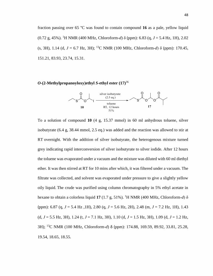
O-(2-Methylpropanoyloxy)ethyl S-ethyl ester (17)36
To a solution of compound 10 (4 g, 15.37 mmol) in 60 ml anhydrous toluene, silver
isobutyrate (6.4 g, 38.44 mmol, 2.5 eq.) was added and the reaction was allowed to stir at
RT overnight. With the addition of silver isobutyrate, the heterogenous mixture turned
grey indicating rapid interconversion of silver isobutyrate to silver iodide. After 12 hours
the toluene was evaporated under a vacuum and the mixture was diluted with 60 ml diethyl
ether. It was then stirred at RT for 10 mins after which, it was filtered under a vacuum. The
filtrate was collected, and solvent was evaporated under pressure to give a slightly yellow
oily liquid. The crude was purified using column chromatography in 5% ethyl acetate in
hexane to obtain a colorless liquid 17 (1.7 g, 51%). 1H NMR (400 MHz, Chloroform-d) δ
(ppm): 6.87 (q, J = 5.4 Hz ,1H), 2.80 (q, J = 5.6 Hz, 2H), 2.48 (m, J = 7.2 Hz, 1H), 1.43
(d, J = 5.5 Hz, 3H), 1.24 (t, J = 7.1 Hz, 3H), 1.10 (d, J = 1.5 Hz, 3H), 1.09 (d, J = 1.2 Hz,
3H); 13C NMR (100 MHz, Chloroform-d) δ (ppm): 174.88, 169.59, 89.92, 33.81, 25.28,
19.54, 18.65, 18.55.

49
[(2-Methylpropanoyl)oxy]ethyl chloroformate (18)36
Compound 17 (1.7 g,7.7 mmol) was cooled to -30 °C in a dry ice-acetonitrile cooling bath
and stirred for 5 mins. A solution of sulfuryl chloride (1.3 g, 10.03 mmol, 1.3 eq.) in 5 ml
dichloromethane was added dropwise to compound 17 and stirred at -30 °C over 10 mins
using a canula under N2 atmosphere. The reaction was stirred at that temperature for 45
mins. The completion of the reaction was monitored by NMR at intervals of 15 mins. On
completion of the reaction, the byproduct, sulfenyl chloride, and excess sulfuryl chloride
were distilled out under a vacuum. The crude product was then purified using vacuum
distillation and fractions were collected at a temperature range starting from 40-80 °C. The
fraction passing over 60 °C was found to contain compound 18 as a pale, yellow liquid
(0.58 mg, 41%). 1H NMR (400 MHz, Chloroform-d) δ (ppm): 6.75 (q, J = 5.4 Hz, 1H),
2.52 (m, J = 7.1 Hz, 1H), 1.52 (d, J = 5.5 Hz, 3H), 1.13 (d, J = 3.0 Hz, 3H), 1.11 (d, J =
3.2 Hz, 3H); 13C NMR (100 MHz, Chloroform-d) δ (ppm): 174.60, 148.82, 93.57, 33.72,
19.16, 18.59.

50
N-[(Acetoxy)methyloxy]carbonyl-2’,3’-bis-O-(tert-butyldimethylsilyl)-MTA (19)
To a stirred solution of compound 6 (200 mg, 0.38 mmol) in 3 ml anhydrous
dichloromethane, 1-methylimidazole (249 mg, 3.04 mmol, 8 eq.) was added and the
solution was stirred at 0 °C for 10 mins. A solution of chloroformate 12 (347 mg, 2.28
mmol, 6 eq.) in 2 ml dichloromethane was added dropwise to compound 6 in intervals of
2 hours, each time adding 2 eq. at 0 °C. Once the addition of the chloroformate was
complete, the reaction mixture was allowed to stir at RT for 24 hours. The progress of the
reaction was monitored by TLC and LCMS. On completion of the reaction, the reaction
mixture was diluted with 20 ml dichloromethane and the organic layer was washed with
brine and saturated NH4Cl once. The organic layer was dried over Na2SO4, filtered and the
solvent was evaporated under reduced pressure to give a yellow colored oily crude product.
The crude was purified using column chromatography in 5% methanol in dichloromethane
to give a colorless white solid 19 (21 mg, 9%). 1H NMR (400 MHz, Chloroform-d) δ (ppm):
8.80 (s, 1H), 8.21 (s, 1H), 8.19 (s, 1H), 5.97 (d, J = 5.5 Hz, 1H), 5.93 (s, 2H), 5.09 – 5.05
(m, 1H), 4.33 – 4.30 (m, 1H), 4.28 (dd, J = 6.7, 3.1 Hz, 1H), 3.06 (dd, J = 14.1, 6.7 Hz,
1H), 2.90 (dd, J = 14.0, 5.6 Hz, 1H), 2.18 (s, 3H), 2.17 (s, 3H), 0.97 (s, 9H), 0.80 (s, 9H),
0.17 (d, J = 9.6 Hz, 6H), -0.05 (s, 3H), -0.30 (s, 3H) ; 13C NMR (100 MHz, Chloroform-d)
δ (ppm): 173.21, 151.62, 147.52, 145.31,143.68, 140.72, 122.16, 92.46, 88.62, 86.41,

51
83.07, 70.19, 36.43, 20.63, 16.84, 14.33 -4.21, -4.62, -5.36, -5.42; LCMS (ESI+): 642.27
[M + H]+.
N-[(Acetoxy)methyloxy]carbonyl-MTA (LH1210)
To a solution of compound 19 (21 mg, 0.032 mmol) in anhydrous THF, a 1M solution of
TBAF in THF (17 mg, 0.065 mmol, 2 eq.) was added dropwise at 0 °C and stirred for 2
hours at RT. The reaction progress was monitored using TLC and LCMS. On
disappearance of the starting material, the solvent was evaporated, and the reaction mixture
was diluted with 5 ml dichloromethane and washed once with brine. The crude product
was purified using 10% methanol in dichloromethane to obtain a sticky white solid
LH1210 (4 mg, 30%). 1H NMR (400 MHz, Methanol-d4) δ (ppm): 8.71 (s, 1H), 8.34 (bs,
1H), 8.12 (s, 1H), 5.91 (d, J = 5.7 Hz, 1H), 5.84 (s, 2H), 4.74 (t, J = 5.5 Hz, 1H), 4.43 (t, J
= 6.0 Hz, 1H), 4.28 (q, J = 6.0 Hz, 1H), 2.84 (dd, J = 14.2 Hz, 5.5 Hz, 1H), 2.72 (dd, J =
14.0 Hz, 5.5 Hz, 1H), 2.16 (s, 3H), 2.08 (s, 3H); 13C NMR (100 MHz, Methanol- d4) δ
(ppm): 171.22, 152.81, 149.63, 147.31, 145.44, 140.72, 122.42, 88.65, 82.53, 76.14, 70.83,
68.11, 35.47, 15.24.

52
N-[(2-Methylpropanoyloxy)methyloxy]carbonyl-2’,3’-bis-O-(tert-
butyldimethylsilyl)-MTA (20)
To a solution of compound 6 (200 mg, 0.3 mmol) in 3 ml anhydrous dichloromethane, 1-
methylimidazole (249 mg, 3.04 mmol, 8 eq.) was added and the solution was stirred at 0
°C for 10 mins. A solution of chloroformate 14 (411 mg, 2.28 mmol, 6 eq.) in 2 ml
dichloromethane was added dropwise to compound 6 in intervals of 2 hours, each time
adding 2 eq. at 0 °C. Once the addition of the chloroformate was complete, the reaction
mixture was allowed to stir at RT for 24 hours. The progress of the reaction was monitored
by TLC and LCMS. The reaction mixture was diluted with 20 ml dichloromethane and the
organic layer was washed with brine and saturated NH4Cl once. The organic layer was
dried over Na2SO4, filtered and the solvent was evaporated under reduced pressure to give
a yellow colored oily crude product. The crude was purified using column chromatography
in 5% methanol in dichloromethane to give a colorless white solid 20 (36 mg, 14%). 1H
NMR (400 MHz, Chloroform-d) δ (ppm): 8.80 (s, 1H), 8.22 (s, 1H), 8.19 (s, 1H), 5.97 (d,
J = 5.5 Hz, 1H), 5.94 (s, 2H), 5.07 (d, J = 4.3 Hz, 1H), 4.32 (d, J = 3.0 Hz, 1H), 4.30 – 4.27
(m, 1H), 3.06 (dd, J = 14.0, 6.6 Hz, 1H), 2.90 (dd, J = 13.9, 5.5 Hz, 1H), 2.65 (sept, J = 7.1
Hz, 1H), 2.18 (s, 3H), 1.23 (d, J = 6.9 Hz, 6H) . 13C NMR (100 MHz, Chloroform-d) δ
(ppm): 13C NMR (100 MHz, Chloroform-d) δ 175.29, 152.40, 151.17, 148.58, 143.90,

53
143.18, 119.53, 89.52, 84.84, 82.31, 75.07, 74.13, 36.62, 34.38, 33.78, 33.69, 18.64, 18.54,
18.20, 18.01, 17.82, 16.68, -4.40, -4.58, -4.63, -5.08; LCMS (ESI+): 670.28 [M + H]+.
N-[(2-Methylpropanoyloxy)methyloxy]carbonyl-MTA(LH1211)
To a solution of compound 20 (36 mg, 0.053 mmol) in anhydrous THF, a 1M solution of
TBAF in THF (18.26 mg, 0.069 mmol, 2 eq.) was added dropwise at 0 °C and stirred for 2
hours at RT. The reaction progress was monitored using TLC and LCMS. On the
disappearance of the starting material, the solvent was evaporated, and the reaction mixture
was diluted with 5 ml dichloromethane and washed once with brine. The crude product
was purified in 10% methanol in dichloromethane to obtain a sticky white solid LH1211
(6 mg, 26%). 1H NMR (400 MHz, Methanol- d4) δ (ppm): 8.82 (s, 1H), 8.31 (bs, 1H), 8.24
(s, 1H), 6.01 (d, J = 5.7 Hz, 1H), 5.91 (s, 2H), 5.14 (t, J = 5.5 Hz, 1H), 4.47 (t, J = 6 Hz,
1H), 4.31 (q, J = 6.4 Hz, 1H), 3.17 (dd, J = 14.1 Hz, 5.3 Hz, 1H), 2.85 (dd, J = 14.0 Hz,
5.5 Hz, 1H), 2.25 (s, 3H), 1.54 (d, J = 3.0 Hz, 3H), 1.50 (d, J = 3.0 Hz, 3H); 13C NMR (100
MHz, Methanol- d4) δ (ppm):172.65, 151.82, 148.32, 147.19, 146.32, 144.72, 121.85,
88.46, 84.27, 76.23, 70.84, 67.28, 35.61, 32.48, 17.28, 13.88.

54
N-[(Acetoxy)ethyloxy]carbonyl-2’,3’-bis-O-(tert-butyldimethylsilyl)-MTA (21)
To a solution of compound 6 (200 mg, 0.38 mmol) in 3 ml anhydrous dichloromethane, 1-
methylimidazole (249 mg, 3.04 mmol, 8 eq.) was added and the solution was stirred at 0
°C for 10 mins. A solution of chloroformate 16 (379 mg, 2.28 mmol, 6 eq.) in 2 ml
dichloromethane was added dropwise to compound 6 in intervals of 2 hours, each time
adding 2 eq. at 0 °C. Once the addition of the chloroformate was complete, the reaction
mixture was allowed to stir at RT for 24 hours. The progress of the reaction was monitored
by TLC and LCMS. The reaction mixture was diluted with 20 ml dichloromethane and the
organic layer was washed with brine and saturated NH4Cl once. The organic layer was
dried over Na2SO4 and filtered. The solvent was evaporated under reduced pressure to give
a yellow colored oily crude product. The crude was purified using column chromatography
in 5% methanol in dichloromethane to give a colorless white solid 21 (18 mg, 10%). 1H
NMR (400 MHz, Chloroform-d) δ (ppm): 8.10 (s, 1H), 8.04 (bs, 1H), 7.98 (s, 1H), 6.75 (q,
J = 5.3 Hz, 1H), 6.01 (d, J = 5.2 Hz, 1H), 4.36 (t, J = 5.5 Hz, 1H), 4.22 (t, J = 6.1 Hz, 1H),
4.16 (q, J = 6.4 Hz, 1H), 3.12 (dd, J = 14.3 Hz, 5.5 Hz, 1H), 2.84 (dd, J = 14.2 Hz, 5.5 Hz,
1H), 2.13 (s, 3H), 1.54 (d, J = 5.6 Hz, 3H), 1.31 (s, 3H), 1.04 (s, 9H), 0.75 (s, 9H), 0.52
(d, J = 7 Hz, 6H), -0.15 (s, 3H), - 0.36 (s, 3H). 13C NMR (100 MHz, Chloroform-d) δ
(ppm): 170.34, 151.42, 148.62, 147.31, 146.38, 144.92, 122.28, 88.41, 86.27, 82.19, 84.76,

55
80.11, 35.42, 25.85, 23.64, 18.36, 15.48, 14.27, -4.14, -4.28, -5.36, -5.62; LCMS (ESI+):
656.23 [M + H]+.
N-[(Acetoxy)ethyloxy]carbonyl-MTA (LH1212)
To a solution of compound 21 (18 mg, 0.027 mmol) in anhydrous THF, a 1M solution of
TBAF in THF (14.12 mg, 0.054 mmol, 2 eq.) was added dropwise at 0 °C and stirred for 2
hours at RT. The reaction progress was monitored using TLC and LCMS. On the
disappearance of the starting material, the solvent was evaporated, and the reaction mixture
was diluted with 5 ml dichloromethane and washed once with brine. The crude product
was purified in 10% methanol in dichloromethane to obtain a sticky white solid LH1212
(6 mg, 55%). 1H NMR (400 MHz, Methanol- d4) δ (ppm): 8.15 (s, 1H), 8.05 (bs, 1H), 7.84
(s, 1H), 6.14 (d, J = 5.7 Hz, 1H), 5.92 (q, J = 6.3 Hz, 1H), 4.32 (t, J = 5.4 Hz, 1H), 4.17 (t,
J = 6.3 Hz, 1H), 4.06 (q, J = 6.1 Hz, 1H), 2.84 (dd, J = 14.0 Hz, 5.5 Hz, 1H), 2.65 (dd, J =
14.2 Hz, 5.5 Hz, 1H), 2.06 (s, 3H), 1.43 (d, J = 5.4 Hz, 3H), 1.28 (s, 3H); 13C NMR (100
MHz, Methanol- d4) δ (ppm): 170.32, 151.41, 148.63, 147.33, 146.28, 144.81, 122.34,
88.36, 86.21, 72.18, 70.38, 35.55, 14.29.

56
N-[(2-Methylpropanoyloxy)ethyloxy]carbonyl-2’,3’-bis-O-(tert-butyldimethylsilyl)-
MTA (22)
To a solution of compound 6 (200 mg, 0.38 mmol,) in 3 ml anhydrous dichloromethane,
1-methylimidazole (249 mg, 3.04 mmol, 8 eq.) was added and the solution was stirred at 0
°C for 10 mins. A solution of chloroformate 18 (411 mg, 2.28 mmol, 6 eq.) in 2 ml
dichloromethane was added dropwise to compound 6 in intervals of 2 hours, each time
adding 2 eq. at 0 °C. Once the addition of the chloroformate was complete, the reaction
mixture was allowed to stir at RT for 24 hours. The progress of the reaction was monitored
by TLC and LCMS. The reaction mixture was diluted with 20 ml dichloromethane and the
organic layer was washed with brine and saturated NH4Cl once. The organic layer was
dried over Na2SO4 and filtered. The solvent was evaporated under reduced pressure to give
a yellow colored oily crude product. The crude was purified using column chromatography
in 5% methanol in dichloromethane to give a colorless white solid 22 (22 mg, 21%). 1H
NMR (400 MHz, Chloroform-d) δ (ppm): 8.79 (s, 1H), 8.18 (s, 1H), 6.99 (q, J = 5.2 Hz,
1H), 5.96 (d, J = 5.5 Hz, 1H), 5.08 (q, J = 5.3 Hz, 1H), 4.34 – 4.27 (m, 2H), 3.06 (dd, J =
16.7, 6.5 Hz, 1H), 2.90 (dd, J = 13.7, 5.5 Hz, 1H), 2.60 (dt, J = 14.0, 6.9 Hz, 1H), 2.18 (s,
3H), 1.61 (dd, J = 5.3, 1.8 Hz, 3H), 1.24 – 1.19 (m, 6H), 0.97 (s, 9H), 0.80 (s, 9H), 0.16 (d,
J = 9.5 Hz, 6H), -0.05 (s, 3H), -0.30 (s, 3H); 13C NMR (100 MHz, Chloroform-d) δ (ppm):

57
174.56, 150.76, 149.62, 148.27, 146.24, 144.29, 122.38, 92.84, 88.52, 86.42, 77.31, 76.42,
35.47, 33.71, 25.67, 19.27, 18.14, 17.57, 14.82, -4.32, -4.67, -5.21, -5.33; LCMS (ESI+):
684.35 [M+H]+ .
N-[(2-Methylpropanoyloxy)ethyloxy]carbonyl-MTA (LH1213)
To a solution of compound 22 (22 mg, 0.032 mmol) in anhydrous THF, a 1M solution of
TBAF in THF (16.71 mg, 0.064 mmol, 2 eq.) was added dropwise at 0 °C and stirred for 2
hours at RT. The reaction progress was monitored using TLC and LCMS. On the
disappearance of the starting material, the solvent was evaporated, and the reaction mixture
was diluted with 5 ml dichloromethane and washed once with brine. The crude product
was purified in 10% methanol in dichloromethane to obtain a sticky white solid LH1213
(8 mg, 56%). 1H NMR (400 MHz, Methanol- d4) δ (ppm): 8.71 (s, 1H), 8.25 (bs, 1H), 8.02
(s, 1H), 5.96 (d, J = 5.7 Hz, 1H), 5.72 (q, J = 6.4 Hz, 1H), 4.46 (t, J = 5.2 Hz, 1H), 4.32 (t,
J = 6.1 Hz, 1H), 4.18 (q, J = 6.0 Hz, 1H), 2.72 (dd, J = 14.1 Hz, 5.5 Hz, 1H), 2.53 (dd, J =
14.2 Hz, 5.5 Hz, 1H), 1.95 (s, 3H), 1.68 (d, J = 5.6 Hz, 3H), 1.24 (d, J = 3.2 Hz, 3H), 1.15
(d, J = 3.1 Hz, 3H); 13C NMR (100 MHz, Methanol- d4) δ (ppm): 174.31, 150.14, 149.35,
148.22, 147.85, 146.73, 122.17, 88.31, 86.42, 71.22, 70.54, 35.61, 33.29, 19.72, 18.34,
15.44.

58
Chloromethyl 4-nitrophenyl ester (23)37
A solution of 4-nitrophenol (600 mg, 4.3 mmol) in 2 ml dichloromethane was cooled in an
ice bath and N,N-Diisopropylethylamine (557 mg, 4.3 mmol) was added dropwise at 0 °C.
A solution of chloromethyl chloroformate (556 mg, 4.3 mmol,) in 8 ml dichloromethane
was added to the solution and the reaction mixture was stirred overnight at RT. The
progress of the reaction was monitored by TLC. On completion of the reaction, the reaction
mixture was diluted with 70 ml dichloromethane and extracted with 30 ml water. The
organic layer was then washed once with 25 ml sat. NaHCO3 and 30 ml brine. The
dichloromethane layer was collected, dried over Na2SO4 and filtered. The solvent
evaporated to give compound 23 (882 mg, 88%) as a white solid that was used in the next
step without further purification. 1H NMR (400 MHz, Chloroform-d) δ (ppm): 8.26 (dd, J
= 14.3 Hz, 9.1 Hz, 2H), 7.40 (dd, J = 28.1 Hz, 9.4 Hz, 2H), 5.8 (s, 2H); 13C NMR (100
MHz, Chloroform-d) δ (ppm):154.84, 145.92, 125.55, 125.45, 121.66, 72.69.

59
(Acetyloxy)methyl 4-nitrophenyl carbonate (24a)37
A solution of ZnO (90.67 mg, 1.11 mmol, 3 eq.) and glacial acetic acid (44 mg, 0.74 mmol,
2 eq.) was stirred in toluene at 105 °C for 1 hour. The temperature was lowered to 60 °C
and compound 23 (86 mg, 0.37 mmol) was added to the solution, followed by the addition
of NaBr (61.12 mg, 0.59 mmol, 1.6 eq.). The reaction mixture was further stirred at that
temperature for 12 hours and the water generated, during the reaction, was removed using
a Dean Stark apparatus. The progress of the reaction was monitored by TLC. The desired
product was not obtained through this route and the starting material was recovered.
(Acetyloxy)methyl 4-nitrophenyl carbonate (24b)37
To the solution of glacial acetic acid (3.36 mg, 0.05 mmol, 1.3 eq.) and TBAHSO4 (20 mg,
0.05 mmol, 1.3 eq.), NaHCO3 (9.8 mg, 0.11 mmol, 2.7 eq.) in dichloromethane: H2O (2:1)
was added and stirred at RT for 2 hours. To the above solution, compound 23 (10 mg, 0.04
mmol) was added at 0 °C and the reaction mixture was stirred at RT for 18 hours. The

60
progress of the reaction was monitored by TLC. The desired product was not obtained
through this route and starting material was recovered.
(Acetyloxy)methyl 4-nitrophenyl carbonate (24c)37
To a solution of compound 23 (500 mg, 2.15 mmol) in acetone, NaI (648 mg, 4.31 mmol,
2 eq.) and NaHCO3 (18.13 mg, 0.21 mmol, 0.1 eq.) were added and the reaction mixture
was stirred overnight at RT. The progress of the reaction was monitored by TLC. On
completion of the reaction, the acetone was evaporated under reduced pressure, 10 ml
diethyl ether was added to the residue and the mixture was filtered. The filtrate was
evaporated under reduced pressure and the residue was diluted with 30 ml dichloromethane
and washed twice with 10 ml 20% Na2S2O3 and 10 ml brine. The organic layer was
collected, dried over Na2SO4 and the solvent was then evaporated under a vacuum. The
crude product was purified using 5% ethyl acetate in hexane to give a white solid (698 mg,
80%). The solid obtained was reacted with silver acetate (721 mg, 4.32 mmol, 2 eq.) in
toluene at RT for 12 hours. On completion of the reaction, the toluene was evaporated,
diethyl ether was added, and the solution was then filtered under a vacuum. The filtrate
was evaporated to give a crude product which was further purified in 10% ethyl acetate in
hexane to give compound 24c as a white solid (164 mg, 68%). 1H NMR (400 MHz,
Chloroform-d) δ (ppm): 8.21 (dd, J = 14.2 Hz, 9.1 Hz, 2H), 7.22 (dd, J = 28.0 Hz, 9.2 Hz,

61
2H), 6.2 (s, 2H), 2.04 (s, 3H); 13C NMR (100 MHz, Chloroform-d) δ (ppm):168.33, 155.38,
153.82, 145.35, 125.19, 122.42, 76.62, 21.08.
2’,3’-bis-O-(Trimethylsilyl)-MTA (25)
To a solution of MTA (50 mg, 0.16 mmol) in anhydrous dichloromethane, 1-
methylimidazole (552.10 mg, 6.7 mmol, 40 eq.) and DMAP (10.27 mg, 0.08 mmol, 0.5
eq.) were added and allowed to cool to 0 °C in an ice bath. To the above solution, a
solution of trimethylsilyl chloride (366 mg, 3.36 mmol, 20 eq.) in dichloromethane was
added and stirred at RT. The reaction progress was monitored by LCMS. Presence of
starting material was detected after 24 hours along with the presence of compound 25.
LCMS (ESI+): 442.50 [M+H]+.
2’,3’-bis-O-(Isopropylidene)-MTA (26)

62
To a solution of MTA (30 mg, 0.1 mmol) in 5 ml acetone, p-toluenesulfonic acid (9.6
mg, 0.05 mmol, 0.5 eq.) was added followed by the addition of anhydrous CuSO4 (32 mg,
0.2 mmol, 2 eq.) and the reaction mixture was stirred at RT for 24 hours. The reaction
mixture was diluted with 20 ml dichloromethane and washed with 10 ml sat. NaHCO3.
The organic layer was dried over Na2SO4 and filtered. The solvent was evaporated to
give a crude product which was further purified in 10% methanol in dichloromethane to
give compound 26 as a white solid (9 mg, 33%). 1H NMR (400 MHz, Chloroform-d) δ
(ppm): 8.42 (s, 1H), 7.86 (s, 1H), 6.02 (d, J = 5.4 Hz, 1H), 5.47 (t, J = 5.5 Hz, 1H), 5.01
(t, J = 5.2 Hz, 1H), 4.48 (q, J = 5.7 Hz, 1H), 2.76 (dd, J = 14.2 Hz, 5.5 Hz, 1H), 2.62 (dd,
J = 14.0 Hz, 6.0 Hz, 1H), 2.01 (s, 1H), 1.54 (s, 1H), 1.32 (s, 1H) ; LCMS (ESI+): 338.11
[M+H]+.
2’,3’-bis-O-(di-tert-butylsilylene)-MTA (27)
To a solution of MTA (8 mg, 0.02 mmol) in 0.5 ml DMF, triethylamine (6 mg, 0.06 mmol,
3 eq.) was added at 0 °C and then, di-tert-butylsilylbis(trifluoromethanesulfonate) (23 mg,
0.04 mmol, 2 eq.) was added dropwise. The reaction mixture was stirred at RT overnight.
DMAP (2 mg, 0.01 mmol, 0.5 eq.) was added as a catalyst. The reaction progress was

63
monitored by TLC and LCMS, however, only the presence of starting material was
observed.
N-[(Acetoxy)methyloxy]carbonyl-2’,3’-bis-O-(tert-butyldimethylsilyl)-MTA (28)
To a solution of compound 6 (100 mg, 0.19 mmol) in 5 ml dichloromethane, DIPEA (50
mg, 0.38 mmol, 2 eq.) was added and stirred at RT for 5 mins. A solution of compound
24c (96 mg, 0.38 mmol, 2 eq.) in 1 ml dichloromethane was added dropwise to the above
solution and stirred for 12 hours at RT. The reaction progress was monitored by TLC and
LC-MS. After 48 hours, no conversion was seen.
N-[(2-Methylpropanoyloxy)ethyloxy]carbonyl-MTA (29)
To a solution of compound 22 (2 mg, 0.0029 mmol) in 0.524 ml of THF, 5.84 μl (0.2 eq.)
of a 0.1 M TBAF buffered solution prepared by diluting 1M TBAF in THF 10 times with
0.1 M K2HPO4 buffer (pH=7.1) was added. The reaction mixture was monitored by

64
LCMS for progress of reaction for 2 hours at RT and the formation of MTA (LCMS
(ESI+): 298.32 [M+H]+) was observed in less than 1 hour.
N-[(2-Methylpropanoyloxy)ethyloxy]carbonyl-MTA (30)
To a solution of compound 22 (2 mg, 0.0029 mmol) in 0.524 ml of THF, 5.84 μl (0.2 eq.)
of a 0.1 M TBAF buffered solution prepared by diluting 1M TBAF in THF 10 times with
0.1 M K2HPO4 buffer (pH=7.4) was added. The reaction mixture was monitored by
LCMS for progress of reaction for 2 hours at RT and the formation of MTA (LCMS
(ESI+): 298.32 [M+H]+) was observed in less than 1 hour.
N-[(2-Methylpropanoyloxy)ethyloxy]carbonyl-MTA (31)
To a solution of compound 22 (2 mg, 0.0029 mmol) in 0.524 ml of THF,5.84 μl (0.2 eq.)
of a 0.1 M TBAF buffered solution prepared by diluting 1M TBAF in THF 10 times with
0.1 M K2HPO4 buffer (pH=6.7) was added. The reaction mixture was monitored by

65
LCMS for progress of reaction for 24 hours at RT. Presence of mono-deprotected form of
compound 22 (LCMS (ESI+): 570.41 [M+H]+) was observed.
N-[(2-Methylpropanoyloxy)ethyloxy]carbonyl-MTA (32)
To a solution of compound 24 (2 mg, 0.0029 mmol) in 0.58 ml DMSO, 8.75 μl of a 0.67
M CsF solution prepared in DMSO:MeOH=1: 2 was added. The progress of the reaction
was monitored by LCMS and stirred at RT for 2 hours and the presence of MTA (LCMS
(ESI+): 298.32 [M+H]+) was detected in less than 1 hour.
N-[(2-Methylpropanoyloxy)ethyloxy]carbonyl-MTA (33)
To a solution of compound 22 (2 mg, 0.0029 mmol) in 0.58 ml DMSO, 8.75 μl of a 0.67
M CsF solution prepared in DMSO:MeOH=1:2 was added. To the above solution, 5.8 μl
of phosphate buffer, pH =7.1 was added. The progress of the reaction was monitored by

66
LCMS. No byproduct MTA was detected with complete conversion to desired product in
2 hours. LCMS (ESI+): 456.13 [M+H]+ .
Preparation of 200 ml 50 mM Phosphate buffer containing 1 mM EDTA
NaH2PO4 (1.38 g, 10 mM) and EDTA (74.4 mg, 0.1 mM) were dissolved in 180 ml
deionized water. The pH was first adjusted to 7.4 with 5 N NaOH and then with 1N NaOH.
The volume was adjusted to 200 ml and the buffer was filtered and stored at 4 °C.
Stability Assay of MTA prodrugs in Phosphate Buffer
The stock solution of the prodrug (10 mM in DMSO) was diluted 10-fold to give a 1 mM
working solution in 10% DMSO. 100 μl of the 1 mM solution was added to 875 μl of 50
mM phosphate buffer containing 1 mm EDTA, pH 7.4 and incubated at 37 °C for 5 minutes
in an eppendorf vial. The vial was then centrifuged at a speed of 9 x 1000g for 5 minutes
and the supernatant was split into 3 fractions of 292.5 μl, one for stability in phosphate
buffer, one for activation in human liver microsomes and one for activation in mouse liver
microsomes.
The fraction for analyzing stability was mixed with 7.5 μl of deionized water and incubated
at 37 °C for 3 days. Time points were collected after every 0 min, 3 min, 5 min, 10 min, 15
min, 30 min, 45 min, 1hr, 3 hr, 5 hr, 8 hr, 10 hr, 12 hr, 24 hr, 48 hr, 72 hr. At each time
point, 20 μl aliquots were taken and diluted with 40 μl cold acetonitrile and centrifuged at
9 x 1000g for 5 mins. 40 μl of the supernatant were taken and diluted with 40 μl deionized

67
water and frozen before injecting on the HPLC. The samples were analyzed using a Perkin
Elmer Series 2000 HPLC with a Phenomenex Kinetix C18 (50 x 4.6 mm) column and a
gradient of 10 - 90% acetonitrile containing 0.1% TFA in 10 min at a flowrate of 0.6
mL/min was used with detection wavelength of 220 and 254 nm.
In-vitro Activation Assay in Human and Mouse Liver Microsomes38
The mouse and human liver microsomes were obtained from Xeno Tech with a protein
concentration of 20 mg/ml and stored in eppendorf vials at – 70 °C. Stock solution of 1
mM compound in 1 ml DMSO was diluted 10-fold to give a 1 mM solution in 10% DMSO.
100 μl of the 1 mM solution was added to 875 μl of 50 mM phosphate buffer containing 1
mm EDTA, pH 7.4 and incubated at 37 °C for 5 minutes in an eppendorf vial. The vial was
then centrifuged at a speed of 9 x 1000g for 5 minutes and the supernatant was split into 3
fractions of 292.5 μl, one for stability in phosphate buffer, one for activation in human liver
microsomes and one for activation in mouse liver microsomes. The fraction for analyzing
activation was mixed with 7.5 μl of either the human or mouse liver microsomes and
incubated at 37 °C for 3 days. Time points were collected after every 0 min, 3 min, 5 min,
10 min, 15 min, 30 min, 45 min, 1hr, 3 hr, 5 hr, 8 hr, 10 hr, 12 hr, 24 hr, 48 hr, 72 hr. At
each time point, 20 μl aliquots were taken and quenched with 40 μl cold acetonitrile and
stored on ice for 30 mins before centrifugation at 9 x 1000g for 5 mins. 40 μl of the
supernatant were taken and diluted with 40 μl deionized water and frozen before injecting
on the HPLC. The samples were analyzed using a Perkin Elmer Series 2000 HPLC with a
Phenomenex Kinetix C18 (50 x 4.6 mm) column and a gradient of 10 - 90% acetonitrile

68
containing 0.1% TFA in 10 min at a flowrate of 0.6 mL/min was used with a detection
wavelength of 220 and 254 nm. The half-life was calculated from the first order rate
equation of the conversion of prodrug to MTA.
In-vitro Oxidative Activation Assay in presence of NADPH
Stock solution (10 mM in DMSO) was diluted 10-fold to give a solution of 1 mM in 10%
DMSO. 100 μl of the 1 mM solution was added to 875 μl of 50 mM phosphate buffer
containing 1 mm EDTA, pH 7.4 and incubated at 37 °C for 5 minutes in an eppendorf vial.
The vial was then centrifuged at a speed of 9 x 1000g for 5 minutes and the supernatant
was split into 2 fractions, each of 292.5 μl, one for oxidative activation in mouse liver
microsomes and one for human liver microsomes. Each of the fractions was mixed with 20
μl of NADPH and either 20 μl of mouse liver microsomes or human liver microsomes and
incubated at 37 °C for 3 days. Time points were collected after every 0 min, 3 min, 5 min,
10 min, 15 min, 30 min, 45 min, 1hr, 3 hr, 5 hr, 8 hr, 10 hr, 12 hr, 24 hr, 48 hr, 72 hr. At
each time point, 20 μl aliquots were taken and quenched with 40 μl cold acetonitrile and
stored on ice for 30 mins before centrifugation at 9 x 1000g for 5 mins. 40 μl of the
supernatant were taken and diluted with 40 μl deionized water and frozen before injecting
on the HPLC. The samples were analyzed using a Perkin Elmer Series 2000 HPLC with a
Phenomenex Kinetix C18 (50 x 4.6 mm) column and a gradient of 10 - 90% acetonitrile
containing 0.1% TFA in 10 min at a flowrate of 0.6 mL/min was used with a detection
wavelength of 220 and 254 nm. The half-life was calculated from the first order rate
equation of the conversion of prodrug to MTA.

69
In-vitro Activation Assay in 50 % Human Serum
Stock solution of compound (10 mM in DMSO) was diluted 10-fold to give a 1 mM
solution in 10 % DMSO. 100 μl of the above solution was added to 400 μl of 50 mM
phosphate buffer, vortexed and incubated at 37 °C for 5 mins. It was then centrifuged at 9
x 1000g for 5 mins and the supernatant was split into 2 fractions of 230 μl each, one for
control in H2O and other for activation in Human Serum. The control fraction was mixed
with 230 μl of water, while the other was mixed with 230 μl of Human Serum. Both
fractions were incubated at 37 °C for 2 days. Time points were calculated after every 0 min,
3 min, 5 min, 10 min, 15 min, 30 min, 45 min, 1hr, 3 hr, 5 hr, 8 hr, 10 hr, 12 hr, 24 hr, 48
hr. For every time point, 20 μl of aliquots were taken and diluted with 40 μl cold
Acetonitrile:Methanol=2:1, vortexed and stored on ice for 30 mins. The samples were then
centrifuged at 9 x 1000g for 5 mins and the supernatant was collected. 40 μl of the
supernatant were diluted with 40 μl of deionized water and stored at – 30 °C before
injection on the HPLC. The samples were analyzed using a Perkin Elmer Series 2000
HPLC with a Phenomenex Kinetix C18 (50x4.6mm) column and a gradient of 10 - 90%
acetonitrile containing 0.1% TFA in 10 min at a flowrate of 0.6 mL/min was used with a
detection wavelength of 220 and 254 nm. The half-life was calculated from the first order
rate equation of the conversion of prodrug to MTA.

70
REFERENCES
1. World Health Organization, Cancer, 2018.
2. National Cancer Institute, Risk Factors for cancer, 2015.
3. Kaye S. B. New antimetabolites in cancer chemotherapy and their clinical impact.
Br. J. Cancer 1998, 78, 1-7.
4. Visentin M.; Zhao R.; & Goldman. The Antifolates. The Antifolates. Hematol.
Oncol. Clin. N. Am. 2012, 26, 629-648.
5. Friedman J. M.; Weber-Schöndorfer C. Antineoplastic drugs. In Drugs During
Pregnancy and Lactation,3rd Ed, Schaefer C.; Peters P.; Miller R. Eds, Elsevier,
2015, 373-399.
6. Salser J. S.; Balis M. E. The Mechanism of Action of 6-Mercaptopurine: I.
Biochemical Effects. Cancer Res. 1965, 25, 539-543.
7. Rahman M.; Hasan M. R. Cancer metabolism and drug resistance. Metabolites 2015,
5, 571-600.
8. Brown C. Targeted therapy: an elusive cancer target. Nature 2016, 537, 106-108.
9. Lubin M.; Lubin A. Selective killing of tumors deficient in methylthioadenosine
phosphorylase: a novel strategy. PloS one 2009, 4, 5735.
10. Tang B.; Testa J. R.; Kruger W. D. Increasing the therapeutic index of 5-fluorouracil
and 6-thioguanine by targeting loss of MTAP in tumor cells. Cancer Biol. Ther.
2012, 13, 1082-1090.
11. Bertino J. R.; Waud W. R.; Parker W. B.; Lubin M. Targeting tumors that lack
methylthioadenosine phosphorylase (MTAP) activity: current strategies. Cancer
Biol. Ther. 2011, 11, 627-632.
12. Fitchen J. H.; Riscoe M. K.; Dana B. W.; Lawrence H. J.; Ferro A. J.
Methylthioadenosine phosphorylase deficiency in human leukemias and solid
tumors. Cancer Res. 1986, 46, 5409-5412.
13. Lubin A.; Lubin M. Therapy of tumors and infectious agents deficient in
methylthioadenosine phosphorylase. US Patent 12675276, 2014.
14. Batova A.; Diccianni M. B.; Omura-Minamisawa M.; Yu J.; Carrera C. J.;
Bridgeman L. J.; Kung F. H.; Pullen J.; Amylon M. D.; Alice L. Y. Use of alanosine
as a methylthioadenosine phosphorylase-selective therapy for T-cell acute
lymphoblastic leukemia in vitro. Cancer Res. 1999, 59, 1492-1497.
15. Chen Z.H.; Olopade O. I.; Savarese T. M. Expression of Methylthioadenosine
Phosphorylase cDNA inp16−, MTAP− Malignant Cells: Restoration of
Methylthioadenosine Phosphorylase-Dependent Salvage Pathways and Alterations
of Sensitivity to Inhibitors of Purine de novo Synthesis. Mol. Pharmacol. 1997, 52,
903-911.
16. Hori H.; Tran P.; Carrera C. J.; Hori Y.; Rosenbach M. D.; Carson D. A.; Nobori T.
Methylthioadenosine phosphorylase cDNA transfection alters sensitivity to
depletion of purine and methionine in A549 lung cancer cells. Cancer Res. 1996,
56, 5653-5658.
17. Kamatani N.; Nelson-Rees W. A.; Carson D. A. Selective killing of human
malignant cell lines deficient in methylthioadenosine phosphorylase, a purine
metabolic enzyme. Proc. Natl. Acad. Sci. U.S.A 1981, 78, 1219-1223.

71
18. Wolford R. W.; MacDonald M. R.; Zehfus B.; Rogers T. J.; Ferro A. J. Effect of 5′-
methylthioadenosine and its analogs on murine lymphoid cell proliferation. Cancer
Res. 1981, 41, 3035-3039.
19. Simile M. M.; Banni S.; Angioni E.; Carta G.; De Miglio M. R.; Muroni M. R.;
Calvisi D. F.; Carru A.; Pascale R. M.; Feo F. 5′-Methylthioadenosine
administration prevents lipid peroxidation and fibrogenesis induced in rat liver by
carbon-tetrachloride intoxication. J. Hepatol. 2001, 34, 386-394.
20. Wolford R.; Riscoe M.; Johnson L.; Ferro A.; Fitchen J. Effect of 5'-
methylthioadenosine (a naturally occurring nucleoside) on murine hematopoiesis.
Exp. Hematol. 1984, 12, 867-871.
21. Stramentinoli G.; Gennari F. Adenosine derivatives of anti-inflammatory and
analgesic activity, and therapeutic compositions which contain them as their active
principle. US Patent 06406010, 1984.
22. Moratti E. M. Pharmaceutical compositions containing 5'-deoxy-5'-
methylthioadenosine s-adenosylmethionine and their salts for reducing seborrhea.
US Patent 07492991, 1998.
23. Riscoe M.; Ferro A. 5-Methylthioribose. Its effects and function in mammalian cells.
J. Biol. Chem. 1984, 259, 5465-5471.
24. Russo G. L.; Della Ragione F.; Utili R.; Andreana A.; Ruggiero G.; Zappia V.
Studies on Human Serum 5′-Deoxy-5′-Methylthioadenosine Phosphorylase:
Molecular Properties and Clinical Perspectives. In Progress in Polyamine Research,
Springer, 1988, 229-238.
25. Kindler H. L.; Burris H. A.; Sandler A. B.; Oliff I. A. A phase II multicenter study
of L-alanosine, a potent inhibitor of adenine biosynthesis, in patients with MTAP-
deficient cancer. Invest. New Drugs 2009, 27, 75-81.
26. Efferth T.; Miyachi H.; Drexler H. G.; Gebhart E. Methylthioadenosine
phosphorylase as target for chemoselective treatment of T-cell acute lymphoblastic
leukemic cells. Blood Cells Mol. Dis. 2002, 28, 47-56.
27. Kung P.P.; Zehnder L. R.; Meng J. J.; Kupchinsky S. W.; Skalitzky D. J.; Johnson
M. C.; Maegley K. A.; Ekker A.; Kuhn L. A.; Rose P. W. Design, synthesis, and
biological evaluation of novel human 5′-deoxy-5′-methylthioadenosine
phosphorylase (MTAP) substrates. Bioorg. Med. Chem. Lett. 2005, 15, 2829-2833.
28. Bloom L.; Boritzki T.; Ogden R.; Skalitzky D.; Kung P.P.; Zehnder L.; Kuhn L.;
Meng J. Combination therapies for treating methylthioadenosine phosphorylase
deficient cells. US Patent 10367366, 2004.
29. Imai T.; Taketani M.; Shii M.; Hosokawa M.; Chiba K. Substrate specificity of
carboxylesterase isozymes and their contribution to hydrolase activity in human
liver and small intestine. Drug Metabol. Dispos. 2006, 34, 1734-1741.
30. Yang Y.H.; Aloysius H.; Inoyama D.; Chen Y.; Hu L. Enzyme-mediated hydrolytic
activation of prodrugs. Acta Pharm. Sin. B 2011, 1, 143-159.
31. Burkhart D. J.; Barthel B. L.; Post G. C.; Kalet B. T.; Nafie J. W.; Shoemaker R. K.;
Koch T. H. Design, synthesis, and preliminary evaluation of doxazolidine
carbamates as prodrugs activated by carboxylesterases. J. Med. Chem. 2006, 49,
7002-7012.

72
32. Hu L. Prodrug Approaches to Drug Delivery. In Drug Delivery: Principles and
Applications, 2nd Ed.; Wang B.;Siahann T.;Eds; John Wiley & Sons: Hoboken, New
Jersey, 2016, 227-271.
33. Desmoulin F.; Gilard V.; Malet-Martino M.; Martino R. Metabolism of
capecitabine, an oral fluorouracil prodrug: 19F NMR studies in animal models and
human urine. Drug Metabol. Dispos. 2002, 30, 1221-1229.
34. Moha-Lerman E. B.; Nidam T.; Cohen M.; Avhar-Maydan S.; Balanov A. Processes
for the preparation and purification of gabapentin enacarbil. US Patent 12626682,
2012.
35. DiLauro A. M.; Seo W.; Phillips S. T. Use of catalytic fluoride under neutral
conditions for cleaving silicon–oxygen bonds. J. Org. Chem. 2011, 76, 7352-7358.
36. Folkmann M.; Lund F. J. Acyloxymethyl carbonochloridates. New intermediates in
prodrug synthesis. Synthesis 1990, 1159-1166.
37. Gallop M. A.; Cundy K. C.; Zhou C. X.; Yao F.; Xiang J.N. Prodrugs of GABA
analogs, compositions and uses thereof. US Patent 10170127, 2004.
38. Shindoh H.; Nakano K.; Yoshida T.; Ishigai M. Comparison of in vitro metabolic
conversion of capecitabine to 5-FU in rats, mice, monkeys and humans-toxicological
implications. J. Toxicol. Sci. 2011, 36, 411-422.