Embed Size (px)
Citation preview

1 Design, Synthesis, and Use of MMP‑2 Inhibitor-Conjugated Quantum2 Dots in Functional Biochemical Assays3 Erika Bourguet,*,† Kristina Brazhnik,‡,§ Alyona Sukhanova,‡,§ Gautier Moroy,∥ Sylvie Brassart-Pasco,⊥
4 Anne-Pascaline Martin,⊥,# Isabelle Villena,# Georges Bellon,⊥ Janos Sapi,† and Igor Nabiev‡,§
5†Institut de Chimie Moleculaire de Reims, UMR 7312-CNRS, SFR Cap-Sante, UFR de Pharmacie, Universite de Reims
6 Champagne-Ardenne, 51 rue Cognacq Jay, 51100 Reims, France
7‡Laboratoire de Recherche en Nanosciences, LRN - EA4682, UFR de Pharmacie, Universite de Reims Champagne-Ardenne, 51 rue
8 Cognacq Jay, 51100 Reims, France
9§Laboratory of Nano-Bioengineering, National Research Nuclear University MEPhI (Moscow Engineering Physics Institute), 31
10 Kashirskoe shosse, 115409 Moscow, Russian Federation
11∥Molecules Therapeutiques In Silico, INSERM UMR-S 973, Universite Paris Diderot, Sorbonne Paris Cite, 35 rue Helene Brion,
12 75013 Paris, France
13⊥Laboratoire de Biochimie et de Biologie moleculaire, MEDyC, UMR CNRS/URCA 7369, SFR Cap-Sante, UFR de Medecine,
14 Universite de Reims Champagne-Ardenne, 51 rue Cognacq Jay, 51100 Reims, France
15#Laboratoire de Parasitologie-Mycologie, EA3800, SFR Cap-Sante, UFR de Medecine, Universite de Reims Champagne-Ardenne, 51
16 rue Cognacq-Jay, 51100 Reims, France
17 ABSTRACT: The development of chemically designed18 matrix metalloprotease (MMP) inhibitors has advanced the19 understanding of the roles of MMPs in different diseases. Most20 MMP probes designed are fluorogenic substrates, often21 suffering from photo- and chemical instability and providing22 a fluorescence signal of moderate intensity, which is difficult to23 detect and analyze when dealing with crude biological samples.24 Here, an MMP inhibitor that selectively inhibits MMP-2 more25 strongly than Galardin has been synthesized and used for26 enzyme labeling and detection of the MMP-2 activity. A27 complete MMP-2 recognition complex consisting of a biotinylated MMP inhibitor tagged with the streptavidin-quantum dot28 (QD) conjugate has been prepared. This recognition complex, which is characterized by a narrow fluorescence emission29 spectrum, long fluorescence lifetime, and negligible photobleaching, has been demonstrated to specifically detect MMP-2 in in30 vitro sandwich-type biochemical assays with sensitivities orders of magnitude higher than those of the existing gold standards31 employing organic dyes. The approach developed can be used for specific in vitro visualization and testing of MMP-2 in cells and32 tissues with sensitivities significantly exceeding those of the best existing fluorogenic techniques.
33 ■ INTRODUCTION
34 Studying biological processes at the molecular level within a35 living cell is a major challenge for cell biology investigations.1,2
36 Similarly, spatiotemporal tracking of small molecules in ex vivo37 or in vivo environments gives better insight into their38 interactions with proteins, rendering target identification39 more efficacious and, in f ine, accelerating drug development.40 The existing techniques based on spectroscopic and biooptical41 methods using organic dyes are mostly limited by the42 difficulties in imaging individual molecules in the optically43 noisy cellular environment and accessing directly the interior of44 living cells.3 As promising alternative tools, semiconductor45 nanocrystal quantum dots (QDs) have emerged for numerous46 biomedical applications,4,5 such as cellular labeling, biochemical47 sensing, probing biocatalyzed reactions, and drug delivery.6
48 QDs are characterized by their narrow composition and size49 dependent emission wavelength,7 extreme brightness, rock-
50solid photostability, and chemical robustness.8 QD fluorescence51covers the optical spectrum from the near-UV to the IR, which52provides a unique possibility for multiplexing, unlike with the53use of organic dyes.9 Even if QDs possess optical properties for54biological applications, nanoparticles need to be functionalized55to achieve a convenient solubility in aqueous media, and in f ine,56hydrophilic ligands may be used as anchor points for57biomolecules such as peptides, DNA, or drugs.10 The58hydrophobic outer shell of QDs may be exchanged or59chemically reacted with bi- or multifunctional molecules60containing capping ligands, cationic polymers, or liposomes61bearing a cell penetrating peptide (CPP),11,12 or with62amphiphilic polymers.13,14 Biomolecules may be attached to
Received: February 3, 2016Revised: March 1, 2016
Article
pubs.acs.org/bc
© XXXX American Chemical Society A DOI: 10.1021/acs.bioconjchem.6b00065Bioconjugate Chem. XXXX, XXX, XXX−XXX
ajk00 | ACSJCA | JCA10.0.1465/W Unicode | research.3f (R3.6.i11:4432 | 2.0 alpha 39) 2015/07/15 14:30:00 | PROD-JCA1 | rq_5070516 | 3/07/2016 14:04:08 | 15 | JCA-DEFAULT

63 their surface by covalent binding via different surface functional64 groups (amino, thiol, carboxyl, etc.), or through noncovalent65 streptavidin/biotin, electrostatic or ionic interactions.15,16
66 Matrix metalloproteinases (MMPs) belong to the family of67 zinc-dependent proteases involved in the degradation and68 remodelling of extracellular matrix proteins. The activity of69 MMPs is controlled by endogenous tissue inhibitors of70 metalloproteinases (TIMPs). The finely tuned MMP/TIMP71 balance found in normal physiological processes (tissue72 modeling, angiogenesis, wound healing, embryonic growth,73 etc.) is disrupted in several pathologies such as atherosclerosis,74 aneurysm, pulmonary emphysema, and cancer. Among more75 than 25 MMPs, MMP-2 (gelatinase A) plays an important role76 in tumorigenesis, particularly in cancer progression17 rendering77 this enzyme a therapeutic and diagnostic target18 for drug78 development and in vivo detection of tumor growth or invasion.79 Thus, the conception of potent and MMP-2 selective inhibitors80 may constitute a valuable therapeutic approach for melanoma.19
81 In the past two decades, several pseudodipeptide-type82 inhibitors, such as batimastat, marimastat, and ilomastat83 (Galardin), were synthesized with the aim to regulate
f1 84 imbalanced proteolytic activity (Figure 1). The first generation85 of MMP inhibitors, in spite of their high potency and86 preliminary clinical trials, have been withdrawn before advanced87 clinical experiments due to severe musculoskeletal side effects.88 They may be considered as starting points for the design and89 development of selective MMP inhibitors with better90 physicochemical and pharmacological profiles. These efforts91 took two directions: (1) exploration of structural differences in92 the substrate-binding regions around the zinc cation and (2)93 design of new zinc-binding groups (ZBG).94 Although matrix metalloproteinases have a great homology95 of sequence, the size, shape, depth, and conformational96 mobility of side pockets (denoted S1, S2, and S3 for the97 nonprimed side and S′1, S′2, and S′3 for the primed side98 around the zinc-containing catalytic site) are determinant of the99 inhibitory potency and selectivity.20,21 Studies on structure−100 activity relationship have shown that the subsite selectivity101 decreases in the following order: S′1 > S2, S′3, S3 > S1 >102 S′2.22,23 Thus, attempts at obtaining selectivity among MMP103 enzymes have concentrated on the S′1 subsite, called the104 specificity pocket.24,25 The S′1 pocket of MMP-2 is hydro-105 phobic, forms a large, nearly bottomless channel, and the S′1106 loop is flexible to accommodate more bulky ligands.26 Thus,107 modifications of the P′1 group in MMP inhibitors (MMPIs)108 ensure selectivity among MMP-family members. Since the109 succinyl hydroxamate backbone is a common feature in the110 most efficient first-generation MMP binders,27 pharmacomo-111 dulation was carried out at this structural unit.112 For this purpose, Miller et al.28 observed the maximum113 specificity of the inhibitor for MMP-2 in comparison to MMP-1114 with addition of a C12 alkyl chain as the P′1 group into the
115structure of batimastat (IC50 = 1 and 50000 nM, respectively).116Broadhurst et al.29 have described a marimastat analogue117containing a C9 linear chain displaying a good MMP-2118inhibitory activity (IC50 < 0.15 nM).30 However, Miller et119al.28 described the best selectivity for MMP-2 versus MMP-1120with a compound containing a C16 alkyl chain (IC50 = 0.6 and1215000 nM, respectively). Levy et al.31 modified ilomastat by122elongating its alkyl chain to eight carbon atoms. Neither123inhibitory activity nor selectivity was ameliorated.124MMP inhibitors contain hydroxamic acid as a zinc-binding125group (ZBG). Although this function has proved to be a very126strong ZBG, toxicity, low bioavailability, and in vivo instability127of this chemical entity associated with severe side effects have128resulted in the development of alternative ZBG such as129carboxylic acid, a precursor of the hydroxamate group.130Batimastat and marimastat analogues bearing a C16 alkyl131chain in the P′1 position with carboxylic acid type ZBG showed132the lowest MMP-2 inhibitory activity (IC50 = 50 and 30 nM,133respectively).134Reliable techniques are crucial for hit detection, biological135evaluation of compound collections, or monitoring enzyme136inhibitory activities. In the past, conventional techniques137including gel electrophoresis,32 fluorescence (near-infra-138red),33−40 or magnetic-resonance-based approaches41−43 were139used to detect MMPs activity, but recently, much attention has140been paid to Forster resonance energy transfert (FRET) for141assay of proteases.44−46
142QD-FRET-based protease sensors exploiting QDs as donor143and rhodamine or gold nanoparticle as acceptor have also been144developed to survey collagenase activity47−49 in normal and145cancer cells.50 QDs can also act as energy acceptors in146bioluminescence resonance energy transfer (BRET) from a147protein energy donor, such as a mutant form of Renilla148luciferase, to detect protease activity.51,52 QD-based methods149have also been used for multiplexed detection and imaging of150several MMPs (MMP-2 and MMP-7)52,53 and multiplexed151protease inhibition and competition (folic acid and MMP-7)152assays.48,54 In these methods, the MMP activity is identified by153changes in QD fluorescence resulting from the cleavage of a154specific enzyme-recognizing peptide attached to the QDs.155Other approaches employ QDs coupled to antibodies or MMP-1569-siRNA to study the enzymatic activity of MMP-955 or the157MMP-9 gene expression in the brain.56
158Herein, we describe the synthesis and preliminary biological159evaluation of a range of ilomastat derivatives bearing carboxylic160acid as a ZBG and alkylidene chains of different lengths at the161P′1 position. Additional pharmacomodulations were made at162the C-2 position of the indole ring to evaluate the impact of a163bulky phenyl substitution, P′2 group, on MMP inhibition164activity. Among these derivatives, the most selective MMP-2165inhibitor (4k) was conjugated to QDs to use it as probe to166identify MMP-2 enzyme activity. The advanced optical
Figure 1. Structure of highly efficient inhibitors: batimastat, marimastat, and ilomastat.
Bioconjugate Chemistry Article
DOI: 10.1021/acs.bioconjchem.6b00065Bioconjugate Chem. XXXX, XXX, XXX−XXX
B

167 properties of streptavidin-QD conjugates, the high binding168 constant for streptavidin−biotin interaction and the technical169 simplicity of mixing streptavidin-QD nanoparticles with the170 biotinylated ligand (4k) make this approach an attractive tool171 to generate imaging probes with potential in vitro applications
s1 172 in advanced biochemical MMP-2 assays (Scheme 1).
173 ■ RESULTS AND DISCUSSION174 Influence of the P′1 Part of Ilomastat on Gelatinase175 Inhibition. For a decade, we have been involved in the176 pharmacomodulation of ilomastat, focusing recently our efforts177 on designing analogue derivatives with a modified P′1 succinic178 component.179 First, incorporation of one unsaturation at the P′1 position180 aiming at increasing the hydrophobicity and conformational181 rigidity was considered. Replacement of the isobutyl group of182 ilomastat with an isobutylidene function 1 with the E geometry183 improved the selectivity for MMP-2 versus MMP-3 (IC50 = 1.3
f2 184 and 179 nM, respectively) (Figure 2).57 Pursuing these185 pharmacomodulations, we synthesized other dehydro and186 didehydro analogues (2a−d).58187 The inhibitory activities of these analogues for all MMPs188 were decreased as compared to ilomastat (Galardin). However,189 analogue 2a with a C7 alkyl chain displayed an interesting190 selectivity for MMP-2 compared to MMP-9 (IC50 = 123 and191 >104 nM, respectively) indicating that the S′1 pocket of MMP-192 2 is sufficiently deep to accommodate long alkyl chains.193 Influence of the P′2 Part of Ilomastat on Gelatinase194 Inhibition. Introduction of an amino-alkyl chain at the C2195 position of the indole ring leads to a decrease in the MMP196 inhibitory activity but greatly improves the selectivity for MMP-197 2.57 In this line, a phenyl group was introduced at the C2
f3 198 position through the Suzuki reaction59,60 (Figure 3), and the199 obtained analogue 3 was shown to have enhanced potency and200 selectivity for MMP-2 versus MMP-1 (IC50 = 0.092 and 0.244201 nM, respectively).61
202 In order to clarify the mode of interaction between MMP-2203 and analogue 3, molecular docking computations have been204 carried out using AutoDock software.62,63 As expected, the205 hydroxamate group chelates the Zn atom in the lowest-energy
f4 206 binding mode (Figure 4a). Moreover, the carbonyl function of
207the hydroxamate group is involved in H-bond interaction with208Ala192. The isobutyl group is located in the S′1 subsite forming209van der Waals interactions with Leu191, Ala192, Val400, His403,210Pro423, and Tyr425. The modified indole ring is located in the211S′2 subsite and is oriented toward the solvent. In addition, four212H-bonds between compound 3 and MMP-2 amino acid213residues located in the S′3 subsite, namely, Gly189, Leu191,
Scheme 1. Noncovalent Coupling of a BiotinylatedCompound and a Fluorescent QD-Streptavidin Conjugate
Figure 2. Pharmacomodulation of the P′1 group.
Figure 3. Pharmacomodulation of the P′2 group.
Figure 4. Docking of analogue 3 in the structure of MMP-2. (a) Thebinding mode of analogue 3 with MMP-2. (b) A detailed view of theH-bonds enabling model stabilization.
Bioconjugate Chemistry Article
DOI: 10.1021/acs.bioconjchem.6b00065Bioconjugate Chem. XXXX, XXX, XXX−XXX
C

214 Pro423, and Tyr425, (highlighted) stabilize this model (Figure215 4b).216 It is well known that the large and solvent-exposed S′2217 pocket is flexible to accommodate bulky and hydrophobic218 groups,30,64 and aromatic groups are in agreement with this219 model as supported by in vitro activity.65
220 Influence of the P′1 and P′2 Parts of Ilomastat on221 Gelatinase Inhibition. In order to further improve the222 selectivity for MMP-2, the alkyl chain was elongated by 8 to 20223 carbon atoms using a method described previously,58 and a224 phenyl group was also introduced at the C2 position of the
f5 225 indole part through the Suzuki reaction (Figure 5).66
226 Replacement of the very strong hydroxamic acid Zn-chelating227 group by a less complexing carboxylate function may also favor228 selectivity to the detriment of affinity. For comparison,229 ilomastat and compound 3b were also included in the series
t1 230 of biological evaluations (Table 1).31,67 As shown in Table 1,231 the carboxylate counterpart 3b is more selective for MMP-1232 (among other MMPs) than ilomastat, although its inhibitory233 activity is decreased.234 In agreement with the literature data, the inhibitory activity235 values for compounds 4a−p are detrimental to affinity236 compared to ilomastat (Galardin) but remain of the same237 order of magnitude as that found for the carboxylic-acid-type238 ilomastat analogue 3b.
239Contrary to our expectations, elongation of the alkyl chain240with an incorporated unsaturation did not improve the241inhibitory activity of the compounds toward MMP-2. However,242we found an increased selectivity of the C10 analogue 4c (n =2438, R = H) for MMP-9 versus MMP-1.244Furthermore, the ilomastat derivative 4k (n = 8, R = Ph) with245a C10 alkyl chain and a phenyl group at the P′2 position246displayed the best selectivity for MMP-2 inhibition (IC50 = 80247nM) in comparison to that of other MMPs (Table 1),248demonstrating that a carboxylate type ZBG may be an249alternative for a finely tuned balance between enzyme activity250and selectivity. In addition, this pharmacomodulation helps to251avoid the toxicity and side effects known about the parent252ilomastat-type molecules.253According to the docking computations, the lowest-energy254binding modes are characterized by chelation of the Zn atom255with the carboxylate group of 4k in the MMP-2 active site. Four256H-bonds are formed between the pseudopeptide backbone of257the 4k analogue and MMP-2 residues, located at the upper258(Gly189 and Leu191) and lower (Pro423 and Tyr425) rims of the259S′3 subsite. The modified P′2 group in the S′2 subsite is260exposed to the solvent. The long alkyl chain is inserted into the261S′1 pocket, inducing van der Waals interactions with Leu399,262Val400, His403, Ala419, Leu420, Ala422, Pro423, Ile424, Tyr425, Thr426,263 f6Thr428, and Phe431 (Figure 6).264We have also investigated the binding mode of compound 4k265with MMP-9. In the lowest-energy docked conformation266predicted by AutoDock, the Zn atom is chelated by the267carboxyl group, and four H-bonds are formed with Gly186,268Leu188, Pro421, and Tyr423 located in the S′3 subsite. The269modified P′2 group in the S′2 subsite is exposed again to the270solvent. The long alkyl group is stabilized in the S′1 subsite by271several van der Waals interactions formed by the following272amino acids: Leu397, Val398, His401, Ala417, Leu418, Tyr420, Met422,273 f7Tyr423, Arg424, and Thr426 (Figure 7).274As a result of preliminary modeling and examination studies,275the most appropriate compound, 4k, was selected for further276evaluation and estimation of its inhibitory activity and effects on277cell migration and invasion. MMP-2 proteolytic activity was278measured in the presence or absence of the inhibitor using a279fluorescent dye quenched substrate (DQ) gelatin.280Concanavalin A treatment of cells induces active MMP-14281expression on the cell surface, ensuring rapid MMP-2 activation282and release into the culture medium.69 In the presence of283compound 4k, the MMP-2 activity was reduced by 20%, while284ilomastat (Galardin) decreased the enzyme activity by 30%285 f8(Figure 8).286HT-1080 cell invasion was also estimated in vitro in the287presence of compound 4k or ilomastat. HT-1080 cells were288tested for their ability to migrate through Matrigel-coated (20289 f9μg/well) filters during 6 h. Compound 4k did not alter cell
Figure 5. Pharmacomodulation of the ZBG, the P′1 and P′2 groups.
Table 1. P′1- and P′2-Modified Ilomastat Inhibitorsa
compds n R MMP-1 MMP-2 MMP-9MMP-13
MMP-14
ilomastat H 1.5 1.1 0.5 13.43b H 9.9b 170c 100c 1260b
4a 6 H >105 7570 838 992 >105
4b 7 H >105 458 241 1280 >105
4c 8 H >105 247 173 925 >105
4d 9 H >105 249 450 243 >105
4e 10 H >105 351 211 244 >105
4f 11 H >105 655 673 515 >105
4g 14 H >105 762 582 155 >105
4h 18 H >105 >105 >105 1277 >105
4i 6 Ph >105 670 297 1573 >105
4j 7 Ph 5182 976 339 1062 >105
4k 8 Ph 1190 80 320 1068 >105
4l 9 Ph 3085 501 554 459 >105
4m 10 Ph 4151 459 581 555 >105
4n 11 Ph >105 568 768 607 >105
4o 14 Ph >105 1235 982 185 >105
4p 18 Ph >105 >105 1395 245 >105
aThe IC50 values are expressed in nanomolar. bUnpublished results.68cKi values are expressed in nanomolar.
Bioconjugate Chemistry Article
DOI: 10.1021/acs.bioconjchem.6b00065Bioconjugate Chem. XXXX, XXX, XXX−XXX
D

290 f9invasion (Figure 9), while ilomastat inhibited cell migration by29125%.
292During the invasion process, HT-1080 cells produce different293types of MMPs (MMP-14, MMP-2, etc.) and serine proteinases294(u-PA, t-PA, etc.), which are involved in cell migration.295Evidently, compound 4k mainly blocks the MMP-2 activity and296is a selective MMP-2 inhibitor, but its involvement in the297migration process is probably insignificant, which explains the298absence of inhibitory effect on HT-1080 cell invasion. Ilomastat299(Galardin) inhibits different types of MMPs, including MMP-30014, which is known to degrade the extracellular matrix, to301activate proMMP-2, and to greatly favor tumor cell invasion.302This could explain the observed inhibitory effect on HT-1080303cell invasion (25%).304Synthesis of a Biotinylated Inhibitor 4k. In line with our305goal for providing tools for imaging, we investigated the306feasibility of MMP inhibitor-QD conjugates. The selected
Figure 6. Docking of analogue 4k in the structure of MMP-2. (a) Thebinding mode of analogue 4k with MMP-2. (b) A detailed view of theH-bonds enabling model stabilization.
Figure 7. Docking of analogue 4k in the structure of MMP-9. (a) Thebinding mode of analogue 4k with MMP-9. (b) A detailed view of theH-bonds enabling model stabilization.
Figure 8. MMP-2 proteolytic activity measurement. HT-1080 cellswere treated overnight with concanavalin A. Cells were thenpreincubated with or without the effector (100 μM 4k or 10 μMGalardin) for 1 h and then with dye quenched substrate (DQ gelatin)(500 ng/300 μL per well). Fluorescence was measured at 525 nm. *: p< 0.05. **: p < 0.01.
Figure 9. HT-1080 cell invasion rate estimated by the ability of cells tomigrate through Matrigel-coated membranes upon treatment withcompound 4k or ilomastat (Galardin). (a) A representative photo-micrograph showing cell invasion after 6 h of incubation. (b) Arepresentative bar graph quantifying cell invasion. *: p < 0.1. Scale bar,50 μm.
Bioconjugate Chemistry Article
DOI: 10.1021/acs.bioconjchem.6b00065Bioconjugate Chem. XXXX, XXX, XXX−XXX
E
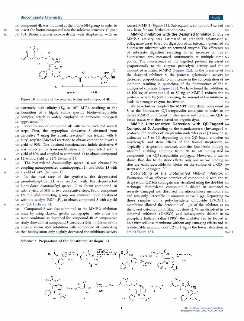
307 compound 4k was modified at the indole NH group in order tof10 308 insert the biotin compound into the inhibitor structure (Figuref10 309 10). Biotin interacts noncovalently with streptavidin with an
310 extremely high affinity (KD ≃ 1015 M−1), resulting in the311 formation of a highly stable specific biotin−streptavidin312 complex, which is widely employed in numerous biological313 approaches.70,71
314 Modification of compound 4k with biotin included several315 steps. First, the tryptophan derivative 8 obtained from316 derivative 7 using the Suzuki reaction72 was treated with t-317 butyl acrylate (Michael reaction) to obtain compound 9 with a318 yield of 90%. The obtained functionalized indole derivative 9319 was subjected to transamidification and deprotected with a320 yield of 80% and coupled to compound 11 to obtain compound
s2 321 12 with a yield of 82% (Scheme 2).322 The biotinylated diaminoalkyl spacer 16 was obtained by323 coupling monoprotected diaminopropan 14 and biotin 15 with
s3 324 a yield of 74% (Scheme 3).325 In the next step of the synthesis, the deprotected326 pseudodipeptide 13 was reacted with the deprotected327 biotinylated diaminoalkyl spacer 17 to obtain compound 18328 with a yield of 38% in two consecutive steps. From compound329 18, the allyl-protecting group was removed upon treatment330 with the catalyst Pd(Ph3P)4 to obtain compound 5 with a yield
s4 331 of 72% (Scheme 4).332 Compound 5 was also submitted to the MMP-2 inhibition333 assay by using classical gelatin zymography made under the334 same conditions as described for compound 4k. A comparative335 study showed that compound 5 ensured a 34% inhibition of the336 enzyme versus 45% inhibition with compound 4k, indicating337 that biotinylation only slightly decreased the inhibitory activity
338 f11toward MMP-2 (Figure 11). Subsequently, compound 5 served339as a basis for our further experiments.340MMP-2 Inhibition with the Designed Inhibitor 5. The341MMP-2 activity was estimated in standard gelatinase/342collagenase assay based on digestion of an internally quenched343fluorescent substrate with an activated enzyme. The efficiency344of substrate digestion resulting in an increase in dye345fluorescence was measured continuously at multiple time346points. The fluorescence of the digested product increased347proportionally to the enzyme proteolytic activity and the348 f12amount of activated MMP-2 (Figure 12a). In the presence of349the designed inhibitor 5, the protease gelatinolytic activity350decreased proportionally to an increase in the concentration of351inhibitor, resulting in quenching of the fluorescence of the352undigested substrate (Figure 12b). We have found that addition353of 100 ng of compound 5 to 50 ng of MMP-2 reduces the354protease activity by 50%. Increasing the amount of the inhibitor355leads to stronger enzyme inactivation.356We have further coupled the MMPI biotinylated compound3575 to the fluorescent QD-streptavidin conjugate in order to358detect MMP-2 in different in vitro assays and to compare QD-359based assays with those based on organic dyes.360MMP-2 Ultrasensitive Detection with QD-Tagged361Compound 5. According to the manufacturer’s (Invitrogen)362protocol, the number of streptavidin molecules per QD may be363estimated as 5 to 10, depending on the QD batch, emission364wavelength, and steric effects of the bound streptavidin.365Typically, a streptavidin molecule contains four biotin binding366sites71,73 enabling coupling from 20 to 40 biotinylated367compounds per QD-streptavidin conjugate. However, it was368shown that, due to the steric effects, only one or two binding369sites are easily accessible for biotin on the surface of a QD-370streptavidin conjugate.73,74
371Dot-Blotting of the Biotinylated MMP-2 Inhibitor.372Formation of an effective complex of compound 5 with the373streptavidin-QD565 conjugate was visualized using the dot-blot374technique. Biotinylated compound 5 diluted in methanol375severely damaged and dissolved the nitrocellulose membrane376and was only detectable in amounts above 2 μg. Depositing377these samples on a polyvinylidene difluoride (PVDF)378membrane allowed the detection of 1 μg of the inhibitor as379the lowest detection limit (data not shown). When dissolved in380dimethyl sulfoxide (DMSO) and subsequently diluted in381phosphate buffered saline (PBS), the inhibitor can be loaded382on a nitrocellulose membrane without any damaging effects and383is detectable in amounts of 0.2 to 1 μg as the lowest detection384 f13limit (Figure 13).
Figure 10. Structure of the resultant biotinylated compound 4k.
Scheme 2. Preparation of the Substituted Analogue 13
Bioconjugate Chemistry Article
DOI: 10.1021/acs.bioconjchem.6b00065Bioconjugate Chem. XXXX, XXX, XXX−XXX
F

385 In Vitro Sandwich-Type Assay for MMP-2 Detection Using386 the Designed QD-Coupled Inhibitor 5. The formation of a387 complete complex consisting of the MMP-2 enzyme,388 biotinylated inhibitor 5, and fluorescent conjugate of389 streptavidin with QD800 nm was performed by means of a390 solid-state sandwich-type analytic biochemical assay. Here, 50391 ng of MMP-2 was used in MMP-2 activity assays since this392 amount of active enzyme was estimated to efficiently and393 quickly digest the gelatin substrate. The proenzyme was394 activated with p-amino phenylmercuric acetate before adsorp-395 tion in order to provide appropriate conformation of the396 inhibitory binding sites. Activated MMP-2, MMP-2, or the397 same quantity of BSA protein control were adsorbed on a398 treated 96-well plate in PBS (pH 7.4). Increasing amounts of399 biotinylated compound 5 and the QD-streptavidin conjugate400 were subsequently incubated with the adsorbed MMP-2. The401 QD fluorescence intensity increased proportionally to the402 biotinylated inhibitor amount in the range from 0 to 100 μg
f14 403 (Figure 14). Thus, the complex of MMP-2 with the designed404 biotinylated compound 5 can be quantitatively detected and405 visualized using compact and bright QD-streptavidin con-406 jugates.407 We performed additional experiments in order to explore the408 photostability of QDs enabling long-term accumulation of their409 fluorescence signal accompanied by order-of-magnitude
410improvements of the signal-to-noise ratios in a fluorogenic411assay and, consequently, increasing the sensitivity for412application of QD-based conjugates to MMP-2 detection in413vitro and to compare the sensitivity of QD-based assays with414those based on organic dyes.415 f15Figure 15 shows that accumulation of the signal from the416fluorogenic assay employing strepta-QD conjugate for visual-
Scheme 3. Preparation of the Biotinylated Linker 17
Scheme 4. Synthesis of Compound 5
Figure 11. Inhibition of MMP-2 gelatinolytic activity. (a) Results ofzymography analysis: 0.4 ng of active MMP-2/well was resolved ingelatin zymography. Each lane was incubated overnight in 50 mM Trisbuffer, pH 7.5, with 200 mM NaCl and 5 mM CaCl2 without (control)or with 10 μM inhibitor (compound 4k or compound 5). (b)Quantification of MMP-2 gelatinolytic activity by densitometry. ***: p< 0.001. **: p < 0.005.
Figure 12. Influence of compound 5 on MMP-2 activity as determinedby dye quenched substrate (DQ gelatin) digestion. (a) MMP-2 activityat different concentrations of preactivated enzyme resulting in anincrease in fluorescein fluorescence. (b) Concentration-dependenteffect of compound 5 on MMP-2 activity. Each analysis has beenperformed in duplicate (p < 0.001).
Figure 13. Detection of biotinylated compound 5 with the fluorescentstreptavidin-QD 565 nm conjugate using the dot-blot technique.
Bioconjugate Chemistry Article
DOI: 10.1021/acs.bioconjchem.6b00065Bioconjugate Chem. XXXX, XXX, XXX−XXX
G

417 ization of MMP-2-inhibitor complexes leads to an increase in418 the signal-to-noise ratio in the given detection scheme by more419 than 2 orders of magnitude and, hence, the corresponding420 increase in the sensitivity of detection of the inhibitor-MMP-2421 complex. This increase may reach even higher values if longer422 signal accumulation times and optimized detection schemes are423 employed. The result shown in Figure 15 was achieved due to424 the rock-solid photostability of QDs enabling long-term QD425 fluorescent signal accumulation without significant photo-426 bleaching of the nanocrystals. Such a photostability is not427 characteristic of the organic dyes used in currently available428 fluorogenic assays. Therefore, we have performed an additional429 comparative study of the model biochemical assays employing430 different organic dyes in parallel with the assays employing
431strepta-QD conjugates in order to compare the changes in their432state during long-term signal detection and accumulation.433 f16As seen from Figure 16, accumulation of the signal from434fluorogenic assays employing strepta-QD for revealing the
435(compound 5)-MMP-2 complex during 10−20 min may result436in an increase in the intensity of the signal by more than two437orders of magnitude, whereas the signals of all typical organic438dyes recorded under exactly the same conditions are photo-439bleached. However, the most important fact is that the440fluorescent signal from the assay employing the strepta-QD441obtained in the course of signal accumulation is more than 2442orders of magnitude stronger than that for the maximal signals443that may be obtained for typical fluorogenic assays employing444the best organic dyes under optimal conditions. This shows that445the use of QD-based labels in typical fluorogenic assays is446decisively advantageous.447Panels a and b show variations of the fluorescence signals448from the model assays during signal accumulation for 20 and 4449min, respectively. In panel a, the signals from organic dyes are450too weak to be visible in comparison with the signals from451strepta-QDs at the equimolar concentration and even after a 1/
Figure 14. Quantitative detection of the MMP-2/biotinylatedcompound 5 complex with the fluorescent streptavidin-QD800 nmconjugate in in vitro sandwich-type biochemical assays. In a controlexperiment, BSA was used instead of MMP-2. Each analysis has beenperformed in duplicate (p < 0.001).
Figure 15. An increase of the fluorescent signal as a result of itsaccumulation during 2, 4, 6, 8, 10, or 20 min for different quantities ofcompound 5 complexed with equimolar quantities of the fluorescentstreptavidin-QD800 conjugate in an in vitro sandwich-type biochemicalassay. The mean fluorescence intensities in the case of immediatesignal recording (0 min of accumulation) in the presence of 10, 50,and 100 μg of compound 5 are 50, 220, and 420 au, respectively,which shows that the signal-to-noise ratios in assays employing QDscan be increased by more than 2 orders of magnitude.
Figure 16. Comparative analysis of variations of fluorescent signals asa result of their accumulation at permanent signal-to-noise ratios fortypical in vitro biochemical sandwich-like fluorogenic assays employingtwo different concentrations of the fluorescent streptavidin-QD800conjugate and the most popular organic dyes.
Bioconjugate Chemistry Article
DOI: 10.1021/acs.bioconjchem.6b00065Bioconjugate Chem. XXXX, XXX, XXX−XXX
H

452 100 dilution; in panel b, the signals from strepta-QD are strong453 and visible only in the initial period of signal accumulation.454 Panel c shows the same data as in panel a but on a demi-455 logarithmic scale. Here, the curves corresponding to the456 evolutions of the fluorescence signals from strepta-QDs and457 from different organic labels are visible in the same graph.
458 ■ CONCLUSIONS
459 Proteolytic enzymes of the MMP family, in particular, MMP-2,460 are overexpressed during cancer pathological processes.461 Activated MMPs have been detected in tissues, plasma,462 serum, and urine of cancer patients at increased levels,; they463 are also positively correlated with the severity of metastatic464 tumors.75−77 Thus, MMPs can serve as selective and specific465 tumor markers for clinical applications.466 Because of the unique potency to combine monitoring and467 therapy, nanoparticle-based techniques can provide an468 advantage over standard diagnostic and therapeutic tools with469 respect to disease diagnosis and monitoring, drug delivery, and470 release. Moreover, nanoparticle-based diagnostic tools can be471 easily modified, combined, and improved to ensure multiplexed472 detection and advanced diagnostics sensitivity. Thus, nano-473 particles (e.g., fluorescent nanocrystals or QDs) are considered474 a useful and indispensable tool for advanced clinical diagnostics,475 imaging, and therapeutic applications.78
476 In this study, a series of new MMP-inhibitors, ilomastat477 derivatives, have been prepared and subjected to preliminary478 biological evaluations in experiments with the main matrix479 metalloproteinases (MMP-1, MMP-2, MMP-9, MMP-13, and480 MMP-14). A systematic enzyme inhibition/selectivity study481 was carried out, with carboxylic acid serving as a ZBG, by482 introducing a phenyl group to the C2 carbon atom of the483 tryptophan indole ring and by incorporating alkylidene chains484 of different lengths at the P′1 position of the succinic acid485 moiety. As a result of structure−activity relationship (SAR)486 studies, we have identified the most selective MMP-2 inhibitor487 with convenient inhibition potency (IC50 = 80 nM) and488 biotinylated it for use in assays employing the QD-streptavidin489 conjugate for visualization. The specific fluorescent complex of490 compound 5-biotin/streptavidin-QD was tested and evaluated491 for MMP-2 inhibition in model in vitro sandwich-type analytic492 biochemical assays and compared with similar model assays493 employing typical organic dyes. The data show that the494 fluorogenic assays employing QDs allows the detection signal495 level to be increased by more than 2 orders of magnitude496 compared to that of typical organic dyes, thus paving the way to497 a considerable increase in the sensitivity of MMP detection in498 in vitro assays. The described ultrasensitive QD-based detection499 approach, exemplified here by MMP-2 detection, is by no500 means limited to it. Designing and using broad-spectrum501 inhibitors will allow this strategy to be efficiently modified for502 detecting both other MMPs and other hydrolytic enzymes for503 different substrates.
504 ■ EXPERIMENTAL PROCEDURES
505 General Synthesis Methods and Materials. Standard506 solvents were purchased from commercial sources and were507 dried by standard procedures and redistilled under N2 prior to508 use. Reactions and products were routinely monitored by thin509 layer chromatography (TLC) on silica gel (KIESELGEL 60510 PF254, Merck). HBTU and 2,4,6-collidine were purchased511 from commercial sources. Pure products were obtained by
512means of flash chromatography using Merck Geduran SI silica513gel 60 (70−230 mesh ASTM). The melting points were514determined using a Reichert Thermovar hot-stage apparatus515and are uncorrected. The NIR-FT spectra (KBr or NaCl film)516were measured using a PerkinElmer Spectrum BX FTIR517instrument. The 1H NMR (300 MHz) and 13C NMR (75518MHz) spectra were recorded by means of a Bruker AC 300519spectrometer using TMS as an internal standard; the chemical520shifts δ were expressed in ppm; the following abbreviations are521used: singlet (s), doublet (d), doublet of doublets (dd), triplet522(t), and multiplet (m). Coupling constants J were expressed in523hertz. Mass spectra were recorded by means of an MSQ524ThermoFinnigan apparatus using the chemical ionization (CI)525method. Electrospray ionization mass spectrometry experi-526ments were carried out using a hybrid tandem quadrupole/527time-of-flight (Q-TOF) instrument equipped with a pneumati-528cally assisted electrospray (Z-spray) ion source (Micromass,529Manchester, UK) operated in the positive mode. Optical530rotations were measured on a PerkinElmer 241 polarimeter531(Na lamp, λ = 589 nm).532Synthesis Procedures. (S)-Ethyl3-(1H-indol-3-yl)-2-(2,2,2-533trifluoroacetamido)propanoate 7. L-Tryptophan (5.0 g, 24.5534mmol) was dissolved in absolute ethanol (80 mL). Thionyl535chloride (3.6 mL, 2 equiv, 48.9 mmol) was added dropwise536during 15 min. The mixture was refluxed for 3.5 h; then, the537solvent was evaporated. The white solid was dissolved in538EtOAc, the organic phase was washed with an aqueous solution539of NaHCO3 (5%), and then dried over MgSO4. After filtration540and concentration of the solvent, a slightly yellow oil was541obtained. Then, CH2Cl2 (110 mL), triethylamine (3.4 mL, 1542equiv, 24.5 mmol), and trifluoroacetic anhydride (8.2 mL, 2.4543equiv, 58.6 mmol) were added at 0 °C. The mixture was stirred544at room temperature overnight. The solvent was evaporated;545the crude product was purified by flash chromatography546(cyclohexane/CH2Cl2: 40/60) to afford the white solid of547compound 7 (5.6 g, 72%). mp: 117−119 °C. [α]21D −21 (c5481.13, DMSO). 1H NMR (DMSO-d6, 300 MHz): δ 1.14 (t, 3H,549J = 7.1 Hz), 3.20−3.35 (m, 2H), 4.12 (q, 2H, J = 7.1 Hz),5504.52−4.60 (m, 1H), 7.02−7.10 (m, 2H), 7.18 (s, 1H), 7.37 (d,5511H, J = 8.1 Hz), 7.55 (d, 1H, J = 7.8 Hz), 9.95 (d, 1H, J = 7.5552Hz), 10.92 (s, 1H). 13C NMR (DMSO-d6, 75 MHz): δ 14.1,55326.2, 54.0, 61.3, 109.4, 110.6, 111.7, 114.4, 118.2, 118.4, 118.7,554121.3, 122, 124.0, 127.1, 136.3, 155.9, 156.4, 156.9, 157.4,555170.5. IR (KBr): ν (cm−1) 3374, 3322, 1736, 1696, 1555, 1304,5561278, 1230, 1176, 1093, 1027, 873, 860, 741, 692. MS (CI): m/557z 328.96 [M+·] (100), 311 (14), 283 (15), 255 (25), 242 (23),558238 (59). Anal. Calcd for C15H15N2O3F3: C 54.88, H 4.61, N,5598.53%. Found: C 54.64, H 4.74, N 8.38%.560( S ) -E thy l 3 - (2 -pheny l -1H- indo l -3 -y l ) -2 - (2 ,2 ,2 -561trifluoroacetamido)propanoate 8. N-Bromosuccinimide (54562mg, 0.3 mmol) was added to a suspension of α-N-563(trifluoroacety1)-L-tryptophan ethyl ester 7 (0.1 g, 0.3 mmol)564in CCl4 (2 mL). The mixture was refluxed for 30 min under565nitrogen, and the solvent was evaporated in vacuo. The filtrate566was purified by flash chromatography (petroleum ether/EtOAc:5679/1) to afford a brominated compound (118 mg, 96%). mp:568134−137 °C. [α]21D −14 (c 1.41, DMSO). 1H NMR (CDCl3,569300 MHz): δ 1.22 (t, 3H, J = 7.1 Hz), 3.29−3.35 (m, 2H),5704.08−4.24 (m, 2H), 4.91 (dd, 1H, J = 13.8 Hz, J = 5.8 Hz), 6.99571(d, 1H, J = 7.1 Hz), 7.09−7.20 (m, 2H), 7.25 (d, 1H, J = 7.4572Hz), 7.46 (d, 1H, J = 7.7 Hz), 8.35 (s, 1H). 13C NMR (CDCl3,57375 MHz): δ 13.8, 27.3, 52.9, 62.5, 109.0, 109.6, 109.8, 110.7,574113.6, 117.4, 117.7, 120.5, 121.2, 122.8, 127.5, 136.0, 156.0,
Bioconjugate Chemistry Article
DOI: 10.1021/acs.bioconjchem.6b00065Bioconjugate Chem. XXXX, XXX, XXX−XXX
I

575 156.5, 157.0, 157.5, 170.2. IR (KBr): ν (cm−1) 3339, 1727,576 1701, 1555, 1450, 1419, 1278, 1194, 1164, 1027, 873, 741, 723,577 648. Anal. Calcd for C15H14N2O3BrF3: C 44.25, H 3.47, N578 6.88%. Found: C 44.41, H 3.34, N 6.78%.579 Phenylboronic acid (52 mg, 1.5 equiv, 0.42 mmol) was580 dissolved in a mixture of toluene and ethanol (1/1) (4 mL), the581 brominated compound (115 mg, 0.28 mmol), NaHCO3 (47582 mg, 2 equiv, 0.56 mmol) dissolved in pure water (1 mL), and583 LiCl (36 mg, 3 equiv, 0.84 mmol); finally, catalyst Pd(PPh3)4584 (32 mg, 0.1 equiv, 0.028 mmol) was added. The yellow solution585 was stirred under reflux for 2 h. The red mixture was586 concentrated; then, CH2Cl2 and an aqueous solution of587 NaHCO3 (10%) were added. The aqueous phase was extracted588 twice with CH2Cl2. The organic phases were collected, dried589 over MgSO4, filtered, and concentrated. The crude product was590 purified by flash chromatography (petroleum ether/EtOAc: 9/591 1) to afford compound 8 (105 g, 92%). mp: 172−174 °C.592 [α]21D −10.5 (c 0.84, DMSO). 1H NMR (CDCl3, 300 MHz): δ593 1.02 (t, 3H, J = 7.1 Hz), 3.56−3.62 (m, 3H), 3.91−3.97 (m,594 1H), 4.81 (dd, 1H, J = 13.6 Hz, J = 5.8 Hz), 6.68 (d, 1H, J = 7.4595 Hz), 7.12−7.57 (m, 9H), 8.22 (s, 1H). 13C NMR (CDCl3, 75596 MHz): δ 13.7, 26.5, 53.4, 61.9, 105.6, 109.0, 111.0, 113.0, 117.0,597 118.5, 120.2, 121.0, 122.7, 128.2, 128.3, 128.9, 129.0, 129.1,598 132.5, 135.6, 136.3, 155.1, 155.7, 156.3, 156.9, 170.2. IR (KBr):599 ν (cm−1) 3374, 3286, 1753, 1705, 1555, 1450, 1278, 1203,600 1172, 1018, 877, 745, 697. MS (CI): m/z [M+1] 405.29 (100),601 405 (60), 339 (15), 218 (20), 206 (15). Anal. Calcd for602 C21H19N2O3F3: C 62.37, H 4.74, N 6.93%. Found: C 62.18, H603 4.66, N 6.82%.604 (S)-Ethyl3-(1-(3-tert-butoxy-3-oxopropyl)-2-phenyl-1H-605 indol-3-yl)-2-(2,2,2-trifluoroacetamido)propanoate 9. tBuOK606 (107 mg, 1.3 equiv, 0.95 mmol) was added to a solution of607 compound 8 (296 mg, 0.73 mmol) in THF (5 mL); the608 solution was stirred for 15 min under nitrogen. Then, tert-butyl609 acrylate (1 mL) was added to the solution dropwise. The610 reaction mixture was incubated upon heating to reflux for 3.5 h.611 The mixture was diluted with a solution of saturated NH4Cl612 and extracted with EtOAc. The organic phase was dried over613 MgSO4 and filtered, and the solvent was evaporated. The crude614 product was purified by flash chromatography (petroleum615 ether/EtOAc: 85/15) to afford compound 9 (351 mg, 90%).616 mp: 84−86 °C. [α]21D −2.4 (c 0.5, MeOH). 1H NMR (CDCl3,617 300 MHz): δ 1.08 (t, 3H, J = 7.1 Hz), 1.32 (s, 9H), 2.37−2.43618 (m, 2H), 3.33 (ddd, 2H, J = 5.7 Hz, J = 14.7 Hz, J = 20.4 Hz),619 3.74−3.80 (m, 1H), 3.98−4.04 (m, 1H), 4.25−4.30 (m, 2H),620 4.71 (dd, 1H, J = 6.1 Hz, J = 13.9 Hz), 6.51 (d, 1H, J = 7.8 Hz),621 7.15−7.61 (m, 9H). 13C NMR (CDCl3, 75 MHz): δ 13.7, 26.6,622 26.8, 27.8, 35.8, 39.5, 53.2, 61.8, 81.0, 106.7, 109.9, 117.2,623 118.4, 118.5, 119.9, 120.0, 121.1, 122.3, 127.8, 128.8, 129.0,624 130.4, 130.9, 136.0, 138.9, 155.0, 155.9, 156.8, 157.7, 170.1,625 170.1. IR (NaCl): ν (cm−1) 3400, 3322, 2976, 2925, 1723,626 1540, 1462, 1361, 1207, 1155, 1018, 739, 700. HRMS (ESI):627 m/z calcd for C28H32N2O5F3 533.2263, found 533.2257 (−1.2628 ppm). Anal. Calcd for C28H31N2O5F3: C 63.09, H 5.82, N629 5.25%. Found: C 62.99, H 6.00, N 5.41%.630 (S)-tert-Butyl 3-(3-(2-amino-3-(methylamino)-3-oxoprop-631 yl)-2-phenyl-1H-indol-1-yl)propanoate 10. A solution of632 compound 9 (304 mg, 0.57 mmol) in absolute ethanol (2.3633 mL), and aqueous methylamine (40%) (2.3 mL) was stirred at634 room temperature overnight. The solvent was evaporated, and635 the crude product was diluted in CH2Cl2. The organic phase636 was washed twice with an aqueous solution of HCl (5%). The637 collected acid phases were then alkalized with an aqueous
638solution of NaHCO3 (10%) to reach pH 8. The aqueous phase639was extracted with CH2Cl2, dried over MgSO4, and filtered.640Evaporation of the solvent gave a white foam (191 mg, 80%).641[α]21D + 9.2 (c 0.5, MeOH). 1H NMR (CDCl3, 300 MHz): δ6421.31 (s, 9H), 2.39−2.44 (m, 2H), 2.68−2.76 (m, 4H), 3.40−6433.45 (m, 1H), 3.60−3.64 (m, 1H), 4.26−4.31 (m, 2H), 7.13−6447.54 (m, 9H), 7.73 (d, 1H, J = 7.7 Hz). 13C NMR (CDCl3, 75645MHz): δ 25.7, 27.8, 29.9, 35.8, 39.5, 55.3, 81.0, 109.7, 109.8,646119.3, 119.7, 122.1, 127.6, 128.6, 128.8, 130.5, 131.4, 136.1,647138.9, 170.1, 175.1. IR (NaCl): ν (cm−1) 3379, 3302, 2914,6481723, 1659, 1527, 1462, 1364, 1150, 845, 742, 703. HRMS649(ESI): m/z calcd for C25H32N3O3 422.2444, found 422.2449650(1.3 ppm). Anal. Calcd for C25H31N3O3·H2O: C 68.33, H 7.06,651N 9.56%. Found: C 68.21, H 7.34, N 9.53%.652(S,E)-Allyl 3-(3-(1-(3-tert-butoxy-3-oxopropyl)-2-phenyl-6531H- indo l -3 -y l ) -1 - (methy lamino) -1 -oxopropan-2 -654ylcarbamoyl)tridec-3-enoate 12. HBTU (300 mg, 1.5 equiv,6550.79 mmol) and 2,4,6-collidine (141 μL, 2 equiv, 10 mmol)656were added to a solution of compound 10 (157 mg, 0.53657mmol) in dry CH2Cl2 (20 mL). The reaction mixture was658stirred for 1 h at room temperature; then, a solution of659compound 11 (223 mg, 0.53 mmol) in dry CH2Cl2 (20 mL)660was added, and the solution was stirred overnight at room661temperature. The solvent was evaporated, and the crude662product was purified by flash chromatography (CH2Cl2/663MeOH: 99/1, 98/2) to afford compound 12 (304 g, 82%).664[α]21D −4.2 (c 0.5, MeOH). 1H NMR (CDCl3, 300 MHz): δ6650.89 (t, 3H, J = 7.1 Hz), 1.24−1.31 (m, 23H), 2.01−2.06 (m,6662H), 2.38 (t, 2H, J = 7.7 Hz), 2.60 (d, 3H, J = 4.8 Hz), 3.13−6673.41 (m, 4H), 4.29 (t, 2H, J = 7.5 Hz), 4.50−4.52 (m, 2H),6684.62 (dd, 1H, J = 7.3 Hz, J = 14.4 Hz), 5.16−5.23 (m, 2H),6695.79−5.85 (m, 2H), 5.96 (t, 1H, J = 7.3 Hz), 6.47 (d, 1H, J =6707.3 Hz), 7.17−7.50 (m, 8H), 7.84 (d, 1H, J = 7.6 Hz). 13C671NMR (CDCl3, 75 MHz): δ 14.1, 22.6, 26.2, 27.2, 27.8, 28.4,67228.5, 29.2, 29.3, 29.4, 29.6, 31.8, 32.5, 35.7, 39.6, 53.8, 65.5,67381.0, 109.0, 109.8, 118.3, 119.4, 120.1, 122.2, 127.8, 128.6,674128.8, 129.4, 130.4, 131.0, 131.7, 136.2, 138.2, 138.6, 168.6,675170.1, 170.9, 171.4. IR (NaCl): ν (cm−1) 3405, 3317, 2925,6762852, 1726, 1659, 1625, 1511, 1462, 1364, 1323, 1253, 1152,677990, 922, 742, 700. HRMS (ESI): m/z calcd for678C42H57N3O6Na 722.4145, found 722.4135 (−1.5 ppm). Anal.679Calcd for C42H57N3O6.H2O: C 70.29, H 7.95, N 5.85%. Found:680C 70.19, H 8.20, N 6.89%.681(S,E)-3-(3-(2-(2-(2-(Allyloxy)-2-oxoethyl)dodec-2-enami-682do)-3-(methylamino)-3-oxopropyl)-2-phenyl-1H-indol-1-yl)-683propanoic Acid 13. TFA (4 mL) was added to a solution of684compound 12 (300 mg, 0.42 mmol) in dry CH2Cl2 (10 mL),685and the mixture was sequentially stirred for 30 min at 0 °C and686for 30 min at room temperature. After evaporation, an orange687oil was obtained (276 mg, 100%). 1H NMR (CDCl3, 300688MHz): δ 0.89 (t, 3H, J = 6.5 Hz), 1.24−1.41 (m, 14H), 2.03−6892.10 (m, 2H), 2.46−2.60 (m, 5H), 3.17−3.31 (m, 4H), 4.33 (t,6902H, J = 7.5 Hz), 4.50 (d, 2H, J = 5.5 Hz), 4.67−4.69 (m, 1H),6915.17−5.28 (m, 2H), 5.78−5.83 (m, 1H), 6.15 (t, 1H, J = 6.8692Hz), 6.56 (s, 1H), 7.13−7.65 (m, 10H), 9.8 (s, 1H). 13C NMR693(CDCl3, 75 MHz): δ 14.0, 22.6, 26.7, 26.9, 28.3, 28.6, 29.2,69429.3, 29.4, 29.6, 31.8, 32.5, 34.3, 39.5, 54.5, 65.9, 107.9, 109.7,695118.6, 118.7, 120.2, 122.5, 127.8, 127.9, 128.9, 130.3, 130.6,696131.4, 135.9, 138.8, 141.3, 170.0, 171.6, 172.9, 172.9. IR697(NaCl): ν (cm−1) 3307, 3054, 2925, 2852, 1731, 1661, 1635,6981527, 1465, 1408, 1359, 1271, 1196, 1176, 1137, 987, 925, 742,699716, 700. HRMS (ESI): m/z calcd for C38H50N3O6 644.3700,700found 644.3702 (0.4 ppm).
Bioconjugate Chemistry Article
DOI: 10.1021/acs.bioconjchem.6b00065Bioconjugate Chem. XXXX, XXX, XXX−XXX
J

701 tert-Butyl 3-(5-(2-oxo-hexahydro-1H-thieno[3,4-d]-702 imidazol-4-yl)pentanamido) propylcarbamate 16. HBTU703 (232 mg, 1.5 equiv, 0.61 mmol) and 2,4,6-collidine (110 μL,704 2 equiv, 0.81 mmol) were added to a solution of biotin 15 (100705 mg, 0.41 mmol) in dry DMF (3 mL). The reaction mixture was706 stirred for 1 h at room temperature; then, a solution of707 compound 14 (107 mg, 1.5 equiv, 0.61 mmol) in dry CH2Cl2708 (3 mL) was added, and the mixture was stirred overnight at709 room temperature. The solvent was evaporated, and the crude710 product was purified by flash chromatography (CH2Cl2/711 MeOH: 9/1, 8/2) to afford compound 16 (121 mg, 74%).712 mp: 93−95 °C. [α]21D + 47.8 (c 0.5, MeOH). 1H NMR713 (DMSO-d6, 300 MHz): δ 1.25−1.53 (m, 17H), 2.05 (t, 2H, J =714 7.3 Hz), 2.56−2.60 (m, 1H), 2.83 (dd, 1H, J = 5.1 Hz, J = 12.4715 Hz), 2.89 (dd, 2H, J = 6.2 Hz, J = 12.8 Hz), 3.02 (dd, 2H, J =716 6.7 Hz, J = 12.9 Hz), 3.10−3.12 (m, 1H), 4.11−4.15 (m, 1H),717 4.29−4.33 (m, 1H), 6.38 (s, 1H), 6.45 (s, 1H), 6.79 (t, 1H, J =718 5.2 Hz), 7.77 (t, 1H, J = 5.4 Hz). 13C NMR (CDCl3, 75 MHz):719 δ 25.3, 27.8, 27.8, 28.1, 29.4, 35.6, 35.9, 37.0, 40.1, 55.3, 59.9,720 61.6, 79.2, 156.7, 164.0, 174.1. IR (NaCl): ν (cm−1) 3297,721 2925, 1687, 1524, 1457, 1361, 1276, 1248, 1165, 842. HRMS722 (ESI): m/z calcd for C18H32N4O4SNa 423.2042, found723 423.2035 (−1.7 ppm). Anal. Calcd for C18H32N4O4S·2H2O:724 C 49.54, H 7.33, N 12.84, S 7.32%. Found: C 49.40, H 7.66, N725 13.68, S 6.78%.726 N-(3-Aminopropyl)-5-(2-oxo-hexahydro-1H-thieno[3,4-d]-727 imidazol-4-yl)pentanamide 17. TFA (1 mL) was added to a728 solution of compound 16 (95.5 mg, 0.24 mmol) in dry CH2Cl2729 (2 mL), and the mixture was sequentially stirred for 30 min at 0730 °C and for 30 min at room temperature. After evaporation, an731 orange oil was obtained (144 mg, 100%). 1H NMR (CDCl3,732 300 MHz): δ 1.41−1.52 (m, 2H), 1.58−1.69 (m, 4H), 1.83−733 1.88 (m, 2H), 2.19−2.27 (m, 2H), 2.72−2.96 (m, 4H), 3.15−734 3.21 (m, 1H), 3.28−3.38 (m, 2H), 4.32−4.36 (m, 1H), 4.52−735 4.58 (m, 1H). 13C NMR (CDCl3, 75 MHz): δ 25.0, 26.8, 27.6,736 27.8, 35.0, 35.3, 36.5, 40.0, 55.3, 60.1, 61.9, 164.2, 175.5. IR737 (NaCl): ν (cm−1) 3291, 2919, 2852, 1661, 1457. HRMS (ESI):738 m/z calcd for C13H25N4O2S 301.1620, found 301.1691.739 (E)-Allyl 3-((S)-1-(methylamino)-1-oxo-3-(1-(3-oxo-3-(3-(5-740 (2-oxo-hexahydro-1H-thieno[3,4-d] imidazol-4-yl ) -741 pentanamido)propylamino)propyl)-2-phenyl-1H-indol-3-yl)-742 propan-2-ylcarbamoyl)tridec-3-enoate 18. HBTU (108 mg,743 1.2 equiv, 0.28 mmol) and 2,4,6-collidine (47 μL, 1.5 equiv,744 0.35 mmol) were added to a mixture of compound 13 (0.23745 mmol) in dry CH2Cl2 (2 mL) and DMF (1 mL). The reaction746 mixture was stirred for 1 h at room temperature; then, a747 solution of compound 17 (0.23 mmol) in dry CH2Cl2 (2 mL)748 and 2,4,6-collidine (47 μL, 1.5 equiv, 0.35 mmol) were added,749 and the solution was stirred overnight at room temperature.750 The solvents were evaporated. The mixture was diluted with751 EtOAc (2 mL). The organic phase was washed sequentially752 with a solution of citric acid (1 N), a solution of NaHCO3 (1753 N), and a solution of NaCl and dried over MgSO4. After754 filtration and evaporation, the crude product was purified by755 flash chromatography (CH2Cl2/MeOH: 9/1, 8/2) to afford756 compound 18 (83 mg, 38% over two steps). mp: 86−88 °C.757 [α]21D + 9.8 (c 0.5, MeOH). 1H NMR (DMSO-d6, 300 MHz):758 δ 0.87 (t, 3H, J = 6.0 Hz), 1.27−1.52 (m, 22H), 2.02−2.09 (m,759 4H), 2.28−2.31 (m, 2H), 2.42 (d, 3H, J = 4.4 Hz), 2.56−2.60760 (m, 1H), 2.79−3.29 (m, 10H), 4.12−4.19 (m, 3H), 4.28−4.47761 (m, 4H), 5.19 (ddd, 2H, J = 1.1 Hz, J = 17.3 Hz, J = 18.2 Hz),762 5.79−5.85 (m, 1H), 6.25 (t, 1H, J = 7.2 Hz), 6.38 (s, 1H), 6.44763 (s, 1H), 7.03−7.19 (m, 2H), 7.40−7.83 (m, 11H). 13C NMR
764(DMSO-d6, 75 MHz): δ 14.2, 22.3, 25.5, 25.8, 27.8, 28.2, 28.4,76528.9, 29.1, 29.2, 31.5, 35.4, 36.0, 36.3, 36.5, 36.5, 39.9, 40.2,76654.8, 55.6, 59.3, 61.2, 64.6, 109.2, 110.1, 117.5, 119.2, 119.3,767121.5, 128.0, 128.7, 129.1, 130.8, 131.3, 132.7, 135.9, 138.2,768138.3, 162.9, 167.3, 169.5, 170.4, 171.7, 172.1. IR (NaCl): ν769(cm−1) 3421, 3302, 2919, 2852, 1695, 1653, 1545, 1532, 1462,7701354, 1325, 1263, 1173, 842, 739. HRMS (ESI): m/z calcd for771C51H71N7O7SNa 948.5033, found 948.5041 (0.8 ppm). Anal.772Calcd for C51H71N7O7S·6H2O: C 59.24, H 6.87, N 9.48, S7733.09%. Found: C 59.18, H 7.04, N 10.17, S 4.14%.774(E)-3-((S)-1-(Methylamino)-1-oxo-3-(1-(3-oxo-3-(3-(5-(2-775oxo -hexahyd ro -1H- th i eno [3 ,4 -d ] im idazo l -4 - y l ) -776pentanamido)propylamino)propyl)-2-phenyl-1H-indol-3-yl)-777propan-2-ylcarbamoyl)tridec-3-enoic Acid 5. Allyl ester 18778(64 mg, 0.07 mmol) was dissolved in THF (2 mL) in a779nitrogen atmosphere and supplied with a catalyst (Pd(PPh3)4,7800.1 equiv) and morpholine (18.6 μL, 3.1 equiv, 0.21 mmol) in781the dark. After incubation for 30 min, the solvent was782evaporated. The organic phase was filtered through the Celite783filter agent, and the resultant solution was concentrated in784vacuo. The concentrate was purified by silica gel column785chromatography with the solvent CH2Cl2/MeOH: 7/3 to786afford acid compound 5 as a white solid (44 mg, 72%). mp:787103−105 °C. [α]21D + 21.2 (c 0.5, MeOH). 1H NMR (DMSO-788d6, 300 MHz): δ 0.87 (t, 3H, J = 6.8 Hz), 1.26−1.49 (m, 22H),7891.99−2.09 (m, 4H), 2.29−2.41 (m, 2H), 2.42 (d, 3H, J = 4.4790Hz), 2.77−3.10 (m, 11H), 4.11−4.40 (m, 5H), 6.14 (t, 1H, J =7917.2 Hz), 6.37 (s, 1H), 6.45 (s, 1H), 6.99−7.14 (m, 2H), 7.41−7927.65 (m, 8H), 7.89 (t, 1H, J = 4.8 Hz), 8.06 (d, 1H, J = 7.8 Hz),7938.41 (bs, 1H). 13C NMR (DMSO-d6, 75 MHz): δ 14.2, 22.3,79425.5, 25.9, 27.8, 28.2, 28.4, 28.6, 28.9, 29.1, 31.5, 35.4, 35.6,79536.0, 39.9, 40.2, 54.5, 55.6, 59.3, 61.2, 109.0, 110.3, 119.0,796119.4, 121.4, 127.7, 128.6, 131.0, 131.4, 131.5, 136.1, 136.4,797138.6, 162.9, 168.5, 170.1, 172.0, 172.3, 173.5. IR (NaCl): ν798(cm−1) 3266, 2919, 2852, 2356, 2330, 1651, 1643, 1555, 1542,7991465, 1457, 1359, 1261, 1106, 742. HRMS (ESI): m/z calcd for800C48H67N7O7SNa 908.4720, found 908.4724 (0.3 ppm). Anal.801Calcd for C48H67N7O7S·4H2O: C 60.18, H 7.00, N 10.24, S8023.34%. Found: C 60.91, H 7.82, N 10.50, S 2.90%.803Molecular Modeling. The AutoDock 4.0 software was804used to perform the computational molecular docking. The805AutoDockTools package was employed to prepare the input806files necessary for the docking procedures and to analyze the807results of docking. Figures were constructed using the PyMOL808software (DeLano, W. L. (2002) PyMol Molecular Graphics809System, Palo Alto, CA. http://www.pymol.org).810Initial Data on MMPs and Ligands. The structures from the811Protein Data Bank (PDB) entries 1CK7 (for MMP-2)79 and8121GKC (for MMP-9)80 were used for docking simulations.813While MMP atoms and the zinc ion in the catalytic site were814retained, all of the other atoms were removed. Residues 31−815109 corresponding to the pro-domain prohibiting the MMP-2816proteolytic activity have been also removed. The protonation817states of all ionizable residues were computed using the818PROPKA software.81,82 The MMP side chains were kept fixed819for all the docking computations. Ligands were built using the820Marvin software (Marvin 5.3.2, 2010, ChemAxon: http://www.821chemaxon.com). The AutoDock module AutoTors was used to822determine the torsion angles of the ligands. All of the flexible823torsions except amide bonds were allowed to rotate during the824docking stage.825Molecular Docking Simulations and Calculations. Affinity826grid maps were calculated for each atom type constituting
Bioconjugate Chemistry Article
DOI: 10.1021/acs.bioconjchem.6b00065Bioconjugate Chem. XXXX, XXX, XXX−XXX
K

827 MMP-2 with the use of the AutoGrid software. Grid maps were828 centered on the MMP catalytic site, with 126 × 126 × 126 grid829 points, and spacings of 0.291 and 0.225 Å between the grid830 points for MMP-2 and MMP-9, respectively. Mehler and831 Solmajer’s83 distance-dependent dielectric permittivity was used832 for the calculations of the electrostatic grid maps. Random833 starting positions on the entire protein surface and random834 orientations and torsions were used for all ligands. The835 AutoDock software, version 4.0, was used for docking836 computations, with a Lamarckian genetic algorithm.62,63 Each837 docking experiment was performed with four runs constituted838 of a series of 250 simulations. Each docking simulation was839 carried out with an initial population of 250 individuals, a840 maximum number of 2,500,000 energy evaluations and a841 maximum number of 27,000 generations. The pseudo-Solis and842 Wets modification methods were used with default parameters.843 The docked conformations of the ligands were clustered with a844 root-mean-square deviation (RMSD) cutoff of 0.5 Å.845 Biological Evaluations. Inhibition Studies. The quenched846 fluorogenic substrates DNP-Pro-Cha-Gly-Cys(Me)-His-Ala-847 Lys(N-Me-Abz)-NH2 for MMP-1 or MMP-9 inhibition848 (where DNP is 2,4-dinitrophenyl; Cha is β-cyclohexylalanyl;849 Abz is 2-aminobenzoyl(antraniloyl)) were purchased from850 Calbiochem (VWR, Strasbourg, France), Mca L-Pro-Leu-Gly-851 Leu-Dpa-Ala-Arg-NH2 for MMP-2, MMP-13, or MMP-14852 inhibition (where Mca is (7-methoxycoumarin-4-yl); Dpa is853 [N-3-(2,4-dinitrophenyl)-L-2,3-diaminopropionyl]) and 6-(7-854 nitro-benzo[1,2,5]oxodiazol-4-ylamino)-hexanoyl-Arg-Pro-Lys-855 Pro-Leu-Ala-Nva-Trp-Lys(7 dimethylaminocoumarin-4-yl)NH2856 for MMP-3 inhibition were from Bachem (Weil am Rhein,857 Germany). The dye quenched fluorogenic substrate (DQ858 gelatin) and EnzCheck Gelatinase/Collagenase assays were859 purchased from Life Technologies (USA).860 Human recombinant pro-MMP-1, pro-MMP-2, pro-MMP-9,861 pro-MMP-13, and catalytic domains of MT1-MMP were862 obtained from Calbiochem. The pro-enzymes (pro-MMP-2 or863 pro-MMP-9) were freshly activated with 1−4 mM p-amino-864 phenylmercuric acetate (APMA, Sigma-Aldrich, Saint Quentin865 Fallavier, France) at 37 °C for 1−2 h. Fluorescent conjugates of866 streptavidin with QDs were purchased from Life Technologies867 (USA).868 In Vitro Fluorogenic Substrate Digestion Assay. Briefly, 1869 nM of MMP-1, 0.95 nM of MMP-2, 0.89 nM of MMP-9, 0.6870 nM of MMP-13, and 3.1 nM of MMP-14 catalytic domain were871 incubated with increasing concentrations of the synthetic872 compounds (from 1 to 5000 nM). Assays were initiated by873 adding the respective fluorogenic substrate (1−10 nM). The874 fluorescence was monitored with a PerkinElmer HT Soft 7000875 plus spectrofluorimeter (PerkinElmer, Courtaboeuf, France).876 Upon cleavage of the fluorogenic peptide by MMP, the initial877 rate of the peptide hydrolysis in the absence (Vo) or presence878 (Vi) of the synthetic molecule was determined. The IC50 was879 calculated after plotting Vi/Vo as a function of the synthetic880 molecule concentration by fitting with a nonlinear regression881 (Grafit Computer software, R. Leatherbarrow, Erithacus882 Software).883 MMP-2 Proteolytic Activity Measurement. MMP-2 activity884 was estimated in EnzCheck Gelatinase/Collagenase standard885 commercial assays based on digestion of an internally quenched886 fluorescent substrate with an activated enzyme according to the887 manufacturer’s protocol. Briefly, 0.35−2 μg of recombinant888 MMP-2 was first preactivated with 2.5 mM APMA in an889 activation buffer (50 mM Tris-HCl, pH 7.5, 150 mM NaCl, 20
890mM CaCl2, and 0.01% Tween-20) during 1.5 h at 37 °C. Upon891activation, 0−100 ng of MMP-2 was mixed with 50 μg/mL DQ892gelatin in a total volume of 200 μL of the activating buffer in a893black nontreated 96-well plate. The samples were incubated at894room temperature, while protected from light for 2 to 24 h.895The activity of MMP-2 was measured fluorimetrically in a896fluorescence microplate reader (Infinite M200 Pro, Tecan,897Switzerland). Since the reaction of quenched fluorescent898substrate digestion with the activated enzyme is continuous,899fluorescence was measured at multiple time points: 0, 20, 40,90060, 80, 100, 120 min, etc. The fluorescence of digested products901of the DQ gelatin was induced at 488 nm and measured at 525902nm in a fluorescence microplate reader equipped with two903monochromatic scanners. Background fluorescence was904corrected by subtracting the value derived from no-enzyme905substrate control. This protocol was also employed for the906detection of compound 5 (the enzyme inhibitor).907In this case, 50 ng of activated MMP-2 was premixed with 0−90850 μg of compound 5 preliminarily dissolved in DMSO in a909total volume of 100 μL of the activating buffer. This enzyme−910inhibitor mixture was supplemented with 50 μg/mL DQ gelatin911in a total volume of 200 μL, and the inhibited MMP-2 activity912was measured fluorimetrically at different time points.913In Vitro Gelatinolytic Assays. HT-1080 cells were grown to914a density of 80% in a 24-well plate and treated with 12.5 μg/mL915concanavalin A overnight to convert pro-MMP-2 into active916MMP-2. Cells were preincubated with or without effectors (100917μM 4k, 10 μM ilomastat (Galardin)) for 1 h and then with DQ918gelatin (500 ng/300 μL per well). Fluorescence was measured919at 525 nm.920In vitro invasion assay was performed in modified Boyden921chambers (tissue culture treated; diameter, 6.5 mm; pore size, 8922μm; Greiner-One, Courtaboeuf, France). Five ×104 HT-1080923cells were suspended in serum-free DMEM with 4.5 g/L924glucose containing 0.2% (w/v) BSA and seeded onto925membranes coated with Matrigel (20 μg/well). The lower926compartment was filled with DMEM supplemented with 10%927(v/v) FBS and 2% (w/v) BSA. After a 6-h incubation period,928the cells were fixed with methanol and stained with crystal929violet for 15 min. The cells remaining on the upper face of the930membrane were scraped. Crystal violet staining of the migrating931cells (on the lower face) was eluted with 10% (v/v) acetic acid,932and absorbance was read at 560 nm.933Quantum Dot-Based Assays. The streptavidin-coated934QDs used in this study were purchased from Invitrogen935Corporation.936Dot-Blotting. The formation of the complex of biotinylated937compound 5 with streptavidin-QD conjugate was visualized938using the dot-blot detection technique. Briefly, different939amounts (0−20 μg) of biotinylated compound 5 dissolved940either in methanol or in DMSO and supplied with PBS, pH 7.4,941were applied onto a nitrocellulose or PVDF membrane in a942total volume of 2 μL. The membrane was dried completely and943blocked from nonspecific binding of the fluorescent conjugate944with 5% (w/v) nonfat dry milk in PBS (pH 7.4) containing9450.05% (v/v) Tween 20 during 1 h at room temperature. Then,946the membrane was incubated with streptavidin-QD 565 nm947fluorescent conjugate (1:100 dilution in PBS (pH 7.4)948containing 0.5% (w/v) casein from bovine milk) during 40949min at room temperature and washed thoroughly three times950with PBS (pH 7.4) containing 0.05% (v/v) Tween 20. The951fluorescence of the inhibitor-QD complexes of different952intensities was detected using a ChemiDoc MP Imaging system
Bioconjugate Chemistry Article
DOI: 10.1021/acs.bioconjchem.6b00065Bioconjugate Chem. XXXX, XXX, XXX−XXX
L

953 (Bio-Rad Laboratories, Inc., USA) equipped with a standard954 emission filter (605/50 filter).955 Sandwich-Type Biochemical Assay. The formation of a956 complete complex consisting of the enzyme MMP-2,957 biotinylated compound 5, and fluorescent conjugate of958 streptavidin with QDs was performed by means of a solid-959 state sandwich-type analytic biochemical assay. Briefly, APMA960 preactivated MMP-2 enzyme or BSA protein control was961 adsorbed in a 96-well plate (Maxisorp Nunc, Thermo Fischer962 Scientific, Denmark) in PBS (pH 7.4) overnight at 4 °C. Then,963 the wells were washed and blocked with 1.5% (w/v) casein in964 PBS (pH 7.4) with gentle shaking for 60 min at room965 temperature. Different amounts of biotinylated compound 5966 (0−200 μg per well) were added into the wells, and the mixture967 was incubated with gentle shaking for 60 min at room968 temperature. Wells were then washed three times with PBS.969 Finally, MMP-2/compound 5 complexes and BSA controls970 were incubated with the fluorescent streptavidin-QD 800 nm971 conjugate (1:100 dilution in PBS (pH 7.4) containing 0.5% (w/972 v) casein) with gentle shaking for 40 min at room temperature.973 The wells were washed, after which QD fluorescence was974 induced at 488 nm and measured at 800 nm using a975 fluorescence microplate reader equipped with two mono-976 chromatic scanners.
977 ■ AUTHOR INFORMATION978 Corresponding Author979 *Institut de Chimie Moleculaire de Reims, UMR 7312-CNRS,980 SFR Cap-Sante, UFR Pharmacie, Universite de Reims981 Champagne-Ardenne 51 rue Cognacq-Jay, 51096 Reims982 Cedex, France. Phone: +33 326913733. Fax: +33 326918029.983 E-mail: [email protected] Notes985 The authors declare no competing financial interest.
986 ■ ACKNOWLEDGMENTS987 I.N. and A.S. acknowledge support of the Agence Nationale de988 Recherche through the ICENAP project of the M-ERA.NET989 EU Programme and the Ministry of Education and Science of990 the Russian Federation, contract no. 4.624.2014/K. Supports991 from the Universite de Reims Champagne-Ardenne, SFR CAP-992 Sante, CNRS, Ministry of Higher Education and Research993 (MESR), Ligue contre le Cancer de la Marne, and EU-994 programme FEDER to the PlAneT CPER project are also995 acknowledged. Dr. W. Hornebeck is warmly thanked for his996 advice. Technical assistance of Mrs. M. Decarme and Dr. F.997 Antonicelli is gratefully acknowledged.
998 ■ ABBREVIATIONS999 APMA, p-aminophenylmercuric acetate; BRET, biolumines-1000 cence resonance energy transfer; BSA, bovine serum albumin;1001 CPP, cell penetrating peptide; DMF, dimethylformamide;1002 DMSO, dimethyl sulfoxide; DMEM, Dulbecco’s modified1003 Eagle’s medium; FBS, fetal bovine serum; FRET, Forster1004 resonance energy transfer; HBTU, O-(benzotriazol-1-yl)-1005 N,N,N′,N′-tetramethyluronium hexafluorophosphate; IC50, the1006 half maximal inhibitory concentration; MMP(s), matrix metal-1007 loproteinase(s); MMPI(s), matrix metalloproteinase inhibi-1008 tor(s); PBS, phosphate buffered saline; PVDF, polyvinylidene1009 difluoride; QD(s), quantum dot(s); TFA, trifluoroacetic acid;1010 THF, tetrahydrofuran; TLC, thin layer chromatography; ZBG,1011 zinc binding group
1012■ REFERENCES(1) 1013Xie, X. S., Yu, J., and Yang, W. Y. (2006) Living cells as test tubes.
1014Science 312 (5771), 228−230.(2) 1015Elf, J., Li, G. W., and Xie, X. S. (2007) Probing transcription
1016factor dynamics at the single-molecule level in a living cell. Science 3161017(5828), 1191−1194.
(3) 1018Evanko, D. (2008) Watching single molecules in cells. Nat.1019Methods 5 (1), 25−25.
(4) 1020Michalet, X., Pinaud, F. F., Bentolila, L. A., Tsay, J. M., Doose, S.,1021Li, J. J., Sundaresan, G., Wu, A. M., Gambhir, S. S., and Weiss, S.1022(2005) Quantum dots for live cells, in vivo imaging, and diagnostics.1023Science 307 (5709), 538−544.
(5) 1024Gill, R., Zayats, M., and Willner, I. (2008) Semiconductor1025Quantum Dots for Bioanalysis. Angew. Chem., Int. Ed. 47, 7602−7625.
(6) 1026Yum, K., Na, S., Xiang, Y., Wang, N., and Yu, M. F. (2009)1027Mechanochemical delivery and dynamic tracking of fluorescent1028quantum dots in the cytoplasm and nucleus of living cells. Nano1029Lett. 9 (5), 2193−2198.
(7) 1030Nabiev, I., Sukhanova, A., Artemyev, M., and Oleinikov, V.1031(2008) Fluorescent Colloidal Particles as Detection Tools in1032Biotechnology Systems, Colloidal Nanoparticles in Biotechnology1033(Elissari, A., Ed.), pp 133−168, Wiley & Sons Inc., London.
(8) 1034Prasad, P. N. (2004) Nanophotonics, Wiley, New York.(9) 1035Bilan, R., Fleury, F., Nabiev, I., and Sukhanova, A. (2015)
1036Quantum dot surface chemistry and functionalization for cell targeting1037and imaging. Bioconjugate Chem. 26 (4), 609−624.
(10) 1038Karakoti, A. S., Shukla, R., Shanker, R., and Singh, S. (2015)1039Surface functionalization of quantum dots for biological applications.1040Adv. Colloid Interface Sci. 215, 28−45.
(11) 1041Delehanty, J. B., Bradburne, C. E., Boeneman, K., Susumu, K.,1042Farrell, D., Mei, B. C., Blanco-Canosa, J. B., Dawson, G., Dawson, P.1043E., Mattoussi, H., et al. (2010) Delivering quantum dot-peptide1044bioconjugates to the cellular cytosol: escaping from the endolysosomal1045system. Integr. Biol. 2 (5−6), 265−277.
(12) 1046Jiang, T., Olson, E. S., Nguyen, Q. T., Roy, M., Jennings, P. A.,1047and Tsien, R. Y. (2004) Tumor imaging by means of proteolytic1048activation of cell-penetrating peptides. Proc. Natl. Acad. Sci. U. S. A. 1011049(51), 17867−17872.
(13) 1050Jan czewski, D., Tomczak, N., Han, M. Y., and Vancso, G. J.1051(2011) Synthesis of functionalized amphiphilic polymers for coating1052quantum dots. Nat. Protoc. 6 (10), 1546−1553.
(14) 1053Sukhanova, A., Devy, J., Venteo, L., Kaplan, H., Artemyev, M.,1054Oleinikov, V., Klinov, D., Pluot, M., Cohen, J. H., and Nabiev, I.1055(2004) Biocompatible fluorescent nanocrystals for immunolabeling of1056membrane proteins and cells. Anal. Biochem. 324, 60−67.
(15) 1057Montenegro, J. M., Grazu, V., Sukhanova, A., Agarwal, S., de la1058Fuente, J. M., Nabiev, I., Greiner, A., and Parak, W. J. (2013)1059Controlled antibody/(bio-) conjugation of inorganic nanoparticles for1060targeted delivery. Adv. Drug Delivery Rev. 65 (5), 677−688.
(16) 1061Medintz, I. L., Uyeda, H. T., Goldman, E. R., and Mattoussi, H.1062(2005) Quantum dot bioconjugates for imaging, labelling and sensing.1063Nat. Mater. 4, 435−446.
(17) 1064Vaisanen, A., Tuominen, H., Kallioinen, M., and Turpeenniemi-1065Hujanen, T. (1996) Matrix metalloproteinase-2 (72 kD type IV1066collagenase) expression occurs in the early stage of human melanocytic1067tumour progression and may have prognostic value. J. Pathol. 180,1068283−289.
(18) 1069Overall, C. M., and Kleifeld, O. (2006) Tumour microenviron-1070ment - opinion: Validating matrix metalloproteinases as drug targets1071and anti-targets for cancer therapy. Nat. Rev. Cancer 6 (3), 227−239.
(19) 1072Bourguet, E., Sapi, J., Emmonard, H., and Hornebeck, W.1073(2009) Control of melanoma invasiveness by anticollagenolytic agents:1074a reappraisal of an old concept. Anti-Cancer Agents Med. Chem. 9 (5),1075576−597.
(20) 1076Terp, G. E., Cruciani, G. C., Christensen, I. T., and Jørgensen, F.1077S. (2002) Structural differences of matrix metalloproteinases with1078potential implications for inhibitor selectivity examined by the GRID/1079CPCA approach. J. Med. Chem. 45 (13), 2675−2684.
Bioconjugate Chemistry Article
DOI: 10.1021/acs.bioconjchem.6b00065Bioconjugate Chem. XXXX, XXX, XXX−XXX
M

(21)1080 Nicolotti, O., Miscioscia, T. F., Leonetti, F., Muncipinto, G., and1081 Carotti, A. (2007) Screening of matrix metalloproteinases available1082 from the protein data bank: insights into biological functions, domain1083 organization, and zinc binding groups. J. Chem. Inf. Model. 47 (6),1084 2439−2448.
(22)1085 Kontogiorgis, C. A., Papaioannou, P., and Hadjipavlou-Litina, D.1086 J. (2005) Matrix metalloproteinase inhibitors: a review on1087 pharmacophore mapping and (Q)SARs results. Curr. Med. Chem. 121088 (3), 339−355.
(23)1089 Pirard, B. (2007) Insight into the structural determinants for1090 selective inhibition of matrix metalloproteinases. Drug Discovery Today1091 12 (15−16), 640−646.
(24)1092 Cuniasse, P., Devel, L., Makaritis, A., Beau, F., Georgiadis, D.,1093 Matziari, M., Yiotakis, A., and Dive, V. (2005) Future challenges facing1094 the development of specific active-site-directed synthetic inhibitors of1095 MMPs. Biochimie 87 (3−4), 393−402.
(25)1096 Rao, G. B. (2005) Recent Developments in the Design of1097 Specific Matrix Metalloproteinase Inhibitors aided by Structural and1098 Computational Studies. Curr. Pharm. Des. 11 (3), 295−322.
(26)1099 Lovejoy, B., Welch, A. R., Carr, S., Luong, C., Broka, C.,1100 Hendricks, R. T., Campbell, J. A., Walker, K. A., Martin, R., Van, Wart1101 H., et al. (1999) Crystal structures of MMP-1 and −13 reveal the1102 structural basis for selectivity of collagenase inhibitors. Nat. Struct. Biol.1103 6 (3), 217−221.
(27)1104 Serra, P., Bruczko, M., Zapico, J. M., Puckowska, A., García, M.1105 A., Martín-Santamaría, S., Ramos, A., and de Pascual-Teresa, B. (2012)1106 MMP-2 Selectivity in Hydroxamate-type Inhibitors. Curr. Med. Chem.1107 19 (7), 1036−1064.
(28)1108 Miller, A., Askew, M., Beckett, R. P., Bellamy, C. L., Bone, E. A.,1109 Coates, R. E., Davidson, A. H., Drummond, A. H., Huxley, P., Martin,1110 F. M., et al. (1997) Inhibition of Matrix Metalloproteinases: An1111 examination of the S1′ pocket. Bioorg. Med. Chem. Lett. 7 (2), 193−1112 198.
(29)1113 Broadhurst, M. J., Brown, P. A., Johnson, W. H., and Lawton, G.1114 (1994) Chem. Abstr. 121, 57993.
(30)1115 Whittaker, M., Floyd, C. D., Brown, P., and Gearing, A. J. H.1116 (1999) Design and therapeutic application of matrix metalloproteinase1117 inhibitors. Chem. Rev. 99 (9), 2735−2776.
(31)1118 Levy, D. E., Lapierre, F., Liang, W., Ye, W., Lange, C. W., Li, X.,1119 Grobelny, D., Casabonne, M., Tyrrell, D., Holme, K., et al. (1998)1120 Matrix Metalloproteinase Inhibitors: A Structure−Activity Study. J.1121 Med. Chem. 41 (2), 199−223.
(32)1122 Zhao, Z., Raftery, M. J., Niu, X. M., Daja, M. M., and Russell, P.1123 J. (2004) Application of in-gel protease assay in a biological sample:1124 characterization and identification of urokinase-type plasminogen1125 activator (uPA) in secreted proteins from a prostate cancer cell line1126 PC-3. Electrophoresis 25 (7−8), 1142−1148.
(33)1127 Funovics, M., Weissleder, R., and Tung, C. H. (2003) Protease1128 sensors for bioimaging. Anal. Bioanal. Chem. 377 (6), 956−963.
(34)1129 Murphy, G., Nguyen, Q., Cockett, M. I., Atkinson, S. J., Allan, J.1130 A., Knight, C. G., Willenbrock, F., and Docherty, A. J. (1994)1131 Assessment of the role of the fibronectin-like domain of gelatinase A1132 by analysis of a deletion mutant. J. Biol. Chem. 269 (9), 6632−6636.
(35)1133 Knight, C. G., Willenbrock, F., and Murphy, G. (1992) A novel1134 coumarin-labelled peptide for sensitive continuous assays of the matrix1135 metalloproteinases. FEBS Lett. 296 (3), 263−266.
(36)1136 Pham, W., Choi, Y., Weissleder, R., and Tung, C. H. (2004)1137 Developing a peptide-based near-infrared molecular probe for protease1138 sensing. Bioconjugate Chem. 15 (6), 1403−1407.
(37)1139 McIntyre, J. O., and Matrisian, L. M. (2003) Molecular imaging1140 of proteolytic activity in cancer. J. Cell. Biochem. 90 (6), 1087−1097.
(38)1141 McIntyre, J. O., Fingleton, B., Wells, K. S., Piston, D. W., Lynch,1142 C. C., Gautam, S., and Matrisian, L. M. (2004) Development of a1143 novel fluorogenic proteolytic beacon for in vivo detection and imaging1144 of tumour-associated matrix metalloproteinase-7 activity. Biochem. J.1145 377 (3), 617−628.
(39)1146 Bremer, C., Bredow, S., Mahmood, U., Weissleder, R., and1147 Tung, C. H. (2001) Optical imaging of matrix metalloproteinase-2
1148activity in tumors: feasibility study in a mouse model. Radiology 2211149(2), 523−529.
(40) 1150Bremer, C., Tung, C. H., and Weissleder, R. (2001) In vivo1151molecular target assessment of matrix metalloproteinase inhibition.1152Nat. Med. 7 (6), 743−748.
(41) 1153Zhao, M., Josephson, L., Tang, Y., and Weissleder, R. (2003)1154Magnetic sensors for protease assays. Angew. Chem., Int. Ed. 42 (12),11551375−1378.
(42) 1156Harris, T. J., von Maltzahn, G., Derfus, A. M., Ruoslahti, E., and1157Bhatia, S. N. (2006) Proteolytic actuation of nanoparticle self-1158assembly. Angew. Chem., Int. Ed. 45 (19), 3161−3165.
(43) 1159Lepage, M., Dow, W. C., Melchior, M., You, Y., Fingleton, B.,1160Quarles, C. C., Pepin, C., Gore, J. C., Matrisian, L. M., and McIntyre, J.1161O. (2007) Noninvasive detection of matrix metalloproteinase activity1162in vivo using a novel magnetic resonance imaging contrast agent with a1163solubility switch. Mol. Imaging 6 (6), 393−403.
(44) 1164Medintz, I. L., Clapp, A. R., Brunel, F. M., Tiefenbrunn, T.,1165Uyeda, H. T., Chang, E. L., Deschamps, J. R., Dawson, P. E., and1166Mattoussi, H. (2006) Proteolytic activity monitored by fluorescence1167resonance energy transfer through quantum-dot-peptide conjugates.1168Nat. Mater. 5 (7), 581−589.
(45) 1169Chang, E., Miller, J. S., Sun, J., Yu, W. W., Colvin, V. L., Drezek,1170R., and West, J. L. (2005) Protease-activated quantum dot probes.1171Biochem. Biophys. Res. Commun. 334 (4), 1317−1321.
(46) 1172Stockholm, D., Bartoli, M., Sillon, G., Bourg, N., Davoust, J., and1173Richard, I. (2005) Imaging calpain protease activity by multiphoton1174FRET in living mice. J. Mol. Biol. 346 (1), 215−222.
(47) 1175Shi, L., De Paoli, V., Rosenzweig, N., and Rosenzweig, Z. (2006)1176Synthesis and application of quantum dots FRET-based protease1177sensors. J. Am. Chem. Soc. 128 (32), 10378−10379.
(48) 1178Kim, Y. P., Oh, Y. H., Oh, E., Ko, S., Han, M. K., and Kim, H. S.1179(2008) Energy transfer-based multiplexed assay of proteases by using1180gold nanoparticle and quantum dot conjugates on a surface. Anal.1181Chem. 80 (12), 4634−4641.
(49) 1182Kim, J. H., and Chung, B. H. (2010) Proteolytic fluorescent1183signal amplification on gold nanoparticles for a highly sensitive and1184rapid protease assay. Small 6 (1), 126−131.
(50) 1185Smith, R., Sewell, S. L., and Giorgio, T. D. (2008) Proximity-1186activated nanoparticles: in vitro performance of specific structural1187modification by enzymatic cleavage. Int. J. Nanomed. 3 (1), 95−103.
(51) 1188Yao, H., Zhang, Y., Xiao, F., Xia, Z., and Rao, J. (2007)1189Quantum dot/bioluminescence resonance energy transfer based highly1190sensitive detection of proteases. Angew. Chem., Int. Ed. 46 (23), 4346−11914349.
(52) 1192Xia, Z., Xing, Y., So, M. K., Koh, A. L., Sinclair, R., and Rao, J.1193(2008) Multiplex detection of protease activity with quantum dot1194nanosensors prepared by intein-mediated specific bioconjugation.1195Anal. Chem. 80 (22), 8649−8655.
(53) 1196Zhang, Y., So, M. K., and Rao, J. (2006) Protease-modulated1197cellular uptake of quantum dots. Nano Lett. 6 (9), 1988−1992.
(54) 1198Sewell, S. L., and Giorgio, T. D. (2009) Synthesis and enzymatic1199cleavage of dual-ligand quantum dots.Mater. Sci. Eng., C 29 (4), 1428−12001432.
(55) 1201Michaluk, P., Mikasova, L., Groc, L., Frischknecht, R., Choquet,1202D., and Kaczmarek, L. (2009) Matrix metalloproteinase-9 controls1203NMDA receptor surface diffusion through integrin beta1 signaling. J.1204Neurosci. 29 (18), 6007−6012.
(56) 1205Bonoiu, A., Mahajan, S. D., Ye, L., Kumar, R., Ding, H., Yong, K.1206T., Roy, I., Aalinkeel, R., Nair, B., Reynolds, J. L., et al. (2009) MMP-91207gene silencing by a quantum dot-siRNA nanoplex delivery to maintain1208the integrity of the blood brain barrier. Brain Res. 1282, 142−155.
(57) 1209Marcq, V., Mirand, C., Decarme, M., Emonard, H., and1210Hornebeck, W. (2003) MMPs inhibitors: New succinylhydroxamates1211with selective inhibition of MMP-2 over MMP-3. Bioorg. Med. Chem.1212Lett. 13 (17), 2843−2846.
(58) 1213Moroy, G., El Mourabit, H., Toribio, A., Denhez, C.,1214Dassonville, A., Decarme, M., Renault, J. H., Mirand, C., Bellon, G.,1215Sapi, J., et al. (2007) Simultaneous presence of unsaturation and long1216alkyl chain at P1′ of Ilomastat confers selectivity for gelatinase A
Bioconjugate Chemistry Article
DOI: 10.1021/acs.bioconjchem.6b00065Bioconjugate Chem. XXXX, XXX, XXX−XXX
N

1217 (MMP-2) over gelatinase B (MMP-9) inhibition as shown by1218 molecular modelling studies. Bioorg. Med. Chem. 15 (14), 4753−4766.
(59)1219 Phillips, R. S., and Cohen, L. A. (1986) Intramolecular general1220 acid and general base catalyses in the hydrolysis of 2-halotryptophans1221 and their analogs. J. Am. Chem. Soc. 108 (8), 2023−2030.
(60)1222 Colletti, S. L., Li, C., Fisher, M., Wyvratt, M., and Meinke, P.1223 (2000) Tryptophan-replacement and indole-modified apicidins: syn-1224 thesis of potent and selective antiprotozoal agents. Tetrahedron Lett. 411225 (41), 7825−7829.
(61)1226 LeDour, G., Moroy, G., Rouffet, M., Bourguet, E., Guillaume,1227 D., Decarme, M., ElMourabit, H., Auge, F., Alix, A. J. P., Laronze, J. Y.,1228 et al. (2008) Introduction of the 4-(4-bromophenyl)benzenesulfonyl1229 group to hydrazide analogs of Ilomastat leads to potent gelatinase-B1230 (MMP-9) inhibitors with improved selectivity. Bioorg. Med. Chem. 161231 (18), 8745−8759.
(62)1232 Huey, R., Morris, G. M., Olson, A. J., and Goodsell, D. S. (2007)1233 A semiempirical free energy force field with charge-based desolvation.1234 J. Comput. Chem. 28 (6), 1145−1152.
(63)1235 Morris, G. M., Goodsell, D. S., Halliday, R. S., Huey, R., Hart,1236 W. E., Belew, R. K., and Olson, A. J. (1998) Automated Docking Using1237 a Lamarckian Genetic Algorithm and an Empirical Binding Free1238 Energy Function. J. Comput. Chem. 19 (14), 1639−1662.
(64)1239 Babine, R. E., and Bender, S. L. (1997) Molecular recognition of1240 protein-ligand complexes: Applications to drug design. Chem. Rev. 97,1241 1359−1472.
(65)1242 Fujisawa, T., Igeta, K., Odake, S., Morita, Y., Yasuda, J., and1243 Morikawa, T. (2002) Highly water-soluble matrix metalloproteinases1244 inhibitors and their effects in a rat adjuvant-induced arthritis model.1245 Bioorg. Med. Chem. 10 (8), 2569−2581.
(66)1246 Bourguet, E., Hornebeck, W., Sapi, J., Alix, A. J. P., and Moroy,1247 G. (2012) Pharmacomodulation of Broad Spectrum Matrix Metal-1248 loproteinase Inhibitors towards Regulation of Gelatinases. Enzyme1249 Inhibition and Bioapplications. InTech Publisher, Rijeka, Croatia.
(67)1250 Yamamoto, M., Tsujishita, H., Hori, N., Ohishi, Y., Inoue, S.,1251 Ikeda, S., and Okada, Y. (1998) Inhibition of membrane-type 1 matrix1252 metalloproteinase by hydroxamate inhibitors: an examination of the1253 subsite pocket. J. Med. Chem. 41 (8), 1209−1217.
(68)1254 LeDour G. (2005) Synthese de Nouveaux Inhibiteurs Potentiels1255 de Metalloproteinases Matricielles (MMPs), Ph.D. Thesis, Universite 1256 de Reims Champagne-Ardenne.
(69)1257 Polette, M., Huet, E., Birembaut, P., Maquart, F. X., Hornebeck,1258 W., and Emonard, H. (1999) Influence of oleic acid on the expression,1259 activation and activity of gelatinase A produced by oncogene-1260 transformed human bronchial epithelial cells. Int. J. Cancer 80 (5),1261 751−755.
(70)1262 Hermanson, G. T. (1996) Bioconjugate Techniques, Academic1263 Press, San Diego, CA.
(71)1264 Green, N. M. (1990) Avidin and streptavidin. Methods Enzymol.1265 184, 51−67.
(72)1266 Schmidt, U., and Weinbrenner, S. (1996) Preparation of 2-1267 Bromo-5-hydroxytryptophans. Synthesis 1996, 28−30.
(73)1268 Mittal, R., and Bruchez, M. P. (2011) Biotin-4-fluorescein based1269 fluorescence quenching assay for determination of biotin binding1270 capacity of streptavidin conjugated quantum dots. Bioconjugate Chem.1271 22 (3), 362−368.
(74)1272 Clarke, S., Pinaud, F., Beutel, O., You, C., Piehler, J., and Dahan,1273 M. (2010) Covalent monofunctionalization of peptide-coated1274 quantum dots for single-molecule assays. Nano Lett. 10 (6), 2147−1275 2154.
(75)1276 La Rocca, G., Pucci-Minafra, I., Marrazzo, A., Taormina, P., and1277 Minafra, S. (2004) Zymographic detection and clinical correlations of1278 MMP-2 and MMP-9 in breast cancer sera. Br. J. Cancer 90 (7), 1414−1279 1421.
(76)1280 Zucker, S., Lysik, R. M., Zarrabi, M. H., and Moll, U. (1993)1281 M(r) 92,000 type IV collagenase is increased in plasma of patients with1282 colon cancer and breast cancer. Cancer Res. 53 (1), 140−146.
(77)1283 Roy, R., Yang, J., and Moses, M. A. (2009) Matrix1284 metalloproteinases as novel biomarkers and potential therapeutic1285 targets in human cancer. J. Clin. Oncol. 27 (31), 5287−5297.
(78) 1286Akinfieva, O., Nabiev, I., and Sukhanova, A. (2013) New1287directions in quantum dot-based cytometry detection of cancer serum1288markers and tumor cells. Crit. Rev. Oncol. Hematol. 86, 1−14.
(79) 1289Morgunova, E., Tuuttila, A., Bergmann, U., Isupov, M.,1290Lindqvist, Y., Schneider, G., and Tryggvason, K. (1999) Structure of1291human pro-matrix metalloproteinase-2: activation mechanism revealed.1292Science 284 (5420), 1667−1670.
(80) 1293Rowsell, S., Hawtin, P., Minshull, C. A., Jepson, H., Brockbank,1294S. M., Barratt, D. G., Slater, A. M., McPheat, W. L., Waterson, D.,1295Henney, A. M., et al. (2002) Crystal structure of human MMP9 in1296complex with a reverse hydroxamate inhibitor. J. Mol. Biol. 319 (1),1297173−181.
(81) 1298Li, H., Robertson, A. D., and Jensen, J. H. (2005) Very fast1299empirical prediction and rationalization of protein pKa values. Proteins:1300Struct., Funct., Genet. 61 (4), 704−721.
(82) 1301Bas, D. C., Rogers, D. M., and Jensen, J. H. (2008) Very fast1302prediction and rationalization of pKa values for protein-ligand1303complexes. Proteins: Struct., Funct., Genet. 73 (3), 765−783.
(83) 1304Mehler, E. L., and Solmajer, T. (1991) Electrostatic effects in1305proteins: comparison of dielectric and charge models. Protein Eng., Des.1306Sel. 4 (8), 903−910.
Bioconjugate Chemistry Article
DOI: 10.1021/acs.bioconjchem.6b00065Bioconjugate Chem. XXXX, XXX, XXX−XXX
O