Embed Size (px)
Citation preview
1
Designated Doctor 101 Webinar
Important Note: Applicable statute and Division rules often address specific exceptions or circumstances that may differ from this general training tool. An applicable statute and/or Division rule is the controlling authority.
2
Disclaimer
The material presented in this workshop is madeavailable by the Texas Department of Insurance -Division of Workers’ Compensation (TDI-DWC) foreducational purposes only. The material is notintended to represent the only method or procedureappropriate for the medical situations discussed.Rather, it is intended to present an approach, view,statement, or opinion of the faculty, which may behelpful to others who face similar situations.

3
Housekeeping
• “Interactive” webinar
• Mute your phone/VOIP audio connection until time to ask questions
• We will mute all attendees during the presentation and unmute all for questions
• Unmute your phone/VOIP connection to ask questions
4
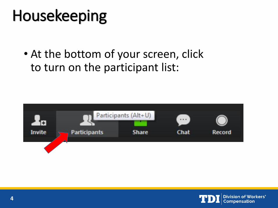
Housekeeping
• At the bottom of your screen, click to turn on the participant list:
5
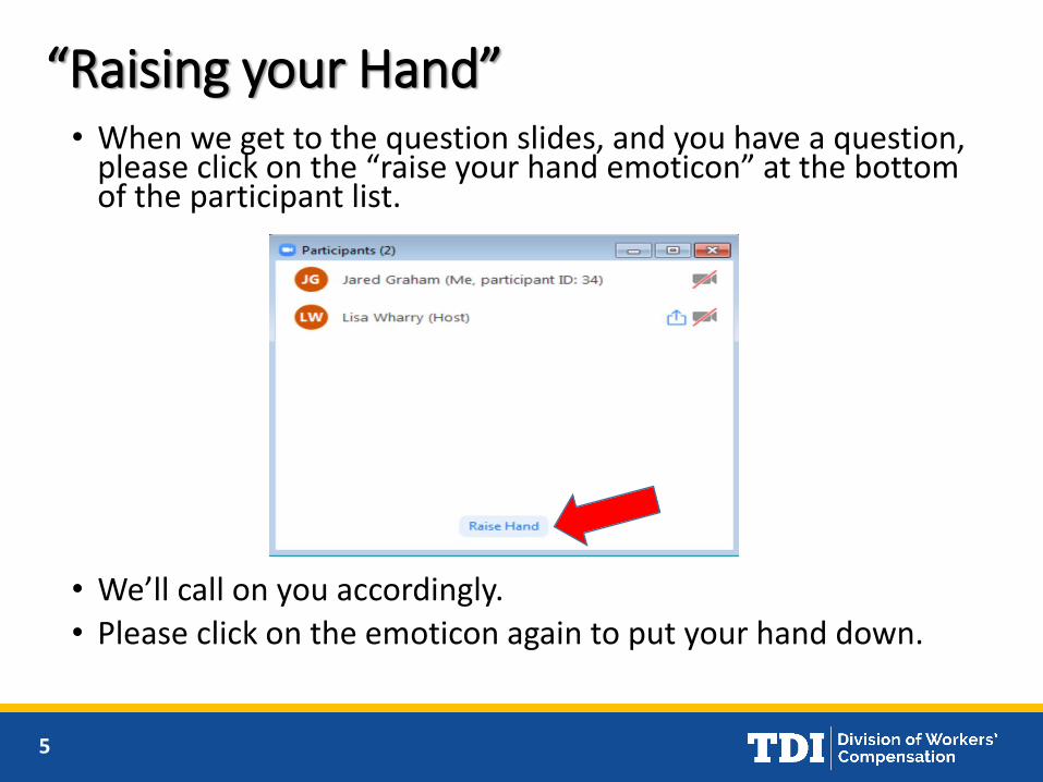
“Raising your Hand”• When we get to the question slides, and you have a question,
please click on the “raise your hand emoticon” at the bottom of the participant list.
• We’ll call on you accordingly.
• Please click on the emoticon again to put your hand down.
6
Today’s Game Plan
• Present Concepts via PowerPoint
• Review 3 cases you were asked to answer
• Q&A for the other cases and topics
• Placeholder slides for questions
• “Any questions…?”
• Raise your hand…
7
Designated Doctor Role
• Objective, neutral medical expert appointed by DWC to
answer specific questions about the medical condition of
the injured employee
• Requires special training and testing
• DD exam may be requested by the insurance carrier, the
injured employee, the Injured employee’s representative,
or DWC
• May not initiate or provide treatment
8
Examinations Conducted by Designated Doctors
Texas Labor Code (TLC) §408.0041 states the specific issues to be
addressed by designated doctors as questions concerning:
• Attainment of Maximum Medical Improvement (MMI)
• Impairment caused by the compensable injury (IR)
• The extent of the employee’s compensable Injury (EOI)
• Whether disability is a direct result of the compensable injury
• Ability to return to work (RTW)
• Issues similar to those described above.
9
Designated Doctor Role
Requirements:
• Active medical license and authorized to practice within their scope of licensure and jurisdiction
• Successful completion of DWC authorized Training and Testing
• Only acting in the capacity of a designated doctor when assigned by the division to do so
10
Designated Doctor Role
• Conduct an exam to address the issue(s) identified in the Commissioner’s Order and DWC Form 32 or Presiding Officer’s Directive (POD)
• Utilize the AMA Guides to the Evaluation of Permanent Impairment, 4th Edition for determining the impairment rating
• Consider ODG or other evidence-based medicine when available and appropriate
11
Designated Doctor Role
• Utilize Medical Disability Guidelines (MDG) as guidelines for the evaluation of expected or average return-to-work time frames
• Timely file required reports/forms
12
Importance of the Designated Doctor’s Opinion
• The report of the designated doctor is given presumptive weight in dispute resolution unless the preponderance of the evidence is to the contrary
• DD’s opinion given presumptive weight
• Significant impact on DWC dispute resolution
• Insurance carrier shall be required to pay income and medical benefits based on the designated doctor ’s opinion during a pending dispute
28 TAC 127.10(h)
13
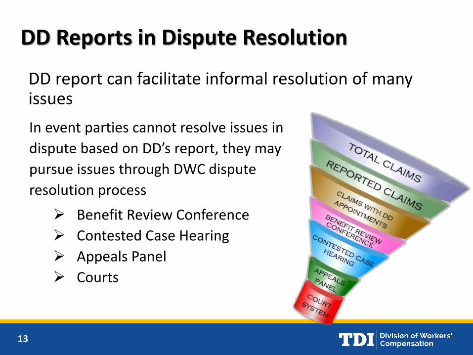
DD report can facilitate informal resolution of many issues
DD Reports in Dispute Resolution
In event parties cannot resolve issues in
dispute based on DD’s report, they may
pursue issues through DWC dispute
resolution process
➢ Benefit Review Conference
➢ Contested Case Hearing
➢ Appeals Panel
➢ Courts
14
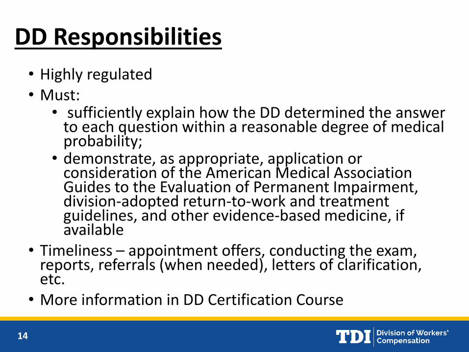
DD Responsibilities
• Highly regulated• Must:
• sufficiently explain how the DD determined the answer to each question within a reasonable degree of medical probability;
• demonstrate, as appropriate, application or consideration of the American Medical Association Guides to the Evaluation of Permanent Impairment, division-adopted return-to-work and treatment guidelines, and other evidence-based medicine, if available
• Timeliness – appointment offers, conducting the exam, reports, referrals (when needed), letters of clarification, etc.
• More information in DD Certification Course
15
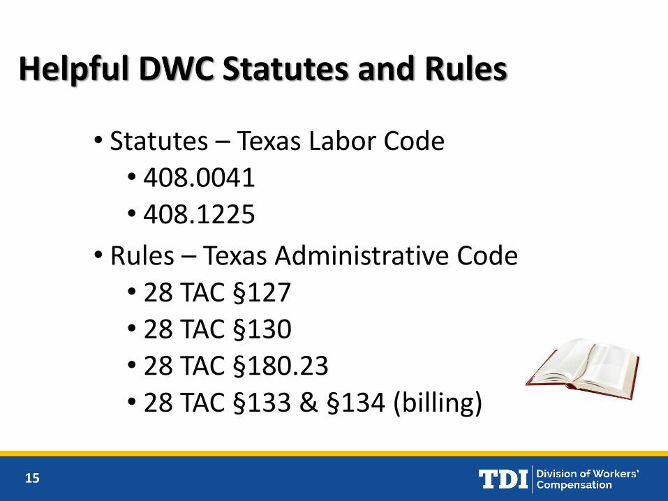
Helpful DWC Statutes and Rules
• Statutes – Texas Labor Code• 408.0041• 408.1225
• Rules – Texas Administrative Code• 28 TAC §127• 28 TAC §130• 28 TAC §180.23• 28 TAC §133 & §134 (billing)

16
Any questions about the role of the designated doctor?
17
Maximum Medical Improvement (MMI)
Question for designated doctor:
Has MMI been reached?
If so, on what date?
18
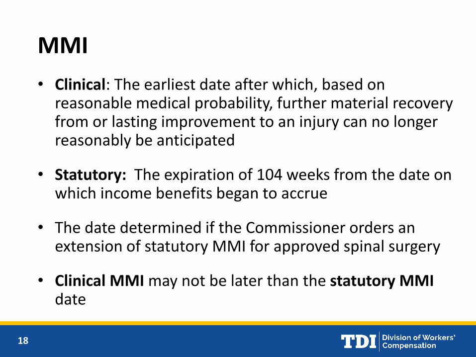
MMI
• Clinical: The earliest date after which, based on reasonable medical probability, further material recovery from or lasting improvement to an injury can no longer reasonably be anticipated
• Statutory: The expiration of 104 weeks from the date on which income benefits began to accrue
• The date determined if the Commissioner orders an extension of statutory MMI for approved spinal surgery
• Clinical MMI may not be later than the statutory MMI date
19
MMI
• Based on the records reviewed and the exam findings, define the compensable injury for certification of MMI and IR and explain this in your report
• Consider the Official Disability Guidelines (ODG), including Appendix D, to determine if, based on reasonable medical probability, additional treatment can be anticipated to result in further material recovery or lasting improvement
• If not at MMI, why not (what is needed to reach MMI)?
20
MMI
• Answer the question from the DWC Form-032
Has MMI been reached; if so, on what date?
• If at MMI, why is the IE at MMI?
• If at MMI, what is the date and why that date?
• “Yes” or “no” and a sufficient explanation why or why not, not just a conclusion!

21
Any questions about MMI concepts or cases?
22
Impairment Rating (IR)
23
Impairment RatingQuestion for Designated Doctor:
On the certified MMI date, what is theimpairment rating?
• Perform thorough, relevant physical examinationof all compensable body areas/systems
• Correlate with the findings in prior medical records
• Make referrals, if necessary, to answer question
• Use 4th Edition of AMA Guides to rate
• Show your work!
24
Impairment Rating
• Assignment of an impairment rating for the current compensable injury shall be based on the injured employee’s condition on the MMI date considering the medical record and the certifying examination
• Assign one whole body impairment rating for the current compensable injury
• Use the rating criteria contained in the appropriate edition of the AMA Guides to the Evaluation of Permanent Impairment

25
Impairment Rating
• Show your work! so that “… any knowledgeable person can compare the clinical findings with the guides criteria and determine whether or not the impairment estimates reflect those criteria.” AMA Guides, page 8
• Document the findings and explain the impairment rating in your narrative report, plus relevant worksheets
• Complete and sign the DWC Form-069
26
Overview of the AMA Guides
• AMA Guides, 4th edition publishedJune 1993
• Effective in the Texas workers’ compensation system October 15, 2001
• 15 Chapters
• Chapters 1 and 2 – Impairment Evaluation; Records and Reports
27
Overview of the AMA Guides
• Chapter 3 – The Musculoskeletal System (Hand and Upper Extremity, Lower Extremity, Spine)
• Approximately 90% of designated doctor examinations involve these 3 body areas

28
Measurements
Consistency of measurements (all measurements, not just ROM)
• Between examiners (pages 7, 8, and 9)
• By the same examiner generally within +/- 10%, (page 9)
• “…plausible and relate to the impairmentbeing evaluated,” (page 8)
• With medical records (page 8)
29
Measurements
• Active, not passive ROM, should be rated(Comparing active with passive may provide useful information)
• Rounding and interpolating are permitted unless the book gives other directions
• DO NOT round impairment rating in DWC system (Not as instructed in the AMA Guides on page 9)
30
Combined Values
Each organ system/body area should be expressedas a whole person impairment, then
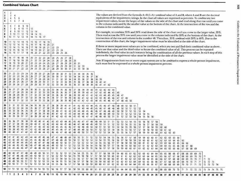
• Whole person impairments should be combinedusing the Combined Values Chart (pp. 322 – 324)
• “Combining” assures that the impairment can’t exceed 100%. It reduces the remaining portion of the whole person that is available for the second impairment
• Example 40% c/w 40% (of the remaining 60%) = 64%
31
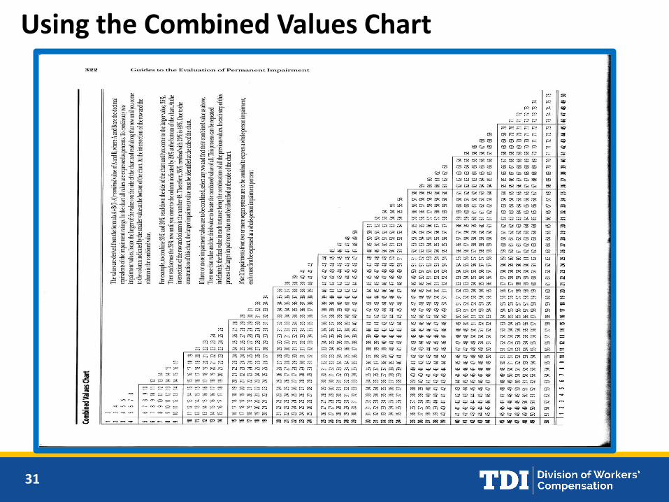
Using the Combined Values Chart
32
Combining 3 or MoreImpairment Values
• “If three or more impairment values are to be combined, select any two and find their combined value as above. Then use that value and the third value to locate the combined value of all. This process can be repeated indefinitely, the final value in each instance being the combination of all the previous values. In each step of this process, the larger impairment value must be identified at the side of the chart.” (page 322)
• Best practice - combine the largest % with the second largest %, then combine with third largest %, etc.

33
Conflict between DWC Statutes/Rules and AMA Guides
DWC Statutes/Rules take precedence
34
Any questions about impairment rating concepts?
35
Spine
• Most common, simplest portion of Ch. 3
• DRE (Diagnosis Related Estimates) aka “the Injury Model” vs. Range of Motion (ROM) model
• DRE is preferred – pp. 94, 99, 101, 112 of the Guides
• DRE should be used for conditions on Table 70 (p. 108) per instructions on p. 94
36
Spine
• Use of the DRE Model is not optional and is to be used unless there is a specific reason why it cannot…Appeal Panel Decision No. 030288
• ROM model – used as a differentiator if DRE does not apply or if there is disagreements between DRE categories - p. 101
• “ROM model” is rarely necessary, however non-uniform loss of ROM is a DRE II differentiator

37
Impairment Rating – SpineDifferentiators - Table 71 p. 109; also pp. 102-107
• Muscle guarding or spasm*
• Non-uniform loss of ROM,
dysmetria
• Non-verifiable radicular complaints
• Loss of relevant reflex(es)
• Decreased muscle
circumference, atrophy (>2 cm)
• Electrodiagnosis (unequivocal
evidence of acute nerve root
compromise)
• Loss of motion segment integrity seen on flexion/extension x-rays
• Loss of bowel or bladder control (rectal exam shows loss of sphincter tone, use of assistive device such as catheter)
• Bladder studies-unequivocal incontinence
• Range of motion model

38
Terminology
• “Cervicothoracic” = Cervical
• “Thoracolumbar” = Thoracic
• “Lumbosacral” = Lumbar
p. 95 Guides
39
SPINE Case 3
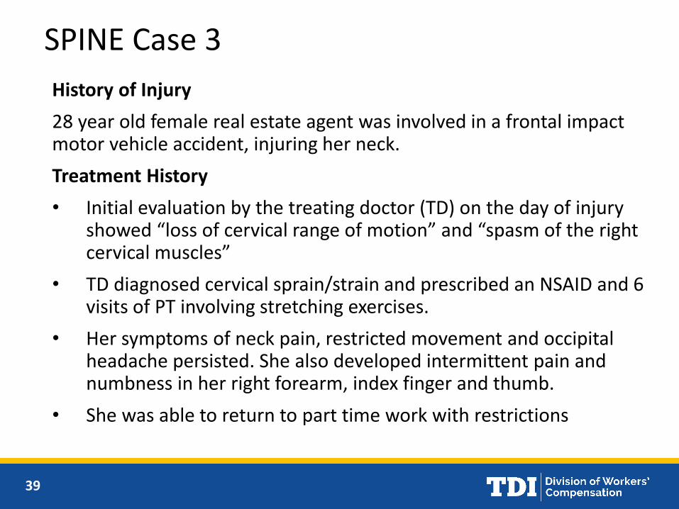
History of Injury
28 year old female real estate agent was involved in a frontal impact motor vehicle accident, injuring her neck.
Treatment History
• Initial evaluation by the treating doctor (TD) on the day of injury showed “loss of cervical range of motion” and “spasm of the right cervical muscles”
• TD diagnosed cervical sprain/strain and prescribed an NSAID and 6 visits of PT involving stretching exercises.
• Her symptoms of neck pain, restricted movement and occipital headache persisted. She also developed intermittent pain and numbness in her right forearm, index finger and thumb.
• She was able to return to part time work with restrictions
40
SPINE Case 3
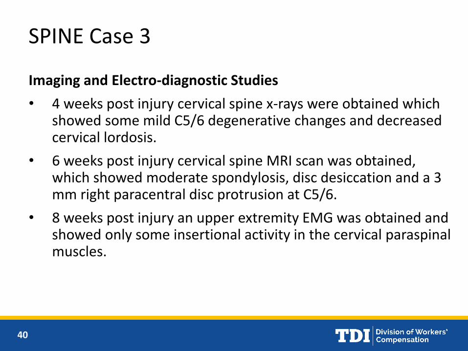
Imaging and Electro-diagnostic Studies
• 4 weeks post injury cervical spine x-rays were obtained which showed some mild C5/6 degenerative changes and decreased cervical lordosis.
• 6 weeks post injury cervical spine MRI scan was obtained, which showed moderate spondylosis, disc desiccation and a 3 mm right paracentral disc protrusion at C5/6.
• 8 weeks post injury an upper extremity EMG was obtained and showed only some insertional activity in the cervical paraspinalmuscles.
41
SPINE Case 3
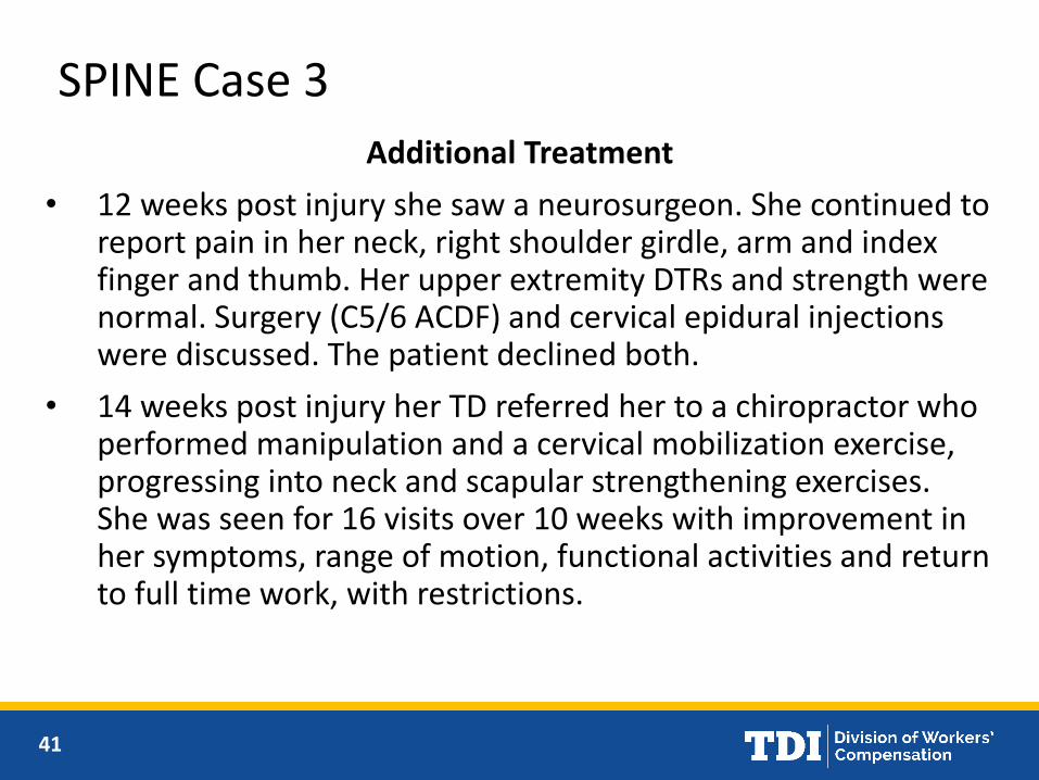
Additional Treatment
• 12 weeks post injury she saw a neurosurgeon. She continued to report pain in her neck, right shoulder girdle, arm and index finger and thumb. Her upper extremity DTRs and strength were normal. Surgery (C5/6 ACDF) and cervical epidural injections were discussed. The patient declined both.
• 14 weeks post injury her TD referred her to a chiropractor who performed manipulation and a cervical mobilization exercise, progressing into neck and scapular strengthening exercises. She was seen for 16 visits over 10 weeks with improvement in her symptoms, range of motion, functional activities and return to full time work, with restrictions.
42
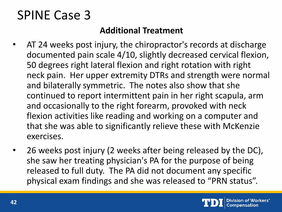
SPINE Case 3Additional Treatment
• AT 24 weeks post injury, the chiropractor's records at discharge documented pain scale 4/10, slightly decreased cervical flexion, 50 degrees right lateral flexion and right rotation with right neck pain. Her upper extremity DTRs and strength were normal and bilaterally symmetric. The notes also show that she continued to report intermittent pain in her right scapula, arm and occasionally to the right forearm, provoked with neck flexion activities like reading and working on a computer and that she was able to significantly relieve these with McKenzie exercises.
• 26 weeks post injury (2 weeks after being released by the DC), she saw her treating physician's PA for the purpose of being released to full duty. The PA did not document any specific physical exam findings and she was released to “PRN status”.
43
SPINE Case 3
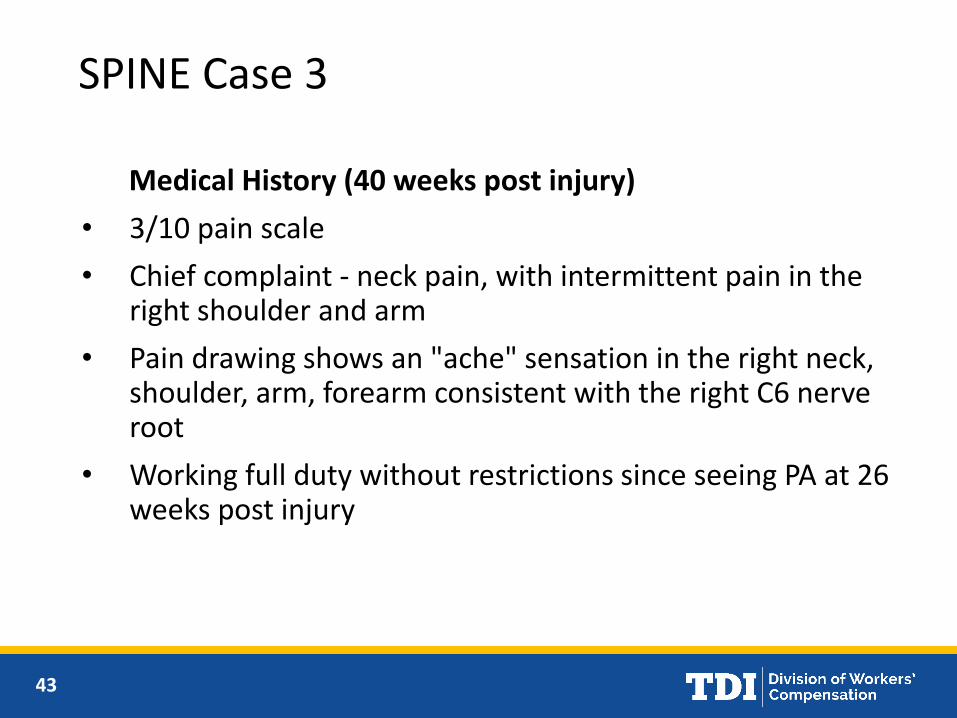
DD Medical History (40 weeks post injury)
• 3/10 pain scale
• Chief complaint - neck pain, with intermittent pain in the right shoulder and arm
• Pain drawing shows an "ache" sensation in the right neck, shoulder, arm, forearm consistent with the right C6 nerve root
• Working full duty without restrictions since seeing PA at 26 weeks post injury
44
SPINE Case 3
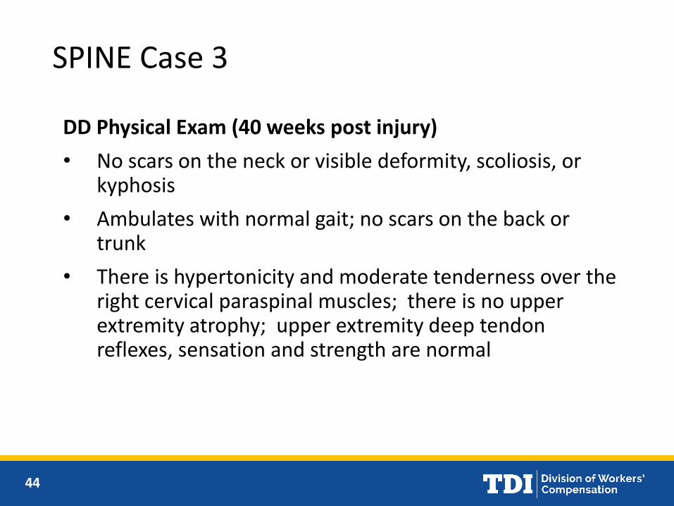
DD Physical Exam (40 weeks post injury)
• No scars on the neck or visible deformity, scoliosis, or kyphosis
• Ambulates with normal gait; no scars on the back or trunk
• There is hypertonicity and moderate tenderness over the right cervical paraspinal muscles; there is no upper extremity atrophy; upper extremity deep tendon reflexes, sensation and strength are normal
45
SPINE Case 3
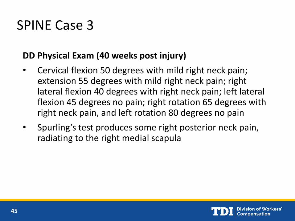
DD Physical Exam (40 weeks post injury)
• Cervical flexion 50 degrees with mild right neck pain; extension 55 degrees with mild right neck pain; right lateral flexion 40 degrees with right neck pain; left lateral flexion 45 degrees no pain; right rotation 65 degrees with right neck pain, and left rotation 80 degrees no pain
• Spurling’s test produces some right posterior neck pain, radiating to the right medial scapula
46
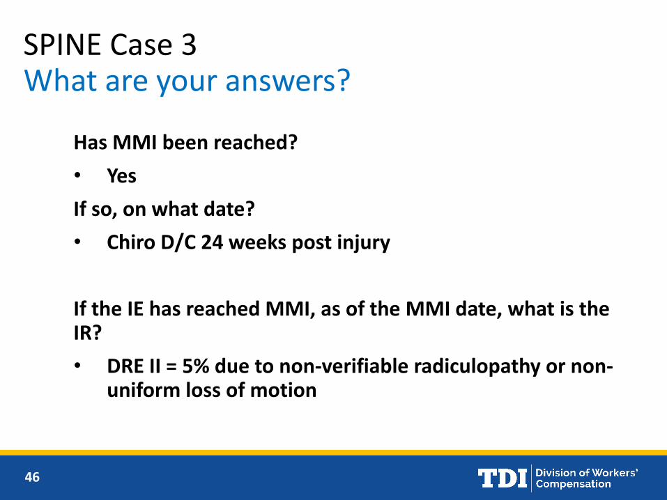
Has MMI been reached?
• Yes
If so, on what date?
• Chiro D/C 24 weeks post injury
If the IE has reached MMI, as of the MMI date, what is the IR?
• DRE II = 5% due to non-verifiable radiculopathy or non-uniform loss of motion
SPINE Case 3What are your answers?
47
Any questions about Spine IR concepts or cases?
48
Hand and Upper Extremity Impairment Sections
Are Different Than The Other Chapters
49
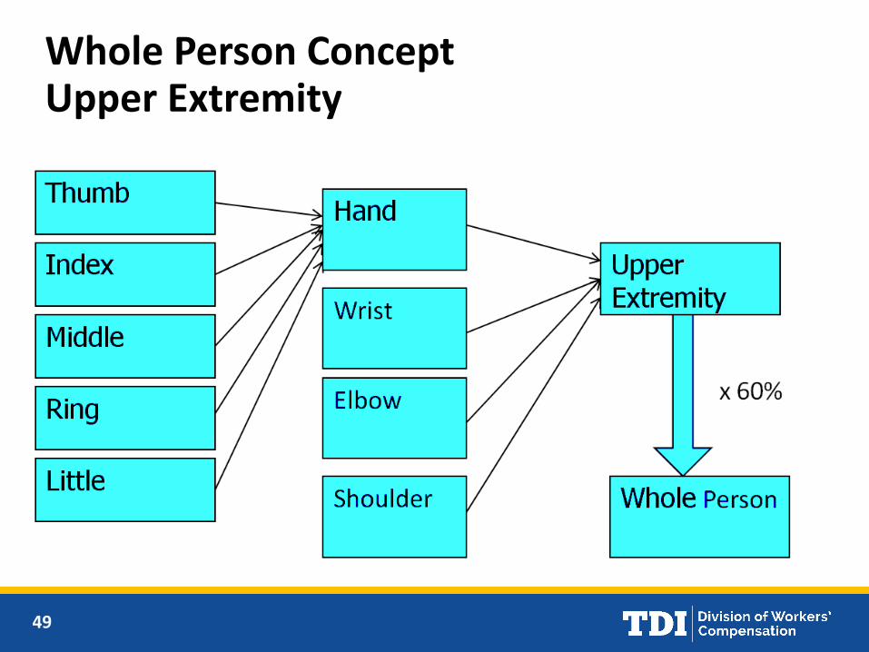
Whole Person ConceptUpper Extremity
50
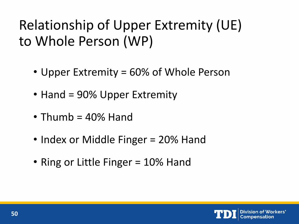
Relationship of Upper Extremity (UE) to Whole Person (WP)
• Upper Extremity = 60% of Whole Person
• Hand = 90% Upper Extremity
• Thumb = 40% Hand
• Index or Middle Finger = 20% Hand
• Ring or Little Finger = 10% Hand

51
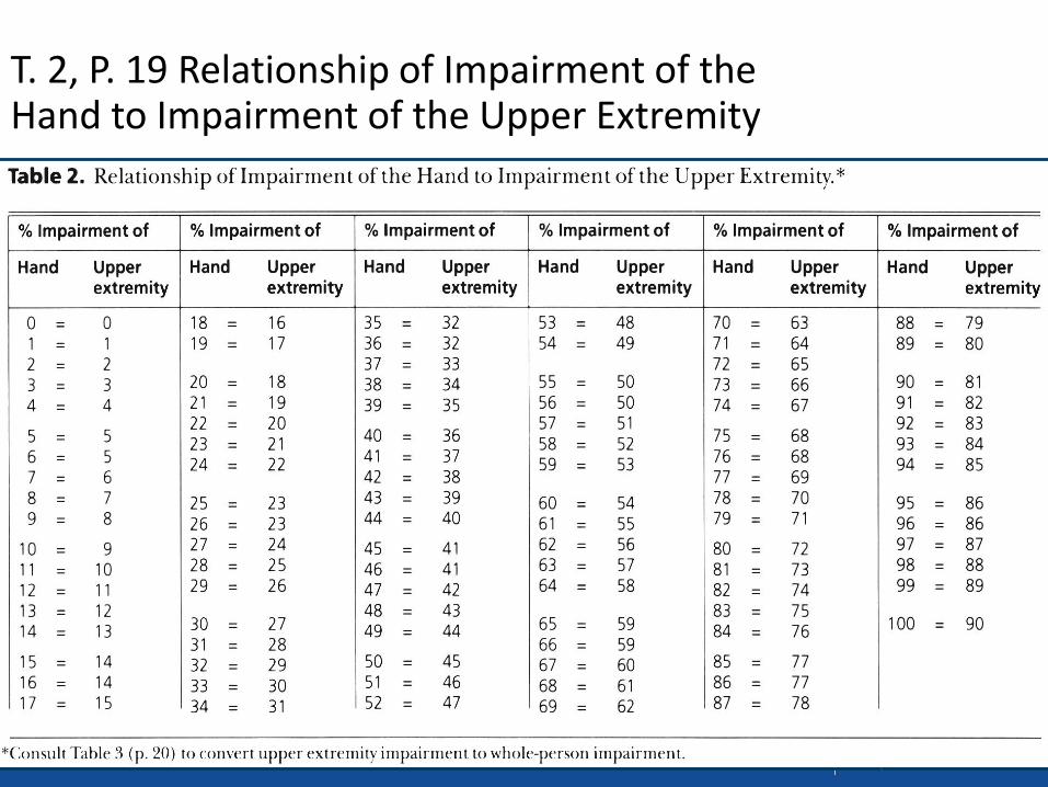
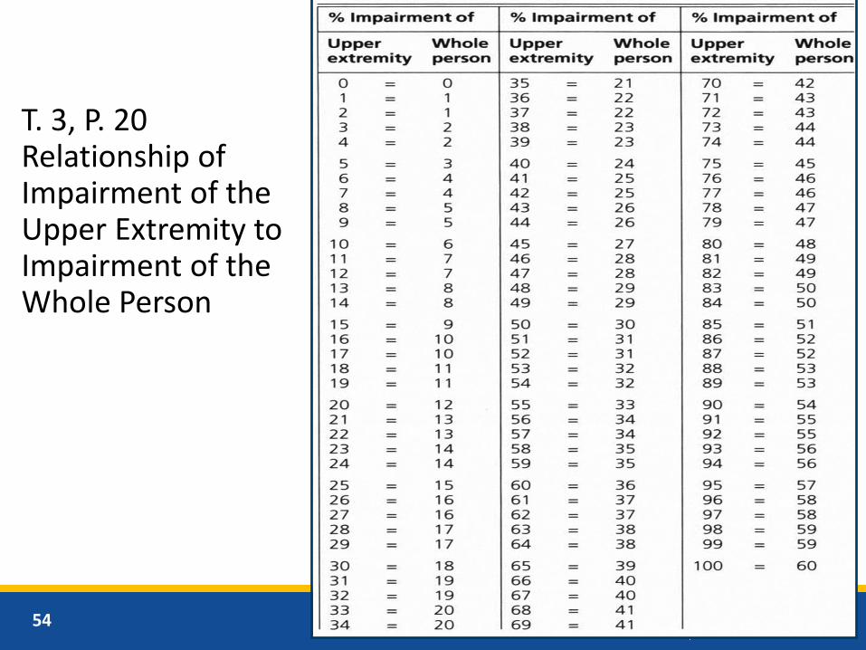
These Impairment Values Have to be Converted to Whole Person by Using:
Table 1, p. 18
Table 2, p. 19
Table 3, p. 20

52
T. 1, P. 18Relationship of Impairment of the Digits to Impairment of the Hand.
53
T. 2, P. 19 Relationship of Impairment of theHand to Impairment of the Upper Extremity
54
T. 3, P. 20Relationship of Impairment of the Upper Extremity to Impairment of the Whole Person
55
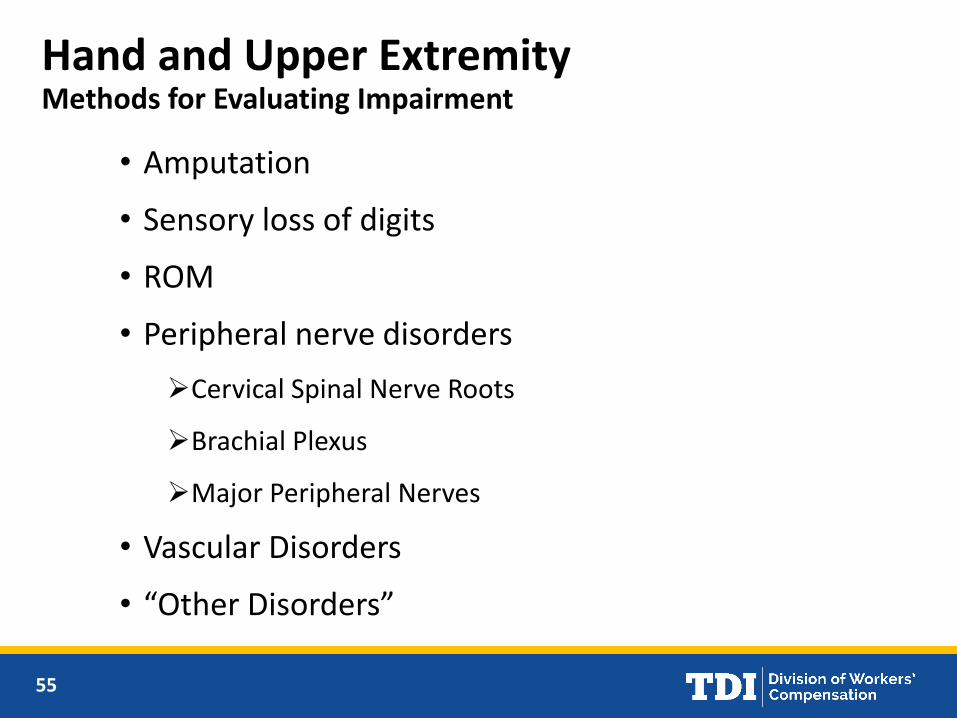
Hand and Upper ExtremityMethods for Evaluating Impairment
• Amputation
• Sensory loss of digits
• ROM
• Peripheral nerve disorders
➢Cervical Spinal Nerve Roots
➢Brachial Plexus
➢Major Peripheral Nerves
• Vascular Disorders
• “Other Disorders”

56
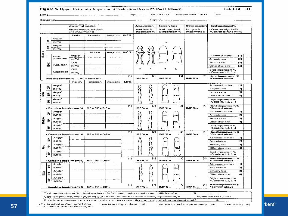
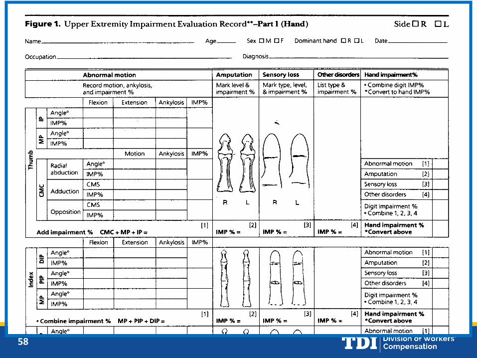
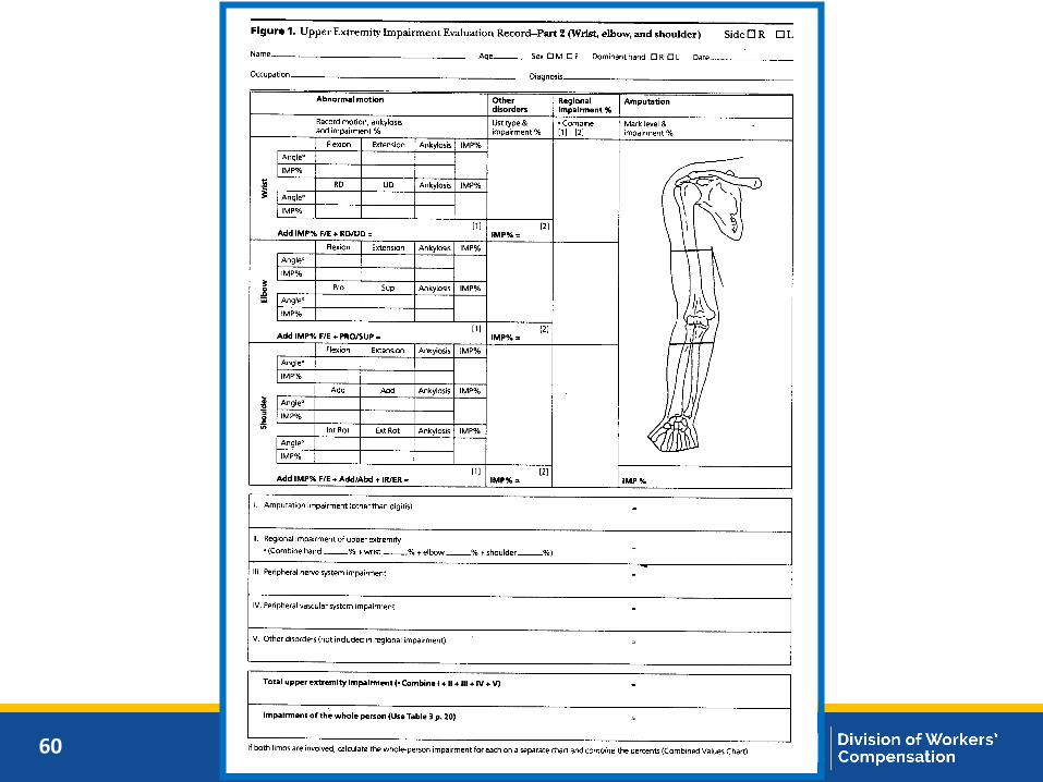
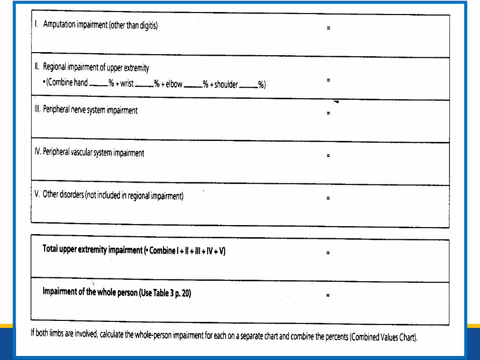
Use Figure 1!!!
57
58

59
60
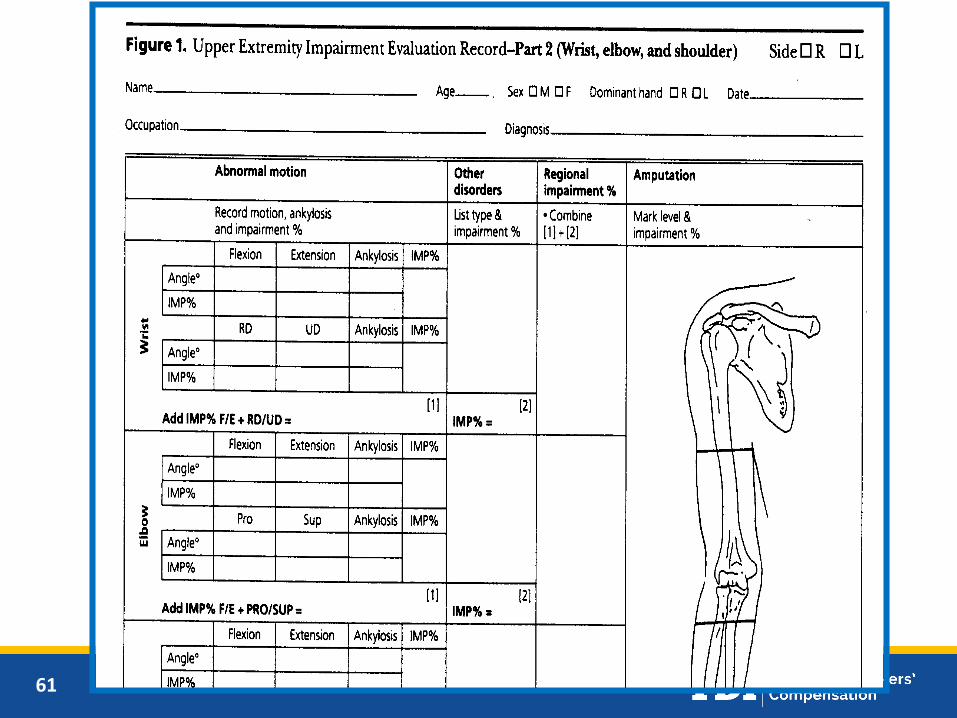
61
62
63
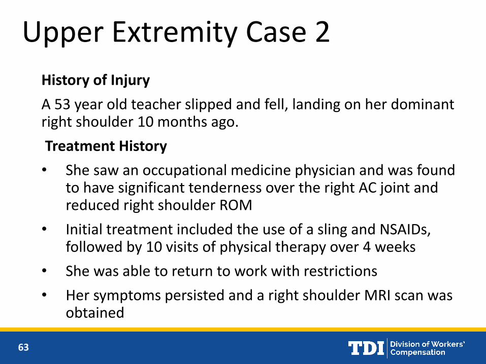
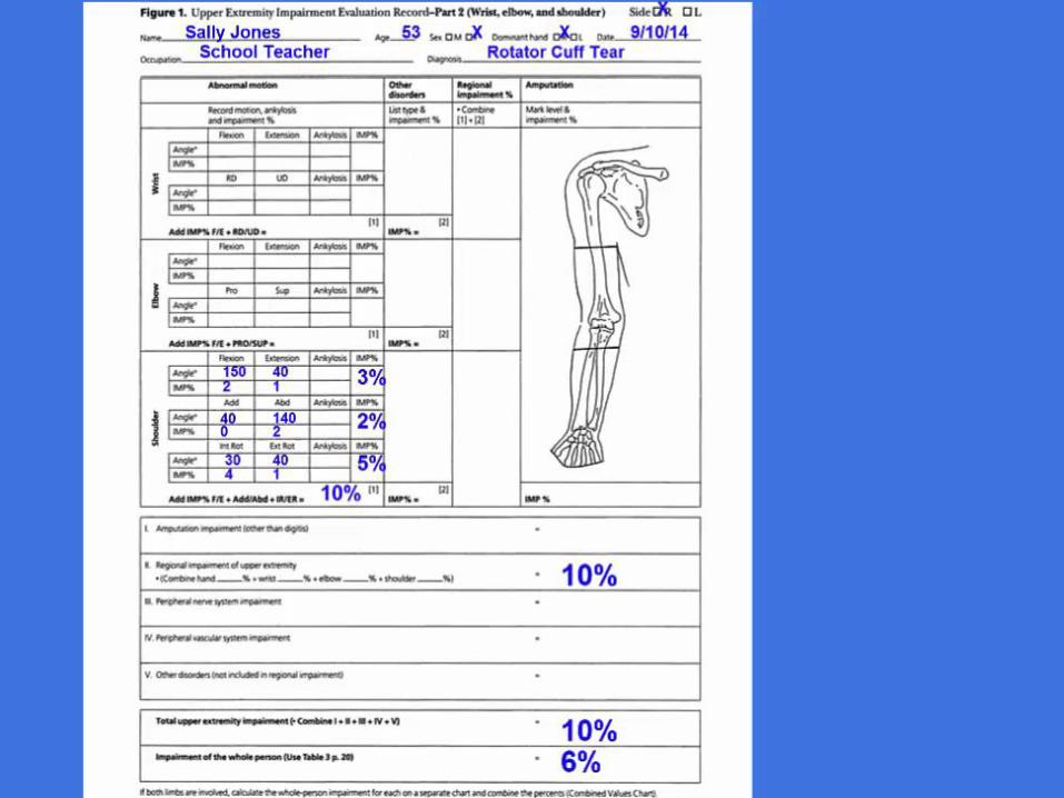
Upper Extremity Case 2
History of Injury
A 53 year old teacher slipped and fell, landing on her dominant right shoulder 10 months ago.
Treatment History
• She saw an occupational medicine physician and was found to have significant tenderness over the right AC joint and reduced right shoulder ROM
• Initial treatment included the use of a sling and NSAIDs, followed by 10 visits of physical therapy over 4 weeks
• She was able to return to work with restrictions
• Her symptoms persisted and a right shoulder MRI scan was obtained
64
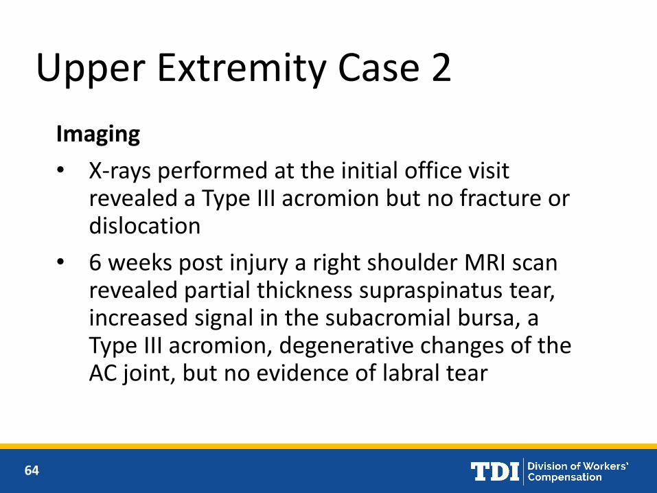
Upper Extremity Case 2
Imaging
• X-rays performed at the initial office visit revealed a Type III acromion but no fracture or dislocation
• 6 weeks post injury a right shoulder MRI scan revealed partial thickness supraspinatus tear, increased signal in the subacromial bursa, a Type III acromion, degenerative changes of the AC joint, but no evidence of labral tear
65
Upper Extremity Case 2
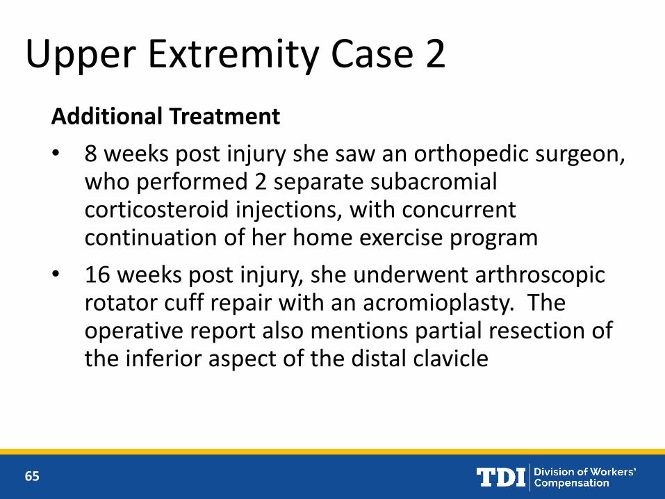
Additional Treatment
• 8 weeks post injury she saw an orthopedic surgeon, who performed 2 separate subacromialcorticosteroid injections, with concurrent continuation of her home exercise program
• 16 weeks post injury, she underwent arthroscopic rotator cuff repair with an acromioplasty. The operative report also mentions partial resection of the inferior aspect of the distal clavicle
66
Upper Extremity Case 2
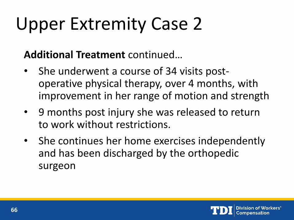
Additional Treatment continued…
• She underwent a course of 34 visits post-operative physical therapy, over 4 months, with improvement in her range of motion and strength
• 9 months post injury she was released to return to work without restrictions.
• She continues her home exercises independently and has been discharged by the orthopedic surgeon

67
Upper Extremity Case 2
DD Medical History (10 months post injury)
• Chief complaint right shoulder pain with overhead activities
• No reported prior history of evaluation or treatment of shoulder condition prior to this work injury
68
Upper Extremity Case 2
DD Physical Exam (10 months post injury)
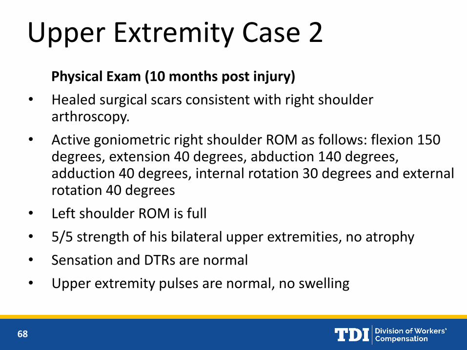
• Healed surgical scars consistent with right shoulder arthroscopy.
• Active goniometric right shoulder ROM as follows: flexion 150 degrees, extension 40 degrees, abduction 140 degrees, adduction 40 degrees, internal rotation 30 degrees and external rotation 40 degrees
• Left shoulder ROM is full
• 5/5 strength of his bilateral upper extremities, no atrophy
• Sensation and DTRs are normal
• Upper extremity pulses are normal, no swelling
69
Upper Extremity Case 2What are your answers?
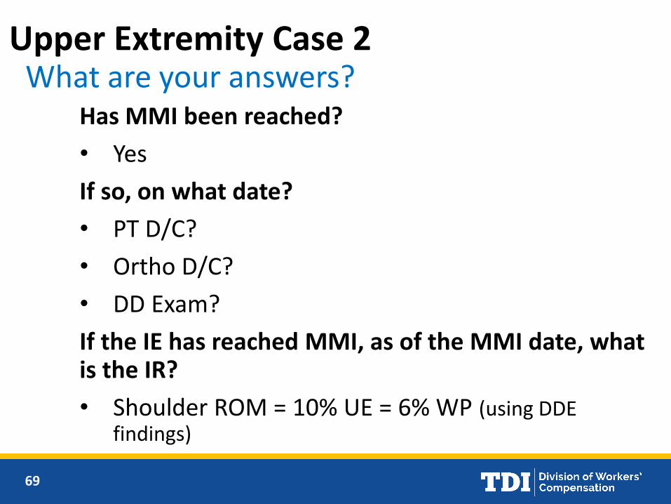
Has MMI been reached?
• Yes
If so, on what date?
• PT D/C?
• Ortho D/C?
• DD Exam?
If the IE has reached MMI, as of the MMI date, what is the IR?
• Shoulder ROM = 10% UE = 6% WP (using DDE findings)
70
71
Any questions about UE IR concepts or cases?
72
How to Determine ImpairmentRating Lower Extremity
• Calculate impairment according to textand tables for each applicable parameterof the 13 possible methods.
• Determine which parameters canbe combined.
• Select largest and most clinically appropriate method for each region.
73
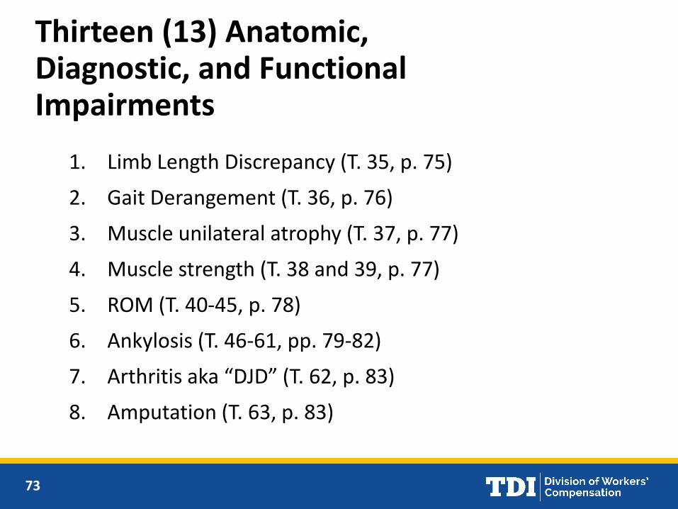
Thirteen (13) Anatomic, Diagnostic, and Functional Impairments
1. Limb Length Discrepancy (T. 35, p. 75)
2. Gait Derangement (T. 36, p. 76)
3. Muscle unilateral atrophy (T. 37, p. 77)
4. Muscle strength (T. 38 and 39, p. 77)
5. ROM (T. 40-45, p. 78)
6. Ankylosis (T. 46-61, pp. 79-82)
7. Arthritis aka “DJD” (T. 62, p. 83)
8. Amputation (T. 63, p. 83)
74
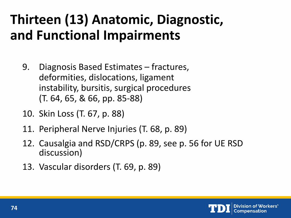
Thirteen (13) Anatomic, Diagnostic, and Functional Impairments
9. Diagnosis Based Estimates – fractures,deformities, dislocations, ligamentinstability, bursitis, surgical procedures(T. 64, 65, & 66, pp. 85-88)
10. Skin Loss (T. 67, p. 88)
11. Peripheral Nerve Injuries (T. 68, p. 89)
12. Causalgia and RSD/CRPS (p. 89, see p. 56 for UE RSD discussion)
13. Vascular disorders (T. 69, p. 89)
75
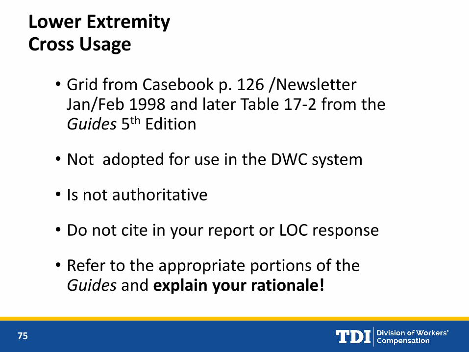
Lower ExtremityCross Usage Tables
• Grid from Casebook p. 126 /NewsletterJan/Feb 1998 and later Table 17-2 from theGuides 5th Edition
• Not adopted for use in the DWC system
• Is not authoritative
• Do not cite in your report or LOC response
• Refer to the appropriate portions of theGuides and explain your rationale!
76
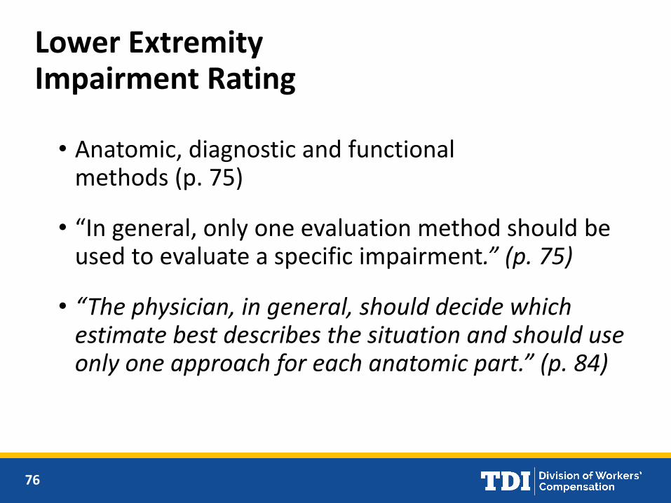
Lower Extremity Impairment Rating Concepts
• Anatomic, diagnostic and functionalmethods (p. 75)
• “In general, only one evaluation method should be used to evaluate a specific impairment.” (p. 75)
• “The physician, in general, should decide which estimate best describes the situation and should use only one approach for each anatomic part.” (p. 84)
77
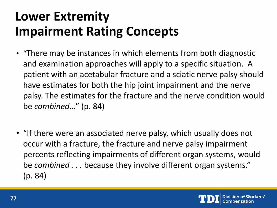
Lower Extremity Impairment Rating Concepts
• “There may be instances in which elements from both diagnostic and examination approaches will apply to a specific situation. A patient with an acetabular fracture and a sciatic nerve palsy should have estimates for both the hip joint impairment and the nerve palsy. The estimates for the fracture and the nerve condition would be combined…” (p. 84)
• “If there were an associated nerve palsy, which usually does not occur with a fracture, the fracture and nerve palsy impairment percents reflecting impairments of different organ systems, would be combined . . . because they involve different organ systems.” (p. 84)
78
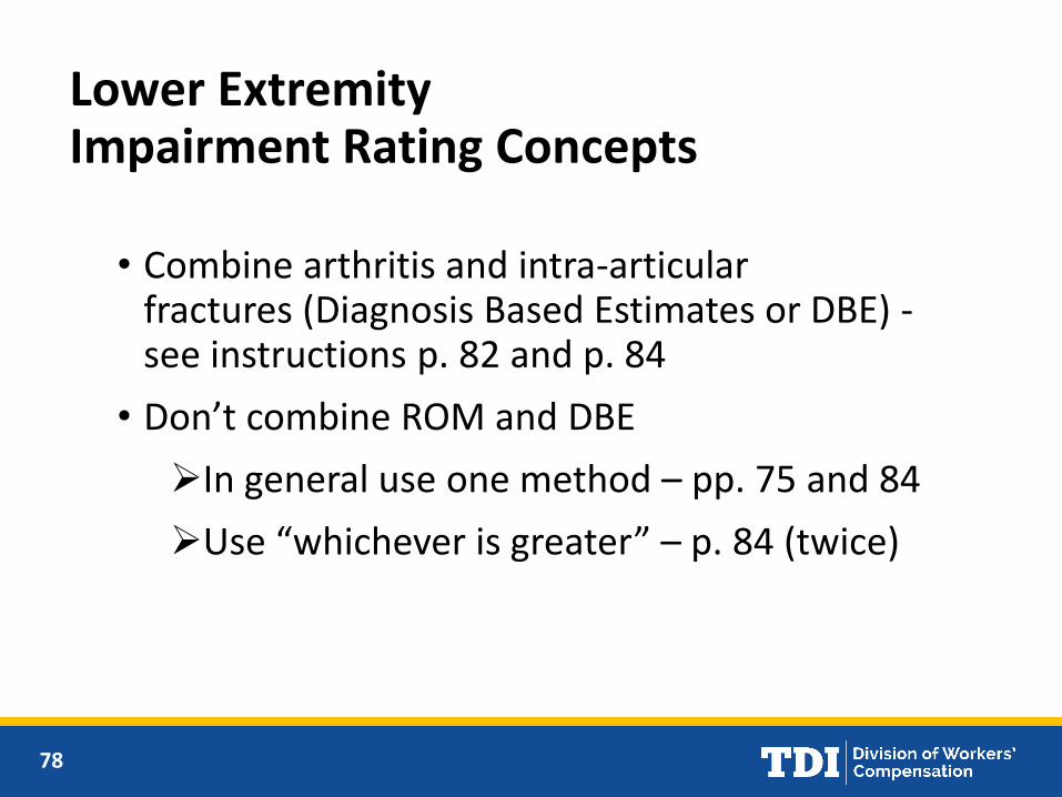
Lower Extremity Impairment Rating Concepts
• Combine arthritis and intra-articularfractures (Diagnosis Based Estimates or DBE) -see instructions p. 82 and p. 84
• Don’t combine ROM and DBE
➢In general use one method – pp. 75 and 84
➢Use “whichever is greater” – p. 84 (twice)
79
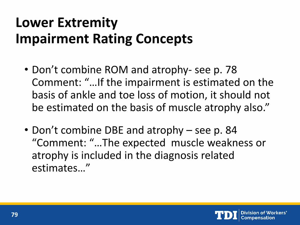
Lower Extremity Impairment Rating Concepts
• Don’t combine ROM and atrophy- see p. 78 Comment: “…If the impairment is estimated on the basis of ankle and toe loss of motion, it should not be estimated on the basis of muscle atrophy also.”
• Don’t combine DBE and atrophy – see p. 84 “Comment: “…The expected muscle weakness or atrophy is included in the diagnosis related estimates…”
80
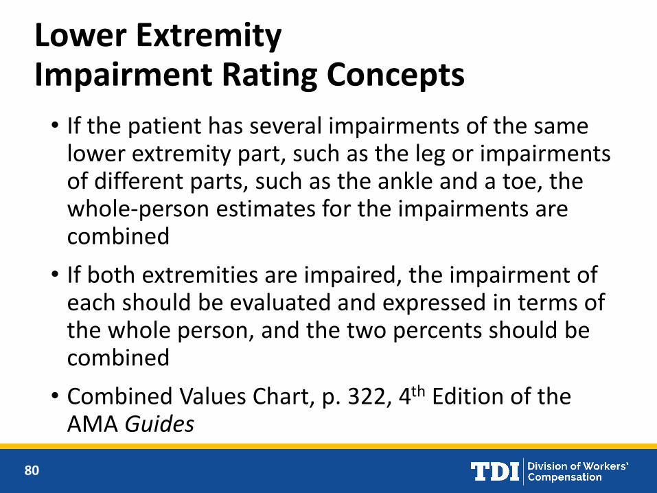
Lower Extremity Impairment Rating Concepts
• If the patient has several impairments of the same lower extremity part, such as the leg or impairments of different parts, such as the ankle and a toe, the whole-person estimates for the impairments are combined
• If both extremities are impaired, the impairment of each should be evaluated and expressed in terms of the whole person, and the two percents should be combined
• Combined Values Chart, p. 322, 4th Edition of the AMA Guides
81
Combined Values Chart
82
Lower Extremity Impairment Rating Concepts
• All tables show percentages in lower extremity (LE) and whole person (WP)
• Lower extremity = 40% WP but impairment values are expressed and combined at WP level, for both same LE part (i.e. ankle), or for different parts of the LE (i.e. ankle and knee)

83
Lower Extremity Impairment Rating Concepts
• The Lower Extremity is weighted at0.40 or 40% Whole Person
• Never exceed amputation value –APD 111720
• Lower Extremity impairmentscalculated to exceed 100% are rated at the amputation value
84
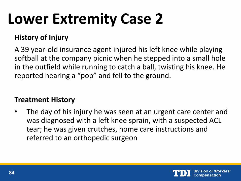
Lower Extremity Case 2History of Injury
A 39 year-old insurance agent injured his left knee while playing softball at the company picnic when he stepped into a small hole in the outfield while running to catch a ball, twisting his knee. He reported hearing a “pop” and fell to the ground.
Treatment History
• The day of his injury he was seen at an urgent care center and was diagnosed with a left knee sprain, with a suspected ACL tear; he was given crutches, home care instructions and referred to an orthopedic surgeon

85
Lower Extremity Case 2
Treatment History continued…
• 2 weeks post injury he saw an orthopedic surgeon, who found him to be on crutches, have significant knee effusion, decreased ROM and a positive Lachman’s sign and significant valgus instability
• 3 weeks post injury a right knee MRI scan was obtained showing grade III tears of the anterior cruciate ligament (ACL) and medial collateral ligament (MCL), and a tear of the posterior horn of the lateral meniscus
• The orthopedic surgeon recommended initial non-operative management including a hinged brace, continuing home care and a trial of physical therapy for 6 weeks, focusing on ROM, and progression of exercises
86
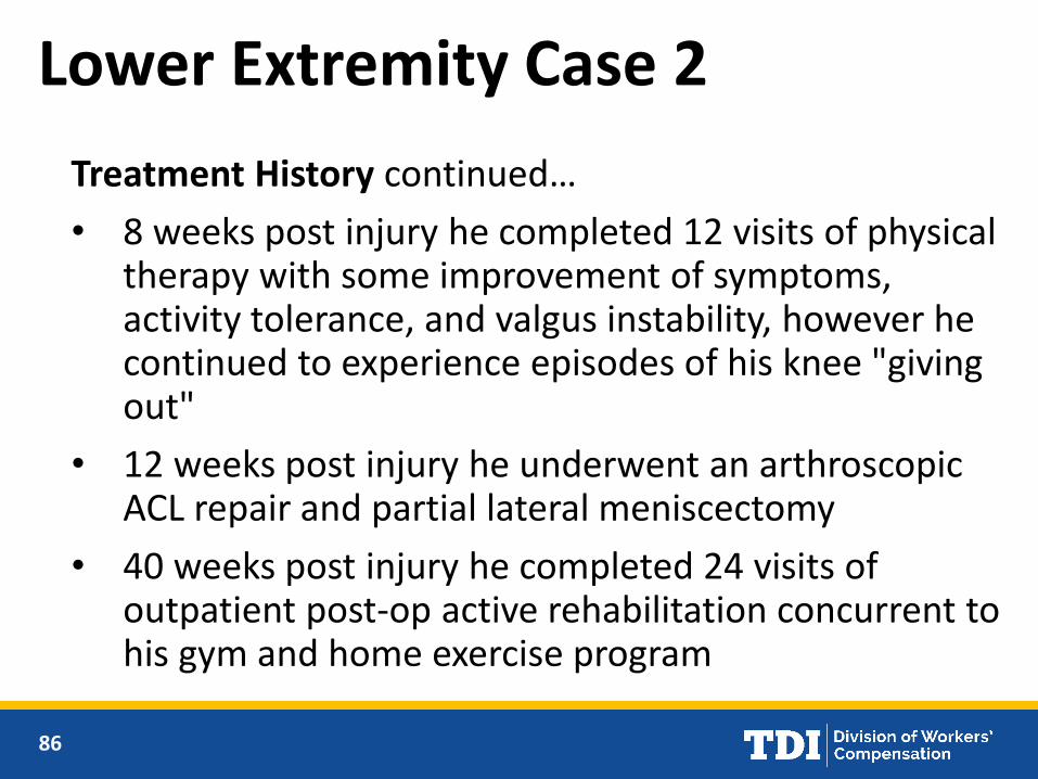
Lower Extremity Case 2
Treatment History continued…
• 8 weeks post injury he completed 12 visits of physical therapy with some improvement of symptoms, activity tolerance, and valgus instability, however he continued to experience episodes of his knee "giving out"
• 12 weeks post injury he underwent an arthroscopic ACL repair and partial lateral meniscectomy
• 40 weeks post injury he completed 24 visits of outpatient post-op active rehabilitation concurrent to his gym and home exercise program
87
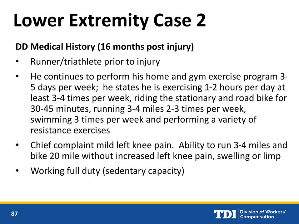
Lower Extremity Case 2DD Medical History (16 months post injury)
• Runner/triathlete prior to injury
• He continues to perform his home and gym exercise program 3-5 days per week; he states he is exercising 1-2 hours per day at least 3-4 times per week, riding the stationary and road bike for 30-45 minutes, running 3-4 miles 2-3 times per week, swimming 3 times per week and performing a variety of resistance exercises
• Chief complaint mild left knee pain. Ability to run 3-4 miles and bike 20 mile without increased left knee pain, swelling or limp
• Working full duty (sedentary capacity)
88
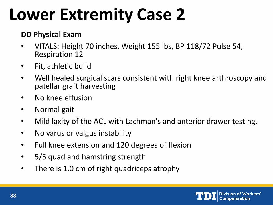
Lower Extremity Case 2DD Physical Exam
• VITALS: Height 70 inches, Weight 155 lbs, BP 118/72 Pulse 54, Respiration 12
• Fit, athletic build
• Well healed surgical scars consistent with right knee arthroscopy and patellar graft harvesting
• No knee effusion
• Normal gait
• Mild laxity of the ACL with Lachman's and anterior drawer testing.
• No varus or valgus instability
• Full knee extension and 120 degrees of flexion
• 5/5 quad and hamstring strength
• There is 1.0 cm of right quadriceps atrophy
89
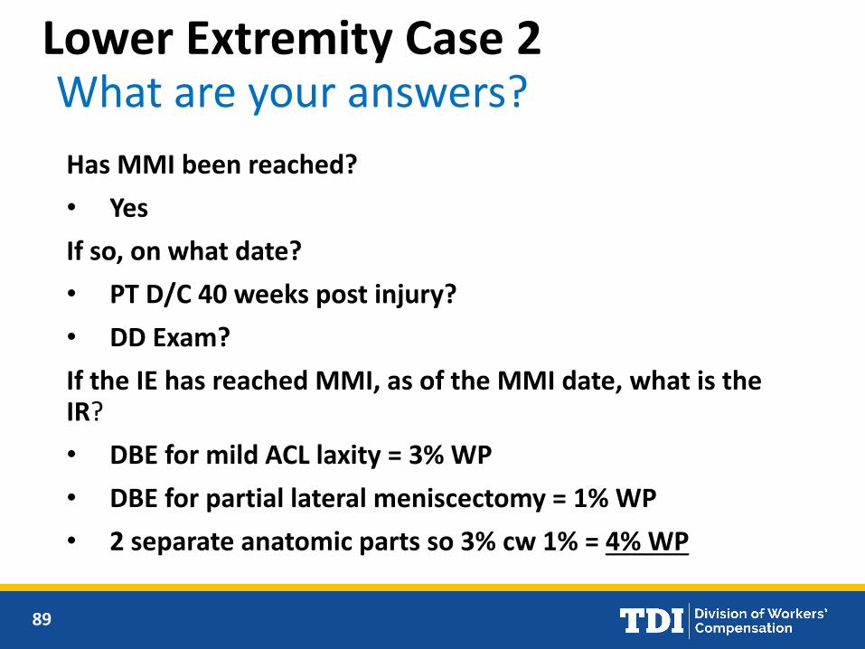
Lower Extremity Case 2What are your answers?
Has MMI been reached?
• Yes
If so, on what date?
• PT D/C 40 weeks post injury?
• DD Exam?
If the IE has reached MMI, as of the MMI date, what is the IR?
• DBE for mild ACL laxity = 3% WP
• DBE for partial lateral meniscectomy = 1% WP
• 2 separate anatomic parts so 3% cw 1% = 4% WP

90
Any questions about LE IR concepts or cases?

91
Extent of Injury (EOI)
92
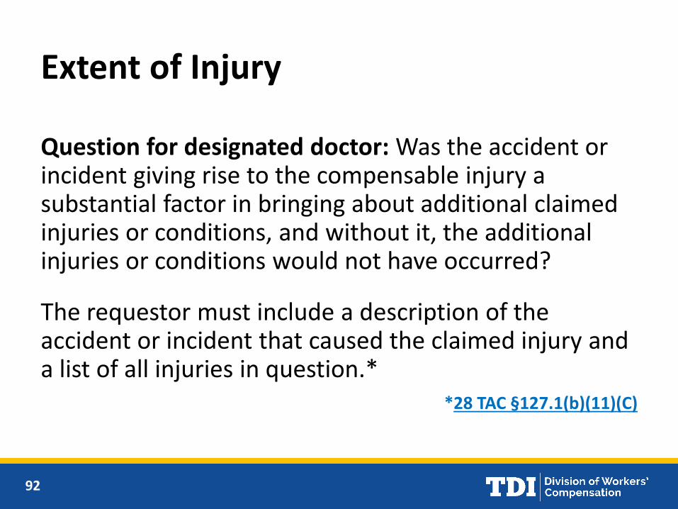
Extent of Injury
Question for designated doctor: Was the accident or incident giving rise to the compensable injury a substantial factor in bringing about additional claimed injuries or conditions, and without it, the additional injuries or conditions would not have occurred?
The requestor must include a description of the accident or incident that caused the claimed injury and a list of all injuries in question.*
*28 TAC §127.1(b)(11)(C)
93
Extent of Injury
More detailed information provided in the DD Certification Course

94
Any questions about EOI?

95
Return to Work (RTW)

96
Return to Work
Questions for designated doctor:
• Is the injured employee able to return to work in any capacity and what work activities can the injured employee perform?
• The designated doctor must also submit one or more DWC Form-073, Work Status Report, to cover the relevant time periods

97
Return to Work
Dates:
The requestor must provide the beginning and ending dates for each period covered by the request if requesting the designated doctor to address a time period other than the present
98
Return to Work (SIBs)
Return to Work for Supplemental Income Benefits (SIBs) – Box 36 Block F
Question for designated doctor: Has the injured employee’s medical condition improved sufficiently to allow the employee to return to work in any capacity for the identified qualifying period(s)?
99
Return to Work (SIBs)
The requestor will provide beginning and endingdates for the SIBs qualifying periods in question.The designated doctor must address all periods in which the injured employee could or could not work
Designated doctors must review and list the medical records (if any exist) for the relevant qualifying period

100
Any questions about RTW?
101
DD Training and Testing
• Training
– Required Certification course
–Optional workshops
• Testing
– PSI test centers http://www.psiexams.com/ or (800) 733-9267
–MD/DO tests – 60 questions
– DC tests - 60 questions
– Up to 5 hours to complete
102
Designated Doctor Resources
• Designated doctor websitehttp://www.tdi.texas.gov/wc/dd/index.html
• Outreach training to system stakeholders
http://www.tdi.texas.gov/wc/events/index.html
103
Designated Doctor Resources
• Presentations from the Designated Doctor Certification Coursehttp://www.tdi.texas.gov/wc/dd/certtraining.html
104
Comp Connection for Health Care Providers
Health care providers can obtain practical information and guidance on issues commonly encountered when treating injured employees such as:
• Treatment guidelines
• Pharmacy Formulary
• Billing and reimbursement
• Workers’ compensation forms
• Licensing and certification requirements

105
800-252-7031 opt. #3

106
Questions?

107


![[d], [o] If one doctor doctors another doctor does the doctor who doctors the doctor doctor the doctor the way the doctor he is doctoring doctors? Or](https://img.pdfslide.net/doc/110x75/56649e995503460f94b9c732/d-o-if-one-doctor-doctors-another-doctor-does-the-doctor-who-doctors-the.jpg)


![Ramon Nazareno vs Maersk Filipinas Crewing [Gr168703 February 26 2013] = Seafarer's Personal Doctor vs Company Designated Doctor](https://img.pdfslide.net/doc/110x75/577cd6a21a28ab9e789cd7d3/ramon-nazareno-vs-maersk-filipinas-crewing-gr168703-february-26-2013-seafarers.jpg)