Embed Size (px)
Citation preview
Designer Fentanyls Drugs that kill and how to detect themCarfentanil
2
Simon Hudson, Sport and Specialised Analytical Services, LGC Standards, Fordham, UK.
Recent communications from Public Health England (PHE) and the National Crime Agency (NCA) have highlighted an increased awareness of the recreational use of fentanyl analogues in the UK with multiple deaths associated with the inadvertent consumption of carfentanil contained in heroin1,2.This is a situation already encountered in other parts of the world, in particular the USA.3
LGC’s drug testing laboratory based in Fordham in the UK performs specialised analyses for forensics laboratories including those doing work for UK coroners. This includes testing for synthetic cannabinoid receptor agonists, other new psychoactive substances (NPS), drugs of abuse and prescription drugs. The technology used is Thermo Scientific™ Orbitrap™-based high-resolution accurate-mass (HRAM) liquid chromatography-mass spectrometry (LCMS) as it enables extremely broad analyte coverage at high sensitivity.
In March 2017, routine testing for NPS and other drugs detected the presence of carfentanil in a post mortem blood case. Over the next 6 months, carfentanil was detected in a further 69 cases in either blood or urine or both. Other fentanyl analogues were often detected in conjunction with carfentanil and these included in order of frequency, fentanyl, alfentanil, isobutyryl/butyrylfentanyl and 4-fluoroisobutyryl/butyrylfentanyl,fluorofentanyl, acetylfentanyl and furanylfentanyl.
Carfentanil is an analogue of fentanyl with a potency of 100 times that of fentanyl and 10,000 times that of morphine. It is believed to be active in humans at doses of 1µg and lethal at doses of 20µg. A sample of heroin tested at LGC for an external private forensic drugs testing laboratory was found to contain carfentanil at a level of 0.2%, i.e. 2µg per 1mg of heroin. A typical dose of heroin is 100mg so this would contain 200µg which is 10 times the supposed lethal dose.
At the time carfentanil was firstdetected by LGC there was very little information in the scientific literature regarding its metabolism. In vitro studies had suggested metabolism through hydroxylation and dealkylation4.
The detection of high potency fentanyl analogues certainly presents a significant analytical challenge to many laboratories and it’s widely held that the true extent of deaths related to the use, either intentional or unintentional, of carfentanil is not certain due to the varied analytical capabilities of laboratories performing the testing of post mortem biological fluids.3
Science for a safer world
The metabolism of carfentanil and its detection in blood and urine.
This paper is intended to share knowledge from LGC’s laboratories regarding:
1. Analytical methodology enabling the detection of low levels of many drugs including fentanyl analogues
2. Carfentanil in vivo metabolism information from post mortem samples.
3
Urine
Urine samples (50µl to 1ml depending on available volume) were prepared for analysis by the addition of 1ml of 1M pH 6.3 phosphate buffer containing D3 morphine glucuronide, and D3 EDDP. The samples were then hydrolysed overnight at 45ºC using beta glucuronidase derived from Helix pomatia to cleave any glucuronidated phase II metabolites. After centrifugation, the samples were extracted for analysis using a generic solid phase extraction methodology on an Agilent Nexus reversed phase polymer sorbent. Cartridges were condition with methanol, then water prior to loading the entire prepared sample. The cartridges were then washed with hexane and then dried under either full vacuum or a steady flow of nitrogen for 30 seconds. The cartridges were then eluted with two sequential 1ml volumes of 10% methanol in ethyl acetate. The resulting elution from the cartridge was washed in 1.5ml of ultrapure water, the organic layer removed and taken to dryness in a centrifugal evaporation system. The dry residue was reconstituted in LCMS mobile phase and submitted for analysis by full scan Orbitrap HRAM LCMS.
Blood
Blood, blood serum or blood plasma samples (50µl to 1ml depending on available volume) were prepared for analysis as above but without the enzyme hydrolysis stage.
A 10 µL portion of the prepared sample (urine or blood) was injected for analysis onto a Thermo ScientificTM UltiMate™ Closed Sampler XRS Ultra-High Performance Liquid Chromatography (UHPLC) system, interfaced to a Thermo ScientificTM Q ExactiveTM Focus hybrid quadrupole-OrbitrapTM mass spectrometer, operating in heated positive ion electrospray mode. Chromatographic separation was achieved in 5.0 minutes on a Waters Atlantis T3 HPLC column maintained at 40°C using a gradient consisting of a mixture of 0.1% acetic acid (A) and acetonitrile containing 0.1% acetic acid (B). Using a flow rate of 400µl/minute, initial solvent conditions were 99% A and 1% B. After 0.3 minutes Solvent B was ramped to 9% over the next 0.9 minutes, to 30% over the next 0.8 minutes, to 43% over the next 0.65 minutes, to 65% over the next 0.35 minutes and then to 99% after a total run time of 3.4 minutes. The final conditions were held until 4.5 minutes at which point the solvent composition reverted to 1% B. These conditions were held for a further 2 minutes for equilibration prior to the next injection.
Data were acquired in full scan mode operating at a mass resolution of 70,000 (FWHM) at m/z 200, across a mass range of 80-550 amu. Data dependent scanning was enabled in ’confirmation’ mode utilising an inclusion list of over 800 compounds derived from the in house accurate mass database. The top 3 ions from the inclusion list in each scan were isolated and subjected to an energy spread HCD scan combining collision energies of 15, 35 and 50, to produce an energy spread mass spectrum. A third scan event using ‘all ion fragmentation’ was performed with a stepped HCD setting of 15, 35 and 50 at a mass resolution of 35,000 across a scan range of 50-500 amu.
Acquired data were processed using Thermo ScientificTM ToxFinderTM software against an in-house database containing over 800 NPS and other pharmaceutical drugs. The presence of reported drugs is confirmed in the data through full scan accurate mass determination of molecular ions, identification of accurate mass qualifier ions in AIF, and the automatic generation of accurate mass MS2 data for comparison with mass spectral libraries.
Semi-quantitation was performed using calibrator samples in duplicate containing carfentanil at 50pg/mL and 500pg/mL in each batch. A mean response factor generated from the peak height for the full scan accurate mass for protonated carfentanil against the corresponding data for method internal marker (EDDP D3) was calculated and applied to all unknown carfentanil results. Where other fentanyls were present, the carfentanil calibrators were used as surrogates for these to calculate approximate levels.
The method capability was approximately 5pg/ml for 1ml of blood or urine.
Analytical methodologyCreated with contributions from
4
Positive findings
In the carfentanil cases seen to date, parent drug was seen in all samples with levels from sub 10pg/ml in both blood plasma and urine up to 4000pg/ml in blood plasma and 20750pg/ml in urine. The presence of metabolites was variable, possibly depending on how quickly death followed consumption.
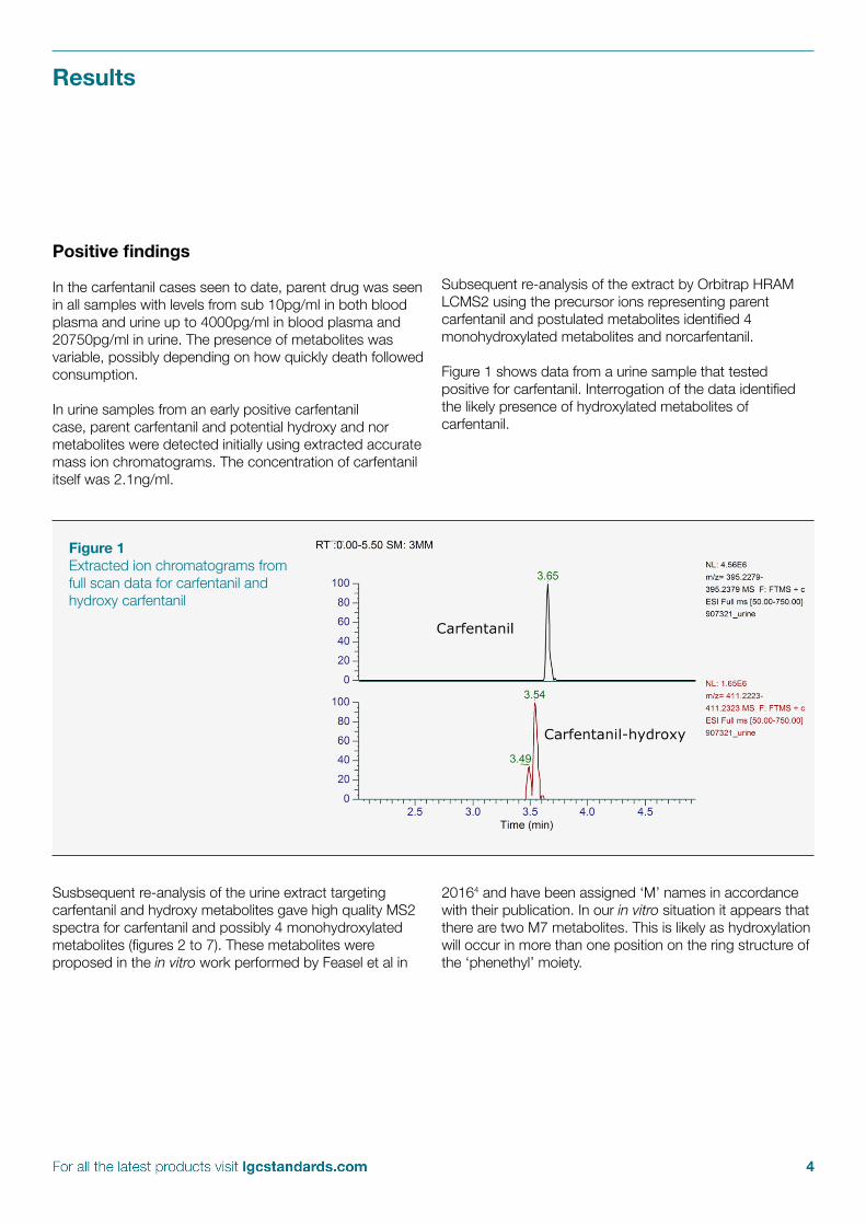
In urine samples from an early positive carfentanil case, parent carfentanil and potential hydroxy and nor metabolites were detected initially using extracted accurate mass ion chromatograms. The concentration of carfentanil itself was 2.1ng/ml.
Subsequent re-analysis of the extract by Orbitrap HRAM LCMS2 using the precursor ions representing parent carfentanil and postulated metabolites identified 4 monohydroxylated metabolites and norcarfentanil.
Figure 1 shows data from a urine sample that tested positive for carfentanil. Interrogation of the data identified the likely presence of hydroxylated metabolites of carfentanil.
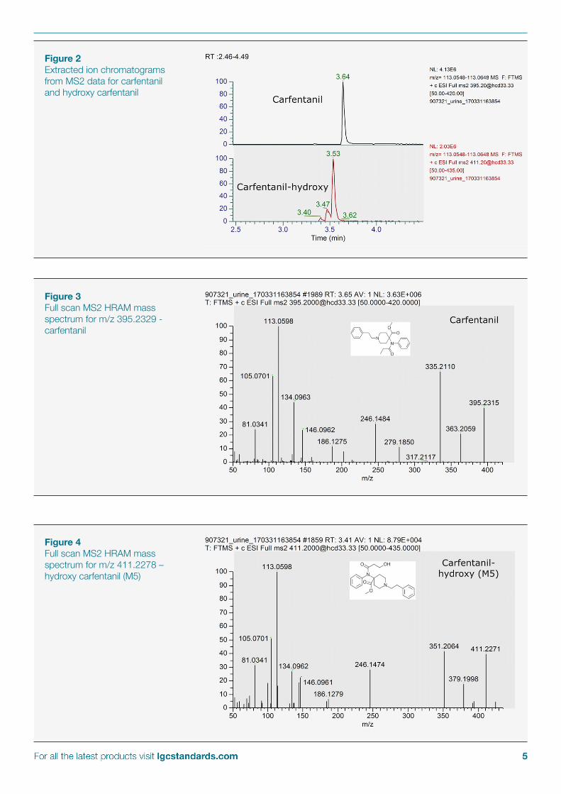
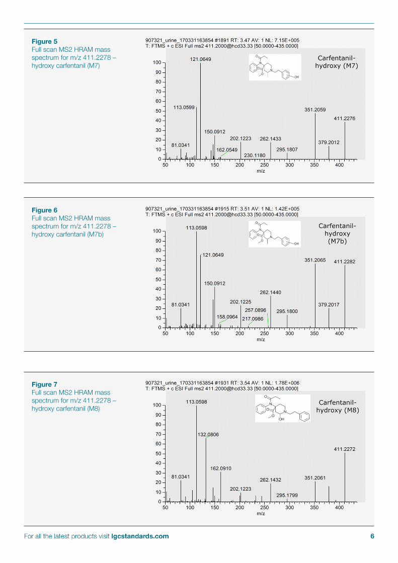
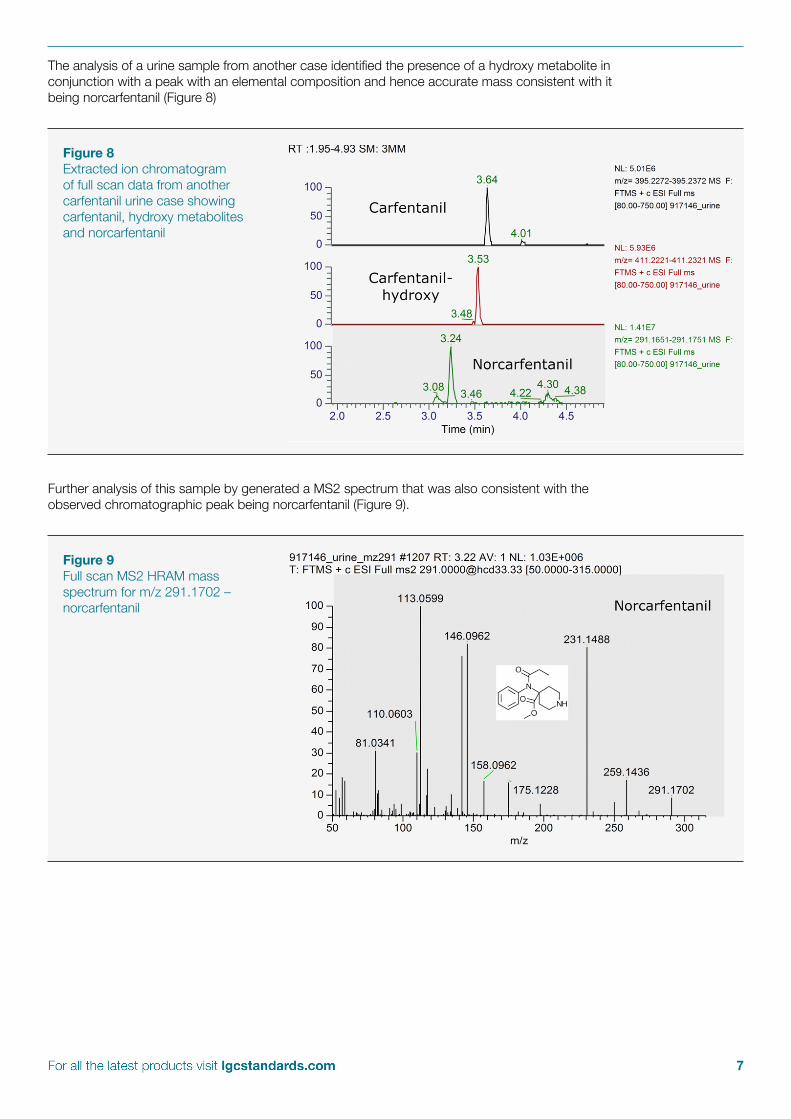
Susbsequent re-analysis of the urine extract targeting carfentanil and hydroxy metabolites gave high quality MS2 spectra for carfentanil and possibly 4 monohydroxylated metabolites (figures 2 to 7). These metabolites were proposed in the in vitro work performed by Feasel et al in
20164 and have been assigned ‘M’ names in accordance with their publication. In our in vitro situation it appears that there are two M7 metabolites. This is likely as hydroxylation will occur in more than one position on the ring structure of the ‘phenethyl’ moiety.
Figure 1Extracted ion chromatograms from full scan data for carfentanil and hydroxy carfentanil
Results
5
Figure 2Extracted ion chromatograms from MS2 data for carfentanil and hydroxy carfentanil
Figure 3Full scan MS2 HRAM mass spectrum for m/z 395.2329 - carfentanil
Figure 4Full scan MS2 HRAM mass spectrum for m/z 411.2278 – hydroxy carfentanil (M5)
6
Figure 5Full scan MS2 HRAM mass spectrum for m/z 411.2278 – hydroxy carfentanil (M7)
Figure 6Full scan MS2 HRAM mass spectrum for m/z 411.2278 – hydroxy carfentanil (M7b)
Figure 7Full scan MS2 HRAM mass spectrum for m/z 411.2278 – hydroxy carfentanil (M8)
7
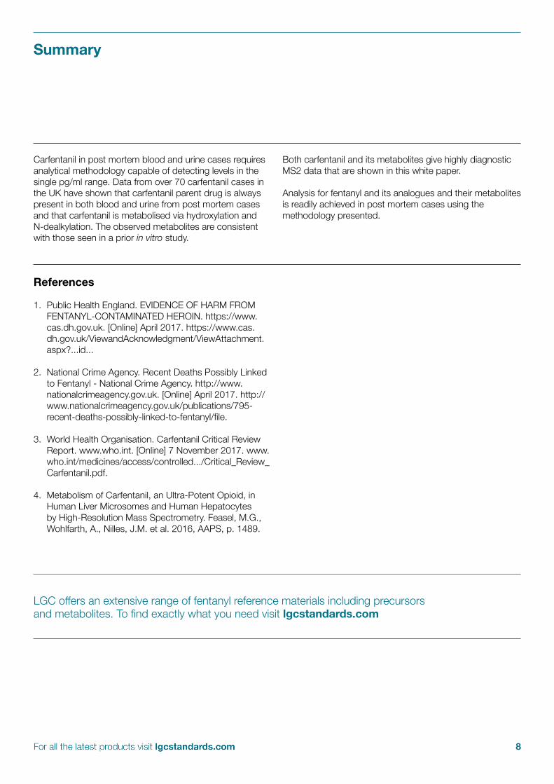
The analysis of a urine sample from another case identified the presence of a hydroxy metabolite in conjunction with a peak with an elemental composition and hence accurate mass consistent with it being norcarfentanil (Figure 8)
Further analysis of this sample by generated a MS2 spectrum that was also consistent with the observed chromatographic peak being norcarfentanil (Figure 9).
Figure 8Extracted ion chromatogram of full scan data from another carfentanil urine case showing carfentanil, hydroxy metabolites and norcarfentanil
Figure 9Full scan MS2 HRAM mass spectrum for m/z 291.1702 – norcarfentanil
8
Carfentanil in post mortem blood and urine cases requires analytical methodology capable of detecting levels in the single pg/ml range. Data from over 70 carfentanil cases in the UK have shown that carfentanil parent drug is always present in both blood and urine from post mortem cases and that carfentanil is metabolised via hydroxylation and N-dealkylation. The observed metabolites are consistent with those seen in a prior in vitro study.
Both carfentanil and its metabolites give highly diagnostic MS2 data that are shown in this white paper.
Analysis for fentanyl and its analogues and their metabolites is readily achieved in post mortem cases using the methodology presented.
References
1. Public Health England. EVIDENCE OF HARM FROM FENTANYL-CONTAMINATED HEROIN. https://www.cas.dh.gov.uk. [Online] April 2017. https://www.cas.dh.gov.uk/ViewandAcknowledgment/ViewAttachment.aspx?...id...
2. National Crime Agency. Recent Deaths Possibly Linked to Fentanyl - National Crime Agency. http://www.nationalcrimeagency.gov.uk. [Online] April 2017. http://www.nationalcrimeagency.gov.uk/publications/795-recent-deaths-possibly-linked-to-fentanyl/file.
3. World Health Organisation. Carfentanil Critical Review Report. www.who.int. [Online] 7 November 2017. www.who.int/medicines/access/controlled.../Critical_Review_Carfentanil.pdf.
4. Metabolism of Carfentanil, an Ultra-Potent Opioid, in Human Liver Microsomes and Human Hepatocytes by High-Resolution Mass Spectrometry. Feasel, M.G., Wohlfarth, A., Nilles, J.M. et al. 2016, AAPS, p. 1489.
Summary
LGC offers an extensive range of fentanyl reference materials including precursors and metabolites. To find exactly what you need visit lgcstandards.com