Embed Size (px)
Citation preview

Detailed methodology
for
dystrophin quantification
Valentina Sardone, PhD PI Prof. Francesco Muntoni
Dubowitz Neuromuscular Centre
UCL Institute of Child Health
London
FDA-NIH Dystrophin Methodology Workshop FDA White Oak Campus
20th of March 2015

Valentina Sardone, PhD PI Prof. Francesco Muntoni
Dubowitz Neuromuscular Centre
UCL Institute of Child Health
30 Guilford Street
WC1N 1EH, London
Disclosure of Conflict of Interest
I am working on the SKIP-NMD project funded by the EU within the FP-7 framework.
Sarepta Therapeutics is one of the participants and co-sponsor.

Muscle samples preparation: fibers orientation and freezing procedure
H&E
Spectrin (NCL-SPEC1,
Novocastra, monoclonal,
anti mouse)
To get appropriate specimens for performing IHC samples MUST be handled and frozen down properly
We follow a strict Standard Operating Procedure (SOP) and all the people involved have been trained
properly successful method 1) SKIP-NMD muscles coming from other centres
2) < 1% of muscle specimens present freezing artefacts
Quality control check: H&E and spectrin staining
Stereomicroscope
(Leica MZ75 with a
separate light source
Leica CLS 100X)
Frozen isopentane
30’’in LN2
OCT Compound
(AGR1180 Agar
Scientific)
7 μm sections
(Leica Cryostat
CM1850UV)

Dystrophin antibodies
Fairclough et al., 2013 Nature Reviews Genetics 14, 373–378
Epitope exon
79 (last 17aa)
α-mouse
Novocastra
Epitope
exons 10-12
α-mouse
Novocastra
Dys 3 Epitope
exons 26-30
α-mouse
Novocastra
Dys 1
Abcam
15277 Epitope
exon 77
α-rabbit
Abcam
N-terminal
actin binding
domain Central rod domain
Cysteine
rich
domain MANDYS106 Epitope
exon43
α-mouse
Gleen
Morris Lab
Dys 2
Over the years, selection and validation of antibodies taking account that each antibody:
i. recognizes different protein epitope (validated in patients deleted for the specific epitope)
ii. has its own epitope affinity
iii. gives different intensity values
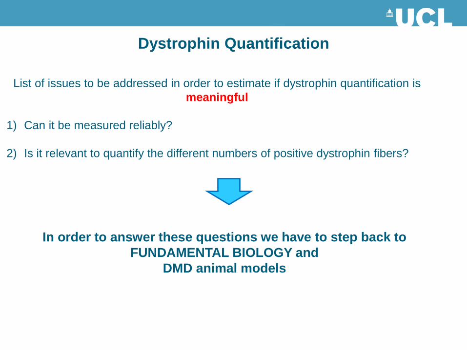
Dystrophin Quantification
List of issues to be addressed in order to estimate if dystrophin quantification is
meaningful
1) Can it be measured reliably?
2) Is it relevant to quantify the different numbers of positive dystrophin fibers?
In order to answer these questions we have to step back to
FUNDAMENTAL BIOLOGY and
DMD animal models

Acquistion
Leica DMR Fluorescence microscope
Serial sections:
1 section single labelling Dys2
1 section single labelling P7
1 section single labelling spectrin
Each section 4 pictures
Software
Metamorph (Molecular Devices)
40 different Region of Interest for each pic
Min = cytoplasm
Max = sarcolemma membrane
Can it be measured reliably?
Analysis
Normalizing factor= average (Max-
min) Spectrin EACH SAMPLE/
average (Max-min) Spectrin ALL
CONTROLS
1

A. Spectrin
B. Dystrophin
C. Spectrin Mask
D. Binary mask
E. Erode
F. Dilate
G. Spectrin
H. Dystrophin
Beekman et al., 2014
Taylor et al., 2012
2
3
Can it be measured reliably?

International Benchmarking of methods for dystrophin quantification
Institution of the Biochemical Outcome Measures Study Group (BOM-SG)
The BOM-SG was formed with a goal to provide data-driven, international standard dystrophin
quantification for DMD clinical trials
BOM Partners Arechavala
method
Taylor
method
Beekman
method
Muntoni Lab, London,
UK
Flanigan Lab, Columbus,
USA
Straub Lab, Newcastle,
UK
Voit Lab, Paris, France
Prosensa Therapeutics,
Leiden,
Immunohistochemistry technique
Results from different labs
using Arechavala method

Comparison of different dystrophin quantification methods
Results using Arechavala et al., 2010a method (5 Labs)
Taylor et al., 2012 method (3 Labs)
Beekman et al., 2014 method (1 Lab)

Western Blot assay
C1 A B C D E F C2
Protocol based on Taylor et al., 2012 published method
- Muscle samples lysed in protein extraction buffer
- 25ug of protein loaded on a 3-8 % Tris-acetate gel
- Wet transfer
- 1st Antibodies abcam 15277 1:400 O/N
α-actinin 1:3000 1hr
- ECL detection with 2nd antibodies (1:15000 α-rabbit, 1:500000 α-
mouse)
- Bands quantification performed by ImageJ or Odyssey
- Data normalized on α-actinin and presented relative to the average of
the 2 ctr used

Immunohistochemistry versus Western Blot
IHC and Western blot give different information
The amount of dystrophin on blot varies in different
subcellular fractions between full length and shorter
isoforms minidystrophins are less associated with
the sarcolemmal fraction compared to wild type
IHC and Western blot are different techniques with different range of sensitivity
IHC is more sensitive in detecting low levels of protein and allows high level of inter lab agreement:
- Takes into account dystrophin distribution, that in DMD and BMD patients is PATCHY
- Provides confirmation of subcellular protein localization (Western Blot is based on a homogenous protein
extraction)
- Anthony et al., 2014 proved reproducibility of this method
In several samples (e.g., BMD
sample A, c.40_41delGA), the level
of dystrophin determined by both
techniques was highly comparable
In others (e.g. BMD sample F, del ex
10-44) the level of dystrophin
quantified by western blotting was
significantly higher than that
determined by
immunohistochemistry.

Is it important to quantify the different numbers of positive dystrophin fibers?
The same level of protein (detected on a blot) had a different effect in different transgenic lines
based on the uniformity of expression:
- Mice with non-uniform expression had more pathology and a less favourable functional
outcome
- Mice with same overall levels but more uniform dystrophin distribution had less pathology and
better functional outcome

Additional important information from IHC:
- Numbers of positive dystrophin fibers per section
- Percentage of the fiber expressing dystrophin
Spectrin +ve fibers
Excluding fibers at the
edges
Dystrophin staining Scoring dystrophin +ve fibers
considering the % of the fiber
expressing dystrophin
1st METHOD : Operator Dependent counting from images of sections
2nd METHOD : Algorithm based counting from scan of entire section
Establishment
of a common method
across the BOM-SG is
in progress
Dystr
ophin
S
pectr
in
DMD

Take home message
Dystrophin quantification
1) Can it be measured reliably?
2) Is it relevant to quantify the different numbers of positive dystrophin
fibers?

MRC Centre for Neuromuscular Diseases
Thanks for your attention
Patients and their families
Acknowledgements