Embed Size (px)
Citation preview
CLINICAL AND VACCINE IMMUNOLOGY, Oct. 2011, p. 1680–1688 Vol. 18, No. 101556-6811/11/$12.00 doi:10.1128/CVI.05099-11Copyright © 2011, American Society for Microbiology. All Rights Reserved.
Detection of Antibodies against Paracoccidioides brasiliensis Melanin inIn Vitro and In Vivo Studies during Infection�
Martha E. Uran,1,2* Joshua D. Nosanchuk,3 Angela Restrepo,1 Andrew J. Hamilton,4Beatriz L. Gomez,1,4† and Luz E. Cano1,2,5†
Medical and Experimental Mycology Group, Corporacion para Investigaciones Biologicas, Medellín, Colombia1; School ofMedicine, Universidad Pontificia Bolivariana, Medellín, Colombia2; Division of Infectious Diseases, Department of Medicine,
Albert Einstein College of Medicine, Bronx, New York3; Dermatology Department, St. Johns Institute of Dermatology,Guy’s Hospital, Guy’s, Kings, and St. Thomas Medical Schools, London, United Kingdom4; and School of Microbiology,
Universidad de Antioquia, Medellín, Colombia5
Received 19 April 2011/Returned for modification 26 May 2011/Accepted 26 July 2011
Several cell wall constituents, including melanins or melanin-like compounds, have been implicated in thepathogenesis of a wide variety of microbial diseases caused by diverse species of pathogenic bacteria, fungi, andhelminthes. Among these microorganisms, the dimorphic fungal pathogen Paracoccidioides brasiliensis pro-duces melanin in its conidial and yeast forms. In the present study, melanin particles from P. brasiliensis wereinjected into BALB/c mice in order to produce monoclonal antibodies (MAbs). We identified five immuno-globulin G1 (IgG1) �-chain and four IgM melanin-binding MAbs. The five IgG1 �-chain isotypes are the firstmelanin-binding IgG MAbs ever reported. The nine MAbs labeled P. brasiliensis conidia and yeast cells bothin vitro and in pulmonary tissues. The MAbs cross-reacted with melanin-like purified particles from other fungiand also with commercial melanins, such as synthetic and Sepia officinalis melanin. Melanization duringparacoccidioidomycosis (PCM) was also further supported by the detection of IgG antibodies reactive tomelanin from P. brasiliensis conidia and yeast in sera and bronchoalveolar lavage fluids from P. brasiliensis-infected mice, as well as in sera from human patients with PCM. Serum specimens from patients with othermycoses were also tested for melanin-binding antibodies by enzyme-linked immunosorbent assay, and cross-reactivities were detected for melanin particles from different fungal sources. These results suggest thatmelanin from P. brasiliensis is an immunologically active fungal structure that activates a strong IgG humoralresponse in humans and mice.
Melanins, or melanin-like compounds, are cell wall constit-uents in a wide variety of microorganisms, including severalspecies of pathogenic bacteria, fungi and helminthes. Thesepolymeric pigments are implicated in the pathogenesis of di-verse microbial diseases (31, 40). Notably, melanin productionhas been demonstrated in a wide range of human pathogenicfungi, including Cryptococcus neoformans (30, 57), Lacazialoboi (52), Scedosporium prolificans (43), Histoplasma capsula-tum (32), Sporothrix schenckii (26), Pneumocystis carinii (Pneu-mocystis jiroveccii) (14), Fonsecaea pedrosoi (1, 2), Blastomycesdermatitidis (36), Aspergillus fumigatus (59), Exophiala derma-titidis (50), Penicillium marneffei (58), Candida albicans (25),and Coccidioides posadasii (37). Importantly, P. brasiliensis alsoproduces melanin (11). In P. brasiliensis, conidia and yeast cellsproduce melanin or melanin-like compounds in vitro and invivo. Notably, P. brasiliensis melanization protects the fungusfrom phagocytosis and increases its resistance to antifungaldrugs (9). In the present study we focus on P. brasiliensismelanin in order to determine its capacity to induce antibodies(Abs) in murine immunization, murine infection, and humandisease.
(The data presented in this study are from a master’s thesisof M. E. Uran, approved by the Faculty of Health Sciences,Universidad Pontificia Bolivariana, Medellín, Colombia.)
MATERIALS AND METHODS
Fungal strain. P. brasiliensis strain ATCC 60855, originally isolated from aColombian patient, was obtained from the American Type Culture Collection(Manassas, VA) and used for all of the experiments.
P. brasiliensis yeast growth with or without L-DOPA. P. brasiliensis ATCC60855 was converted from the mycelium to the yeast form in solid Difco Sab-ouraud dextrose (Becton Dickinson, Co., Le Pont de Claix, France) with 10%L-asparagine (Sigma Chemical Co., St. Louis, MO) and 10% thiamine hydro-chloride (Sigma). To obtain melanized yeast cells, the fungus was grown for 15days in the synthetic defined liquid minimal McVeigh-Morton medium (41) atpH 5.5 with or without 1.0 mM L-DOPA (Sigma) at 37°C in a rotary shaker at 150rpm. All cultures were kept in the dark to prevent photopolymerization, asreported previously (53). Cells were collected by centrifugation at 3,000 rpm for30 min at 4°C (refrigerated centrifuge, IEC Centra, GP8R; Thermo FisherScientific, Inc.), autoclaved, washed with 1� phosphate-buffered saline (PBS),and stored at 4°C until used (11, 46).
P. brasiliensis mycelial growth and conidium production. P. brasiliensis ATCC60855, known to sporulate on special culture media, was used for the productionof conidia (42). Standard techniques were used to grow the mycelial form, and tocollect and dislodge conidia (12, 42). Conidial melanization does not require theaddition of exogenous phenolic or other compounds (11).
Conidia used to infect mice for the in vivo model were obtained by thetraditional glass wool method previously described (12). Conidia were counted ina hemacytometer, and their viability was evaluated by the fluorescein diacetateand ethidium bromide staining procedure, as described previously (6).
Melanin particles: isolation and purification from P. brasiliensis conidia andyeast cells. Melanin particles were isolated from wild-type conidia and yeast cellsinduced with L-DOPA using published methodologies (26, 49). Briefly, conidia
* Corresponding author. Mailing address: Medical and Experimen-tal Mycology Unit, Corporacion para Investigaciones Biologicas, Car-rera 72A No. 78B-141, A.A. 73-78, Medellín, Colombia. Phone: 57 4403 59 50. Fax: 57 4 441 55 14. E-mail: [email protected].
† L.E.C. and B.L.G. are senior coauthors.� Published ahead of print on 3 August 2011.
1680
on March 2, 2020 by guest
http://cvi.asm.org/
Dow
nloaded from
and yeast cells were collected by centrifugation, autoclaved, and treated withlysing enzymes (of Aspergillus spp. [Sigma]) to generate protoplasts that werethen collected by centrifugation, washed, and incubated overnight in denaturantsolution (26, 49). Cell debris was collected by centrifugation, washed, and treatedwith recombinant proteinase K (PCR grade from Roche Applied Science, Indi-anapolis, IN); the resultant materials were washed and then boiled in 6 M HCl.The materials remaining after acid digestion were collected by centrifugation,washed extensively with PBS, dialyzed against distilled water for 10 days at 4°C,and then lyophilized (26). The melanin particles (ghosts) from yeast cells wereused to generate monoclonal Abs (MAbs), and particles from both conidia andyeast cells were used as antigens for the coating of the enzyme-linked immu-nosorbent assay (ELISA) plates for detection of melanin-binding Abs (seebelow).
Pulmonary paracoccidioidomycosis (PCM) experimental model. We utilizedthe mouse model previously described by our group (12, 24). All murine exper-iments were approved by the animal use committees of the Corporacion paraInvestigaciones Biologicas (CIB) and the Guy’s, Kings, and St. Thomas MedicalSchools. The animals were obtained from the CIB breeding colony and used inall experiments. They were divided into two groups depending on the inoculum:(i) the infected group (n � 7 for each time interval assessed), consisting of6-week-old male BALB/c mice that were inoculated intranasally with 4 � 106
conidia/60 �l of 1� sterile PBS buffer, and (ii) the control group (n � 5 for eachtime point), 6-week-old male BALB/c mice inoculated intranasally with 60 �l of1� sterile PBS buffer. Mice were sacrificed at 0, 48, and 96 h and 1, 2, 3, 4, 6, 8,10, 12, and 16 weeks after infection. After sacrifice, blood and bronchoalveolarlavage fluid (BAL) samples were collected. Samples were kept individually, andalso a pool for each group was made. Serum was separated from blood bycentrifugation at 1,500 rpm for 5 min and then frozen at �20°C. For BALsamples, the trachea was exposed and a blunt-tipped needle inserted. One mil-liliter of cold RPMI 1640 medium was instilled into the lungs, aspirated, andcentrifuged at 1,500 rpm for 5 min at 4°C; the cells were discarded, and thesupernatants were frozen at �20°C.
Production of MAbs against P. brasiliensis yeast cells’ melanin particles.BALB/c mice (6- to 8-week-old females) from Harlam Olac (United Kingdom)were maintained at Guy’s and King’s Campus. Mice were bled from the tail veinprior to immunizations to define baseline antibody concentrations. At 1-weekintervals, animals received five intraperitoneal injections of 50 �g or 100 �l ofmelanin particles extracted from P. brasiliensis L-DOPA-induced yeast cells sus-pended in a 1:1 (vol/vol) emulsion of incomplete Freund adjuvant (Difco, EastMolesey, United Kingdom) in PBS. A week before sacrifice, the mice were bledto measure polyclonal antibody responses against melanin by an ELISA (proto-col 1, see below); the spleen from the most responsive mouse was used togenerate hybridomas using the sp2/0 myeloma cells fusion partner as describedpreviously (13, 60). Supernatants from each hybridoma obtained were screenedfor the presence and classification of melanin-binding MAbs by ELISA (protocol1, see below). The MAbs were subclassified using a mouse MAb isotyping test kit(MMT1; Serotec, Kidlington, United Kingdom).
ELISAs (protocols 1, 2, and 3) for melanin-binding Ab determination: coatingof plates. Techniques used to develop ELISAs (protocols 1, 2, and 3, see below)were as follows. The plates were coated with a suspension of 50 �g of melaninderived from P. brasiliensis L-DOPA yeast or wild-type conidia. Plates were alsogenerated using melanins from Sepia officinalis (Sigma M-2649), S. schenckiiconidia (26), P. marneffei conidia (58), A. fumigatus conidia (59), Aspergillus nigerconidia (59), C. neoformans yeast cells (35), or C. albicans yeast cells (25). Inaddition, plates were made using synthetic melanin (Sigma M-8631). Each mel-anin was mixed with distilled water, and 50 �g of the polymer was plated inindividual wells of a polystyrene 96-well ELISA plate (catalog no. 3591; Costar,Corning, NY). Control wells were not coated with melanin particles. The plateswere kept at room temperature till dry and then heat fixed by incubating theplates at 60°C for 1 h as described previously (48). The plates were subsequentlyused for the different ELISA protocols.
ELISA protocol 1: ELISA to detect melanin-binding polyclonal and MAbs andto determine the sensitivity and specificity of MAbs. The plates coated withdifferent melanins were blocked overnight with SuperBlock (Pierce Biotechnol-ogy, Rockford, IL) blocking buffer in Tris-buffered saline (TBS) at 4°C to preventnonspecific binding. After each step in the ELISA, plates were washed threetimes with 0.1% Tween 20 in TBS. Sera from mice inoculated with P. brasiliensisyeast cell melanin or hybridoma supernatants were diluted 1:10 in SuperBlockblocking buffer in PBS, and 100 �l of each sample was incubated in triplicate for1.5 h at 37°C. After washes, 100 �l of a 1:1,000 peroxidase-conjugated goatanti-mouse immunoglobulin G (IgG) or goat anti-mouse IgM (Jackson Immu-noresearch, West Grove, PA) diluted in SuperBlock was added for 1.5 h at 37°C.The plates were washed, and o-phenylenediamine in 0.01 M sodium citrate buffer
(pH 5.0) was used as the enzyme substrate. The plates were incubated for 10 minin the dark, and the reaction was stopped with 0.01 M H2SO4. The solutions weretransferred to clear ELISA plates, and their optical densities (OD) weremeasured at 490 nm with an ELISA plate reader (Microplate Reader 450/550;Bio-Rad, Hercules, CA) (26). The transfer of solutions was performed as theadherent melanin particles interfered with OD determinations. As controls,we used melanized wells without primary antibody, melanized wells in whichthe primary antibody had been replaced by the culture medium used forhybridoma growth (histone acetyltransferase [HAT], supplemented RPMI1640 [Gibco-Invitrogen, Carlsbad, CA]), or wells without melanin (secondaryantibody alone). OD readings higher than that of the controls (0.1 OD) wereconsidered positive.
ELISA protocol 2: determining melanin-binding Ab titers during experimen-tal infection. ELISA plates were blocked and washed as detailed above. To testfor Ab production, mouse sera and BALs from the experimental pulmonaryPCM model were diluted 1:10 in SuperBlock blocking buffer in PBS, and 100-�lportions were added to triplicate wells and processed as described for ELISAprotocol 1, except that the enzyme substrate was p-nitrophenylphosphate at 1mg/ml (Sigma Chemical Co.) in 98% diethanolamine 0.5 mM MgCl2 buffer. Theenzymatic reaction was stopped with 3 N NaOH and read spectrophotometricallyat 405 nm. As controls, we used melanized wells incubated with serum or BALsamples from uninfected animals, melanized wells without sera or BALs (sec-ondary Ab only), or wells without melanin (secondary Ab only). OD readingshigher than those of controls (0.5 OD) were considered positive. One of thegenerated P. brasiliensis melanin-binding MAbs, 8C1-IgG, was used as a positivecontrol.
ELISA protocol 3: determining melanin-binding Ab titers in human sera. Atotal of 27 serum specimens from patients with different clinical forms of PCM(14 with the chronic form and 13 with the subacute form) were tested individ-ually. As a positive control, we utilized a pool consisting of 64 sera from newlydiagnosed PCM patients (i.e., untreated) from the CIB’s Biobank collection. Asnegative controls, we used pooled sera from six tuberculosis patients, and fourpools from healthy subjects provided by two local blood bank laboratories, eachpool consisting of more than 40 samples from healthy subjects without historiesof pulmonary diseases. The use of the sera was approved by the institutionalreview board of the CIB.
To measure human melanin-binding Abs, wells were blocked and washed asdescribed for ELISA protocol 1. Sera were diluted 1:500 in SuperBlock blockingbuffer in PBS, and 100 �l of each sample was incubated in triplicate for 1.5 h at37°C. After washes, 100 �l of peroxidase-conjugated goat anti-human IgG (Jack-son Immunoresearch) (2) diluted in SuperBlock at 1:5,000 was added for 1.5 h at37°C. After washes, the reaction was developed with o-phenylenediamine in 0.01M sodium citrate buffer (pH 5.0) as the enzyme substrate. Plates were incubatedfor 10 min in the dark, and the reaction was stopped with 0.01 M H2SO4. Thesolutions were transferred to clear the ELISA plates, and their OD were mea-sured at 490 nm with an ELISA plate reader (iMark microplate absorbancereader; Bio-Rad, Hercules, CA). In addition to the non-PCM sera, negativecontrols included uncoated wells and coated wells from which the primary anti-body had been omitted.
Immunofluorescence assays from cultured samples. Reactivity of P. brasilien-sis MAbs was assessed by immunofluorescence according to methods describedin Youngchim et al. (59). Briefly, P. brasiliensis conidia or melanized yeasts(cultured with L-DOPA) were heat fixed and then blocked overnight withSuperBlock blocking buffer in PBS at 4°C. MAbs generated from the immunizedmouse were diluted in SuperBlock (10 �g/ml) and applied for 1 h at 37°C. Afterwashing, a 1:100 dilution of fluorescein isothiocyanate-conjugated (FITC)Affinipure goat anti-mouse IgG or IgM (Jackson Immunoresearch), was usedas a secondary Ab. The slides were mounted using 50% glycerol–50% PBS,coverslips were applied, and the samples were examined by using a Zeissimmunofluorescence microscope. As a negative control, slides and particleswere incubated with PBS instead of the primary Ab, followed by incubationwith FITC-conjugated goat anti-mouse as described above. The IgM melanin-binding MAb, 6D2, generated in response to C. neoformans melanin (49), wasused as a positive control.
Immuno-transmission electron microscopy (immuno-TEM). Samples of pul-monary tissues from P. brasiliensis-infected mice 24 weeks postinfection andmelanin ghosts obtained from conidia or yeast cells (as described above) werefixed by incubation with formaldehyde (4%) and glutaraldehyde (0.1%) solutionsovernight. The paraffin-embedded pulmonary tissues were passed through gra-dients of decreasing xylol and alcohol (100 to 50%) solutions before initiating theprocess. Frozen samples were then transferred to a Leica EM AFS freezesubstitution unit (Leica Microsystems, Vienna, Austria), and ultrathin lung tissuesections were placed on nickel grids and processed as described previously (29).
VOL. 18, 2011 P. BRASILIENSIS MELANIN-BINDING ANTIBODIES 1681
on March 2, 2020 by guest
http://cvi.asm.org/
Dow
nloaded from
The samples were then fixed in 2% glutaraldehyde in 0.1 M cacodylate at roomtemperature overnight. P. brasiliensis melanin-binding 8C3-IgG or 2GA-IgMMAbs (10 �g/ml) diluted in SuperBlock were incubated overnight at room tem-perature. The grids were washed, and 2.5 �g of biotin-conjugated goat anti-mouseIgG or IgM (5 �g/ml, conjugated with streptavidin and 10-nm gold; GoldmarkBiologicals, Phillipsburg, NJ) in SuperBlock was added for 2 h at room temperature.The grids were fixed in 2% glutaraldehyde. Samples were viewed on a JEOL (Tokyo,Japan) 1200EX transmission electron microscope at 80 kV.
Statistical analyses. Results were collected in a database and analyzed byone-way analysis of variance with GraphPad Prism version 3.02 for Windows(GraphPad Software, San Diego, CA). Methods such as analysis of variance andthe Student t test were applied to the data, and the results are expressed as themean standard error of the mean of two or three independent experiments, withsignificance defined as P � 0.05. To analyze microscopic procedures such asfluorescence, TEM, or histopathology photographic studies, percentage compar-isons were done.
RESULTS
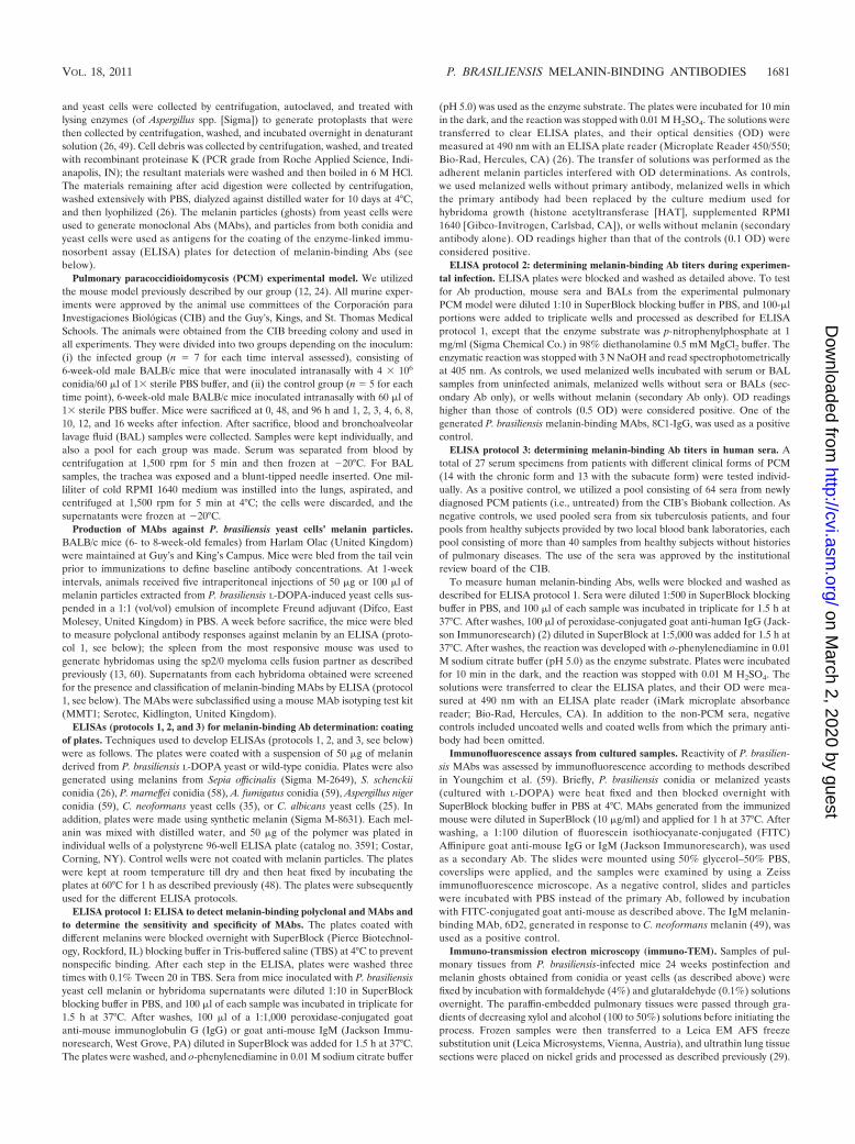
Generation of MAbs against P. brasiliensis yeast cells mel-anin particles. A total of nine P. brasiliensis melanin-bindingMAbs were produced (Fig. 1). The four IgM isotype MAbswere considered reactive (�0.1 OD greater than the controls)at dilutions to 102 for MAbs 8GA and 8GD and 103 for MAbs2GA and 7FC (Fig. 1A). The five IgG1 �-chain MAbs weresignificantly more reactive than the IgMs, since the reactivitiesfor MAbs 8C1 and 8C3 occurred to 105 dilutions and for MAbs7E1, 7E2, and 7E3 to 107 dilutions (Fig. 1B).
Table 1 shows the reactivities of the MAbs using the differ-ent melanin ELISAs. In each ELISA, the IgG MAbs produced
FIG. 1. ELISA reactivity of the nine P. brasiliensis melanin-binding MAbs produced (four IgMs and five IgG1s) against P. brasiliensis yeastmelanin (50 �g/well), supernatants were diluted 1:10. The negative controls were wells incubated with secondary antibody, RPMI, and HAT media,and wells without melanin were incubated with secondary antibody. This panel show a representative reaction of IgM MAbs (A) and IgG MAbs(B) against P. brasiliensis yeast melanin. The experiment was performed twice with similar results.
TABLE 1. ELISA reactivities for P. brasiliensis melanin-binding MAbs that reacted with different sources of melanina
MAb againstP. brasiliensis melanin
Mean OD observed with various melanin sources
Synthetic S. officinalis P. brasiliensisyeast cell
P. brasiliensisconidia
A. fumigatusconidia
A. nigerconidia
S. schenckiiconidia
P. marneffeiconidia
C. neoformansyeast cell
IgG7E1 0.412 0.414 0.422 0.401 0.380 0.369 0.369 0.371 0.4187E2 0.394 0.390 0.422 0.389 0.398 0.361 0.429 0.384 0.4227E3 0.437 0.435 0.471 0.436 0.405 0.381 0.431 0.403 0.4358C1 0.441 0.444 0.425 0.459 0.454 0.384 0.469 0.419 0.4348C3 0.441 0.477 0.444 0.438 0.459 0.417 0.503 0.480 0.408Bk-IgG 0.190 0.043 0.015 0.090 0.105 0.167 0.152 0.105 0.166
IgM8GA 0.149 0.074 0.172 0.207 0.018 0.311 0.225 0.105 0.0628GD 0.145 0.081 0.176 0.210 0.043 0.269 0.249 0.090 0.0627FC 0.215 0.038 0.195 0.205 0.093 0.421 0.303 0.186 0.2752GA 0.239 0.064 0.231 0.184 0.110 0.447 0.361 0.184 0.281Bk-IgM 0.047 0.001 0.005 0.111 0.024 0.173 0.102 0.026 0.130
a The fungal melanin was included at 50 �g per well. Numbers in boldface indicate the highest mean OD against each type of melanin. Negative controls were wellswith melanin incubated with the secondary antibody or without melanin (blank reactions were subtracted from each value). The ELISAs were repeated at least twicewith similar results. Supernatants were diluted 1:10.
1682 URÁN ET AL. CLIN. VACCINE IMMUNOL.
on March 2, 2020 by guest
http://cvi.asm.org/
Dow
nloaded from
higher OD than the IgM MAbs, except for IgMs 2GA and7FC, which produced the highest OD in response to the A.niger melanin. Wells without melanin or with melanin but in-cubated with either the growth media (supplemented RPMI)or the secondary Ab were nonreactive. Based on the reactivi-ties observed in these experiments, we used IgG MAbs 8C1and 8C3 and IgM MAb 2GA for further studies.
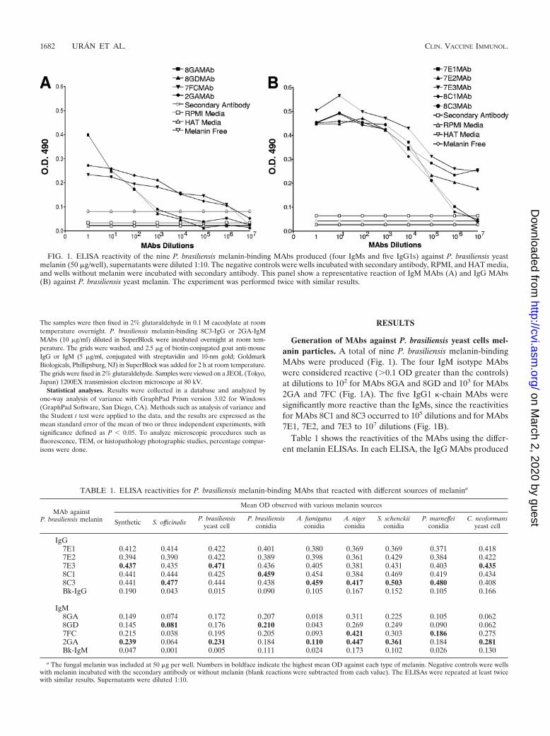
Binding of melanin in vitro and in vivo. The melanin-bindingMAbs (IgG and IgM) reacted with P. brasiliensis yeast onlyafter cultures had been induced with L-DOPA. Reactivity pre-dominantly occurred along the yeast cell wall (Fig. 2A). Whenthese yeast cells were analyzed by immuno-TEM, MAb label-ing occurred on the interior and exterior aspects of the cell wall(Fig. 2C). The conidia from mycelial cultures grown in wateragar without L-DOPA induction were also labeled by bothMAb isotypes, as demonstrated by fluorescence and immuno-TEM (Fig. 2B and D). Controls incubated only with the fluo-rescein-conjugated secondary Ab or with the conjugated con-
trol Ab were nonreactive (data not shown). In addition,reactivity was demonstrated in the lungs of the P. brasiliensisconidium-infected mice using immuno-TEM with IgG (Fig.2E) and IgM MAbs (Fig. 2F).
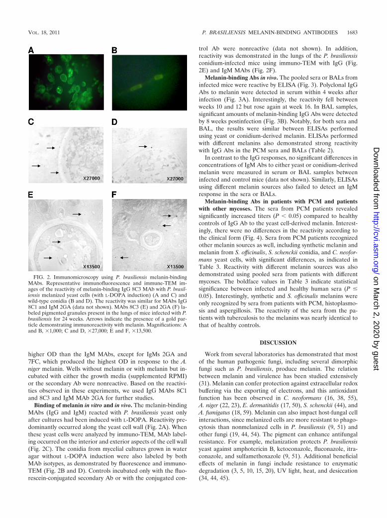
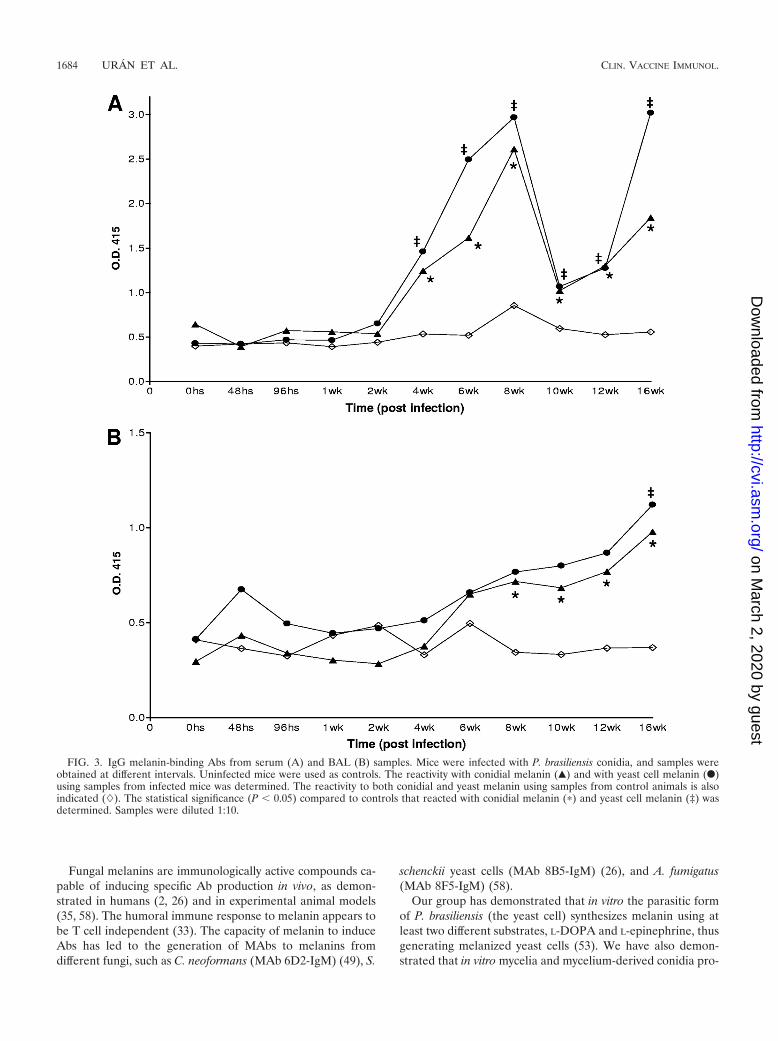
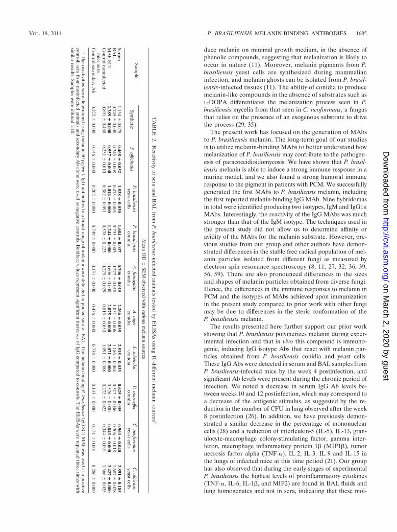
Melanin-binding Abs in vivo. The pooled sera or BALs frominfected mice were reactive by ELISA (Fig. 3). Polyclonal IgGAbs to melanin were detected in serum within 4 weeks afterinfection (Fig. 3A). Interestingly, the reactivity fell betweenweeks 10 and 12 but rose again at week 16. In BAL samples,significant amounts of melanin-binding IgG Abs were detectedby 8 weeks postinfection (Fig. 3B). Notably, for both sera andBAL, the results were similar between ELISAs performedusing yeast or conidium-derived melanin. ELISAs performedwith different melanins also demonstrated strong reactivitywith IgG Abs in the PCM sera and BALs (Table 2).
In contrast to the IgG responses, no significant differences inconcentrations of IgM Abs to either yeast or conidium-derivedmelanin were measured in serum or BAL samples betweeninfected and control mice (data not shown). Similarly, ELISAsusing different melanin sources also failed to detect an IgMresponse in the sera or BALs.
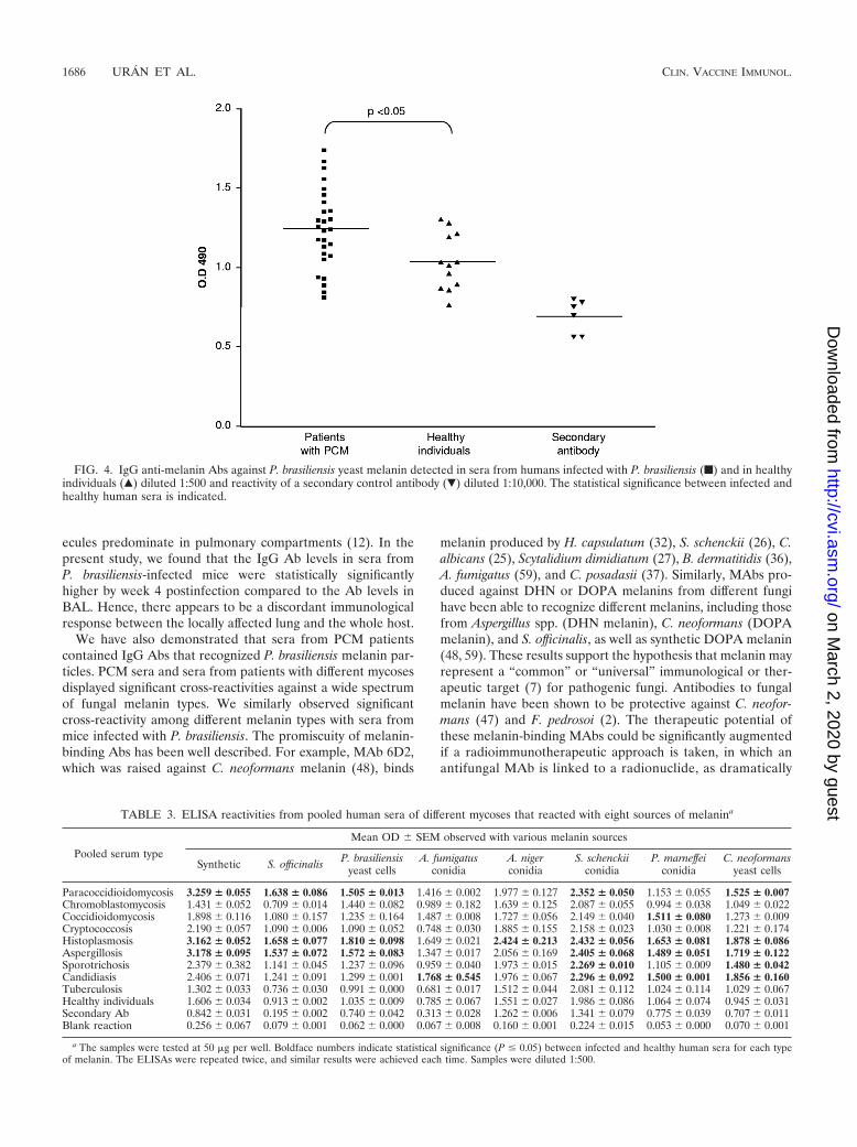
Melanin-binding Abs in patients with PCM and patientswith other mycoses. The sera from PCM patients revealedsignificantly increased titers (P � 0.05) compared to healthycontrols of IgG Ab to the yeast cell-derived melanin. Interest-ingly, there were no differences in the reactivity according tothe clinical form (Fig. 4). Sera from PCM patients recognizedother melanin sources as well, including synthetic melanin andmelanin from S. officinalis, S. schenckii conidia, and C. neofor-mans yeast cells, with significant differences, as indicated inTable 3. Reactivity with different melanin sources was alsodemonstrated using pooled sera from patients with differentmycoses. The boldface values in Table 3 indicate statisticalsignificance between infected and healthy human sera (P �0.05). Interestingly, synthetic and S. officinalis melanins wereonly recognized by sera from patients with PCM, histoplasmo-sis and aspergillosis. The reactivity of the sera from the pa-tients with tuberculosis to the melanins was nearly identical tothat of healthy controls.
DISCUSSION
Work from several laboratories has demonstrated that mostof the human pathogenic fungi, including several dimorphicfungi such as P. brasiliensis, produce melanin. The relationbetween melanin and virulence has been studied extensively(31). Melanin can confer protection against extracellular redoxbuffering via the exporting of electrons, and this antioxidantfunction has been observed in C. neoformans (16, 38, 55),A. niger (22, 23), E. dermatitidis (17, 50), S. schenckii (44), andA. fumigatus (18, 59). Melanin can also impact host-fungal cellinteractions, since melanized cells are more resistant to phago-cytosis than nonmelanized cells in P. brasiliensis (9, 51) andother fungi (19, 44, 54). The pigment can enhance antifungalresistance. For example, melanization protects P. brasiliensisyeast against amphotericin B, ketoconazole, fluconazole, itra-conazole, and sulfamethoxazole (9, 51). Additional beneficialeffects of melanin in fungi include resistance to enzymaticdegradation (3, 5, 10, 15, 20), UV light, heat, and desiccation(34, 44, 45).
FIG. 2. Immunomicroscopy using P. brasiliensis melanin-bindingMAbs. Representative immunofluorescence and immune-TEM im-ages of the reactivity of melanin-binding IgG 8C3 MAb with P. brasil-iensis melanized yeast cells (with L-DOPA induction) (A and C) andwild-type conidia (B and D). The reactivity was similar for MAbs IgG8C1 and IgM 2GA (data not shown). MAbs 8C3 (E) and 2GA (F) la-beled pigmented granules present in the lungs of mice infected with P.brasiliensis for 24 weeks. Arrows indicate the presence of a gold par-ticle demonstrating immunoreactivity with melanin. Magnifications: Aand B, �1,000; C and D, �27,000; E and F, �13,500.
VOL. 18, 2011 P. BRASILIENSIS MELANIN-BINDING ANTIBODIES 1683
on March 2, 2020 by guest
http://cvi.asm.org/
Dow
nloaded from
Fungal melanins are immunologically active compounds ca-pable of inducing specific Ab production in vivo, as demon-strated in humans (2, 26) and in experimental animal models(35, 58). The humoral immune response to melanin appears tobe T cell independent (33). The capacity of melanin to induceAbs has led to the generation of MAbs to melanins fromdifferent fungi, such as C. neoformans (MAb 6D2-IgM) (49), S.
schenckii yeast cells (MAb 8B5-IgM) (26), and A. fumigatus(MAb 8F5-IgM) (58).
Our group has demonstrated that in vitro the parasitic formof P. brasiliensis (the yeast cell) synthesizes melanin using atleast two different substrates, L-DOPA and L-epinephrine, thusgenerating melanized yeast cells (53). We have also demon-strated that in vitro mycelia and mycelium-derived conidia pro-
FIG. 3. IgG melanin-binding Abs from serum (A) and BAL (B) samples. Mice were infected with P. brasiliensis conidia, and samples wereobtained at different intervals. Uninfected mice were used as controls. The reactivity with conidial melanin (Œ) and with yeast cell melanin (F)using samples from infected mice was determined. The reactivity to both conidial and yeast melanin using samples from control animals is alsoindicated (�). The statistical significance (P � 0.05) compared to controls that reacted with conidial melanin (�) and yeast cell melanin (‡) wasdetermined. Samples were diluted 1:10.
1684 URÁN ET AL. CLIN. VACCINE IMMUNOL.
on March 2, 2020 by guest
http://cvi.asm.org/
Dow
nloaded from
duce melanin on minimal growth medium, in the absence ofphenolic compounds, suggesting that melanization is likely tooccur in nature (11). Moreover, melanin pigments from P.brasiliensis yeast cells are synthesized during mammalianinfection, and melanin ghosts can be isolated from P. brasil-iensis-infected tissues (11). The ability of conidia to producemelanin-like compounds in the absence of substrates such asL-DOPA differentiates the melanization process seen in P.brasiliensis mycelia from that seen in C. neoformans, a fungusthat relies on the presence of an exogenous substrate to drivethe process (29, 35).
The present work has focused on the generation of MAbsto P. brasiliensis melanin. The long-term goal of our studiesis to utilize melanin-binding MAbs to better understand howmelanization of P. brasiliensis may contribute to the pathogen-esis of paracoccidioidomycosis. We have shown that P. brasil-iensis melanin is able to induce a strong immune response in amurine model, and we also found a strong humoral immuneresponse to the pigment in patients with PCM. We successfullygenerated the first MAbs to P. brasiliensis melanin, includingthe first reported melanin-binding IgG MAb. Nine hybridomasin total were identified producing two isotypes, IgM and IgG1�MAbs. Interestingly, the reactivity of the IgG MAbs was muchstronger than that of the IgM isotype. The techniques used inthe present study did not allow us to determine affinity oravidity of the MAbs for the melanin substrate. However, pre-vious studies from our group and other authors have demon-strated differences in the stable free radical population of mel-anin particles isolated from different fungi as measured byelectron spin resonance spectroscopy (8, 11, 27, 32, 36, 39,56, 59). There are also pronounced differences in the sizesand shapes of melanin particles obtained from diverse fungi.Hence, the differences in the immune responses to melanin inPCM and the isotypes of MAbs achieved upon immunizationin the present study compared to prior work with other fungimay be due to differences in the steric conformation of theP. brasiliensis melanin.
The results presented here further support our prior workshowing that P. brasiliensis polymerizes melanin during exper-imental infection and that in vivo this compound is immuno-genic, inducing IgG isotype Abs that react with melanin par-ticles obtained from P. brasiliensis conidia and yeast cells.These IgG Abs were detected in serum and BAL samples fromP. brasiliensis-infected mice by the week 4 postinfection, andsignificant Ab levels were present during the chronic period ofinfection. We noted a decrease in serum IgG Ab levels be-tween weeks 10 and 12 postinfection, which may correspond toa decrease of the antigenic stimulus, as suggested by the re-duction in the number of CFU in lung observed after the week8 postinfection (26). In addition, we have previously demon-strated a similar decrease in the percentage of mononuclearcells (28) and a reduction of interleukin-5 (IL-5), IL-13, gran-ulocyte-macrophage colony-stimulating factor, gamma inter-feron, macrophage inflammatory protein 1� (MIP1�), tumornecrosis factor alpha (TNF-), IL-2, IL-3, IL-9 and IL-15 inthe lungs of infected mice at this time period (21). Our grouphas also observed that during the early stages of experimentalP. brasiliensis the highest levels of proinflammatory cytokines(TNF-, IL-6, IL-1�, and MIP2) are found in BAL fluids andlung homogenates and not in sera, indicating that these mol-
TA
BL
E2.
Reactivity
ofsera
andB
AL
fromP
.brasiliensis-infectedanim
alstested
byE
LISA
susing
10different
melanin
sourcesa
Sample
Mean
OD
SE
Mobserved
with
variousm
elaninsources
SyntheticS.officinalis
P.brasiliensisyeast
cellsP
.brasiliensisconidia
A.fum
igatusconidia
A.niger
conidiaS.schenckii
conidiaP
.marneffei
conidiaC
.neoformans
yeastcells
C.albicans
yeastcells
Serum1.154
0.078
0.468�
0.0521.538
�0.030
1.684�
0.0370.706
�0.015
2.266�
0.0352.515
�0.033
0.625�
0.0350.963
�0.040
2.891�
0.185B
AL
0.749
0.0080.245
0.008
0.475
0.0050.733
0.003
0.277
0.0181.051
0.006
1.186
0.0040.317
0.036
0.306
0.0181.687
0.028
MA
b8C
12.289
�0.000
0.557�
0.0001.016
�0.000
1.244�
0.0000.606
0.000
2.075�
0.0002.071
�0.000
0.526
0.0000.843
�0.000
2.427�
0.000C
ontrolnoninfectedm
icesera
0.891
0.4380.231
0.010
0.387
0.0910.578
0.223
0.275
0.0290.815
0.053
1.091
0.3880.272
0.022
0.344
0.0911.366
0.035
Controlsecondary
Ab
0.272
0.0000.146
0.000
0.202
0.0000.749
0.000
0.131
0.0000.436
0.000
0.738
0.0000.143
0.000
0.151
0.0010.286
0.000
aT
hereactivities
were
determined
usingm
elanin50
�g/w
ell.IgGantibodies
toa
broadrange
ofmelanin
were
detectedin
pooledsera
orB
AL
.The
melanin-binding
P.brasiliensis
IgG8C
1M
Ab
was
usedas
apositive
control;serafrom
noninfectedanim
alsand
secondaryA
balone
were
usedas
negativecontrols.B
oldfacevalues
representsignificant
increasesof
IgGcom
paredto
controls.The
EL
ISAs
were
repeatedthree
times
with
similar
results.Samples
were
diluted1:10.
VOL. 18, 2011 P. BRASILIENSIS MELANIN-BINDING ANTIBODIES 1685
on March 2, 2020 by guest
http://cvi.asm.org/
Dow
nloaded from
ecules predominate in pulmonary compartments (12). In thepresent study, we found that the IgG Ab levels in sera fromP. brasiliensis-infected mice were statistically significantlyhigher by week 4 postinfection compared to the Ab levels inBAL. Hence, there appears to be a discordant immunologicalresponse between the locally affected lung and the whole host.
We have also demonstrated that sera from PCM patientscontained IgG Abs that recognized P. brasiliensis melanin par-ticles. PCM sera and sera from patients with different mycosesdisplayed significant cross-reactivities against a wide spectrumof fungal melanin types. We similarly observed significantcross-reactivity among different melanin types with sera frommice infected with P. brasiliensis. The promiscuity of melanin-binding Abs has been well described. For example, MAb 6D2,which was raised against C. neoformans melanin (48), binds
melanin produced by H. capsulatum (32), S. schenckii (26), C.albicans (25), Scytalidium dimidiatum (27), B. dermatitidis (36),A. fumigatus (59), and C. posadasii (37). Similarly, MAbs pro-duced against DHN or DOPA melanins from different fungihave been able to recognize different melanins, including thosefrom Aspergillus spp. (DHN melanin), C. neoformans (DOPAmelanin), and S. officinalis, as well as synthetic DOPA melanin(48, 59). These results support the hypothesis that melanin mayrepresent a “common” or “universal” immunological or ther-apeutic target (7) for pathogenic fungi. Antibodies to fungalmelanin have been shown to be protective against C. neofor-mans (47) and F. pedrosoi (2). The therapeutic potential ofthese melanin-binding MAbs could be significantly augmentedif a radioimmunotherapeutic approach is taken, in which anantifungal MAb is linked to a radionuclide, as dramatically
FIG. 4. IgG anti-melanin Abs against P. brasiliensis yeast melanin detected in sera from humans infected with P. brasiliensis (f) and in healthyindividuals (Œ) diluted 1:500 and reactivity of a secondary control antibody (�) diluted 1:10,000. The statistical significance between infected andhealthy human sera is indicated.
TABLE 3. ELISA reactivities from pooled human sera of different mycoses that reacted with eight sources of melanina
Pooled serum typeMean OD SEM observed with various melanin sources
Synthetic S. officinalis P. brasiliensisyeast cells
A. fumigatusconidia
A. nigerconidia
S. schenckiiconidia
P. marneffeiconidia
C. neoformansyeast cells
Paracoccidioidomycosis 3.259 � 0.055 1.638 � 0.086 1.505 � 0.013 1.416 0.002 1.977 0.127 2.352 � 0.050 1.153 0.055 1.525 � 0.007Chromoblastomycosis 1.431 0.052 0.709 0.014 1.440 0.082 0.989 0.182 1.639 0.125 2.087 0.055 0.994 0.038 1.049 0.022Coccidioidomycosis 1.898 0.116 1.080 0.157 1.235 0.164 1.487 0.008 1.727 0.056 2.149 0.040 1.511 � 0.080 1.273 0.009Cryptococcosis 2.190 0.057 1.090 0.006 1.090 0.052 0.748 0.030 1.885 0.155 2.158 0.023 1.030 0.008 1.221 0.174Histoplasmosis 3.162 � 0.052 1.658 � 0.077 1.810 � 0.098 1.649 0.021 2.424 � 0.213 2.432 � 0.056 1.653 � 0.081 1.878 � 0.086Aspergillosis 3.178 � 0.095 1.537 � 0.072 1.572 � 0.083 1.347 0.017 2.056 0.169 2.405 � 0.068 1.489 � 0.051 1.719 � 0.122Sporotrichosis 2.379 0.382 1.141 0.045 1.237 0.096 0.959 0.040 1.973 0.015 2.269 � 0.010 1.105 0.009 1.480 � 0.042Candidiasis 2.406 0.071 1.241 0.091 1.299 0.001 1.768 � 0.545 1.976 0.067 2.296 � 0.092 1.500 � 0.001 1.856 � 0.160Tuberculosis 1.302 0.033 0.736 0.030 0.991 0.000 0.681 0.017 1.512 0.044 2.081 0.112 1.024 0.114 1.029 0.067Healthy individuals 1.606 0.034 0.913 0.002 1.035 0.009 0.785 0.067 1.551 0.027 1.986 0.086 1.064 0.074 0.945 0.031Secondary Ab 0.842 0.031 0.195 0.002 0.740 0.042 0.313 0.028 1.262 0.006 1.341 0.079 0.775 0.039 0.707 0.011Blank reaction 0.256 0.067 0.079 0.001 0.062 0.000 0.067 0.008 0.160 0.001 0.224 0.015 0.053 0.000 0.070 0.001
a The samples were tested at 50 �g per well. Boldface numbers indicate statistical significance (P � 0.05) between infected and healthy human sera for each typeof melanin. The ELISAs were repeated twice, and similar results were achieved each time. Samples were diluted 1:500.
1686 URÁN ET AL. CLIN. VACCINE IMMUNOL.
on March 2, 2020 by guest
http://cvi.asm.org/
Dow
nloaded from
shown for C. neoformans where radioimmunotherapy out-performed amphotericin B in experimental murine crypto-coccosis (4).
In summary, our results confirmed that P. brasiliensis is ableto synthesize melanin in vitro and in vivo and support the viewthat this ability could be related to virulence. Melanin from P.brasiliensis L-DOPA-induced yeast cells was shown to be animmunogenic particle that allowed melanin-binding MAbs tobe produced for this pathogenic fungus. Moreover, five of theMAbs were of the IgG type. The polymerized melanin formedduring the in vivo conidium-yeast transition was also immuno-genic, as demonstrated in serum and BAL samples from in-fected animals with Abs that were able to bind to isolatedmelanin particles from conidia and yeast cells when tested byELISA. In addition, PCM patients have circulating Abs thatreact with P. brasiliensis melanin and also melanins from dif-ferent sources. We also characterized the antibody response tomelanin induced during other infectious diseases and foundthat the melanin-binding Abs cross-reacted with the additionalmelanins tested. These results support the hypothesis of “com-mon” or “universal” targets for different fungal species thatinduce Abs. Future studies will explore the effects of the mel-anin-binding P. brasiliensis MAbs on the pathogenesis of PCM.
ACKNOWLEDGMENTS
This study was supported by COLCIENCIAS (Colombia) project1115-04-11823, the CIB, and the Universidad de Antioquia. M.E.U. issupported by the CIDI-UPB Master-Fellow and the Bancolombia-CIBprograms, Medellín, Colombia. J.D.N. was supported in part by NIHgrant AI52733.
We thank E. Caro, R. Morris-Jones, and S. Youngchim for theirtechnical support, as well as members of the Department of Physiologyand Biophysics, Albert Einstein College of Medicine, Bronx, NY, fortheir help with the TEM studies.
REFERENCES
1. Alviano, C. S., S. R. Farbiarz, W. De Souza, J. Angluster, and L. R. Travas-sos. 1991. Characterization of Fonsecaea pedrosoi melanin. J. Gen. Micro-biol. 137:837–844.
2. Alviano, D. S., et al. 2004. Melanin from Fonsecaea pedrosoi induces pro-duction of human antifungal antibodies and enhances the antimicrobialefficacy of phagocytes. Infect. Immun. 72:229–237.
3. Bloomfield, B. J., and M. Alexander. 1967. Melanins and resistance of fungito lysis. J. Bacteriol. 93:1276–1280.
4. Bryan, R. A., et al. 2010. Radioimmunotherapy is more effective than anti-fungal treatment in experimental cryptococcal infection. J. Infect. Dis. 202:633–637.
5. Bull, A. T. 1970. Inhibition of polysaccharases by melanin: enzyme inhibitionin relation to mycolysis. Arch. Biochem. Biophys. 137:345–356.
6. Calich, V. L., A. Purchio, and C. R. Paula. 1979. A new fluorescent viabilitytest for fungi cells. Mycopathologia 66:175–177.
7. Casadevall, A., and L.-A. Pirofski. 2007. Antibody-mediated protectionthrough cross-reactivity introduces a fungal heresy into immunologicaldogma. Infect. Immun. 75:5074–5078.
8. Cunha, M. M., et al. 2010. Melanin in Fonsecaea pedrosoi: a trap for oxida-tive radicals. BMC Microbiol. 10:80.
9. da Silva, M. B., et al. 2006. Melanin in the dimorphic fungal pathogenParacoccidioides brasiliensis: effects on phagocytosis, intracellular resistanceand drug susceptibility. Microbes Infect. 8:197–205.
10. Dixon, D. M., and A. Polak-Wyss. 1991. The medically important dematia-ceous fungi and their identification. Mycoses 34:1–18.
11. Gomez, B. L., et al. 2001. Detection of melanin-like pigments in the dimor-phic fungal pathogen Paracoccidioides brasiliensis in vitro and during infec-tion. Infect. Immun. 69:5760–5767.
12. Gonzalez, A., J. H. Sahaza, B. L. Ortiz, A. Restrepo, and L. E. Cano. 2003.Production of proinflammatory cytokines during the early stages of experi-mental Paracoccidioides brasiliensis infection. Med. Mycol. 41:391–399.
13. Hamilton, A. J., M. A. Bartholomew, L. E. Fenelon, J. Figueroa, and R. J.Hay. 1990. A murine monoclonal antibody exhibiting high species specificityfor Histoplasma capsulatum var. capsulatum. J. Gen. Microbiol. 136:331–335.
14. Icenhour, C. R., T. J. Kottom, and A. H. Limper. 2003. Evidence for a
melanin cell wall component in Pneumocystis carinii. Infect. Immun. 71:5360–5363.
15. Jacobson, E. S. 2000. Pathogenic roles for fungal melanins. Clin. Microbiol.Rev. 13:708–717.
16. Jacobson, E. S., and J. D. Hong. 1997. Redox buffering by melanin andFe(II) in Cryptococcus neoformans. J. Bacteriol. 179:5340–5346.
17. Jacobson, E. S., E. Hove, and H. S. Emery. 1995. Antioxidant function ofmelanin in black fungi. Infect. Immun. 63:4944–4945.
18. Jahn, B., et al. 1997. Isolation and characterization of a pigmentless-conid-ium mutant of Aspergillus fumigatus with altered conidial surface and re-duced virulence. Infect. Immun. 65:5110–5117.
19. Kozel, T. R. 1983. Dissociation of a hydrophobic surface from phagocytosisof encapsulated and non-encapsulated Cryptococcus neoformans. Infect. Im-mun. 39:1214–1219.
20. Kuo, M. J., and M. Alexander. 1967. Inhibition of the lysis of fungi bymelanins. J. Bacteriol. 94:624–629.
21. Lopera, D. 2011. Structural and topographic dynamics of pulmonary histo-pathology and local cytokine profiles in mice infected with Paracoccidioidesbrasiliensis conidia, p. 140–167. In Reconocimiento y caracterizacion de lospatrones radiologicos, histopatologicos e inmunologicos durante la paracoc-cidioidomicosis pulmonar experimental y en respuesta al tratamiento inmu-nomodulador con pentoxifilina solo o en combinacion con el tratamiento conel antimicotico itraconazol. University of Antioquia, Medellín, Columbia.
22. Lukiewicz, S. 1972. The biological role of melanin. I. New concepts andmethodical approaches. Folia Histochem. Cytochem. 10:93–108.
23. Malama, A. A., and L. A. Smirnova. 1975. Effect of cyclic compounds onpigment formation in Aspergillus niger cultures. Prikl. Biokhim. Mikrobiol.11:57–62. (In Russian.)
24. McEwen, J. G., V. Bedoya, M. M. Patino, M. E. Salazar, and A. Restrepo.1987. Experimental murine paracoccidiodomycosis induced by the inhalationof conidia. J. Med. Vet. Mycol. 25:165–175.
25. Morris-Jones, R., et al. 2005. Synthesis of melanin pigment by Candidaalbicans in vitro and during infection. Infect. Immun. 73:6147–6150.
26. Morris-Jones, R., et al. 2003. Synthesis of melanin-like pigments by Sporo-thrix schenckii in vitro and during mammalian infection. Infect. Immun.71:4026–4033.
27. Morris-Jones, R., et al. 2004. Scytalidium dimidiatum causing recalcitrantsubcutaneous lesions produces melanin. J. Clin. Microbiol. 42:3789–3794.
28. Naranjo, T. W., et al. 2010. Histopathologic and immunologic effects of theitraconazole treatment in a murine model of chronic pulmonary paracoc-cidioidomycosis. Microbes Infect. 12:1153–1162.
29. Nosanchuk, J. D., and A. Casadevall. 2003. Budding of melanized Crypto-coccus neoformans in the presence or absence of L-DOPA. Microbiology149:1945–1951.
30. Nosanchuk, J. D., and A. Casadevall. 1997. Cellular charge of Cryptococcusneoformans: contributions from the capsular polysaccharide, melanin, andmonoclonal antibody binding. Infect. Immun. 65:1836–1841.
31. Nosanchuk, J. D., and A. Casadevall. 2003. The contribution of melanin tomicrobial pathogenesis. Cell. Microbiol. 5:203–223.
32. Nosanchuk, J. D., et al. 2002. Histoplasma capsulatum synthesizes melanin-like pigments in vitro and during mammalian infection. Infect. Immun. 70:5124–5131.
33. Nosanchuk, J. D., A. L. Rosas, and A. Casadevall. 1998. The antibodyresponse to fungal melanin in mice. J. Immunol. 160:6026–6031.
34. Nosanchuk, J. D., J. Rudolph, A. L. Rosas, and A. Casadevall. 1999. Evi-dence that Cryptococcus neoformans is melanized in pigeon excreta: impli-cations for pathogenesis. Infect. Immun. 67:5477–5479.
35. Nosanchuk, J. D., P. Valadon, M. Feldmesser, and A. Casadevall. 1999.Melanization of Cryptococcus neoformans in murine infection. Mol. Cell.Biol. 19:745–750.
36. Nosanchuk, J. D., et al. 2004. Blastomyces dermatitidis produces melanin invitro and during infection. FEMS Microbiol. Lett. 239:187–193.
37. Nosanchuk, J. D., J. J. Yu, C. Y. Hung, A. Casadevall, and G. T. Cole. 2007.Coccidioides posadasii produces melanin in vitro and during infection. FungalGenet. Biol. 44:517–520.
38. Nyhus, K. J., A. T. Wilborn, and E. S. Jacobson. 1997. Ferric iron reductionby Cryptococcus neoformans. Infect. Immun. 65:434–438.
39. Paolo, W. F., Jr., et al. 2006. Effects of disrupting the polyketide synthasegene WdPKS1 in Wangiella (Exophiala) dermatitidis on melanin productionand resistance to killing by antifungal compounds, enzymatic degradation,and extremes in temperature. BMC Microbiol. 6:55.
40. Plonka, P. M., and M. Grabacka. 2006. Melanin synthesis in microorgan-isms–biotechnological and medical aspects. Acta Biochim. Polon. 53:429–443.
41. Restrepo, A., and B. E. Jimenez. 1980. Growth of Paracoccidioides brasiliensisyeast phase in a chemically defined culture medium. J. Clin. Microbiol.12:279–281.
42. Restrepo, A., M. E. Salazar, L. E. Cano, and M. M. Patino. 1986. A tech-nique to collect and dislodge conidia produced by Paracoccidioides brasilien-sis mycelial form. J. Med. Vet. Mycol. 24:247–250.
43. Revankar, S. G., J. E. Patterson, D. A. Sutton, R. Pullen, and M. G. Rinaldi.
VOL. 18, 2011 P. BRASILIENSIS MELANIN-BINDING ANTIBODIES 1687
on March 2, 2020 by guest
http://cvi.asm.org/
Dow
nloaded from
2002. Disseminated phaeohyphomycosis: review of an emerging mycosis.Clin. Infect. Dis. 34:467–476.
44. Romero-Martinez, R., M. Wheeler, A. Guerrero-Plata, G. Rico, and H.Torres-Guerrero. 2000. Biosynthesis and functions of melanin in Sporothrixschenckii. Infect. Immun. 68:3696–3703.
45. Rosas, A. L., and A. Casadevall. 1997. Melanization affects susceptibility ofCryptococcus neoformans to heat and cold. FEMS Microbiol. Lett. 153:265–272.
46. Rosas, A. L., and A. Casadevall. 2001. Melanization decreases the suscepti-bility of Cryptococcus neoformans to enzymatic degradation. Mycopathologia151:53–56.
47. Rosas, A. L., J. D. Nosanchuk, and A. Casadevall. 2001. Passive immuniza-tion with melanin-binding monoclonal antibodies prolongs survival of micewith lethal Cryptococcus neoformans infection. Infect. Immun. 69:3410–3412.
48. Rosas, A. L., et al. 2000. Synthesis of polymerized melanin by Cryptococcusneoformans in infected rodents. Infect. Immun. 68:2845–2853.
49. Rosas, A. L., et al. 2000. Isolation and serological analyses of fungal mela-nins. J. Immunol. Methods 244:69–80.
50. Schnitzler, N., et al. 1999. Effect of melanin and carotenoids of Exophiala(Wangiella) dermatitidis on phagocytosis, oxidative burst, and killing by hu-man neutrophils. Infect. Immun. 67:94–101.
51. Taborda, C. P., M. B. da Silva, J. D. Nosanchuk, and L. R. Travassos. 2008.Melanin as a virulence factor of Paracoccidioides brasiliensis and other di-morphic pathogenic fungi: a minireview. Mycopathologia 165:331–339.
52. Taborda, V. A. 1999. Constitutive melanin in cell wall of etiologic agent ofLobo’s disease. Rev. Inst. Med. Trop. Sao Paulo 41:210. (Author Reply.)
53. Uran, M. E., L. M. Castaneda, A. Restrepo, and L. E. Cano. 2008. Expresionde melanina en Paracoccidioides brasiliensis: induccion química con L-DOPAy L-epinefrina. Medicina UPB 27:17–24.
54. van Duin, D., A. Casadevall, and J. D. Nosanchuk. 2002. Melanization ofCryptococcus neoformans and Histoplasma capsulatum reduces their suscep-tibilities to amphotericin B and caspofungin. Antimicrob. Agents Che-mother. 46:3394–3400.
55. Wang, Y., P. Aisen, and A. Casadevall. 1995. Cryptococcus neoformans mel-anin and virulence: mechanism of action. Infect. Immun. 63:3131–3136.
56. Wang, Y., P. Aisen, and A. Casadevall. 1996. Melanin, melanin “ghosts,” andmelanin composition in Cryptococcus neoformans. Infect. Immun. 64:2420–2424.
57. Wang, Y., and A. Casadevall. 1994. Growth of Cryptococcus neoformans inpresence of L-DOPA decreases its susceptibility to amphotericin B. Antimi-crob. Agents Chemother. 38:2648–2650.
58. Youngchim, S., R. J. Hay, and A. J. Hamilton. 2005. Melanization of Peni-cillium marneffei in vitro and in vivo. Microbiology 151:291–299.
59. Youngchim, S., R. Morris-Jones, R. J. Hay, and A. J. Hamilton. 2004.Production of melanin by Aspergillus fumigatus. J. Med. Microbiol. 53:175–181.
60. Zola, H., and D. Brooks. 1982. Techniques for the production and charac-terization of monoclonal hybridoma antibodies, p. 1–57. In J. G. Hurrell(ed.), Monoclonal hybridoma techniques and applications. CRC Press, Inc.,Boca Raton, FL.
1688 URÁN ET AL. CLIN. VACCINE IMMUNOL.
on March 2, 2020 by guest
http://cvi.asm.org/
Dow
nloaded from