Embed Size (px)
Citation preview
Detection of Antilymphocyte Antibody withTwo-Color Method in Systemic Lupus Erythematosusand Its Heterogeneous Specificities against HumanT-Cell Subsets
KUNIO OKUDAIRA, HIDENORI NAKAI, TETSUO HAYAKAWA,TAKAMICHI KASHIWADO, KIYOAKI TANIMOTO, YOSHIHIKo HORIUCHI,and TAKEO JujI, Department of Internal Medicine and Physical Therapy,and the Transfusion Service, School of Medicine, University ofTokyo, Tokyo, Japan
A B S T R A C T The two-color method originally de-scribed by Van Rood et al. (Van Rood, J. J., A. VanLeeuwen, and J. S. Ploen. 1976. Simultaneous detec-tion of two cell populations by two-color fluorescenceand application to the recognition of B-cell deter-minants. Nature (Lond.). 262: 795-797) for the typingof homologous leukocytic antibodies, D-region wasused for the detection of antilymphocyte antibody(ALA) in systemic lupus erythematosus. In this method,surface immunoglobulin-bearing cells were identi-fied with fluorescein isothiocyanate-labeled anti-immunoglobulin and nuclei of killed cells werestained with ethidium bromide. Therefore, cell type(T or B) of the target cells can be identified withoutfractionating them. ALA was detected in 87% of lupussera and had a preferential reactivity with T cells. Itsmajor immunoglobulin class was shown to be immuno-globulin (Ig)M.The subspecificity of ALA was further analyzed
using fractionated T-cell subsets as target cells. WhenT lymphocytes were separated into Fc receptor-bear-ing (Ty) and lacking (Ty[-]) cells, 64% ofALA showedpreferential reactivity with Ty cells and 14% withTy(-) cells. The remainder had no selective reactivityagainst Ty or Ty(-) cells. Ty cells were shown to havesuppressor activity, whereas Ty(-) cells were indi-cated to contain helper cells. The above finding was inagreement with the observation that treatment of Tcells with ALA that preferentially react with Ty cellsconsiderably enhanced immunoglobulin synthesis invitro, whereas treatment of T cells with ALA reactive
Received for publication 6 February 1979 and in revisedform 2 July 1979.
with Ty(-) cells clearly suppressed the formation ofimmunoglobulins. Treatment ofALA with no selectivereactivity showed variable effects on in vitro immuno-globulin synthesis.These results indicate that ALA in lupus have
heterogeneous specificities against human T-cellsubsets.
INTRODUCTION
Antilymphocyte antibodies (ALA)' are frequentlydetected in the sera from patients with systemic lupuserythematosus (SLE) (1, 2). The isotype of most ALAwas shown to be immunoglobulin (Ig)M (3, 4). Theyare mainly reactive with T lymphocytes, and coldreactivity has been reported as one of their char-acteristic features (5, 6). The studies of natural thymo-cytotoxic antibody in New Zealand Black mice andtheir Fl hybrids New Zealand Black/White (7-9)suggested a critical importance of ALA in the patho-genesis of SLE. The specificities of ALA againstT-cell subsets in humans have not been establishedyet, and the pathogenetic role ofALA remains unclear.In the present study, the two-color fluorescent method,which was originally devised for the typing of homol-ogous leukocytic antibodies, D-region antigen by VanRood et al. (10), was introduced to explore thespecificities ofALA. This method enabled us to identifythe target cell type (T or B) of ALA without fraction-
'Abbreviations used in this paper: ALA, antilymphocyteantibody; Con A, concanavalin A; FITC, fluorescein isothio-cyanate; HBSS, Hanks' balanced salt solution; PBL, peri-pheral blood lymphocytes; RA, rheumatoid arthritis; SLE,systemic lupus erythematosus.
1213J. Clin. Invest. © The American Society for Clinical Investigation, Inc. * 0021-9738/79111/1213/08 $1.00Volume 64 November 1979 1213 -1220
ating the cells. The result indicated that ALA in thelupus sera were preferentially reactive with T cells.Attempts were made to further define the specificity
ofALA using fractionated Ty cells and Ty(-) cells (11).The effect of the treatment of T cells with differentspecificities of ALA on in vitro immunoglobulinsynthesis was also examined.
METHODS
Subjects. Serum samples were obtained from 71 patientswith SLE, 14 with rheumatoid arthritis (RA), 17 withSjogren's syndrome, and 19 with progressive systemicsclerosis in our clinic. 25 synovial fluid samples were aspiratedfrom patients with RA and used in the same manner as serumsamples.Lymphocyte preparation. Peripheral blood lymphocytes
(PBL) were isolated from the heparinized blood of normalindividuals by differential centrifugation with Ficoll-Conraysolution (Pharmacia Fine Chemicals, Uppsala, Sweden) aspreviously described (12, 13). Mononuclear cells on theinterface were aspirated, washed three times with Hanks'balanced salt solution (HBSS), and used as lymphocytepreparations for the following experiments.Procedures oftwo-colorfluorescent method in the lympho-
cyte cytotoxicity test. The method described by Van Roodet al. (10) was essentially followed. 100 ,ul of 6 x 10⁶ cells/mlof separated lymphocytes were incubated with an equalvolume of fluorescein isothiocyanate (FITC)-labeled rabbitanti-human immunoglobulin purchased from Behring-WerkeAG Marburg/Lahn, West Germany, at 37°C for 10 min. Theywere washed three times with HBSS and the cell number wasadjusted to 8 x 10⁶/ml. 0.05 pu each of the lymphocytesuspensions and undiluted heat-inactivated serum sampleswere incubated in a Kissmeyer tray at 15°C for 30 min.Then, 2.5 ,ul ofrabbit complement were added to each well inthe tray. The mixtures were further incubated at 15°C for 150min. At the end ofthe incubation, 0.5 p1 of2 ,ug/ml ofethidiumbromide was added to the tray, and the stainings by FITC andethidium bromide were observed under fluorescein micros-copy (Vanox, AHB-LB, Olympus, Tokyo).Concanavalin A (Con A) treatment ofSLE serum. Immu-
noglobulins other than IgG were removed from SLE sera byusing Con A (14, 15). 1 ml of serum samples was incubatedwith 10 mg of Con A coupled to Sepharose beads (PharmaciaFine Chemicals) at 37°C for 60 min. The supemates wereseparated from the beads by centrifugation at 1,500 rpm for10 min and assayed for the cytotoxicity test. Con A treatmentof the serum resulted in maximal reduction of IgM to unde-tectable levels and IgA to -40% of original serum value asassessed by single radial immunodiffusion, whereas the levelof IgG remained unchanged (15).Separation ofTy and Ty(-) lymphocytes. Sheep erythro-
cytes were treated with neuraminidase and E rosettes wereformed with normal PBL by the standard method (16). Erosette-forming cells were separated from nonrosette-formingcells by the differential centrifugation on Ficoll-Conraysolution as described above. In this procedure E rosette-forming cells sank to the bottom and sheep erythrocytescontained in the E rosette-forming fractions were lysed withTris-ammonium chloride buffer at 370C for 20 min (17). Thecell suspensions were washed three times with HBSS andused as T cells. Non-E rosette-forming cells were recoveredfrom the interface and used as B cells. Ox erythrocytes weresensitized at 37°C for 20 min with rabbit IgG antibodyseparated from IgM antibody by Sephadex G 200 gel-filtration
(Pharmacia Fine Chemicals). The sensitized ox erythrocytes(EAox) were washed three times with HBSS. EAox andseparated T cell preparations were incubated at 37°C for 30min to form EA rosettes. The reaction mixtures were appliedto Ficoll-Conray solution and the rosette-forming cells andnonrosette-forming cells were separated in the same manneras described above. Ox erythrocytes in the EA rosette-forming fractions on the bottom were lysed with Tris-ammo-nium chloride buffer and used as Ty cells. The fractions at theinterface were washed three times with HBSS and used asTy(-) cells. These cell fractions were used as the targets ofthe lymphocyte microcytotoxicity test (18).Treatment of T lymphocytes with SLE serum and
complement. Equal volumes of8 x 106/ml ofT-cell prepara-tions and serial dilutions of heat-inactivated lupus sera wereincubated at 150C for 30 min. Fivefold volume of rabbitcomplement was added to the mixture and incubated furtherat 150C for 150 min. The treated cells were washed three timeswith HBSS and used for in vitro immunoglobulin synthesisexperiments.In vitro immunoglobulin synthesis. 1 million PBL in
RPMI-1640 (Grand Island Biological Co., Grand Island, N. Y.)supplemented with 300 ,ug/ml of L-glutamine, 100 U/mlpenicillin, 100 ,ug/ml streptomycin, and 10% heat-inactivatedfetal calf serum were cultured with or without pokeweedmitogen 10 ,ug/ml, (Grand Island Biological Co.) at 370C in thepresence of humidified 5% CO2 atmosphere for 7 d in13 x 100 mm plastic tubes (Falcon Labware, Div. Becton,Dickinson & Co. Oxnard, Calif.). After 7 d, the supernateswere separated from the cells by centrifugation at 400 g for10 min, and the levels of IgG and IgM in the supemates weremeasured.Measurements of IgG and IgM levels in the culture
supernates. The method of Gleich et al. (19) was used. 100jl of culture supernates were mixed with an equal volume of20 ng/ml of 125I-labeled human IgG in pH 7.5, 0.01 M phos-phate buffer containing 1% of bovine serum albumin and1:3,000 diluted rabbit anti-human IgG (Behring-Werke AG),and incubated at 4°C for 48 h. Then, 100 pJ each of 1:6 dilutedgoat anti-rabbit IgG and 1:150 diluted normal rabbit serumwere added to the mixtures and incubated at 4°C for anadditional 24 h. After the reaction was completed the mix-tures were centrifuged at 4,000 g for 30 min, and the super-nates were separated from the precipitates. The radio-activities in the supernates and precipitates were countedin a gamma scintillation counter. Percent radioactivity of theprecipitates was calculated, and the values of IgG con-centration were figured out in comparison with the standardcurve made from purified IgG preparations. The IgMconcentrations in the culture supernates were measured in thesame manner as IgG except for the use of 125I-labeled humanIgM preparations and rabbit anti-human IgM antiserum.
RESULTS
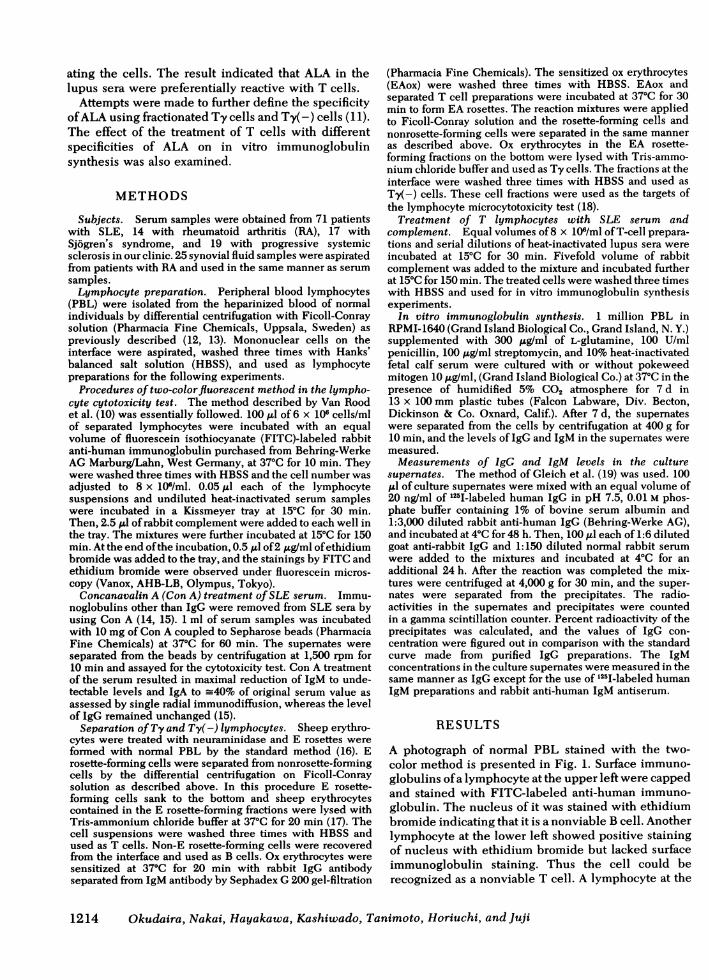
A photograph of normal PBL stained with the two-color method is presented in Fig. 1. Surface immuno-globulins ofa lymphocyte at the upper left were cappedand stained with FITC-labeled anti-human immuno-globulin. The nucleus of it was stained with ethidiumbromide indicating that it is a nonviable B cell. Anotherlymphocyte at the lower left showed positive stainingof nucleus with ethidium bromide but lacked surfaceimmunoglobulin staining. Thus the cell could berecognized as a nonviable T cell. A lymphocyte at the
1214 Okudaira, Nakai, Hayakawa, Kashiwado, Tanimoto, Horiuchi, and Juji
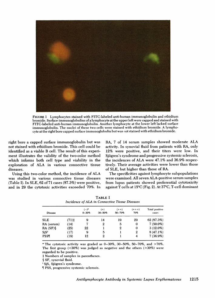
FIGURE 1 Lymphocytes stained with FITC-labeled anti-human immunoglobulin and ethidiumbromide. Surface immunoglobulins ofa lymphocyte at the upper left were capped and stained withFITC-labeled anti-human immunoglobulin. Another lymphocyte at the lower left lacked surfaceimmunoglobulin. The nuclei of these two cells were stained with ethidium bromide. A lympho-cyte at the right bore capped surface immunoglobulin but was iot stained with ethidium bromide.
right bore a capped surface immunoglobulin but wasnot stained with ethidium bromide. This cell could beidentified as a viable B cell. The result of this experi-ment illustrates the validity of the two-color methodwhich informs both cell type and viability in theexploration of ALA in various connective tissuediseases.Using this two-color method, the incidence of ALA
was studied in various connective tissue diseases(Table I). In SLE, 62 of 71 cases (87.3%) were positive,and in 29 the cytotoxic activities exceeded 70%. In
RA, 7 of 14 serum samples showed moderate ALAactivity. In synovial fluid from patients with RA, only12% were positive, and their titers were low. InSj6gren's syndrome and progressive systemic sclerosis,the incidences of ALA were 47.1% and 36.9% respec-tively. Their average activities were lower than thoseof SLE, but higher than those of RA.The specificities against lymphocyte subpopulations
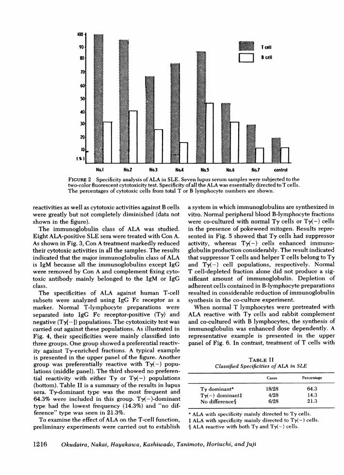
were examined. All seven ALA-positive serum samplesfrom lupus patients showed preferential cytotoxicityagainst T cells at 15°C (Fig. 2). At 37°C, T-cell dominant
TABLE IIncidence ofALA in Connective Tissue Diseases
(H)* (+) (++) (+++) Total positiveDisease 0-30% 30-50% 50-70% 70% cases
SLE (71)t 9 14 19 29 62 (87.3%)RA (serum) (14) 7 2 5 0 7 (50.0%)RA (SF)§ (25) 22 1 2 0 3 (12.0%)SjS"l (17) 9 5 1 2 8 (47.1%)PSS¶ (19) 12 2 1 4 7 (36.9%)
* The cytotoxic activity was graded as 0-30%, 30-50%, 50-70%, and >70%.The first group (<30%) was judged as negative and the others (>30%) wereregarded to be positive.t Numbers of samples in parentheses.§ SF, synovial fluid."SjS, Sjogren's syndrome.¶ PSS, progressive systemic sclerosis.
Antilymphocyte Antibody in Systemic Lupus Erythematosus 1215
No.1 No.2 No.3 No.4 No,5 No.6 No.7 control
FIGURE 2 Specificity analysis ofALA in SLE. Seven lupus serum samples were subjected to thetwo-color fluorescent cytotoxicity test. Specificity of all the ALA was essentially directed to T cells.The percentages of cytotoxic cells from total T or B lymphocyte numbers are shown.
reactivities as well as cytotoxic activities against B cellswere greatly but not completely diminished (data notshown in the figure).The immunoglobulin class of ALA was studied.
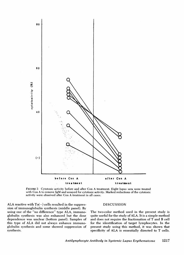
Eight ALA-positive SLE sera were treated with Con A.As shown in Fig. 3, Con A treatment markedly reducedtheir cytotoxic activities in all the samples. The resultsindicated that the major immunoglobulin class of ALAis IgM because all the immunoglobulins except IgGwere removed by Con A and complement fixing cyto-toxic antibody mainly belonged to the IgM or IgGclass.The specificities of ALA against human T-cell
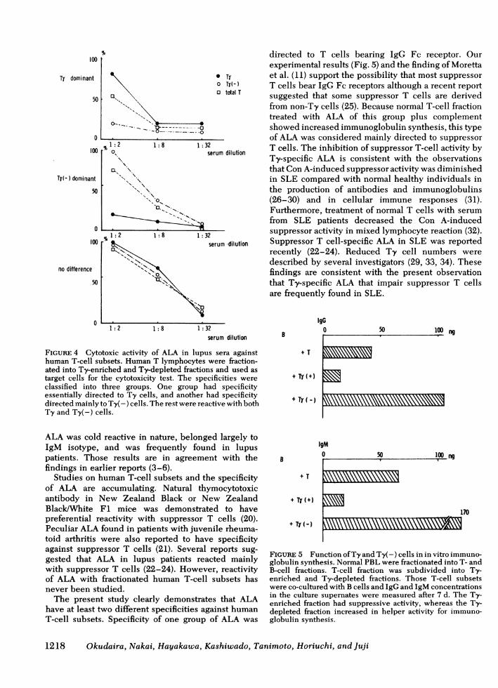
subsets were analyzed using IgG Fc receptor as amarker. Normal T-lymphocyte preparations wereseparated into IgG Fc receptor-positive (Ty) andnegative (Ty[-]) populations. The cytotoxicity test wascarried out against these populations. As illustrated inFig. 4, their specificities were mainly classified intothree groups. One group showed a preferential reactiv-ity against Ty-enriched fractions. A typical exampleis presented in the upper panel of the figure. Anothergroup was preferentially reactive with Ty(-) popu-lations (middle panel). The third showed no preferen-tial reactivity with either Ty or Ty(-) populations(bottom). Table II is a summary of the results in lupussera. Ty-dominant type was the most frequent and64.3% were included in this group. Ty(-)-dominanttype had the lowest frequency (14.3%) and "no dif-ference" type was seen in 21.3%.To examine the effect ofALA on the T-cell function,
preliminary experiments were carried out to establish
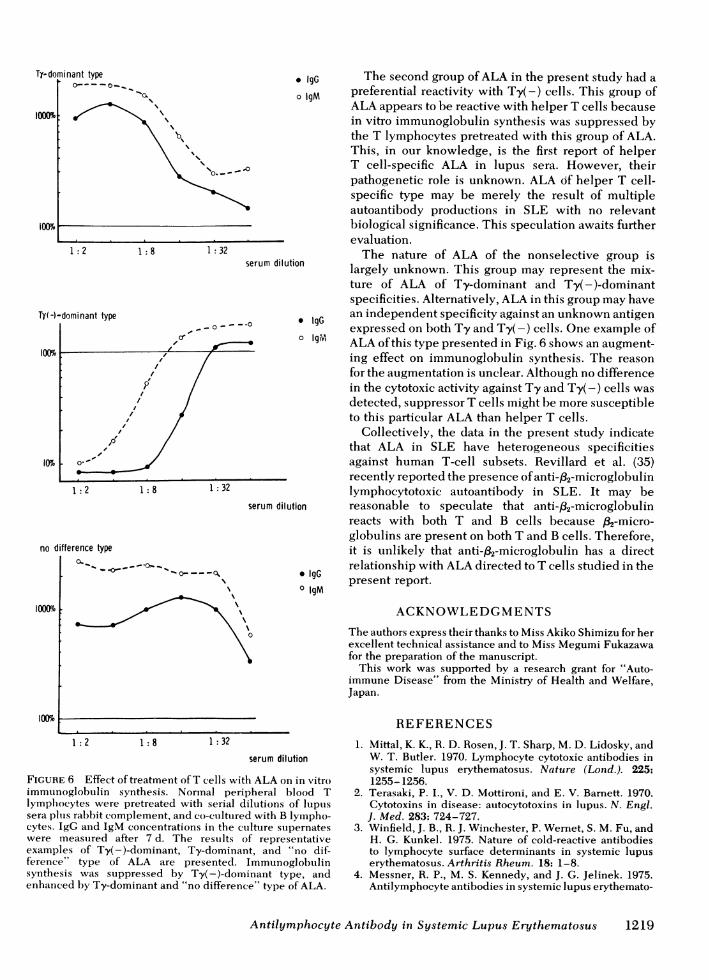
a system in which immunoglobulins are synthesized invitro. Normal peripheral blood B-lymphocyte fractionswere co-cultured with normal Ty cells or Ty(-) cellsin the presence of pokeweed mitogen. Results repre-sented in Fig. 5 showed that Ty cells had suppressoractivity, whereas Ty( -) cells enhanced immuno-globulin production considerably. The result indicatedthat suppressor T cells and helper T cells belong to Tyand Ty(-) cell populations, respectively. NormalT cell-depleted fraction alone did not produce a sig-nificant amount of immunoglobulin. Depletion ofadherent cells contained in B-lymphocyte preparationsresulted in considerable reduction of immunoglobulinsynthesis in the co-culture experiment.When normal T lymphocytes were pretreated with
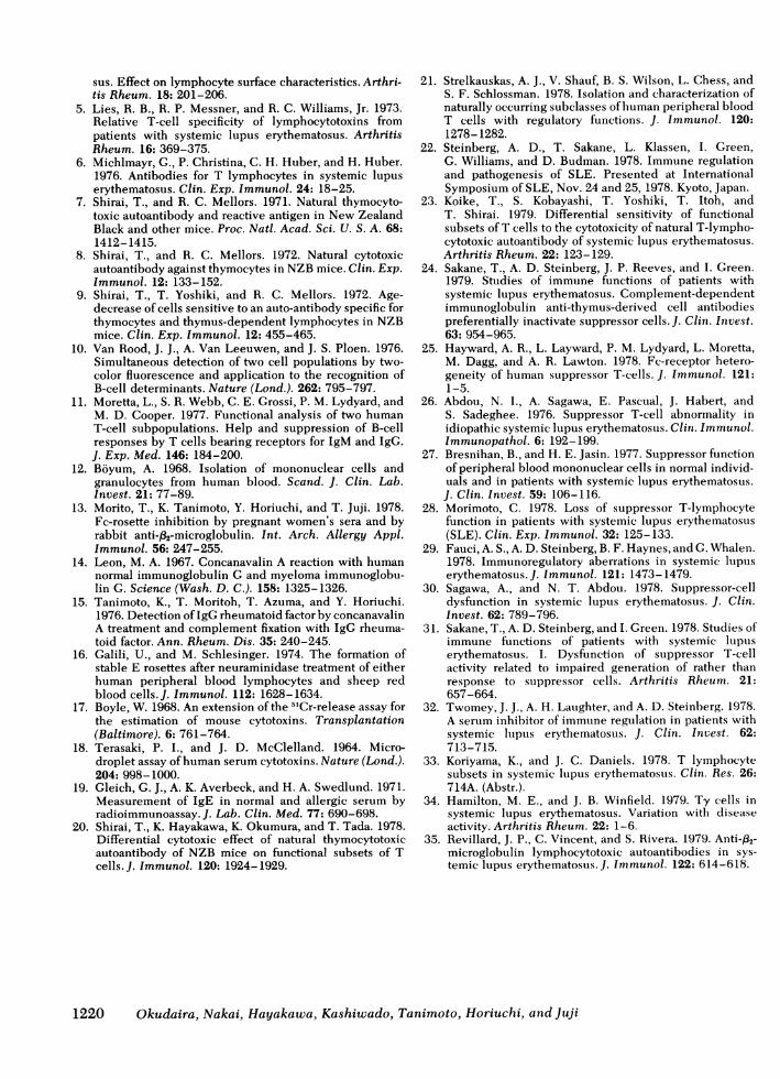
ALA reactive with Ty cells and rabbit complementand co-cultured with B lymphocytes, the synthesis ofimmunoglobulin was enhanced dose dependently. Arepresentative example is presented in the upperpanel of Fig. 6. In contrast, treatment of T cells with
TABLE IIClassified Specificities ofALA in SLE
Cases Percentage
Ty dominant* 18/28 64.3Ty(-) dominant4 4/28 14.3No difference§ 6/28 21.3
* ALA with specificity mainly directed to Ty cells.t ALA with specificity mainly directed to Ty(-) cells.§ ALA reactive with both Ty and Ty(-) cells.
1216 Okudaira, Nakai, Hayakawa, Kashiwado, Tanimoto, Horiuchi, and Juji
'''' Os \;st \
* \
a_ \
- \
v Q \.. ; . \ >..... ,; , m. ..... \., \
., ^ _
>
Q-.__., __
\_
., . _.before Con A
trea tme nt
after Con A
treatment
FIGURE 3 Cytotoxic activity before and after Con A treatment. Eight lupus sera were treatedwith Con A to remove IgM and assayed for cytotoxic activity. Marked reductions of the cytotoxicactivity were observed after Con A treatment in all cases.
ALA reactive with Ty(-) cells resulted in the suppres-sion of immunoglobulin synthesis (middle panel). Byusing one of the "no difference" type ALA, immuno-globulin synthesis was also enhanced but the dosedependence was unclear (bottom panel). Samples ofthis type of ALA did not always enhance immuno-globulin synthesis and some showed suppression ofsynthesis.
DISCUSSION
The two-color method used in the present study isquite useful for the study ofALA. It is a simple methodand does not require the fractionation of T and B cellfor the identification of target lymphocytes. In thepresent study using this method, it was shown thatspecificity of ALA is essentially directed to T cells.
Antilymphocyte Antibody in Systemic Lupus Erythematosus
80
60
-
x0
0U>1
(i
40
(-)
1217
100
Ty dominant
%a
50 [
o
100
Ty(- ) dominant
50
0
100
no difference
50
n
0K~~~~~~
0-- Bzyz~~~-- -..-%l:2
01: 8 1: 3
o,
I.\I\\
" O.
.~~~~~~~~~~~~~-_ 11s
% 1:2 1:8 1:3
K.NX*
:k L-
1:2 1:8 1:32serum dilution
FIGURE 4 Cytotoxic activity of ALA in lupus sera againsthuman T-cell subsets. Human T lymphocytes were fraction-ated into Ty-enriched and Ty-depleted fractions and used astarget cells for the cytotoxicity test. The specificities wereclassified into three groups. One group had specificityessentially directed to Ty cells, and another had specificitydirected mainly to Ty(-) cells. The rest were reactive with bothTy and Ty(-) cells.
ALA was cold reactive in nature, belonged largely toIgM isotype, and was frequently found in lupuspatients. Those results are in agreement with thefindings in earlier reports (3-6).
Studies on human T-cell subsets and the specificityof ALA are accumulating. Natural thymocytotoxicantibody in New Zealand Black or New ZealandBlack/White Fl mice was demonstrated to havepreferential reactivity with suppressor T cells (20).Peculiar ALA found in patients with juvenile rheuma-toid arthritis were also reported to have specificityagainst suppressor T cells (21). Several reports sug-gested that ALA in lupus patients reacted mainlywith suppressor T cells (22-24). However, reactivityof ALA with fractionated human T-cell subsets hasnever been studied.The present study clearly demonstrates that ALA
have at least two different specificities against humanT-cell subsets. Specificity of one group of ALA was
directed to T cells bearing IgG Fc receptor. Ourexperimental results (Fig. 5) and the finding of Moretta
* Ty et al. (11) support the possibility that most suppressoro TY(-) T cells bear IgG Fc receptors although a recent reporto total T suggested that some suppressor T cells are derived
from non-Ty cells (25). Because normal T-cell fractiontreated with ALA of this group plus complementshowed increased immunoglobulin synthesis, this typeof ALA was considered mainly directed to suppressor
32 T cells. The inhibition of suppressor T-cell activity byserum dilution Ty-specific ALA is consistent with the observations
that Con A-induced suppressor activity was diminishedin SLE compared with normal healthy individuals inthe production of antibodies and immunoglobulins(26-30) and in cellular immune responses (31).Furthermore, treatment of normal T cells with serumfrom SLE patients decreased the Con A-induced
32 suppressor activity in mixed lymphocyte reaction (32).serum dilution Suppressor T cell-specific ALA in SLE was reported
recently (22-24). Reduced Ty cell numbers weredescribed by several investigators (29, 33, 34). Thesefindings are consistent with the present observationthat Ty-specific ALA that impair suppressor T cellsare frequently found in SLE.
IgG0
+ TY (+)
+ TY( -)
B
+ TY(+)
+ TY (-)
50 100 ng
IgM0 50 100 ng
170
FIGURE 5 Function ofTy and Ty( -) cells in in vitro immuno-globulin synthesis. Normal PBL were fractionated into T- andB-cell fractions. T-cell fraction was subdivided into Ty-enriched and Ty-depleted fractions. Those T-cell subsetswere co-cultured with B cells and IgG and IgM concentrationsin the culture supernates were measured after 7 d. The Ty-enriched fraction had suppressive activity, whereas the Ty-depleted fraction increased in helper activity for immuno-globulin synthesis.
1218 Okudaira, Nakai, Hayakawa, Kashiwado, Tanimoto, Horiuchi, and Juji
3
Ty-dominant typeoI - e- _ S
'%
'ooOo%
0o. __-
1: 2 1: 8 1: 32serum diluti
Ty(-)-dominant type
serum dilut
no difference type
I X-.o-- - Or S~~~~~o- Q
100
1~~~~~~~~~~~~0
* IgG The second group ofALA in the present study had ao IgM preferential reactivity with Ty(-) cells. This group of
ALA appears to be reactive with helper T cells becausein vitro immunoglobulin synthesis was suppressed bythe T lymphocytes pretreated with this group of ALA.This, in our knowledge, is the first report of helperT cell-specific ALA in lupus sera. However, theirpathogenetic role is unknown. ALA of helper T cell-specific type may be merely the result of multipleautoantibody productions in SLE with no relevantbiological significance. This speculation awaits furtherevaluation.The nature of ALA of the nonselective group is
ionl largely unknown. This group may represent the mix-ture of ALA of Ty-dominant and Ty(-)-dominantspecificities. Alternatively, ALA in this group may have
* IgG an independent specificity against an unknown antigenexpressed on both Ty and Ty(-) cells. One example ofo IgM ALA ofthis type presented in Fig. 6 shows an augment-
ing effect on immunoglobulin synthesis. The reasonfor the augmentation is unclear. Although no differencein the cytotoxic activity against Ty and Ty(-) cells wasdetected, suppressor T cells might be more susceptibleto this particular ALA than helper T cells.
Collectively, the data in the present study indicatethat ALA in SLE have heterogeneous specificitiesagainst human T-cell subsets. Revillard et al. (35)recently reported the presence ofanti-,82-microglobulinlymphocytotoxic autoantibody in SLE. It may be
tion reasonable to speculate that anti-,32-microglobulinreacts with both T and B cells because p2-micro-globulins are present on both T and B cells. Therefore,it is unlikely that anti-f32-microglobulin has a direct
IgG relationship with ALA directed to T cells studied in the* lgG present report.o IgM
ACKNOWLEDGMENTSThe authors express their thanks to Miss Akiko Shimizu for herexcellent technical assistance and to Miss Megumi Fukazawafor the preparation of the manuscript.This work was supported by a research grant for "Auto-
immune Disease" from the Ministry of Health and Welfare,Japan.
1: 2 1: 8 1: 32serum dilution
FIGURE 6 Effect of treatment ofT cells with ALA on in vitroimmunoglobuilin synthesis. Normal peripheral blood Tlymphocytes were pretreated with serial dilutions of lupuissera plus rabbit complement, and co-cutltured with B lympho-cytes. IgG and IgM concentrations in the culture supernateswere measuired after 7 d. The results of representativeexamples of Ty(-)-dominant, Ty-dominant, and "no dif-ference" type of ALA are presented. Immunoglobulinsynthesis was suppressed by Ty(-)-dominant type, andenhanced by Ty-dominant and "no difference" type of ALA.
REFERENCES1. Mittal, K. K., R. D. Rosen, J. T. Sharp, M. D. Lidosky, and
W. T. Butler. 1970. Lymphocyte cytotoxic antibodies insystemic lupus erythematosus. Nature (Lond.). 225:1255- 1256.
2. Terasaki, P. I., V. D. Mottironi, and E. V. Barnett. 1970.Cytotoxins in disease: autocytotoxins in lupus. N. Engl.J. Med. 283: 724-727.
3. Winfield, J. B., R. J. Winchester, P. Wernet, S. M. Fu, andH. G. Kunkel. 1975. Nature of cold-reactive antibodiesto lymphocyte surface determinants in systemic lupuserythematosus. Arthritis Rheum. 18: 1-8.
4. Messner, R. P., M. S. Kennedy, and J. G. Jelinek. 1975.Antilymphocyte antibodies in systemic lupus erythemato-
Antilymphocyte Antibody in Systemic Lupus Erythematosus 1219
sus. Effect on lymphocyte surface characteristics. Arthri-tis Rheum. 18: 201-206.
5. Lies, R. B., R. P. Messner, and R. C. Williams, Jr. 1973.Relative T-cell specificity of lymphocytotoxins frompatients with systemic lupus erythematosus. ArthritisRheum. 16: 369-375.
6. Michlmayr, G., P. Christina, C. H. Huber, and H. Huber.1976. Antibodies for T lymphocytes in systemic lupuserythematosus. Clin. Exp. Immunol. 24: 18-25.
7. Shirai, T., and R. C. Mellors. 1971. Natural thymocyto-toxic autoantibody and reactive antigen in New ZealandBlack and other mice. Proc. Natl. Acad. Sci. U. S. A. 68:1412- 1415.
8. Shirai, T., and R. C. Mellors. 1972. Natural cytotoxicautoantibody against thymocytes in NZB mice. Clin. Exp.Immunol. 12: 133-152.
9. Shirai, T., T. Yoshiki, and R. C. Mellors. 1972. Age-decrease of cells sensitive to an auto-antibody specific forthymocytes and thymus-dependent lymphocytes in NZBmice. Clin. Exp. Immunol. 12: 455-465.
10. Van Rood, J. J., A. Van Leeuwen, and J. S. Ploen. 1976.Simultaneous detection of two cell populations by two-color fluorescence and application to the recognition ofB-cell determinants. Nature (Lond.). 262: 795-797.
11. Moretta, L., S. R. Webb, C. E. Grossi, P. M. Lydyard, andM. D. Cooper. 1977. Functional analysis of two humanT-cell subpopulations. Help and suppression of B-cellresponses by T cells bearing receptors for IgM and IgG.J. Exp. Med. 146: 184-200.
12. Boyum, A. 1968. Isolation of mononuclear cells andgranulocytes from human blood. Scand. J. Clin. Lab.Invest. 21: 77-89.
13. Morito, T., K. Tanimoto, Y. Horiuchi, and T. Juji. 1978.Fc-rosette inhibition by pregnant women's sera and byrabbit anti-f32-microglobulin. Int. Arch. Allergy Appl.Immunol. 56: 247-255.
14. Leon, M. A. 1967. Concanavalin A reaction with humannormal immunoglobulin G and myeloma immunoglobu-lin G. Science (Wash. D. C.). 158: 1325-1326.
15. Tanimoto, K., T. Moritoh, T. Azuma, and Y. Horiuchi.1976. Detection ofIgG rheumatoid factor by concanavalinA treatment and complement fixation with IgG rheuma-toid factor. Ann. Rheum. Dis. 35: 240-245.
16. Galili, U., and M. Schlesinger. 1974. The formation ofstable E rosettes after neuraminidase treatment of eitherhuman peripheral blood lymphocytes and sheep redblood cells. J. Immunol. 112: 1628-1634.
17. Boyle, W. 1968. An extension of the 51Cr-release assay forthe estimation of mouse cytotoxins. Transplantation(Baltimore). 6: 761-764.
18. Terasaki, P. I., and J. D. McClelland. 1964. Micro-droplet assay ofhuman serum cytotoxins. Nature (Lond.).204: 998-1000.
19. Gleich, G. J., A. K. Averbeck, and H. A. Swedlund. 1971.Measurement of IgE in normal and allergic serum byradioimmunoassay. J. Lab. Clin. Med. 77: 690-698.
20. Shirai, T., K. Hayakawa, K. Okumura, and T. Tada. 1978.Differential cytotoxic effect of natural thymocytotoxicautoantibody of NZB mice on functional subsets of Tcells. J. Immunol. 120: 1924-1929.
21. Strelkauskas, A. J., V. Shauf, B. S. Wilson, L. Chess, andS. F. Schlossman. 1978. Isolation and characterization ofnaturally occurring subclasses ofhuman peripheral bloodT cells with regulatory functions. J. Immunol. 120:1278-1282.
22. Steinberg, A. D., T. Sakane, L. Klassen, I. Green,G. Williams, and D. Budman. 1978. Immune regulationand pathogenesis of SLE. Presented at InternationalSymposium of SLE, Nov. 24 and 25, 1978. Kyoto, Japan.
23. Koike, T., S. Kobayashi, T. Yoshiki, T. Itoh, andT. Shirai. 1979. Differential sensitivity of functionalsubsets of T cells to the cytotoxicity of natural T-lympho-cytotoxic autoantibody of systemic lupus erythematosus.Arthritis Rheum. 22: 123-129.
24. Sakane, T., A. D. Steinberg, J. P. Reeves, and I. Green.1979. Studies of immune functions of patients withsystemic lupus erythematosus. Complement-dependentimmunoglobulin anti-thymus-derived cell antibodiespreferentially inactivate suppressor cells. J. Clin. Invest.63: 954-965.
25. Hayward, A. R., L. Layward, P. M. Lydyard, L. Moretta,M. Dagg, and A. R. Lawton. 1978. Fc-receptor hetero-geneity of human suppressor T-cells. J. Immunol. 121:1-5.
26. Abdou, N. I., A. Sagawa, E. Pascual, J. Habert, andS. Sadeghee. 1976. Suppressor T-cell abnormality inidiopathic systemic lupus erythematosus. Clin. Immunol.Immunopathol. 6: 192-199.
27. Bresnihan, B., and H. E. Jasin. 1977. Suppressor functionof peripheral blood mononuclear cells in normal individ-uals and in patients with systemic lupus erythematosus.J. Clin. Invest. 59: 106-116.
28. Morimoto, C. 1978. Loss of suppressor T-lymphocytefunction in patients with systemic lupus erythematosus(SLE). Clin. Exp. Immunol. 32: 125-133.
29. Fauci, A. S., A. D. Steinberg, B. F. Haynes, and G. Whalen.1978. Immunoregulatory aberrations in systemic lupuserythematosus. J. Immunol. 121: 1473-1479.
30. Sagawa, A., and N. T. Abdou. 1978. Suppressor-celldysfunction in systemic lupus erythematosus. J. Clin.Invest. 62: 789-796.
31. Sakane, T., A. D. Steinberg, and I. Green. 1978. Studies ofimmune functions of patients with systemic lupuserythematosus. I. Dysfunction of suppressor T-cellactivity related to impaired generation of rather thanresponse to siuppressor cells. Arthritis Rheuin. 21:657-664.
32. Twomey, J. J., A. H. Laughter, and A. D. Steinberg. 1978.A serum inhibitor of immune regulation in patients withsystemic lupus erythematosus. J. Clin. Invest. 62:713-715.
33. Koriyama, K., and J. C. Daniels. 1978. T lymphocytesubsets in systemic lupus erythematosus. Clin. Res. 26:714A. (Abstr.).
34. Hamilton, M. E., and J. B. Winfield. 1979. Ty cells insystemic lupus erythematosus. Variation with diseaseactivity. Arthritis Rheum. 22: 1-6.
35. Revillard, J. P., C. Vincent, and S. Rivera. 1979. Anti-X82-microglobulin lymphocytotoxic autoantibodies in sys-temic lupus erythematosus. J. Immunol. 122: 614-618.
1220 Okudaira, Nakai, Hayakawa, Kashiwado, Tanimoto, Horiuchi, and Juji


![Tumor Detection with 131I-labeledHuman Monoclonal Antibody ... · [CANCER RESEARCH 53, 5920-5928, December 15, 1993] Tumor Detection with 131I-labeledHuman Monoclonal Antibody COU-1](https://img.pdfslide.net/doc/110x75/603cdf8783e7021f98686577/tumor-detection-with-131i-labeledhuman-monoclonal-antibody-cancer-research.jpg)