Embed Size (px)
Citation preview
Earl! Human. DeveloDment .
ELSEVIER Early Human Development 47 SUPPI. (1996) ~47-~48
Detection of p-core fragment in second trimester Down’s syndrome pregnancies
Laurence Cole”‘“, Taichi Isozaki”, Glen Palomakib, Jacob Canick’, Ray Ilesd, Leonard Kellner’, Deverux Sailer”, Howard Cuckle”
“Yale University, New Haven, CT, USA hFobundation for Blood Research, Scarborough, ME, USA
‘Brown Univertity, Providence, RI, USA “St. Bartholomew’s Hospital, London, UK
‘Universi~ of Leeds, Leeds, UK
Keywords: Down’s syndrome; P-Core fragment; Second trimester
p-Core fragment (/?-core) accounts for about two thirds of hCG P-subunit-related molecules in second trimester pregnancy urine. In 1994, Cuckle et al. [l], measured p-core in second trimester pregnancy urines. A large difference was found in 7 cases with Down’s syndrome and 67 with normal karyotype fetuses (6.1 MOM). They indicated that p-core might replace determination of serum antigens. In 1995, the same group published a study with 24 Down’s syndrome samples. They again found 6.0 MOM, and an 80% detection rate with 5% false positive rate [2]. In 1995 Canick et al. [3] reported like results with 14 Down’s syndrome samples, 5.3 MOM, with 88% detection at 5% false positive rate. These detection rates using urine p-core tests may be greatly superior to those for serum hCG or free P-subunit, or triple screen tests. One other paper has been published by Hayashi and Kozu suggesting less favorable results [4]. In this paper urines were tested with the Wako p-core kit, that primarily detects free P-subunit, and secondarily measures p-core. Choice of the correct p-core assay may be very important for screening applications.
Here we test the findings of Cuckle et al. and Canick et al., [l-3] with three p-core only, two p-core with free P-subunit crossreactivity ( < 25%), and one free p-
* Corresponding author.
037%3782/96/$15.00 Copyright 0 1996 Elsevier Science Ireland Ltd. All rights reserved PIZ SO378-3782(96)01819-l
S48 L. Cole et al. I Eurly Human Developmenr 47 Suppl. (1996) S47-S48
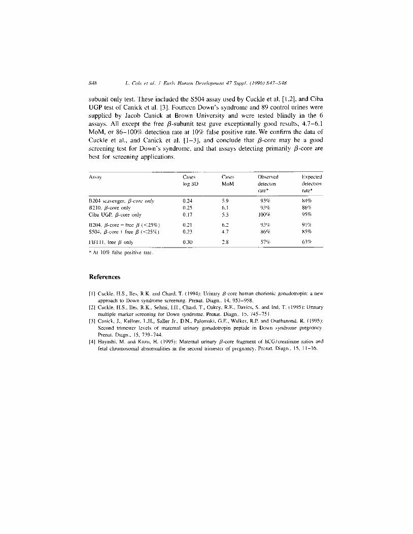
subunit only test. These included the S504 assay used by Cuckle et al. [ 1,2], and Ciba UGP test of Canick et al. [3]. Fourteen Down’s syndrome and 89 control urines were supplied by Jacob Canick at Brown University and were tested blindly in the 6 assays. All except the free P-subunit test gave exceptionally good results, 4.7-6.1 MOM, or 86- 100% detection rate at 10% false positive rate. We confirm the data of Cuckle et al., and Canick et al. [l-3], and conclude that p-core may be a good screening test for Down’s syndrome, and that assays detecting primarily p-core are best for screening applications.
Assay Cases
log SD
Cases
MOM Observed detecton
rate*
Expected detection rate*
B204 scavenger, p-cow only B210, p-core only
Ciba UGP. p-core only
8204, p-core + free p (<2S%)
S.504, p-core + free p (<25%)
FBTI I, free p only
0.24 5.9 93% 84%
0.2.5 6.1 93% 868
0.17 5.3 100% 95%
0.21 6.2 93% 91%
0.23 4.7 86% 85%
0.30 2.8 57% 63%
* At 10% false positive rate
References
[I] Cuckle, H.S., Iles, R.K. and Chard. T. (1994): Urinary p-core human chorionic gonadotropin: a new approach to Down syndrome screening. Prenat. Diagn., 14, 953-958.
[2] Cuckle, H.S., Iles, R.K.. Sehmi, I.H., Chard, T., Oakey, R.E., Davies, S. and Ind, T. (1995): Urinary multiple marker screening for Down syndrome. Prenat. Diagn., 15, 745-751.
[3] Canick. I., Kellner, L.H., Saller Jr., D.N., Palomaki, G.E., Walker, R.P. and Osathanond, R. (199.5):
Second trimester levels of maternal urinary gonadotropin peptide in Down syndrome pregnancy. Prenat. Diagn., IS, 739-744.
[4] Hayashi, M. and Kozu, H. (1995): Maternal urinary p-core fragment of hCG/creatinine ratios and fetal chromosomal abnormalities in the second trimester of pregnancy. Prenat. Diagn., IS. 1 I-16.