Embed Size (px)
Citation preview
APPLIED MICROBIOLOGY, Aug. 1974, P. 306-311Copyright ( 1974 American Society for Microbiology
Vol. 28, No. 2Printed in U.S.A.
Detection of Escherichia coli Antigens by a LatexAgglutination Test
K. HECHEMY, R. W. STEVENS, AND H. A. GAAFAR
Division of Laboratories and Research, New York State Department of Health, Albany, New York 12201
Received for publication 9 April 1974
A latex particle agglutination technique to detect ethylenediaminetetraace-tate-solubilized extracts from Escherichia coli and whole E. coli cells isdescribed. The sensitivity of the serological test was found to be 0.5 to 2.5 ng forthe solubilized antigens and 1.5 x 10⁶ to 5.7 x 10⁶ cells per ml for the particulateantigens. The test was 100 to 1,000 times more sensitive than the standardbacterial agglutination test. Furthermore, it detected E. coli antigens during allphases of bacterial growth, whereas the bacterial test detected the antigens onlyafter the mid-log phase. No significant cross-reactivity was observed betweenlatex-anti-E. coli preparations and heterologous bacterial'strains used in theexperimental procedure. A buffer formula containing fatty acid-free bovinealbumin prevented nonspecific aggregation of the latex particles.
Standard test procedures for the identifica-tion of Escherichia coli serotypes involve ag-glutination (3) or immunofluorescence (2) withwhole cells, or gel precipitation (5) with solubleantigens. The relative insensitivity or complex-ity of these tests is a major disadvantage whenthe quantity of antigen or the time available islimited. This led us to study the application ofthe latex particle technique to E. coli. Latexparticles have been used as inert carriers ofantigens for detecting the corresponding serumantibodies (9, 12, 13) and, conversely, as vehi-cles for antibodies for detecting antigen in bio-logical fluids (1, 10).This paper reports the development and eval-
uation of a test using anti-E. coli globulinadsorbed to latex, which makes possible therapid as well as sensitive detection of both E.coli cells and ethylenediaminetetraacetate-solubilized extracts from those cells.
MATERIALS AND METHODSBuffers. Four buffers were used: 0.12 M tris(hy-
droxymethyl)aminomethane-hydrochloride, pH 8.0;0.1 M glycine-buffered saline (GBS), pH 8.2; andGBS containing 1 or 0.1% bovine albumin, type F(BAF; Sigma Chemical Co., St. Louis, Mo.).
Cultures. Stock strains from the Division's culturecollection were maintained as stab cultures in nutri-ent agar at room temperature.
Antigen preparation. A 0.1-ml volume of an 18-hbroth culture was inoculated into 600 ml of beef heartinfusion broth containing 1% glucose and incubatedon a rotary shaker for 16 h at 37 C. The cells wereharvested and washed three times by centrifugation
at 10,000 x g with the tris(hydroxymethyl)aminome-thane buffer.
Soluble antigens (SAg) were prepared essentially asdescribed by Leive and Shovlin (8). Stock SAg solu-tions (10 mg/ml) were prepared by dissolving lyophi-lized material in GBS with 0.1% BAF. These werestored at -20 C.
Particulate antigens (PAg) were suspensions ofresting cells diluted in membrane-filtered (MilliporeCorp.) saline. The maximum dilution that reacted(2+) in the latex agglutination test was determined,and the cells were counted with a Coulter counter(Coulter Electronics, Inc., Hialeah, Fla.)
Globulins. Suspensions of various serotypes of E.coli cells from 18- to 24-h cultures were adjusted to anoptical density (at 540 nm) of 350 Klett units in 0.3%formalin solution in saline. Rabbits were immunized(6), and the antisera obtained were tested for bacte-rial agglutination with the homologous culture. Glob-ulins from preimmune and immunized rabbits wereprecipitated by ammonium sulfate and separatedfurther on a diethylaminoethyl-cellulose column (7).The purity of the fraction obtained was determined byimmunoelectrophoresis, and the protein was adjustedto 1 mg/ml with saline by the biuret method (4).
Latex-IgG suspension. Stock suspensions of latexparticles (Difco Laboratories, Detroit, Mich.) werefirst adjusted for adsorption of a single globulin asdescribed by Newman et al. (10). The latex-singleimmunoglobulin G (IgG) suspensions were preparedby mixing the reagents in the following order: latex,IgG, GBS, and GBS with 1% BAF. After eachaddition the mixture was shaken for 30 s. For tests ofSAg and PAg, the ratios by volume of the reagentswere: for E. coli serotype 0127:B8, 3:1:1.2:0.8;for serotype 0128 :B12, 3:0.5: 1.5: 1; for serotype0124: B17, 4:2: 1:1; and for the six remaining sero-
,06
on May 19, 2018 by guest
http://aem.asm
.org/D
ownloaded from
LATEX AGGLUTINATION TEST
types, 3:1: 1: 1. For tests of all whole cultures, thelatex-IgG composition was latex-IgG-GBS, 3:1:2.
For adsorption of several globulins to a single latexpreparation, 0.3-ml volumes of stock latex were cen-trifuged at 4,000 x g and all the supernatants weredecanted and pooled. For each suspension, the sedi-ment from 0.3 ml of latex was mixed with 0.3 ml of anIgG mixture (0.1 ml from each of three IgG solutions)and shaken for 2 min. To restore the smooth and evensuspension, 0.4 ml of the original latex supernatantwas returned to the mixture, followed by 0.1 ml ofGBS with 1% BAF. The final suspension was shakenfor an additional 2 min.
Latex test. Forty microliters of an SAg solution orPAg suspension was mixed with 20 gliters of latexglobulin suspension on a glass slide. The slide washand-tilted for 2 min. The degree of agglutination wasread macroscopically and recorded as 4+, 3+, 2+, or1+.
Specificity of latex tests. The following controlsfor nonspecific aggregation were included with alltests: (i) latex-IgG plus GBS with 0.1% BAF; (ii)latex-normal globulin plus GBS with 0.1% BAF; (iii)latex-normal globulin plus antigen; and (iv) latex plusantigen buffer.The specificity of the test with the soluble anti-
gen(s) was determined after finding the concentrationof SAg which, when mixed with homologous IgG,resulted in particle agglutination. Antigen at thisconcentration was then mixed with each of eightremaining IgG preparations. Cross-reactivity was re-corded if agglutination was 2+ or greater.
Test specificity with whole cultures was also stud-ied. A number of organisms (see Table 1) were grownwith shaking at 37 C for 16 h in beef heart infusion-glucose. The cultures were then mixed with latex-anti-E. coli IgG suspensions and with anti-E. coli IgGsolutions (1 mg of protein/ml). At the end of theincubation period, the degree of specific and heterolo-gous reactivity was recorded as described above.
Sensitivity of latex and bacterial tests. SAg so-lutions were diluted and tested by the particle agglu-tination test. In addition, latex and bacterial agglutina-tion tests of resting cells and growing cultures wereperformed. Resting E. coli 055: B5 cells (10-folddilutions of 5.57 x 10' PAg/ml) were mixed withlatex-IgG suspensions or with IgG solutions (1 mg ofprotein per ml) for the bacterial agglutination test.The growing culture was initially prepared from a16-h beef heart infusion-glucose culture. The cellswere harvested by centrifugation and suspended in 10ml of beef heart infusion-glucose, and 1 ml of thissuspension was inoculated into 5 ml of the samemedium. The culture was then incubated with shak-ing at 37 C, and samples were withdrawn at intervalsand examined by latex and bacterial agglutinationtests. The test mixtures in both systems were incu-bated for 2 min at room temperature; the results wereread macroscopically.
RESULTSEthylenediaminetetraacetate-solubilized
antigen(s). The ethylenediaminetetraacetate
extraction procedure yielded 35 to 50 mg of SAgper gram (dry weight) of E. coli cells. Thelyophilized extracts were fluffy white powderswith a faint greenish tinge. They were soluble inwater to a concentration of 10 mg/ml at 25 C.
Specificity of the latex test. The appearanceof test and control mixtures is illustrated in Fig.1. Aggregation was not observed with controlmixtures. Moreover, latex buffer suspensionalone with either PAg suspension or SAg solu-tion did not cause clumping (not illustrated).These results indicate that the agglutinationshown is an immunological reaction.Reactions obtained with nine globulin-coated
particle preparations and the corresponding E.coli extracts were strain specific. In addition,tests of anti-E. coli and latex-anti-E. coli sus-pensions against a number of pathogenic orga-nisms indicated the specificity of this procedure(Table 1). With the exception of Staphylococcusaureus, cross-reactivity in both the bacterialand particle agglutination tests was minimal(<1+) and similar.Aggregation of S. aureus cultures by la-
tex-IgG was noted with all the antibody particlepreparations available but not with any of theglobulins in solution. Cultures of S. aureus weretested against latex-anti-E. coli 0111:B4, la-tex-normal globulin, and latex-GBS with 0.1%BAF. Maximum reactivity (Table 2) was ob-served with both particle-globulins and un-diluted cultures, although no agglutination wasnoted with the particle-buffer preparation.When S. aureus culture was diluted 1:8, noreaction was observed. Cultures of E. coli0111: B4, however, were reactive in a dilution of1: 1,000.
Sensitivity of the latex test. The optimumconcentration of the IgG prepared for this studywas 0.17 mg of protein per ml in latex-single IgGsuspensions, except for IgG against serotypes0124: B17 and 0128: B12, which were optimumat 0.125 and 0.08 mg/ml, respectively. The limitof detection of antigen(s) by the particle ag-glutination test is summarized in Table 3. Withthe latex-single IgG preparation, the minimumdetectable concentration of SAg varied between0.5 and 2.5 ng; with the latex-multiple IgG, itvaried between 1 and 5 ng. The number of cellsthat could be detected with either preparationranged between 1.5 and 5.7 x 10⁶ per ml.The comparative sensitivity of the latex and
bacterial agglutination tests was shown by twoexperiments. The first indicated that a suspen-sion of PAg (5.57 x 10' cells/ml) was visiblyaggregated when tested with either latex-IgG orIgG in solution. However, agglutination of PAg
307VOL. 28, 1974
on May 19, 2018 by guest
http://aem.asm
.org/D
ownloaded from

HECHEMY, STEVENS, AND GAAFAR
A B.A
FIG. 1. Latex agglutination. (A, B) Tests with soluble (SAg) and particulate (PAg) antigens, respectively. Therest are control mixtures: (C) latex IgG and buffer, (D) latex-normal globulin and PAg, (E) latex-normal globulinand SAg and (F) latex-normal globulin and buffer.
TABLE 1. Latex particle and bacterial agglutination tests with heterologous bacterial cultures
Degree of agglutination of immune globulins from E. coli serotypes
Culturea 026: B6 0111: B4 0119: B14 0126: B16 0124: B17
Lb B L B L B L B L B
Proteus rettgeri ................±.........4 1+ - - -
Enterobacter hafniae ................... - 1+ - - _ _ _ 1+Pseudomonas aeruginosa ........-........ 1+ _ - - - -
Staphylococcus aureus .............. .... 4+ - 4+ - 4+ - 4+ - 4+Enterobacter cloacae .......... ......... - - - - - - 1+ -
Arizona henshawii ...................... _ - - 1+ -
Neisseria meningiditis ..................-4 _ 1+ 1+ _ - - - -
Salmonella cubana .. . ...- - - 1+ - _ _E. coli 026: B6.4+ 4+ - - - - - _ _ _E. coli Olll:B4 .. 4+ 4+ - - - _ _ _E. coli 0119:B14. _ _ _ 4+ 4+ - - _ _E. coli 0126: B16 - - - - - 4+ 4+ - _E. coli 0124:B17. 4+ 4+
a No reactions occurred with the following cultures: Yersinia enterolitica, Citrobacter freundii, Enterobacteraerogenes, Provendicia alcalifaciens, Shigella dysenteriae, Proteus morganii, Edwardsiella tarda, Alkalescensdispar, E. coli 055:B5, E. coli 0127:B8, and E. coli 0128:B12.
h L, Latex agglutination test; B, bacterial agglutination test.c, Tests were made and gave negative results.
308 APPL. MICROBIOL.
on May 19, 2018 by guest
http://aem.asm
.org/D
ownloaded from

LATEX AGGLUTINATION TEST
TABLE 2. Agglutination of S. aureus and E.-coli culture by latex-globulin
Agglutination
Latex-globulin S. aureus E. coli 0111: B4
1:la 1:2 1:4 1:8- 1:1 1:2 1:4- 1:1,0001:1,000 1:100
Latex-anti-E. coliOlll:B4 4+ 2+ 4 _b 4+ 4+ 4+ 2+Latex-normal globulin ......... ......... 4+ 1+ J _ ± -_Latex-GBS with 0.1%BAF.___j.......- -
a Antigen dilution.-, Negative results.
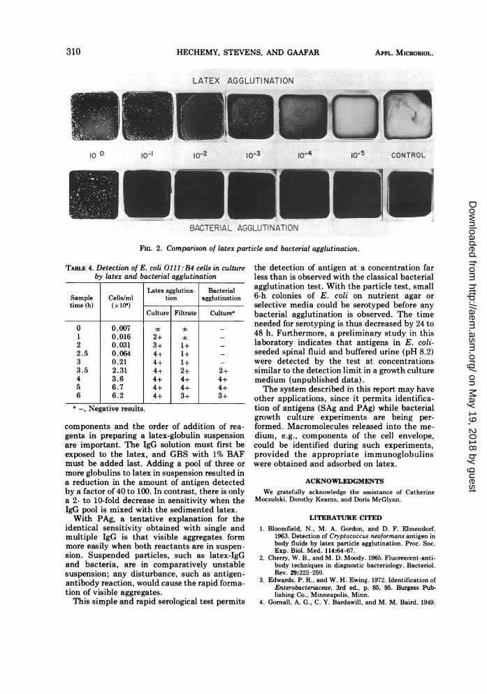
by IgG in solution was not visible after a 10-folddilution of the cells, whereas reactivity in thelatex procedure was observed after a 1,000-folddilution (Fig. 2). The second experiment (Table4) used portions taken from a culture at inter-vals over a period of 6 h. At the start ofincubation, the mixtures of culture media andlatex-IgG and IgG in solution were not visiblyagglutinated. After 1 h, while the cells were stillin lag phase, the latex reagent was aggregated.The reagent was also specifically aggregated bycell-free filtrates, although the reaction wasminimal. After 3.5 h, with the cells in thelogarithmic phase, aggregation was observedwith IgG solution. The minimum number ofcells (x109) per ml, as detected by the Coultercounter technique, was 0.016 by the latex testand 2.31 by bacterial agglutination, a differencein sensitivity of approximately 150-fold.
DISCUSSIONThe colloidal protective effect of serum pro-
tein on the hydrophobic latex particles was firstreported by Singer et al. (11), who found that, ofthe various proteins tested, Cohn fraction V(albumin) contributed a maximum stability tosuspended particles. Bloomfield et al. (1) firstused GBS -bovine serum albumin as a stabilizerin the latex test for Cryptococcus antigen. In ourstudy, a similar atypical aggregation with latexparticles mixed with GBS-bovine serum al-bumin suggested that the phenomenon is notdue to adsorbed globulin. When the nondefat-ted serum albumin was replaced by BAF inGBS, whether freshly prepared or stored forperiods up to 6 months at 5 C, the nonspecificaggregation was eliminated. Whenever wholecultures were tested, however, BAF was omit-ted. The effect of lipid from culture media onthe reaction was minimal, presumably becauseof the overriding effect of the large amount ofprotein present in culture media. Therefore,consideration of the protein and lipid content ofthe reaction mixtures is essential in establishingoptimum test reactivity.
TABLE 3. Minimum sensitivity of latex testa
Cells/ml EDTAb(X 10") extract
Pool E. coli (ng/ml)erotype
SingleMulti-
SingleMulti-
Single pie Single piegG IgG gO IgG
I 0111:B4 5.53 5.53 1.0 5.0055:B5 5.73 5.73 2.5 5.0026:B6 3.78 3.78 1.0 5.0
II 087:B7 5.63 5.63 0.5 5.00127:B8 5.23 5.23 1.0 5.00128: B12 5.70 5.70 0.5 1.0
III 0119:B14 2.87 2.87 0.5 1.00126:B16 5.68 5.68 0.5 1.00124:B17 1.57 1.57 1.0 1.0
a Latex-single IgG and latex-multiple IgG werereacted at optimum conditions against SAg or PAg.The lowest concentration of SAg or PAg which gave a2+ aggregation was recorded.
b EDTA, Ethylenediaminetetraacetate.
The GBS-with-BAF formula provided a uni-form, nongranular dispersion of latex in bufferand greatly enhanced the reliability andreproducibility of the test. BAF may also beuseful in other latex test preparations where anamount of antigen or antibody sufficient to keepthe particles in suspension would interfere withthe aggregation of the particles.A second cause of nonspecific aggregation was
a low concentration of anti-S. aureus in bothimmunized and nonimmunized rabbits whichwas solved by diluting the serum. When the E.coli extracts and antisera were tested, typecross-reactivity was not observed, even betweenthe closely related 086 and 0127 types (3).Although a large number of E. coli serotypeswere not studied with the latex test, it isprobable that cross-reactivity within the genusis comparable to that obtained by the bacterialtest.To achieve maximum sensitivity, the ratio of
VOL. 28, 1974 309
on May 19, 2018 by guest
http://aem.asm
.org/D
ownloaded from
HECHEMY, STEVENS, AND GAAFAR
LATEX AGGLUTI NATION
10-2 -10-3 1:0-4 IO5 CONTROL
BACTERIAL AGGLUTINATION
FIG. 2. Comparison of latex particle and bacterial agglutination.
TABLE 4. Detection of E. coli 0111 :B4 cells in cultureby latex and bacterial agglutination
Latex agglutina- BacterialSample Cells/ml tion agglutinationtime (h) (x 109)
Culture Filtrate Culturea
0 0.007 ± 4 _1 0.016 2+ ± _2 0.031 3+ 1+ _2.5 0.064 4+ 1+ _3 0.21 4+ 1+ -
3.5 2.31 4+ 2+ 2+4 3.6 4+ 4+ 4+5 6.7 4+ 4+ 4+6 6.2 4+ 3+ 3+
a -, Negative results.
components and the order of addition of rea-gents in preparing a latex-globulin suspensionare important. The IgG solution must first beexposed to the latex, and GBS with 1% BAFmust be added last. Adding a pool of three or
more globulins to latex in suspension resulted ina reduction in the amount of antigen detectedby a factor of 40 to 100. In contrast, there is onlya 2- to 10-fold decrease in sensitivity when theIgG pool is mixed with the sedimented latex.With PAg, a tentative explanation for the
identical sensitivity obtained with single andmultiple IgG is that visible aggregates formmore easily when both reactants are in suspen-sion. Suspended particles, such as latex-IgGand bacteria, are in comparatively unstablesuspension; any disturbance, such as antigen-antibody reaction, would cause the rapid forma-tion of visible aggregates.This simple and rapid serological test permits
the detection of antigen at a concentration farless than is observed with the classical bacterialagglutination test. With the particle test, small6-h colonies of E. coli on nutrient agar orselective media could be serotyped before anybacterial agglutination is observed. The timeneeded for serotyping is thus decreased by 24 to48 h. Furthermore, a preliminary study in thislaboratory indicates that antigens in E. coli-seeded spinal fluid and buffered urine (pH 8.2)were detected by the test at concentrationssimilar to the detection limit in a growth culturemedium (unpublished data).The system described in this report may have
other applications, since it permits identifica-tion of antigens (SAg and PAg) while bacterialgrowth culture experiments are being per-formed. Macromolecules released into the me-
dium, e.g., components of the cell envelope,could be identified during such experiments,provided the appropriate immunoglobulinswere obtained and adsorbed on latex.
ACKNOWLEDGMENTSWe gratefully acknowledge the assistance of Catherine
Moczulski, Dorothy Kearns, and Doris McGlynn.
LITERATURE CITED
1. Bloomfield, N., M. A. Gordon, and D. F. Elmendorf.1963. Detection of Cryptococcus neoformans antigen inbody fluids by latex particle agglutination. Proc. Soc.Exp. Biol. Med. 114:64-67.
2. Cherry, W. B., and M. D. Moody. 1965. Fluorescent-anti-body techniques in diagnostic bacteriology. Bacteriol.Rev. 29:222-250.
3. Edwards, P. R., and W. H. Ewing. 1972. Identification ofEnterobacteriaceae, 3rd ed., p. 85, 95. Burgess Pub-lishing Co., Minneapolis, Minn.
4. Gornall, A. G., C. Y. Bardawill, and M. M. Baird. 1949.
lo 0 l0-'
310 APPL. MICROBIOL.
on May 19, 2018 by guest
http://aem.asm
.org/D
ownloaded from
LATEX AGGLUTINATION TEST
Determination of serum proteins by means of the biuretreaction. J. Biol. Chem. 177:751-766.
5. Grados, O., and W. H. Ewing. 1969. Technics for charac-terization of soluble antigens of Enterobacteriaceae.Public Health Service and Mental Health Administra-tion Publication, National Communicable DiseaseCenter, Atlanta, Ga.
6. Harris, H. H., and M. B. Coleman (ed.). 1963. Diagnosticprocedures and reagents, 4th ed., p. 308-309. AmericanPublic Health Association, New York.
7. King, T. P. 1968. Cellulose ion exchange chromatogra-phy, p. 154-162. In C. A. Williams and M. W. Chase(ed.), Methods in immunology and immunochemistry,vol. 2. Academic Press Inc., New York.
8. Leive, L., and J. K. Shovlin. 1968. Physical, chemical andimmunological properties of lipopolysaccharide re-
leased from Escherichia coli by ethylenediaminetet-raacetate. J. Biol. Chem. 243:6384-6391.
9. Muraschi, T. F., N. Bloomfield, and R. B. Newman. 1962.A slide latex-particle agglutination test for trichinosis.Tech. Bull. Registry Med. Technologist 32:9-13 (Re-printed in Amer. J. Clin. Pathol. 37:227-231, 1962).
10. Newman, R. B., R. W. Stevens, and H. A. Gaafar. 1970.Latex agglutination test for the diagnosis of Haemophi-lus influenzae meningitis. J. Lab. Clin. Med. 76:107-113.
11. Singer, J. M., G. Altmann, I. Oreskes, and C. M. Plotz.1961. The mechanism of particulate carrier reactions.III. The stabilizing effect of serum proteins. Amer. J.Med. 30:772-778.
12. Singer, J. M., and C. M. Plotz. 1956. The latex fixationtest. I. Application to the serologic diagnosis of rheu-matoid arthritis. Amer. J. Med. 21:888-892.
13. Stevens, R. W. 1965. Agglutination of Reiter protein-coated latex particles. Amer. J. Clin. Pathol.43:490-493.
VOL. 28, 1974 311
on May 19, 2018 by guest
http://aem.asm
.org/D
ownloaded from