Embed Size (px)
Citation preview

ORIGINAL ARTICLE
Determinants of bone mass, density and growthin growing dogs with normal and osteopenic bones
K. Kumar & I. V. Mogha & H. P. Aithal & Amarpal &P. Kinjavdekar & G. R. Singh & A. M. Pawde & H. C. Setia
Accepted: 6 June 2008 /Published online: 17 July 2008# Springer Science + Business Media B.V. 2008
Abstract Survey radiographs of all the growing dogs aged up to 6 months, which werepresented to the IVRI polyclinics during the 10 year period were screened to study thedeterminants of bone mass, density and growth. On the basis of clinical history andradiographic evaluation of long bones, the cases were categorized as normal or osteopenic.The relative cortical density (RCD), cortical index (CI), diameter of bone at the distalmetaphysis (DDFM) and the width of the growth plate (WFGP) were determined by takingthe femur as a model bone in German shepherd, Doberman and Spitz breeds of dogs atdifferent age groups. The results showed that the RCD was the least in 0–2 month old normalgrowing dogs in all the breeds. As the age advanced up to 6 months the RCD increased 20–25%, and at 6 months, Spitz and Doberman showed significant increase (P<0.05) in the RCD.In osteopenic bones, RCD remained less (25–50%) than that of normal animals at all agegroups, and at 2–6 months of age, RCD in osteopenic bones was significantly lesser than innormal animals in GSD and Spitz breeds. The CI was also the least at 0–2 months of age innormal dogs. The CI increased about 50% at 4–6 months of age in GSD and Spitz. Whereasin Dob., there was no appreciable change in the CI at different age groups, and at 2–6 months itwas significantly (P<0.05) lesser than that of Spitz. In osteopenic bones, the CI was 25–75%lesser than that of normal animals at different age groups, and at 4–6 months there wassignificant difference (P<0.05) between the normal and osteopenic bones in GSD and Spitz.The DDFM was the least in 0–2 month old normal growing dogs, and as the age advanced, itincreased 10–20% up to 6 months. However, no significant difference in the DDFM was seenbetween breeds and also between the normal and osteopenic bones at different age groups. Innormal animals, theWFGPwas highest in the early age, subsequently it reduced 50–75% and at4–6 months there was significant decrease (P<0.05) in all the breeds of dogs. And at 4–6 months, there was significant (P<0.05) difference in the WFGP between breeds, it was theleast in Spitz andmaximum in Dob., suggesting faster growth plate closure in Spitz than in GSDand Dob. breeds. In osteopenic bones, WFGP was generally more than in normal animals, andat 4–6 months (about 3–5 times more) there was significant difference (P<0.05) between the
Vet Res Commun (2009) 33:57–66DOI 10.1007/s11259-008-9072-8
K. Kumar : I. V. Mogha : H. P. Aithal (*) : . Amarpal : P. Kinjavdekar :G. R. Singh : A. M. Pawde :H. C. SetiaDivision of Surgery, Indian Veterinary Research Institute, Izatnagar- 243 122 (UP), Bareilly, Indiae-mail: [email protected]
K. Kumar . I. V. Mogha . H. P. Aithal (*) . Amarpal . P. Kinjavdekar .G. R. Singh . A. M. Pawde . H. C. SetiaDivision of Surgery, Indian Veterinary Research Institute, Izatnagar- 243 122 (UP), Bareilly, Indiae-mail: [email protected]; [email protected]

normal and osteopenic bones in all breeds, indicating that physeal closure may be delayed inosteopenic bones. The results indicate that among different breeds Doberman breed has the leastbone mass and may be more prone to osteopenia; whereas Spitz has the strongest bone.
Keywords Bone density . Bone growth . Growing dogs . Osteopenic bones
AbbreviationsANOVA analysis of varianceCI cortical indexCWT cortical wall thicknessD mid-diameter of boneDDFM diameter of distal femoral metaphysisDob. DobermanGSD German shepherdmm millimeter% per centRCD cortical densityWFGP width of femoral growth plate
Introduction
Several reports available in the literature on the occurrence of fractures indicate that younggrowing dogs are more prone to fractures (Maala and Celo 1975; Phillips 1979; Singh et al.1983; Balagopalan et al. 1995; Aithal et al. 1999a; Kumar et al. 2007). Automobile accidentsand fall from height are reported to be the common causes of fractures. The inherent weaknessof bone associated with decreased mineralization and osteopenia due to many developmentaland metabolic diseases may make young growing dogs more prone to fractures.
Generalized osteopenia has been reported to occur in conditions like rickets, osteomalaciaand nutritional secondary hyperparathyroidism, due to deficiency or imbalance in the dietarycalcium, phosphorus or vitamin-D. In these conditions, there is impairment in themineralization of newly formed osteoid tissue (Grubb and Talmage 1983), consequentlythe bones become weak and can not bear mechanical stress adequately leading to fractures.In a study conducted in our Institute (Aithal et al. 1999b), about 10% of total fractures wererecorded in the osteopenic bones. In another study, 29% of total fractures in growing dogs(aged up to one year) were recorded in osteopenic bones (Kumar et al. 2007). Theknowledge of bone density and mass at different age groups of growing dogs of differentdog breeds may help to detect osteopenia in an early stage. However, there seems to be nosystematic study and record of the different radiographic determinants of bone mass,density and growth in growing dogs of different age groups and different breeds. The studywas aimed to measure and compare the determinants both in normal and osteopenic bonesof growing dogs, which would help in early detection and management of osteopenia.
Materials and methods
Survey radiographs of all the growing dogs aged up to 6 months, which were presented tothe IVRI polyclinics during the 10 year period were screened. The cases were categorizedas normal or osteopenic based on the clinical history records and by studying the
58 Vet Res Commun (2009) 33:57–66

radiographs. The cases which showed clinical any signs like hind quarter weakness,bowing/bending of long bones, pain on palpation of long bones, reduced exercise tolerance,enlarged metaphyses of long bones or any other sign of fracture/lameness wereradiographed, and the cases showing clear radiographic signs of osteopenia were consideredas osteopenic. Whereas the cases which neither showed any sign of osteopenia clinicallynor radiographically were categorized as normal (Kumar et al. 2007). The radiographs of150 normal cases and 62 osteopenic cases in growing dogs of different breeds like GSD,Dob. and Spitz, aged up to 6 months were analyzed and compared for differentdeterminants of bone. The relative cortical density, cortical index, diameter of bone at thedistal metaphysis and width of the growth plate were determined by taking the femur as thestandard bone. Radiographs of all the animals were digitized using digital camera and aCCD scanner (Armburst 2007). Both radiographs and their digitized images were used forradiographic interpretation.
The relative cortical density (RCD) was measured from the radiographs subjectively andscored as: 4 (normal density of adult bone), 3 (slight reduction in bone density as comparedto normal), 2 (moderate reduction in bone density, but more than surrounding soft tissuedensity) or 1 (severe reduction in bone density, almost similar to soft tissue density).
The cortical index (CI) was measured as the ratio of combined cortical thickness to bonediameter at the mid-diaphysis, i.e. CI= 2 CWT/D, where CWT=cortical wall thickness(mm) and D is the mid-diameter (mm) of bone.
The diameter of distal femoral, metaphysis (in mm) (DDFM) was also measured at itsmaximum width and the mean ± SE values were obtained.
The width of femoral growth plate/physis (WFGP) was measured (in mm) at threeplaces, i.e. at the centre and at two sides. Average of these values was measured in eachcase. Further, mean ± SE values were determined for the dogs of different age groups anddifferent breeds.
The scores for RCD and the values for CI, DDFM and WFGP were recorded for thedogs in different age groups like 0–2 months, 2–4 months and 4–6 months. At least threeobservations were made for each parameter at each age group (range 3–48). Theseobservations were made by the same persons who were blinded to the design of theexperiment. The values of these parameters were compared in different breeds of dogs likeGSD, Dob. and Spitz. These breeds were selected based on the availability of the cases. Thecomparison was also made between the normal and osteopenic bones.
The data for the CI, DDFM and the WFGP were analysed using one way analysis ofvariance (ANOVA), while the scores of RCD were analyzed using chi square test (Snedecorand Cochran 1967).
Results
Relative cortical density
Breed-wise scoring for relative cortical density among the growing dogs of different agegroups is presented in Table 1. The mean RCD in normal bones was the least in the agegroup of 0–2 months in all the breeds of dogs studied. Among the different breeds, Dob.had insignificantly (P>0.05) lower values of RCD as compared to others. As the ageincreased, the RCD also increased (20–25%), the increase was significant at 4–6 months ofage in Dob. and Spitz breeds. However, at 4–6 months, there was no significant (P>0.05)difference in the values of RCD among the different breeds.
Vet Res Commun (2009) 33:57–66 59
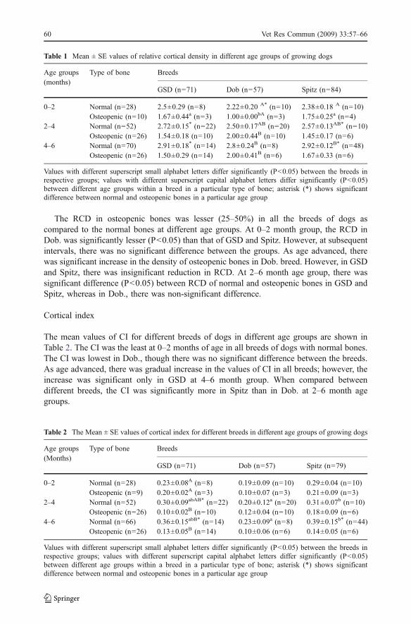
The RCD in osteopenic bones was lesser (25–50%) in all the breeds of dogs ascompared to the normal bones at different age groups. At 0–2 month group, the RCD inDob. was significantly lesser (P<0.05) than that of GSD and Spitz. However, at subsequentintervals, there was no significant difference between the groups. As age advanced, therewas significant increase in the density of osteopenic bones in Dob. breed. However, in GSDand Spitz, there was insignificant reduction in RCD. At 2–6 month age group, there wassignificant difference (P<0.05) between RCD of normal and osteopenic bones in GSD andSpitz, whereas in Dob., there was non-significant difference.
Cortical index
The mean values of CI for different breeds of dogs in different age groups are shown inTable 2. The CI was the least at 0–2 months of age in all breeds of dogs with normal bones.The CI was lowest in Dob., though there was no significant difference between the breeds.As age advanced, there was gradual increase in the values of CI in all breeds; however, theincrease was significant only in GSD at 4–6 month group. When compared betweendifferent breeds, the CI was significantly more in Spitz than in Dob. at 2–6 month agegroups.
Table 1 Mean ± SE values of relative cortical density in different age groups of growing dogs
Age groups(months)
Type of bone Breeds
GSD (n=71) Dob (n=57) Spitz (n=84)
0–2 Normal (n=28) 2.5±0.29 (n=8) 2.22±0.20 A* (n=10) 2.38±0.18 A (n=10)Osteopenic (n=10) 1.67±0.44a (n=3) 1.00±0.00bA (n=3) 1.75±0.25a (n=4)
2–4 Normal (n=52) 2.72±0.15* (n=22) 2.50±0.17AB (n=20) 2.57±0.13AB* (n=10)Osteopenic (n=26) 1.54±0.18 (n=10) 2.00±0.44B (n=10) 1.45±0.17 (n=6)
4–6 Normal (n=70) 2.91±0.18* (n=14) 2.8±0.24B (n=8) 2.92±0.12B* (n=48)Osteopenic (n=26) 1.50±0.29 (n=14) 2.00±0.41B (n=6) 1.67±0.33 (n=6)
Values with different superscript small alphabet letters differ significantly (P<0.05) between the breeds inrespective groups; values with different superscript capital alphabet letters differ significantly (P<0.05)between different age groups within a breed in a particular type of bone; asterisk (*) shows significantdifference between normal and osteopenic bones in a particular age group
Table 2 The Mean ± SE values of cortical index for different breeds in different age groups of growing dogs
Age groups(Months)
Type of bone Breeds
GSD (n=71) Dob (n=57) Spitz (n=79)
0–2 Normal (n=28) 0.23±0.08A (n=8) 0.19±0.09 (n=10) 0.29±0.04 (n=10)Osteopenic (n=9) 0.20±0.02A (n=3) 0.10±0.07 (n=3) 0.21±0.09 (n=3)
2–4 Normal (n=52) 0.30±0.09abAB* (n=22) 0.20±0.12a (n=20) 0.31±0.07b (n=10)Osteopenic (n=26) 0.10±0.02B (n=10) 0.12±0.04 (n=10) 0.18±0.09 (n=6)
4–6 Normal (n=66) 0.36±0.15abB* (n=14) 0.23±0.09a (n=8) 0.39±0.15b* (n=44)Osteopenic (n=26) 0.13±0.05B (n=14) 0.10±0.06 (n=6) 0.14±0.05 (n=6)
Values with different superscript small alphabet letters differ significantly (P<0.05) between the breeds inrespective groups; values with different superscript capital alphabet letters differ significantly (P<0.05)between different age groups within a breed in a particular type of bone; asterisk (*) shows significantdifference between normal and osteopenic bones in a particular age group
60 Vet Res Commun (2009) 33:57–66

Osteopenic bones showed decreased CI (25–75% less) in all the breeds of dogs, thoughto a variable extent, as compared to normal bones. In 0–2 month age group dogs, CI wasrelatively (non-significantly) lesser in Dob. than others. In general, reduced CI wasrecorded in older age group animals of all breeds. However, there was no significant changein the CI in different breeds at different age groups, except in GSD where the CI wassignificantly less in animals aged 4–6 months, than those aged 0–2 months. At 4–6 monthsthere was significant difference (P<0.05) in CI between the normal and osteopenic bones inGSD and Spitz.
Diameter of distal metaphysis
The mean values of diameter of distal metaphysis of femur for different breeds of dogs indifferent age groups are shown in Table 3.
The diameter of distal metaphysis was the least in dogs with normal bones in the agegroup of 0–2 months and increased slightly (10–20%), and non-significantly (P>0.05) upto 6 months. Among the different breeds, GSD and Dob. showed non significantly morewidening at the distal metaphysis than Spitz. Similar trend was noticed in osteopenic bones.However, no significant difference in the DDFM was observed between breeds and alsobetween the normal and osteopenic bones at different age groups.
Width of femoral growth plate (WFGP)
The mean values of the WFGP for different breeds of dogs in different age groups areshown in Table 4. The WFGP was the maximum in the age group of 0–2 months in normalanimals of all the breeds. Among the different breeds, Spitz and Dob. recorded relatively (P>0.05) lesser WFGP than German shepherd. The WFGP reduced gradually and significantly(P<0.05)) in older age group animals of the breeds. The reduction was about 3 to 4 times inmost of the breeds, except in Dob., where the decrease in WFGP was less than 50%. And at4–6 months, there was significant (P<0.05) difference in the WFGP between breeds, it wasthe least in Spitz and maximum in Dob., suggesting that growth plate closure may be faster inSpitz than in GSD and Dob. breeds.
In osteopenic bones, the WFGP varied among different age groups and different breeds(Table 4). In 0–2 month group, the WFGP did not show significant difference between thebreeds. Contrary to the trend of reduction in WFGP seen in normal dogs, in osteopenic
Table 3 The Mean ± SE values of diameter of distal metaphysis (in mm) for different breeds in different agegroups of growing dogs
Age groups(months)
Type of bone Breeds
GSD (n=71) Dob (n=57) Spitz (n=84)
0–2 Normal (n=28) 18.12±2.20 (n=8) 17.00±2.40 (n=10) 15.67±3.33 (n=10)Osteopenic (n=10) 19.20±3.14 (n=3) 20.00±2.61 (n=3) 17.00±1.95 (n=4)
2–4 Normal (n=52) 18.97±2.18 (n=22) 18.00±1.97 (n=20) 16.40±2.22 (n=10)Osteopenic (n=26) 22.00±3.20 (n=10) 21.00±2.73 (n=10) 17.30±2.34 (n=6)
4–6 Normal (n=70) 22.00±2.33 (n=14) 20.02±2.12 (n=8) 16.65±1.72 (n=48)Osteopenic (n=26) 25.50±3.41 (n=14) 23.00±2.71 (n=6) 20.50±3.20 (n=6)
Values between the breeds, between the age groups or between osteopenic and normal bones do not differsignificantly (P>0.05)
Vet Res Commun (2009) 33:57–66 61
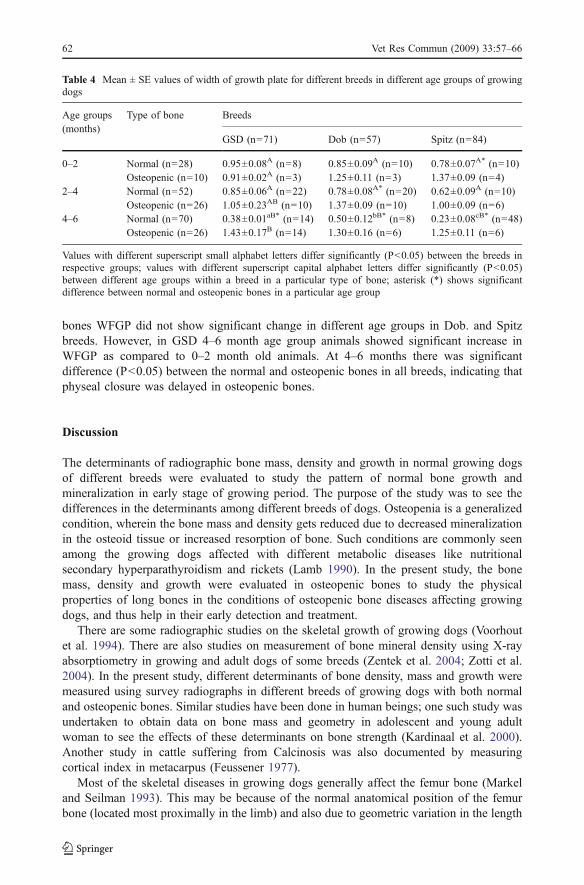
bones WFGP did not show significant change in different age groups in Dob. and Spitzbreeds. However, in GSD 4–6 month age group animals showed significant increase inWFGP as compared to 0–2 month old animals. At 4–6 months there was significantdifference (P<0.05) between the normal and osteopenic bones in all breeds, indicating thatphyseal closure was delayed in osteopenic bones.
Discussion
The determinants of radiographic bone mass, density and growth in normal growing dogsof different breeds were evaluated to study the pattern of normal bone growth andmineralization in early stage of growing period. The purpose of the study was to see thedifferences in the determinants among different breeds of dogs. Osteopenia is a generalizedcondition, wherein the bone mass and density gets reduced due to decreased mineralizationin the osteoid tissue or increased resorption of bone. Such conditions are commonly seenamong the growing dogs affected with different metabolic diseases like nutritionalsecondary hyperparathyroidism and rickets (Lamb 1990). In the present study, the bonemass, density and growth were evaluated in osteopenic bones to study the physicalproperties of long bones in the conditions of osteopenic bone diseases affecting growingdogs, and thus help in their early detection and treatment.
There are some radiographic studies on the skeletal growth of growing dogs (Voorhoutet al. 1994). There are also studies on measurement of bone mineral density using X-rayabsorptiometry in growing and adult dogs of some breeds (Zentek et al. 2004; Zotti et al.2004). In the present study, different determinants of bone density, mass and growth weremeasured using survey radiographs in different breeds of growing dogs with both normaland osteopenic bones. Similar studies have been done in human beings; one such study wasundertaken to obtain data on bone mass and geometry in adolescent and young adultwoman to see the effects of these determinants on bone strength (Kardinaal et al. 2000).Another study in cattle suffering from Calcinosis was also documented by measuringcortical index in metacarpus (Feussener 1977).
Most of the skeletal diseases in growing dogs generally affect the femur bone (Markeland Seilman 1993). This may be because of the normal anatomical position of the femurbone (located most proximally in the limb) and also due to geometric variation in the length
Table 4 Mean ± SE values of width of growth plate for different breeds in different age groups of growingdogs
Age groups(months)
Type of bone Breeds
GSD (n=71) Dob (n=57) Spitz (n=84)
0–2 Normal (n=28) 0.95±0.08A (n=8) 0.85±0.09A (n=10) 0.78±0.07A* (n=10)Osteopenic (n=10) 0.91±0.02A (n=3) 1.25±0.11 (n=3) 1.37±0.09 (n=4)
2–4 Normal (n=52) 0.85±0.06A (n=22) 0.78±0.08A* (n=20) 0.62±0.09A (n=10)Osteopenic (n=26) 1.05±0.23AB (n=10) 1.37±0.09 (n=10) 1.00±0.09 (n=6)
4–6 Normal (n=70) 0.38±0.01aB* (n=14) 0.50±0.12bB* (n=8) 0.23±0.08cB* (n=48)Osteopenic (n=26) 1.43±0.17B (n=14) 1.30±0.16 (n=6) 1.25±0.11 (n=6)
Values with different superscript small alphabet letters differ significantly (P<0.05) between the breeds inrespective groups; values with different superscript capital alphabet letters differ significantly (P<0.05)between different age groups within a breed in a particular type of bone; asterisk (*) shows significantdifference between normal and osteopenic bones in a particular age group
62 Vet Res Commun (2009) 33:57–66

of the bone (Markel and Seilman 1993). Hence in the present study the determinants wereevaluated taking femur as a model bone. Further, the study was focused mostly on GSD,Dob. and Spitz breeds of dogs, as these breeds were the most common descript breedsreared in and around this locality.
Mean scores of RCD was the least in the normal growing pups aged between 0–2 months in all the breeds of dogs, and it increased gradually up to 6 months. This mayindicate that mineralization of bone is less in the very early stage of growth and it graduallyincreases with the age. In a similar study, femur bone showed increased cortical densitywith increased age (Delaquerriere et al. 1982). The RCD values recorded in different breedsaffected with osteopenia was less in comparison to normal bones at different age groups.The reduced RCD clearly indicates decreased mineralization of bone. Several earlier reportsindicate that hypocalcemia caused either due to imbalance in the calcium-phosphorus ratioin the diets of growing dogs or deficiency of vitamin D, is primarily responsible forosteopenia induced by stimulation of parathyroid (Rosol and Capen 1997).
The CI has been measured using thickness of cortex in both sides in bone and bone massby different workers (Feussener 1977; Suittie et al. 1983; Kardinaal et al. 2000). In thepresent study, all the normal dogs showed relatively less CI in younger age group and itincreased gradually up to six months. This indicates that as the age advances, the growthand remodeling of long bones increase. In dogs, the bone growth is reported to be rapidduring the first 6 months of life, mostly by endochondral ossification (Braden 1993;Chambers 1993). Remodelling of bone is also reported to occur during this period at theperiosteal and endosteal sites in response to mechanical stress (Baron 1993; Braden 1993;Canalis 1993). Among the different breeds of dogs, Spitz has shown maximal CIthroughout the growing period, which was significantly more than that of Dob. during 2–6 months of age. This suggests that Spitz has relatively more cortical bone thickness thanother breeds studied. Whereas Dob. has the least cortical bone thickness and hence is moreprone to get osteopenic.
In dogs affected with osteopenia, the CI was lesser than the normal bones, especially in 2–6 month age groups, indicating that the dogs in this age group are more susceptible to developosteopenia. This may be attributed to reduced mineralization of growing skeleton orcontinued and prolonged drainage of minerals from the skeleton. In majority of the animalsthe CI was reduced up to 50–75% of normal bones, and there was significant difference in theCI between the normal and osteopenic bones at 4–6 months of age; this indicates that CI is areliable indicator of osteopenia, and it may also help to assess the severity of the condition.Among the different breeds, Dob. affected with osteopenia showed the least CI than others.This was due to the fact that the CI recorded in normal Dob. was also the least among differentbreeds. This indicates that Dob. pups may be more prone to develop osteopenia duringgrowth. Further, more care has to be taken while judging the bone as osteopenic in Dob. as thenormal bone density is also relatively less. As there is very little difference in the CI of normaland osteopenic bones (no significant difference at 4–6 month), this breed of dogs might showpathological fractures even without showing apparent radiographic signs of osteopenia.
The DDFM did not show any significant difference (P>0.05) between the dogs aged 0–2 months and those aged 4–6 months, indicating that most of the growth (metaphysealwidth) occurs very early in the growing period (within 2 months). Among the differentbreeds, GSD and Dob. showed slightly (non significantly) larger DDFM than Spitz. Thiswas possibly due to the fact that GSD and Dob. are relatively large breed dogs with largerlong bones than Spitz.
The variation in the diameter of distal metaphysis is one of the most important factors indetermining the change in bone geometry in osteopenic bones. In weak bones, the
Vet Res Commun (2009) 33:57–66 63

mechanical stress is concentrated at the distal metaphysis (Canalis 1993; Baron 1993;Braden 1993). Under such conditions in hind limbs the diameter of distal metaphysis offemur is more affected than many other bones (Markel and Seilman 1993). In the presentstudy also increased DDFM was observed in osteopenic bones in different breeds of dogs atdifferent age groups. Among the different breeds, GSD showed slightly (but non-significantly) more increase in the DDFM, probably because of relatively heavy weightof animals leading to more stress concentration. This broadening of metaphysis generallygives either “saucer” or “cupping” effect (Krook et al. 1971; Olsson 1972).
Growth plate is responsible for longitudinal growth of a long bone by a process calledendochondral ossification, where the cartilage tissue is slowly replaced by osseous tissue(Braden 1993; Chambers 1993; Olsson 1993). In young and growing animals, the growthplate remains open, as the age increases the width of the growth plate slowly decreases andat the stage of maturity it ceases to exist (Brighton 1985). Similar observations were madein the present study. The WFGP was greatest in the normal animals at the age of 0–2 months. At 4–6 months, WFGP reduced substantially in most of the breeds, indicatingthat maximum growth of long bone occurs before six months. Among the different breeds,the WFGP was slightly (but non-significantly) more in GSD than in Dob. and Spitz in theearly period of growth. However, the physeal width in GSD and Dob. breeds remainedrelatively more open than in Spitz at 4–6 months of age. This difference in the WFGP maybe due to variations in the bone length and growth in different breeds. Open, wide physismay indicate the ability of the bone to grow in length, and it is possible that in large breedsof dogs (GSD and Dob.) the longitudinal growth of bones may continue for relativelylonger period than small breeds (Spitz) of dogs.
The WFGP in osteopenic bones remained greater than that of normal bones at differentintervals. This suggests that in osteopenic bones in general there is a delay in calcification/ossification of physeal cartilage, which may affect the longitudinal growth of long bones.Delay in calcification/ossification may be attributed to mineral deficiency, especially ofcalcium, and/or vitamin D3. Increase in the physeal width is also a characteristic sign ofrickets (Kealy 1979). In such cases, failure of mineralization of cartilage leads touninterrupted growth of chondrocytes resulting in increased physeal width. The results ofthe present study also suggest that osteopenic bone condition in growing dogs may also beattributed to rachitic condition. Unlike in normal bones, osteopenic bones did not show thetrend of decreasing WFGP with advancing age. Instead, a large physeal gap was seen atdifferent levels; and at the age of 4–6 months there was a significant difference between theWFGP in normal and osteopenic bones in different breeds of dogs. This indicated that theclosure of physis is delayed in osteopenic bones.
Conclusions
The results of this study showed that the relative cortical density, cortical index and thediameter of long bones of normal growing dogs increases gradually, and physeal widthreduces as age advances (up to 6 months). Doberman has the least cortical index among thedifferent breeds studied; hence a normal bone is more likely to be diagnosed as osteopenicand thus special care has to be taken while diagnosing osteopenia in this breed. Whereas inSpitz, the CI is very good, thus may be less prone to develop osteopenia; and the growthplate closure occurs relatively faster. In dogs with osteopenic bones, cortical density andcortical index decreases markedly and widening of distal metaphysis occurs during thegrowth period. The WFGP either increases or remains the same in the growth period in
64 Vet Res Commun (2009) 33:57–66

different breeds with osteopenia, and in such cases the closure of physis occurs relativelylater than in normal bones.
References
Aithal, H. P., Singh, G. R. and Bisht, G. S., 1999a. Fractures in dogs: a survey analysis of 402 cases. IndianJournal of Veterinary Surgery, 20, 15–21
Aithal, H. P., Singh, G. R., Amarpal, Kinjavdekar, P. and Setia, H. C., 1999b. Fractures secondary tonutritional bone disease in dogs: A review of 38 cases. Journal of Veterinary Medicine, 46-A, 483–487
Armburst, L. J., 2007. Digital images and digital radiographic image capture. In: D. E. Thrall (ed.), Textbookof Veterinary Diagnostic Radiology, 5th edn, (Saunders Elsevier, Philadelphia), 22–37
Balagopalan, T. P., Devanand, C. B., Rajankutty, K., Sarada Amma, T., Nayar, S. R., Varkey, C. A.,Jalaluddin, A. M., Nayar, K. N. M. and George, P. O., 1995. Fractures in dogs: A review of 208 cases.Indian Journal of Veterinary Surgery, 16, 41–43
Baron, R., 1993. Anatomy and ultra structure of bone. In: M. J. Favus (ed.), Primer on Metabolic BoneDiseases and the Disorders of Mineral Metabolism, (Raven Press, New York), 3–9
Braden, T. D., 1993. Histophysiology of the growth plate and growth plate injuries. In: M. J. Bojrab (ed.),Disease Mechanisms in Small Animal Surgery, 2nd edn, (Lea and Febiger, Philadelphia), 1027–1041
Brighton, T. C., 1985. Normal bone formation. In: C. D. Newton and D. M. Nunamaker (eds.), Text Book ofSmall Animal Orthopaedics, (J. B. Lippincott, Philadelphia, London), 21
Canalis, E., 1993. Regulation of bone remodeling. In: M. J. Favus (ed.), Primer on Metabolic Bone Diseasesand the Disorders of Mineral Metabolism, (Raven Press, New York), 33–40
Chambers, J. N., 1993. Developmental and congenital problems of antebrachium and adjacent joints. In: M.J. Bojrab (ed.), Disease Mechanisms in Small Animal Surgery, 2nd edn, (Lea and Febiger, Philadelphia),834–840
Delaquerriere, R. L., Anderson, C., Jorch, U. M. and Cook, M., 1982. Radiographic morphometry andradiographis photodensitometry of the femur in the Beagle at 13 and 21 months. American Journal ofVeterinary Research, 43, 2255–2258
Feussener, H., 1977. Usefulness of Radiographic Diagnosis in the Recognition of Enzootic Calcinosis inCattle, 85
Grubb, S. A. and Talmage, R. V., 1983. Metabolic bone diseases. In: F.C. Wilson (ed.), The MusculoskeletalSystem, Basic Processes and Disorders, (J.B. Lippincott, Philadelphia).
Kardinaal, A. F., Hoorneman, G., Vaananen, K., Chartes, P., Ando, S., Maggiolini, M., Charzewska, J.,Rotily, M., Deloraine, A., Heikkinen, J., Juvin, R. and Schaafsma, G., 2000. Determinants of bone massand bone geometry in adolescent and young adult women. Calcified Tissue International, 66, 81–89doi:10.1007/PL00005834
Kealy, J. K., 1979. Bones and joints. In: J. K. Kealy (ed.), Diagnostic Radiology of the Dog and Cat, (W.B.Saunders, Philadelphia), 325
Krook, L., Lutwalk, L., Henrickson, P. A., Kallfelz, F., Hirsch, C., Romanas, B., Belanger, L. F., Marier, J. R.and Sheffy, B. E., 1971. Reversibility of nutritional osteoporosis: physicochemical data on bones froman experimental study in dogs. Journal of Nutrition, 101, 233–246
Kumar, K., Mogha, I. V., Aithal, H. P., Kinjavdekar, P., Amarpal, Singh, G. R., Pawde, A. M. and Kushwaha,R. B., 2007. Occurrence and pattern of long bone fractures in growing dogs with normal and osteopenicbones. Journal of Veterinary Medicine, 54A, 484–490
Lamb, C. R., 1990. The double cortical line: a sign of osteopenia. Journal of Small Animal Practice, 31,189–192 doi:10.1111/j.1748–5827.1990.tb00768.x
Maala, C. P. and Celo, E. M., 1975. A study on the anatomical locations, incidence and causes of fractures indogs. Phillipine Journal of Veterinary Medicine, 14, 137–143
Markel, M. D. and Seilman, E., 1993. Radiographic study of homotypic variation of long bones in dogs.American Journal of Veterinary Research, 54, 2000–2003
Olsson, S. E., 1972. Radiology in veterinary pathology. A review with special reference to hypertrophicosteodystrophy and secondary hyperparathyroidism in the dog. Acta Radiol. Suppl. 319, 255–270
Olsson, S. E., 1993. Pathophysiology, morphology and clinical signs of osteochondrosis in dogs. In: M. J.Bojrab (ed.), Disease Mechanism in Small Animal Surgery, 2nd edn, (Lea and Febiger, Philadelphia),776–796
Phillips, I. R., 1979. A survey of bone fractures in the dog and cat. Journal of Small Animal Practice, 20,661–674
Vet Res Commun (2009) 33:57–66 65

Rosol, T. J. and Capen, C. C., 1997. Calcium–regulating hormones and diseases of abnormal mineral(calcium, phosphorus, magnesium) metabolism. In: J. J. Kaneko, J. W. Harvey and M. L. Bruss (eds.),Clinical Biochemistry of Domestic Animals, 5th edn, (Academic Press, San Diego), 619–687
Singh, A. P., Mirakhur, K. K. and Nigam, J. M., 1983. A study on the incidence and anatomical locations offractures in canine, caprine, bovine, equine and camel. Indian Journal of Veterinary Surgery, 4, 61–66
Snedecor, G. W. and Cochran, W. G., 1967. Statistical Methods, 6th edn, (Oxford and IBH PublishingCompany, New Delhi)
Suittie, J. M., Wenham, G. and Kay, N. B., 1983. Simple in-vitro method of determining calcium andphosphorus content of the metacarpus of red deer using radiography. Veterinary Record, 113, 393–394
Voorhout, G., Nap, R. C. and Hazewinkel, H. A. W., 1994. A radiographic study on the development of theantebrachium in Great Dane pups, raised under standard conditions. Veterinary Radiology andUltrasound, 35, 271–276 doi:10.1111/j.1740–8261.1994.tb02040.x
Zentek, J., Liesegang, A., Mayrhofer, E., Schneider, S., Breit, S. M., Grampp, S. and Künzel, W. W. F., 2004.Comparative assessment of bone mineral measutements obtained by use of dual-energy x-rayabsorptiometry, peripheral quantitative computed tomography, and chemical-physical analysis in femursof juvenile and adult dogs. American Journal of Veterinary Research, 65, 891–900 doi:10.2460/ajvr.2004.65.891
Zotti, A., Isola, M., Sturaro, E., Menegazzo, L., Piccinini, P. and Bernardini, D., 2004. Vertebral mineraldensity measured by dual-energy X-ray absortiometry (DEXA) in a group of healthy Italian boxer dogs.Journal of Veterinary Medicine, 51A, 254–258
66 Vet Res Commun (2009) 33:57–66