Embed Size (px)
Citation preview
Loyola University ChicagoLoyola eCommons
Master's Theses Theses and Dissertations
1980
Determination of the Gingival Crevicular FluidVolumes Associated with Restored (Fixed andRemovable Prosthodontics) and Non-RestoredTeethPamela Denise GolaszLoyola University Chicago
This Thesis is brought to you for free and open access by the Theses and Dissertations at Loyola eCommons. It has been accepted for inclusion inMaster's Theses by an authorized administrator of Loyola eCommons. For more information, please contact [email protected].
This work is licensed under a Creative Commons Attribution-Noncommercial-No Derivative Works 3.0 License.Copyright © 1980 Pamela Denise Golasz
Recommended CitationGolasz, Pamela Denise, "Determination of the Gingival Crevicular Fluid Volumes Associated with Restored (Fixed and RemovableProsthodontics) and Non-Restored Teeth" (1980). Master's Theses. Paper 3088.http://ecommons.luc.edu/luc_theses/3088
DETERMINATION OF THE GINGIVAL CREVICULAR FLUID VOLUMES
ASSOCIATED WITH RESTORED (FIXED AND REMOVABLE PROSTHODONTICS)
AND NON-RESTORED TEETH
By
Pamela Denise Golasz, D.D.S.
A Thesis Submitted to the Faculty of the Graduate School
of Loyola University of Chicago in Partial Fulfillment
of the Requirements for the Degree of
Master of Science
June 1980
L!?R~.r::-;:
LOYOU~~ UNIVERSIT'( MEDiCAL CEJ~IW.-<.
DEDICATION
To Virginia, my Mother, to thank her for the years of unselfish
love, patient guidance and gentle encouragement, I dedicate this thesis.
In loving memory of Victoria Bugaj, my Grandmother, for sharing
her wisdom and love; and to thank her for the lilacs and other miracles
of Nature.
To my dear sister, Roxane, not only for her love and understanding,
but most importantly, for her friendship.
it
ACKNOWLEDGEMENTS
r would like to express my sincerest gratitude to Dr. William
F.P. Malone, Thesis Director, for his intuition and guidance during
the preparation of this thesis. His own professional competence and
superior command of Prosthodontics will continue to motivate and in~
spire.
Grateful acknowledgement is given to Dr. Patrick Toto and Dr.
Boleslaw Mazur for serving as members of the Advisory Committee.
iii
VITA
The author, Pamela Denise Golasz, is the daughter of Daniel
and Virginia (Bugaj) Golasz. She was born June 12, 1953 in Chicago,
Illinois.
Her elementary education was obtained from the Franz Peter
Schubert School i.n Chicago, Illinois; and her secondary education
from the Maine Township High School South of Park Ridge, Illinois
where she graduated in 1971,
In May of 1975, she graduated from DePaul University, Chicago,
Illinois where she received the degree of Bachelor of Science, major
ing in Biology.
In September of 1975, she entered Loyola University School of
Dentistry, Maywood, Illinois and 1 n May of 1979, received the degree
of Doctor of Dental Surgery.
Following graduation, she entered the post graduate clinical
specialty training program in Fixed Prosthodontics and the graduate
program in Oral Biology at Loyola University School of Dentistry,
Maywood, Illinois.
iv
TABLE.OF CONTENTS
DEDICATION ••••• . . . . . . . . . . . . ACKNOWLEDGEMENTS •• . . . . . . . . . . . .. . . . . . . . . VITA. • • • • • • • . . . . . . . . . . . . . LIST OF ILLUSTRATIONS
LIST OF TABLES •••••
. . . . . . . . . . . . . . . .
. . . . . . . . . . . . . . . . . CONTENTS OF APPENDIX. . . . . . . . . . . . . . . Chapter
I. INTRODUCTION AND STATEMENT OF PURPOSE ••• . . . . II. REVIEW OF THE LITERATURE ••••• . . . . . . . . . . .
Historical Perspective ••••••••••
Classical Literature •••••••••••.
Recent Literature •• . . . . . . . . . . . .
. . . . . . . . . . . . . .
Clinical Literature ••••••• . . . . . . . . . . . . III. MATERIALS AND METHOD •• . . . . . • • • . . . . . . IV. EXPERIMENTAL RESULTS ••••••••• • • •
V. DISCUSSION •••.• . . . . . . . . . . VI. SUMMARY AND CONCLUSIONS •••.• . . . . . . . . . . . .
BIBLIOGRAPHY ••••• . . . . . . . . . . . . . . APPENDIX •••• . . . . . . . . . . . . . .
v
PAGE
ii
iii
iv
vi
vii
viii
1
5
5
6
14
18
24
36
54
59
61
67
· LIST OF ILLUSTRATIONS
Fisure
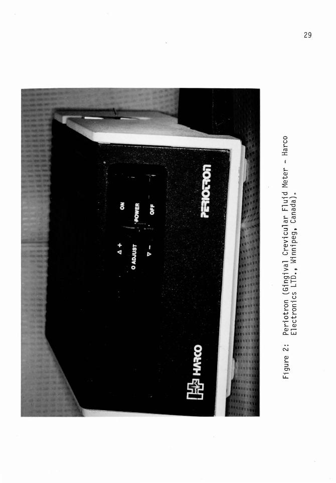
1. Diagram of the dentinoepithelial junction •••• . . . •. 2. Periotron (Gingival Crevicular Fluid Meter-Harco
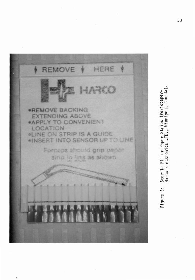
Electronics LTD., Winnipeg, Canada) ••••••• . . . . 3. Sterile Filter Paper Strips (Periopaper-Harco
Electronics LTD., Winnipeg, Canada} ••••••
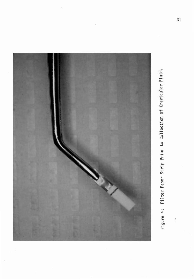
4. Filter Paper Strip Prior to Collection of Crevicular Fluid •••••••••••••••
. . . . .
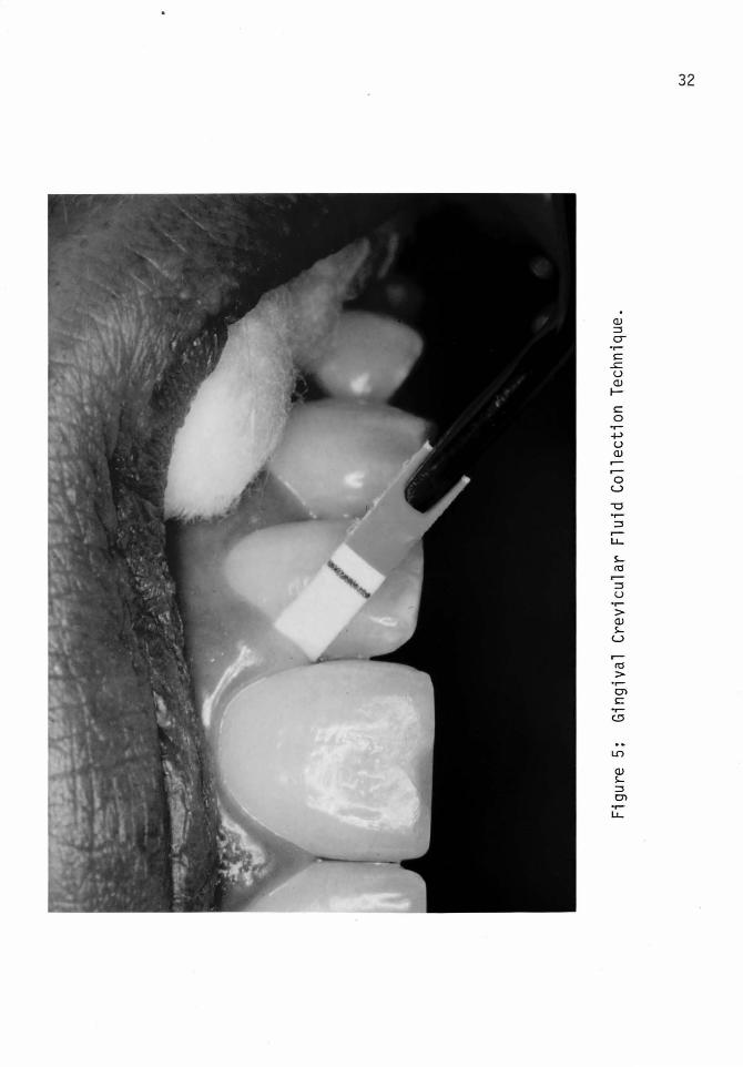
. . . . . 5. Gingival Crevicular Fluid Collection Technique •• . . . 6. Placing Filter Paper Strip Between Recording
Sensors of the Gingival Crevicular Fluid Meter (Periotron) ••••••••••••••• . . . . .
7. Measuring the Depth of the Gingival Sulcus • • • • • • •
8. Pathway of Complement Activation in Gingival Tissue. • •
vt
Page
28
29
30
31
32
33
34
35
Table
1.
2.
3.
LIST OF TABLES
Compiled Data from Individual Data Sheets
Fixed Prosthodontic Categories, frequency distribution and mean values •••••••
• • • • • • • •
• •
Removable Prosthodontic Categories, frequency distribution and mean values •••••••••
. . . . . .
. . . . .. . 4. Fixed Prosthodontic Categories, frequency
distribution and mean values for individual categories ••••• ~ •••••••••••• . . . . . .
5.
6.
7.
Removable Prosthodontic Categories, frequency distribution and mean values for individual categ_ori es. • • • • • • • • • • • • • • • • • • • •
Summary of mean values for all comparison groups ••
T-Test analysis: Single restored unit compared with restored abutment under existing partial denture ••
• • •
. . . • • •
8. T-Test analysis: Restored bridge abutment (Fixed Prosthodontics) compared with restored abutment under existing partial denture •••••••••• . . . .
9. Regression Analysis •••••••••••••• . . . . .
vii
Page
40
46
47
48
49
50
51
52
53
CONTENTS OF APPENDIX
Patient Data Sheet (Fixed Prosthodontics) , •••• . . . . . . Patient Data Sheet (Removable Prosthodontics) •••• • • • • •
viii
Page
68
69
CHAPTER I
INTRODUCTION AND STATEMENT OF PURPOSE
The primary objective of restorative dentistry is the replacement
of lost teeth or portions of carious and/or fractured teeth. Tissue
trauma is an inherent problem (Jameson, 1979) even for the most accom
plished and exacting operator. Utilization of the most delicate and in
nocuous mani pul ati on of the supporting structures can sti 11 result in an
inflammatory response of soft tissues. One controversial aspect within
restorative dentistry is the deleterious effect of applied stress on
abutment teeth for both fixed and removable prosthesis. The length of
the span of the prosthesis has never been objectively assessed.
Studies (Waerhaug, 1960) have indicated subgingival margin termin
ation and subsequent plaque accumulation were critical considerations in
tissue response. The actual contour of the crown proved to be a major
determinant in the type of tissue response noted. Youdelis (1973) stated
"overcontourtng encourages the accumulation of particulate and microbial
matter in an area inaccessible for cleaning by the patient". Caruso
(1979) determined the morphologic contour of an artificial tooth and/or
of a fixed prosthesis should be undercontoured rather than overcontoured.
The type of dental materials employed and their relative degree of sur
face roughness (Volchansky, 1974) were critical to inflammatory response
observed in the soft tissue. Jameson (1979) stated margin termination, .
independent of the dental material employed to construct the crown was
1
crucial, and felt chronic inflammation was indigenous to subgingival
placement of full coverage margins.
Investigators (Tylman, 1978) have eluded to the fact that stresses
promoted by 11 free ended 11 removable partial dentures are ultimately the
cause of loss of abutment, Schuyler, 1941, stated 11 the semi edentulous
mouth presents to the dentist one of his most complex problems. Fixed
restorations supplying one, two or a greater number of teeth have served
more adequately in maintaining oral function and health than the remova
ble partial appliance as commonly planned and constructed... Cohn (1956)
sited 11 a common existing cause of periodontal disease is the faulty re
movable partial denture, or fixed partial denture, in which anchorages
are improperly selected or incorrectly fabricated. · A physiologic result
can be obtained from a dental prosthesis only when the strain exerted on
abutment teeth is considered in relation to the biologic union of the
2
tooth to the supporting structures, and the mechanical principles involved ...
These implications as to the detrimental nature of partial denture
prosthesis command additional credence when partial dentures are utilized
in patients whose dentitions are handicapped by periodontal disease. Com
parisons by restorative dentists have been difficult because removable
partial dentures are usually placed in mouths in which restoration via
fixed prosthodontics is contraindicated. Despite this fact, some evidence
should be demonstrated to validate the concept that removable partial den
ture abutments respond poorly to the increased occlusal load and torque.
Until recently, no quantitative measurement could be obtained to
assess the degree of response (inflammation) of the soft tissue adjacent
to the abutments. Traditionally, the measurement of gingival response
to restorative procedures has been evaluated by subjective clinical in~
dices. Loe and Si1ness {1967} correlated various degrees of observable
inflammation by numerical assessment of gingival inflammation on a scale
ranging from zero (Q .. - no observable inflammation} to three (3-- evidence
of severe gingival inflammation). Consistency from one researchers ob
servations and recordings to anothers has proven to be a deficient aspect
of this clinical procedure. Recently, the introduction of an instrument*
that utilizes vo1umetrtc quantitation of crevicular fluid as a diagnostic
measuring tool of gingival inflammation has lead to a more objective and
consistent assessment of soft tissue response to chronic irritation.
Crevicular fluid flow i·ncreases with an increase in inflammation, and in
creased crevicular fluid flow occurs prior to clinical manifestations.
Brill (1959) first suggested using crevicular fluid as a measure
of gingival inflammation to provide a quantitative means to monitor the
physiology of the sulcular region. He noted a greater fluid flow from
patients with extensive restorative work compared to clinically healthy,
non-restored marginal gingiva. Egelberg (1964} found a correlation be
tween the amount of gingival exudate collected on filter paper strips
and the area of inflammatory cell infiltration noted in histological ex
amination. There remains some dispute as to the nature of crevicular
fluid, be it exudative or transudative. The conclusion of Bang and Ci
masoni (1971} stated "because the gingival fluids have a capacity to
* Periotron, Harco Electronics Limited, Winnipeg, Canada.
3
carry high molecular weight compounds such as proteins, this is confirm
ation that gingival fluid is an inflammatory exudate that passes the
crevicular barrier as a result of increased capillary permeability."
Cimasoni (1974) qualitatively determined the following components were
present in crevicular fluid: Immunoglobulins G, A, and M, Globulins alpha
1 and 2 and beta 1, albumin, fibrinolytic factors, lactic acid, urea, hy
droxyproline, endotoxins, bradykinins, and lysosomal enzymes.
The purpose of this investigation is to determine and compare the
inflammatory changes in the marginal gingiva of abutment teeth for fixed
and removable prostheses.
4
CHAPTER II
REVIEW OF THE LITERATURE
A. Historical Perspective
In G.V. Black's (1887) histological assessment of the periodontal
membrane, he noted the existence of a gingival fluid. "Close clinical
examination makes it apparent that there is a slight secretion at this
point that is not quite satisfactorily explained even yet by microscopic
study of the part." Black noted the existence of a system of cells lo
cated about the principle fibers of the periodontal membrane. Referring
to these cells as salivary corpuscles, he noted that this tissue was
affected first tn salivation with certain drugs such as mercury and
iodine. Black (1899} described a plexus of loops of glandular tissue
associated tn a parallel fashion to the root and anastomosing freely.
Black (1922} postulated that subgingival calculus formation was augumen
ted by the constant fluid present in the subgingival space.
Stillman and McCall (1927) sited 11 the cleansing of the enamel sur
face over which it lies by the serous secretion from its crevicular sur
face" as a function of the marginal gingiva.
McCall (1924} proposed that teeth in traumatic occlusion resulted
in an acidic exudate of the gingival crevice by alteration of the perio
dontal tissue.
Boedecker (1931) disagreed with Black's (1899) description of a
plexus of gingival glands based upon histological findings. Boedecker
·s

did concur with McCall's theory and utilized litmus paper to examine
human gingival fluid and to determine "the relation of an acid exudate
of the gingival crevice to erosion."
Miller (1938) spoke of "crevicular exudate" as a "discharge from
the gingival crevice". He postulated alterations in the crevicular
exudate as evidence of subclinical signs of periodontal disease 11 Since
microscopic examination of the crevicular fluid reveals the presence of
an unusual number of pus cells. 11 Miller noted increased amounts of
gingival fluid occurred in crevices adjacent to teeth with erosion.
B. Classical Literature
Waerhaug's (1952) investigations indicated after injection of
India ink into the gingival sulcus of young dogs, an increased trans
udation of fluids and emigration of leukocytes through the sulcular
epithlium could be observed within one hour; and the majority of the
India ink particles had been removed by this fluid within two hours.
Waerhaug postulated saliva could not penetrate below the gingival mar
gin and felt the gtngi'va could defend itself against injury due to the
close approximation of the "epithelial cuff" around the tooth.
Waerhaug and Steen (1952) observed the histologic tissue reaction
over a forty-eight hour time period after the deposition of pure cul
tures of pathogenic bacteria into bacteria free gingival pockets of dogs.
Conclusions drawn included: 1. Bacteria within the gingival sulcus re
sult in epithelial necrosis and inflammation of the connective tissue
with a subsequent exudate formation. 2. "From all pockets, there is a
6
7
constant flow of cellular elements and tissue fluid. 11
Brill and Bjorn (1959) in attempting to differentiate crevicular
epithelium from other oral and nasal epithelium; studied the permeability
of human crevicular epithelium to orally administered fluorescein molecules.
Both the epithelial lining of the gingival crevice and the nasal mucosa
proved to be permeable to fluorescein molecules. A correlation between the
amount of inflammation present and the quantity of fluorescein recovered on
filter strips placed in the gingival sulcus was observed in that patients
with extensive restorations as opposed to non-restored teeth lead to the
recovery of a greater amount of fluorescein.
Brill and Krasse (1959) demonstrated an increased flow of tissue
fluid from the gingival sulcus that followed short term mechanical stim
ulation (externally and within the sulcus) of clinically healthy marginal
gingiva that returned to the original flow rate within ten minutes in their
studies with dogs. Two explanations for the increased fluid flow were
postulated: 1. Mechanical stimulation of a vascular bed results in arter
iole dilation with subsequent increased pressure and increased permeability
allowing the egress of plasma, and thereby increasing the concentration of
interstitial fluid. 2. A sol < ) gel mechanism exists for the ground
substance of the connective tissue matrix that remains a gel unless mechan
ical stimulation is applied and the sol state results.
Brill (1959) injected dogs with Evan's Blue (vital dye T-1824 that
binds to plasma albumin in low concentration and a globulin in higher
concentrations) to evaluate the modification of gingival fluid flow under
the influence of histamine, mechanical stimulation and the inflammatory
state. Brill postulated the dye bound to plasma proteins could be em
ployed to evaluate changes in the gingival vessel permeability, and
this change could be measured by the quantity of dye that was recovered
vta filter paper strips placed into the sulcus. Conclusions included
protein bound Evan's Blue from plasma can pass through capillary walls
in minute concentrations and is observed in the gingival sulcus follow
ing mechanical stimulation, IV injection of histamine, and inflammation.
Brill (1959} demonstrated chewing paraffin in human subjects lead
to an increase in the gingival tissue fluid flow and would affect the
state of the gingival health. "When gingival structures are stimulated
by chewing, the antimicrobial effect may be increased, because mechanical
stimulation of the gingival vascular bed stimulates escape of fluid from
the vessels and plasma contains several antimicrobial substances."
Brill (1959) observed the flushing action of gingival fluid on
non vital particles and living bacteria. "The flow of tissue fluid from
sub epithelial structures of marginal gingiva is able to remove particu
late matter, including bacteria, from gingival pockets." Brill coined
the term'pocket•to be that space located between the gingival epithelium
and the tooth substance.
Brill and Bronnestan (1960) utilized immunoelectrophoretic analysis
of human sulcular fluid to determine the presence of seven different
serum protein components including alpha 2 macroglobulin, beta and gamma
globulin. They postulated the source of the gingival fluid is tissue
8
9
and is formed extracellularly via capillary filtration.
Gavin and Collins, (1961} disputed Waerhaug and Steen's 1952 con
clusion that the healthy gingiva is sterile through their investigation
into the bacteriologic status of the clinically healthy human gingiva.
"The clinically healthy gtngiva1 crevice appears to contain microorgan
isms in the majority of cases." A subsequent study (l96lb} lead to the
conclusion human gingival fluid from clinically healthy gingival crevices
exhibits neither bacteriostatic nor bacteriocidal effects on oral bacteria
including Neisseria, Diptheroides, Streptococci, and Staphylococci.
Loe (1961) in his studies of the epithelial cell turn over rate
made the following conclusions upon histologic examination: l."Mitotic
figures along the entire length of the epithelial lining of the pocket
and the desquamation of the surface cells support the view that the ep
ithelial cuff is constantly renewed". 2, The aggregation of neutrophilic
leukocytes withtn the gt-ngival sulcus indicated "they migrate through the
epithelial lining under physiological conditions,•• and 3. "There is a
continuous transudation of tissue fluid into the clinically normal gin
gival pockets."
Gustafsson and Nilssen (1961) noted in their studies of gingival
fluid from clinically healthy human gingiva since fibrinolysis could be
observed, plasmin and activator must be present in the crevicular fluid.
"The fibrinolytic factors in the crevicular fluid might be of signifi
cance in counteracting the deposition of fibrin and other proteins at
the junction between the gingival epithelium and the tooth."
Harvey {1962} upon observation of the physiologic flow of tissue
fluid in flushing extraneous matter from the sulcus of dogs postulated
11 the normal gingival crevice maintains its hygienic state by constant
flushing with tissue fluid which is increased as a result of an acute
inflammatory response to trritation."
Krasse and Egelberg (1962) utilized flame photometry to analyze
the Na/K ratio of human crevicular fluid and concluded "gingival pocket
fluid cannot be regarded as a simple filtration product but rather as
an inflammatory exudate."
10
Egelberg (1963a} in his comparison of cellular content of gingival
sulcus fluid from clinically healthy gingiva to fluid from chronically
inflamed gingi'va observed that although no difference in quality of cells
could be detected, an increase in inflammatory cells compared to epithe
lial cells from the inflamed gingival samples could be observed. Again,
his conclusion was 11 fluid in healthy pockets may be regarded as an in
flammatory exudate."
Egelberg (1963b) demonstrated that topical application of histamine
solution to marginal and attached gingiva of both human subjects and dogs
was able to penetrate the gingival crevicular epithelium but not the at
tached gingival epithelium. This contradicted Waerhaug (1955) and Brill
(1959c) who concluded the gingival fluid was capable of preventing pene
tration of substances into the crevicular area.
Mann (1963) collected gingival fluid from human subjects on filter
paper strips after oral administration of fluorescein sodium (325 mg.}.
Results indicated as the severity of the gingival inflammation increased,
the crevi'cular fluid flow increased, and 11 inf1arm~ation, (not pocket
depth}, was the main factor contributing to the rate of fluid flow. 11
Egelberg (1964) studied crevicular fluid (human and canine) to
determine if a relationship existed between clinical and histologic cri
teria for gingival inflammati-on and the quantity of gingival exudate re
covered by filter paper strips. Conclusions included: 1. A higher de
gree of inflammation was observed in the gingival papilla area. 2. A
correlation existed between the quantity of gingival exudate recovered
on filter paper strips and the area of inflammatory cell infiltrate.
11
Brandtzaeg and Mann (1964) in their study of the lysozymal activity
of human gingival fluid, serum and saliva (from patients with either gin
givitis or periodontitis) concluded crevicular lysozymal activity increased
wtth the severity of inflammation; whereas serum and saliva did not. 11 The
enzyme in gingival fluid is assumed to be primarily of local origin, pos
sibly derived from leukocytes ...
. Weinstein and Mandel (1964) attempted to classify crevicular fluid
as specifically altered transudate from serum sine~: 1. Sulcular cells
are constantly sloughed off and the intracellular contents become part
of the transudate. 2. An active ionic transport mechanism may be ex
hibited by the cells. 3. A cytocellular fluid may be contributed by
crevicular cells through micropores in the cell walls.
Loe and Holm-Pedersen (1965) employed both an extracrevicular tech
nique and an intracrevicular technique to measure crevicular fluid.
Biomicroscopic techniques lead to the revelation that deep insertion of
filter paper strips into the sulcus resulted in capillary compression.
Conclusions included the healthy human gingival crevices do not exhibit
fluid flow (disputing the conclusion of Brill, 1962) and "mechanical
stimulation of the periodontium does not produce fluid from such crevi
ces (disputing the conclusions of Brill, l959b and Brill and Krasse,
1959). 11 In addition, crevicular fluid flow commences pri'or to clinical
manifestations and persists for some time after clinical inflammation
indices have disappeared. "Gingival fluid ts an inflammatory exudate
and that the absence or presence of f1utd may represent the definite
clintcal criterion tn the reftned distinction between normal and inflamed
gtngi'va. 11
12
Sueda's, et al~, (1966} histochemical study of human gingival fluid,
blood serum and saliva lead to confirmation of gingival fluid components;
proteins, lipids, and polysaccharides bound to proteins (muco-, lipo-, or
glyco-proteins). The results of their study seemed to confirm Cimasoni's
(1966} hypothesis that the mucopoly-saccharide substance identified by
Toto and Sicher (1964} located between enamel and crevicular epithelium
fn humans could represent a condensation of gingival fluid.
Stallard (1967} utilized the tnjecti'on of plastic microspheres
i'nto the external carotid artery of squirrel monkeys to study periodon
tal microcirculation. Microspheres became trapped tn areas of chronic
inflammation "possibly associated with abnormal coiling of microvessels
observed beneath the 11 col 11 and sulcular epithelium ... Stallard noted the
existence of a correlation between the integrity of microvasculature
within the sulcular epithelium and the presence of gingival crevicular
fluid. 11 1nitially, the inflanunatory reaction, with its characteristic
vascular alterations, is a physiologic defense mechanism; however, it
appears that in the cases of periodontal disease, the inflammatory re
sponse eventually becomes pathologic."
Weinstein, et al., (1967} demonstrated and characterized the
presence of gingival creviculat fluid in human gingival sulcus labelled,
"clinically normal 11• The authors cited deviation in criteria for 11 clin
i ca lly norma 111 and uncontro 11 ed differences in techntque for their con
tradiction of the findings of Loe, et al,, (1965). The technique util
ized by Weinstein (fluorescei-n label1tngl was one hundred times more
sensitive in detecting proteins than the tedmique employed by Loe
(ninhydrin staining}.
Nagao (1967) studied crev~.cular fluid by weighing filter paper
strips before and after fluid collection and by ni'nhydrin staining. He
concluded that crevicular fluid flow increases following the insertion
of crowns and higher crevicular fluid levels were associated with crowns
that did not fit well as ppposed to a well fitting prosthesis.
Sandalli and Wade {1969} concluded the following from their exam
ination of crevicular fluid volume following periodontal surgery: Gin.
gival fluid flow was reduced following scaling, polishing, and supervis.ed
oral hygiene. (Gingival fluid flow decreased four weeks post operatively
in concurrence with L()e and Ho lm-Pedersen' s fi'ndi ngs, 1965). 11 There is
13
a relation between the depth of pocket and the amount of gingival
fluid: measuring this amount may be of value in scoring the perio
dontal status ...
Orban and Stallard (1969} from their studies to determine if a
correlation between fluid volume and clinical scoring techniques and
biopsy specimens existed; postulated alteration of the intercellular
cementing substance occurs through numerous factors (hyaluronidase,
chewing, brushing, gingival massage, circadian rhythm and hormones)
with the overall result being an increase in the permeability of the
sulcular epithelium. In addition, crevicular fluid scores did not
correlate with biopsy scores based upon inflammatory infiltrate pre
sent. 11 A better indication of inflammatory status of the gingival
tissues, as revealed by biopsies, is the evaluation or measurement
of dental plaque ...
Oliver, et al., (1969) in human studies to determine if are
lation between gingival index (Loe and Silness, 1967), gingival fluid
volumes, and histologic inflammatory cell densities existed, concluded
correlation exists between gingival index system and the volum~ of
gingival exudation (ninhydrin staining technique). In patients with
11 no clinical evidence of ~ingival inflammation; there is no exudate
in the vast majority of crevices ...
C. Recent Literature
Egelberg and Attstrom (1973) in human and canine studies to
14
evaluate the orifice and intracrevicular methods for measuring gin
gival fluid concluded both methods were "comparable for evaluation of
intra-individual changes of gingival inflammation 11 and "the orifice
method showed statistically significant differences at a few more
time point comparisons than the intra crevicular method ... The orifice
method demonstrated less variation between samples.
Alfano (1974) in an attempt to explain the mechanism and origins
of gingival fluid be it a physiologic transudate or a pathologic in
flammatory exudate concluded gingival crevicular fluid arises via two
pathways: 1. Generation of a standing osmotic gradient by macro-molec
ular by-products of the bacteria present in the subgingival plaque and
2. The initiation of the classical inflammatory pathway. 11 At various
times or in different areas of the mouth, gingival fluid may progress
from an initial osmotically modulated to a secondary inflammatory exu
date with consequent alterations in its composition ... Borden, et al.,
(1974) utilized a fluid meter (Harco Electronics LTD.~ Winnipeg, Canada)
to compare extra crevicular and intra crevicular collection techniques
and note any correlation between gingival fluid flow and the gingival
index (Loe and Silness, 1967). Results obtained indicate repeated intra
crevicular measurements yielded similar gingival fluid flow levels but
did not significantly stimulate the flow of fluid within the sulcus. The
technique recommended for measurement of gingival fluid with the gingival
crevicular fluid meter includes depleting the crevicular fluid pool pre
sent with a sterile filter paper strip followed by a thirty second
15
interval and placement of a new sterile filter strip for three seconds
to measure the gingival crevicular fluid flow. The three second intra
crevicular measurement of gingival crevicular fluid proved to be more
sensitive than the extracreviuclar technique.
Shern, et al., (1974) in comparison studi'es of ninhydrin staining
method and the crevicular fluid flow meter (Harco) for quantification
of human crevicular fluid flow found 11 precision, accuracy and reliabil ..
ity of measuring crevicular flow proved greater using a flow meter than
using the ninhydrin dye method 11, A combination of gingival crevicular
fluid meter fluid flow measurements and cytological smears was suggested
as 11 Valuable physiologic measurements for cli'nical trials. 11
Golub, et al., (1974) in studies on collagenolytic activity of
human gingival crevicular fluid found collagenolysi's in human gingival
crevicular fluid was associated wtth gingiva that exhibited an increase
in inflammation.
Lie and Selvig (1975} constructed an experimental dental cuticle
on surfaces of human enamel, dentin, cementum, and a control material
(epoxy resin with exposed synthetic hydroxyapatite crystals). They
postulated the dental cuticle is partly formed by 11 adsorption of ma
terial from serum and tissue fluid which may have seeped through the
junctional epithelium as gingival exudate 11; and the cuticle is not an
anatomical structure but is derived from 11 the ubiquitous presence of
inflammation of the gingiva and exudation through the junctional epi
thelium ...
16
Holm--Pedersen, et al., (1975) in induced human gingivitis studies
determined gingival inflammation was more rapid and aggressive in the
elderly and was accompanied by a slightly slower tissue recovery once
oral hygiene was reinstated. This age differential was more pronounced
with gingival crevicular flow measurements than with the gingival index
(Loe and Silness, 1967) and was therefore deemed more sensitive. 11 0b
served differences in development of gingivitis suggest an altered host
response to the plaque microorganisms with age 11•
Squier and Johnson (1975) in discussing the nature of the per
meability of the oral mucosa noted most substances utilize diffusion
and obeyed Fick's law in which 11 the rate of penetration is directly
proporUonal to the concentration of the penetrant'• i'n movement across
skin and oral mucosa. The skin and oral mucosa were found to differen
tiate based upon polarity; thereby, only facilitating the diffusion of
polar molecules and macromolecules. It was postulated foreign sub~
stances utilize intercellular pathways to enter junctional ep-ithelium
as no intercellular barrier exists in junctional epithelium as opposed
to the membrane coating granules present in oral epithelium that affect
permeability.
Golub and Kleinberg (1976) in thei'r review of the literature per
tinent to crevicular fluid and clinical applications in periodontal
therapy agreed with Alfano (1974) in concluding by-products of plaque
modulate crevicular fluid; but the mechanism postulated was plaque macro
molecule by-products increase crevicular fluid flow di'rectly by
17
alteration of the crevicular epithelium and connective tissue cells.
Based upon the fact that gingival crevicular fluid flow increases prior
to clinical observation of gingival pathology, they suggested collection
of gingival fluid on filter paper strips as an aid to screening indi
viduals for systemic diseases following the development of biochemical
and microbiological analyses of components of gingival crevicular fluid.
Suppipat (1976) utilized the HAR-600 Gingival Crevice Fluid Meter
(Harco Electronics LTD., Winnipeg, Canada) in clinical research and
determined if the orifice collectton method is employed (Loe and Holm
Pedersen, 1965), clinically healthy gingiva yielded at best very slight
amounts of gingival fluid and a greater correlation exists between gin~
giva1 inflammation and fluid flow than with gingival inflammation and
pocket depth (Mann, 1963).
D. Clinical Literature
Waerhaug (1960) in histological evaluations of crown margins in
relation to gingival inflammation determined plaque to be the etiology
of gingival inflammation irregardless of the material employed in the
construction of the restoration or its degree of surface roughness.
While subgingival margin determination was found to be contributory, the
cause of the inflammatory response was plaque and by-products of com
ponent microorganisms. Decisions regarding termination of a restoration
margin must include an accurate assessment of incidence of caries, oral
hygiene, esthetics and potential for periodontal disease.
Morris (1962) discussed crown contour and its influence on gingival
18
health and concluded (contrary to Amsterdam, et al., 1959) artificial
buccal and lingual crown contours contribute to gingival pathology
rather than prevent it. In conclusion, he re-emphasized the fact
crown contour should contribute to proper muscle action and gingival
health.
Loe (1962} discussed the concept of extension for prevention in
relati'on to crown restorations and concluded 11 any known type of dental
. restoration extending into the subgingival area causes damage to the
periodontal tissues; either by providing possi'bi'li'ties for bacterial re ..
tention and/or by a dtrect irritational effect of the material £!:.!. ~. 11
Marcum (1967) concluded from histological margin termination
studies on dogs crown margins terminated at the gingival crest resulted
in a minimal inflammatory response; however, a slight to severe in
flammatory response was associated with crown margins terminated either
above or below the gingival crest. Cfting 11 plaque formation and ad
herence of food debris 11 as an explanation for inflammation associated
w1th margins above the crest of the marginal gingiva, he also concluded
11 better marginal finish and a better crown contour that deflects food
away from the gingival crevice 11 contributed to the minimal inflammatory
response associated with margin termination at the crest of the marginal
gingiva.
Waerhaug (1968} discussed the fact margtn termination of crowns
should be supragingival to minimize the contributory effect of plaque
retention on crown margins to periodontal di'sease. nMany patients would
19
be better off if bridges or. partial dentures were not constructed ...
Mount (1970) in an analysis of literature pertinent to crown
gingival margin termination concluded adequate preparation is the
most critical component when the gingival crevice is involved.
Berman (1973) stated "the anatomy and physiology of the sulcus,
coupled with refractory vision, present an insurmountable task in prep
aration.•• Clinicians do not possess an innocuous technique of tooth
preparation that is non injurious to the delicate gingival soft tissues.
Trivedi and Talim (1973) performed clinical and histologic exam
inations on fifty-four human teeth in which Class V restorations of
polished amalgam, unpolished amalgam, si'licate cement, acrylic resin or
gold alloy were placed. Clinically, no changes in the gingiva were
20
noted adjacent to the restorations; however, histologic examination re
vealed the greatest degree of inflammation in tissue adjacent to those
teeth restored with silicate cements and acrylic resin, and no inflam
mation was associated with the gold alloy restoration. One third of all
histological samples did not reveal any incidence of pathology irregard
less of the material employed in the restoration. 11Gingival response
appeared to be caused by chemical injury, unpolished restorative materials,
poor margin adaptation and inadequate oral hygiene ...
Volchansky, et al., (1974) performed scanning electron microscopic
studies to evaluate the surface topography of enamel, calculus, cementum,
gold inlay, porcelain fused to gold, and amalgam. In addition, a Taylor
Hobson Talysurf model 3 surface roughness testing machine was employed
to determine five distinct levels of surface roughness and concluded
"enamel is probably the smoothest and most acceptable surface in the
mouth, and all natural and restored surfaces should be compared to it."
21
Mormann (1974) observed.the effect either polished or roughened
inlays had upon the crevicular fluid rate (utilizing ninhydrin staining
technique} and plaque formation (utilizing the Loe .. Silness scoring
technique, 1967). The margins were well adapted and terminated at least
one millimeter into the sulcus. Mean gingival fluid flow rates for non
restored teeth were significantly lower than fluid flow rates from the
restored teeth. In addition, the rough surface served as a nidus for
plaque formation to a greater extent than did smooth polished surfaces.
In conclusion, "even perfectly adapted and well polished proximal gold
inlays cause gingival inflammation."
Ranfjord, et al., (1974) in his review of a longitudinal study from
the University of Oslo that pertained to margin termination of bridge
abutments emphasized the following: 1. Subgingival margin termination
was directly correlated to periodontal pathology and did not yield the
anticipated protection against recurrent caries., 2. Gingival recession
will occur subsequent to long term provisional uti'lization., and 3. If
the sulcular epithelium is invaded or removed, the potential for increased
sulcular epithelial permeability ts increased.
Newcomb (1974) employed clinical studies to assess the degree of
gingival inflammation in relation to subgingival margin termination util
izing Gingival Index, Loe Silness Gingival Plaque Indes, 1967, and pocket
depth determination. Conclusions obtained were; 1. "The nearer a sub
gingival crown margin approaches the base of the gingival crevice, the
more likely it is that severe gingival inflammation will occur," and 2.
"Least amount of inflammation is observed when subgingival crown margins
are placed at the gingival crest or just into the gingival crevice."
22
Strauss, et al., (1975} employed the Gingival Crevice Meter (Harco}
to examine subcltntcal inflammatory changes under major connectors of
mandibular partial dentures and determined changes in crevicular fluid
flow can be measured with no changes tn pocket depth or tn Loe~Silness
Gingival Index, 1967, "Removable partial denture components can modify·
the crevicular flow of adjacent gingival crevices."
Burch (1975} emphasized the necessity for supragingival margin
termination wherever possible citing buccal, lingual, proximal and tran
sitional line contours in restorations as mandatory aspects in the main
tenance of gingival health.
Mahajan (1976} performed histological evaluations of inter dental
papillae adjacent to full coverage restorations and non restored teeth.
Conclusions included: 1, Clinically normal tissue adjacent to non restored
teeth exhibited few inflammatory cells compared to normal tissue adjacent
to restored teeth which showed inflammatory cell infiltrate and dilated
blood vessels., 2. Inflamed gingiva next to restored teeth demonstrated
heavy inflammatory cell density and dilated blood vessels., 3. A loss
of collagen fibers around blood vessels was observed fn inflamed areas.,
and 4. The squamous epithelium mitotic index increased with full coverage
restorations.
23
Caruso (1979) in histological evaluation of crown contour of cast
full coverage restorations in Rhesus monkey studies utilizing parameters
of gingival index, plaque index, crevicular fluid volume and inflammatory
cell density concluded both overcontoured and undercontoured cast crowns
exhibited adverse gingival tissue response. "Undercontoured cast crowns
· with margins terminating at or slightly above the gingival crest did not
effect the gingival tissue as.much as overcontoured crowns."
CHAPTER III
MATERIALS AND METHOD
The purpose of this investigation is to determine and compare
the inflammatory changes in the marginal gingiva of abutment teeth
for fixed and removable prostheses.
Data was obtained from those patients seeking treatment at the
Loyola Dental School clintc. Thirty pattents that had an existing
partial denture with at least one restored abutment (full coverage)
and a non restored tooth in the same dental arch were utilized as
candidates for the thesis~ Parameters of Gingival Crevicular Fluid
volume, Loe-Silness Gingival Index (1967}, and determination of the
depth of the gingival sulcus were recorded for the restored abutment
under the existing partial denture, the non restored tooth (control),
as well as for a second abutment of the partial denture. In addition,
the type of material utilized to fabricate the full coverage restora
tion, the margin termination of the restoration, the total sum of the
edentulous span, the type of partial denture and type of opposing den
tition were recorded for each candidate.
Data was also obtained from thirty patients that had fixed pros
thodontic restorations including a terminal abutment for a fixed pros
thesis, a single full coverage restoration and a non restored tooth
(control} in the same dental arch. The parameters recorded included
the Gingival Crevicular Fluid volume, Uie-Silness Gingival Index (1967),
24
and detenninatton of the depth of the gingival sulcus.
The method of Gingival Crevicular Fluid collection employed in
volved initially isolating the area to be measured with a sterile
cotton roll and gently drying the marginal gingiva with a warm air
syringe.
The existing pool of crevicular fluid present in the gingival
sulcus was emptied by gently inserting a sterile filter paper strip
(see Figure 4) into the orifice of the gingival sulcus and allowing
it to remain in place for an elapsed time of three seconds before it
was removed and disgarded. A second sterile filter paper strip was
then inserted into the .orifice of the gingival sulcus (see Figure 5),
allowed to remain in place for twenty-seven seconds, removed and im
mediately inserted between the recording sensors (see Figure 6) of
the Gingtval Crevicular Fluid Meter so that the entire moistened area
of the filter paper was in contact with the sensors (the filter paper
strip is inserted to the line indicated on each strip as indicated
tn Figure 4},
The digital read out value of the Gingival Crevicular Fluid
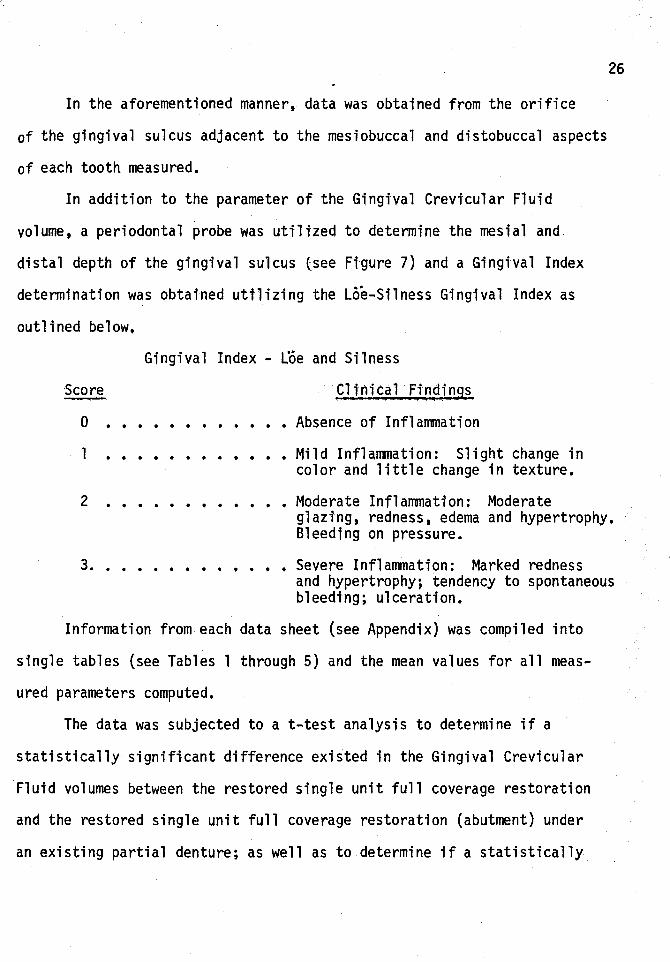
Meter (Periotron-Harco Electronics LTD., Winnipeg, Canada) rises
to a maximum level and then decreases. The value recorded was the
highest numerical reading obtained. Digital numerical values are
converted to fluid vo 1 ume (microliters) by di vi ding the readings by
a conversion factor of 200 (eg. a digital read out of 10 = 0.05 ul)
Following each measurement, the sensors were dried with a sterile
cotton roll.
25
In the aforementioned manner, data was obtained from the orifice
of the gingival sulcus adjacent to the mesiobuccal and distobuccal aspects
of each tooth measured.
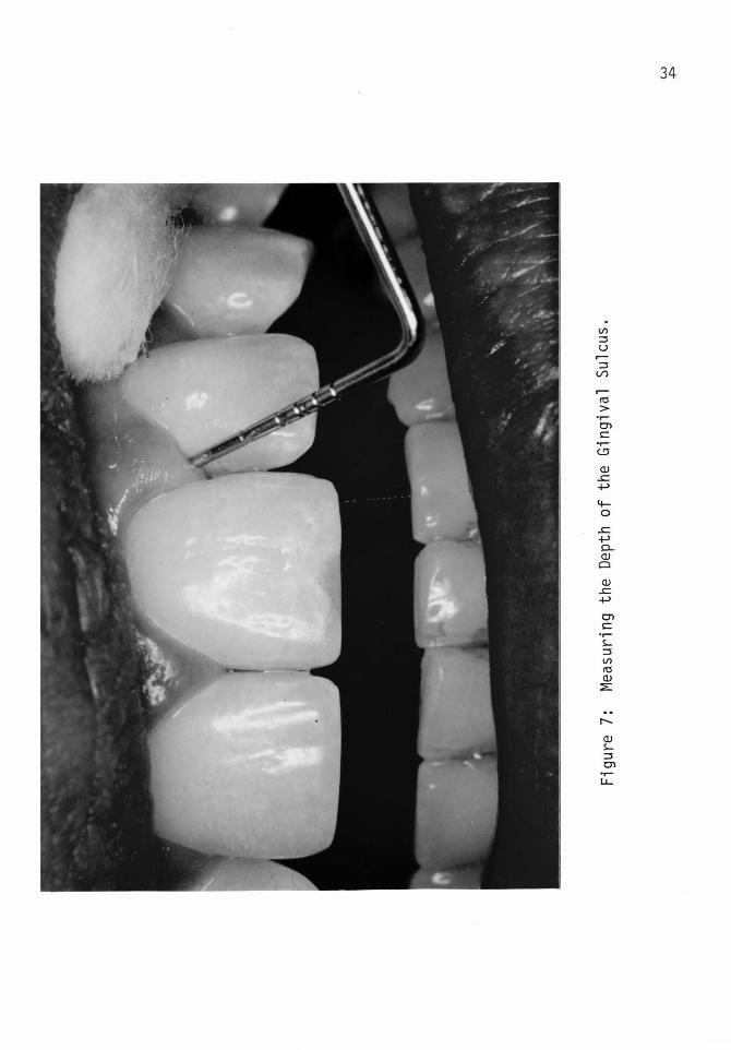
In addition to the parameter of the Gingival Crevicular Fluid
volume, a periodontal probe was utili'zed to determine the mesial and
distal depth of the gingival sulcus tsee Ftgure 7) and a Gingival Index
determination was obtained utilizing the Loe-Silness Gingival Index as
outlined below.
Score
0
1
Gingival Index - Loe and Silness
• • • • • • . . . . . . . . . . • • •
Clinical Findings I . -
• Absence of Inflammation
• Mild Inflammation: Slight change in color and little change in texture.
26
2 . . . . . . . . . . . . Moderate Inflammation: Moderate glazing, redness, edema and hypertrophy. Bleeding on pressure.
3. • ••••••••••• Severe Inflammation: Marked redness and hypertrophy; tendency to spontaneous bleeding; ulceration.
Information from each data sheet (see Appendix) was compiled into
single tables (see Tables 1 through 5) and the mean values for all meas
ured parameters computed.
The data was subjected to a t-test analysis to determine if a
statistically significant difference existed in the Gingival Crevicular
Fluid volumes between the restored single unit full coverage restoration
and the restored single unit full coverage restoration (abutment) under
an existing partial denture; as well as to determine if a statistically
significant difference existed in the Gingival Crevicular Fluid volumes
between the restored abutment for fixed prosthesis and the restored
abutment under an existing removable prosthesis (partial denture).
Regression analysis was also employed to determine which variables
of an abutment for a removable prosthesis contributed significantly to
the Gingival Crevicu1ar Fluid volumes obtained.
27
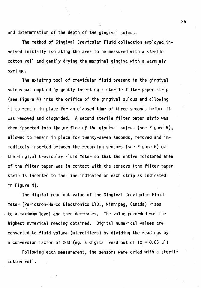
-s:: 0 Vl
~ co 'J
0
::E
s:: 0 ......
+-> u s:: :::::1 .,..., r-IO ...... rQ) ~ +-> ....... c... Q) 0 s:: ......
+-> s:: Q)
"'0
Q) ~
+-> 4-0
E co S-0'1 co ....... Cl
Q) S:::::1 0'1 .,.....
1:1...
28
0 u ~ ttl :I:
.,..... ......... :::::Jttl r--"0 L.L.. ttl
c ~ttl ttiU ,...... :::::J u en
.,..... Q)
> 0.. Q)•r~c u c .,.... ,...... 3: ttl > .,.... eno Cl
•r- _J c.!J ._.., Vl
u C•r-0 c ~0 ~~ 0.j..l
..... u ~Q) Q),...... a_ w
N
Q) ~ :::::J en .,....
LL..
29
1<'0 s.... "0 Q)<'O CLC <'0<'0 CLU 0 ....- .. s...rn Q)Q)
0.... CL ............... c
V'IC CL•r•r-3 s.... +> V)
0 S....fQ)_J CL <'OV'l
0.... u ...... s....c Q) 0 +> s.... r- +> ·...- u l.J....Q)
r-Q)Ll.J
.,..... 0 s...u Q) s... +'<'0 V) :c
...... l.J....
30
.--::::l u
> (!) Su
4-0
s::: 0
·..-+-> u (!) .-.--0 u
0 +-> So
·r-· So... 0-
•r-u...
31
Q) ::::! CJ
c ..s::: u Q)
1--
c 0 ..,.... +-' u Q)
,....... ,....... 0 u
-o or::! ,.......
L.L.
~ <tl
....::::! u ..,.... > Q) ~
u ,....... <tl > O'l c
32
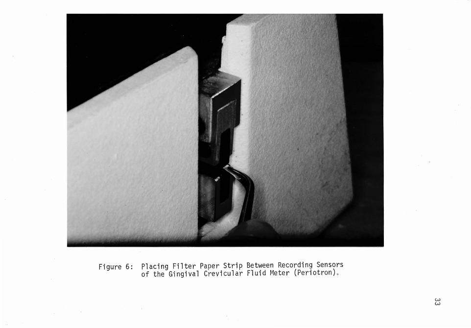
Figure 6: Placing Filter Paper Strip Between Recording Sensors of the Gingival Crevicular Fluid Meter (Periotron) o
w w
Vl ::::5 u
34
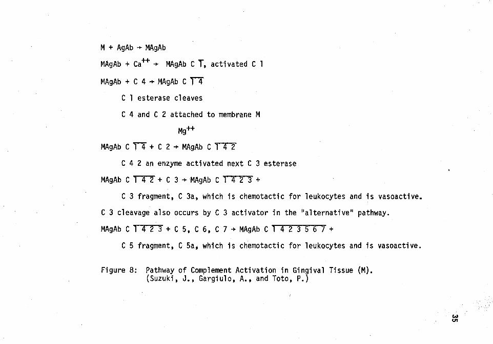
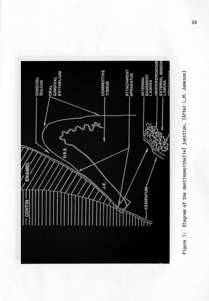
M + AgAb + MAgAb
MAgAb + ca++ + MAgAb C T, activated C 1
MAgAb + C 4 + MAgAb C r-4 C 1 esterase cleaves
C 4 and C 2 attached to membrane M
Mg++
MAgAb C ~ + C 2 + MAgAb C ~2
C 4 2 an enzyme activated next C 3 esterase
MAgAb C Tlf~- + C 3 + MAgAb C 1 4 2 "3 +
C 3 fragment, C 3a, which is chemotactic for leukocytes and is vasoactive.
C 3 cleavage also occurs by C 3 activator in the "alternative .. pathway.
MAgAb C T4-z-r + C 5, C 6, C 7 + MAgAb C 14-2-~--s-o/ +
C 5 fragment, C Sa, which is chemotactic for leukocytes and is vasoactive.
Figure 8: Pathway of Complement Activation in Gingival Tissue (M). (Suzuki, J., Gargiulo, A., and Toto, P.)
w U1
CHAPTER IV
EXPERIMENTAL RESULTS
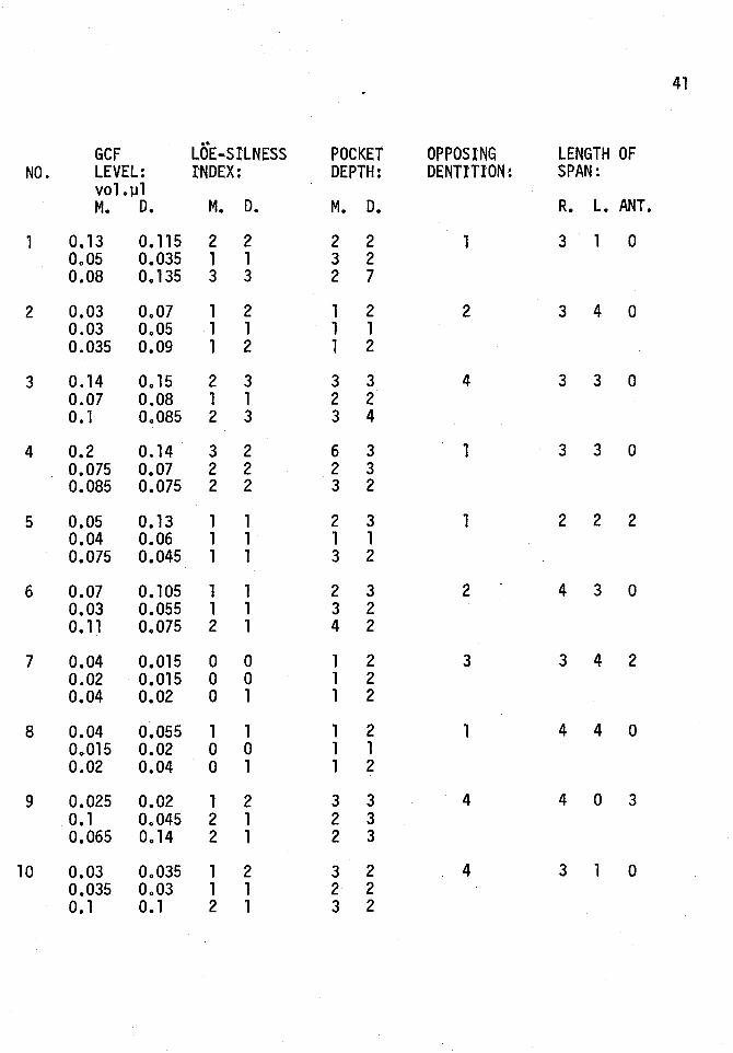
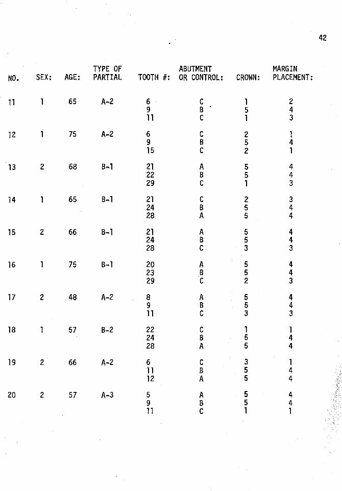
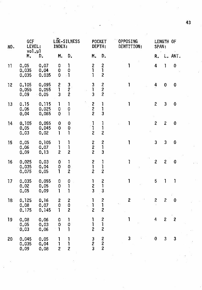
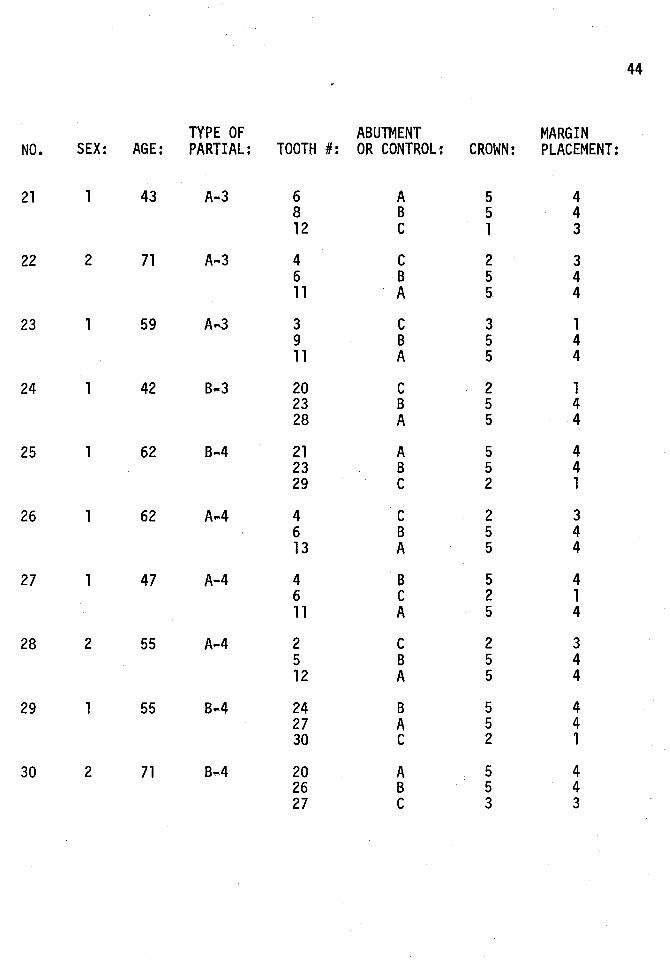
Table one is the compiled data from individual data sheets (see
Appendix) listing the sex, age, type of existing partial denture, teeth
involved in the collection of data, designation of type of abutment or
control, type of restorative material, margin termination, gingival
crevicular flutd volumes, Loe-Silness index, pocket depth, opposing
dentition and length of edentulous span.
Table two lists the frequency distribution for sex, material
utili'zed for crown restoration; and mean values for age, gingival cre
vicular fluid volume, Loe-Silness index and pocket depth for the fixed
prosthodontic non restored tooth (control), restored bridge abutment,
and single restored tooth,
Table three lists the frequency distribution for sex, type of
partial denture, type of opposing dentition; and mean values for age,
gingival crevicular fluid volume, Loe-Silness index, pocket depth, and
total length of edentulous span for the removable prosthodontic non
restored tooth (control), restored abutment under a partial denture,
and non restored abutment under a partial denture.
Table four summarizes the mean values for the various categories
under the fixed prosthodontic groups. The lowest mean gingival crevi
cular fluid volume values, Loe-Silness index, and pocket depth deter
minations were associated with the non restored (control) group. These
36
values increased for the restored single crown; however, the highest
mean values were associated with the restored bridge abutment.
Table five summarizes the mean values for the various categories
under the removable prosthodontic groups. The lowest mean gingival
crevicular fluid volume values, Loe"Silness index, and pocket depth
determinations were associated with the non restored (control) group.
These values increased for the non restored partial denture abutment
group; however, the highest mean values were associated with the re
stored partial denture abutments.
Table six summarizes mean values of gingival crevicular fluid
volume, Loe-Silness index and pocket depth determination for all com
parison groups,
Table seven summarizes the t .. test analysis comparing the gingival
crevicular fluid volume, Loe-Silness index and pocket depth determina
tton between the stngle unit restored tooth (Fixed Prosthodontics) and
the restored abutment under an existing partial denture. The gingival
crevicular fluid volume is statistically higher (p < .001) for there
stored abutment under a partial denture as compared to a single re
stored tooth.
Table eight summarizes the t-test analysis comparing the gingival
crevicular fluid volume, Loe-Silness index and pocket depth determina
tion between the restored bridge abutment (Fixed Prosthodontics) and
the restored abutment under an existing partial denture. No statistic
ally significant difference (p < .001) occurs between the two groups
37
for either total fluid volume, Loe-Silness index or pocket depth de
termination.
The data from the Removable Prosthodontic categories was sub
jected to a multiple regression analysis using Mesial and Distal
gingival crevicular fluid volumes, and Lo·e-Si·lness index as dependent
variables and employing the type of opposing dentition, material for
the crown, margin termination, type of .partial denture and total length
of edentulous span as independent variables. Table nine highlights
the statistically signi'ficant (p < .001) independent variables of type
of material used in construction of the crown and margin termination
that had the greatest influence upon the increased gingival crevicular
fluid volumes and Loe-Silness index.
38
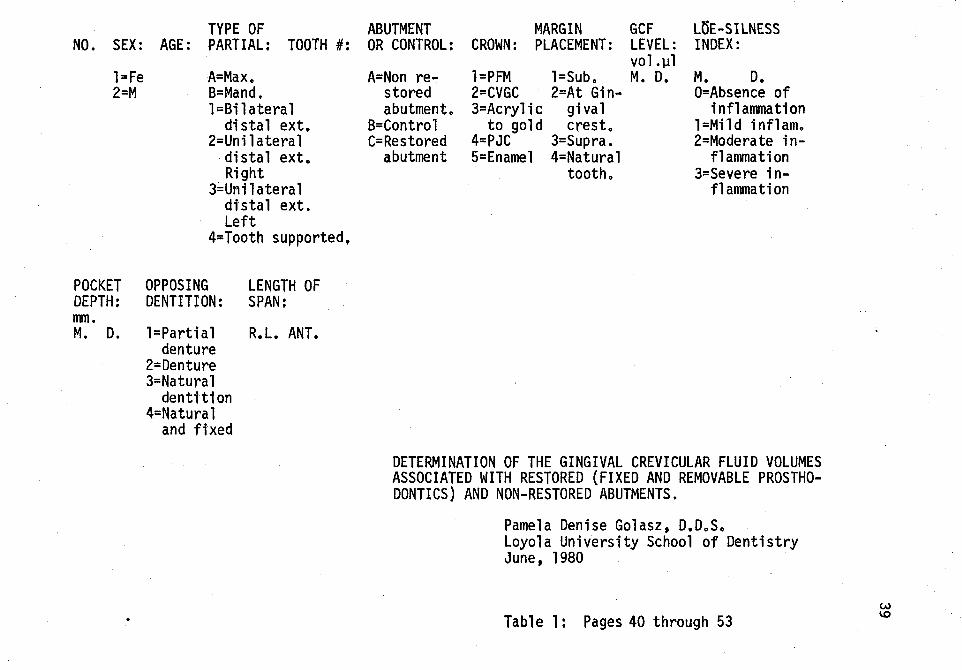
TYPE OF ABUTMENT MARGIN GCF LfiE-SILNESS INDEX: NO. SEX: AGE: PARTIAL: TOOTH #: OR CONTROL: CROWN: PLACEMENT: LEVEL:
l=Fe 2=M
A= Max. B=Mand. 1 =Bi 1 atera 1
distal ext. 2=Unil atera 1
·distal ext. Right
3;,.Uni 1 atera 1 distal ext. Left
4=Tooth supported.
POCKET OPPOSING LENGTH OF DEPTH: DENTITION: SPAN; mm. M. D. l=Partial R.L. ANT.
denture 2=Denture 3=Natural
dentttion 4=Natural
and fixed
A=Non restored abutment.
B=Control C=Restored
abutment
vol.~l l=PFM l=Sub. M. D. 2=CVGC 2=At Gin-3=Acrylic gival
to gold crest. 4=PJC 3=Supra. 5=Enamel 4=Natural
tooth.
M. D. O=Absence of
inflammation l=Mild inflam. 2=Moderate in
flammation 3=Severe in
flammation
DETERMINATION OF THE GINGIVAL CREVICULAR FLUID VOLUMES ASSOCIATED WITH RESTORED (FIXED AND REMOVABLE PROSTHODONTICS) AND NON-RESTORED ABUTMENTS.
Pamela Denise Golasz, D.D.S. Loyola University School of Dentistry June, 1980
Table 1: Pages 40 through 53 w \0
40
TYPE OF ABUTMENT MARGIN NO. SEX: AGE: PARTIAL: TOOTH #: OR CONTROL: CROWN: PLACEMENT:
2 58 A-1 5 c 1 1 8 B 5 4 14 c 1 1
2 2 62 B-1 21 c 2 3 24 B 5 4 29 c 2 3
3 1 67 B-1 21 c 3 2 24 B 5 4 28 c 3 2
4 1 49 B-1 22 c 1 1 24 B 5 4 27 c 1 1
5 1 49 A-1 5 c 1 1 6 B 5 4 11 c 1 3
6 1 66 B-1 21 c 3 1 23 B 5 4 27 c 3 1
7 1 66 A-1 6 c 1 1 8 B 5 4 11 c 1 1
8 2 63 B-1 22 c 2 3 24 B 5 4 27 c 1 1
9 1 68 A-2 8 B 5 4 10 c 1 1 14 c 2 3
10 1 62 B-2 20 c 1 3 23 B 5 4 28 c 1 1
41
•• GCF LOE .. SILNESS POCKET OPPOSING LENGTH OF NO. LEVEL: I'NDEX: DEPTH: DENTITION: SPAN:
VOLJ.Il M. D. M. D. M. D. R. L. ANT.
1 0.13 0.115 2 2 2 2 1 3 1 0 0.05 0.035 1 1 3 2 0.08 0.135 3 3 2 7
2 0.03 0.07 1 2 1 2 2 3 4 0 0.03 0.05 1 1 1 1 0.035 0.09 1 2 1 2
3 0.14 0.15 2 3 3 3 4 3 3 0 0.07 0.08 1 1 2 2 o. 1 0.085 2 3 3 4
4 0.2 0.14 3 2 6 3 1 3 3 0 0.075 0.07 2 2 2 3 0.085 0.075 2 2 3 2
5 0.05 0.13 1 1 2 3 1 2 2 2 0.04 0.06 1 1 1 1 0.075 0.045 1 1 3 2
6 0.07 0.105 1 1 2 3 2 4 3 0 0.03 0.055 1 1 3 2 0.11 0.075 2 1 4 2
7 0.04 0.015 0 0 1 2 3 3 4 2 0.02 0.015 0 0 1 2 0.04 0.02 0 1 1 2
8 0.04 0.055 1 1 1 2 1 4 4 0 0.015 0.02 0 0 1 1 0.02 0.04 0 1 1 2
9 0.025 0.02 1 2 3 3 4 4 0 3 o. 1 0.045 2 1 2 3 0.065 0.14 2 1 2 3
10 0.03 0.035 1 2 3 2 4 3 1 0 0.035 0.03 1 1 2 2 o. 1 0.1 2 1 3 2
42
TYPE OF ABUTMENT MARGIN NO. SEX: AGE: PARTIAL TOOTH #: OR CONTROL: CROWN: PLACEMENT:
11 1 65 A-.2 6 c 1 2 9 B 5 4 11 c 1 3
12 1 75 A ... 2 6 c 2 1 9 B 5 4 15 c 2 1
13 2 68 B ... 1 21 A 5 4 22 B 5 4 29 c 1 3
14 1 65 B ... 1 21 c 2 3 24 B 5 4 28 A 5 4
15 2 66 s ... 1 21 A 5 4 24 B 5 4 28 c 3 3
16 1 75 B-.1 20 A 5 4 23 B 5 4 29 c 2 3
17 2 48 A-2 8 A 5 4 9 B 5 4 11 c 3 3
18 1 57 B-2 22 c 1 1 24 B 5 4 28 A 5 4
19 2 66 A-2 6 c 3 1 11 B 5 4 12 A 5 4
20 2 57 A .. 3 5 A 5 4 9 B 5 4 11 c 1 1
43
.. GCF LOE ... SIL.NESS POCKET OPPOSING LENGTH OF
NO. LEVEL: I'NDEX: DEPTH: DENTITION: SPAN: vo1.ll1 M. D. M. D. M. D. R. L. ANT.
11 0~05 0.07 0 1 2 2 1 4 1 0 0.035 0.04 0 0 1 1 0.035 0.035 0 1 1 2
12 0.105 0.095 2 3 3 2 1 4 0 0 0.055 0.055 1 2 1 2 0.09 0.05 3 2 3 2
13 0.15 0.115 1 1 2 1 1 2 3 0 0.06 0~025 0 0 2 1 0.04 0.065 0 1 2 3
14 0.105 0.055 0 0 1 1 1 2 2 0 0,05 0.045 0 0 1 1 0.03 0.02 1 1 2 2
15 0.05 0.105 1 1 2 2 1 3 3 0 0.06 0.07 1 1 2 1 0.09 0.13 2 2 2 3
16 0.025 0.03 0 1 2 1 1 2 2 0 0.035 0.04 0 0 1 1 0.075 Oo05 1 2 2 2
17 0.035 Oo055 0 0 1 2 1 5 1 1 0.02 0.05 0 1 2 1 0.05 0.09 1 1 3 3
18 0.125 0.16 2 2 1 2 2 2 2 0 0.08 0~07 0 0 1 1 0.175 0.145 1 2 2 2
19 0.08 0.06 0 1 1 2 1 4 2 2 Oo05 0.03 0 0 1 1 Oo03 0.06 1 1 2 2
20 Oo045 0.05 1 1 3 2 3 0 3 3 0.035 0.04 1 1 2 2 0.09 0.08 2 2 3 2
44
TYPE OF ABUTMENT MARGIN NO. SEX: AGE: PARTIAL: TOOTH #: OR CONTROL: CROWN: PLACEMENT:
21 1 43 A-3 6 A 5 4 8 B 5 4 12 c 1 3
22 2 71 A-3 4 c 2 3 6 B 5 4 11 A 5 4
23 1 59 A .. 3 3 c 3 1 9 B 5 4 11 A 5 4
24 1 42 B-3 20 c 2 1 23 B 5 4 28 A 5 4
25 1 62 s .. 4 21 A 5 4 23 B 5 4 29 c 2 1
26 1 62 A-4 4 c 2 3 6 B 5 4 13 A 5 4
27 1 47 A-4 4 B 5 4 6 c 2 1 11 A 5 4
28 2 55 A-4 2 c 2 3 5 B 5 4 12 A 5 4
29 1 55 B-4 24 B 5 4 27 A 5 4 30 c 2 1
30 2 71 B ... 4 20 A 5 4 26 B 5 4 27 c 3 3
45
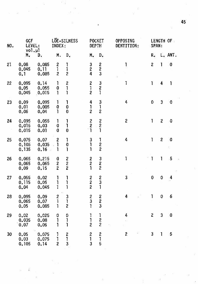
GCF LQE ... SILNESS POCKET OPPOSING LENGTH OF NO. LEVEL: INDEX: DEPTH DENTITION: SPAN:
vo1.p1 M. D. M. D. M. D. R. L. ANT.
21 0.08 Oo085 2 1 3 2 1 2 1 0 0.045 0.11 1 1 2 2 o. 1 0.085 2 2 4 3
22 0.095 0.14 1 2 2 3 1 1 4 1 0.05 0.055 0 1 1 2 0.045 0.015 1 1 2 l
23 0.09 0.095 1 1 4 3 4 0 3 0 o. 01 0.005 0 0 1 1 0.06 0.04 1 0 2 2
24 0.095 0.055 1 1 2 2 2 1 2 0 0.015 0.03 0 1 2 2 0.015 0.01 0 0 1 1
25 0.075 0.07 2 1 3 1 4 1 2 0 0.105 0.035 1 0 1 2 0.135 0.16 1 1 1 2
26 0.065 0.215 0 2 2 3 1 1 1 5 0.065 0.065 2 2 2 2 0.09 o. 15 2 2 1 2
27 0.055 0 .. 02 1 1 2 2 3 0 0 4 0.115 0.05 1 1 2 3 0.04 0.045 1 1 2 1
28 0.095 0.09 2 3 2 2 4 1 0 6 0.065 0.07 1 1 3 2 0.05 0.085 1 2 1 3
29 0.02 0.025 0 0 1 1 4 2 3 0 0.035 0.08 1 1 1 2 0.07 0.06 1 1 2 2
30 0.05 0.075 1 2 2 2 2 3 1 5 0.03 0 .. 075 1 1 1 1 0.105 0.14 2 3 3 5
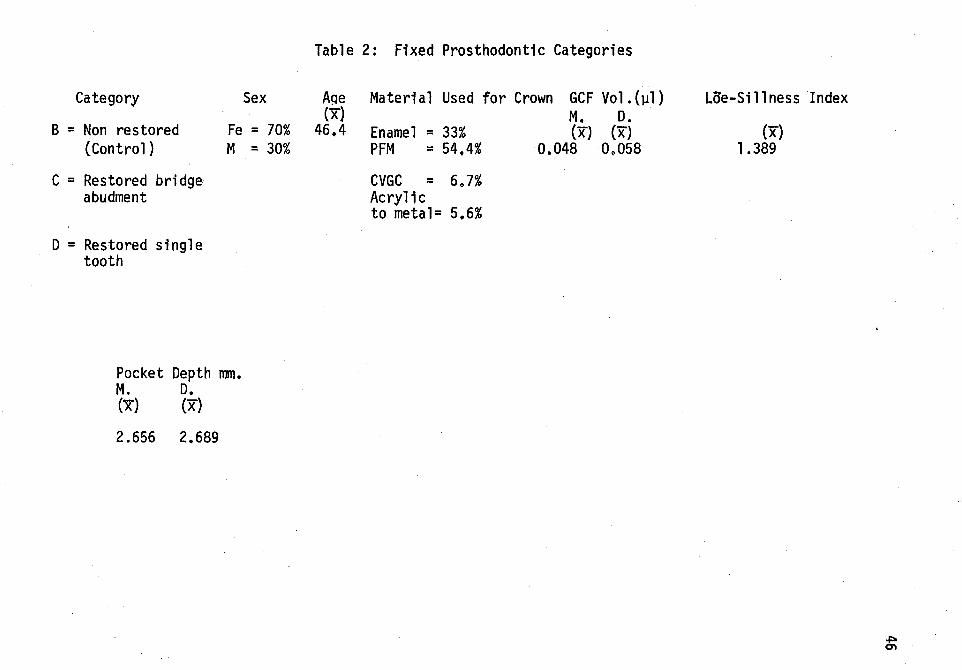
Category
B = Non restored (Control}
C = Restored bridge abudment
D = Restored single tooth
Table 2: F1xed Prosthodont1c Categories
Sex A~e Material Used for Crown GCF Vol.(~l} (x) M. D.
Fe = 70% 46.4 Enamel = 33% (x) (x) M = 30% PFM = 54.4% 0.048 09058
CVGC = 6.7% Acrylic to metal= 5.6%
Pocket Depth mm. M. D. (x) (x)
2.656 2.689
Loe-Sillness Index
(x) 1.389
~ 0"1
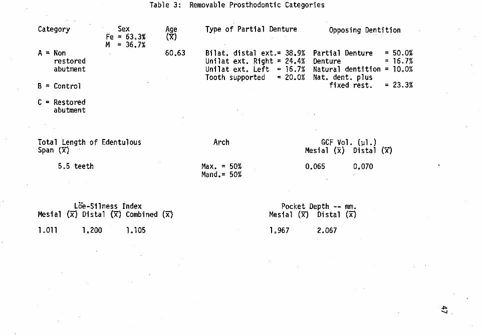
Table 3: Removable Prosthodontic Categories
Category
A = Non restored abutment
B = Control
C = Restored abutment
Sex Fe = 63.3% M = 36.7%
Total Length of Edentulous Span {x)
5.5 teeth
Lcie-Silness Index
Age (x)
60.63
Mesial (X} Distal {x) Combined (x}
1 . 011 1.200 1.105
Type of Partial Denture
Bilat. distal ext.= 38.9% Unilat ext. Right = 24.4% Unilat ext. Left = 16.7% Tooth supported = 20.0%
Arch
Max, = 50% Mand.= 50%
Opposing Dentition
Partial Denture = 50.0% Denture = 16.7% Natural dentition = 10.0% Nat. dent. plus
fixed rest. = 23.3%
GCF Vol. (lll.} Mesial (x) Distal (x)
0.065 0.070
Pocket Depth -- mm. Mesial (x) Distal (x)
1 o967 2.067
~ .......
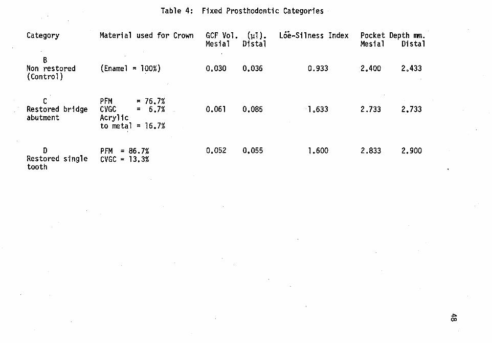
Table 4:
Category Material used for Crown
B Non restored (Enamel ~ 100%) (Control)
c PFM ;:: 76.7% Restored bridge CVGC ~ 6.7% abutment Acryl1c
to metal = 16.7%
D PFM = 86.7% Restored single CVGC = 13.3% tooth
Fixed Prosthodontic Categories
GCF Vol. (lJ1). Loe~Silness Index Mesial Distal
0.030 0.036 0.933
0.061 0,085 1.633
0.052 0.055 1.600
Pocket Depth mm. Mesial Distal
2.400 2.433
2.733 2.733
2.833 2.900
~ (X)
Category
A Non restored
B Non restored single tooth (control)
c Restored partial denture abutment
Table 5: Removable Prosthodontic Categories
Material used for Crown GCP Vol, (~1}. Loe-Silness Index Mesial Distal Mesial Distal Combined
(Enamel = 100%} 0.064 0,071 1.200 1.267 1.500
(Enamel = 100%} 0,044 0.046 0.633 0,767 0.800
PFM = 36.7% 0.087 0.093 1.200 1.567 1.600 CVGC = 40.0%
Acrylic to metal = 23.3%
Margin Placement
Sub Ging. = 50.0% At crest = 6.7% Supra Ging = 43.3%
Pocket Depth mm. Mesial Distal
2,033 2.100
lo633 1.600
2.233 2.500
~ \0
Table 6: Summary of Mean Values for all Comparison Groups
GINGIVAL CREVICULAR FLUID VOL.(~l.) ..
POCKET DEPTH (mm,) COMPARISON GROUPS LOE-SILNESS INDEX (Mesial) (Distal) (Mesial) (Distal)
FIXED PROSTHODONTICS: Non Restored Tooth (Control) 0.030 0.036 0.933 2.400 2.433
vs. Restored Single Tooth 0.052 0.055 1.600 2.833 2.900
vs. Restored Bridge Abutment 0.061 0.085 1.633 2.733 2.733
REMOVABLE PROSTHODONTICS: Non Restored Tooth (Control} 0,044 09046 0.800 1.633 1.600
vs. Non Restored Abutment 0.064 0.071 1.500 2.033 2.100
vs. Restored Partial Denture
Abutment 0.087 0.093 1.600 2.233 2.500 - - - - - - ~ - - ~ -- -- ~ - ~ - ~ - - ~ ~ - " ~ " - ---- - - - - - - - - - - - - - - - - - -FIXED PROSTHODONTICS:
Restored Single Tooth 0,052 0.055 1.600 2.833 2. 900 . vs.
REMOVABLE PROSTHODONTICS: Restored Partial Denture 0.087 0.093 1.600 2.233 2.500
Abutment - - - - - - ~ - ~ - - - - --- - - -- - ~ ---- - ~ -- - - - - -- - - - - - - - - - - - - - -FIXED PROSTHODONTICS:
Restored Bridge Abutment 0.061 0,085 1.633 2.733 2.733 vs.
REMOVABLE PROSTHODONTICS: Restored Partial Denture 0.087 0.093 1.600 2.233 2.500
Abutment
01 0
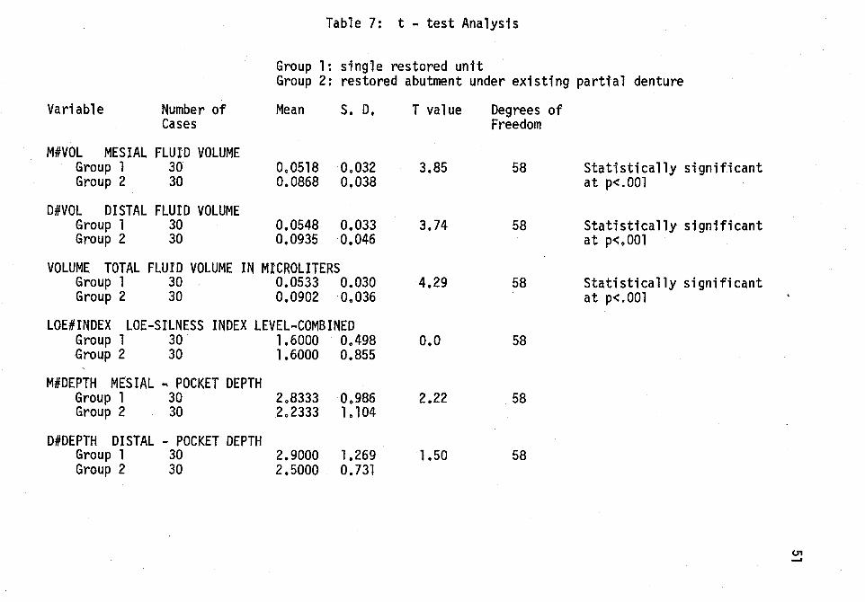
Table 7: t - test Analysis
Group 1: single restored unit Group 2: restored abutment under existing partial denture
Variable Number of Cases
M#VOL MESIAL FLUID VOLUME Group 1 30 Group 2 30
D#VOL DISTAL FLUID VOLUME Group 1 30 Group 2 30
Mean S. D.
0.0518 0.032 0.0868 0.038
0.0548 0.033 0,0935 0.046
VOLUME TOTAL FLUID VOLUME IN MICROLITERS Group 1 30 0.0533 0.030 Group 2 30 0.0902 0.036
LOE#INDEX LOE-SILNESS INDEX LEVEL-COMBINED Group 1 30 1.6000 0.498 Group 2 30 1.6000 0.855
M#DEPTH MESIAL .. POCKET DEPTH Group 1 30 2.8333 0.986 Group 2 30 2.2333 1.104
D#DEPTH DISTAL - POCKET DEPTH Group 1 30 2.9000 1.269 Group 2 30 2.5000 0.731
T value
3.85
3.74
4.29
o.o
2.22
1.50
Degrees of Freedom
58
58
58
58
58
58
Statistically significant at p<.OOl
Statistically significant at p<.OOl
Statistically significant at p<.OOl
01 -
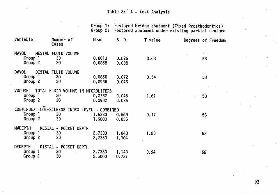
Table 8: t - test Analysis
Group 1: restored bridge abutment (Fixed Prosthodontics) Group 2: restored abutment under existing partial denture
Variable Number of Mean S. D. T value Degrees of Freedom Cases
M#VOL MESIAL FLUID VOLUME Group 1 30 0.0613 0.026 3.03 58 Group 2 30 0.0868 0.038
D#VOL DISTAL FLUID VOLUME Group 1 30 0.0850 0,072 0.54 58 Group 2 30 0.0936 0.046
VOLUME TOTAL FLUID VOLUME IN MICROLITERS Group 1 30 0,0732 0.045 1.61 58 Group 2 30 0.0902 0.036 ..
LOE#INDEX LOE-SILNESS INDEX LEVEL ~ COMBINED Group 1 30 1.6333 0.669 0,17 58 Group 2 30 1.6000 0,855
M#DEPTH MESIAL - POCKET DEPTH Group 1 30 2.7333 1.048 1.80 58 Group 2 30 2.2333 1.104
D#DEPTH DISTAL - POCKET DEPTH Group 1 30 2.7333 1.143 0.94 58 Group 2 30 2o5000 0.731
U'1 N
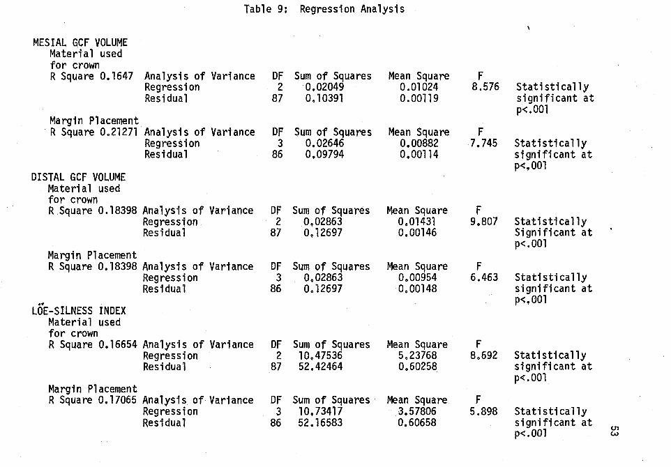
Table 9: Regression Analysis
MESIAL GCF VOLUME Materi·a 1 used for crown R Square 0.1647 Analysis of Variance DF Sum of Squares Mean Square F
Regression 2 0.02049 0.01024 8.576 Statistically Residual 87 0.10391 0.00119 significant at
p<.OOl Margin Placement R Square 0.21271 Analysis of Variance DP Sum of Squares Mean Square F
Regression 3 0.02646 0.00882 7.745 Statistically Residual 86 0,09794 0,00114 significant at
p<,OOl DISTAL GCF VOLUME
Material used for crown R.Square 0.18398 Analysts of Variance DF Sum of Squares Mean Square F
Regression. 2 0.02863 0.01431 9.807 Stati sti ca lly Residual 87 0.12697 0.00146 Significant at
p<.OOl Margin Placement R Square 0.18398 Analysis of Variance DF Sum of Squares Mean Square F
Regression 3 0.02863 0.00954 6.463 Statistically Residual 86 0.12697 0.00148 significant at
•• p<.OOl LOE-SILNESS INDEX
Material used for crown R Square 0.16654 Analysis of Variance DF Sum of Squares Mean Square F
Regression 2 10.47536 5.23768 8.692 Statistically Residual 87 52,42464 0.60258 significant at
p<.OOl Margtn Placement R Square 0.17065 Analysts of Variance OF Sum of Squares Mean Square F
Regression 3 10,73417 3.57806 5.898 Statistically Residual 86 52.16583 0.60658 significant at
U1 p<.OOl w
CHAPTER V
DISCUSSION
The fragile, cellular microcosm of the dentoepithelium and the
gingival sulcus function to maintain periodontal integrity. With the
advent of high speed instrumentation, even the most highly skilled
operator employing innocuous and delicate techniques of soft tissue
manipulation encounters the inherent problem of tissue trauma. An
inflammatory response of the marginal gingiva may occur subsequent
to the intrinsic irritation provided by full coverage restorations and
the various levels of gingival termination, Another detrimental aspect
of restorative procedures is the adaptation of the full coverage re
storation to the preparation. The cement interface between termination
of the restorative material and natural tooth structure provides a ni
dus for plaque accumulation.
The response to inherent tissue trauma and bacterial by products
has been inflammation of the soft tissues. Gingival inflammation is
the manifestation of an attempt on the part of ~he host to dilute, iso
late and remove, and render benign substances deemed foreign. They may
be of microbial origin or due to some noxious stimuli. A correlation
between an increase in gingival crevicular fluid flow and an increase
in inflammation has been observed; and crevicular fluid as postulated
by Brill, 1960, can be quantitatively measured and thereby serve as an
indicator of the level of inflammation present that is far more consistent
54
than subjective clinical indices that have been utilized historically.
Inflammatory changes have been observed by investigators on a
histological basis, Alterred microcirculation and increased inflam
matory cell density will ultimately lead to increased vascular per
meability that permtts an egress of flutd from the vascular compartment
(Grant. Stetn. and Everett, 1972} into the gingival sulcus. Presence
of increased gingival crevicular fluid flow in the sulcus will reflect
the level of periodontal inflammation prior to clinical manifestation
(Loe and Holm-Pedersen, 1965) of pathology. Subclinical inflammation
can thus be monitored and the inflammatory process interrupted to limit
the amount of irreversible ttssue damage.
The controversy over whether gingival crevicular fluid flow can
be demonstrated in clinically healthy gingiva has lead to the theory
that crevicular f1utd is a reflection of the host's response to noxious
or exogenous sti·mult and his capacity to release mediators of inflamma
tion. The host's response to foreign invasion will vary with the in
dtvi·dual and may be affected by age, genetic capacity for response and
predisposition. and the general health of the individual.
Investigators have discussed the relative value of abutments for
both fixed and removable prosthodontics. Cohn (1956) stated "a common
cause of periodontal disease is the faulty removable partial denture,
or fixed partial denture, in which anchorages are improperly selected
or incorrectly fabricated." Waerhaug {1968) in emphasizing that margin
termination should be supragingival to minimize the contributory effect
55
of plaque retention on crown margins toward periodontal disease stated 11many patients would be better off tf brtdges or partial dentures were
not constructed ...
This investigation was performed tn an attempt to lend statisti
cal credence to the inflammation levels of restored abutment teeth for
both fixed and removable prosthesis. A determination and comparison of
the inflammatory changes in the marginal gingiva of abutment teeth for
fixed and removable prosthesis was performed. Data was collected from
thirty tndtvtduals (mean age = 46.4) that exhibited a restored bridge
abutment, a restored single unit crown and at least one non restored
tooth {control}. Thirty additional individuals (mean age = 60.63)
served as candidates for the collection of data from at least one re
stored abutment under an existing partial denture and a non restored
tooth (control}.
The gingival crevicular fluid volume mean, Loe-Silness Index,
and pocket depth mean were determined and the lowest values were cor
related with the fixed prosthodontic non restored tooth (control)
category. These values tended to increase with the single restored
tooth category and were the highest with the restored abutment for a
bridge category.
Further increases in the aforementioned parameters occurred in the
control group for the removable prosthesis group, increased with the non
restored abutment, and were the highest for the restored abutment under
an existing partial denture.
A statistically significant increase is observed in gingival
56
crevicular fluid volume associated with a restored abutment under a re
movable prosthesis when compared with a single restored crown. In part,
this can be attributed to the additional plaque accumulation and associ
ated bacterial by products related to the components and materials used
tn the fabrication of the partial denture, This increase may also re
flect the compromised dentitions (periodontally involved) that neces
sitate partial denture prostheses, In addition, the statistics obtained
may actually be a reflectton of the a1terred host response to a foreign
material, in this case, a removable prosthesis. It may well be that an
alterred host response tn the form of an autoimmune response that is the
body's response to a prosthesis deemed 11 foreign 11 or not recognized as
11 self11 is reflected i'n the data. Further studies to varify the presence
of C 3 and IgG in human gingival crevicular fluid would be indicated.
(See Figure 8: Pathway of Complement activation, page 35).
The fact that no statistically significant increase in gingival
crevicular fluid volumes could be measured between a restored bridge
abutment and a restored partial denture abutment appears to be explained
by the fact that the restored bridge abutment gingival crevicular fluid
values increased to meet the increased gingival crevicular fluid values
of the restored partial denture abutment. This increased gingival cre
vicular fluid volume associated with a restored bridge abutment may well
reflect the margin termination, material utilized to construct the pros
thesis, the contour of the bridge abutment, and the existence of inade
quate embrasure that would have facilitated the maintenance of the pros
thesis by the patient.
57
From the regression analysis of the variables of partial denture
prosthesis, the factors that contributed the most to the increased level
of gingival crevicular fluid volume were margin placement and type of
restorative material employed.
The findings in this study correlate with those of Brill and Bjorn,
(1959) who observed increased gingival crevicular fluid volume with full
coverage restorations as compared to non restored teeth: Maruyama and
associates, (1976) who observed dilation of capillary loops related to
inflammation in gingiva classified as clinically normal; and Mahajan,
(1976) whose histologic observation of the interdental papilla adjacent
to full coverage restorations indi'cated that both normal and inflamed
gingiva adjacent to restored teeth exhibited an inflammatory cell in
filtrate and dilated blood vessels.
58
CHAPTER VI
SUMMARY AND CONCLUSIONS
The dynamic aspects of the dento-gingival junction and sulcus
were presented, and inherent problems in restorative dentistry that
contrtbute to soft tissue inflammation were discussed. Components of
gingival crevicular fluid and their possible implications in relation
to periodontal pathology have been correlated with a response on the
part of the host to exogenous or noxious stimuli.
A determination and comparison of the inflammatory changes in
the marginal gingiva of abutment teeth for fixed and removable pros
theses as well as for non restored teeth was performed. Data was
collected from thirty individuals (mean age = 46.4) that exhibited a
restored bridge abutment, a restored single unit crown, and at least
one non restored tooth (control). Thirty additional individuals
(mean age = 60.63) served as candidates for the collection of data
from at least one restored abutment under an existing partial denture
and a non restored tooth (control). The gingival crevi·cular fluid vol
ume, Loe-Silness index, and pocket depth were recorded; data was sub
jected to a t-test analysis between the two populations, and a re
gression analysis was performed on the variables recorded for the partial
denture prostheses.
The following conclusions can be made in view of the results obtained
during this study: 59
1. In a comparison of the marginal gingiva inflammatory changes
associated with restored teeth versus the non restored teeth;
for both the Fixed Prosthodontic categories and the Removable
Prosthodontic categories, the least amount of inflammation as
measured by the parameters of gingtval crevicular fluid volume,
Loe-Silness index and pocket depth tn all cases, were associated
wfth the non restored groups.
2. In a comparison of the marginal gingtva inflammatory changes of
the single restored tooth from the Fixed Prosthodontic population
versus the restored tooth under an exi'sting partial denture from
the Removable Prosthodontic population; the least amount of in
flammation as measured by the gingival crevicular fluid volume
was associated with the single restored tooth from the Fixed
Prosthodontic population.
3. In a comparison of the margtna1 gingiva inflammatory changes of
the restored bridge abutments (Fixed Prosthodontics) to the
restored abutments under a partial denture (Removable Prostho
dontics}; the least amount of inflammation as measured by the
gingival crevicular fluid volume was associated with the restored
bridge abutment.
4. Loe-Silness index and pocket depth determination may not be as
valid a determi'nant of inflammatory levels of soft tissue as is
determination of the gingival crevicular'fluid volume.
5. Teeth with subgingival margin placement from both populations were
accompanied by predictable inflammatory levels.
60
BIBLIOGRAPHY
Amsterdam, M., and Fox, L. "Provisional Splinting-Principles .2.!!.9_ Technigues." Dent. Cl in. No. Am., (1"959), 73-79.
Alfano,.Michael C. 11 The ~4jgil of Gingival Fluid. 11 J. Theor. B1ol., Vol. 47,lT9 , 2/-136.
Band, J. and Cimasoni, G. "Total Protein fn Human Crevicular Flutd." J. Dent. Res., Vol. 5o, (19m, 1683.
Black, G.V. "The Periosteum and Peridental Membrarie-Interfibrous E1ementsO'T" the PeriaentaT Membrane. 11 The Dent, Review, Vo 1 • I, N07 O,(Apri 1 15, 1887) , 289-302.
Black, G.V. "The Fibers and Glands of the Peridental Membrane. 11
The Dent:l:osmos, VOT7 XLI, No~, (Feb., 1899}, 101-122.
Black, G.V. Special Dental Pathologx, 2nd ed. Chicago: MedicoDental Publishing Co., 1920.
Bodecker, Charles F., and Cahn, Lester R •. 11 The Histologx and Function of the Gingiva... Dent. ItemsO'T" Interest, VOT. 53, (Feb.--;-193T), 94.
Borden, S.M., Golub, L.M., and Kleinberg, I. 11 An Intracrevicular Technt~ue for Monitoring Gingival Crevicufar Fluid TGrrr Flow. J:D"ent. Res., Vol. 53, (Feb., 1 974), Abstract mr, l'75:"
Brandtzaeg, Per, and Mann, Wallace v., Jr. 11A Com~arative s~adx_ of the Lysozxme Activity of Human Gi n~ival ocket Fl u1 , 'Seri:i'n17and Saliva." A"ctaOdont. Scan., Vol. 22, (1964), 441-4ss:-- ·
Brill, Niels, and Bjorn, Hilding. 11 Passage of Tissue Fluid fnto Human Ginfival Pockets. 11 Acta Odont. Scand., Vol. 17,(1959), 1 -21.
Brill, Niels, and Krasse, Bo., "Effect of Mechanical Stimulation .Q!!. Flow of Tissue Fluid Throufh Gingival "Pocket Epithelium.;' Actaoaont. Scanainav., Vol. 7, (1959), 115-130.
Bri 11, ~i e 1 s., 11 Influence of Cap ill ar,t Permeabi 1 i ty .!?.!2. Flow of T1ssue Fluid 1nto Gin~ival Pockets." Acta Odont. Scandinav., Vol. 17, {1959a). 23- 3.
61
Br111, Niels,. 11 ~ffect of ChewinA .Q!!. Flow of Tissue Fluid into Human G1n~1 va 1 PoCKets. 11 eta Odont. Scandi nav., Vo I.'T7, ~b), 77-281.
Brill, Niels, and Bronnestam, Rolf. 11 Immuno-Electrophoretic §tud~.of Tissue Fluid from Gin~ival Pockets. 11 Acta Odont.
1nav., Vol. 18, (m), 9 -1oo.
Burch, James G. 11 Periodontal Considerations in O~erative Dentistry. 11
J. Prosth. Dent., Vol. 34, No.2, (Aug.-;19 5), 156-63.
Caruso, J.L., 11 Histological Evaluation of Cast Crown Contours in Experimental Animals 11
, Onpub1ishe<:0f.'S71hesls, Loyola Un1v. Dental School, 1979.
Cimasoni, G. Monogralhs in Oral Science, Vol. 3- The Crevicular Fluid. Switzer and;s:-Targer AG, 1m. - -
Cohn, L. A., 11 The Physiologic Basis for Tooth Fixation in PrecisionAttachedPartial 'Dentures. 11 J:Prosth. Dent., Vol. 6, No. 2, (1956) ' 220-244.
Egelberg, Jan. 11 Cellular Elements in Gingtval Pocket Fluid. 11
Acta Odont. Scandinav., Vol.~' (l o3a}, 280-287.
Egelberg, Jan. "Diffusion of Histamine into the Gingival Crevice and Throuf the CreviCU'J a r Epithe l'flifii.'"'1Acta dont. Scandi nav., VOT, 21, 19030), 271-282.
Egelberg, Jan. "Gin~ival Exudate Measurements for Evaluation of Inflammatory C anges of tne Gi'ngivae. 11 Odont. Rev., Vo1:15, (1964)' 381:'398. -- . .
Gavin, J.B., and Collins, A.A., 11 The Occurrence of Bacteria Wtthin the Clinically Healthy Gi'ngival Crevice. 11 J. Perio-dont., Vol. 32, (l96la), 198-201. ---·
Gavin, J.B., and Collins, A.A., 11 An Evaluation of the Antimicrobial Effect of the Fluid Exudate from the Clinically Healthy Gingival Crevice ... J. Periodont., Vol. 32, (196lb), 99-101. -
Gli'ckman, I., Stein, S., and Smulow, Jerome, 11 The Effect of Increased Functional Forces Upon the Periodontium of Splinted and Non-Splinted Teeth 11
, J. of Periodont., Vol. 32, (1961), 290-300. - -
62
Glickman, Irving. Clinical Periodontologx, Philadelphia, W.B. Saunders Co., 1972.
Golub, L.M., Borden, S.M., and Kleinberg, I., "Urea Content of Gingival Crevicular Fluid and Its Relation to Periodontal Disease in Humans. 11 J. Periodont. Res., Vol. 6, (1971), 243-251. - -
Grant, Daniel A., Stern, Irving, B., and Everett, Frank G. Orban's Periodontics, St. Louis, The C.V. Mosby Co., 1972.
Gustaf~son, Gunnar T., and Nilsson, Inga Marie, hFibrinolytic Activity in Fluid from Gingival Crevice." Soc. Exper. Biol. and Med. Proc., Vol. 106, (1961), 277-280. - -
Harvey, Paul M. 11 Elimination of Extraneous Material from the Gingival Crevice." !L· Periodont., Vol. 33, (1962), 231-237.
Holm-Pedersen, Paul, Agerbaek, Nina, and Theilade, Else, "Experimental Gingivitis in Young and Elderly Individuals. 11
!L· Clin. Periodont., Vol. 2, (1975), 14-24.
Jameson, Lee M., 11 Comparison of the Volume of Crevicular Fluid From Restored and Non Restored Teeth. 11 J. Prosth. Dent., Vol. 41, No.2, (Feb., 1979), 209-214. - ----
Krasse, Bo, and Egelberg, Jan., "The Relative Proportions of Sodium, Potassium and Calcium in Gingival Pocket Fluid." Acta Odont. Scandinav., Vol. 20, (1962), 143-152.
Loe, Harald, 11 Physiological Aspects of the Gingival P·ocket. 11
Acta Odont. Scandinav., Vol. 19, (1961), 387-395.
Loe, Harald, and Holm-Pedersen Paul, 11Absence and Presence of Fluid from Normal and Inflamed Gingivae. 11 Periodontics, Vol. 3, No. 4, (July/Aug., 1965), 171-177.
Mahajan, Meera, "Histological Evaluation of Gingiva in Complete Crown Restorations ... Unpublished M.S. Thesis, Loyola University Dental School, 1976.
McCall, John Oppie, 11 Studies in the Etiology of Apprbximal and Gingival Caries. 11 !L· Dent. Res., Vol. 6, (Dec., 1924), 461.
Maruyama, Takao, Simoosa, Takaji, and Ojima, Hiroo, 11 Morphology of Gingival Capillaries Adjacent to Complete Crowns. 11
, J. Prosth. ~.,Vol. 35, No. 2, (Feb., 1976), 179-184. -
63
Mann, Wallace V., Jr., 11 The Correlation of Gingivitis Pocket Depth and Exudate from the Gingival Crevice. 11 J. Perio-dont., Vol. 32, (1963), 379-387. -
Miller, Samuel Charles, Textbook of Periodontia (Oral Medicine), York, Pa., The Maple Press Co., {1938). -----
Mormann, W., Regolati, B., and Renggli, J.J., 11 Gingival Reaction To Well-Fitted Subgingival Proximal Gold Inlays." J. Clini-cal Periodont., Vol. 1, (1974), 120-125. -
Morris, Melvin L. "Artificial Crown Contours and Gingival Health." ~· Prosth. Dent., Vol. 12, No. 6, (1962), 1146-1156.
Mount, G.J. "Crowns and the Gingival Tissues." Austral. Dent. J., Vol. 15, No, 4, (Aug., 1970), 253-258. - -
Nagao, M., "Influence of Prosthetic Appliances Upon the Flow of Crevicular Tissue Fluid", Bull. Tokyo Med. and Dent. Univ., Vol. 14, (1967), 21. - - --
Nemetz, Harold, "Tissue Management in Fixed Prosthodontics." ~· Prosth. Dent., Vol. 31, No.6, (June, 1974), 628-636.
Newcomb, G.M., 11 The Relationship Between the Location of- Subgingival Crown Margins and Gingival Inflammation," J. Pertodont., Vol. 45, (1974), 151-154. -
Oliver, R.C., Holm-Pedersen., and Loe, H., "The Correlation Between Clinical Scoring, Exudate Measurements and Microscopic Evaluation of Inflammation in the Gingiva." J. Perjodont. Res., Vol. 40, (1969), 201-209. -
Orban, J. E., and Sta 11 ard, R. E., 11 Gi ngi va 1 Crevicular Fluid: A Reliable Predictorof Gingival Health?" J. Periodont., Vol. 40, (1969), 231-235. _ -
Ramfjord, S.P., Berry, J.M., Charbeneau, G.T., Lee, R.E., Pavone, B.W., and Phillips, R.W., 11 Report of the Committee on Scientific Investigation of the American Academy of Restorative Dentistry." J. Prosth. Dent., Vol. 32, No. 2. (Aug. 1974) t 198-221. - -
Renggli, H.H., and Regolati, B., 11 Intracrevicular Sampling of Leukocytes Using Plastic Strips. 11 Helvet. Odont. Acta, Vol. 16, {Oct., 1972), 93-99.
64
Sandalli, Peter, and Wade, A. Bryan, 11Alterations in Crevicular Fluid Flow During Heali'ng Following Gingivectomy and Flap Procedures ... J. Periodont. Res., Vol. 4, (1969), 314-318. - .
Schuyler, C.H., 11 The Partial Denture as a Means of Stabilizing Abutment Teeth 11
, Jour. A.D.A., Vol. 28, (July, 1941), 1121-1125. ----
Shern, R.J., Von Mohr, G., and Joly, 0., 11 Crevicular Fluid Flow and Cytopathology as Objective Clinical Measurements. 11
~· Dent. Res., Vol. 53, (Feb., 1974), 175.
Sicher, Harry, Orban's Oral Histology and Embryologx. St. Louis, The c.v. Mosby Co:;-f96o.
Squier, ~.A., and Johnson, N.W., 11 Permeabilit.y of Oral Mucosa 11,
Bnt. Med. Bull., Vol. 31, No.2, (1975}, 169-175.
Stallard, Richard E. 11 Periodontal Microcirculation and the Gingival Crevicular Fluid. 11 Parodontologie and Academx Rev., Vol. 1, No.1, (Mar., 1967), "34-45.
Stillman, Paul R., and McCall, John Oppie. A Textbook of Clinical Periodontia. New York, Macmillan Co., 1927.--
Strauss, S., Goldberg, A., and Zarb, B.A., 11 Subclinical Inflammatory Changes Under Mandibular Partial Dentures. 11
J. Dent. Res., Vol. 54, Special Issue A, (1975) Abstract 1173, 88.-
Sueda, T., Cimasoni, G., and Held, A.J., 11 Histochemical Study of Human Gingival Fluid. 11 Parodontologie, Vol. 20, No. 4, (Dec. , 1966) , 141-146.
Suppipat, Nophadol, 11 The Use of the HAR-600 Gingival Crevice Fluid Meter in Clinical Research. 11 Unpublished M.S. Thesis, University of Oslo, 1976. ·
Suzuki, J., Gargiulo, A., and Toto, P., 11 Immunoglobulins and Complement in Gingiva from Human Periodontal Disease. 11
Developments~ Industrial Microbiolo~, Vol. 20, 687-694.
Toto, Patrick D., and Sicher, Harry. 11 The Epithelial Attachment. 11
Periodontics, Vol. 2, No. 4, (July/Aug., 1964), 154-156.
Trivedi, S.C., and Talim, S.T., 11 The Response of Human Gingiva to Restorative Materials. 11 J. Prosth. Dent., Vol. 29, No.1, (Jan., 1973), 222-230. - ----
65
Tylman, s., and Malone, w., Theory and Practice of Fixed Prosthodontics. C.V. Mosby Co., St. Louis, 1978.
Vo1chansky, A., Cleaton-Jones, P., and Retief, D.H. 11 Study of Surface Characteristi-cs of Natural Teeth and Restorations Adjacent to Gingiva. 11 J, Prosth. Dent., Vol. 31, No.4, (April, 1974), 411-4217' -
Waerhaug, J., and Steen, E. 11 The Presence or Absence of Bacteria in Gingival Pockets and the Reaction in Healthy Pockets to Certain Pure Cultures. 11 Odont. Tskr., Vol. 60, (1952), 1-24.
Waerhaug, J. 11 The Gingival Pocket: Anatomy, Pathology, Deepening, and Elimination. 11 Odont. Tskr., Vol. 60, Suppl. 1, (1952).
Waerhaugh, J. 11 Histologic Considerations Which Govern Where the Margins of Restorations Should Be Located in Relation to the Gingiva. 11 Dent. Clin. No. Am., Symposium on Practical Periodontal Therapy, (March,-r96QT, 161-176,
Waerha·ugh, J. 11 Periodontology and Partial Prosthesis. 11 Internat. Dent.~ •• Vol. 18, No.1, (March, 1968), 101-105.
Weinstein, Ephraim, and Mandel, Irwin D. 11 The Fluid of the Gingival Sulcus. 11 Periodontics, Vol. 2, No.4, (July/Aug., 1964), 147-153.
Weinstein, Ephraim, Mandel, Irwin D., Salkin, Albert, Oshrain, Herbert I., and Pappas, George D., 11 Studies of Gingival Fluid. 11
Periodontics, Vol. 5, No. 4, (July/Aug., 1967), 161-166.
Youdelis, R.A., Weaver, J.D., and Sapkos, S., 11 Facial and Lingual Contours of Artificial Complete Crown Restorations and Their Effects on the periodontium. 11 J. Prosth. Dent., Vol. 29, No. 1, (Jan., 1973). - -
66

APPENDIX
67
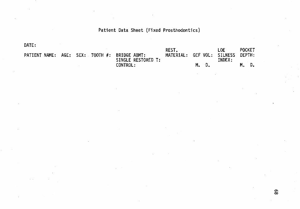
Patient Data Sheet (Fixed Prosthodontics)
DATE:
PATIENT NAME: AGE: SEX: TOOTH #: BRIDGE ABMT: SINGLE RESTORED T: CONTROL:
REST. LOE POCKET MATERIAL: GCF VOL: SILNESS DEPTH:
INDEX: M. D. M, D.
en (X)
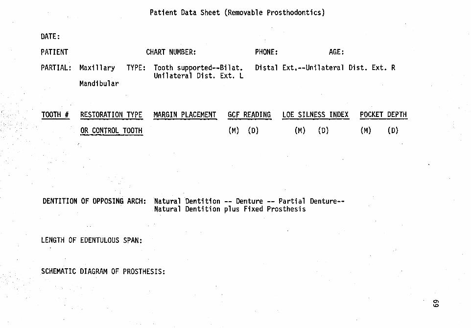
Patient Data Sheet (Removable Prosthodontics)
DATE:
PATIENT CHART NUMBER: PHONE: AGE:
PARTIAL: Maxillary TYPE: Tooth supported~-Bilat. Distal Exto--Unilateral Dist. Ext. R Unilateral Dist. Ext. L
Mandibular
TOOTH # RESTORATION TYPE MARGIN PLACEMENT GCF READING LOE SILNESS INDEX POCKET DEPTH
OR CONTROL TOOTH (M) (D) (M) (D)
DENTITION OF OPPOSING ARCH: Natural Dentition -- Denture -- Partial Denture-Natural Dentition plus Fixed Prosthesis
LENGTH OF EDENTULOUS SPAN:
SCHEMATIC DIAGRAM OF PROSTHESIS:
(M) (D)
0'1 ~
APPROVAL SHEET
The thesis submitted by Pamela Denise Golasz, D.D.S., has been read and approved by the following committee:
William F. Malone, D.D.S., M.S., Ph.D., Director of the Graduate Prosthodontic Program Professor of Fixed Prosthodontics, Loyola
Patrick D. Toto, D.D.S., M.S., Professor and Chairman, Department of General and Oral Biology, Loyola
Boleslaw Mazur, D.D.S., M.S., Professor of Fixed Prosthodontics, Loyola
The final copies have been examined by the director of the thesis and the signature which appears below verifies the fact that any necessary changes have been incorporated and that the thesis is now given final approval by the Committee with reference to content and form.
The thesis is therefore accepted in partial fulfillment of the requirements for the degree of Master of Science in Oral Biology.
Date Director's Signature
70

![Review Article Identification of Gingival Crevicular Fluid ...downloads.hindawi.com/journals/dm/2016/1804727.pdf · precise chair-side analytical test [, ]. ... and oral biomarkers](https://img.pdfslide.net/doc/110x75/5aa6a10e7f8b9a2f048ee63b/review-article-identification-of-gingival-crevicular-fluid-chair-side-analytical.jpg)
![Microbial and metabolomic analysis of gingival crevicular ... · 2]. Periodontitis affects the majority of adults worldwide and may cause various systemic diseases, including dia-betes](https://img.pdfslide.net/doc/110x75/5ffe1bd5f6ee7b7ad975583c/microbial-and-metabolomic-analysis-of-gingival-crevicular-2-periodontitis.jpg)