Embed Size (px)
Citation preview
TitleDetermining circulating endothelial cells using CellSearchsystem during preoperative systemic chemotherapy in breastcancer patients.
Author(s)Ali, Arwa M; Ueno, Takayuki; Tanaka, Sunao; Takada,Masahiro; Ishiguro, Hiroshi; Abdellah, Ashraf Z; Toi,Masakazu
Citation European journal of cancer (Oxford, England : 1990) (2011),47(15): 2265-2272
Issue Date 2011-10
URL http://hdl.handle.net/2433/148015
Right © 2011 Elsevier Ltd.
Type Journal Article
Textversion author
Kyoto University

1
Determining circulating endothelial cells using CellSearch system during
preoperative systemic chemotherapy in breast cancer patients
Arwa M Alia, b
, Takayuki Uenoa, Sunao Tanaka
a, Masahiro Takada
a, Hiroshi Ishiguro
c,
Ashraf Z Abdellahb, and Masakazu Toi
a
aDepartment of Surgery (Breast Surgery), Graduate School of Medicine, Kyoto University,
54 Kawaracho, Shogoin, Sakyo-ku, Kyoto 606-8507, Japan
bMedical Oncology, South Egypt Cancer Institute, Asyut University, Egypt
cOut-patients Oncology Unit, Kyoto University Hospital, 54 Kawaracho, Shogoin, Sakyo-
ku, Kyoto 606-8507, Japan
Corresponding author:
Takayuki Ueno
Department of Breast Surgery, Graduate School of Medicine, Kyoto University
54 Kawaracho, Shogoin, Sakyo-ku, Kyoto 606-8507, Japan
Tel: +81-75-751-3660; Fax: +81-75-751-3616
Sources of support (Grants)
Japan’s Ministry of Health, Labor, and Welfare for a study on construction of an algorithm
for multimodality therapy with biomarkers for primary breast cancer during the formulation
of the decision-making process, led by Masakazu Toi (H18-3JIGAN-IPPAN-007, H19-
3JIGAN-IPPAN-007)The Innovative Techno-Hub for Integrated Medical Bio-imaging
Project of the Special Coordination Fund for Promoting Science and Technology, from the
Ministry of Education, Culture, Sports, Science and Technology, Japan
*Manuscript
2
ABSTRACT
Background: Circulating endothelial cells (CECs) have been studied as a biomarker for
tumor progression and monitoring therapeutic effects. The CellSearch system is a semi-
automated system that allows standardized analysis of CECs. This study assessed the
clinical implications of CECs determined by the CellSearch system in breast cancer patients.
Methods: Seventy-six consecutive breast cancer patients (53 operable and 23 metastatic or
recurrent) were enrolled for the study. Thirty-five patients with operable breast cancer
received preoperative chemotherapy with a regimen based on anthracycline and/or taxane.
CECs are defined as CD146+CD105
+CD45
−DAPI
+ cells in the system. CD34 expression
was examined using the additional channel in the system.
Results: A majority (4539 of 5183 cells, 88%) of CECs from patients with operable breast
cancer were CD34-positive. Triple-negative cancers showed higher baseline CEC and
CD34+CEC counts than the other types (P = 0.0387 and 0.0377, respectively). Low
baseline CEC and CD34+CEC counts, and a low CD34 positive rate were associated with
pathological complete response (pCR) of preoperative chemotherapy in patients with
primary breast cancer (P = 0.046, 0.027 and 0.01, respectively). In multivariate analyses,
the CD34 positive rate was significant for pCR (P = 0.021). During preoperative
chemotherapy, CEC and CD34+CEC counts before each cycle of chemotherapy increased
with taxane-based regimens (P = 0.0018 and 0.0008, respectively) but not with
anthracycline-based regimens.
Conclusions: Baseline CEC, in particular CD34+CEC, counts and the CD34 positive rate
might be useful for the prediction of treatment response of preoperative chemotherapy in
patients with operable breast cancer.

3
Key words: circulating endothelial cells, CellSearch system, breast cancer, chemotherapy,
pathological response
4
INTRODUCTION
Circulating endothelial cells (CECs) and their progenitors, endothelial progenitor
cells (EPCs), are being studied with increasing interest in oncology, particularly in relation
to tumor angiogenesis. Recent studies have demonstrated elevated CEC count in patients
with malignant diseases compared with healthy controls (1-7). Several pioneering studies
have demonstrated that CEC elevations are associated with tumor stage, tumor
characteristics and prognosis (4, 8-10). It has been experimentally demonstrated that
chemotherapy causes a rapid induction of EPCs into the systemic circulation of mice,
irrespective of the presence of tumor (11). EPC mobilization may support tumor cell
survival even during anticancer chemotherapy.
CECs and EPCs are currently determined by several different assay systems
including the flow cytometry and immunomagnetic detection system using endothelial cell
markers including CD31, CD34, and CD146, and progenitor cell markers including
CD133(12). However, the markers and criteria that are used differ among studies (13, 14).
The flow cytometry analysis has some limitations including standardization between
different laboratories and difficulties in fresh blood shipping. Recently, a semi-automated
system for the detection of CECs was developed. The CellSearch system (Veridex LLC,
Raritan, NJ) is mostly automated but enables researchers to detect endothelial cells visually
using the immunofluorescence system. This system allows standardized analyses in
different laboratories and shipment of blood samples in special tubes containing
preservatives.
In this study, we used the CellSearch system to examine baseline CEC count and
CEC alterations during systemic chemotherapy in association with clinicopathological

5
parameters and treatment responses to preoperative chemotherapies.
6
PATIENTS AND METHODS
Patients
We enrolled 76 consecutive patients with histologically confirmed breast cancer
who were treated at Kyoto University Hospital between 2007 and 2009, comprising 53
patients with operable breast cancer and 23 patients with metastatic or recurrent breast
cancer. Other inclusion criteria were age 20–70 years, performance status (ECOG) <3, and
estimated survival time >3 months. Blood samples were drawn before the initiation of any
treatment in the operable breast cancer group and before the initiation of treatment for the
metastatic or recurrent breast cancer in the metastatic or recurrent breast cancer group.
Thirty-five patients with operable breast cancer received preoperative chemotherapy with a
regimen based on anthracycline, taxane or a combination of both. The anthracycline-based
regimen comprises four cycles of FEC (5-FU 500 mg/m2, epirubicin 100 mg/m
2,
cyclophosphamide 500 mg/m2) tri-weekly or four cycles of EC (epirubicin 100 mg/m
2,
cyclophosphamide 500 mg/m2) tri-weekly. The taxane-based regimen comprised four
cycles of docetaxel alone (75 mg/m2) tri-weekly or four cycles of TC (docetaxel 75 mg/m
2,
cyclophosphamide 600 mg/m2) tri-weekly. The combination regimen comprises four cycles
of an anthracycline-based regimen tri-weekly and four cycles of a taxane-based regimen tri-
weekly. Trastuzumab was not administered preoperatively but after surgery to patients with
HER2-positive tumors. We analyzed alterations in CEC count during treatment in 17
patients who received preoperative chemotherapy by collecting blood samples before each
cycle of chemotherapy and 24 hours after administration of chemotherapy. For combination
regimen, blood samples were drawn during four cycles of the first regimen. Clinical
response to chemotherapy was assessed according to the Response Evaluation Criteria in
7
Solid Tumors (RECIST). The study protocol was approved by the Ethics Committee of
Kyoto University, and written informed consent was obtained from all patients.
Evaluation of CECs by the CellSearch system
Blood samples were drawn into CellSave tubes (Veridex, LLC, NJ) containing a cell
preservative. Samples were maintained at room temperature and processed within 24 hours
of collection. All evaluations were performed without prior knowledge of the clinical status
of the patient. The CellSearch system, used for endothelial cell detection, consists of
CellSave tubes, CellTracks AutoPrep, a fully automated sample preparation system, the
Endothelial Cell Reagent Kit, and the CellSpotter Analyzer II, a semi-automated
fluorescence microscope.
In brief, 4 ml blood was mixed with 10 ml buffer, centrifuged at 800 ×g for 10 min,
and placed in the sample preparation system. The instrument aspirated the plasma/buffer
layer, and antiCD146 ferrofluids were added. After incubation and subsequent magnetic
separation, unbound cells and the remaining plasma were aspirated. The enriched cells were
fluorescently labeled with the nuclear stain 4,6-diamidino-2-phenylindole (DAPI). Staining
reagents (<0.0006% mouse monoclonal antibodies specific to CD105 conjugated to
phycoerythrin; <0.0013% mouse antiCD45 monoclonal antibodies conjugated to
allophycocyanin in phosphate-buffered saline containing 0.5% BSA and 0.1% sodium
azide) together with antiCD34 antibody conjugated to FITC (clone AC136, Miltenyi,
Biotech GmbH, Germany) were added in conjunction with a permeabilization buffer to
label the cells fluorescently. After incubation, magnetic separation was repeated to remove
the excess staining reagent. After the final processing step, the cells were re-suspended in
8
300 µL of buffer and transferred to a chamber placed between two magnets that orient the
immunomagnetically labeled cells in a monolayer for analyses. The cells were then
examined with a four-color semi-automated fluorescent microscope, the CellSpotter
Analyzer II. A gray-scale charge-coupled device camera was used to scan the entire
chamber surface, and each captured frame was then evaluated for potential CEC candidates
by image analysis software. CECs were defined as CD146+CD105
+CD45
−DAPI
+ cells.
CECs were stained with an additional antibody against CD34 and its expression was
evaluated using an extra channel in the system.
Pathological analyses
Tumor biopsy specimens before preoperative chemotherapy were examined
pathologically for tumor grade according to the Scarff–Bloom–Richardson grading system.
Tumor specimens were also examined for estrogen receptor (ER), progesterone receptor
(PgR), and human epidermal growth factor receptor type 2 (HER2). The antibodies for ER,
PgR, and HER2 were ER(SP1), PGR(1E2), and HER2(4B5), respectively (all from Roche
Diagnostics, Tokyo, Japan). ER and PgR statuses were defined as positive for tumors
having 10% or more positive tumor cells. HER2 positivity was determined by a strong
expression (3+) of HER2 by the HercepTest or by an HER2:CEP17 ratio >2.2 by
fluorescence in situ hybridization (FISH). Triple negative was defined as ER negative, PgR
negative, and HER2 negative tumors.
The pathological response was assessed after surgery following preoperative
chemotherapy. A pathological complete response (pCR) was defined as no residual invasive
tumor cells in mammary glands and lymph nodes.
9
The MIB1/Ki67 labeling index was calculated by counting positively stained tumor
cells per 1000 tumor cells in the hot spots. Tumors having an MIB1/Ki67 index >20% were
categorized as rapidly proliferative (positive), and those having an index <20% were
defined as slowly proliferative (negative).
Statistical analyses
Correlation analyses were performed to assess the associations between baseline
CEC counts and tumor size, nodal status, grade, stage, ER, PgR and HER2 statuses, tumor
phenotype, and tumor response. Correlation analysis was performed using the Mann–
Whitney test for two independent samples and the Kruskal–Wallis test for more than two
independent samples. Logistic regression analysis was used to identify parameters
associated with pathological response. Changes in CEC and CEP numbers were analyzed
using repeated measures ANOVA. Statistical analyses were performed using JMP (ver.
8.0.1; SAS Institute Japan, Tokyo, Japan). P values of <0.05 were considered statistically
significant.
10
RESULTS
Characteristics of CECs detected by the CellSearch system
The expression of CD34, which is a commonly used marker for endothelial cells, was
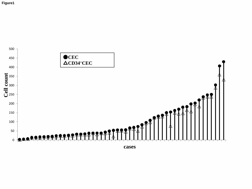
examined in CECs detected by the CellSearch system. As shown in Figure 1, 88% (4539
of 5183 cells) of CECs from patients with operable breast cancer before treatment were
CD34 positive.
Patient characteristics and correlations with clinicopathological parameters
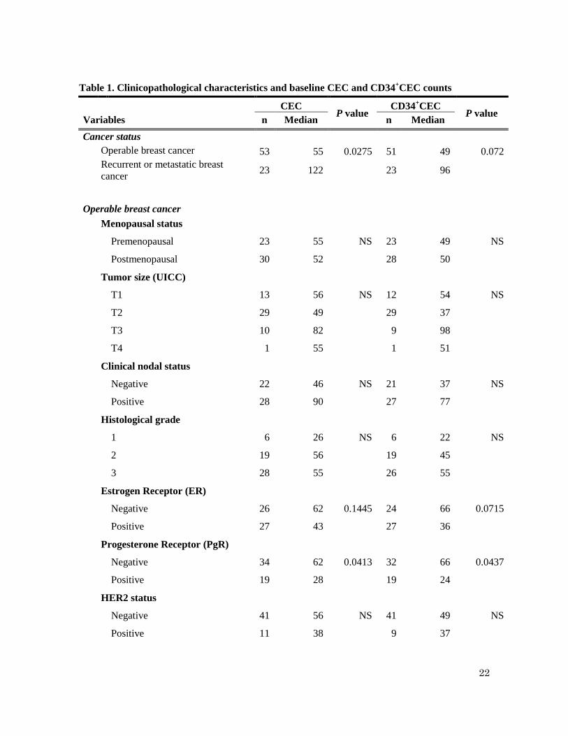
Table 1 shows the characteristics of the patients and their baseline CEC and
CD34+CEC counts in relation to clinicopathological parameters. CD34 expression was not
measured in two patients with operable breast cancer. CEC count was higher in metastatic
or recurrent breast cancer patients than in patients with operable breast cancer (P = 0.0275).
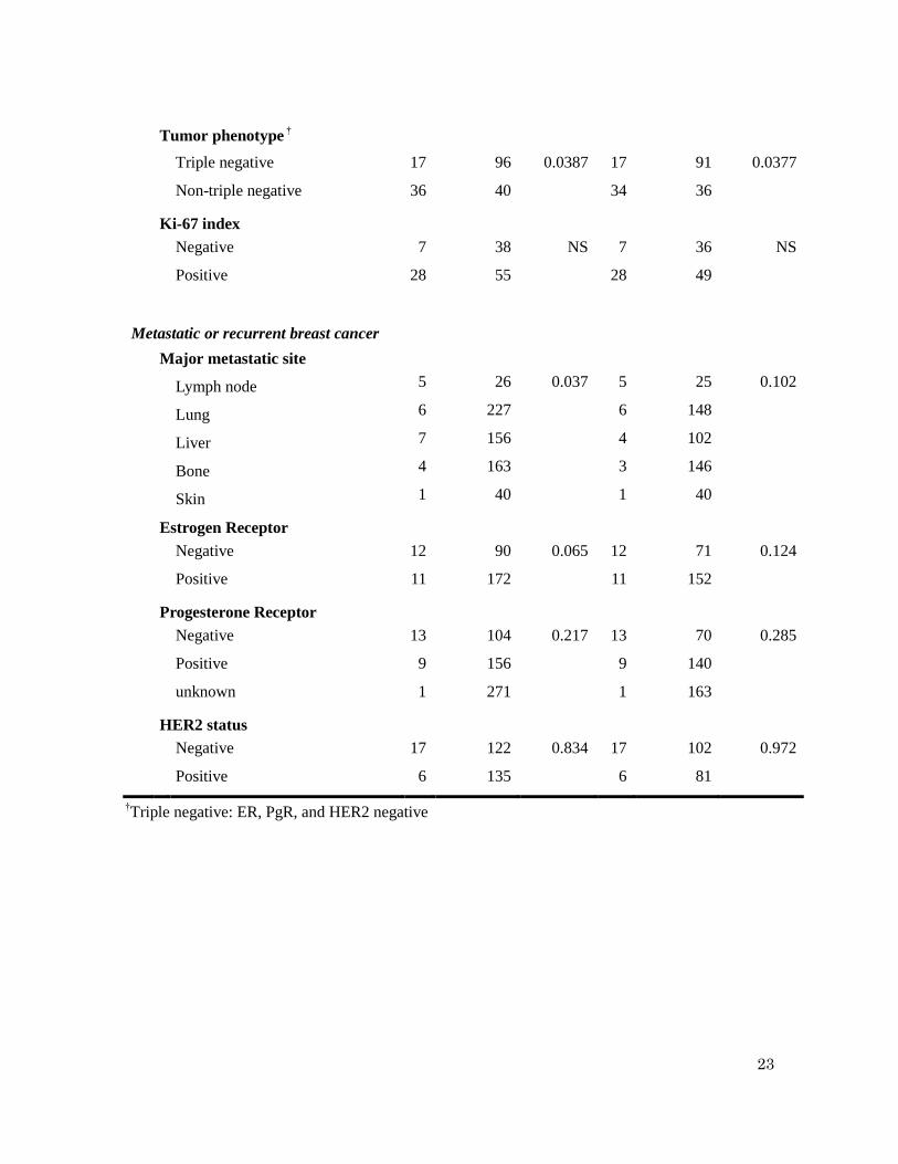
Among patients with operable breast cancer, those with triple-negative cancers had
significantly higher CEC and CD34+CEC counts than those with other types of cancer (P =
0.0387 and 0.0377, respectively). Similarly, patients with PR-negative cancers showed
higher CEC and CD34+CEC counts than those with PR-positive cancers (P = 0.0413 and
0.0437, respectively). In patients with metastatic or recurrent breast cancer, patients with
lung, liver or bone metastasis showed higher CEC counts than those with lymph node or
skin metastasis (P = 0.037).
CEC and CD34+ CEC counts and responses to chemotherapy
In 35 patients with operable breast cancer, CEC and CD34+CEC counts were
examined according to pathological and clinical responses to preoperative chemotherapy.
11
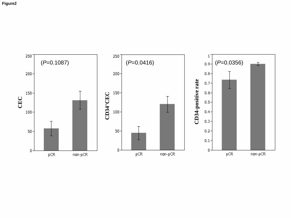
The pCR group showed lower numbers of baseline CD34+CEC counts than the non-pCR
group (P = 0.0416) (Figure 2). In addition, the pCR group showed a lower CD34-positive
rate (CD34+CEC count/total CEC count) than the non-pCR group (P = 0.0356) (Figure 2).
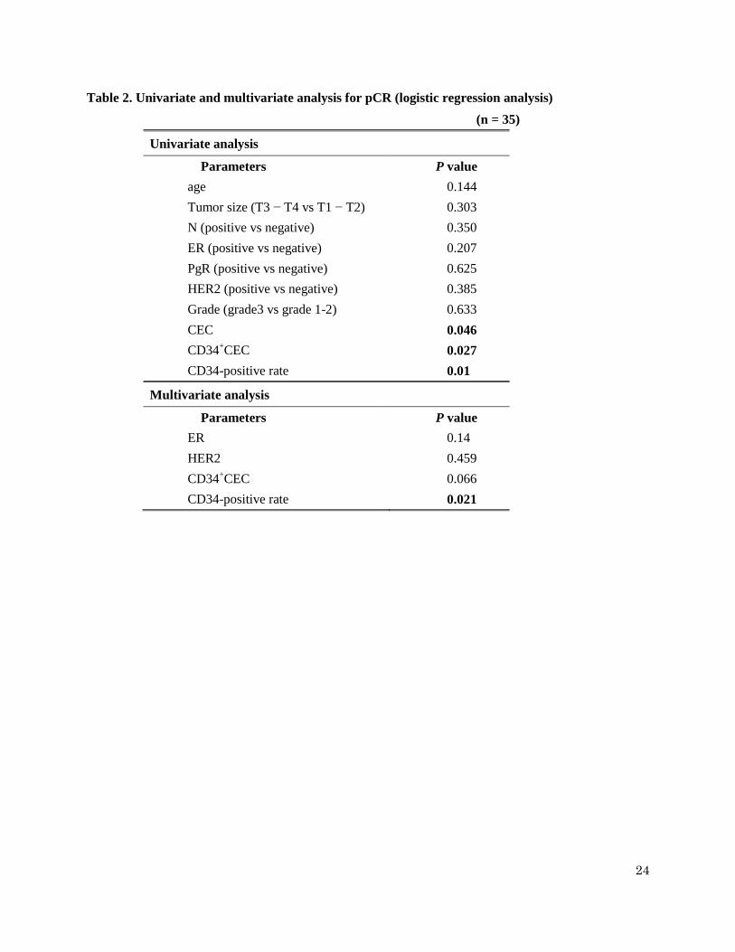
In the logistic regression analysis, CEC, CD34+CEC, and CD34-positive rates were
significantly associated with pCR in univariate analyses (P = 0.046, 0.027, and 0.01,
respectively) (Table 2). In multivariate analyses, the CD34-positive rate remained
significant for pCR (P = 0.021) (Table 2). CEC counts, CD34+CEC counts, and CD34-
positive rate did not show any association with clinical responses (data not shown).
Changes in CEC and CD34+CEC counts during systemic chemotherapy
Alterations in CEC and CD34+CEC counts during the first four cycles of
chemotherapy were analyzed in 17 patients with operable breast cancer who received
preoperative chemotherapy as either a taxane-based or an anthracycline-based regimen.
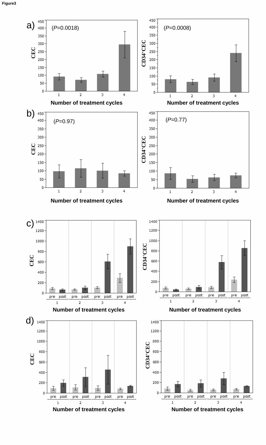
Patients who received taxane-based regimens showed increasing numbers of pretreatment
CECs and CD34+CECs during the treatment cycles (P = 0.0018 and 0.0008, respectively)
(Figure 3a) whereas those who received anthracycline-based regimens did not show such
increases (P = 0.97 and 0.77, respectively) (Figure 3b). This indicates that changes in CEC
and CD34+CEC counts depend on the type of chemotherapy. CEC and CD34
+CEC counts
showed a rapid increase 24 hours after each cycle of chemotherapy. Unlike anthracycline-
based regimens (Figure 3d), taxane-based regimens showed an incremental pattern in CEC
count after repeated cycles of chemotherapy (Figure 3c).
12
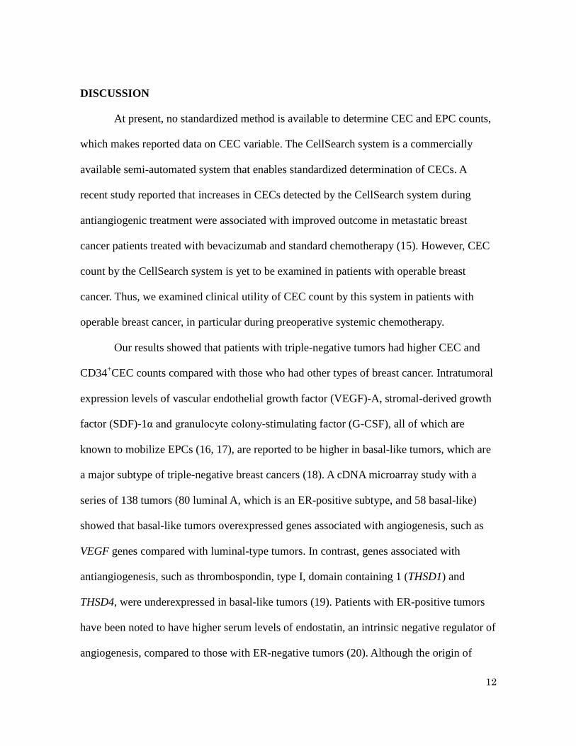
DISCUSSION
At present, no standardized method is available to determine CEC and EPC counts,
which makes reported data on CEC variable. The CellSearch system is a commercially
available semi-automated system that enables standardized determination of CECs. A
recent study reported that increases in CECs detected by the CellSearch system during
antiangiogenic treatment were associated with improved outcome in metastatic breast
cancer patients treated with bevacizumab and standard chemotherapy (15). However, CEC
count by the CellSearch system is yet to be examined in patients with operable breast
cancer. Thus, we examined clinical utility of CEC count by this system in patients with
operable breast cancer, in particular during preoperative systemic chemotherapy.
Our results showed that patients with triple-negative tumors had higher CEC and
CD34+CEC counts compared with those who had other types of breast cancer. Intratumoral
expression levels of vascular endothelial growth factor (VEGF)-A, stromal-derived growth
factor (SDF)-1α and granulocyte colony-stimulating factor (G-CSF), all of which are
known to mobilize EPCs (16, 17), are reported to be higher in basal-like tumors, which are
a major subtype of triple-negative breast cancers (18). A cDNA microarray study with a
series of 138 tumors (80 luminal A, which is an ER-positive subtype, and 58 basal-like)
showed that basal-like tumors overexpressed genes associated with angiogenesis, such as
VEGF genes compared with luminal-type tumors. In contrast, genes associated with
antiangiogenesis, such as thrombospondin, type I, domain containing 1 (THSD1) and
THSD4, were underexpressed in basal-like tumors (19). Patients with ER-positive tumors
have been noted to have higher serum levels of endostatin, an intrinsic negative regulator of
angiogenesis, compared to those with ER-negative tumors (20). Although the origin of
13
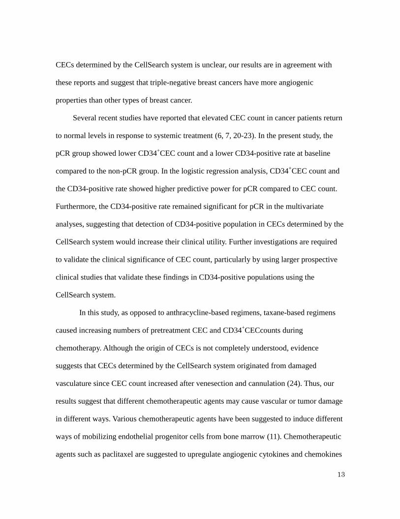
CECs determined by the CellSearch system is unclear, our results are in agreement with
these reports and suggest that triple-negative breast cancers have more angiogenic
properties than other types of breast cancer.
Several recent studies have reported that elevated CEC count in cancer patients return
to normal levels in response to systemic treatment (6, 7, 20-23). In the present study, the
pCR group showed lower CD34+CEC count and a lower CD34-positive rate at baseline
compared to the non-pCR group. In the logistic regression analysis, CD34+CEC count and
the CD34-positive rate showed higher predictive power for pCR compared to CEC count.
Furthermore, the CD34-positive rate remained significant for pCR in the multivariate
analyses, suggesting that detection of CD34-positive population in CECs determined by the
CellSearch system would increase their clinical utility. Further investigations are required
to validate the clinical significance of CEC count, particularly by using larger prospective
clinical studies that validate these findings in CD34-positive populations using the
CellSearch system.
In this study, as opposed to anthracycline-based regimens, taxane-based regimens
caused increasing numbers of pretreatment CEC and CD34+CECcounts during
chemotherapy. Although the origin of CECs is not completely understood, evidence
suggests that CECs determined by the CellSearch system originated from damaged
vasculature since CEC count increased after venesection and cannulation (24). Thus, our
results suggest that different chemotherapeutic agents may cause vascular or tumor damage
in different ways. Various chemotherapeutic agents have been suggested to induce different
ways of mobilizing endothelial progenitor cells from bone marrow (11). Chemotherapeutic
agents such as paclitaxel are suggested to upregulate angiogenic cytokines and chemokines
14
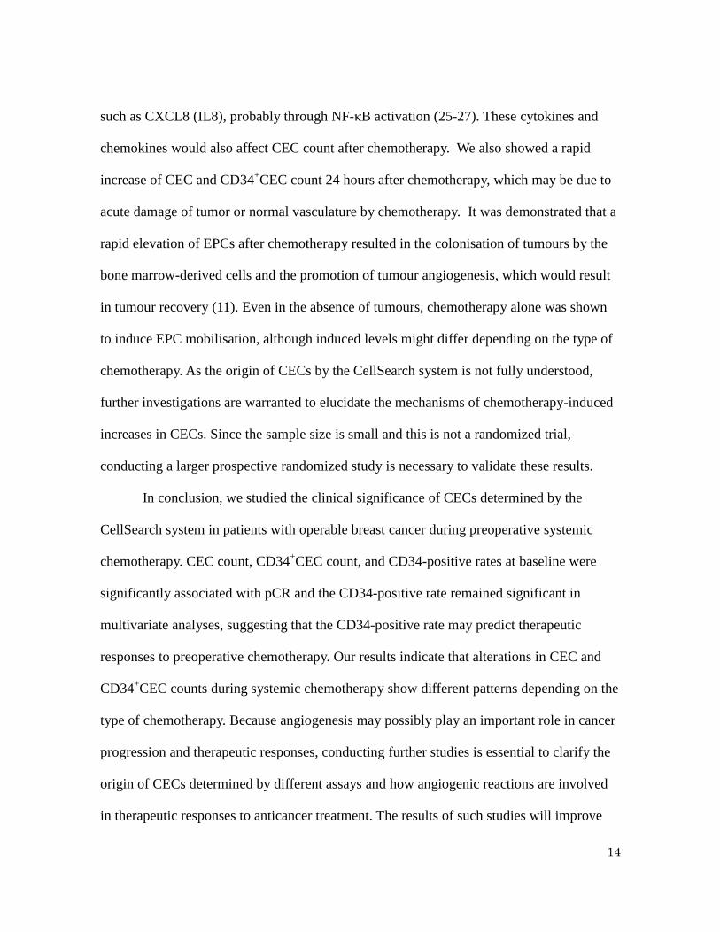
such as CXCL8 (IL8), probably through NF-κB activation (25-27). These cytokines and
chemokines would also affect CEC count after chemotherapy. We also showed a rapid
increase of CEC and CD34+CEC count 24 hours after chemotherapy, which may be due to
acute damage of tumor or normal vasculature by chemotherapy. It was demonstrated that a
rapid elevation of EPCs after chemotherapy resulted in the colonisation of tumours by the
bone marrow-derived cells and the promotion of tumour angiogenesis, which would result
in tumour recovery (11). Even in the absence of tumours, chemotherapy alone was shown
to induce EPC mobilisation, although induced levels might differ depending on the type of
chemotherapy. As the origin of CECs by the CellSearch system is not fully understood,
further investigations are warranted to elucidate the mechanisms of chemotherapy-induced
increases in CECs. Since the sample size is small and this is not a randomized trial,
conducting a larger prospective randomized study is necessary to validate these results.
In conclusion, we studied the clinical significance of CECs determined by the
CellSearch system in patients with operable breast cancer during preoperative systemic
chemotherapy. CEC count, CD34+CEC count, and CD34-positive rates at baseline were
significantly associated with pCR and the CD34-positive rate remained significant in
multivariate analyses, suggesting that the CD34-positive rate may predict therapeutic
responses to preoperative chemotherapy. Our results indicate that alterations in CEC and
CD34+CEC counts during systemic chemotherapy show different patterns depending on the
type of chemotherapy. Because angiogenesis may possibly play an important role in cancer
progression and therapeutic responses, conducting further studies is essential to clarify the
origin of CECs determined by different assays and how angiogenic reactions are involved
in therapeutic responses to anticancer treatment. The results of such studies will improve

15
the understanding of how antiangiogenic treatment should be combined with conventional
chemotherapies for improved treatment efficacy and ultimately lead to the achievement of
personalized treatment.

16
Acknowledgments
This study was funded by a research grant from Japan’s Ministry of Health, Labor,
and Welfare for a study on constructing an algorithm for multimodality therapy with
biomarkers for primary breast cancer during the formulation of the decision-making
process, led by Masakazu Toi (H18-3JIGAN-IPPAN-007, H19-3JIGAN-IPPAN-007).
This work was supported in part by the Innovative Techno-Hub for Integrated
Medical Bio-imaging Project of the Special Coordination Fund for Promoting Science and
Technology, from the Ministry of Education, Culture, Sports, Science and Technology,
Japan.

17
Conflict of interest statement
None declared.
18
References
1. Brunner M, Thurnher D, Heiduschka G, Grasl M, Brostjan C, Erovic BM. Elevated
levels of circulating endothelial progenitor cells in head and neck cancer patients. J Surg Oncol
2008;98:545-50.
2. Go RS, Jobe DA, Asp KE, Callister SM, Mathiason MA, Meyer LA, et al. Circulating
endothelial cells in patients with chronic lymphocytic leukemia. Ann Hematol 2008;87:369-73.
3. Greenfield JP, Jin DK, Young LM, Christos PJ, Abrey L, Rafii S, et al. Surrogate
markers predict angiogenic potential and survival in patients with glioblastoma multiforme.
Neurosurgery 2009;64:819-26.
4. Ho JW, Pang RW, Lau C, Sun CK, Yu WC, Fan ST, et al. Significance of circulating
endothelial progenitor cells in hepatocellular carcinoma. Hepatology 2006;44:836-43.
5. Kawaishi M, Fujiwara Y, Fukui T, Kato T, Yamada K, Ohe Y, et al. Circulating
endothelial cells in non-small cell lung cancer patients treated with carboplatin and paclitaxel.
J Thorac Oncol 2009;4:208-13.
6. Mancuso P, Burlini A, Pruneri G, Goldhirsch A, Martinelli G, Bertolini F. Resting and
activated endothelial cells are increased in the peripheral blood of cancer patients. Blood
2001;97:3658-61.
7. Mancuso P, Colleoni M, Calleri A, Orlando L, Maisonneuve P, Pruneri G, et al.
Circulating endothelial-cell kinetics and viability predict survival in breast cancer patients
receiving metronomic chemotherapy. Blood 2006;108:452-9.
8. Beerepoot LV, Mehra N, Vermaat JS, Zonnenberg BA, Gebbink MF, Voest EE. Increased
levels of viable circulating endothelial cells are an indicator of progressive disease in cancer
patients. Ann Oncol 2004;15:139-45.
9. DePrimo SE, Bello C. Surrogate biomarkers in evaluating response to anti-angiogenic
agents: focus on sunitinib. Ann Oncol 2007;18 Suppl 10:x11-19.
10. Dome B, Timar J, Dobos J, Meszaros L, Raso E, Paku S, et al. Identification and
clinical significance of circulating endothelial progenitor cells in human non-small cell lung
cancer. Cancer Res 2006;66:7341-7.
11. Shaked Y, Henke E, Roodhart JM, Mancuso P, Langenberg MH, Colleoni M, et al.
Rapid chemotherapy-induced acute endothelial progenitor cell mobilization: implications for
antiangiogenic drugs as chemosensitizing agents. Cancer Cell 2008;14:263-73.
12. Strijbos MH, Gratama JW, Kraan J, Lamers CH, den Bakker MA, Sleijfer S.
Circulating endothelial cells in oncology: pitfalls and promises. Br J Cancer 2008;98:1731-5.
13. Bertolini F, Shaked Y, Mancuso P, Kerbel RS. The multifaceted circulating endothelial
19
cell in cancer: towards marker and target identification. Nat Rev Cancer 2006;6:835-45.
14. Yoder MC, Ingram DA. Endothelial progenitor cell: ongoing controversy for defining
these cells and their role in neoangiogenesis in the murine system. Curr Opin Hematol
2009;16:269-73.
15. Bidard FC, Mathiot C, Degeorges A, Etienne-Grimaldi MC, Delva R, Pivot X, et al.
Clinical value of circulating endothelial cells and circulating tumor cells in metastatic breast
cancer patients treated first line with bevacizumab and chemotherapy. Ann Oncol
2010;21:1765-71.
16. Asahara T, Takahashi T, Masuda H, Kalka C, Chen D, Iwaguro H, et al. VEGF
contributes to postnatal neovascularization by mobilizing bone marrow-derived endothelial
progenitor cells. EMBO J 1999;18:3964-72.
17. Jin DK, Shido K, Kopp HG, Petit I, Shmelkov SV, Young LM, et al. Cytokine-mediated
deployment of SDF-1 induces revascularization through recruitment of CXCR4+
hemangiocytes. Nat Med 2006;12:557-67.
18. Van den Eynden GG, Smid M, Van Laere SJ, Colpaert CG, Van der Auwera I, Bich TX,
et al. Gene expression profiles associated with the presence of a fibrotic focus and the growth
pattern in lymph node-negative breast cancer. Clin Cancer Res 2008;14:2944-52.
19. Bertucci F, Finetti P, Cervera N, Charafe-Jauffret E, Buttarelli M, Jacquemier J, et al.
How different are luminal A and basal breast cancers? Int J Cancer 2009;124:1338-48.
20. Furstenberger G, von Moos R, Lucas R, Thurlimann B, Senn HJ, Hamacher J, et al.
Circulating endothelial cells and angiogenic serum factors during neoadjuvant chemotherapy of
primary breast cancer. Br J Cancer 2006;94:524-31.
21. Wierzbowska A, Robak T, Krawczynska A, Pluta A, Wrzesien-Kus A, Cebula B, et al.
Kinetics and apoptotic profile of circulating endothelial cells as prognostic factors for induction
treatment failure in newly diagnosed acute myeloid leukemia patients. Ann Hematol
2008;87:97-106.
22. Norden-Zfoni A, Desai J, Manola J, Beaudry P, Force J, Maki R, et al. Blood-based
biomarkers of SU11248 activity and clinical outcome in patients with metastatic imatinib-
resistant gastrointestinal stromal tumor. Clin Cancer Res 2007;13:2643-50.
23. Zhang H, Vakil V, Braunstein M, Smith EL, Maroney J, Chen L, et al. Circulating
endothelial progenitor cells in multiple myeloma: implications and significance. Blood
2005;105:3286-94.
24. Strijbos MH, Verhoef C, Gratama JW, Sleijfer S. On the origin of (CD105+) circulating
endothelial cells. Thromb Haemost 2009;102:347-51.

20
25. Camp ER, Li J, Minnich DJ, Brank A, Moldawer LL, MacKay SL, et al. Inducible
nuclear factor-kappaB activation contributes to chemotherapy resistance in gastric cancer. J
Am Coll Surg 2004;199:249-58.
26. Nakanishi C, Toi M. Nuclear factor-kappaB inhibitors as sensitizers to anticancer
drugs. Nat Rev Cancer 2005;5:297-309.
27. Uslu R, Sanli UA, Dikmen Y, Karabulut B, Ozsaran A, Sezgin C, et al. Predictive value
of serum interleukin-8 levels in ovarian cancer patients treated with paclitaxel-containing
regimens. Int J Gynecol Cancer 2005;15:240-5.
21
Figure legends
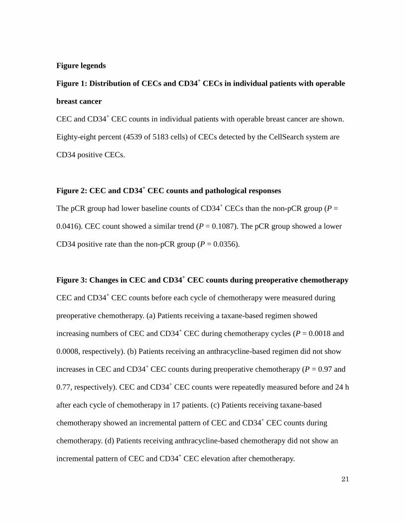
Figure 1: Distribution of CECs and CD34+ CECs in individual patients with operable
breast cancer
CEC and CD34+ CEC counts in individual patients with operable breast cancer are shown.
Eighty-eight percent (4539 of 5183 cells) of CECs detected by the CellSearch system are
CD34 positive CECs.
Figure 2: CEC and CD34+ CEC counts and pathological responses
The pCR group had lower baseline counts of CD34+ CECs than the non-pCR group (P =
0.0416). CEC count showed a similar trend (P = 0.1087). The pCR group showed a lower
CD34 positive rate than the non-pCR group (P = 0.0356).
Figure 3: Changes in CEC and CD34+ CEC counts during preoperative chemotherapy
CEC and CD34+ CEC counts before each cycle of chemotherapy were measured during
preoperative chemotherapy. (a) Patients receiving a taxane-based regimen showed
increasing numbers of CEC and CD34+ CEC during chemotherapy cycles (P = 0.0018 and
0.0008, respectively). (b) Patients receiving an anthracycline-based regimen did not show
increases in CEC and CD34+ CEC counts during preoperative chemotherapy (P = 0.97 and
0.77, respectively). CEC and CD34+ CEC counts were repeatedly measured before and 24 h
after each cycle of chemotherapy in 17 patients. (c) Patients receiving taxane-based
chemotherapy showed an incremental pattern of CEC and CD34+ CEC counts during
chemotherapy. (d) Patients receiving anthracycline-based chemotherapy did not show an
incremental pattern of CEC and CD34+ CEC elevation after chemotherapy.
22
Table 1. Clinicopathological characteristics and baseline CEC and CD34+CEC counts
CEC P value
CD34+CEC
P value Variables n Median n Median
Cancer status
Operable breast cancer 53 55 0.0275 51 49 0.072
Recurrent or metastatic breast
cancer 23 122
23 96
Operable breast cancer
Menopausal status
Premenopausal 23 55 NS 23 49 NS
Postmenopausal 30 52 28 50
Tumor size (UICC)
T1 13 56 NS 12 54 NS
T2 29 49 29 37
T3 10 82 9 98
T4 1 55 1 51
Clinical nodal status
Negative 22 46 NS 21 37 NS
Positive 28 90 27 77
Histological grade
1 6 26 NS 6 22 NS
2 19 56 19 45
3 28 55 26 55
Estrogen Receptor (ER)
Negative 26 62 0.1445 24 66 0.0715
Positive 27 43 27 36
Progesterone Receptor (PgR)
Negative 34 62 0.0413 32 66 0.0437
Positive 19 28 19 24
HER2 status
Negative 41 56 NS 41 49 NS
Positive 11 38 9 37
23
Tumor phenotype†
Triple negative 17 96 0.0387 17 91 0.0377
Non-triple negative 36 40 34 36
Ki-67 index
Negative 7 38 NS 7 36 NS
Positive 28 55 28 49
Metastatic or recurrent breast cancer
Major metastatic site
Lymph node 5 26 0.037 5 25 0.102
Lung 6 227 6 148
Liver 7 156 4 102
Bone 4 163 3 146
Skin 1 40 1 40
Estrogen Receptor
Negative 12 90 0.065 12 71 0.124
Positive 11 172 11 152
Progesterone Receptor
Negative 13 104 0.217 13 70 0.285
Positive 9 156 9 140
unknown 1 271 1 163
HER2 status
Negative 17 122 0.834 17 102 0.972
Positive 6 135 6 81
†Triple negative: ER, PgR, and HER2 negative
24
Table 2. Univariate and multivariate analysis for pCR (logistic regression analysis)
(n = 35)
Univariate analysis
Parameters P value
age 0.144
Tumor size (T3 − T4 vs T1 − T2) 0.303
N (positive vs negative) 0.350
ER (positive vs negative) 0.207
PgR (positive vs negative) 0.625
HER2 (positive vs negative) 0.385
Grade (grade3 vs grade 1-2) 0.633
CEC 0.046
CD34+CEC 0.027
CD34-positive rate 0.01
Multivariate analysis
Parameters P value
ER 0.14
HER2 0.459
CD34+CEC 0.066
CD34-positive rate 0.021
Cel
l co
un
t
cases
●CEC
△CD34+CEC
0
50
100
150
200
250
300
350
400
450
500
CEC
CEP
Figure1
(P=0.1087) (P=0.0416) (P=0.0356)
CD
34
+C
EC
CE
C
CD
34
-po
siti
ve
rate
Figure2
a)
b)
(P=0.0018)
(P=0.97) (P=0.77)
(P=0.0008)
CD
34
+C
EC
CE
C
CD
34
+C
EC
CE
C
c)
d)
Number of treatment cycles
Number of treatment cyclesNumber of treatment cycles
Number of treatment cycles
CD
34
+C
EC
CE
C
CD
34
+C
EC
CE
C
Number of treatment cyclesNumber of treatment cycles
Number of treatment cyclesNumber of treatment cycles
Figure3

Conflict of interest statement
None declared
*Conflict of Interest statement