Embed Size (px)
Citation preview

Determining the Role of Proteins in the Molecular Properties of
Equine Synovial Fluid
Marsha Lampi
Advisor: Dr. Skip RochefortOregon State University
School of Chemical, Biological and Environmental EngineeringSummer 2009

Synovial Fluid
Found in diarthrotic, freely moveable joints.
Responsible for nutrient distribution, lubrication, and shock absorption.
Used for diagnosis of joint diseases.
Synovial Joint Cavity
Articular Cartilage
Articulating Bone
Synovial Membrane
http://edugen.wiley.com/edugen/student/mainfr.uni

Hyaluronic Acid (HA)
Largest molecular component of synovial fluid. Molecular weight ranges from 0.2 – 10 million g/mol.
Some joint diseases have been linked to the breakdown of HA.
HA injections and oral supplements are currently available and being studied as treatments for joint diseases.
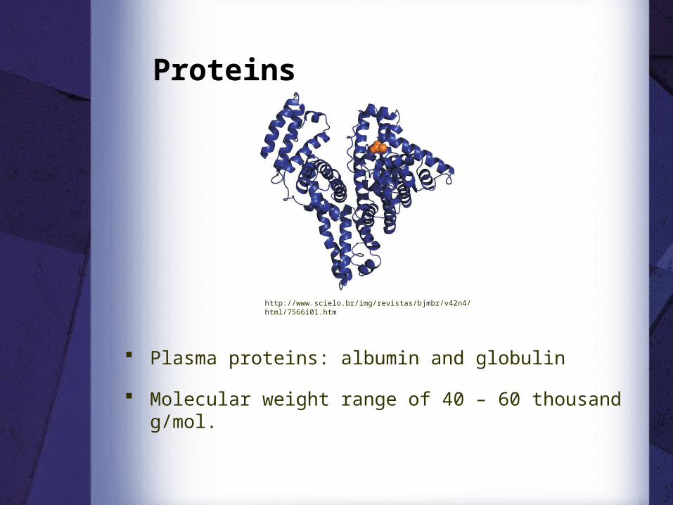
Plasma proteins: albumin and globulin
Molecular weight range of 40 – 60 thousand g/mol.
Proteins
http://www.scielo.br/img/revistas/bjmbr/v42n4/html/7566i01.htm

Equine Synovial Fluid
Stifle (knee)
Hock (ankle)
http://www.ucmp.berkeley.edu/education/lessons/xenosmilus/skeletal_res_manual2.html

Objective
Develop a protocol to digest the protein in synovial fluid while leaving the hyaluronic acid unchanged.
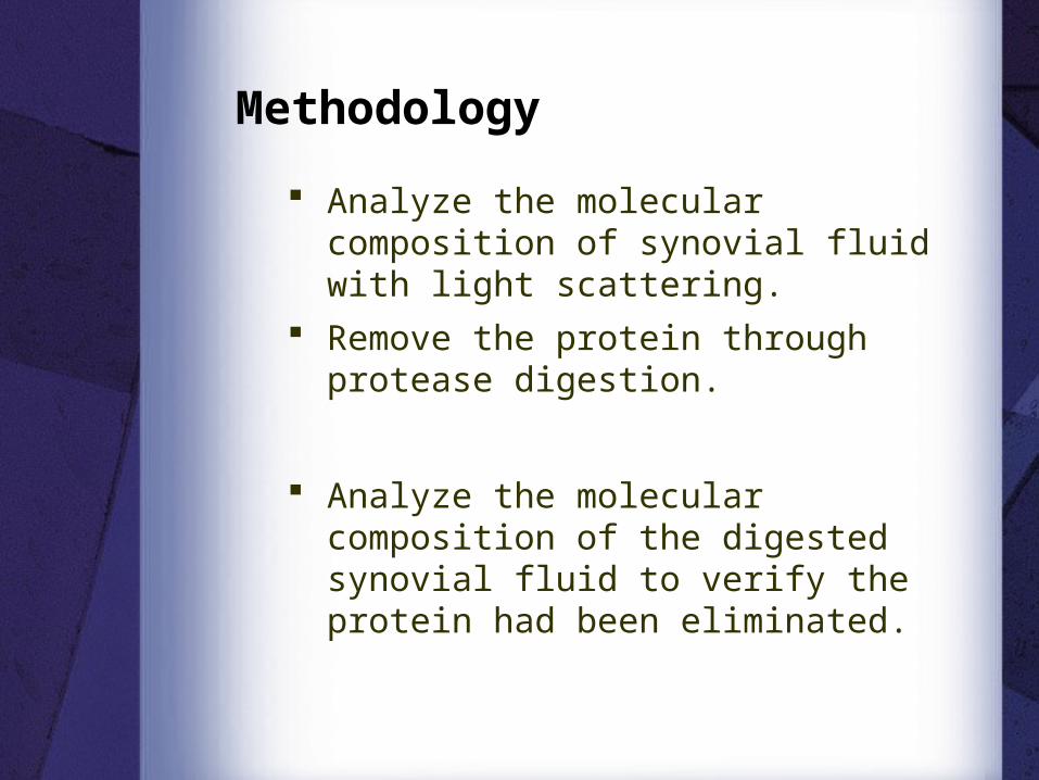
Methodology
Remove the protein through protease digestion.
Analyze the molecular composition of synovial fluid with light scattering.
Analyze the molecular composition of the digested synovial fluid to verify the protein had been eliminated.

Analysis of Molecular
Composition

Gel Permeation Chromatography(GPC)
Separates particles based on size.
Small particles get stuck in the packed interior and move through the column slower.
http://www.waters.com/waters/partDetail.htm?locale=en_US&partNumber=WAT045915
http://www.ap-lab.com/images/LS_setup.gif

Multi-Angle Laser Light Scatter(MALLS)
Light intensity is measured as a function of the deflection angle and concentration.
Allows for molecular weight determination.
Detector, I()Detector, Io
Polymer SolutionLight Source

Refractive Index (RI) Detector Determines concentration based on
the bending of light in comparison to a reference cell.
http://www.polygen.com.pl/viscotek/refractive_index_detector.html

Experimental Set-Up
http://www.ap-lab.com/images/LS_setup.gif

HA Peak
Protein Peak
Before digestion, both the HA and protein peaks are detected.
GPC-MALLS Graph
-- Light Scatter-- Refractive Index

Protein Digestion
Protease Bacillus polymyxa, 1.2 U/mg
Preliminary digestion:− Dilute synovial fluid sample 1:3 − 2 units of protease per mL synovial fluid− 30 minute incubation in water bath− Filtration and phenol-chloroform
extraction to remove proteins
Kvam, Catrine, Granese Daniela, Flaibani, Antonella, Zanetti, Flavio, and Paoletti, Sergio (1993). “Purification and Characterization of Hyaluronan form Synovial Fluid”. Analytical Biochemistry 1993, 211, 44-49.

Undigested Synovial Fluid
Digested Synovial Fluid
Protein Molecular Weight:6-8 x 10⁴ g/mol
Protein Molecular Weight:3-6 x 10³ g/mol
-- Light Scatter-- Refractive Index
-- Light Scatter-- Refractive Index

Protease Concentration
Increase to 4 units/mL Synovial Fluid
Same elution time = no gain in digestion
Low HA concentration
Protein Molecular Weight:3-6 x 10³ g/mol
-- Light Scatter-- Refractive Index
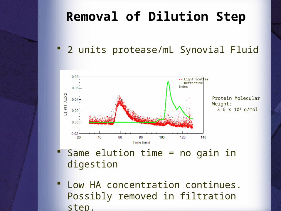
Removal of Dilution Step
2 units protease/mL Synovial Fluid
Same elution time = no gain in digestion
Low HA concentration continues. Possibly removed in filtration step.
Protein Molecular Weight:3-6 x 10³ g/mol
-- Light Scatter-- Refractive Index

Removal of Filtration Step
2 units protease/mL Synovial Fluid
Same elution time = no gain in digestion
Low HA concentration continues. Possibly removed in phenol-chloroform extraction.
Protein Molecular Weight:3-6 x 10³ g/mol
-- Light Scatter-- Refractive Index

Addition of Protease Only
2 units protease/mL Synovial Fluid
Earlier elution time = Less digestion
There is no change between the digested and undigested samples when treated with the protease.
Protein Molecular Weight:5-7 x 10⁴ g/mol
-- Light Scatter-- Refractive Index

Conclusion
The protease Bacillus polymyxa currently being used is not effective in digesting the equine synovial fluid proteins. The protein removal may only be due to phenol-chloroform extraction.
Protein digestion always resulted in a reduction of HA. This suggests that there may be an interaction between the proteins and HA in synovial fluid.

Test a new protease, either Protease K or Pronase E to digest the protein.
Perform rheological analysis on synovial before and after protein digestion to analyze the effects of the proteins on lubrication and shock absorption.
This would allow us to further determine the interaction between HA and the proteins in synovial fluid.
Future Work

Acknowledgments Howard Hughes Medical Institute (HHMI)
URISC
Oregon State University
Dr. Skip Rochefort
Dr. Kevin Ahern
Dr. Jill Parker, OSU School of Veterinary Medicine
Shannon Cahill-Weisser, Project Assistant


Viscosity Indication of lubrication
capabilities.
Viscosity = Shear Stress Shear Rate
Sheer Rate(Rotation Speed)
Sheer Stress(Torque Measurement)

Elasticity
A measure of shock absorption capabilities
Oscillating cone measures stored energy when fluid compressed.

![Un’ora da astronomi A caccia di lampi gamma - INAF€¦ · Un’ora da astronomi A caccia di lampi gamma. Lampi gamma: la scoperta Satelliti Vela [NASA HEASARC] Lampi gamma: la](https://img.pdfslide.net/doc/110x75/600ffc0bac37d83fa21f2fe5/unaora-da-astronomi-a-caccia-di-lampi-gamma-unaora-da-astronomi-a-caccia-di.jpg)