Embed Size (px)
Citation preview
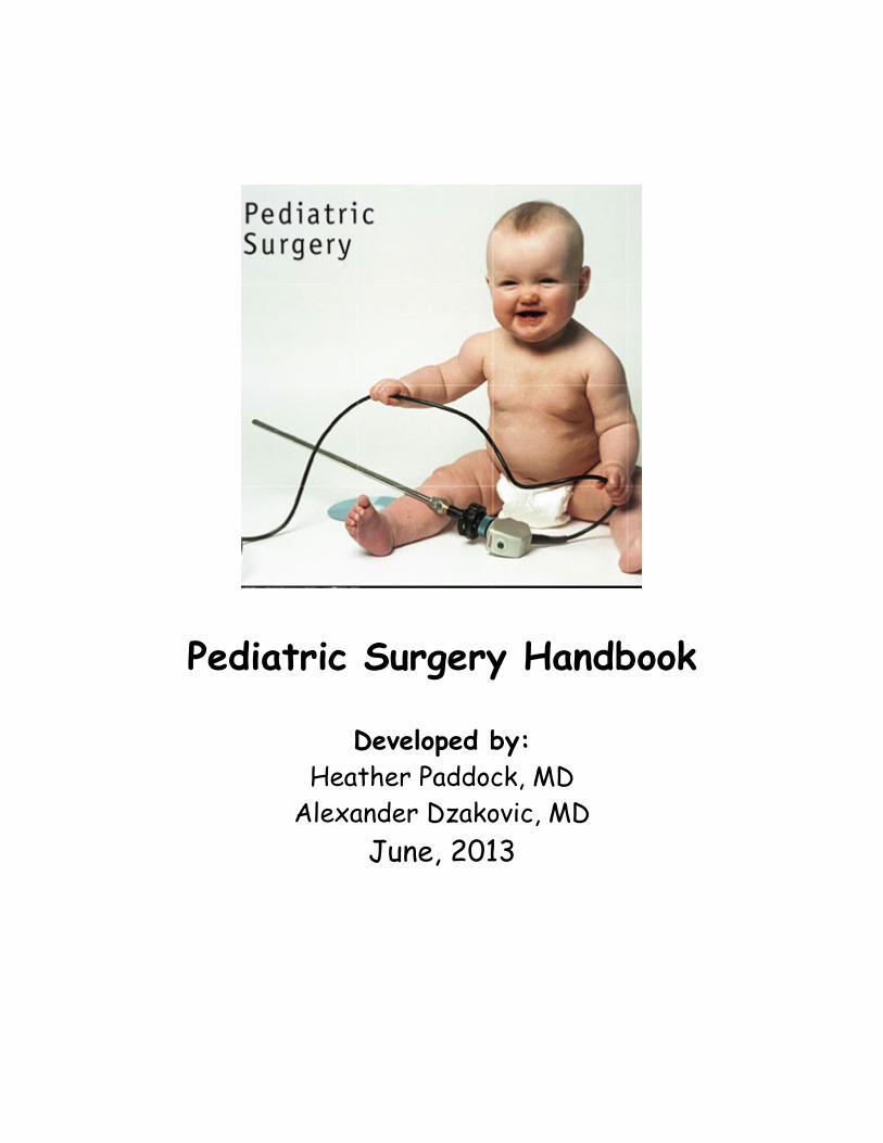
Pediatric Surgery Handbook
Developed by: Heather Paddock, MD
Alexander Dzakovic, MD June, 2013
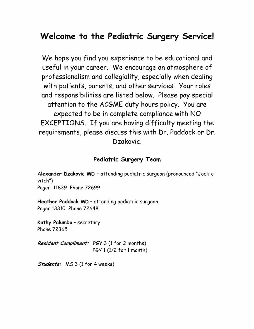
Welcome to the Pediatric Surgery Service!
We hope you find you experience to be educational and useful in your career. We encourage an atmosphere of professionalism and collegiality, especially when dealing with patients, parents, and other services. Your roles
and responsibilities are listed below. Please pay special attention to the ACGME duty hours policy. You are
expected to be in complete compliance with NO EXCEPTIONS. If you are having difficulty meeting the requirements, please discuss this with Dr. Paddock or Dr.
Dzakovic.
Pediatric Surgery Team Alexander Dzakovic MD – attending pediatric surgeon (pronounced “Jock-o-vitch”) Pager 11839 Phone 72699 Heather Paddock MD – attending pediatric surgeon Pager 13310 Phone 72648 Kathy Palumbo – secretary Phone 72365 Resident Compliment: PGY 3 (1 for 2 months) PGY 1 (1/2 for 1 month) Students: MS 3 (1 for 4 weeks)

Roles and Responsibilities Senior Resident (PGY 3) • Supervise all patient management including communications with
consulting services (especially Pediatrics). • Run work rounds in the morning. • Delegate responsibilities on the service. • Teach intern and medical students. • Be familiar with history, exam and results of studies for all patients
preoperatively. • Scrub on all cases (you may delegate some of the cases to the intern if
you think appropriate). • Attend Loyola Outpatient clinics Monday mornings and Friday afternoons. • Attend Oakbrook Terrace clinic 1st and 3rd Tuesday afternoons, 2nd and
4th Thursday mornings. • Present a Pediatric Surgery topic at department of pediatrics noon
conference on 3rd
Thursday of each month (You can discuss topic with Dr. Dzakovic/Paddock)
• Present Pediatric Surgery case at Monday afternoon conference (ACS, PICU, Peds Surg joint conference). Schedule made between the services.
• Present at M&M • Follow up with Nurse Practitioner or Social Work for discharge planning • Prepare discharge paperwork/orders and make follow up appointments Intern • Attend work rounds every morning. • Prepare to present patients on attending rounds. • Attend Loyola Outpatient clinics Monday mornings and Friday afternoons. • Teach medical students. • Call per PGY 1 call schedule. • Take calls from floor. • Enter orders. • Schedule case when directed to by attendings. • Obtain informed consent when directed by attendings. • Give a brief (15 minute) presentation on a topic of your choice relating to
pediatric surgery last Thursday of the month. • Follow up with Nurse Practitioner or Social Work for discharge planning.
• Prepare discharge paperwork/orders and make follow up appointments. Medical Student • Attend work rounds. • Present patients to residents on rounds. • Prepare for rounds (pre-rounding or obtaining vitals) as directed by
senior resident. • Assist resident with patient care duties as directed by intern or senior
resident. • Scrub on all OR cases • Be familiar with patient’s history and important findings preop. • Read about disease process and procedure in advance if case is not an
emergency. • Prepare a 15 minutes presentation on a topic of your choice, relating to
pediatric surgery, last Thursday of the month. Prepare a small handout with your name, date of presentation and references.
• Attend Loyola clinics and Oakbrook clinics. • Take call per student call schedule
Call Schedule Chief residents are encouraged to review evening consults for index cases with the consult resident prior to calling the attending; you should specify this with the consult resident. You are also encouraged to come into the hospital for emergency index cases at night. If you do not wish to come in, the in-house senior resident should scrub on the case. If the pediatric surgery chief resident scrubs on the case, he/she must evaluate the patient and review the studies prior to starting. On nights and weekends you may sign out to the resident on call. During the day, consults (Peds, PCCU, NICU, and ER), and new admissions to the pediatric surgery service, the senior resident should see the patient with the intern and then call the attending. Calls from the floor regarding pediatric surgery patients will go to the pediatric surgery intern during the day and the in-house intern on call at night. Calls regarding NICU patients will all go to the pediatric surgery senior resident during the day and to the in-house resident at night. Rounds Work rounds are conducted in the mornings by the residents and students. The senior resident runs work rounds. Teaching rounds are done every day with the attending. All residents and students, as well as the nurse practitioner participate in teaching rounds. Additionally, we will attempt to coordinate our rounds with General Pediatric Residents and Nursing Staff. The pediatric residents will be involved in an assisting role, to address general pediatric/social/discharge issues. OR Assignments Residents and students are assigned to scrub cases by the senior resident on the service, as appropriate for their level of training. When they scrub, they see the patient and mark them in the pre-op holding area, review their history and exam, and meet the parents. In the OR they participate in the set-up and timeout. During the operation they perform tasks, under the supervision and direction of he attending, that are appropriate for their abilities. Operative decision making is discussed during the case when appropriate. Post-operativley, the plan is reviewed with the attending. The resident and student each write a note documenting the encounter.
Clinic Assignments All residents and students are assigned to the LOC clinic. In the clinic each student pairs up with a resident. The student performs the encounter with the resident present. Feedback is given to the students by the resident. The student then presents the patient to the attending. The differential diagnosis and plan of care is then discussed by the attending, resident and student together. The resident and student each write a note documenting the encounter. Conference Schedule Mondays 3:00-4:00 pm Pediatric Acute Care Surgery Conference, 4th Floor Conf room A, Trauma, Pediatric and PICU service to prepare cases. Mondays 5:00-6:00 pm Surgery M & M Conference, Department of Surgery Conference Room Wednesdays 7:30-8:30 am Department of Surgery Grand Rounds Thursday (2rd Thursday of the month) Pediatrics Noon Conference Maguire 3rd Floor Department of Pediatrics Conference Room Pediatric Surgery Senior Resident to present (see Dr Dzakovic/Paddock for schedule of topics)
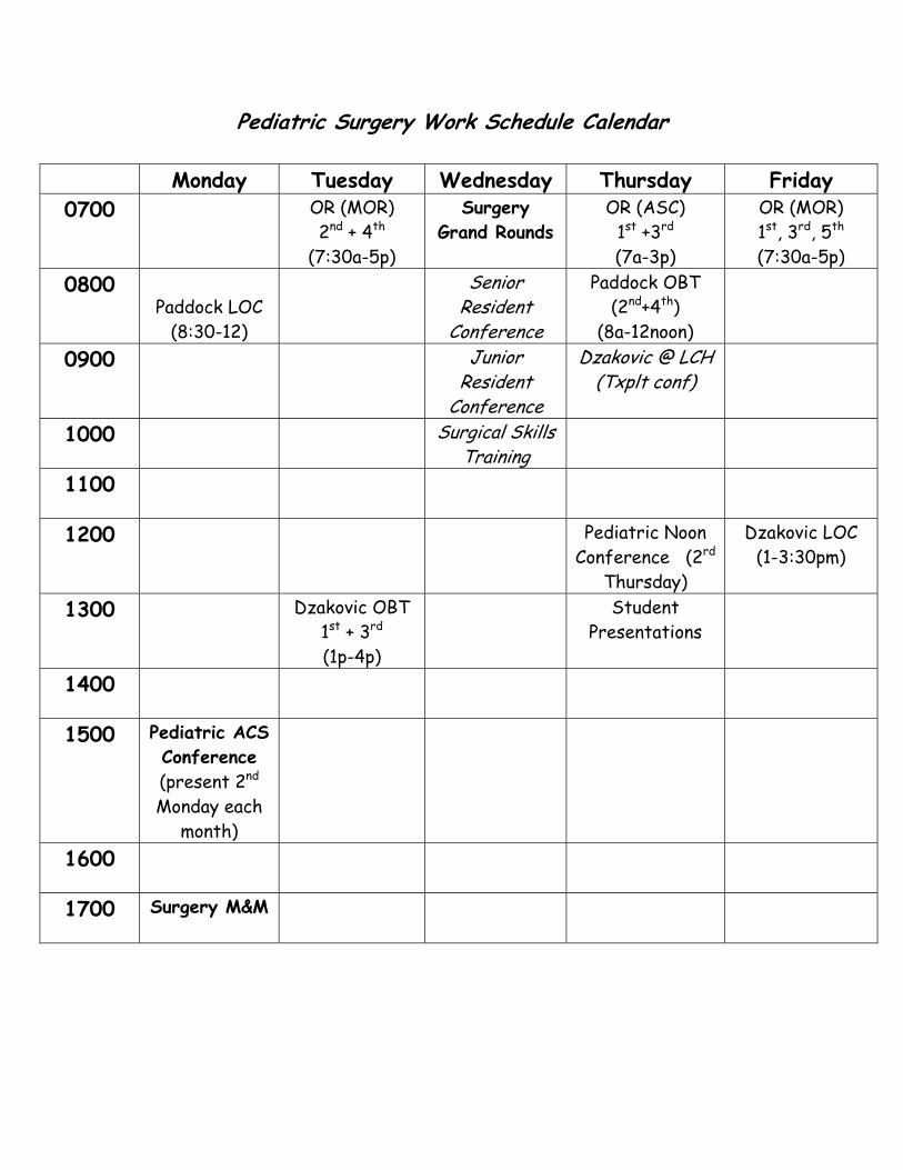
Pediatric Surgery Work Schedule Calendar
Monday Tuesday Wednesday Thursday Friday 0700 OR (MOR)
2nd + 4th (7:30a-5p)
Surgery Grand Rounds
OR (ASC) 1st +3rd (7a-3p)
OR (MOR) 1st, 3rd, 5th (7:30a-5p)
0800 Paddock LOC
(8:30-12)
Senior Resident
Conference
Paddock OBT (2nd+4th)
(8a-12noon)
0900 Junior Resident
Conference
Dzakovic @ LCH (Txplt conf)
1000 Surgical Skills Training
1100
1200 Pediatric Noon Conference (2rd
Thursday)
Dzakovic LOC (1-3:30pm)
1300 Dzakovic OBT 1st + 3rd (1p-4p)
Student Presentations
1400
1500 Pediatric ACS Conference (present 2nd Monday each
month)
1600
1700 Surgery M&M
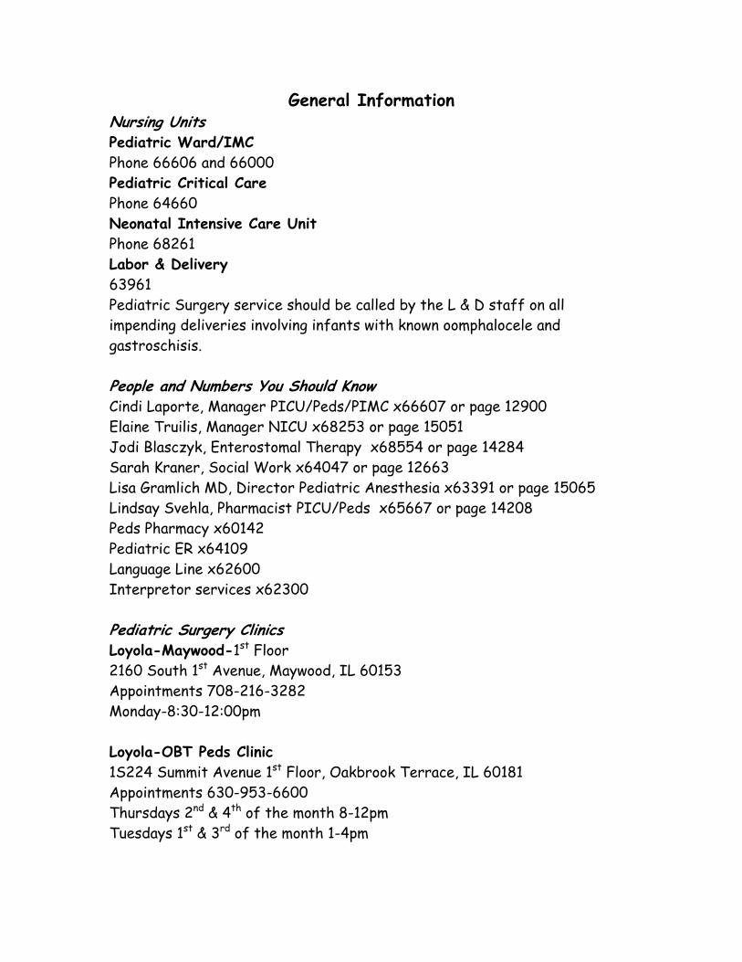
General Information Nursing Units Pediatric Ward/IMC Phone 66606 and 66000 Pediatric Critical Care Phone 64660 Neonatal Intensive Care Unit Phone 68261 Labor & Delivery 63961 Pediatric Surgery service should be called by the L & D staff on all impending deliveries involving infants with known oomphalocele and gastroschisis. People and Numbers You Should Know Cindi Laporte, Manager PICU/Peds/PIMC x66607 or page 12900 Elaine Truilis, Manager NICU x68253 or page 15051 Jodi Blasczyk, Enterostomal Therapy x68554 or page 14284 Sarah Kraner, Social Work x64047 or page 12663 Lisa Gramlich MD, Director Pediatric Anesthesia x63391 or page 15065 Lindsay Svehla, Pharmacist PICU/Peds x65667 or page 14208 Peds Pharmacy x60142 Pediatric ER x64109 Language Line x62600 Interpretor services x62300 Pediatric Surgery Clinics Loyola-Maywood-1st Floor 2160 South 1st Avenue, Maywood, IL 60153 Appointments 708-216-3282 Monday-8:30-12:00pm Loyola-OBT Peds Clinic 1S224 Summit Avenue 1st Floor, Oakbrook Terrace, IL 60181 Appointments 630-953-6600 Thursdays 2nd & 4th of the month 8-12pm Tuesdays 1st & 3rd of the month 1-4pm
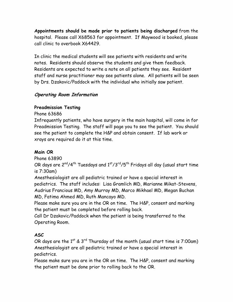
Appointments should be made prior to patients being discharged from the hospital. Please call X68563 for appointment. If Maywood is booked, please call clinic to overbook X64429. In clinic the medical students will see patients with residents and write notes. Residents should observe the students and give them feedback. Residents are expected to write a note on all patients they see. Resident staff and nurse practitioner may see patients alone. All patients will be seen by Drs. Dzakovic/Paddock with the individual who initially saw patient. Operating Room Information Preadmission Testing Phone 63686 Infrequently patients, who have surgery in the main hospital, will come in for Preadmission Testing. The staff will page you to see the patient. You should see the patient to complete the H&P and obtain consent. If lab work or xrays are required do it at this time. Main OR Phone 63890 OR days are 2nd/4th Tuesdays and 1st/3rd/5th Fridays all day (usual start time is 7:30am) Anesthesiologist are all pediatric trained or have a special interest in pediatrics. The staff includes: Lisa Gramlich MD, Marianne Mikat-Stevens, Audrius Francious MD, Amy Murray MD, Marco Mikhael MD, Megan Buchan MD, Fatima Ahmed MD, Ruth Mancayo MD. Please make sure you are in the OR on time. The H&P, consent and marking the patient must be completed before rolling back. Call Dr Dzakovic/Paddock when the patient is being transferred to the Operating Room. ASC OR days are the 1st & 3rd Thursday of the month (usual start time is 7:00am) Anesthesiologist are all pediatric trained or have a special interest in pediatrics. Please make sure you are in the OR on time. The H&P, consent and marking the patient must be done prior to rolling back to the OR.
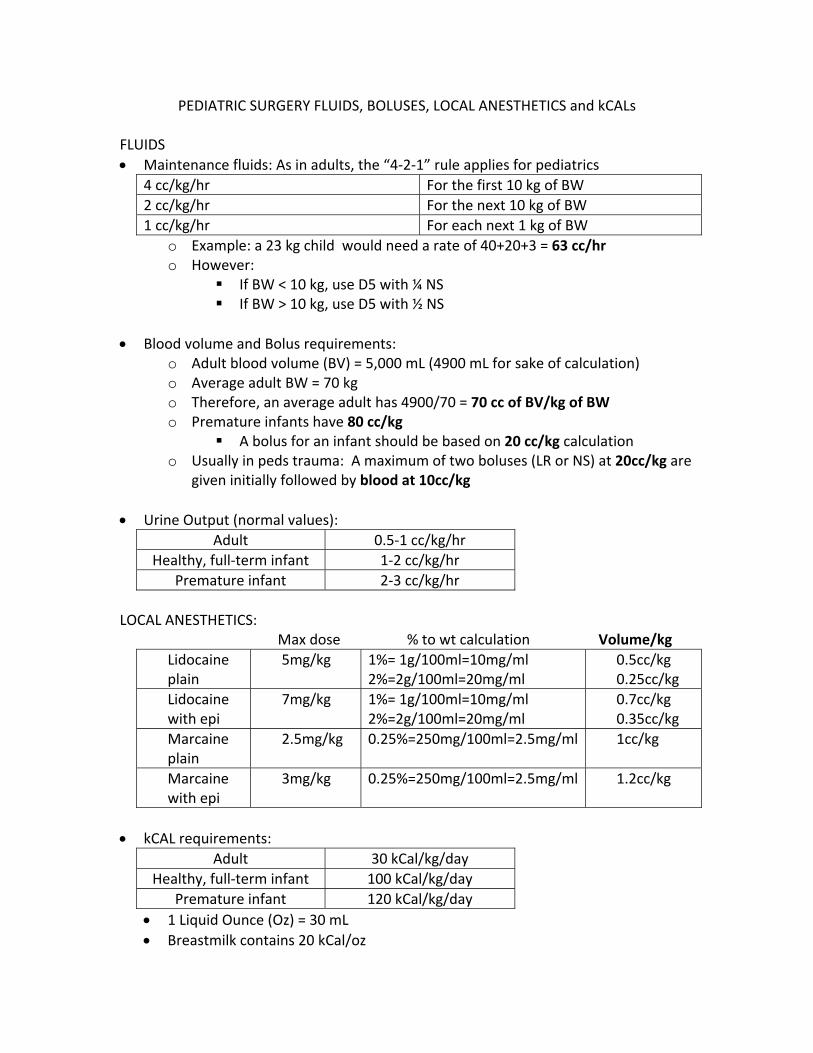
PEDIATRIC SURGERY FLUIDS, BOLUSES, LOCAL ANESTHETICS and kCALs FLUIDS • Maintenance fluids: As in adults, the “4‐2‐1” rule applies for pediatrics
4 cc/kg/hr For the first 10 kg of BW 2 cc/kg/hr For the next 10 kg of BW 1 cc/kg/hr For each next 1 kg of BW
o Example: a 23 kg child would need a rate of 40+20+3 = 63 cc/hr o However:
If BW < 10 kg, use D5 with ¼ NS If BW > 10 kg, use D5 with ½ NS
• Blood volume and Bolus requirements:
o Adult blood volume (BV) = 5,000 mL (4900 mL for sake of calculation) o Average adult BW = 70 kg o Therefore, an average adult has 4900/70 = 70 cc of BV/kg of BW o Premature infants have 80 cc/kg
A bolus for an infant should be based on 20 cc/kg calculation o Usually in peds trauma: A maximum of two boluses (LR or NS) at 20cc/kg are
given initially followed by blood at 10cc/kg • Urine Output (normal values):
Adult 0.5‐1 cc/kg/hr Healthy, full‐term infant 1‐2 cc/kg/hr
Premature infant 2‐3 cc/kg/hr LOCAL ANESTHETICS: Max dose % to wt calculation Volume/kg
Lidocaine plain
5mg/kg 1%= 1g/100ml=10mg/ml 2%=2g/100ml=20mg/ml
0.5cc/kg 0.25cc/kg
Lidocaine with epi
7mg/kg 1%= 1g/100ml=10mg/ml 2%=2g/100ml=20mg/ml
0.7cc/kg 0.35cc/kg
Marcaine plain
2.5mg/kg 0.25%=250mg/100ml=2.5mg/ml 1cc/kg
Marcaine with epi
3mg/kg 0.25%=250mg/100ml=2.5mg/ml 1.2cc/kg
• kCAL requirements:
Adult 30 kCal/kg/day Healthy, full‐term infant 100 kCal/kg/day
Premature infant 120 kCal/kg/day • 1 Liquid Ounce (Oz) = 30 mL • Breastmilk contains 20 kCal/oz
Common Medications
***ALL Pediatric mediations are given as mg/kg***
Antibiotics
Amoxicillin 10 mg/kg PO every 8 hours Ampicillin 25 mg/kg IV every 6 hours Ancef 25mg/kg IV every 8 hours Augmentin 10 mg/kg PO every 8 hours Bactrim 4 mg/kg PO every 12 hours Cefoxitine 40 mg/kg IV every 6 hours Ceftriaxone 50 mg/kg IV every 12 hours Clindamycin 10 mg/kg IV every 6 hours Flagyl 7.5 mg/kg IV every 6 hours Gentamicin 2.5 mg/kg IV every 8 hours Keflex 10 mg/kg PO every 6 hours Linezolid 10 mg/kg IV every 8 hours Ticarcillin 75 mg/kg IV every 6 hours Tobramycin 2.5mg/kg IV every 8 hours Unasyn 40 mg/kg IV every 6 hours Vancomycin 10-15mg/kg IV every 6 hours Zosyn 80mg/kg IV every 6-8 hours
Analgesics and Sedatives Ativan 0.5-1 mg/kg IV every 4 hours Fentanyl 1-2 mcg/kg IV 1 hour Morphine 0.05-0.1 mg/kg IV every 1-2 hours Motrin 10-15 mg/kg PO every 4-6 hours Toradol 0.5 mg/kg IV every 6 hours X 48
hours Tylenol 10-15 mg/kg PO/PR every 4 hours Tylenol with codeine (2.5mg codeine/ml)
0.5-1 mg/kg codeine every 4-6 hours 0.2 X kg = dose in cc’s

Pediatric Surgery Surgical Protocols
Minimal ages for outpatient surgical cases Main OR Premies < 32 weeks at birth ------> need to be 60 weeks PCA 32-36 weeks at birth-----> need to be 55 weeks PCA > 36 weeks at birth ------> need to be 44 weeks PCA
PCA = postconceptual age If they have had any sedation, general anesthesia, or anything except straight local anesthetic in their regional. With complete regional they may go home 2 hours post-operatively, BUT they should have received NO sedation NOR additives to their local anesthetic. ASC Premies <36 weeks--------> must be >60 weeks PCA >36 weeks--------> must be >44 weeks PCA There are several preprinted order sets for pediatric surgery, they include: Pediatric Surgery Admission Pediatric Post Pyloromyotomy Pediatric Surgery Appendicitis Admit Pediatric Surgery Laparoscopic Appendectomy Pediatric Gastrostomy Tube Post op Orders Preoperative Labs/Studies Not routine for simple cases such as hernias, orchiopexy, cyst removal. Obtain or check CBC for children who are having lines placed or removed (usually these are heme/onc patients-Heme/Onc PNP generally orders-check to make sure labs OK) CBC may also be done for anemic patients or infected patients. Coagulation profile for patients with liver disease, sepsis or on TPN, or risk for bleeding.
CMP if history of vomiting, diarrhea, fluid shifts, renal disease or liver disease. Preoperative Xrays/Studies Not routine on simple outpatient cases. UGI is done for any patient scheduled for Gastrostomy tube with or without fundoplication, and/or for workup of bilious vomiting. US is done to r/o pyloric stenosis, ovarian cysts or disease, appendicitis, intussusception. For young or thin children an US may be done to rule out appendicitis. (For all children 3 years and under, regardless of the sex of the patient, if an ultrasound is being ordered it must be ordered as two studies: complete abdomen and complete pelvis) CT is done to evaluate for masses, appendicitis, or abscess. Obstructive series is done if you suspect free air/constipation or bowel obstruction. Barium Enema is done if you suspect intussusception or stricture. It can be therapeutic for intussusception. Preoperative Antibiotics None for clean cases such as hernias, nonifected cysts/lesions. Ancef for most cases (line placement, gastrostomy tube placement, incarcerated hernia, thoracotomy/thoracoscopy, pyloromyotomy). Ancef for all other cases. Amp/Gent/Flagyl or Zosyn for bowel cases and NEC in Preemies. Zosyn for bowel cases including appendectomies in pediatric patients Clindamycin is the choice of antibiotics for soft tissue infections. This should cover community-acquired MRSA. Bactrim may also be used. For all MRSA positive patients Vancomycin 15mg/kg IV on call to OR and redose if surgery >12 hours or Linezolid 10mg/kg IV on call to OR and redose if surgery >8 hours. Preoperative Bowel Prep Stomas will need to have clear liquids for 24 hours before surgery If patient is impacted Enemas 1-3 times a day for 1-3 days For patients who need colonic prep golytely 40cc/kg/hr continuously via NG tube until clear. Do not forget to turn off when patient is to be NPO! Rectal or colonic irrigation may be done until clear in the Operating Room
Postoperative General Orders Vital signs routine every four hours O2 sat monitor and CR monitor for infants less than four weeks or former preemies requiring overnight observation Strict I & O Drains as needed CT to be at 20 cm suction NG to LCS Foley catheter not usually necessary in children (preemie babies keep in for 24-48 hours or if patient has epidural catheter). Laparoscopic cases should have foley placed in OR and removed before leaving OR. In the OR preemies should have the 6FR foley placed and 8FR replogle. Day of surgery patient should have D5LR at maintenance and switch to D5 0.25ns w/20KCL or D5 0.45ns w/20KCL. Bolus with 0.9NS 20cc/kg. Always check with attending before administering blood transfusions. Postoperative Antibiotics Clean-contaminated cases, (non-perforated appendicitis, Gastrostomy tube placement, elective small bowel resection) continue for 24 hours if afebrile and stable. For contaminated cases, (stomas, resection of unprepped bowel) continue for 48 hours. For dirty cases (bowel perforation) continue for 7-14 days. Patients must be afebrile for 48 hours, have a normal WBC and not have an ileus. Postoperative Pain Mangement Infants Tylenol 15 mg/kg PO/PR every 4 hours Morphine 0.05-0.1mg/kg IV every 1-2 hours prn Consider Epidural for cases with large incisions Toddlers Tylenol 15 mg/kg PO/PR every 4 hours Morphine 0.05-0.1mg/kg IV every 1-2 hours prn Tylenol with codeine 0.5-1 mg/kg codeine every 4-6 hours prn
Toradol 0.5mg/kg IV every 6 hours for up to 72 hours. Consider Epidural for cases with large incisions Older Children/Adolescents Tylenol 15 mg/kg PO/PR every 4 hours Morphine 0.05-0.1mg/kg IV every 1-2 hours prn Tylenol with codeine 0.5-1 mg/kg codeine every 4-6 hours prn Morphine PCA basal and PCA dosing 1/3 basal rate and 2/3 hourly dose to divide in bolus dosing (0.1 mg/kg/hr) Toradol 0.5 mg/kg (max 30 mg) IV every 6 hours for up to 72 hours Consider Epidural for cases with large incisions **For Dr Paddock’s ASC surgical patients: DO NOT discharge ASC patients with script for narcotic pain medications unless specifically told to do so. The usual pain control regimen is alternating pediatric Tylenol with Advil every 4 hours as soon as they get home for 24 hours (yes, the parents will need to wake them up in the middle of the night to give meds), then as needed. Postoperative Dressing Management For all cases keep incision dry for 48 hours. Do not immerse in water for 1-2 weeks. Subcuticular Closure- Octyseal or steri-strips, Telfa and Tegaderm. Sutures - Telfa and Tegaderm Incisions with drains in place – dry gauze on top and change 2-4 times a day and prn. Open Wounds – Repack with wet to dry dressings 2-4 times a day. Gastrostomy tubes – One split 2 X 2 under flange and tape down securely. Clean site at least daily with soap and water or ½ strength hydrogen peroxide. DO NOT pull the flange back-it should be flush with the skin. Anal cases –A nursing order and sign should be posted on the patient’s bed that reads “ABSOLUTELY NOTHING PER RECTUM”. This includes temperatures and suppositories. Cleanse site with ½ strength hydrogen peroxide and apply bacitracin ointment with every diaper change. Pyloric Feeding Protocol – start when infant arrives on the pediatric floor Pedialyte up to 60cc’s, if tolerated then
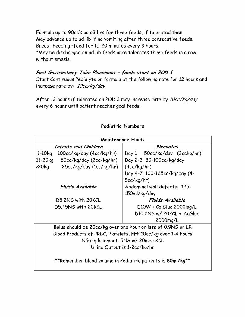
Formula up to 90cc’s po q3 hrs for three feeds, if tolerated then May advance up to ad lib if no vomiting after three consecutive feeds. Breast Feeding –feed for 15-20 minutes every 3 hours. *May be discharged on ad lib feeds once tolerates three feeds in a row without emesis. Post Gastrostomy Tube Placement – feeds start on POD 1 Start Continuous Pedialyte or formula at the following rate for 12 hours and increase rate by: 10cc/kg/day After 12 hours if tolerated on POD 2 may increase rate by 10cc/kg/day every 6 hours until patient reaches goal feeds.
Pediatric Numbers
Maintenance Fluids Infants and Children
1-10kg 100cc/kg/day (4cc/kg/hr) 11-20kg 50cc/kg/day (2cc/kg/hr) >20kg 25cc/kg/day (1cc/kg/hr)
Fluids Available
D5.2NS with 20KCL D5.45NS with 20KCL
Neonates Day 1 50cc/kg/day (3cckg/hr) Day 2-3 80-100cc/kg/day (4cc/kg/hr) Day 4-7 100-125cc/kg/day (4-5cc/kg/hr) Abdominal wall defects: 125-150ml/kg/day
Fluids Available D10W + Ca Gluc 2000mg/L
D10.2NS w/ 20KCL + CaGluc 2000mg/L
Bolus should be 20cc/kg over one hour or less of 0.9NS or LR Blood Products of PRBC, Platelets, FFP 10cc/kg over 1-4 hours
NG replacement .5NS w/ 20meq KCL Urine Output is 1-2cc/kg/hr
**Remember blood volume in Pediatric patients is 80ml/kg**
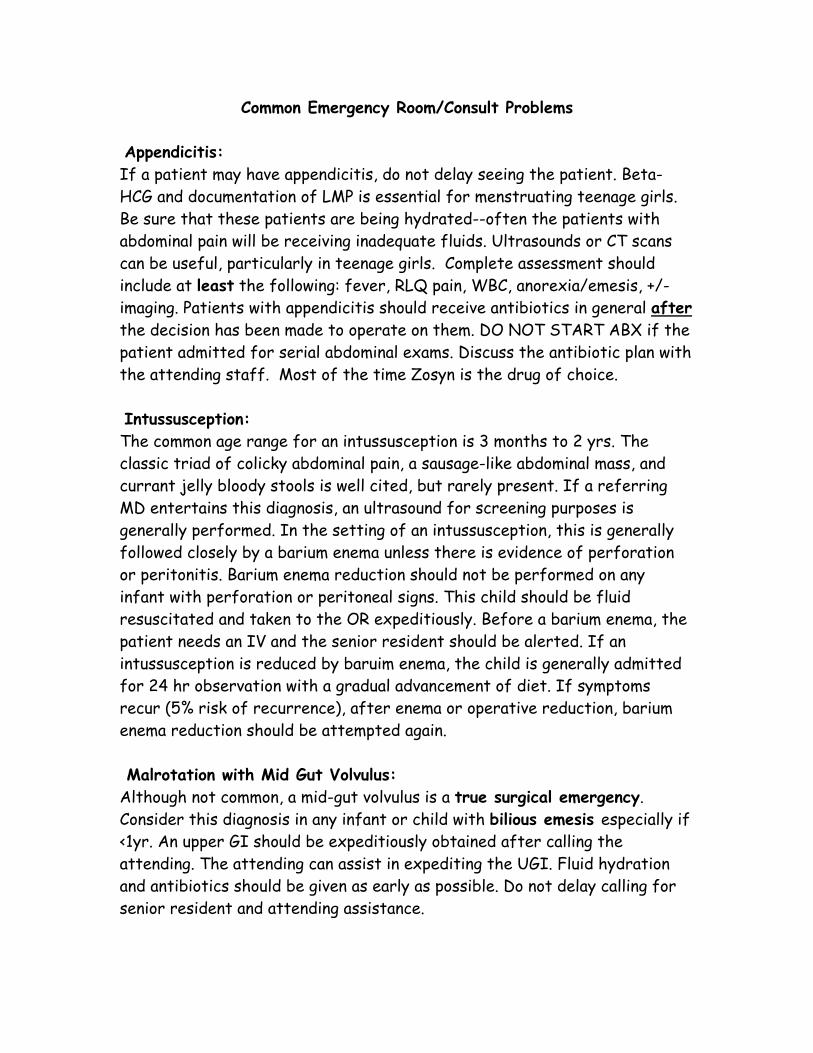
Common Emergency Room/Consult Problems Appendicitis: If a patient may have appendicitis, do not delay seeing the patient. Beta-HCG and documentation of LMP is essential for menstruating teenage girls. Be sure that these patients are being hydrated--often the patients with abdominal pain will be receiving inadequate fluids. Ultrasounds or CT scans can be useful, particularly in teenage girls. Complete assessment should include at least the following: fever, RLQ pain, WBC, anorexia/emesis, +/- imaging. Patients with appendicitis should receive antibiotics in general after the decision has been made to operate on them. DO NOT START ABX if the patient admitted for serial abdominal exams. Discuss the antibiotic plan with the attending staff. Most of the time Zosyn is the drug of choice. Intussusception: The common age range for an intussusception is 3 months to 2 yrs. The classic triad of colicky abdominal pain, a sausage-like abdominal mass, and currant jelly bloody stools is well cited, but rarely present. If a referring MD entertains this diagnosis, an ultrasound for screening purposes is generally performed. In the setting of an intussusception, this is generally followed closely by a barium enema unless there is evidence of perforation or peritonitis. Barium enema reduction should not be performed on any infant with perforation or peritoneal signs. This child should be fluid resuscitated and taken to the OR expeditiously. Before a barium enema, the patient needs an IV and the senior resident should be alerted. If an intussusception is reduced by baruim enema, the child is generally admitted for 24 hr observation with a gradual advancement of diet. If symptoms recur (5% risk of recurrence), after enema or operative reduction, barium enema reduction should be attempted again. Malrotation with Mid Gut Volvulus: Although not common, a mid-gut volvulus is a true surgical emergency. Consider this diagnosis in any infant or child with bilious emesis especially if <1yr. An upper GI should be expeditiously obtained after calling the attending. The attending can assist in expediting the UGI. Fluid hydration and antibiotics should be given as early as possible. Do not delay calling for senior resident and attending assistance.
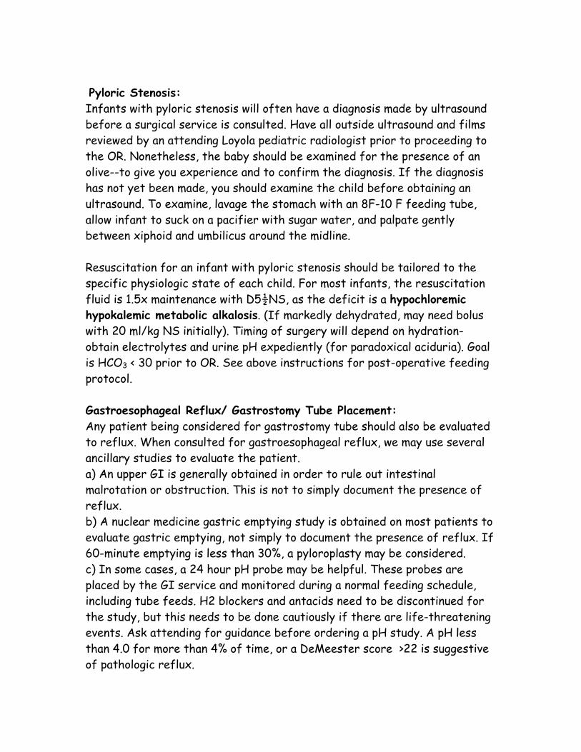
Pyloric Stenosis: Infants with pyloric stenosis will often have a diagnosis made by ultrasound before a surgical service is consulted. Have all outside ultrasound and films reviewed by an attending Loyola pediatric radiologist prior to proceeding to the OR. Nonetheless, the baby should be examined for the presence of an olive--to give you experience and to confirm the diagnosis. If the diagnosis has not yet been made, you should examine the child before obtaining an ultrasound. To examine, lavage the stomach with an 8F-10 F feeding tube, allow infant to suck on a pacifier with sugar water, and palpate gently between xiphoid and umbilicus around the midline. Resuscitation for an infant with pyloric stenosis should be tailored to the specific physiologic state of each child. For most infants, the resuscitation fluid is 1.5x maintenance with D5½NS, as the deficit is a hypochloremic hypokalemic metabolic alkalosis. (If markedly dehydrated, may need bolus with 20 ml/kg NS initially). Timing of surgery will depend on hydration-obtain electrolytes and urine pH expediently (for paradoxical aciduria). Goal is HCO3 < 30 prior to OR. See above instructions for post-operative feeding protocol. Gastroesophageal Reflux/ Gastrostomy Tube Placement: Any patient being considered for gastrostomy tube should also be evaluated to reflux. When consulted for gastroesophageal reflux, we may use several ancillary studies to evaluate the patient. a) An upper GI is generally obtained in order to rule out intestinal malrotation or obstruction. This is not to simply document the presence of reflux. b) A nuclear medicine gastric emptying study is obtained on most patients to evaluate gastric emptying, not simply to document the presence of reflux. If 60-minute emptying is less than 30%, a pyloroplasty may be considered. c) In some cases, a 24 hour pH probe may be helpful. These probes are placed by the GI service and monitored during a normal feeding schedule, including tube feeds. H2 blockers and antacids need to be discontinued for the study, but this needs to be done cautiously if there are life-threatening events. Ask attending for guidance before ordering a pH study. A pH less than 4.0 for more than 4% of time, or a DeMeester score >22 is suggestive of pathologic reflux.

Postoperative management: During the first 24 hours the tube should be to straight drain and then advance feeds as tolerated. Cleaning around the tube should be half strength peroxide and saline for the first week then transition to soap and water. Prior to placement of a g-tube determine who will be following the care of the tube postoperativly, ie who will be providing home health supplies and nutritional plan. The surgical NP will provide education to the family, they need to be notified 48hrs prior to discharge. Gastrostmy Tube Replacements: a. Do not attempt to replace tubes that have been in place for less than 30 days--call the attending for advice. b. Replace tubes carefully. Relux of gastric fluid after placement is adequate to confirm correct placement. However, if in questions/no reflux/less than 1 month post-op then obtain a contrast study to confirm. This should be an AP and lateral abdominal film with half-strength water-soluble contrast in a volume proportionate to the size of the child. Incarcerated Hernias: Inguinal hernias can often be reduced in the ER and repaired within 24-48 hrs, when the swelling has resolved. Call a senior resident or attending to assist. Sedation may be needed. Trendelenberg position also may help. Esophageal/Airway Foreign Bodies: Esophageal foreign bodies are often coins that can be seen on an X-ray. Recent AP and lateral views should be obtained for all patients, even if they have outside films, to identify the current location and number of coins. Foreign bodies which pass into the stomach will usually pass through the GI tract in several days and therefore need not be removed. An exception is batteries that do not pass within several hours. Objects caught within the esophagus need to be removed, either in radiology under fluoroscopy, or in the OR with a rigid esophagoscope and fluoroscopy. Airway foreign bodies are suspected either after witnessed aspiration, recurrent pneumonia, or new onset wheezing or stridor. Inspiration and expiration X-rays may document air trapping, and fluoroscopy can be helpful. Suspicion of aspiration of a foreign body generally mandates endoscopy. After removal of any foreign body, a CXR should be obtained to evaluate for pneumothorax or
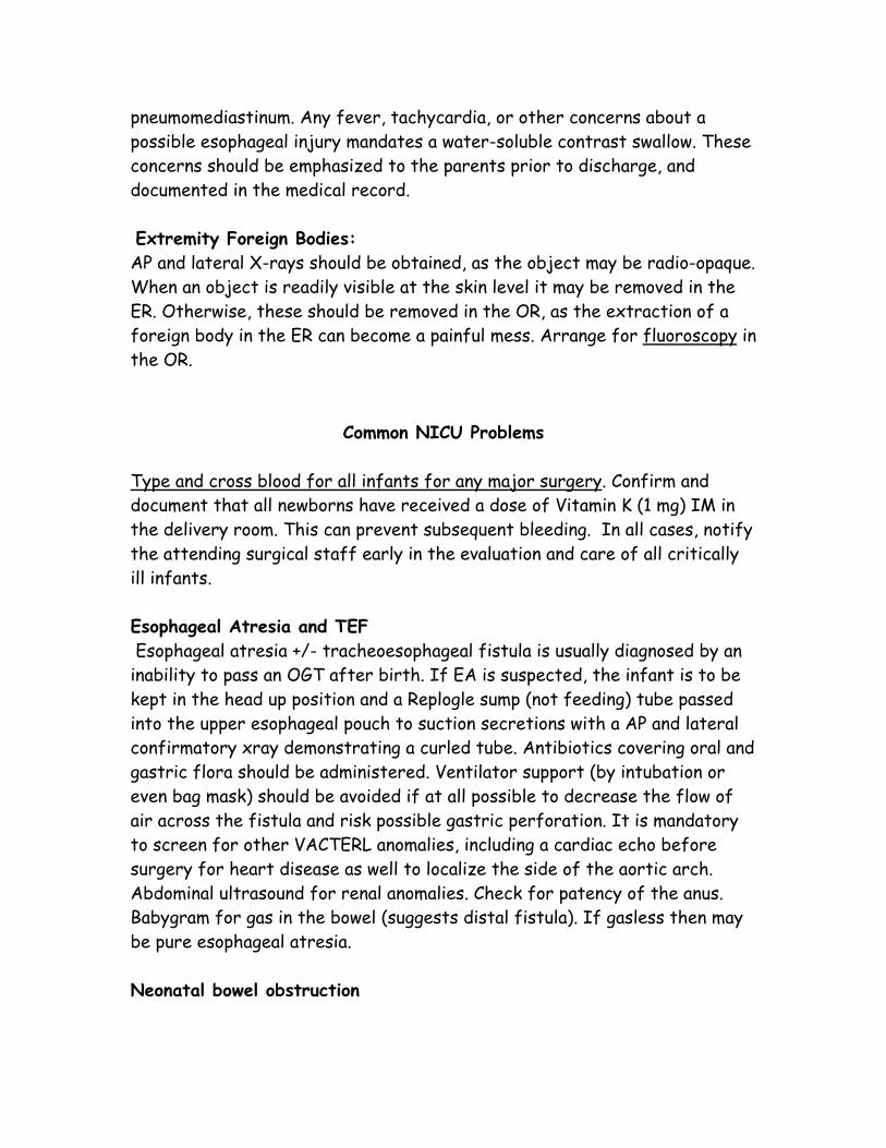
pneumomediastinum. Any fever, tachycardia, or other concerns about a possible esophageal injury mandates a water-soluble contrast swallow. These concerns should be emphasized to the parents prior to discharge, and documented in the medical record. Extremity Foreign Bodies: AP and lateral X-rays should be obtained, as the object may be radio-opaque. When an object is readily visible at the skin level it may be removed in the ER. Otherwise, these should be removed in the OR, as the extraction of a foreign body in the ER can become a painful mess. Arrange for fluoroscopy in the OR.
Common NICU Problems Type and cross blood for all infants for any major surgery. Confirm and document that all newborns have received a dose of Vitamin K (1 mg) IM in the delivery room. This can prevent subsequent bleeding. In all cases, notify the attending surgical staff early in the evaluation and care of all critically ill infants. Esophageal Atresia and TEF Esophageal atresia +/- tracheoesophageal fistula is usually diagnosed by an inability to pass an OGT after birth. If EA is suspected, the infant is to be kept in the head up position and a Replogle sump (not feeding) tube passed into the upper esophageal pouch to suction secretions with a AP and lateral confirmatory xray demonstrating a curled tube. Antibiotics covering oral and gastric flora should be administered. Ventilator support (by intubation or even bag mask) should be avoided if at all possible to decrease the flow of air across the fistula and risk possible gastric perforation. It is mandatory to screen for other VACTERL anomalies, including a cardiac echo before surgery for heart disease as well to localize the side of the aortic arch. Abdominal ultrasound for renal anomalies. Check for patency of the anus. Babygram for gas in the bowel (suggests distal fistula). If gasless then may be pure esophageal atresia. Neonatal bowel obstruction
Causes for bowel obstruction in a newborn differ from those in an older child or an adult. Common causes include intestinal atresia, annular pancreas , Hirschprung’s disease, meconium ileus, and malrotation. Bilious emesis implies a surgical emergency until proven otherwise. In general, this mandates an urgent UGI +/- BE to r/o malrotation with a volvulus, which can be initiated and ordered prior to attending staffing the consult. All infants with suspected bowel obstruction need an NGT and IVF. On plain films (required in all cases of suspected obstruction), the newborn colon cannot be differentiated from small bowel. Most infants will require a barium enema and an UGI study to evaluate the etiology and extent of bowel obstruction. In cases of pneumoperitoneum, no contrast films should be obtained, and the child should be brought quickly to the operating room after resuscitation. Intestinal atresia occurs in the following frequency: duodenal,> jejunal,> multiple,> and colon. Plain films of the abdomen should be obtained in all cases. When a bowel atresia is suspected, a contrast enema should be obtained prior to surgery. Duodenal atresia Duodenal atresia results from failure of recanalization of the foregut. It occurs with Down’s syndrome (30% of patients with DA have Down’s), malrotation, annular pancreas, congenital heart disease, other biliary anomalies, and VACTERL anomalies. Prenatally, DA can be diagnosed by polyhydramnios and a double bubble. Postnatally, these infants present with bilious or non-bilious emesis and a proximal bowel obstruction by X-ray, often with a “double bubble” radiograph. Gas can be present distally (with an incomplete atresia or bifid bile duct). The most important initial consideration is to exclude midgut volvulus, with an UGI. Preoperative care involves volume resuscitation, screening for other anomalies, cardiac echocardiogram, and renal ultrasound. Operative care involves laparotomy, and usual reconstruction is a duodenoduodenostomy. If case of malrotation, a Ladd procedure is done. Hirschsprung’s disease Hirschsprung’s disease (HD) is a frequent cause of newborn bowel obstruction. The disease involves an absence of ganglion cells in the rectum,
and can extend more proximally to involve the entire colon, or even the small bowel. Symptoms are non-specific, such as episodic abdominal distention or constipation, but failure to pass meconium in first 24hr of life is very common (just not specific). A barium enema (in an older infant) will often show a narrow rectum and a dilated colon. Retained barium past 24 hrs is suggestive of HD. Diagnosis is confirmed by suction rectal biopsy. HD is usually managed by initial colostomy, followed by a reconstructive pull-through several months later. Meconium ileus Meconium ileus occurs in 15% of infants with cystic fibrosis (CF). Plain abdominal films can show bowel loops with a soap bubble appearance of meconium. Calcification on abdominal films or ultrasound implies meconium peritonitis resulting from a previous (pre-natal) bowel perforation. Contrast enema may be contraindicated if plain films show calcifications. Initial treatment is non-operative with gastrograffin enema. Success results in passage of meconium, and may need frequent enemas. Surgery is indicated if enemas fail to relieve the obstruction, calcifications are present on plain film, or an infant is too ill to delay operation. Omphalocele and Gastroschisis In case of abdominal wall defect, hypothermia and fluid loss are the immediate life threatening problems. The bowel should be covered with warm, moist dressings (gauze sponges or xeroform/sponges) and the baby placed in a bowel bag. The bowel in gastroschisis (no sac, emanates to the right of the umbilicus) can become gangrenous with compression, and the bowel should be supported to reduce tension on the mesentery. If the bowel appears ischemic, a bedside midline fascial incision can improve mesenteric blood flow. The bowel should be examined frequently. In general, most newborns with gastroschisis will need aggressive fluid resuscitation, ie the usual rate of D10W @60 ml/kg/day is inadequate to account for both fluid and electrolyte needs. Precise fluid needs depend on the physiology of the baby, but generally range from 120-150 ml/kg/day for the first day of D5 1/2NS with added potassium, per electrolyte measurements. Discuss all fluid choices with the NICU staff early in the care of the baby. In most cases of abdominal wall defects, we will place a temporary spring-loaded silo over the intestinal contents to protect them and allow for partial reduction prior to operative closure. In the case of an intact omphalocele sac, the sac can be
used as a silo. This usually requires sedation with intubation, with the assistance of the ICN medical staff. A 10 or 8 Fr. Replogle OGT should be placed to minimize GI distention. IVF at 1 - 1.5x maintenance rate is required with IVF with salt. Antibiotics (amp/gent) are given to reduce risk of infection (though this is difficult to prove). In case of omphalocele, need to rule out chromosomal, cardiac and renal anomalies. Cardiac echo and renal ultrasound should be obtained preoperatively if possible. In case of gastroschisis, there are concerns for intestinal atresias. Necrotizing enterocolitis NEC is the most common surgical emergency in the ICN. This is a lethal disease characterized by ischemic necrosis of the GI tract. NEC is associated with a history of prematurity, low birth weight, RDS, apneic spells, and perinatal asphyxia. Time of onset is usually between days 3-10 of life, generally with an antecedent history of feeding before the onset of symptoms. Common symptoms include bloody diarrhea, feeding intolerance, abdominal distention, apneic spells, and lethargy. Work-up includes abdominal X-rays, NPO, NGT, and IV antibiotics. Early radiographic findings include bowel dilation, and later findings include intramural gas (pneumatosis intestinalis) and portal venous air. Falling PLT counts are an ominous finding suggestive of progressive sepsis in any infant. Free intraperitoneal air signifies intestinal perforation. Infants are monitored very closely, especially for the first 1-2 days of NEC for disease progression. Serial labs and X-rays are obtained every 6-8 hours. Indications for surgery are usually separated into absolute and relative indicators. Absolute indications for surgery include pneumoperitoneum (intestinal perforation) or clinical deterioration in spite of medical therapy. Relative indicators of surgery include advanced pneumatosis, portal venous air, thrombocytopenia, acidosis, or worsening abdominal wall edema or erythema. Congenital diaphragmatic hernia (CDH) Infants with CDH have among the most complex physiology in pediatric surgery but is related to bilateral lung hypoplasia and pulmonary
hypertension. The best management is geared toward minimizing lung injury and reduce pulmonary HTN. Initial Management At delivery: Immediate intubation and placement of a Replogle tube. Initial PIP’s should not exceed 24, although for initial breaths, higher pressures may be necessary. Pressures between 24-30 are reserved for short-term use. Early use of ventilators rather than hand bagging offers information about volumes and compliance, and limits pressures. Radiographs must be used to verify tube positions. Placement of umbilical catheters is standard. Monitoring patients Pre- and post-ductal O2 saturation monitors, especially necessary for those on HFJV or HFOV, pre- and post-ductal arterial access (if possible). Cardiac echoes: Echo is required to assess cardiac anatomy. Ventilator Management Initial goals: conventional ventilation (CV) and pre- and post-ductal O2 sats > 95%. If PIP > 26 and/or MAP > 12 are required to achieve PaCO2 < 65 mmHg, HFV: Start HFV if PIP > 26 with CMV to maintain ideal patient parameters. Initial HFV will be with jet ventilation unless PPHN is apparent. Surgery criteria Surgery in the first few days of life is encouraged for infants who are stable on conventional ventilation. A period of 24 - 48 hours at this range of support is desirable. Blood pressure management Optimize calcium, check cortisol level and consider hydrocortisone if dopamine up to 10 mcg/kg/min has not achieved or maintained acceptable mean arterial pressure (MAP), or cortisol level is low. Optimize hematocrit 40 - 50% as polycythemia may lead to pulmonary hypertension. MAP for term infants with CDH without significant pulmonary hypertension will be acceptable as low as 40, as long as tissues are well perfused, urine output is adequate, post-ductal pH is > 7.25 with a low serum lactate. When cardiovascular performance is suboptimal, pressors will be added, beginning
with dopamine, which can be increased to 20 µg /kg/min. If MAP remains low, hydrocortisone will be added (stress dose if cortisol level is low, maintenance dose if level is high). If above does not provide relief, epi will be used. Volume pushes are applied only in the setting of decreased intravascular volume. Post-operative diuresis should be limited unless oxygenation is compromised and/or ventilator weaning is limited. Diuresis should be gradual, achieving negative fluid balance of no greater than 100 - 200 cc/day, while following measures of intravascular volume (weight, electrolyte, BUN). iNO: NO (Nitric Oxide) should be considered if pulmonary hypertension is evident by cardiac echo/saturations, and the patient is on DA up to 10 µg /kg/min, iNO may be tried. If there is no response after 1 hour, the iNO should be weaned. If pulmonary hypertension persists, and therapy has increased to epi- and alkalinization, iNO may be tried again. If the patient has preductal O2 saturations < 85% with pulmonary hypertension, iNO may be used to bring the patient to acceptable levels, or as a bridge to ECMO. ECMO criteria Inability to consistently maintain postductal PaO2 of 30 mm Hg, preductal O2 saturation of 85%, or post-ductal pH < 7.25, suggest failed medical management. The combination of iNO or alkalinization + necessary to maintain acceptable blood gases and SaO2is a bridge to ECMO. OI's of > 40 x 2 in one hour should lead to consideration of ECMO. (OI= MAPx100xFiO2/PaO2, and MAP 20 on HFOV, FiO2 1.0, and PaO2 of 50 gets you an OI of 40). In addition to O2 saturations and a pH less than 7.25, rising serum lactates should lead to scrutiny of management. Biliary Atresia The infant with biliary atresia, even after a successful hepatic portoenterostomy, will have decreased amounts of bile flow into the intestine and poor fat absorption. This may lead to essential fatty acid deficiency and inadequate absorption of fat soluble vitamins, resulting in a
lack of bone mineralization and failure to thrive. The goals for such an infant are to provide adequate calories using a formula that maximizes fat intake. We favor the use of Pregestimil, which has less medium chain triglycerides than Portagen (60% vs. 80%), but a higher percentage (11%) of its calories is derived from the essential fatty acid linoleic acid. Other options include Portagen, because of its high content of medium chain triglycerides, as infants depend less on bile acids for absorption compared to long chain fatty acids. Breast feeding, although generally ideal in infancy, may be detrimental in patients with biliary atresia because breast milk has a much higher fat content than commercially available formulas. Please discuss feeding issues with the attending and GI staff prior to feeds. Vitamin supplementation is critical in patients with biliary atresia. The addition of fat and water soluble vitamins above those provided in standard infant formulas should be administered via a multi-vitamin preparation. Short Bowel Syndrome The nutritional support of a child with short bowel syndrome (SBS) is quite complex. The care of such an infant can be divided into three stages. The first stage begins after the postoperative period. This period is associated with increased gastric output, due to loss of intrinsic intestinal negative feedback, and increased stool output, which often leads to losses of fluids and electrolytes. During this time a central venous catheter is usually inserted, and since the child will need long-term venous access, each access site must be carefully protected. Enteral feedings should be initiated early in the postoperative period, and will both stimulate small bowel adaptation and limit the development of TPN-associated cholestasis. In infants with SBS, we generally initiate feeds with Pregestamil, and in children over one year of age Peptamen Junior. High stool output is associated with excessive losses of zinc; thus, zinc supplements should be provided. The loss of sodium and bicarbonate can be dramatic, and total body sodium depletion is associated with failure to thrive. A simple way to detect sodium deficit is to measure a spot urine sodium. Urine sodium of less than 10 meq/L indicates sodium depletion, and oral supplementation should be given. During the second phase, intestinal adaptation begins, and assessment of electrolytes, liver function and protein status (total protein, albumin and total iron binding capacity) should be done on a weekly basis. Fat soluble
vitamin levels should be assessed every six months. Patients with loss of the terminal ileum should have a vitamin B12 level assessed yearly. The stool should intermittently be assessed for pH, reducing substances and fecal fats. A stool pH less than 5.5 or a reducing substance level greater than one-half percent indicates malabsorption of carbohydrates. Elevation in fecal fats suggests fat malabsorption, which may require an increase in medium chain triglycerides. Formulas with sucrose will not yield reducing substances despite carbohydrate malabsorption. The final phase is one of long-term therapy, and consists of weaning the patient off of parenteral nutrition. This stage may last from months to years, and during this period, monitoring the development of suck and swallow reflexes is critical. Children without a terminal ileum should be evaluated for renal oxalate stones, and should avoid a high oxalate diet. The Handicapped Child Between 10 and 20% of children in the United States have special needs because of developmental disorders, and many of these disorders require care of a pediatric surgeon. Often the pediatric surgeon is responsible for providing nutritional access as well as for maintaining nutritional care after surgery. Potential factors that may contribute to poor nutrition include feeding disorders, poorly coordinated swallowing reflexes, gastroesophageal reflux, and increased energy expenditure due to muscle spasticity. Children with athetosis (mixed pattern of too much and too little muscle tone) may require a higher than normal calorie intake. Children with myelomeningocele are inactive compared to their peers and may need only 50 to 60% of estimated energy needs of normal children.
Cancer Essentials Neuroblastoma Second most common childhood solid tumor (behind brain tumor). Associated with neural crest abnormalities, Hirschprung’s disease, Beckwith-Weidemann. Varied presentations, depend on site of origin. Infant tumors have improved prognosis, higher rate of Stage IV-S disease. Survival depends on age (< 1 yr improved survival) and tumor stage. Workup involves both a CT scan as well as MRI (if concern of spinal extension). 24 hr urine
for VMA, HVA, as well as serum for neuron-specific enolase, ferritin. Shimada pathologic classification most common-divides tumor into favorable or unfavorable histology based on stroma content, mitosis index, and patient age. Important to distinguish any maturation into ganglioneuroblastoma or ganglioneuroma. N-myc amplification measured. Staging-International Neuroblastoma Staging System (INSS) Stage I: Localized tumor, complete excision, nodes negative Stage IIA: Unilateral tumor, incomplete excision, nodes negative. Stage IIB: Unilateral tumor, ± complete excision, ipsilateral nodes positive. Stage III: Tumor across midline ± nodes, unilateral tumor + contralateral nodes, or midline tumor with bilateral nodes. Stage IV: Mets to distant nodes, bone, bone marrow, liver, or other organs. Stage IV-S: Stage I or II primary with only liver, skin, or bone marrow (< 10% of cells) as site of met. No bone cortex. Wilms’ tumor WT is an embryonal renal tumor. Most common childhood abdominal tumor, incidence 1:1-2,000,000. Associated with WT-1, WT-2 genes. Can be associated with aniridia, hemihypertrophy, Beckwith-Wiedemann, neurofibromatosis, GU abnormalities. Care by NWTS protocol. Usually presents between ages of 1-5 years old with an abdominal mass, hematuria, hypertension. Work-up includes abdominal X-rays, ultrasound (to r/o IVC tumor extension), CXR, abdominal and chest CT. Staging: I Unilateral, no capsule or nodes, no spillage II Unilateral, capsule or fat involvement, no nodes or spillage III Unilateral, (+) regional nodes, tumor rupture or spillage, incomplete resection or tumor biopsy only IV Mets to lung, bone, brain, liver, distant nodes V Bilateral tumor Treatment based on stage and histology. Resection for stage I-III primary. Histology divided into favorable or unfavorable, with anaplastic, clear-cell, or rhabdoid variants less common. Overall survival 90% for all FH tumors. Central Lines Hickman = Broviac => tunneled CVL with external port
Infusaport = Portacath => tunneled CVL with subcutaneous port Insertion and care of central lines is an integral part of our service. Evaluation of a child for a potential central line procedure should be focused on the indications for the line, evaluation of medical comorbidity, discussion with the primary team in terms of specific needs for type and timing of line, and preparation of the child for surgery. In general, the parents need to be notified, consent obtained, and a procedure note should be dictated. Attendings should be informed prior to any procedure on any child. The senior resident or attending will supervise the placement of all lines. The type of sedation should be discussed with senior staff prior to any procedure. Broviac/Hickman/Infusaport/cental line placement: In general, these will be performed in the OR, either via a percutaneous or cutdown approach. Standard postoperative procedures mandate obtaining of a CXR in the recovery room. No child will leave the recovery room without the CXR being checked for line position, to evaluate for a pneumothorax or hemothorax prior to line use. Broviac Removal: Check with senior staff before removing any catheter. For all patients, consent of the parents and documentation by the primary service should be obtained before removing any catheters. Broviac catheters can often be removed in the treatment areas in infants, and may require sedation. Older children may not tolerate removal of catheters at the bedside and must be done in the OR. Broviac Repair: When a Broviac catheter has broken beyond 3 cms from the skin level it can usually be repaired. Repair kits are available from central supply and are specific to the size and type of the catheter. The kits contain instructions, but contact one of the senior resident or the NP if you are not familiar with repair or check uTube for an instructional. Broviac Obstruction: If a catheter will not flush or withdraw, a contrast vascular study may be helpful to identify any obstruction. In the presence of a fibrin sheath, sometimes the use of TPA (Altiplase) can be helpful. Instill 0.5-1.5 ml of TPA (protocol per pharmacy, exact dose per weight-call pharmacy for guidance), leave in place for 5-10 minutes, and aspirate. Continue aspiration over 30-60 minutes. Dose can be repeated if necessary. If a medication precipitate is suspected, call pharmacy for guidance.
Generally instill 0.5-1.5 ml of 0.1 N HCl into lumen (depending on size of catheter). Contact attending prior to doing this. Broviac/Infusaport Infection: Line infection can often be cleared with IV antibiotics. If infection persists, there may be a fibrin sheath at the tip of the catheter that is harboring organisms. This can be documented by a contrast study of the line. If an infection remains resistant, the catheter may have to be pulled, and we will discuss these options with the primary team staff. When there is pus at the exit site, the catheter will usually have to be removed.
Nutrition
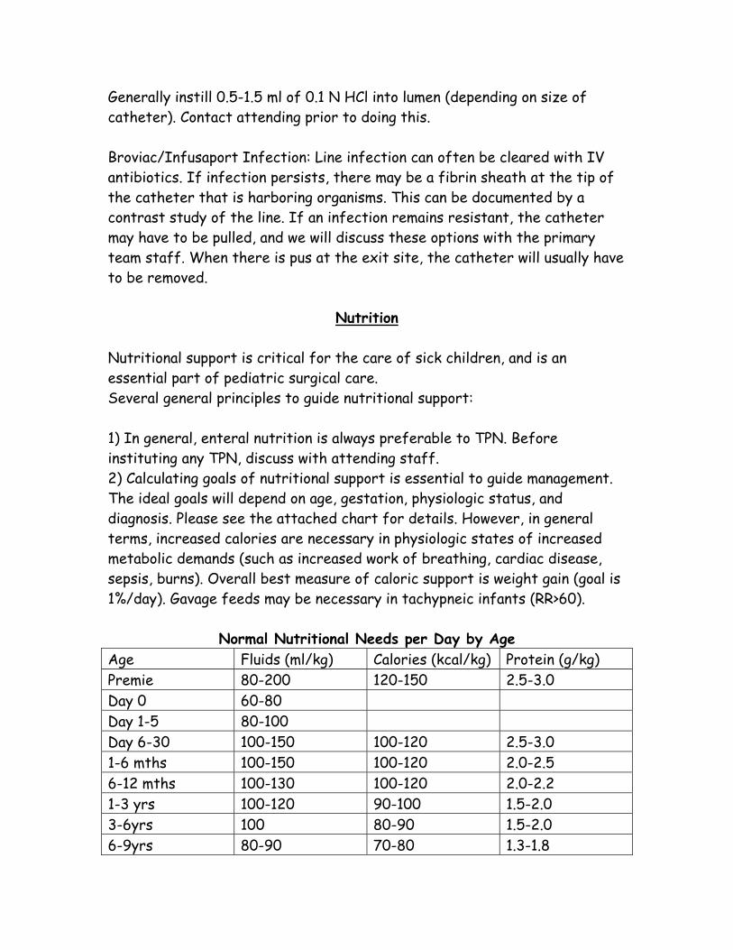
Nutritional support is critical for the care of sick children, and is an essential part of pediatric surgical care. Several general principles to guide nutritional support: 1) In general, enteral nutrition is always preferable to TPN. Before instituting any TPN, discuss with attending staff. 2) Calculating goals of nutritional support is essential to guide management. The ideal goals will depend on age, gestation, physiologic status, and diagnosis. Please see the attached chart for details. However, in general terms, increased calories are necessary in physiologic states of increased metabolic demands (such as increased work of breathing, cardiac disease, sepsis, burns). Overall best measure of caloric support is weight gain (goal is 1%/day). Gavage feeds may be necessary in tachypneic infants (RR>60).
Normal Nutritional Needs per Day by Age Age Fluids (ml/kg) Calories (kcal/kg) Protein (g/kg) Premie 80-200 120-150 2.5-3.0 Day 0 60-80 Day 1-5 80-100 Day 6-30 100-150 100-120 2.5-3.0 1-6 mths 100-150 100-120 2.0-2.5 6-12 mths 100-130 100-120 2.0-2.2 1-3 yrs 100-120 90-100 1.5-2.0 3-6yrs 100 80-90 1.5-2.0 6-9yrs 80-90 70-80 1.3-1.8
9-12yrs 60-80 60-70 1.3-1.8 >12yrs 40-60 30-60 1.0-1.5 Enteral nutrition Choice of enteral formulation, discussed below, depends on age, medical status, and many other factors, including parental preference. Breast Milk: In most cases, breast milk is the best feed for infants. Breast milk is tailored to fit the nutritional needs of a developing infant: Protein: Breast milk proteins include mucins, caseins, and whey proteins. Over the course of normal lactation, the protein in breast milk is approximately 60% whey proteins (primarily alpha-lactalbumin) and 40% casein, although the protein composition changes during lactation. Early in lactation, the concentration of whey proteins is high, whereas casein is virtually undetectable. Subsequently, casein concentration increases, and the concentration of whey proteins decreases. The major casein in breast milk is ß-casein, a highly phosphorylated protein. Clusters of phosphorylated serine and threonine residues are capable of complexing Ca2+ ions, which facilitate Ca2+ absorption. Mucins, known as milk fat globule membrane proteins, surround the lipid globules in milk and contribute only a small percentage of the protein. The protein content of breast milk is 14–16 g/L during early lactation, 8–10 g/L at 3–4 mo, and 7–8 g/L at 6 mo and later. Carbohydrates: The main carbohydrate in breast milk is lactose, and contributes approximately 42% of calories. There is considerable difference in carbohydrate content of human milk during the different phases of lactation. Initially, monosaccharides represent a small percentage of the carbohydrate content. During lactation, there is a rise in lactose and a decrease in oligosaccharides. The exact role for oligosaccharides is unknown, but may represent a low osmolar source of calories. Oligosaccharidases on small intestinal epithelial cells may degrade milk oligosaccharides. The different monosaccharides thus obtained (such as fucose and sialic acid) can be used for synthesis of glycoproteins and glycolipids. Within the gut, ligosaccharides, together with lactose, stimulate the growth of Lactobacillus, the bifidus flora that is protective against GI infections. Oligosaccharides may also inhibit bacterial adhesion to epithelial surfaces.
Amylase: Breast milk contains a high concentration of {alpha} amylase, which is relatively stable against pepsin degradation. Amylase from breast milk may compensate for low salivary and pancreatic amylase activity in newborns and aid in the digestion of complex carbohydrates when complementary foods are being fed in close proximity to breastfeeding. {alpha}1-Antitrypsin: The protease inhibitors {alpha}1-antitrypsin and anti-chymotrypsin in human milk may limit the activity of pancreatic enzymes, acting as natural "brake" molecules. Bile salt–stimulated lipase: Bile salt–stimulated lipase in human milk may aid in lipid digestion, particularly in preterm infants, who have low lipase activity and poor lipid utilization. These lipases hydrolyze acylglycerols, cholesterol esters, diacylphosphatidylglycerols; and micellar as well as water-soluble substrates. Haptocorrin: Virtually all of vitamin B-12 in human milk is bound to haptocorrin. There is more haptocorrin than vitamin B-12 on a molar basis, resulting in the protein being largely in the unsaturated form, which adds to antimicrobial activity. Intrinsic factor is present in the stool of breastfed infants at young age, but its concentration may not be adequate to facilitate the uptake of vitamin B-12. Insulin-like growth factor– binding proteins: Insulin-like growth factors (IGFs) I and II are present in human milk, and are associated with IGF-binding proteins. These binding proteins may protect IGF-I and IGF-II from being digested, and modulate their interaction with intestinal receptors. After the binding of IGF-I and IGF-II to enterocytes, they may exert activity locally and systemically. Immunoglobulins: All types of immunoglobulins are found in human breast milk, but the major type is secretory (s)IgA (> 90%)—a dimer of IgA linked together with a secretory component and a joining chain. Immunoglobulin levels are particularly high in colostrum. Secretory IgA attaches to the lining of the nose, mouth, and throat and retards attachment of microorganisms. Immunoglobulin levels are remarkably high in breast milk, {approx} 1–2 g/L in early lactation, and remain at 0.5–1 g/L up to 2 y of lactation. Specific antibody-mediated immunity increases in response to maternal exposure can be transferred to her breastfed infant, mediated via the so-called enteromammary pathway. Lactoferrin: Lactoferrin is an iron-binding protein that limits the availability of iron to bacteria in the intestines, and is found in highest concentration in
colostrum. Lactoferrin may withhold iron from iron-requiring pathogens. Lactoferrin also has bactericidal activity as a result of the formation of lactoferricin, which inhibits the attachment of enteropathogenic E. coli to ntestinal cells. Lactobacilli: Human breast milk encourages the growth of lactobacilli, which can inhibit gram-negative bacteria and parasites within the gut. Breastfed infants have lactobacillus levels typically 10 times greater than formula-fed infants. Lysozyme: One major components of human milk whey is lysozyme, an enzyme which degrades the outer cell wall of gram-positive bacteria. Interestingly, although other contents of breast milk vary widely between well-nourished and poorly nourished mothers, the amount of ysozyme is strongly conserved. Lysozyme has been shown to kill gram-negative bacteria in synergy with lactoferrin. Standard formulas (Enfamil® LIPIL /Similac) are the cheapest and most widely available standard formulas, and should be used in the absence of breast milk unless there are other concerns. Premature infants (<34 weeks) usually get premature fomulas, such as PF20 (premature formulation, 20 kcal/oz) or PF 24 (24 kcal/oz). For newborns up to 4-6 months we use Enfamil LIPIL. Usual feed for children older than 6 months of age is Pediasure (30 kcal/oz). Protein: The protein of these standard formulas is derived from cow milk, and includes a whey protein to casein ratio similar to that in human breast milk. Fat: The fat concentration in Enfamil is 48% of total calories, similar to breast milk, and is composed of vegetable oils, consisting of 45% palm olein, 20% soy oil, 20% coconut oil, and 15% high oleic sunflower oil. These fats include monounsaturated fatty acid (38% of total fats) and linoleic acid levels similar to that in breast milk. Carbohydrate: The carbohydrate in Enfamil is lactose, similar to breast milk. Lactose contributes 44% of the total calories. Micronutrients: Enfamil LIPIL contains docosahexaenoic acid (DHA) as well as arachidonic acid (ARA). Both of these agents are found in similar levels to breast milk, and are thought to contribute to normal growth of eye and
other tissues, particularly in the CNS. The nucleotide levels in Enfamil are patterned after free nucleotide levels found in breast milk, and include adenylic acid (AMP), guanylic acid (GMP), cytidylic acid (CMP), and uridylic acid (UMP). Calcium and Phosphorus: The levels of calcium and phosphorus in Enfamil are similar to breast milk. Enfamil provides 500 mg of calcium per quart and 340 mg of phosphorus per quart (Ca:P ratio of about 1.5:1)—a balance which is important for mineral utilization. The Ca:P ratio of breast milk is approximately 1.6:1. Iron: Enfamil with Iron provides 11.5 mg of iron per quart. Low-iron Enfamil is contraindicated for infants beyond 4 months of age. The American Academy of Pediatrics recommends the use of supplemental iron in the first year of life. The Recommended Dietary Allowance for iron is 6 mg/day until 6 months of age and 10 mg/day between 6 months and 3 years of age. Soy formulas (Prosoybee/Isomil) can be used for infants who are intolerant to breast milk or cow milk protein (with malabsorptive symptoms). The soy protein-based formulas currently on the market are all free of cow milk-protein as well as glucose. They are prepared at similar caloric density to both breast milk and standard formulas (20 kcal/ounce). The fat content of soy protein-based formulas is derived from vegetable oils, including soy, palm, sunflower, olein, safflower, and coconut oils. The quantity of fats is similar to that in the corresponding cow milk-based formula, and ranges from 3.6 to 3.8 g/dL. The carbohydrates are lactose free, such as corn starch, corn starch hydrolysate, tapioca starch, or sucrose, with content ranging from 6.7 to 6.9 g/dL. All soy formulas are iron-fortified, and meet specifications from the American Academy of Pediatrics and established by the US Food and Drug Administration. To address the use of soy formulas, the AAP concensus statement, (Pediatrics, 101 (1), 148-153, 1998) states: 1) In term infants whose nutritional needs are not being met from breast milk or cow milk-based formulas, soy protein-based formulas are safe alternatives. Isolated soy protein-based formula has no advantage over cow milk protein-based formula as a supplement for the breastfed infant. 2) Because soy protein-based formulas are lactose-free, they are helpful for galactosemia and hereditary lactase deficiency. 3) Parents seeking a vegetarian-based diet for a term infant can be advised to use isolated soy protein-based formula.
4) Most previously well infants with acute gastroenteritis can be managed after rehydration with breast milk or standard dilutions of cow milk-based formulas. Isolated soy protein-based formulas are indicated when lactose intolerance has been documented. 5) The routine use of isolated soy protein-based formula has no proven value in the management of infantile colic or in the prevention of atopic disease in healthy or high-risk infants. 6) Infants with documented cow milk protein-induced enteropathy or enterocolitis frequently are as sensitive to soy protein and should not be given isolated soy protein-based formula routinely. They should be provided formula derived from hydrolyzed protein or synthetic amino acid. 7) Most infants with documented IgE-mediated allergy to cow milk protein will do well on soy protein-based formula. 8) Soy protein-based formulas are not designed or recommended for preterm infants who weigh <1800 g. Elemental or semi-elemental formulas are hypoallergenic, lactose free, and have predigested proteins (primarily hydrolyzed caseins). These formulas are used for infants with milk protein allergies, malabsorption, short bowel, and cystic fibrosis. In general, these formulas are used when there is a concern for an allergy to either cow’s milk proteins or soy proteins. Nutramigen LIPIL should be the first elemental formula used, because it is widely available and least costly. Pregestimil, which uses MCT as part of its fat source, may be better tolerated than Nutramigen (especially for infants with CF), but it is more expensive and must be obtained from a pharmacy. Alimentum is a similar formula that is not as widely available as Pregestimil. Neocate is a special elemental formula designed to provide nutritional support for infants with cow milk allergy and food protein intolerance. Neocate is primarily used in premature infants, but may be helpful for term infants as well. Neocate is used in various clinical situations (eg. short gut syndrome) in which there is impaired intestinal absorption. The caloric distribution is 12% protein, 47% carbohydrate, 41% fat, 100% free amino acids including taurine and carnitine. The carbohydrates are lactose-free to avoid lactose-induced diarrhea. Neocate is more expensive and harder to obtain as an outpatient than other elemental formulas, and should be limited to situations when other formulas are not acceptable. Initiation and progression of feeds for premature infants:
The initiation of feeds should be based on bowel function. Feeds may be started if umbilical lines are present, and continued until feeds reach 40cc/kg/d. Use birth weight to calculate enteral feeds until infant regains birth weight, and then use actual weight. At 80-100 cc/kg/d of enteral feeds for infants > 1000 gms and at 100 cc/kg/d for infants = 1000 gms, change to clear IVF with electrolytes and protein added. Once feeds reach 100cc/kg/d, consider D/C of IVF. HMF is not be added to breast milk until 150cc/kg/d, with the second packet added after HMF is tolerated for 3 days. If slow growth is noted on 150-160 cc/kg/d, consider increasing kcal/oz or volume. Infants with CLD require a minimum of 140kcal/kg/d. ELBW infants should be D/C on 24 or 27 cal feeds. ELBW infants on breast feeds may need to alternate with bottle feeds until growth is established. TPN TPN orders need to be modified daily to account for changing fluid and electrolyte needs. The residents will write all the TPN orders, but must review with nutrition support service and pharmacy staff. All TPN orders should be discussed on morning rounds with the senior resident. Nutrition services (see telephone numbers) can assist you, and contact them at the beginning of the rotation to learn basics. All TPN orders must be entered by noon, so the bag can be hung ~9PM. Volume: The volume of TPN is generally based on the 100/50/25 or 4/2/1 rule for maintenance fluids (see table below). Euvolemic patients should receive maintenance fluids, which include the TPN and lipid volume. When writing TPN orders, first calculate the lipid volume, and subtract that from the total fluids to determine the TPN volume. TPN is infused continuously over 24 hrs, lipids over 20 or 24 hrs. Calories: Given in the form of glucose and lipids as follows: Dextrose (glucose): Start with the dextrose concentration from previous IVF (usually D10). You may increase dextrose by 2.5% daily as long as hyperglycemia or long term starvation and thus risk of refeeding syndrome is not present. Higher serum glucose levels may be acceptable (140s-150s) as long as no more than trace glucosuria is present. Hyperglycemia is poorly tolerated in infants, and requires a decrease in glucose infusion. Maximum dextrose concentration in peripheral line is 12.5%; maximum in central line (UVC or Broviac) is that which gives 12-15 mg/kg/min of dextrose (usually D20 to D25).
Lipid: Lipids are an efficient caloric source (9 kcal/gm), and are given in the form of 20% intralipid. Start infants at 0.5-1 gm/kg/day, and increase by 0.5-1.0 gm/kg/day to max of 3 g/kg/day. Serum triglyceride levels should be monitored at least weekly. Do not advance if triglyceride levels are > 180. If greater than 200, hold intralipids for that day. Intralipids are ordered as cc's to be given over 20 or 24 hours as a continuous infusion. Using 20% intralipids, multiply the g/kg/day by 5 to determine the volume required per day. Protein: Given as trophamine. It is controversial whether protein should contribute to caloric support calculations, as in the non-catabolic state, protein are used preferentially as tissue substrate for growth. Begin as 0.5 g/kg/day, and increase 0.5 gm/kg/day to max of 3 gm/kg/day. Electrolytes: Given to account for daily requirements, dependent on age and clinical status. Sodium requirements (3-5 mEq/kg/day) are higher for premies than older infants. No salt necessary for first 24 hrs of life (obligate salt wasting period). Acetate is converted in the liver to bicarbonate, and the ratio of acetate to chloride is based on serum bicarbonate level. In addition to Na, K and Ca, PO4 (0.5-1 mmol/kg/day) and Mg (0.2 to 0.5 mEq/kg/day). Try to keep the Ca:PO4 ratio around 2:1 (mEq:mmol) to limit calcium salt precipitation. Check Ca and Mg twice weekly. Minor changes in TPN will be made based on daily electrolytes. Monitoring: Daily ICN panel, QID glucose checks, and weekly or biweekly LFTs. TPN-Associated Cholestasis There is a close association between cholestasis and prolonged use of parenteral nutrition, particularly in neonates. Histologically, the liver shows bile duct proliferation in the portal triad region, with the formation of large tracts of fibrosis between normal appearing hepatocytes. Several factors associated with TPN-associated cholestasis include low birth weight, prematurity, and duration of TPN administration. Although reversible in its early stage, eventually irreversible cirrhosis will develop. There is also a higher infection rate in these patients due to decreased T-lymphocyte function and lymphocytic proliferation. Infants with cholestasis have a 10x higher mortality compared to those on TPN without cholestasis. The exact etiology of TPN-related cholestasis is unknown. Virtually every possible factor in TPN has been implicated as a causative agent. Among the more
likely factors is a lack of taurine, which prevents the conjugation and excretion of bile salts. More recently, phytosterols found in lipid compounds have been suspected to have a role in cholestasis. One clear cause of liver injury is overfeeding, commonly with excessive carbohydrates. The liver pathology in this condition is different in that the only finding is hepatic steatosis, without bile duct proliferation. By limiting the amount and type of delivered calories, this problem can be avoided. The incidence of bacterial translocation is higher in a fasting state. During bacterial translocation, a release of endotoxins can lead to the secretion of cytokines including TNF and interferon-gamma, which may lead to liver injury. The use of metronidazole may reduce cholestasis by reducing the colonization of the GI tract. During prolonged periods of fasting, the GI tract lacks sufficient enteral stimulation for the release of a variety of hormones which promote bile flow. Such hormones may be critical in preventing biliary stasis. Use of cholecystokinin has been tried in both animals as well as in clinical trials with moderate success. ELECTROLYTE / ACID-BASE Calcium chloride (CaCl2): 10 - 20 mg/kg (Max: 500 mg) Calcium gluconate: 100 mg/kg (Max: 1 gm) Sodium bicarbonate (NaHCO3): 1 - 2 mEq/kg Sodium phosphate (NaH2PO4): 0.1 - 0.3 mMol/kg over 3 - 6h; max: 10 mMol THAM (Tromethamine): 3 cc/kg ~ 1 mEq/kg base Potassium Chloride (KCl): if K+ = 2.5 - 3.5, then 0.5 mEq/kg over 1 hr (cardiac monitoring); if K+ < 2.5, then 1 mEq/kg over 2hr (cardiac monitoring) Potassium Phosphate (KH2PO4): 0.1 - 0.3 mMol/kg over 3 - 6h; max: 10 mMol (1 mMol KH2PO4 = 1.5 mEq K+)
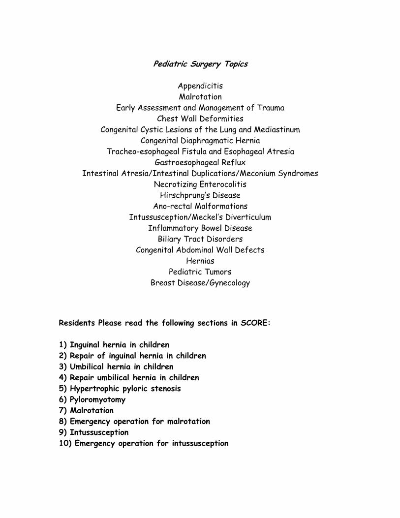
Pediatric Surgery Topics
Appendicitis Malrotation
Early Assessment and Management of Trauma Chest Wall Deformities
Congenital Cystic Lesions of the Lung and Mediastinum Congenital Diaphragmatic Hernia
Tracheo-esophageal Fistula and Esophageal Atresia Gastroesophageal Reflux
Intestinal Atresia/Intestinal Duplications/Meconium Syndromes Necrotizing Enterocolitis
Hirschprung’s Disease Ano-rectal Malformations
Intussusception/Meckel’s Diverticulum Inflammatory Bowel Disease
Biliary Tract Disorders Congenital Abdominal Wall Defects
Hernias Pediatric Tumors
Breast Disease/Gynecology
Residents Please read the following sections in SCORE: 1) Inguinal hernia in children 2) Repair of inguinal hernia in children 3) Umbilical hernia in children 4) Repair umbilical hernia in children 5) Hypertrophic pyloric stenosis 6) Pyloromyotomy 7) Malrotation 8) Emergency operation for malrotation 9) Intussusception 10) Emergency operation for intussusception
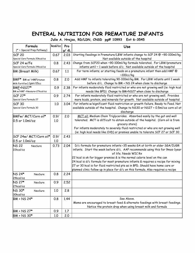
ENTERAL NUTRITION FOR PREMATURE INFANTS
Julie A. Hrejsa, RD/LDN, CNSD pg# 10993 Ext 6-3545
Formula (* = Special Prep Pathway)
kcal/cc Pro g/ dL
Use
SCF 20 Special Care Formula 20kcal/oz
0.67 2.03 Starting feedings in Premature/LBW infants change to SCF 24 @ ~90-100ml/kg. Not available outside of the hospital
SCF 24 w/Fe Special Care Formula 24kcal/oz
0.8 2.43 Change from SCF20 when ~90-100ml/kg formula tolerated. For LBW/premature infants until ~ 1 week before d/c. Not available outside of the hospital
BM (Breast Milk) 0.67 1.1 For term infants; or starting feeds on a premature infant then add HMF @ ~100cc/kg
BMF* BM w/ HMF(Human Milk Fortifier) 1pkt/25cc
0.8 2.0 Add HMF to infants tolerating 90-100ml/kg BM. For LBW infants until 1 week before d/c. Change to BM + NS 24 when close to discharge
BMF+NS27* BM w/HMF +Neosure=27kcal/oz
0.9 2.38 For infants moderately fluid restricted or who are not growing well (ie: high kcal needs like BPD) Change to BM+NS27 when close to discharge
SCF 27* Special Care Formula 27
0.9 2.74 For infants moderately fluid restricted or who are not growing well. Provides more kcals, protein, and minerals for growth. Not available outside of hospital
SCF 30 Special Care Formula 30
1.0 3.04 For infants w/significant fluid restriction or growth failure. Ready to Feed. Not available outside of the hospital. Change to NS30 or NS27 + 0.5ml/oz corn oil at
discharge BMFw/ MCT/Corn oil* 0.5 or 1.0ml/oz
0.9/ 1.0
2.0 MCT oil: Medium Chain Triglycerides. Absorbed easily by the gut and well tolerated. MCT is difficult to obtain outside of the hospital. (Corn oil is from
grocery store) For infants moderately to severely fluid restricted or who are not growing well (ie: high kcal needs like CHD) or premies unable to tolerate SCF 27 or SCF 30
SCF 24w/ MCT/Corn oil* 0.5 or 1.0ml/oz
0.9/ 1.0
2.43
NS 22 NeoSure 22kcal/oz
0.73 2.04 D/c formula for premature infants <35 weeks GA at birth or older SGA/IUGR infants. Start the week before d/c. AAP recommends using this for 9mos-1year
of life. Needs WIC Rx 22 kcal is ok for bigger premies & is the normal calorie level on the can 24 kcal is d/c formula for most premature infants & requires a recipe for mixing 27 or 30 kcal is for fluid restricted pts as in BPD. Should have home care or planned clinic follow up in place for d/c on this formula. Also requires a recipe
NS 24* NeoSure 24kcal/oz
0.8 2.24
NS 27* NeoSure 27kcal/oz
0.9 2.52
NS 30* NeoSure 30kcal/oz
1.0 2.8
BM + NS 24* 0.8 1.44 See Above. Moms are encouraged to breast feed & alternate feedings with breast feedings.
Notice the protein drop when using breast milk and formula BM + NS 27* 0.9 1.7 BM + NS 30* 1.0 2.0
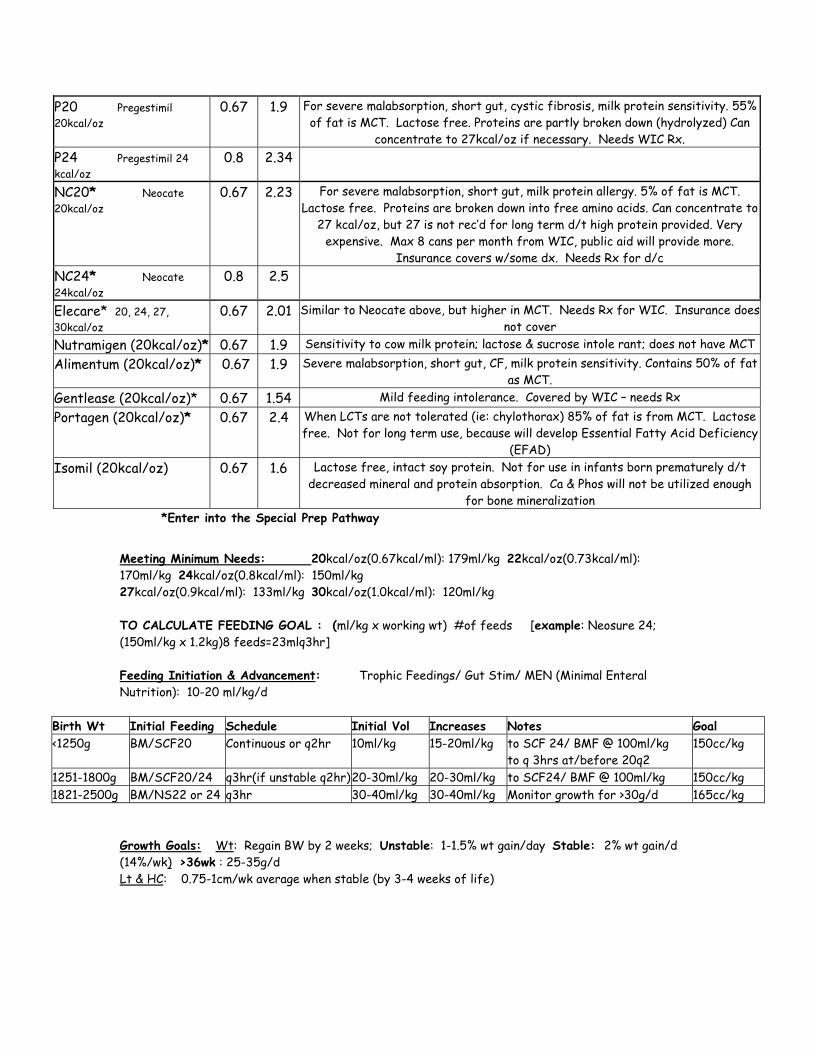
P20 Pregestimil 20kcal/oz
0.67 1.9 For severe malabsorption, short gut, cystic fibrosis, milk protein sensitivity. 55% of fat is MCT. Lactose free. Proteins are partly broken down (hydrolyzed) Can
concentrate to 27kcal/oz if necessary. Needs WIC Rx. P24 Pregestimil 24 kcal/oz
0.8 2.34
NC20* Neocate 20kcal/oz
0.67 2.23 For severe malabsorption, short gut, milk protein allergy. 5% of fat is MCT. Lactose free. Proteins are broken down into free amino acids. Can concentrate to
27 kcal/oz, but 27 is not rec’d for long term d/t high protein provided. Very expensive. Max 8 cans per month from WIC, public aid will provide more.
Insurance covers w/some dx. Needs Rx for d/c NC24* Neocate 24kcal/oz
0.8 2.5
Elecare* 20, 24, 27, 30kcal/oz
0.67 2.01 Similar to Neocate above, but higher in MCT. Needs Rx for WIC. Insurance does not cover
Nutramigen (20kcal/oz)* 0.67 1.9 Sensitivity to cow milk protein; lactose & sucrose intole rant; does not have MCT Alimentum (20kcal/oz)* 0.67 1.9 Severe malabsorption, short gut, CF, milk protein sensitivity. Contains 50% of fat
as MCT. Gentlease (20kcal/oz)* 0.67 1.54 Mild feeding intolerance. Covered by WIC – needs Rx Portagen (20kcal/oz)* 0.67 2.4 When LCTs are not tolerated (ie: chylothorax) 85% of fat is from MCT. Lactose
free. Not for long term use, because will develop Essential Fatty Acid Deficiency (EFAD)
Isomil (20kcal/oz) 0.67 1.6 Lactose free, intact soy protein. Not for use in infants born prematurely d/t decreased mineral and protein absorption. Ca & Phos will not be utilized enough
for bone mineralization *Enter into the Special Prep Pathway
Meeting Minimum Needs: 20kcal/oz(0.67kcal/ml): 179ml/kg 22kcal/oz(0.73kcal/ml): 170ml/kg 24kcal/oz(0.8kcal/ml): 150ml/kg 27kcal/oz(0.9kcal/ml): 133ml/kg 30kcal/oz(1.0kcal/ml): 120ml/kg TO CALCULATE FEEDING GOAL : (ml/kg x working wt) #of feeds [example: Neosure 24; (150ml/kg x 1.2kg)8 feeds=23mlq3hr] Feeding Initiation & Advancement: Trophic Feedings/ Gut Stim/ MEN (Minimal Enteral Nutrition): 10-20 ml/kg/d
Birth Wt Initial Feeding Schedule Initial Vol Increases Notes Goal <1250g BM/SCF20 Continuous or q2hr 10ml/kg 15-20ml/kg to SCF 24/ BMF @ 100ml/kg
to q 3hrs at/before 20q2 150cc/kg
1251-1800g BM/SCF20/24 q3hr(if unstable q2hr) 20-30ml/kg 20-30ml/kg to SCF24/ BMF @ 100ml/kg 150cc/kg 1821-2500g BM/NS22 or 24 q3hr 30-40ml/kg 30-40ml/kg Monitor growth for >30g/d 165cc/kg
Growth Goals: Wt: Regain BW by 2 weeks; Unstable: 1-1.5% wt gain/day Stable: 2% wt gain/d (14%/wk) >36wk : 25-35g/d Lt & HC: 0.75-1cm/wk average when stable (by 3-4 weeks of life)
Goals and Objectives Pediatric Surgery Rotation for Medical Students Addendum MS 3 Length of Rotation - 4 weeks Goals and Objectives 1) Medical Knowledge Student will demonstrate an understanding of the following fluid balance in neonates maintenance of temperature in neonates signs, symptoms, diagnosis and treatment of acute appendicitis in children signs, symptoms, diagnosis and treatment of bowel obstruction in neonates etiology, anatomy, signs and treatment of inguinal hernia in children 2) Patient Care The student will demonstrate the ability to take an complete history perform a thorough physical examination construct an appropriate differential diagnosis formulate an appropriate diagnostic and treatment plan 3) Communication Skills The student will Present cases in an organized, succinct fashion Communicate treatment plans to patients, parents, nurses, residents and attendings 4) Professionalism The student will display professional behavior at all times including Appropriate grooming and dress Use of appropriate and respectful language Respect for patients and families in all interactions Respect for other members of the healthcare team Honesty 5) Practice-based and Lifelong Learning
The student will demonstrate a commitment to self-directed learning and improvement through research and presentation of a chosen topic in pediatric surgery and regular reading on subjects pertaining to patient care. 6) Systems-based Practice The student will demonstrate an understanding of how health professionals work together with parents to accomplish patient care goals through participation in rounds and discussions with other members of the healthcare team. The student will demonstrate an understanding of quality improvement through participation in rounds and attendance at morbidity and mortality conference.