Embed Size (px)
Citation preview

Development and Analysis of a Novel Nanotherapeutic for the Treatment of Multi-drug Resistant Ovarian Cancer
A Major Qualifying Project Report
Submitted to the Faculty of the WORCESTER POLYTECHNIC INSTITUTE
in partial fulfillment of the requirements for the Degree of Bachelor of Science
by:
___________________________________
Jake Brown Biology and Biotechnology
___________________________________
Libbi Richardson Biology and Biotechnology
Date: April 2015
Approved:
____________________________________
Professor Mike Buckholt, PhD Worcester Polytechnic Institute
Biology and Biotechnology
____________________________________
Professor Jill Rulfs, PhD Worcester Polytechnic Institute
Biology and Biotechnology

1
Table of Contents Table of Figures ............................................................................................................................................. 3
Table of Tables .............................................................................................................................................. 4
Acknowledgements ....................................................................................................................................... 5
Authorship .................................................................................................................................................... 6
Abstract ......................................................................................................................................................... 7
Background ................................................................................................................................................... 8
Introduction .............................................................................................................................................. 8
Part 1: Ovarian cancer ............................................................................................................................... 8
Disease Statistics ................................................................................................................................... 8
Types of Ovarian Cancer ....................................................................................................................... 8
Stages .................................................................................................................................................. 10
Existing Treatments ............................................................................................................................ 11
Part 2: Cell Death .................................................................................................................................... 12
Apoptosis ............................................................................................................................................ 13
Necrosis ............................................................................................................................................... 13
Autophagy ........................................................................................................................................... 14
Part 3: Emerging, Innovative Cancer Therapies ...................................................................................... 14
Cytotoxics ............................................................................................................................................ 14
Ceramide ............................................................................................................................................. 15
Kinase Inhibitors .................................................................................................................................. 16
Nanomedicines ................................................................................................................................... 17
Folate Targeting .................................................................................................................................. 19
Part 4: Hypothesis and Specific Aims ...................................................................................................... 21
Materials and Methods ............................................................................................................................... 22
General Manufacturing Nanomedicines ................................................................................................. 22
HPLC Method of Detecting DTX .............................................................................................................. 23
Quantification of APIs in Nanomedicines ............................................................................................... 23
Cell Lines ................................................................................................................................................. 23
Cytotoxicity Assay ................................................................................................................................... 24
Results ......................................................................................................................................................... 27
Manufacturing and Monitoring Stability of Nanomedicines .................................................................. 27
HPLC Detection of DTX ............................................................................................................................ 29

2
Cytotoxicity Assays .................................................................................................................................. 31
Discussion.................................................................................................................................................... 34
References .................................................................................................................................................. 37

3
Table of Figures Figure 1. The stages of ovarian cancer. ..................................................................................................... 10
Figure 2.The types of cell death.. ............................................................................................................... 13
Figure 3. Docetaxel. .................................................................................................................................... 15
Figure 4. Ceramide.. .................................................................................................................................... 16
Figure 5. Staurosporine. ............................................................................................................................. 17
Figure 6. Diagram of Generic Nanoparticle. a. Nanoemulsion.. ............................................................. 19
Figure 7. Cytotoxicity Results for DTX, DTX-CER, and DTX-CER-STP nanoemulsions. .............................. 32

4
Table of Tables Table 1. The plate layout for cells dose with docetaxel nanoemulsions. ................................................ 25
Table 2. The plate layout for cells dose with staurosporine and/or combination nanoemulsions. ....... 26
Table 3. Hydrodynamic Diameter and Polydispersity Index of Nanoemulsions. ..................................... 28
Table 4. Zeta Potential of Nanoemulsions.. .............................................................................................. 29
Table 5. Docetaxel HPLC Loading Efficiency. ............................................................................................. 30
Table 6. Average IC50 Values for Nanoemulsions Tested on SKOV-3 cells. ............................................. 31

5
Acknowledgements
Our team would like to recognize those who were involved in making this project a
possibility and who supported us throughout the duration of the project. We would like to
thank the following people from our sponsor, Nemucore Medical Innovations, for providing
us with direction, supplies, lab equipment, and their expertise throughout the year: Dr. Tim
Coleman, President and CEO; Dr. Nirav Patel, Principal Scientist; and Dr. Alex Piroyan,
Principal Scientist. We would also like to thank our project advisors Dr. Mike Buckholt and
Dr. Jill Rulfs (Worcester Polytechnic Institute) for their guidance, support, and
encouragement throughout the year.

6
Authorship
Both team members spent equal amounts of time working in the lab as well as on the
report for this project.
7
Abstract
Ovarian cancer, when treated with platinum and taxanes, is frequently detected in
later stages and often develops multi-drug resistant properties. Therefore, chemotherapy
regiments can become ineffective, leaving clinicians with less efficacious therapies. Our
hypothesis is that a nanotherapy approach, utilizing nanoemulsions (NE) to deliver a
combination of drugs with synergistic mechanisms of action, will likely reduce the spread
of ovarian cancer and deliver the combination with reduced systemic toxicity. Results
suggest that combination NEs can be manufactured and show some promise to kill ovarian
cancer cells more efficiently.

8
Background
Introduction
Despite substantial improvements in ovarian cancer treatments, optimizing drug
efficacy and minimizing drug toxicity continues to be an important concern. Initially, most
ovarian cancers are sensitive to chemotherapy, but with continued use can result in multi-
drug resistance of cancers. In order to overcome multi-drug resistance while enhancing
potency, a novel way of delivering therapies is needed.
Part 1: Ovarian cancer
Disease Statistics
Ovarian cancer accounts for 3% of cancer in women. (American Cancer Society,
2014). It was projected that for 2014, 21,980 women would receive a new diagnosis of
ovarian cancer and 14,270 would die from the disease (American Cancer Society, 2014). In
the course of a woman’s lifetime, the risk of being diagnosed with ovarian cancer is about 1
in 73, although it mainly develops in older white women, particularly 60 years or older
(American Cancer Society, 2014). In the past, ovarian cancer has tended to remain clinically
silent until the disease had reached an advanced stage that predominantly remained
intraperitoneal throughout its course (Longo & Young, 1981). The percent of the
population affected by ovarian cancer is relatively low and the detection of the disease is
difficult, the 5-year survival rate is extremely low (Parkin, 2011), thus making it a large
unmet clinical need.
Types of Ovarian Cancer
Far too often, ovarian cancer is detected in its late stages, making it extremely
difficult to treat. Often, the disease is identified clinically as tumors that grow in the ovaries.
These tumors can either be benign, borderline, low malignant potential (LMP), or
9
malignant (Johns Hopkins Pathology, 2001). Benign tumors will not metastasize; however,
malignant cells tend to metastasize in two ways. First malignant cells can metastasize by
directly migrating to other organs in the pelvic or abdominal region (Ovarian Cancer
National Alliance, 2014). The second and less common way for malignant tumors to spread
throughout the body is through the bloodstream or lymph nodes (Ovarian Cancer National
Alliance, 2014). Part of the difficulty of detecting ovarian cancer early is due to the
unknown initial causes. Some theories that may explain the cause of ovarian cancer include
genetic errors due to “wear and tear” of the monthly release of the egg or increased
hormone levels before and during ovulation that can stimulate the growth of abnormal
cells (Ovarian Cancer National Alliance, 2014). The ovaries, which are involved in both the
reproductive and endocrine body systems, are small organs that are responsible for
maintaining the health of the female reproductive system.
The ovaries are very complex. Because of their complexity, there are over 30
different types of ovarian cancer, all affecting and localizing different parts of the ovaries
(Ovarian cancer National Alliance, 2014). The most common forms of ovarian cancer can be
found in three distinct cell types: epithelium, germ cells, or stromal cells. The first of these
is the ovarian surface epithelium. Surface epithelium cells are a modified mesothelium that
can give rise to human ovarian carcinomas. The surface epithelium cells can quite often
assume atypical morphologies, known as abnormal cells, which make it extremely difficult
to identify normal epithelial cells (Auersperg et al. 1994). Surface epithelial tumors tend to
account for about 60% of all ovarian cancer neoplasms (Johns Hopkins Pathology, 2001).
Second, ovarian cancer tumors can be found in germ cells. These cells are totipotent
progenitors that are destined to become female germ cells, or oocytes (Bukovsky et al.

10
2006). Germ cell neoplasms make up the second largest amount of ovarian neoplasms at
about 20% (Johns Hopkins Pathology, 2001). The third cell type in which ovarian cancer
tumors can be found are stromal cells. Stromal cells are spindle-shaped morphologically,
similar to fibroblasts, organized into a whorled texture (King, 2013). The highly vascular
tissues comprised of stromal cells account for about 10% of all ovarian neoplasms (Johns
Hopkins Pathology, 2001).
Stages
Ovarian cancer severity is clearly defined in four stages: Stage I (A, B, and C), Stage II
(A, B, and C), Stage III (A, B, and C), and Stage IV (National Ovarian Cancer Coalition, 2014).
Each stage designates the spread or progression of the disease. The intensity ranges from
Stage I, limiting the growth of cancer to the ovary or ovaries, to Stage IV in which the
ovarian cancer has spread out of the abdomen towards the liver (Texas Oncology, 2014).
Figure 1 shows the intensity of cancer in each of the four stages. The smaller green dots
represent the cancer, and as the stages increase, the amount of cancer as well as the surface
area covered by cancer increases.
Figure 1. The stages of ovarian cancer. From left to right, the stages of ovarian cancer are shown starting with no cancer and ending with Stage IV ovarian cancer (Cannistra, 1993).
11
Existing Treatments
The treatment of ovarian cancer continues to be difficult today due to difficulty in
diagnosing the disease until it has reached an advanced stage. However, the regimen of
drug and delivery method has changed to reduce side effects and improve tumor reduction.
In the 1980s, scientist believed the primary forms of treating the disease were either
surgery to remove tumors or radiation therapy (Longo & Young, 1981). Other typical
treatment forms were chemotherapy using single agents as well as combination therapies.
Single agents included melphalan, 5-fluorouracil, and hexamethylmelamine (Long & Young,
1981).Melphalan is a nitrogen mustard alkylating agents that attach an alkyl group to DNA
to induce apoptosis; fluoricil works to induce apoptosis by inhibiting cell synthases; and
hexamethylmelamine also works to induce apoptosis through alkylating agents (Long &
Young, 1981). Chemotherapy using combined therapies would consist of a strict regimen of
particular schedules and concentrations of drugs formulated for intravenous infusion into
the body.
The treatments that show efficacy in ovarian cancer therapy have greatly advanced
in the last 30 years. Generally, ovarian cancer is a chemosensitive disease and the many
chemotherapies have been tested to improve progression free survival (Ozols, 2006).
Chemotherapy can be administered using either an intravenous (IV) or intraperitoneal (IP)
delivery method. IV methods are administered through the vein whereas IP methods are
delivered directly into the peritoneal cavity (Ozols, 2006). The IP method is able to deliver
more drug directly to tumor sites than IV administration (Ozols, 2006). Since the late 90s,
ovarian cancer has been most commonly treated with cytoreductive surgery followed by
combination chemotherapy (Eisenkop et al., 1998). The cytoreductive surgery removes as
12
much of the tumor as possible. Naturally, cells will remain behind, leaving the ability for the
tumor to regrow and possibly spread. In order to keep the ovarian cancer progression free
for as long as possible, a combination chemotherapy method is used. Usually, the
combination is based on a platinum compound and a taxane. The most common platinum
based compounds used are carboplatin and cisplatin, while the standard taxanes used are
paclitaxel or docetaxel (American Cancer Society, 2014). Platinum is mediated by an active
species that interacts with DNA, RNA, and protein. The platinum-induced DNA damage
causes the cell to undergo apoptosis (Agarwal, 2003). Taxanes act by binding to β-tubulin,
which stabilizes the microtubule and induces apoptosis because the cell cannot divide
(Agarwal, 2003). A common regimen of chemotherapy, particularly for epithelial ovarian
cancer, consists of three to six cycles. The platinum-taxane combination is administered IV
every three to four weeks (American Cancer Society, 2014). If the disease is in its later
stages, III or IV, the chemotherapy regimen is usually administered IP. It is very well known
that the method of cytoreductive surgery followed by strict chemotherapy has improved 5-
year survival in patients diagnosed with ovarian cancer (Vasey et al., 2004).
Part 2: Cell Death Programmed cell death is an intrinsic mechanism for cell death that is regulated by
the cell (Edinger, 2004). In order to destroy tumor cells, cell death has to be initiated in the
cells. Three known types of cell death include apoptosis, necrosis, and autophagy. Figure 2
illustrates the differences among the different types of cell deaths. Autophagic cell death
(2b) shows the cell eating itself; apoptotic cell death (2c) shows the cell intrinsically
destroying itself; and necrotic cell death (2d) shows catastrophic cell death.

13
Figure 2.The types of cell death. a. Normal cell. b. Autophagic cell death. c. Apoptotic cell death. d. Necrotic cell death. (Edinger, 2004).
Apoptosis
Apoptosis is a programmed cell death that requires ATP and cellular signaling in
order to initiate cell suicide. In order for apoptotic death to occur, the cell must undergo
nuclear condensation and fragmentation as well as cleave chromosomal DNA into
internucleosomal fragments (Edinger, 2004). The cell morphs into apoptotic bodies when a
ligand attaches to a death receptor or when the mitochondria releases apoptotic mediators
and activates cysteine proteases (Edinger, 2004). After apoptotic cell suicide, phagocytes
remove apoptotic bodies, thus eliminating inflammation from the site of cell death
(Edinger, 2004).
Necrosis
Necrosis is a passive form of cell death that is the result of a bioenergetic
catastrophe. Cellular accidents such as toxins or physical damage initiate ATP depletion to
a level incompatible with cell survival, thus causing the catastrophe to occur (Edinger,
2004). In necrosis, a vacuole encapsulates the cytoplasm and breaks down the plasma

14
membrane; as a result, inflammation surrounds the dying cell and cellular contents are
released (Edinger, 2004).
Autophagy
Autophagy is a passive form of cell death in which the cell digests itself as a suicide
strategy. In autophagy, cells switch to a catabolic program in which cellular constituents
are degraded for energy production as a survival mechanism during periods of nutrient
stress (Edinger, 2004). Cells utilize a double membrane vesicle in the cytosol to
encapsulate the organelles and cytoplasm, and then the vesicle fuses with a lysosome in
order to degrade and recycle the cell contents (Edinger, 2004). Autophagy is unlike
apoptosis and necrosis in that it does not use up ATP destroying the cell.
Part 3: Emerging, Innovative Cancer Therapies
Cytotoxics
Docetaxel
Certain chemotherapies, such as docetaxel, a potent anticancer drug used to treat
various types of cancers, is being used in safer more effective ways. Docetaxel is typically
given as a free drug that has been formulated for IV infusion, and it inhibits cell growth by
binding to microtubules, stabilizing the microtubules, and preventing their
depolymerization (Feng, 2011). More specifically, docetaxel binds to the β-tubulin,
stabilizes the cell’s microtubule, prevents cell depolymerization, and causes cell cycle
arrest (Huynh, 2009). However, ovarian cancer cells are susceptible to taxane-resistance.
Docetaxel resistance may occur because of an overexpression of the p-glycoprotein gene on
a cell or because of mutations on the β-tubulin (Bush, 2013).
Docetaxel has been chosen over other taxanes such as paclitaxel because docetaxel
has a 19-fold greater potency and a 3-fold lower efflux rate than paclitaxel (Feng, Mumper,

15
2013). Docetaxel, shown in Figure 3a, differs structurally from paclitaxel, shown in in
Figure 3b, because it has a hydroxyl group on the 10-position, and it has –OC(CH3) moiety
at the 3’ position (Feng, 2013). Docetaxel is also 10-fold more soluble in water than
paclitaxel, making it easier to dissolve and encompass into various chemotherapeutic
treatments.
a. b.
Figure 3. Docetaxel. a. Docetaxel has a hydroxyl functional group on carbon 10 and a tert-butyl carbamate ester on the phenylpropionate side chain. b. Paclitaxel has an acetate ester on carbon 10 and a tert-butyl carbamate ester on the benzyl amide side chain.
Ceramide
Ceramide is a waxy lipid molecule composed of sphingosine and a fatty acid, as
shown in Figure 4. Ceramide is involved in various types of cell signaling that may regulate
apoptosis, differentiation, or proliferation. Ceramide uses cellular signaling to initiate
programmed cell death in cancer cells; ceramide sends signals to cells to induce the tumor
necrosis factor (TNF), which in turn reduces cellular nutrients as well as causes DNA
fragmentation (Obeid, 1993). Ceramide, specifically C6-ceramide, works well with the anti-
tumor effects of taxols because it sensitizes taxane induced cancer cell death (Ji, 2010).
Decreasing cellular ceramide levels increase tumor growth and metastasis (Guenther,
2008). Cancer cells have the ability to suppress autophagy and facilitate metabolic
quiescence – state of reversible cell cycle arrest (Guenther, 2008). Therefore,

16
incorporating ceramide with docetaxel treatment could overcome drug resistance and
sensitize the cells to docetaxel.
Figure 4. Ceramide. Ceramide consists of a long sphingosine linked to a fatty acid.
Kinase Inhibitors
Plasticity of the Kinome
Protein kinases are enzymes that are responsible for much of cell signaling. The
enzymes are responsible for mitotic progression and spindle assembly checkpoint function
(SAC silencing) (Bush, 2013). The body contains about 518 different kinases, which are
grouped into families by the homology of their catalytic domain, making up the kinome
(Midland, 2012). The kinome is unique in that it is capable of rapidly responding and
remodeling to selective pressures (Graves, 2013). The plasticity of the kinome can interfere
with various chemotherapeutic techniques because the kinases develop resistance to
kinase inhibitors (Barouch-Bentov, R., 2011).
Kinase inhibitor resistance may occur if there is protein kinase overexpression or
expression of inhibitor-resistant mutation; alterations in drug import/export that affect
intracellular drug levels; clonal evolution as the result of additional genetic abnormalities;
or upregulation of alternative signaling pathways (Cooper, 2013). Therefore, it is important
to introduce kinase-inhibiting chemotherapeutics that can target multiple kinases in order
to reduce resiliency (Midland, 2012).
17
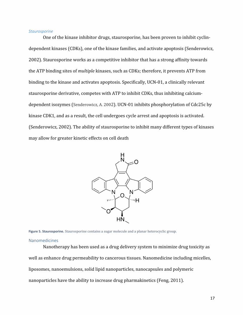
Staurosporine
One of the kinase inhibitor drugs, staurosporine, has been proven to inhibit cyclin-
dependent kinases (CDKs), one of the kinase families, and activate apoptosis (Senderowicz,
2002). Staurosporine works as a competitive inhibitor that has a strong affinity towards
the ATP binding sites of multiple kinases, such as CDKs; therefore, it prevents ATP from
binding to the kinase and activates apoptosis. Specifically, UCN-01, a clinically relevant
staurosporine derivative, competes with ATP to inhibit CDKs, thus inhibiting calcium-
dependent isozymes (Senderowicz, A. 2002). UCN-01 inhibits phosphorylation of Cdc25c by
kinase CDK1, and as a result, the cell undergoes cycle arrest and apoptosis is activated.
(Senderowicz, 2002). The ability of staurosporine to inhibit many different types of kinases
may allow for greater kinetic effects on cell death
Figure 5. Staurosporine. Staurosporine contains a sugar molecule and a planar heterocyclic group.
Nanomedicines
Nanotherapy has been used as a drug delivery system to minimize drug toxicity as
well as enhance drug permeability to cancerous tissues. Nanomedicine including micelles,
liposomes, nanoemulsions, solid lipid nanoparticles, nanocapsules and polymeric
nanoparticles have the ability to increase drug pharmakinetics (Feng, 2011).

18
Nanomedicines are capable of longer circulation of delivery vehicles as well as longer
retention of anticancer agents (Feng, 2011). In addition, nanoparticle surfaces can be
modified with ligands which are able to target specific receptors in the tumor and increase
drug uptake.
Nanoemulsions are nanomedicine platforms that have a lipid core with an aqueous
surrounding, stabilized by an amphiphilic phospholipid monolayer (Figure 6a). The lipid
core consists of flaxseed oil that is made up of 57% Omega-3 and 17% Omega-6 fats. The oil
core has the ability to hold highly toxic, non-water-soluble drugs and protects the drugs
from degradive factors both in the blood and in tumor cells. The aqueous solution that
coats the oil core consists of dissolved egg lecithin, PEG2000DSPE, and glycerol water,
distilled water with a small amount of glycerol in it. The polyethylene glycol (PEG), shown
in Figure 6b, that coats the oil core prevents clearance by the mononuclear phagocyte
system. Cancer specific targeting ligands can be attached to the PEG on the surface of the
molecule, thus directing the NEs through leaky vasculature of the tumor and specifically to
desired cancerous tissues. The desirable formulation characteristics of nanoemulsions
include a monodisperse particle size of less than 200nm, apparent drug entrapment
efficiency, and a negative surface charge of repulsion/attraction between particles known
as a negative zeta potential (Feng, 2011).

19
a. b.
Figure 6. Diagram of Generic Nanoparticle. a. Nanoemulsion. The combined aqueous and oil phase contains the cytotoxic drugs on the inside and the targeting ligand on the outside. b. PEG. The PEG that is on the outside of Figure 6a that the targeting ligand attaches to.
Folate Targeting
Surface Folates
Folate receptors (FR) can be found on the surfaces of certain epithelial cells
throughout the body. In particular, FRα is a receptor coded by the folate receptor 1 gene
and binds to folic acid with high affinity. FRα is a protein overexpressed and unregulated in
ovarian cancer due to a high dependency on folate metabolism, yet is often absent from
normal healthy tissue (Wen et al, 2015). FRα transports folate through membranes by
receptor-mediated endocytosis (Wen et al, 2015). Essentially, drugs targeted with folate
conjugates have become a novel mechanism for delivering nanoparticles to tumor sites
because of their absorption through the plasma membrane. This process is known as folate
targeting.
Research has been applied to create folic-acid (FA) conjugates and link them to
drugs. Specifically, FA can be attached polyethylene glycol-phosphatidylethanolamine
(PEG-PE) and used as a targeting ligand (Sawant & Torchilin, 2010). This allows a targeted
therapy that includes a FA-PEG-PE combination to exploit the cytotoxic effects of the

20
encapsulated drug while simultaneously lowering the collateral damage in healthy tissue
(Dongen et al, 2014).
In fact, a nanoemulsion is a useful nanotherapy platform that can be targeted with
folate. As explained earlier, a nanoemulsion is comprised of an oil core with lipid and PEG-
PE outer layer. The combination of different length PEG in the outer layer gives folate a
significant freedom of rotation. There are many different forms of PEG, ranging from 500
Daltons to 60,000 Daltons. The greater length of PEG is proportional to a greater freedom
of rotation of folate, however if PEG is too large, it may override the hydrophobicity of the
combined lipid and pull itself out of the nanoemulsion. Because of therapies like
nanoemulsions, there are now many opportunities to readily target common
chemotherapies by intravenously injecting folate-targeted nanotherapies which can bypass
p-glycoprotein, which naturally pumps hydrophobic drugs out of the tumor cell.
Theoretically, using folate targeting as part of the delivery method in a nanoemulsion
would deliver more drug to the tumor than previous free drug chemotherapy methods.
Avidity and Affinity
Folate targeted drugs have an obvious affinity for folate receptors found on tumor
cells. This affinity can be measured by the dissociation constant (Kd). Typically, the Kd of
folic acid when binding to folate receptors is about 10-9 M. In the previous section “Surface
Folates”, monovalent folate-targeted drugs were said to increase the amount of drug that
reaches the tumor. However, it has been seen that dissociation has a direct correlation with
the average valency of FA conjugates. Therefore, as the average valency of FA conjugates to
FRα is increased, the dissociation of the folate-targeted drug has been seen to decrease,
thus increasing the affinity of folate targeted nanotherapies (Dongen et al, 2014).

21
Swanson et al used gadolinium-loaded dendrimer nanoparticles targeted with folate
to show the affinity that folate-targeted nanoparticles have for the overexpressed FRα
cancer cells. Gadolinium (Gd) is detected in quantity through MRI, as it is an imaging agent.
The study conducted by Swanston et al showed enhanced gadolinium accumulation in KB
human tumors over 24 hours for targeted dendrimers when compared to non-targeted
dendrimers. This further supported the idea that targeting a drug with FA increases the
affinity and avidity of the therapy.
Part 4: Hypothesis and Specific Aims
It is our hypothesis that the combination of docetaxel, ceramide, and staurosporine
encapsulated in a folate targeted nanoemulsion will deliver a safer and more effective
treatment to ovarian cancer. A problem that occurs in trying to treat ovarian cancer is the
speed at which multi drug resistance develops. The current delivery methods damage the
body while not always treating the tumor(s). The encapsulation technique used to engineer
nanoemulsions prevents this effect by protecting the enclosed drugs from metabolic
deactivation and nonspecific drug interactions. If the nanoemulsions can remain stable by
exhibiting no significant changes in size, zeta potential, and drug payload, then the
nanomedicines can be successful treatment options.
Multidrug resistance can be combated through the use of three different drugs in
combination. The combination of drugs targets a variety of pathways that create a superior
apoptotic effect. At the same time, the nanoemulsion controls the delivery of a highly toxic
payload because of the folate targeting and encapsulation of the drugs. The optimal
combinational therapies at minimal toxicity levels are determined through IC50 testing.

22
Materials and Methods
General Manufacturing Nanomedicines Nanoemulsions consist of an aqueous phase as well as an oil phase. Hydrophobic
active pharmaceutical ingredients (APIs) were loaded into the oil phase of each
nanoemulsion. The hydrophobic APIs used in this project included docetaxel (DTX),
ceramide (CER), and staurosporine (STP). A total of 5 nanoemulsions were made by
loading the following combinations of APIs into oil phases: docetaxel; docetaxel and
ceramide; docetaxel, ceramide, and staurosporine; staurosporine; and staurosporine and
ceramide. The APIs were loaded in a concentration of 50:10:1 ceramide: docetaxel:
staurosporine. Polyethylene Glycol (PEG), the pegylating agent, and Egg Lecithin, an
emulsifying agent, were added to the aqueous phase with glycerol, an agent necessary for
maintaining isotonicity in nanoemulsions. The aqueous phase also contained folate-
targeting ligands conferring specificity to the NEs to receptors, often overexpressed on
ovarian cancer cells. The aqueous phase was kept on a stir plate until all ingredients were
dissolved.
Once the phases were prepared, the two were combined and vortexed to create an
emulsion. The combined emulsion was processed through an LV1 Microfluidizer for 10
cycles at 25,000 PSI. The final concentration of docetaxel drug was determined using an
HPLC instrument, and the final concentration of the drug usually ranged from 1mM to
2mM. The size and surface charge of the nanoparticles were read on the ZetaSizer. The
sizes ranged from 130 – 160nm, and the zeta potential ranged from -40 to -60mV.
Nanoemulsions were stored at 4°C.

23
HPLC Method of Detecting DTX
The concentration of docetaxel loaded into each of the nanoemulsions was
determined via High Performance Liquid Chromatography (HPLC). The Water HPLC
contained a Malvern 1525 Binary HPLC Pump with a Thermo Scientific Hypersil gold
column (C18 5 µM, Size – 150x4.6 mm) and a Malvern 2487 Dual Absorbance Detector UV
detector. The flow rate was always set to 1 ml/min. Standard curves consisting of 75 μg/ml,
50 μg/ml, 40 μg/ml, 25 μg/ml, 12 μg/ml, and 6.25 μg/ml of docetaxel in acetonitrile (ACN)
were established at the beginning of each assay. In between samples, the HPLC system was
washed with 100% ACN.
The mobile phase used to quantify docetaxel was comprised of 60% ACN and 40%
water. The UV detector was set to 270 nm. The retention time of docetaxel was
approximately 2-3 minutes.
The remaining drugs that were used in this study – ceramide and staurosporine –
were not analyzed using the HPLC. The amount of staurosporine loaded into
nanoemulsions was too small to detect, and a method to determine the amount of ceramide
in the nanoemulsion had not been determined.
Quantification of APIs in Nanomedicines
Nanoparticle samples were diluted 1,000 fold in ACN, vortexed for 2 minutes, and
then spun down in a centrifuge for 10 minutes at 10,000rpm. Supernatant was then
analyzed on HPLC using the method above.
Cell Lines The human ovarian cancer cell line, SKOV-3 was obtained from the ATCC (Manassas,
VA). SKOV-3 cells were continuously cultured in RPM1 1640 media with L-glutamine
24
containing 10% fetal bovine serum and 1% penicillin/streptomycin, in a humidified
incubator at 37°C with 5% CO2. All cell culture media as well as sterile equipment used was
purchased from Fisher Scientific.
Cytotoxicity Assay Cell viability and 50% inhibitory concentration (IC50) values were generated using
the 3-(4,5-dimethylthiazol-2-yl)-2,5-diphenyltetrazolium bromide (MTT) assay. SKOV-3
cells were seeded into 96-well plates at a density of 3 X 10³ cells/well in normal growth
media and incubated 24 hours incubation for proper attachment and cell cycle
synchronization. Following incubation, media was removed and replaced with proper
dosing of drug containing media. Table 1 shows the plate layout for cells dosed with just
docetaxel. The IC50 for cells plated with just docetaxel or docetaxel, ceramide loaded
nanoemulsions were higher than other nanoemulsions thus the different concentrations in
the plate layout. The plates of cells dosed with staurosporine and combinational therapies
were dosed according to the plate layout in Table 2. Staurosporine was effective at lesser
concentrations, so the doses were 10-fold less in Table 2.

25
Table 1. The plate layout for cells dose with docetaxel nanoemulsions. This plate layout was used for nanoemulsions loaded with docetaxel only or docetaxel and ceramide.
1 2 3 4 5 6 7 8 9 10 11 12
A PEI Untreated 0.00001 µM
0.0001 µM
0.001 µM
0.01 µM
0.1 µM
1 µM
10 µM
100 µM
Untreated Untreated
B PEI Untreated 0.00001 µM
0.0001 µM
0.001 µM
0.01 µM
0.1 µM
1 µM
10 µM
100 µM
Untreated Untreated
C PEI Untreated 0.00001 µM
0.0001 µM
0.001 µM
0.01 µM
0.1 µM
1 µM
10 µM
100 µM
Untreated Untreated
D PEI Untreated 0.00001 µM
0.0001 µM
0.001 µM
0.01 µM
0.1 µM
1 µM
10 µM
100 µM
Untreated Untreated
E Blank Untreated 0.00001 µM
0.0001 µM
0.001 µM
0.01 µM
0.1 µM
1 µM
10 µM
100 µM
Untreated Untreated
F Blank Untreated 0.00001 µM
0.0001 µM
0.001 µM
0.01 µM
0.1 µM
1 µM
10 µM
100 µM
Untreated Untreated
G Blank Untreated 0.00001 µM
0.0001 µM
0.001 µM
0.01 µM
0.1 µM
1 µM
10 µM
100 µM
Untreated Untreated
H Blank Untreated 0.00001 µM
0.0001 µM
0.001 µM
0.01 µM
0.1 µM
1 µM
10 µM
100 µM
Untreated Untreated
26
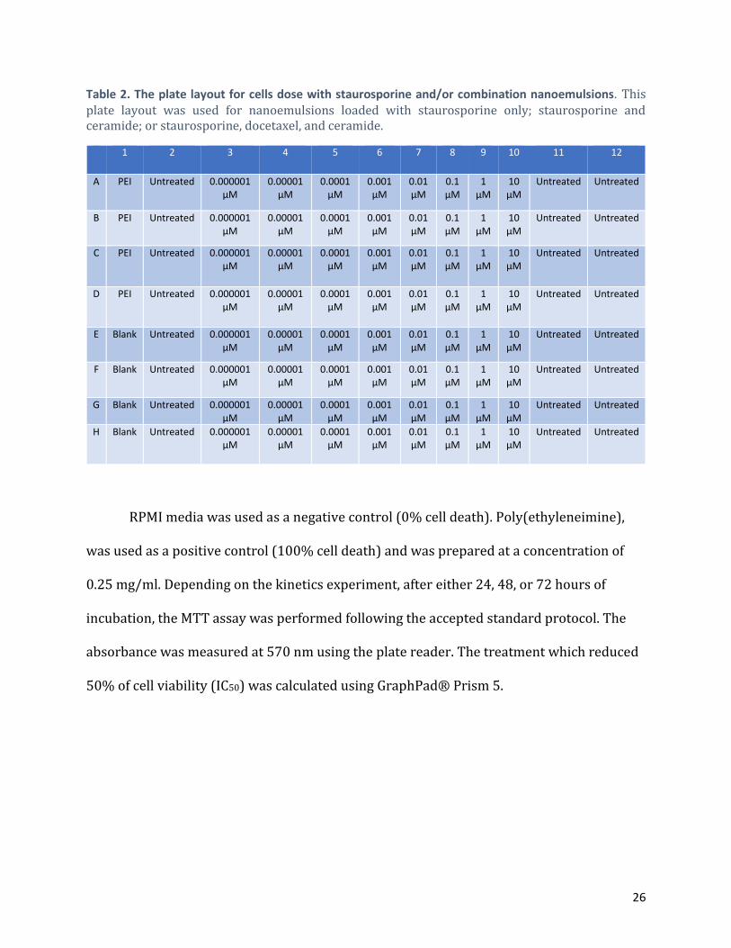
Table 2. The plate layout for cells dose with staurosporine and/or combination nanoemulsions. This plate layout was used for nanoemulsions loaded with staurosporine only; staurosporine and ceramide; or staurosporine, docetaxel, and ceramide.
1 2 3 4 5 6 7 8 9 10 11 12
A PEI Untreated 0.000001 µM
0.00001 µM
0.0001 µM
0.001 µM
0.01 µM
0.1 µM
1 µM
10 µM
Untreated Untreated
B PEI Untreated 0.000001 µM
0.00001 µM
0.0001 µM
0.001 µM
0.01 µM
0.1 µM
1 µM
10 µM
Untreated Untreated
C PEI Untreated 0.000001 µM
0.00001 µM
0.0001 µM
0.001 µM
0.01 µM
0.1 µM
1 µM
10 µM
Untreated Untreated
D PEI Untreated 0.000001 µM
0.00001 µM
0.0001 µM
0.001 µM
0.01 µM
0.1 µM
1 µM
10 µM
Untreated Untreated
E Blank Untreated 0.000001 µM
0.00001 µM
0.0001 µM
0.001 µM
0.01 µM
0.1 µM
1 µM
10 µM
Untreated Untreated
F Blank Untreated 0.000001 µM
0.00001 µM
0.0001 µM
0.001 µM
0.01 µM
0.1 µM
1 µM
10 µM
Untreated Untreated
G Blank Untreated 0.000001 µM
0.00001 µM
0.0001 µM
0.001 µM
0.01 µM
0.1 µM
1 µM
10 µM
Untreated Untreated
H Blank Untreated 0.000001 µM
0.00001 µM
0.0001 µM
0.001 µM
0.01 µM
0.1 µM
1 µM
10 µM
Untreated Untreated
RPMI media was used as a negative control (0% cell death). Poly(ethyleneimine),
was used as a positive control (100% cell death) and was prepared at a concentration of
0.25 mg/ml. Depending on the kinetics experiment, after either 24, 48, or 72 hours of
incubation, the MTT assay was performed following the accepted standard protocol. The
absorbance was measured at 570 nm using the plate reader. The treatment which reduced
50% of cell viability (IC50) was calculated using GraphPad® Prism 5.
27
Results
Manufacturing and Monitoring Stability of Nanomedicines Five different nanoemulsions were engineered throughout this project: one
encapsulating docetaxel (DTX); one encapsulating docetaxel and ceramide (CER); one
encapsulating docetaxel, ceramide, and staurosporine (STP); one encapsulating
staurosporine; and one encapsulating staurosporine and ceramide. The nanoemulsions
were engineered using Microfluidics High Pressure Homogenizer LV1. The LV1
Microfluidizer takes the crude mixture of an aqueous phase and an oil phase and puts it
through tiny porous membranes at a high pressure. The LV1 Microfluidizer takes the size of
the nanoemulsions down to the nano-scale and produces a homogenous colloidal
suspension. After the nanoemulsions have gone through the suggested number of cycles in
the LV1 Microfluidizer, the size and the zeta potential was measured on the Malvern SV90
ZetaSizer, a machine that uses Dynamic Light Scattering to calculate the average size and
distribution of the nanoemulsions.
The stability of the nanoemulsions was monitored to ensure the reliability of the
molecules for an extended period of 28 days. Table 3 shows the hydrodynamic diameter
and polydispersity index (PDI) for each of the five nanoemulsions measured every 7 days
for 28 days, starting with the day the nanoemulsion was synthesized (day 0). The PDI is a
numeric representation of the dispersion of sizes, therefore the higher the PDI, the more
variety in particle size. Table 3 shows that there was little variability in the hydrodynamic
diameters for each of the nanoemulsions, and there was no significant variance in PDI. The
hydrodynamic diameter is expected to be less than 180nm to ensure safety when injected
into the blood. A nanoparticle greater than 180nm could be too large and cause an

28
embolism. Each of the five nanoemulsions synthesized had hydrophobic diameters less
than 180nm that remained stable over the 28 days.
Table 3. Hydrodynamic Diameter and Polydispersity Index of Nanoemulsions. Each nanoemulsion size distribution was determined every 7 days for 28 days using a ZetaSizer. DTX, CER and DTX, CER, and STP nanoemulsions do not have data for day 28 because the nanoemulsion was used up by this time in cytotoxicity assays.
NE
Hydrodynamic Diameter (nm)
Polydispersity Index (±)
Day Day
0 7 14 21 28 0 7 14 21 28
DTX 139.2 138.8 136.7 136.7 141.7 0.021 0.050 0.035 0.074 0.073
DTX, CER
132.4 132.2 133.4 130.5 N/A 0.030 0.037 0.043 0.002 N/A
DTX, CER, STP
129.4 131.4 125.9 121.3 N/A 0.051 0.037 0.063 0.071 N/A
STP 150.7 164.0 161.0 154.0 158.5 0.039 0.079 0.059 0.038 0.047
STP, CER
128.1 134.6 137.5 135.4 132.1 0.081 0.089 0.105 0.096 0.094
The ZetaSizer was also used to measure the zeta potential of each nanoemulsion.
The nanoemulsions are expected to be slightly negative so that upon entry to the body, the
concentrations of cations such as sodium and potassium in the body do not make the
particles too positive, which could cause an embolism. Table 4 shows the zeta potentials
obtained for each nanoemulsion measured every 7 days for a period of 28 days. The zeta
potentials stayed stable in a range of -30 to -50mV, which is what is expected for
nanoemulsion charge. The standard deviations (SD) are included in the table.

29
Table 4. Zeta Potential of Nanoemulsions. Each nanoemulsion zeta potential was determined every 7 days for 28 days using a ZetaSizer. DTX, CER and DTX, CER, and STP nanoemulsions do not have data for day 28 because the nanoemulsion was used up by this time in cytotoxicity assays.
NE
Zeta Potential ± SD (mV)
Day
0 7 14 21 28
DTX -48.7 ± 10.4
-45.8 ± 9.02
-51.5 ± 5.39
-44.5 ± 6.34
-49.3 ± 11
DTX, CER -46.5 ±
8.51 -47.7 ±
9.72 -50.7 ±
10.6 -51.6 ±
9.27 N/A
DTX, CER, STP -42.5 ±
8.16 -43.1 ±
9.47 -46.9 ±
8.79 -48.2 ±
12.2 N/A
STP -43.0 ±
9.75 -43.2 ±
10.2 -46.0 ±
10.6 -48.4 ±
12.0 -42.1 ±
7.37
STP, CER -48.2 ±
10.2 -41.2 ±
10.7 -39.7 ±
11.4 -43.1 ±
9.63 -41.0 ±
7.74
HPLC Detection of DTX
Each nanoemulsion containing docetaxel was assayed to confirm the concentration
of the docetaxel loaded into the nanoemulsion using a Waters HPLC Pump. The mobile
phase of the HPLC was set to 30% Acetonitrile and 20% distilled water, with a flow rate of
1 mL/min. The HPLC mobile phase was only set to a total of 50% to prevent clogging in the
column. A standard curve was constructed with docetaxel dissolved in Acetonitrile. The
nanoparticles were diluted 1:1000 in Acetonitrile and vortexed for approximately 2 min in
order to lyse the particles and free the docetaxel. The supernatant from the dilution was
put through the HPLC to measure drug concentration.
Table 5 shows the milligram of docetaxel loaded into each nanoemulsion, the
amount of docetaxel detected in the HPLC assay (in milligrams per milliliter), the amount of
docetaxel detected in the HPLC assay (in micrograms per milliliter), and the percentage of
loading efficiency for each nanoemulsion. Molarity was used to determine treatment
30
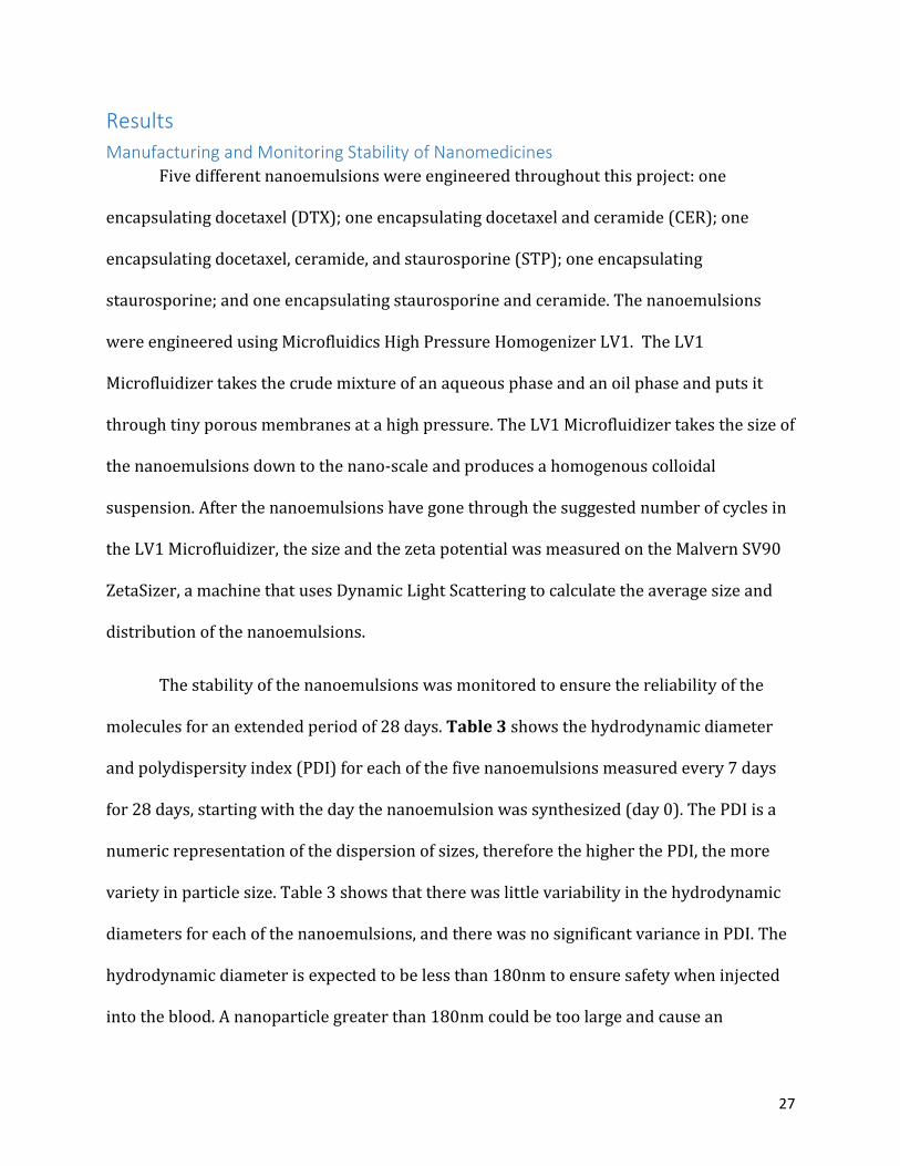
concentrations for the plates. The docetaxel nanoemulsion had an 80.25% loading
efficiency, indicating that of the 2mg/ml put into the nanoemulsion, 80.25% of the
docetaxel actually loaded into the nanoemulsion. The docetaxel, ceramide nanoemulsion
had a 106.3% loading efficiency, meaning that of the 0.8 mg/ml put into the nanoemulsion,
106.3% actually loaded into the emulsion. Having a loading efficiency over 100% is
plausible because the sample we took to assay may have contained slightly more docetaxel
than another sample in the emulsion, suggesting that the concentration may not have been
completely uniform. Lastly, our docetaxel, ceramide, and staurosporine nanoemulsion had
an 83.13% loading efficiency.
Table 5. Docetaxel HPLC Loading Efficiency. Each nanoemulsion that contained docetaxel was run through HPLC to determine docetaxel loading efficiency.
The staurosporine and staurosporine, ceramide nanoemulsions were not assayed
using the HPLC because the small amount of staurosporine in the nanoemulsions would be
nearly impossible to detect.

31
Cytotoxicity Assays
Each of the five nanoemulsions were tested for cytotoxicity on an SKOV-3 cell line.
The kinetics of the mechanism of the drug were tested at 24, 48, and 72 hours. Each
nanoemulsion was tested on four 96-well plates containing SKOV-3 cells and were
administered according to the plate layouts seen in Tables 1 and 2. Cytotoxicity data was
collected for 24, 48, and 72 hours for each nanoemulsion with the exception of the
docetaxel nanoemulsion. The 48 hour plates were infected with fungus and thus did not
produce any conclusive or reportable data resulting in not reported data (N.D.). Both the
staurosporine and staurosporine-ceramide nanoemulsions showed inconclusive data that
cannot be reported. In the cytotoxicity assay, values are presented in both constrained and
non-constrained cell viability percentages. Due to the lack of cell death in both the
staurosporine and staurosporine-ceramide nanoemulsions, the constrained and non-
constrained cytotoxicity data was extremely inconsistent and can therefore not be
reported. The resulting reportable IC50 values for the docetaxel, docetaxel-ceramide, and
docetaxel-ceramide-staurosporine nanoemulsions can be seen in Table 6.
Table 6. Average IC50 Values for Nanoemulsions Tested on SKOV-3 cells. Each nanoemulsion was tested on four 96-well plates of SKOV-3 cells.
IC50 (Average)
NE 24 hour (nM) 48 hour (nM) 72 hour (nM)
DTX NE >100 μM N.D. 4.0085
DTX, CER >100 μM 66.6785 1.809
DTX, CER, STP >100 μM 91.706 3.558
32
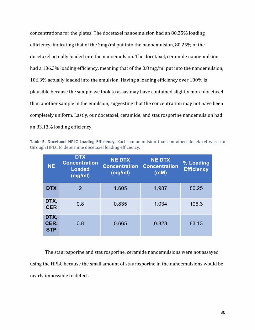
Based on these results, there was not an observable or significant decrease in IC50
values with the addition of any drug into a nanoemulsion. Although both the DTX-CER, and
DTX-CER-STP nanoemulsions showed lower IC50 values than the DTX nanoemulsion alone,
there is no conclusive data supporting the hypothesis predicting a triple or double
combination nanoemulsion with this specific ratio of drugs leads to higher cytotoxicity.
However, based on this data, it is seen that at 72 hours, maximum cytotoxicity is observed
for each of the three FA-targeted nanoemulsions investigated.
Figure 7. Cytotoxicity Results for DTX, DTX-CER, and DTX-CER-STP nanoemulsions. Maximum cytotoxicity is observed at 72 hours for all nanoemulsions. a. Docetaxel nanoemulsion cytotoxicity data. b. Docetaxel-ceramide cytotoxicity data. c. Docetaxel-ceramide-staurosporine cytotoxicity data.
For each of the nanoemulsions, the drug activity at 24 hours showed almost no cell
death, indicating that at this time point, the drug is relatively ineffective at killing the SKOV-
3 cells. For both the DTX-CER and DTX-CER-STP nanoemulsions, an improvement was seen
at 48 hours, displaying higher cytotoxicity and a lower nanomolar kill. All three

33
nanoemulsions showed the most effective and overall maximum cytotoxicity at 72 hours.
This indicates that the mechanism of action of this drug is optimum at 72 hours and
effectively kills SKOV-3 cells better when compared to the mechanisms at 24 and 48 hours.

34
Discussion Ovarian cancer accounts for ~3% of all cancers in women (American Cancer Society,
2014). Although the percentage may seem small, the 5-year survival rate for those suffering
with ovarian cancer is very low (Parkin, 2011). Ovarian cancer is a difficult disease to
detect with a clear unmet clinical need. Two of the main issues that arise with ovarian
cancer are late diagnosis and multi-drug resistance. Ovarian cancer typically is not detected
until it is in the third or fourth stage of the disease where tumors have metastasized to all
areas of the peritoneal space (Cannistra, 1993). Treatment includes surgical removal of
tumors followed by chemotherapy. Unfortunately, the p-glycoprotein in the ovarian cancer
cell lines resist apoptosis and acquire multi-drug resistance by hydrophobic
chemotherapeutic drug efflux or simply not allowing the drugs to enter the cell (Gottesman,
2002).
The use of targeted nanoemulsions has the ability to overcome many of the
problems facing current ovarian cancer treatment. Encapsulating active pharmaceutical
ingredients (API) such as docetaxel, ceramide, and staurosporine, in a nanoemulsion, and
targeting the nanoemulsion towards folate-receptors, has the potential to overcome multi-
drug resistance and deliver the chemotherapeutics in a less toxic way. The goal of this MQP
was to synthesize and characterize 5 combination nanoemulsions, determine
nanoemulsion cytotoxicity on ovarian cancer cells, and determine the kinetics of cell death
of each nanoemulsion. Overall, we wanted to determine if drug combinations enhance
potency.
The five nanoemulsions we synthesized maintained stability in their hydrophobic
diameters and zeta potentials (see Tables 3 and 4, respectively) over the course of 28 days.

35
The consistency among nanoemulsions concluded that it is possible to encapsulate three
chemotherapeutic drugs into a nanoemulsion and that the drugs encapsulated stay within
the nanoemulsion for at least a month. The HPLC analysis further confirmed that docetaxel
is successfully encapsulated into nanoemulsions.
It was previously hypothesized that the addition of staurosporine, a versatile kinase
inhibitor, could alter the kinetics effects of a nanoemulsion also encapsulating docetaxel
and ceramide. This triple combination drug was thought to not only alter the kinetic effects,
but effectively increase cytotoxicity, essentially killing SKOV-3 cells with a lower
concentration of drug. With increased cytotoxicity, less drug could be administered to a
patient and decrease tumor size. However, based on the results seen in Figure 7, the
combination of chemotherapeutics and pro-apoptotic factors to create a triple combination
nanoemulsion does not significantly decrease the IC50 values as was hypothesized.
However, based on the consistency of the data presented in Figure 7 and Table 6, the
maximum cytotoxicity for the nanoemulsions tested was observed at 72 hours in the MTT
assay. Although this specific combination and ratio of chemotherapeutics and pro-
apoptotic agents did not significantly lower the IC50 values, it is thought that other ratios of
the drugs in the nanoemulsions may still hold potential to achieve improved cytotoxicity,
which remains to be tested.
The aim of this study was to develop and test a novel therapy for the treatment of
multidrug resistant ovarian cancer. The results discussed in this paper provide a
foundation for further experimentation and development of the therapy. The next step will
include extensive in vivo studies in order to properly conclude the effectiveness, toxicity,

36
and specificity of the nanoemulsions. Further studies may include testing the
staurosporine activity, adjusting the combination ratio for the cytotoxic drugs, utilizing
different ovarian cancer cell lines, and testing the effectiveness of overcoming the
multidrug resistance. For example, further experimentation could include trying to recover
any cells that had received treatment in the MTT assays followed by trying to “re-kill” the
recovered cells. Further experimentation may also include testing the media from the cells
that received treatment in the MTT assays for any drug showing effluxion from the cells or
for drugs that had not entered the cell in the first place. This will reveal the effects the
drugs had on the cells such as any drug resistance or drug impermeability.

37
References Agarwal, R., & Kaye, S. B. (2003). Ovarian cancer: strategies for overcoming resistance to
chemotherapy. Nature Reviews Cancer, 3(7), 502 - 516. Auersperg N., Maines-Bandiera SL., Dyck HG., Kruk PA. (1994). Characterization of cultured
human ovarian surface epithelial cells: phenotypic plasticity and premalignant changes. Laboratory investigation; a journal of technical methods and pathology 71(4): 510-518.
Bandara NA., Hansen MJ., Low PS. (2014). Effect of Receptor Occupancy on Folate Receptor
Internalization. Molecular Pharmaceutics 11: 1007-1013. doi: 10.1021/mp400659t. Barouch-Bentov, R., & Sauer, K. (2011). Mechanisms of drug resistance in kinases. Expert
Opinion on Investigational Drugs, 153-208. Bukovsky A., Virant-Klun I., Svetlikova M., Willson I. (2006). Ovarian Germ Cells. Methods in
Enzymology; Adult Stem Cells, (419): 208-258. doi: 10.1016/S0076-6879(06)19010-2.
Bush, T. L., Payton, M., & Heller, S. (2013). AMG 900, a Small-Molecule Inhibitor of Aurora Kinases, Potentiates the Activity of Microtubule-Targeting Agents in Human Metastatic Breast Cancer Models. Molecular Cancer Tehrapeutics, 12, 2356-2366.
Cannistra, S. (1993). Cancer of the Ovary. New England Journal of Medicine, 1550-1559. Cooper, M. J., Graves, L. M., Roy, S. G., Jones, L. S., Nguyen, T. A., Whittle, M. C., et al. (2013).
Application of Multiplexed Kinase Inhibitor Beads to Study Kinome Adaptations in Drug-Resistant Leukemia. PLoS ONE, 8(6), e66755.
Dongen MA., Silpe JE., Dougherty CA., Kanduluru AK., Choi SK., Orr BG., Low PS., Holl MM.
(2014). Avidty Mechanism of Dendrimer-Folic Acid Conjugates. Molecular Pharmaceutics 11: 1696-1706. doi: 10.1021/mp5000967.
Edinger, A., & Thompson, C. (2004). Death by design: Apoptosis, necrosis and autophagy.
Current Opinion in Cell Biology, 16, 663-669. Esenkop SM., Friedman RL., Wang H. (1998). Complete Cytoreductive Surgery Is Feasible
and Maximizes Survival in Patients with Advanced Epithelial Ovarian Cancer: A Prospective Study. Gynecologic Oncology 69: 103-108.
Feng, L., Benhabbour, S. R., & Mumper, R. J. (2013). Oil-Filled Lipid Nanoparticles
Containing 2′-(2-Bromohexadecanoyl)-Docetaxel for the Treatment of Breast Cancer. Advanced Healthcare Materials, 2(11), 1451-1457.

38
Feng, L., & Mumper, R. J. (2013). A critical review of lipid-based nanoparticles for taxane delivery. Cancer Letters, 334(2), 157-175.
Feng, L., Wu, H., Ma, P., Mumper, R. J., & Benhabbour, S. R. (2011). Development and
optimization of oil-filled lipid nanoparticles containing docetaxel conjugates designed to control the drug release rate in vitro and in vivo.. International Journal of Nanomedicine, 6, 2545-2556.
Ganta, S., et. al. (2014). Formulation development of a novel targeted theranostic
nanoemulsion of docetaxel to overcome multidrug resistance in ovarian caner. Drug Delivery, 1-13.
Gottesman, M. & Pastan, I. (2002). Mechanisms of drug resistance in cancer therapy.
European Journal of Pharmacology, 17-18. Ji, C., Yang, B., Yang, Y., He, S., Miao, D., He, L., et al. (2010). Exogenous cell-permeable C6
ceramide sensitizes multiple cancer cell lines to Doxorubicin-induced apoptosis by promoting AMPK activation and mTORC1 inhibition. Oncogene, 29(50), 6557-6568.
King, D. (2013). Ovary. Southern Illinois University School of Medicine. From http://
www.siumed.edu/~dking2/erg/ovary.htm. Longo DL., Young RC. (1981). The Natural History and Treatment of Ovarian cancer. Annual
Reviews of Medicine, 1(31): 475-490. Midland, A. A., Gomez, S. M., Johnson, G. L., Graves, L. M., Iii, H. S., Carey, L. A., et al.
(2012). Defining the expressed breast cancer kinome. Cell research, 22(4), 620-623. Ozols RF. (2006). Systemic Therapy for Ovarian Cancer: Current Status and New
Treatments. Seminars in Oncology 33(6): S3-S11. Parkin, M., & Pasini, P. (1999). Erratum: Global Cancer Statistics. CA: A Cancer Journal for
Clinicians, 134-134. Senderowicz, A. M. (2002). The Cell Cycle as a Target for Cancer Therapy: Basic and Clinical
Findings with the Small Molecule Inhibitors Flavopiridol and UCN-01. The Oncologist, 7(90003), 12-19.
Swanson, S. D., Kukowska-Latallo, J. F., Patri, A. K., Chen, C., Ge, S., Cao, Z., ... & Baker, J. R.
(2008). Targeted gadolinium-loaded dendrimer nanoparticles for tumor-specific magnetic resonance contrast enhancement.International journal of nanomedicine, 3(2), 201.
Vasey PA., Jayson GC., Gordon A., Gabra H., Coleman R., Atkinson R., Parkin D., Paul J., Hay A.,

39
Kaye SB. (2004). Phase III Randomized Trial of Docetaxel-Carboplatin Versus Paclitaxel-Carboplatin as First-line Chemotherapy for Ovarian Carcinoma. Journal of the National Cancer Institute 96(22): 1682-1691. doi: 10.1093/jnci/djh323.
Vlashi E., Kelderhouse LE., Sturgis JE., Low PS. (2013). Effect of Folate-Targeted Nanoparticle Size on Their Rates of Penetration into Solid Tumors. American Chemical Society 7(10): 8573-8582. doi: 10.1021/nn402644g.
Wen, Y., Graybill, W. S., Previs, R. A., Hu, W., Ivan, C., Mangala, L. S., ... & Sood, A. K. (2015). Immunotherapy targeting folate receptor induces cell death associated with autophagy in ovarian cancer. Clinical Cancer Research, 21(2), 448-459.