Embed Size (px)
Citation preview
Development and clinical translation of tubular constructs fortracheal tissue engineering: a review
Luis Soriano1,2,3,8, Tehreem Khalid1,2,4,8, Derek Whelan5, Niall O’Huallachain1, Karen C. Redmond6,Fergal J. O’Brien2,3,4,7, Cian O’Leary1,2,3,4,7,9 and Sally-Ann Cryan1,2,3,4,7,9
1School of Pharmacy and Biomolecular Sciences, RCSI University of Medicine and Health Sciences, Dublin, Ireland. 2Tissue EngineeringResearch Group, Dept of Anatomy and Regenerative Medicine, RCSI University of Medicine and Health Sciences, Dublin, Ireland. 3SFICentre for Research in Medical Devices (CÚRAM), RCSI University of Medicine and Health Sciences, Dublin, Ireland. 4SFI AdvancedMaterials and Bioengineering Research (AMBER) Centre, RCSI University of Medicine and Health Sciences and Trinity College Dublin,Dublin, Ireland. 5Dept of Mechanical, Biomedical and Manufacturing Engineering, Munster Technological University, Cork, Ireland.6National Cardio-thoracic Transplant Unit, Mater Misericordiae University Hospital and UCD School of Medicine, Dublin, Ireland. 7TrinityCentre for Biomedical Engineering, Trinity College Dublin, Dublin, Ireland. 8Joint first authors. 9Both authors contributed equally.
Corresponding author: Sally-Ann Cryan ([email protected])
Shareable abstract (@ERSpublications)A review focusing on the state of the art and clinical translation of tissue engineering and currentapproaches aimed at developing tubular substitutes for tracheal regeneration and restorationhttps://bit.ly/37kCB5z
Cite this article as: Soriano L, Khalid T, Whelan D, et al. Development and clinical translation oftubular constructs for tracheal tissue engineering: a review. Eur Respir Rev 2021; 30: 210154[DOI: 10.1183/16000617.0154-2021].
AbstractEffective restoration of extensive tracheal damage arising from cancer, stenosis, infection or congenitalabnormalities remains an unmet clinical need in respiratory medicine. The trachea is a 10–11 cm longfibrocartilaginous tube of the lower respiratory tract, with 16–20 tracheal cartilages anterolaterally and adynamic trachealis muscle posteriorly. Tracheal resection is commonly offered to patients suffering fromshort-length tracheal defects, but replacement is required when the trauma exceeds 50% of total length ofthe trachea in adults and 30% in children. Recently, tissue engineering (TE) has shown promise tofabricate biocompatible tissue-engineered tracheal implants for tracheal replacement and regeneration.However, its widespread use is hampered by inadequate re-epithelialisation, poor mechanical properties,insufficient revascularisation and unsatisfactory durability, leading to little success in the clinical use oftissue-engineered tracheal implants to date. Here, we describe in detail the historical attempts and thelessons learned for tracheal TE approaches by contextualising the clinical needs and essential requirementsfor a functional tracheal graft. TE manufacturing approaches explored to date and the clinical translation ofboth TE and non-TE strategies for tracheal regeneration are summarised to fully understand the big pictureof tracheal TE and its impact on clinical treatment of extensive tracheal defects.
IntroductionTracheal damage associated with narrowing, weakening or discontinuity of the airways is often alife-threatening condition. Defects in the conducting portion of the lower respiratory tract may arise from awide variety of pathologies or clinical conditions. In particular, there is a significant unmet clinical need interms of treatment options for long and extensive tracheal defects [1]. Currently, short-length trachealdamage is typically corrected by thoracic surgery including tracheal resection and end-to-end anastomosis,although this is not advised for those defects exceeding 2 cm in children and 5 cm in adults [2]. In suchcases, patients are treated using slide tracheoplasty, tracheal reconstruction and novel artificial prostheses.Several complications, however, including granulation tissue formation and weakening of the implant overtime, critically endanger their performance [1, 3]. Therefore, there is an urgent need to develop novelsolutions to promote the regeneration of native tracheal tissues [4].
In light of this unmet need and the evolving initiatives to potentially solve it, this review seeks to highlightthe historical attempts at tracheal replacement and how the lessons learnt have impacted and directed
Copyright ©The authors 2021
This version is distributed underthe terms of the CreativeCommons AttributionNon-Commercial Licence 4.0.For commercial reproductionrights and permissions [email protected]
Received: 2 July 2021Accepted: 26 July 2021
https://doi.org/10.1183/16000617.0154-2021 Eur Respir Rev 2021; 30: 210154
EUROPEAN RESPIRATORY REVIEWREVIEW
L. SORIANO ET AL.

current tissue engineering (TE) research attempts. First, we contextualise clinical needs with an overviewof relevant epidemiology and physiological characteristics of the functioning trachea and thus the designrequirements of a tracheal graft. Thereafter, an overview of current repair strategies is outlined, beforesummarising the significant experiences of clinical trials of various technologies.
The clinical need for tracheal replacement and regenerationTracheal damage or dysfunction in adults is commonly caused by traumatic injury, several types of cancerand benign conditions including tracheobronchomalacia (TBM) (table 1) [2, 5–16]. Although exact casenumbers are unknown and challenging to estimate, tracheobronchial injuries are reported to represent 2.5–3.2% of trauma-associated deaths and 0.5–2% of admitted trauma patients with chest and neck injuries [5, 6].Malignant tumours affecting the airways can cause tracheal obstruction and collapse leading to the needfor tracheal resection and reconstruction, including primary tracheal tumours such as squamous cellcarcinoma and adenoid cystic carcinoma [9, 10]. Another pathology that can result in tracheal damage isTBM, an under-reported disease characterised by the weakening of airway walls due to softening ofcartilage tissue covering the trachea and bronchi [11]. Finally, intensive care unit patients with prolongedintubation may develop laryngotracheal injuries due to endotracheal intubation procedures andtracheotomies. The incidence of post-intubation tracheal stenosis has been estimated as 6–21% of patientsthat underwent intubation [12, 13]. Moreover, recent studies suggest that around 10–15% of coronavirusdisease 2019 patients require invasive mechanical ventilation and could develop tracheal stenosis onceextubated [17].
In infants, congenital abnormalities present another unmet need for novel tracheal replacement solutionsincluding tracheoesophageal fistulas, oesophageal atresia and birth abnormalities [14]. On the other hand,tracheal agenesis, a rarer abnormality, affects fewer than one in 50000 births [14–16]. Airway stenosis ininfants and children is more challenging to treat than in adults, as resection and anastomosis are notfeasible when the defect affects more than one third of the total tracheal length (>2 cm [1]). A range ofstents have been used in attempts to support such patients, including Palmaz stents and biodegradablestents with limited success [18]. Although the reported incidence of pathologies affecting the trachea mightseem low, factors such as death before reaching the hospital and common symptoms similar to otherdiseases (such as asthma) masks the true rate at which these conditions affect patients worldwide [19].Furthermore, this misdiagnosis often leads to a high morbidity or mortality rate. Therefore there is asignificant clinical need for tracheal regeneration [20].
Replacing the trachea: an overview of its characteristics and implant design requirementsIn order to develop new implants as tracheal substitutes that replicate physiological form and function, thecomplex biological and anatomical characteristics of the trachea must be understood, including cellularfunction, mechanical properties and vascularisation.
Although the tracheal structure appears to be a simple tube, in reality it is composed of challengingstructural and compositional features to recapitulate. The trachea is the conducting portion of therespiratory tract, connecting the upper and lower respiratory tract, and is responsible for allowing air flowto and from the lungs, mucociliary clearance, and humidification of inspired air. It is composed of 18–22
TABLE 1 Epidemiology of tracheal defects
Pathology Epidemiology Reference
Traumatic tracheobronchial injuries Cause of 2.5–3.2% of trauma-associated deathsPresent in 0.5–2.0% of trauma patients showing chest/neck injuries<1% of elective orotracheal intubations0.05–0.37% of endotracheal intubations
[5–8]
Tracheal tumours Tracheal malignancies present in <1% of peoplePrimary tracheal carcinomas present in <1% people each year
[9, 10]
TBM Overall epidemiology is unknown, but TBM is present in 23% of COPD patients, 44% of patientswith chronic bronchitis and 1% of all patients undergoing bronchoscopy
[11]
Post-intubation tracheal stenosis 6–21% of patients that underwent intubation [12, 13]Oesophageal atresia <1% of births [14]Tracheal agenesis <1% of births [15]Tracheo–oesophageal fistulas <1% of births [16]
COPD: chronic obstructive pulmonary disease; TBM: tracheobronchomalacia.
https://doi.org/10.1183/16000617.0154-2021 2
EUROPEAN RESPIRATORY REVIEW TRACHEAL TISSUE ENGINEERING | L. SORIANO ET AL.
C-shaped hyaline cartilaginous rings that comprise around 80% of the tracheal circumference and areconnected by the annular ligament longitudinally, and horizontally by the trachealis muscle [1]. Theinnermost layer of the tracheal wall at the interface with the lumen, the mucosal layer, is formed by therespiratory epithelium and the lamina propria. The ciliated pseudostratified columnar epithelium isresponsible for mucociliary clearance, attraction of inflammatory cells during airway injury, secretion of awide variety of mediators and represents the first barrier against pathogens and particles during breathing[21]. Therefore, any proposed construct must be airtight and hold the potential to regeneratepseudostratified respiratory epithelium to prevent bacterial infection [22].
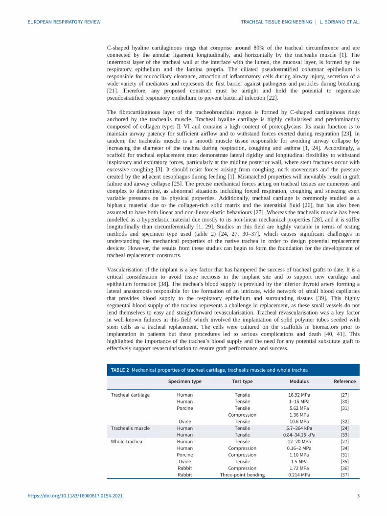
The fibrocartilaginous layer of the tracheobronchial region is formed by C-shaped cartilaginous ringsanchored by the trachealis muscle. Tracheal hyaline cartilage is highly cellularised and predominantlycomposed of collagen types II–VI and contains a high content of proteoglycans. Its main function is tomaintain airway patency for sufficient airflow and to withstand forces exerted during respiration [23]. Intandem, the trachealis muscle is a smooth muscle tissue responsible for avoiding airway collapse byincreasing the diameter of the trachea during respiration, coughing and asthma [1, 24]. Accordingly, ascaffold for tracheal replacement must demonstrate lateral rigidity and longitudinal flexibility to withstandinspiratory and expiratory forces, particularly at the midline posterior wall, where stent fractures occur withexcessive coughing [3]. It should resist forces arising from coughing, neck movements and the pressurecreated by the adjacent oesophagus during feeding [1]. Mismatched properties will inevitably result in graftfailure and airway collapse [25]. The precise mechanical forces acting on tracheal tissues are numerous andcomplex to determine, as abnormal situations including forced respiration, coughing and sneezing exertvariable pressures on its physical properties. Additionally, tracheal cartilage is commonly studied as abiphasic material due to the collagen-rich solid matrix and the interstitial fluid [26], but has also beenassumed to have both linear and non-linear elastic behaviours [27]. Whereas the trachealis muscle has beenmodelled as a hyperelastic material due mostly to its non-linear mechanical properties [28], and it is stifferlongitudinally than circumferentially [1, 29]. Studies in this field are highly variable in terms of testingmethods and specimen type used (table 2) [24, 27, 30–37], which causes significant challenges inunderstanding the mechanical properties of the native trachea in order to design potential replacementdevices. However, the results from these studies can begin to form the foundation for the development oftracheal replacement constructs.
Vascularisation of the implant is a key factor that has hampered the success of tracheal grafts to date. It is acritical consideration to avoid tissue necrosis in the implant site and to support new cartilage andepithelium formation [38]. The trachea’s blood supply is provided by the inferior thyroid artery forming alateral anastomosis responsible for the formation of an intricate, wide network of small blood capillariesthat provides blood supply to the respiratory epithelium and surrounding tissues [39]. This highlysegmental blood supply of the trachea represents a challenge in replacement, as these small vessels do notlend themselves to easy and straightforward revascularisation. Tracheal revascularisation was a key factorin well-known failures in this field which involved the implantation of solid polymer tubes seeded withstem cells as a tracheal replacement. The cells were cultured on the scaffolds in bioreactors prior toimplantation in patients but these procedures led to serious complications and death [40, 41]. Thishighlighted the importance of the trachea’s blood supply and the need for any potential substitute graft toeffectively support revascularisation to ensure graft performance and success.
TABLE 2 Mechanical properties of tracheal cartilage, trachealis muscle and whole trachea
Specimen type Test type Modulus Reference
Tracheal cartilage Human Tensile 16.92 MPa [27]Human Tensile 1–15 MPa [30]Porcine Tensile 5.62 MPa [31]
Compression 1.36 MPaOvine Tensile 10.6 MPa [32]
Trachealis muscle Human Tensile 5.7–364 kPa [24]Human Tensile 0.84–34.15 kPa [33]
Whole trachea Human Tensile 12–20 MPa [27]Human Compression 0.16–2 MPa [34]Porcine Compression 1.10 MPa [31]Ovine Tensile 1.5 MPa [35]Rabbit Compression 1.72 MPa [36]Rabbit Three-point bending 0.214 MPa [37]
https://doi.org/10.1183/16000617.0154-2021 3
EUROPEAN RESPIRATORY REVIEW TRACHEAL TISSUE ENGINEERING | L. SORIANO ET AL.
In summary, due to the high complexity of the tracheobronchial region, any tubular approach aimed atregenerating tracheal tissue must be able to offer a suitable 3D environment for the formation of arespiratory epithelium and hyaline cartilage while allowing the development of a vascularised plexus toavoid necrotic tissue formation. Moreover, it should allow the infiltration of immune cells from the hostimmune system to avoid infection by inhaled pathogens during the regeneration of the respiratoryepithelium. This significant challenge has seen the use of a wide range of strategies; the most historicallysignificant and currently promising will be discussed at length in this review.
Tracheal replacement strategiesThe first recorded attempts in tracheal resection and regeneration date back to the last decade of thenineteenth century [42–44]. After these early ground-breaking approaches, primary anastomosis was firstperformed in humans in 1886 [45]. Other approaches including the use of autogenous skin [46], fascial[47] and costal cartilage [48] grafts were later investigated, coupled with the first use of solid materials inprostheses [49]. The development of tubular tracheal substitutes has been further investigated using a widevariety of approaches including the use of transplanted bioprostheses [50], cadaveric tissue flaps [51],intestinal-derived tubes [52] and aortic homografts [53]. The success and shortcomings of these trachealreplacement strategies in clinical trials will be discussed at length below; however, attempts to replace longsegmental tracheal defects have had limited success in long-term studies and current treatment options facesubstantial limitations. As a result, in recent years, research has shifted towards investigating TEapproaches as a potential option to address this unmet medical need.
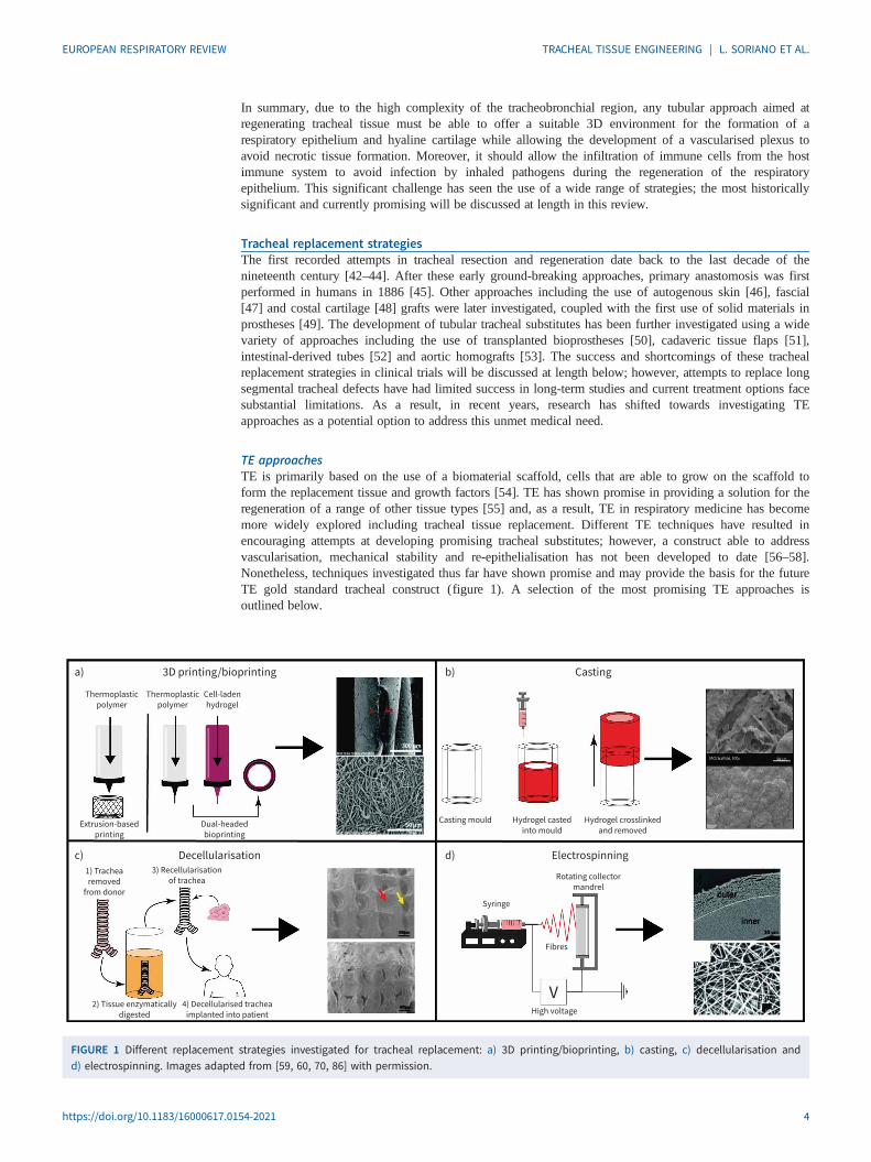
TE approachesTE is primarily based on the use of a biomaterial scaffold, cells that are able to grow on the scaffold toform the replacement tissue and growth factors [54]. TE has shown promise in providing a solution for theregeneration of a range of other tissue types [55] and, as a result, TE in respiratory medicine has becomemore widely explored including tracheal tissue replacement. Different TE techniques have resulted inencouraging attempts at developing promising tracheal substitutes; however, a construct able to addressvascularisation, mechanical stability and re-epithelialisation has not been developed to date [56–58].Nonetheless, techniques investigated thus far have shown promise and may provide the basis for the futureTE gold standard tracheal construct (figure 1). A selection of the most promising TE approaches isoutlined below.
3D printing/bioprinting
Thermoplastic
polymer
1) Trachea
removed
from donor
3) Recellularisation
of trachea
Extrusion-based
printing
Dual-headed
bioprinting
Casting mould
Syringe
Rotating collector
mandrel
Fibres
High voltage
Hydrogel casted
into mould
Hydrogel crosslinked
and removed
Thermoplastic
polymer
Cell-laden
hydrogel
a)
Decellularisationc) Electrospinningd)
Castingb)
2) Tissue enzymatically
digested
4) Decellularised trachea
implanted into patient
FIGURE 1 Different replacement strategies investigated for tracheal replacement: a) 3D printing/bioprinting, b) casting, c) decellularisation andd) electrospinning. Images adapted from [59, 60, 70, 86] with permission.
https://doi.org/10.1183/16000617.0154-2021 4
EUROPEAN RESPIRATORY REVIEW TRACHEAL TISSUE ENGINEERING | L. SORIANO ET AL.

Decellularised tracheaDecellularised tracheas (DTs) have been widely explored as a TE approach for tracheal replacement due tothe advantages allografts provide including the correct airtight structure and the ideal extracellular matrix(ECM) for cellular attachment and growth. Tracheal decellularisation is accomplished by subjectingharvested donor trachea tissue to repeated cycles of various detergents and enzymes over a long period oftime to remove genetic material and avoid immune reactions [36, 61]. Promising results from a smallnumber of animal studies have observed revascularisation and re-epithelialisation in certain areas of grafts,although the majority of attempts have failed as a result of graft collapse or stenosis due to an immuneresponse [62]. Complete removal of genetic material and cellular debris is not possible with existingtechniques, which is likely to trigger an adverse host response in vivo. Complete removal would result insignificant damage to the tracheal tissue, compromising its integrity as a matrix for cell growth and furtherweakening its mechanical properties [57]. While DT approaches show promise by providing an establishedarchitecture for cellular growth, they are severely limited by long expensive processing methods anddonor–recipient matching limitations [36, 63]. However, the natural architecture of DT provides anexcellent base for tissue growth over synthetic materials for mechanical integrity and could be combinedwith other TE techniques with promising initial results [64, 65].
ElectrospinningElectrospinning is a versatile technique that can be used with both synthetic and natural polymers torapidly fabricate customisable multi-layered 3D constructs using a high voltage to eject the polymer ofchoice onto a collector plate forming nanofibrous structures [66–68]. Electrospinning has become widelyused in respiratory TE applications due to its ability to produce fibrous scaffolds with a similar size-scaleto native respiratory ECM using polycaprolactone (PCL), polylactic acid, polyethylene terephthalate andpolyurethane (PU) [25, 66, 69–71]. Successful animal trials of different electrospun tracheal substitutematerials are outlined in table 3 [72–85], although data from a number of studies have observed a morebiocompatible response in cell-loaded electrospun scaffolds than in cell-free constructs [25, 69, 70].Moreover, assessments of different material strength with this technique has typically ranged frommatching the human trachea to being mechanically superior [66, 68]. Electrospinning shows promise as apotential TE technique for tracheal replacement, but long-term animal studies have been limited.
CastingCasting of materials has been widely used in a large range of TE applications allowing better control ofscaffold geometries in 3D structures and reducing variability [35]. Casting methods in TE applicationsusually utilise distinctive blends of hydrogels that are highly customisable and able to provide a range ofbiochemical compositions, architecture and mechanical properties [86]. Hydrogels have been shown tofacilitate cellular infiltration and vascularisation and have been used successfully in applications such asabdominal wall reconstruction [87], skin wound healing [88] and bone regeneration [89]. Patterned2-hydroxyethyl methacrylate hydrogels were found to closely match the mechanical properties of neonatalovine tracheas [39], while fibrin and agarose type I collagen hydrogels were proven to support growth ofcultured ciliated epithelial cells and vascular networks [56]. The use of hydrogels as tracheal substitutesremains limited, but initial investigations on the basis of the requirements for tracheal substitutes have beenpromising although further research is needed to assure mechanical integrity and feasibility ofhydrogel-based tubular constructs.
3D printingIn recent years, 3D printing (3DP) has been utilised for research purposes in the medical field as it allowsfor rapid fabrication of custom scaffolds for tissue replacement that can be tuned to alter propertiesincluding architecture, mechanical properties and rate of degradation [90, 91]. 3DP also facilitates thegeneration of complex multi-layered designs that can be personalised to meet a specific patient’s tracheafeatures [91]. The most commonly explored material in 3DP TE applications has been PCL due to itsexcellent mechanical properties, long-term stability and slow in vivo degradation rate. Recently, 3DPexternal tracheal stents made from PCL were granted United States Food and Drug Administration (FDA)approval for emergency use in paediatric surgery to correct TBM successfully [92]. Thus, the use of 3DPPCL in tracheal replacement research efforts has been widespread with studies fabricating tracheal scaffoldsthat display excellent resistance to compressive stresses and support cartilage tissue formation [2, 93].However, PCL scaffolds have had very little success in animal studies due to an inflammatory responseresulting in the formation of granulation tissue and stenosis [2, 94]. The use of novel materials in 3DP havebeen investigated, such as water-based biodegradable PU, which displayed adequate mechanical propertiesand supported the growth of cartilage tissue [95]. Attempts at 3DP of tracheal scaffolds with variousmaterials to create unique tubular designs are still ongoing and have shown promising initial results but
https://doi.org/10.1183/16000617.0154-2021 5
EUROPEAN RESPIRATORY REVIEW TRACHEAL TISSUE ENGINEERING | L. SORIANO ET AL.
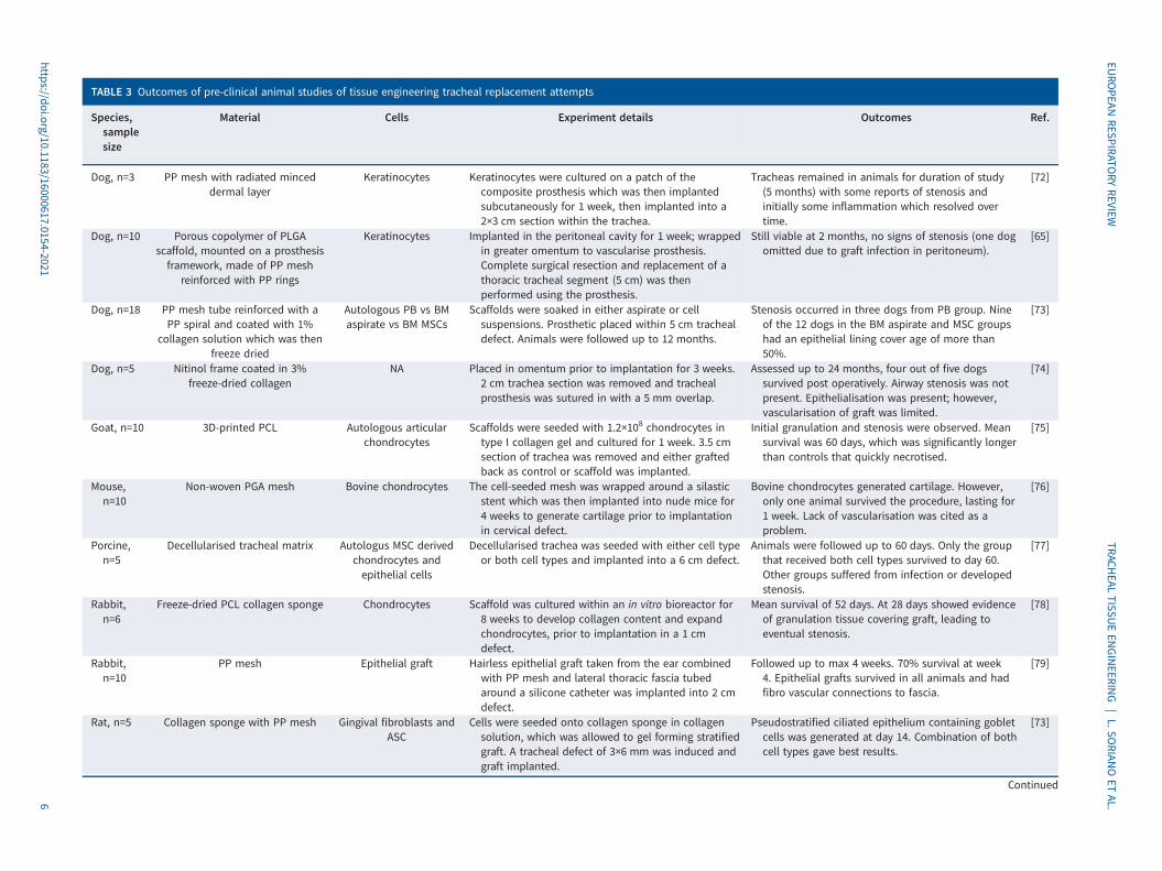
TABLE 3 Outcomes of pre-clinical animal studies of tissue engineering tracheal replacement attempts
Species,samplesize
Material Cells Experiment details Outcomes Ref.
Dog, n=3 PP mesh with radiated minceddermal layer
Keratinocytes Keratinocytes were cultured on a patch of thecomposite prosthesis which was then implantedsubcutaneously for 1 week, then implanted into a2×3 cm section within the trachea.
Tracheas remained in animals for duration of study(5 months) with some reports of stenosis andinitially some inflammation which resolved overtime.
[72]
Dog, n=10 Porous copolymer of PLGAscaffold, mounted on a prosthesisframework, made of PP mesh
reinforced with PP rings
Keratinocytes Implanted in the peritoneal cavity for 1 week; wrappedin greater omentum to vascularise prosthesis.Complete surgical resection and replacement of athoracic tracheal segment (5 cm) was thenperformed using the prosthesis.
Still viable at 2 months, no signs of stenosis (one dogomitted due to graft infection in peritoneum).
[65]
Dog, n=18 PP mesh tube reinforced with aPP spiral and coated with 1%
collagen solution which was thenfreeze dried
Autologous PB vs BMaspirate vs BM MSCs
Scaffolds were soaked in either aspirate or cellsuspensions. Prosthetic placed within 5 cm trachealdefect. Animals were followed up to 12 months.
Stenosis occurred in three dogs from PB group. Nineof the 12 dogs in the BM aspirate and MSC groupshad an epithelial lining cover age of more than50%.
[73]
Dog, n=5 Nitinol frame coated in 3%freeze-dried collagen
NA Placed in omentum prior to implantation for 3 weeks.2 cm trachea section was removed and trachealprosthesis was sutured in with a 5 mm overlap.
Assessed up to 24 months, four out of five dogssurvived post operatively. Airway stenosis was notpresent. Epithelialisation was present; however,vascularisation of graft was limited.
[74]
Goat, n=10 3D-printed PCL Autologous articularchondrocytes
Scaffolds were seeded with 1.2×10⁸ chondrocytes intype I collagen gel and cultured for 1 week. 3.5 cmsection of trachea was removed and either graftedback as control or scaffold was implanted.
Initial granulation and stenosis were observed. Meansurvival was 60 days, which was significantly longerthan controls that quickly necrotised.
[75]
Mouse,n=10
Non-woven PGA mesh Bovine chondrocytes The cell-seeded mesh was wrapped around a silasticstent which was then implanted into nude mice for4 weeks to generate cartilage prior to implantationin cervical defect.
Bovine chondrocytes generated cartilage. However,only one animal survived the procedure, lasting for1 week. Lack of vascularisation was cited as aproblem.
[76]
Porcine,n=5
Decellularised tracheal matrix Autologus MSC derivedchondrocytes andepithelial cells
Decellularised trachea was seeded with either cell typeor both cell types and implanted into a 6 cm defect.
Animals were followed up to 60 days. Only the groupthat received both cell types survived to day 60.Other groups suffered from infection or developedstenosis.
[77]
Rabbit,n=6
Freeze-dried PCL collagen sponge Chondrocytes Scaffold was cultured within an in vitro bioreactor for8 weeks to develop collagen content and expandchondrocytes, prior to implantation in a 1 cmdefect.
Mean survival of 52 days. At 28 days showed evidenceof granulation tissue covering graft, leading toeventual stenosis.
[78]
Rabbit,n=10
PP mesh Epithelial graft Hairless epithelial graft taken from the ear combinedwith PP mesh and lateral thoracic fascia tubedaround a silicone catheter was implanted into 2 cmdefect.
Followed up to max 4 weeks. 70% survival at week4. Epithelial grafts survived in all animals and hadfibro vascular connections to fascia.
[79]
Rat, n=5 Collagen sponge with PP mesh Gingival fibroblasts andASC
Cells were seeded onto collagen sponge in collagensolution, which was allowed to gel forming stratifiedgraft. A tracheal defect of 3×6 mm was induced andgraft implanted.
Pseudostratified ciliated epithelium containing gobletcells was generated at day 14. Combination of bothcell types gave best results.
[73]
Continued
https://doi.org/10.1183/16000617.0154-20216
EURO
PEANRESPIRATO
RYREVIEW
TRACHEAL
TISSUEEN
GINEERIN
G|L.SO
RIANOET
AL.
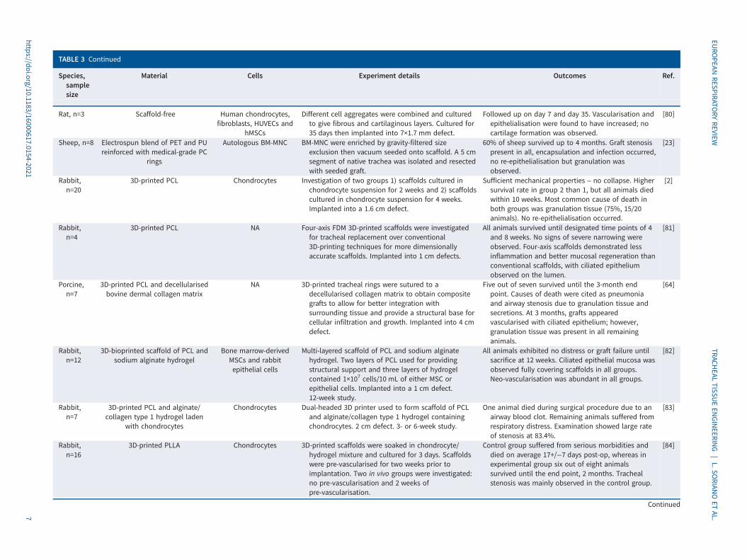
TABLE 3 Continued
Species,samplesize
Material Cells Experiment details Outcomes Ref.
Rat, n=3 Scaffold-free Human chondrocytes,fibroblasts, HUVECs and
hMSCs
Different cell aggregates were combined and culturedto give fibrous and cartilaginous layers. Cultured for35 days then implanted into 7×1.7 mm defect.
Followed up on day 7 and day 35. Vascularisation andepithelialisation were found to have increased; nocartilage formation was observed.
[80]
Sheep, n=8 Electrospun blend of PET and PUreinforced with medical-grade PC
rings
Autologous BM-MNC BM-MNC were enriched by gravity-filtered sizeexclusion then vacuum seeded onto scaffold. A 5 cmsegment of native trachea was isolated and resectedwith seeded graft.
60% of sheep survived up to 4 months. Graft stenosispresent in all, encapsulation and infection occurred,no re-epithelialisation but granulation wasobserved.
[23]
Rabbit,n=20
3D-printed PCL Chondrocytes Investigation of two groups 1) scaffolds cultured inchondrocyte suspension for 2 weeks and 2) scaffoldscultured in chondrocyte suspension for 4 weeks.Implanted into a 1.6 cm defect.
Sufficient mechanical properties – no collapse. Highersurvival rate in group 2 than 1, but all animals diedwithin 10 weeks. Most common cause of death inboth groups was granulation tissue (75%, 15/20animals). No re-epithelialisation occurred.
[2]
Rabbit,n=4
3D-printed PCL NA Four-axis FDM 3D-printed scaffolds were investigatedfor tracheal replacement over conventional3D-printing techniques for more dimensionallyaccurate scaffolds. Implanted into 1 cm defects.
All animals survived until designated time points of 4and 8 weeks. No signs of severe narrowing wereobserved. Four-axis scaffolds demonstrated lessinflammation and better mucosal regeneration thanconventional scaffolds, with ciliated epitheliumobserved on the lumen.
[81]
Porcine,n=7
3D-printed PCL and decellularisedbovine dermal collagen matrix
NA 3D-printed tracheal rings were sutured to adecellularised collagen matrix to obtain compositegrafts to allow for better integration withsurrounding tissue and provide a structural base forcellular infiltration and growth. Implanted into 4 cmdefect.
Five out of seven survived until the 3-month endpoint. Causes of death were cited as pneumoniaand airway stenosis due to granulation tissue andsecretions. At 3 months, grafts appearedvascularised with ciliated epithelium; however,granulation tissue was present in all remaininganimals.
[64]
Rabbit,n=12
3D-bioprinted scaffold of PCL andsodium alginate hydrogel
Bone marrow-derivedMSCs and rabbitepithelial cells
Multi-layered scaffold of PCL and sodium alginatehydrogel. Two layers of PCL used for providingstructural support and three layers of hydrogelcontained 1×10⁷ cells/10 mL of either MSC orepithelial cells. Implanted into a 1 cm defect.12-week study.
All animals exhibited no distress or graft failure untilsacrifice at 12 weeks. Ciliated epithelial mucosa wasobserved fully covering scaffolds in all groups.Neo-vascularisation was abundant in all groups.
[82]
Rabbit,n=7
3D-printed PCL and alginate/collagen type 1 hydrogel laden
with chondrocytes
Chondrocytes Dual-headed 3D printer used to form scaffold of PCLand alginate/collagen type 1 hydrogel containingchondrocytes. 2 cm defect. 3- or 6-week study.
One animal died during surgical procedure due to anairway blood clot. Remaining animals suffered fromrespiratory distress. Examination showed large rateof stenosis at 83.4%.
[83]
Rabbit,n=16
3D-printed PLLA Chondrocytes 3D-printed scaffolds were soaked in chondrocyte/hydrogel mixture and cultured for 3 days. Scaffoldswere pre-vascularised for two weeks prior toimplantation. Two in vivo groups were investigated:no pre-vascularisation and 2 weeks ofpre-vascularisation.
Control group suffered from serious morbidities anddied on average 17+/−7 days post-op, whereas inexperimental group six out of eight animalssurvived until the end point, 2 months. Trachealstenosis was mainly observed in the control group.
[84]
Continued
https://doi.org/10.1183/16000617.0154-20217
EURO
PEANRESPIRATO
RYREVIEW
TRACHEAL
TISSUEEN
GINEERIN
G|L.SO
RIANOET
AL.
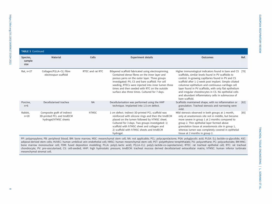
TABLE 3 Continued
Species,samplesize
Material Cells Experiment details Outcomes Ref.
Rat, n=27 Collagen/P(LLA–CL) fibreelectrospun scaffold
RTEC and rat RTC Bilayered scaffold fabricated using electrospinning.Contained dense fibres on the inner layer andporous yarns on the outer layer. Three groupsinvestigated: PV, CS and bare scaffold. For cellseeding, RTECs were injected into inner lumen threetimes and then seeded with RTC on the outsidesurface also three times. Cultured for 7 days.
Higher immunological indicators found in bare and CSscaffolds, similar levels found in PV scaffolds tocontrol. In-growing capillaries found in PV and CSscaffold after 1–2 week post implant. Simple ciliatedcolumnar epithelium and continuous cartilage celllayer found in PV scaffolds, with only flat epitheliumand irregular chondrocytes in CS. No epithelial cellsand abundant inflammatory cells in submucosa ofbare scaffold.
[70]
Porcine,n=6
Decellularised trachea NA Decellularisation was performed using the HHPtechnique. Implanted into 1.5 cm defect.
Scaffolds maintained shape, with no inflammation orgranulation. Tracheal stenosis and narrowing weremild.
[62]
Rabbit,n=20
Composite graft of indirect3D-printed PCL and tmdECM
hydrogel/hTMSC sheets
hTMSC 1 cm defect. Indirect 3D-printed PCL scaffold wasreinforced with silicone rings and then the tmdECMplaced on the lumen followed by hTMSC sheet.Cultured for 3 days. Two groups investigated: 1)scaffold with hTMSC sheet and collagen and2) scaffold with hTMSC sheets and tmdECMhydrogel.
Mild stenosis observed in both groups at 1 month,only at anastomosis site not in middle, but becamemore severe in group 1 at 2 months compared togroup 2. Thin epithelial layer formed abovegranulation tissue at anastomosis site in group 1,whereas lumen was completely covered in epithelialtissue at 2 months in group 2.
[85]
PP: polypropylene; PB: peripheral blood; BM: bone marrow; MSC: mesenchymal stem cell; NA: not applicable; PCL: polycaprolactone; PGA: polyglycolic acid; PLGA: D,L-lactide-co-glycolide; ASC:adipose-derived stem cells; HUVEC: human umbilical vein endothelial cell; hMSC: human mesenchymal stem cell; PET: polyethylene terephthalate; PU: polyurethane; PC: polycarbonate; BM-MNC:bone marrow mononuclear cell; FDM: fused deposition modelling; PLLA: poly(L-lactic acid); P(LLA–CL): poly(L-lactide-co-caprolactone); RTEC: rat tracheal epithelial cell; RTC: rat trachealchondrocyte; PV: pre-vascularised; CS: cell-seeded; HHP: high hydrostatic pressure; tmdECM: tracheal mucosa derived decellularised extracellular matrix; hTMSC: human inferior turbinatemesenchymal stromal cell.
https://doi.org/10.1183/16000617.0154-20218
EURO
PEANRESPIRATO
RYREVIEW
TRACHEAL
TISSUEEN
GINEERIN
G|L.SO
RIANOET
AL.

studies have been limited by single cell type approaches, poor mechanical characterisation and insufficientre-vascularisation [84].
The rise of 3DP in medical research has seen an exploration into direct cell-laden biomaterial printing,referred to as bioprinting, which holds promise in developing scaffolds that are capable of supportingmultiple cell types while being reproducible and patient specific [96]. Bioprinting with cell-loaded bioinksresults in poor biomechanical properties because of low bioink melting points and the resultant weakstructures [93, 97]. To overcome this, studies have used dual-headed 3D printers to produce structurallystrong scaffolds by combining thermoplastic polymers with hydrogels such as alginate and collagen type Ihydrogel with PCL [83]. PCL has also been used in combination with sodium alginate hydrogels andwhen implanted in an animal model growth of ciliated respiratory cells, cartilage formation andvascularisation was observed [82]. Although bioprinting holds potential in fabricating adequate trachealsubstitutes, further research is crucial to overcome cell survival outside physiological conditions during theprinting process, maintaining an appropriate temperature profile as well as withstanding mechanical stressapplied during bioink extrusion [96, 97].
Translation to clinical trialsPre-clinical outcomes of tissue-engineered tracheal scaffoldsTranslation into animal studies has been performed to investigate tissue-engineered tracheas with somelimited success (table 3). However, many of these attempts suffer from limited follow-up time of theanimals, typically 1–3 months [77, 79]. Early attempts often ended in severe inflammatory responseleading to granulation and stenosis of graft or graft failure through infection [76, 78]. Maintainingairtightness and promoting vascularisation significantly adds to the survival prospects of the graft, whichcan be achieved through pre-implantation strategies [65, 78]. Wrapping the construct in omentum providesboth a vascular network and a source of tissue to seal and maintain airtightness of the graft, thus creating abarrier to bacterial colonisation. Pre-implantation in the abdominal cavity facilitates omentum adherence tothe graft and integration of the vascular network [74]. Pre-vascularisation prior to implantation has shownhigher survival rate of animals, lower rates of stenosis and increased re-epithelisation [70, 82].Furthermore, when combined with a cell seeded construct, this technique further supports cell survival andgraft integration. Construct pre-seeding has also been reported to increase the survival rate of animals andthere was a lack of tracheal stenosis when combining pre-seeding with chondrocytes andpre-vascularisation [70]. Moreover, the inclusion of pre-seeded epithelial cells also enhanced graftacceptance and overall implant success in conjunction with mesenchymal stem cells (MSCs), resulting infull epithelial coverage of the graft and no signs of distress or graft failure in all animals for 3 monthsoccurring [82]. The longest running preclinical study to date was performed in a canine model with a24-month follow-up using a collagen-coated nitinol frame [74]. The implant was first wrapped in omentumand implanted into the abdominal cavity for 3 weeks, after which it was implanted following a 20 mmtracheal resection with a survival rate of 80% reported over 18 months. Histological examinationdemonstrated stable epithelialisation in a non-stratified monolayer with no secretory glands present and nomuscular regeneration, although stenosis was not observed [74]. Other studies using this approach havealso observed similar results after 2 months, unfortunately longer-term follow-ups of the study were notpublished [65]. Several other studies in canines have been followed up to 5–12 months [72, 73].
Attempts to implant synthetic tissue-engineered grafts into humans has resulted in significant controversyand led to several fatalities [25]. Since then, further pre-clinical studies in large animal models have beenconducted to better comprehend some of the root cause for these synthetic graft failures [25]. Aspreviously highlighted, the lack of functional epithelium resulted in significant risk of infection,inflammation and/or graft encapsulation. Ongoing inflammation was present in all animals within the studyleading to stenosis formation most evident at proximal and distal locations of the graft [25]. Graft seedingwith MSCs was associated with delayed onset of respiratory distress showing the importance ofestablishing a functional epithelium prior to implantation. These in vivo attempts, although mostlyshort-term studies, have highlighted significant challenges that need to be addressed for a potential trachealgraft. The inclusion of a pre-seeded epithelial layer appears to mitigate bacterial colonisation and thepre-vascularisation of grafts also enhances graft acceptance and survival, however there are still concernsdue to the presence of granulation tissue in some attempts.
Clinical outcome of non-TE approachesProsthesesAfter the limited success of early attempts at treating large tracheal defects, efforts focused onreconstructing the trachea with the use of polymer prosthesis in either solid or porous form. The Nevilleprosthesis, a solid prothesis manufactured using the synthetic polymer siloxane demonstrated success in
https://doi.org/10.1183/16000617.0154-2021 9
EUROPEAN RESPIRATORY REVIEW TRACHEAL TISSUE ENGINEERING | L. SORIANO ET AL.
animal studies but this could not be replicated in humans due to stenosis and graft failure [98–101]. Thiswas caused by the mismatch of mechanical properties of the prosthesis in comparison to the softersurrounding tissue and erosion of adjacent blood vessels and oesophagus due to its rigidity. Furthermore,their inability to become incorporated within the surrounding tissue led to dislodgement during coughingas they become loose and obstructed the airway [49, 90, 100]. Other solid prostheses have consisted ofstainless-steel wire [102], polyethylene (PE) [103], tantalum [104] and silicone coupled with a Dacron™ring [99].
As issues with solid protheses became apparent, research shifted towards investigating porous prostheses toallow for better ingrowth of cells and tissue formation. The most promising development of porousprostheses arose in 1961 with the success of the Marlex™ mesh prothesis, a high-density PE andpolypropylene, showing 16 months’ graft survival and patency in an animal study [101]. However, thisprosthesis performed disappointingly in humans due to stenosis and erosion of the surrounding bloodvessels [105]. The Marlex prosthesis reinforced the importance of the growth of respiratory epithelium onthe inner lumen to act as a protective barrier against foreign bodies in inhaled air [49, 90, 100].
Other attempts at porous protheses have included wire-enforced dermal grafts [106], stainless steel wiremesh [107], silicone with Dacron™ [98], PE [108] and silicone tubes [109], but these have had similarpoor results. Current treatment options for treatment of most tracheobronchical complications have beenwith commercially available stents including the Ultraflex™ [110], the silicone-based self-expandingPolyFlex™ [111] from Boston Scientific, and Novatech’s silicone Dunmon® and Gold Studded Stents®[112]. The use of silicone stents has been shown clinically to improve symptoms and quality of life in thevast majority of cases. Although stents have been shown to be tolerated well in the majority of cases,long-term follow-ups have highlighted serious drawbacks. Stent migration, the formation of granulationtissue, obstruction and mucus retention with infections have all been recorded in long-term studies [113–116].
Aortic allograftsAllograft transplantation for large tracheal defects has primarily focused on two donor tissue sites: tracheasand aortas. Fresh and cryopreserved aortas have been investigated as potential tracheal replacement graftsshowing regenerated cartilage and respiratory epithelium in an ovine model, albeit cartilage regeneration inother animal studies has not been observed [117–119]. Nevertheless, the use of aortas as potential donortissues for trachea replacement was first used in a clinical attempt in a 68-year-old male in 2006, whichresulted in severe complications and ultimately death [120]. A cryopreserved aortic allograft was implantedin a 78-year-old male supported with a stent although no cartilage formation was observed and the patientsuffered from lower forced expiratory volume [121]. These concerns were also present in another studywith six patients using cryopreserved thoracic aortas that were pre-vascularised and stent-supported [122].Serious complications arose during long-term follow-ups including blood vessels erosion, stent migrationand tracheoesophageal fistulas, with no cartilage formation or respiratory epithelium observed in biopsiedtissue [119]. The most extensive trial using aortic allografts involved 20 patients with a 76.9% survival rateafter airway transplantation with regeneration of respiratory epithelium and cartilage formation. However,the authors highlighted the need to further assess both the efficacy and the safety of this procedure [123].
Tracheal allograftsThe first successful human tracheal transplant was reported in 1979 showing no signs of rejection or anycomplication 9 weeks post-op; however, no long-term outcomes were ever made available [124]. Anotherearly success in tracheal allografts was in 1990, when a one-stage tracheal transplantation was successfullyconducted but needed to be supported with a stent and extensive immunosuppressive therapy. A follow-up2 years later reported no issues [125]. Following the early successes, attempts at trachea transplantationhave seen very little clinical success [53, 100]. Recent developments by DELAERE et al. [40, 41, 126] sawthe development of a two-step technique in which the donor trachea is revascularised in the patient’sforearm before implantation [40, 41, 127]. Despite tissue necrosis being observed in some patients, thetechnique remains a promising approach [40, 41, 100].
Although allograft transplantation provides a great airtight analogue structurally, success with allografts hasbeen mixed. The most successful cases used extensive immunosuppressive therapy, which isnot suitablefor patients with malignant tumours, furthermore allografts have to be supported with stents due toinadequate mechanical properties and cartilage regeneration [3, 100]. However, stents have been shown tolead to granulation, erosion of adjacent tissue and blood vessels and infection. Revascularisation ofallografts is therefore poor and allografts require vascularisation by implantation into a second surgical site,increasing risk of infection and hospitalisation time [33, 128]. Although the use of “traditional” organreplacement methods has had little success in tracheal replacement, previous attempts have provided
https://doi.org/10.1183/16000617.0154-2021 10
EUROPEAN RESPIRATORY REVIEW TRACHEAL TISSUE ENGINEERING | L. SORIANO ET AL.

TABLE 4 Translation of non-tissue engineering (TE) and TE approaches for tracheal restoration and regeneration in human clinical trials
Patient(s)details
Technology Material(s) Cells Procedure details Outcome Ref.
Non-TE approaches43-year-oldfemale
Autograft Forearm freeflapsupported with anUltraflex stent
NA A freeflap was harvested and wrappedaround a stent after implantation in a6 cm tracheal defect arising for acarcinoma.
Died 16 months after the procedure frompre-existing conditions.
[131]
63-year-oldfemale
Autograft Forearm freeflap with anexternal mesh support
NA Radial forearm fasciocutaneous flap with aHemashield vascular graft and PolyMaxresorbable mesh.
Patient remains symptom-free at6 months and has returned to normalactivities. Bronchoscopy at 6 monthsshowed slight migration but healedflap with no obstruction.
[132]
16 patients(37–68 years)
Autograft Cartilage rib and fascialskin pad
NA Segments of the patient’s own cartilagewere inserted within a skinpad harvestedfrom the forearm. Construct waswrapped around silicone tube forsuturing and implanted within thedefect.
Three deaths following surgicalprocedure due to lung infections,acute respiratory distress syndromeand myocardial infarction. Long-termfollow-up showed a 65% survival rate.
[129,133]
21-year-oldmale
Allotransplantation Donor cadaveric trachea NA or autologous patientcells
Patient suffering from extensive trachealstenosis. A cadaveric trachea with intactblood supply was implantedheterotopically into thesternocleidomastoid muscle for 3 weeks.It was then implanted orthopically intothe trachea defect with a vascularisedmuscular section of thesternocleidomastoid.
Integration of the trachea graft andproper function for 9 weeks withoutevidence of rejection, ischaemia orinfection.
[124]
24-year-oldfemale
Allotransplantation Donor cadaveric trachea NA Patient suffering from tracheal stenosis andthird stage respiratory insufficiency. Theallograft was implanted and wrappedwith omentum and the patient wasplaced on immunosuppressive therapy.
Uneventful postoperative course withsigns of graft rejection, necrosis,bacterial and viral infection which leadto the need of silicon endoprosthesis.Signs for rejection reduced and 1-yearfollow-up suggested patient was stillalive with a restored tracheal lumen.
[125]
Four patients(17–64 years;three males,one female)
Allotransplantation Human cadaverictrachea
Recipient’s buccal mucosaand/or ingrown recipient
cells
Patients suffering from tracheal stenosis ortracheal chondrosarcoma. Decellularisedtracheas were implanted in the forearmand grafted with buccal mucosa orwrapped in forearm fascia to improvevascularisation.
Tracheal necrosis after withdrawal ofimmunosuppression and poorvascularisation around the graftsleading to partial loss of theallotransplant in three patients whileadditional approaches for recipient cellrepopulation of the construct in onepatient allowed a vascularisedallotransplant and normal airways6 months after transplantation.
[128]
Continued
https://doi.org/10.1183/16000617.0154-202111
EURO
PEANRESPIRATO
RYREVIEW
TRACHEAL
TISSUEEN
GINEERIN
G|L.SO
RIANOET
AL.

TABLE 4 Continued
Patient(s)details
Technology Material(s) Cells Procedure details Outcome Ref.
68-year-oldmale
Stent-supportedaortic autograft
Aorta autograft andsilicone Dumon stent
NA Patient presenting tracheal squamous cellcarcinoma. A 7 cm abdominal aortaautograft was harvested and replacedwith a Dracon graft. The aortic graft wasimplanted coupled with a siliconeDumon stent to avoid aortic wall injury.
Granulation tissue formation led to acuterespiratory distress syndrome whichwas treated by introduction of anadditional tracheal stent. Stent wasremoved due to migration albeit noairway collapse was detected.Pneumonia, respiratory distress andpneumothorax lead to death of patientdue to septic shock.
[120]
Six patients(17–52 years;five males,one female)
Stent-supportedaortic allograft
Fresh or cryopreservedaortic grafts and silicone
stent
NA Patients suffering from mucoepidermoidand adenoid cystic carcinoma. Aorticallografts were wrapped with wellvascularised pectoral muscle orthymopericardial fat flaps and implantedusing a silicone stent.
Complete resection achieved in 83% ofpatients. Major morbidity, fistulas anduneventful outcomes. All grafts showedadequate vascularisation and fourpatients are disease-free.
[122]
78-year-oldmale
Stent-supportedaortic allograft
Cryopreserved aorticgraft and custom-made
nitinol stent
NA Patient suffering from extensivebronchopulmonary malignant tumourpre-treated with chemotherapy. Lungcancer was resected, and astent-supported graft was implanted.
Well-functioning re-implanted lobe found1 year post procedure. Patientrecovered baseline activity withsatisfying health quality of life.
[121]
20 patients(24–79 years;13 males,sevenfemales)
Stent-supportedaortic allograft
Cryopreserved aorticgraft and custom-made
nitinol stent
NA Patients presenting proximal lung tumoursand malignant or benign lesions of thetrachea and bronchi. Radial tumourresections were performed and thestent-supported graft was implanted andcovered circumferentially with a localmuscle flap.
Patient follow-up of 90 days detecting a5% mortality rate. No adverse effect ofthe surgical technique used showing a76.5% survival at a median follow-up of3 years and 11 months. Regeneration ofcartilage and respiratory epitheliumobserved.
[123]
TE approaches26-year-oldmale
Decellularisation Decellularised porcinejejunum containingautologous cellpopulation
Recipient’s mvECs andskMCs
Patient suffering from extensive tracheal andoesophageal defect. Porcine cell-freevascularised scaffolds were obtainedthrough a decellularisation process andseeded to ensure re-endothelialisationwith recipient’s cell prior to implantationinto the 5×2 cm defect. The construct wascharacterised prior to implantation toensure safety and maximum performance.
The postoperative period was uneventfuland the transplanted bioengineeredconstruct was fully integratedpresenting a fully functional respiratoryepithelium on the lining of the airwaywithout tissue scar formation or tissuededifferentiation.
[134]
Continued
https://doi.org/10.1183/16000617.0154-202112
EURO
PEANRESPIRATO
RYREVIEW
TRACHEAL
TISSUEEN
GINEERIN
G|L.SO
RIANOET
AL.
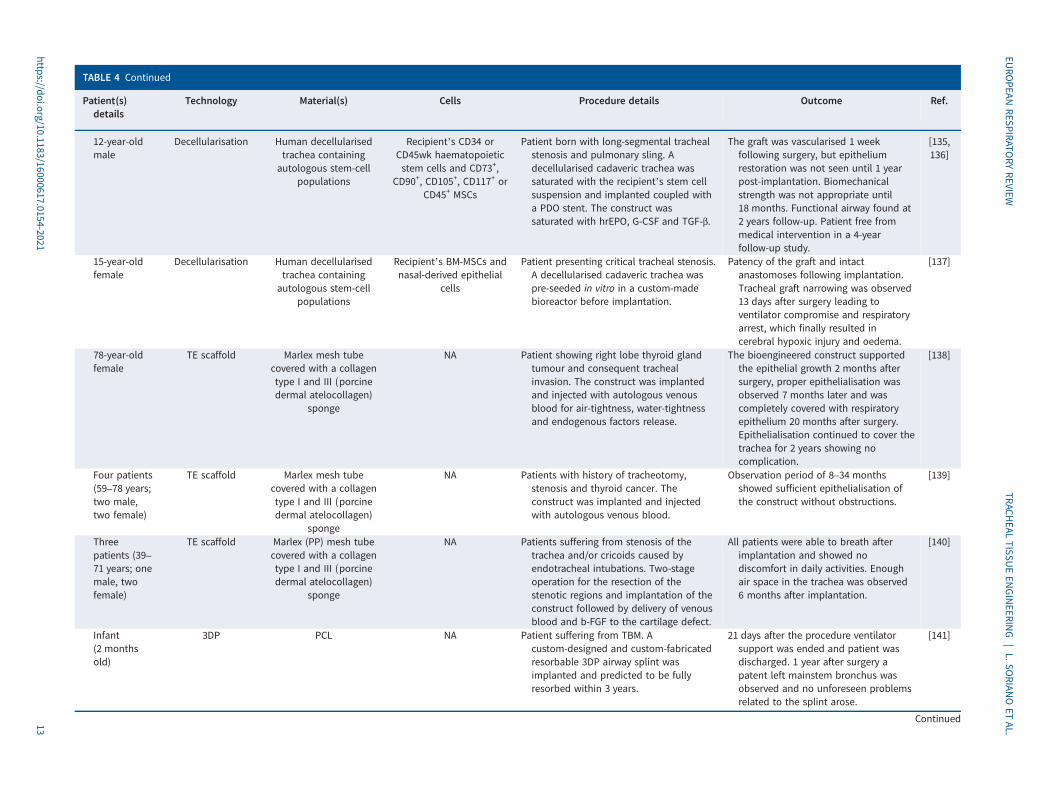
TABLE 4 Continued
Patient(s)details
Technology Material(s) Cells Procedure details Outcome Ref.
12-year-oldmale
Decellularisation Human decellularisedtrachea containingautologous stem-cell
populations
Recipient’s CD34 orCD45wk haematopoieticstem cells and CD73+,
CD90+, CD105+, CD117+ orCD45+ MSCs
Patient born with long-segmental trachealstenosis and pulmonary sling. Adecellularised cadaveric trachea wassaturated with the recipient’s stem cellsuspension and implanted coupled witha PDO stent. The construct wassaturated with hrEPO, G-CSF and TGF-β.
The graft was vascularised 1 weekfollowing surgery, but epitheliumrestoration was not seen until 1 yearpost-implantation. Biomechanicalstrength was not appropriate until18 months. Functional airway found at2 years follow-up. Patient free frommedical intervention in a 4-yearfollow-up study.
[135,136]
15-year-oldfemale
Decellularisation Human decellularisedtrachea containingautologous stem-cell
populations
Recipient’s BM-MSCs andnasal-derived epithelial
cells
Patient presenting critical tracheal stenosis.A decellularised cadaveric trachea waspre-seeded in vitro in a custom-madebioreactor before implantation.
Patency of the graft and intactanastomoses following implantation.Tracheal graft narrowing was observed13 days after surgery leading toventilator compromise and respiratoryarrest, which finally resulted incerebral hypoxic injury and oedema.
[137]
78-year-oldfemale
TE scaffold Marlex mesh tubecovered with a collagentype I and III (porcinedermal atelocollagen)
sponge
NA Patient showing right lobe thyroid glandtumour and consequent trachealinvasion. The construct was implantedand injected with autologous venousblood for air-tightness, water-tightnessand endogenous factors release.
The bioengineered construct supportedthe epithelial growth 2 months aftersurgery, proper epithelialisation wasobserved 7 months later and wascompletely covered with respiratoryepithelium 20 months after surgery.Epithelialisation continued to cover thetrachea for 2 years showing nocomplication.
[138]
Four patients(59–78 years;two male,two female)
TE scaffold Marlex mesh tubecovered with a collagentype I and III (porcinedermal atelocollagen)
sponge
NA Patients with history of tracheotomy,stenosis and thyroid cancer. Theconstruct was implanted and injectedwith autologous venous blood.
Observation period of 8–34 monthsshowed sufficient epithelialisation ofthe construct without obstructions.
[139]
Threepatients (39–71 years; onemale, twofemale)
TE scaffold Marlex (PP) mesh tubecovered with a collagentype I and III (porcinedermal atelocollagen)
sponge
NA Patients suffering from stenosis of thetrachea and/or cricoids caused byendotracheal intubations. Two-stageoperation for the resection of thestenotic regions and implantation of theconstruct followed by delivery of venousblood and b-FGF to the cartilage defect.
All patients were able to breath afterimplantation and showed nodiscomfort in daily activities. Enoughair space in the trachea was observed6 months after implantation.
[140]
Infant(2 monthsold)
3DP PCL NA Patient suffering from TBM. Acustom-designed and custom-fabricatedresorbable 3DP airway splint wasimplanted and predicted to be fullyresorbed within 3 years.
21 days after the procedure ventilatorsupport was ended and patient wasdischarged. 1 year after surgery apatent left mainstem bronchus wasobserved and no unforeseen problemsrelated to the splint arose.
[141]
Continued
https://doi.org/10.1183/16000617.0154-202113
EURO
PEANRESPIRATO
RYREVIEW
TRACHEAL
TISSUEEN
GINEERIN
G|L.SO
RIANOET
AL.
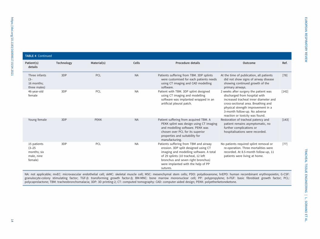
TABLE 4 Continued
Patient(s)details
Technology Material(s) Cells Procedure details Outcome Ref.
Three infants(3–16 months;three males)
3DP PCL NA Patients suffering from TBM. 3DP splintswere customised for each patients needsusing CT imaging and CAD modellingsoftware.
At the time of publication, all patientsdid not show signs of airway diseaseshowing continued growth of theprimary airways.
[78]
46-year-oldfemale
3DP PCL NA Patient with TBM. 3DP splint designedusing CT imaging and modellingsoftware was implanted wrapped in anartificial pleural patch.
2 weeks after surgery the patient wasdischarged from hospital withincreased tracheal inner diameter andcross-sectional area. Breathing andphysical strength improvement in a3-month follow-up. No adversereaction or toxicity was found.
[142]
Young female 3DP PEKK NA Patient suffering from acquired TBM. APEKK splint was design using CT imagingand modelling software. PEKK waschosen over PCL for its superiorproperties and suitability formanufacturing.
Restoration of tracheal patency andpatient remains asymptomatic, nofurther complications orhospitalisations were recorded.
[143]
15 patients(3–25months; sixmale, ninefemale)
3DP PCL NA Patients suffering from TBM and airwayerosion. 3DP split designed using CTimaging and modelling software. A totalof 29 splints (10 tracheal, 12 leftbronchus and seven right bronchus)were implanted with the help of PPsutures.
No patients required splint removal orre-operation. Three mortalities wererecorded. At 8.5-month follow-up, 11patients were living at home.
[77]
NA: not applicable; mvEC: microvascular endothelial cell; skMC: skeletal muscle cell; MSC: mesenchymal stem cells; PDO: polydioxanone; hrEPO: human recombinant erythropoietin; G-CSF:granulocyte-colony stimulating factor; TGF-β: transforming growth factor-β; BM-MNC: bone marrow mononuclear cell; PP: polypropylene; b-FGF: basic fibroblast growth factor; PCL:polycaprolactone; TBM: tracheobronchomalacia; 3DP: 3D printing-2; CT: computed tomography: CAD: computer-aided design; PEKK: polyetherketoneketone.
https://doi.org/10.1183/16000617.0154-202114
EURO
PEANRESPIRATO
RYREVIEW
TRACHEAL
TISSUEEN
GINEERIN
G|L.SO
RIANOET
AL.
insight on the requirements for a successful tracheal graft. In more recent years, research has shifted toovercoming these challenges by using TE technology.
Tracheal autograftsThe use of a patient’s own tissue has also been investigated clinically for tracheal transplant. This two-stepsurgical approach uses the patient’s own cartilage, which are segments that have been harvested from theirribs [129]. These segments are inserted within a fascial skin pad, also harvested from the patient’s forearm,and wrapped around a temporary silicone tube to suture together a tracheal conduit for implantation. Greatcare is taken to keep the radial artery and superficial and deep drainage veins of the skin pad intact torevascularise the conduit upon implantation. This technique alleviates the need for immunosuppressants bynot introducing synthetic or foreign bodies within patients [129, 130]. However, due to the absence of arespiratory epithelial layer and use of autologous cartilage tissue, tracheal autografts are not suitable for allpatients. Autograft transplantation is based on acceptable respiratory function and healthy cartilage tissueas secretion obstruction and cartilage fracture can cause implant failure [129, 130].
Clinical translation of TE approachesFew TE approaches for tracheal regeneration have made it to human clinical trials, but significant findingshave been demonstrated in TE that have reached this milestone (table 4). The first report of a successfulcase dates to 2005 [138] when a Marlex prostheses covered in type I and II porcine-derived collagen wassuccessfully implanted and demonstrated regeneration of the respiratory epithelium. Histologicalexamination showed the successful incorporation of the scaffold within surrounding tissues, regeneratingthe subglottic and tracheal epithelium and achieving optimal epithelial growth 20 months post-operationcovering the implanted material. Moreover, mechanical studies confirmed the patency of the airway [138].This technique was further employed in a wider set of patients demonstrating a well-epithelialised lumenwith no apparent airway obstructions in follow-up examinations making it a promising approach to treattracheal stenosis and malignancy [139]. The combination of this approach with growth factor delivery toenhance graft performance showing optimal regeneration in all patients was also evaluated. Addition ofbasic fibroblast growth factor (bFGF) to the cartilage defect, was hypothesised to improve blood vesselformation, and maintained airway patency in all patients. However, the use of bFGF may be of limited usein cases of cancer resection due to increased tumour recurrence [140].
Surgical implantation of DTs was first successfully performed in 2010. A cadaveric trachea wasdecellularised, implanted in the forearm of the recipient and seeded with buccal mucosa to enhancevascularisation prior to implantation [127]. This approach was further explored with four additionalpatients, but tracheal necrosis and poor vascularisation hampered the outcome of this clinical trial [139]. Inanother set of attempts, cadaveric tracheas were decellularised and pre-seeded prior to implantation withthe recipient’s stem cells and growth factors to ensure vascularisation of the graft and to promotechondrogenesis with mixed outcomes due to graft narrowing. However, one DT successfully restoredepithelium around the damaged section with no further complications observed in a 4-year follow-up study[135–137]. Even though some of these TE approaches have demonstrated a certain degree of promise,further and more extensive follow-up studies and a wider number of patients will be needed in order toensure this technology can become an alternative approach for tracheal restoration and regeneration.
As highlighted earlier, 3DP has emerged as a promising technique to treat tracheal defects in paediatricpatients as 3D printed airway splints have been graded FDA approval recently [144]. The implantation of a3D-printed splint to treat a severe case of TBM was first reported in 2013 [141] and, since then, has beenapplied to a wider set of patients reporting low general mortalities [91, 92, 142, 143]. However, implantationof 3D-printed airway splints has been mostly restricted to paediatric patients suffering from TBM. In thisway, airway growth is guided during the early stages of development and leads to natural resolution of TBM.Apart from paediatric patients, there is only one reported attempt of using a 3D-printed airway splint in anadult individual with maintained airway patency 3 months after the procedure. Additionally, custom-madeairway stents by a number of groups, including for example researchers at the Toulouse University Hospitaland AnatomikModeling have shown promise in ongoing clinical trials. Patient-specific sacrificial mouldswere 3D-printed from CT-scan images to create patient-specific tracheal prosthetics from silicone elastomer.Several patients have successfully received these implants in clinical trials, with reports of improved qualityof life and no complications thus far. This personalised approach shows promise, but long-term results havenot been made available as of yet [145]. Albeit early promising attempts to use 3D-printing technologies toovercome tracheal stenosis and malignancies are emerging, there is a need to extend follow-up studies toasses splint resorption as well as establish optimal designs and validated manufacturing processes in order toassure safety and efficacy during future clinical trials [146].
https://doi.org/10.1183/16000617.0154-2021 15
EUROPEAN RESPIRATORY REVIEW TRACHEAL TISSUE ENGINEERING | L. SORIANO ET AL.
The first step for the successful development of a tissue-engineered trachea is a thorough characterisationof the proposed construct in terms of material performance, degradation, mechanical strength andflexibility. Moreover, the manufacturing process employed should allow where possible the manufacturingof individualised constructs to meet specific patient’s needs, in addition to being anti-bacterial,anti-proliferative, anti-tussive and non-migrating [2]. One of the main limitations in tracheal TE is the lackof consistent and relevant data of airway biomechanics, lack of standardised tests to assess mechanicalproperties as well as a wide range of mechanical values that a potential graft should possess as seen intable 2 [147]. Therefore, a standardised procedure for assessing mechanical strength should be developedto better understand the needs of a TE graft. This standard procedure should also include the differences intracheal biomechanics between different age-group patients as well as gender differences [27, 148].
Several scaffolds have been designed to support epithelialisation in a graft as the lining of a maturepseudostratified epithelium is needed in order to avoid stenosis, tissue outgrowth and granulation [22].Different approaches have been investigated both in vitro and in vivo but the lack of long-termassessment and the granulation and inflammatory reaction following transplantation have hindered furthertranslation. This highlights the need for better TE approaches that support full and functionalre-epithelialisation early on in the development graft process [81, 84, 149].
Vascularisation of a tissue-engineered trachea stands as the most important challenge that needs to beovercome for TE tracheal scaffolds to translate into clinical trials. It has been widely reported thatvascularisation is mandatory in order to achieve a functional graft as the lack of a vasculature formationwill inevitably lead to high morbidity, poor outcomes and no benefit for the patient [41]. Although severalapproaches have shown promise in revascularising the implanted graft, further long-term assessment of thepatients and an increased number of trials is needed to demonstrate that these approaches are suitable fortracheal regeneration and restoration [127, 128, 135, 136].
Overall, the few TE approaches to tracheal replacement that have made it to clinical trials have providedsubstantial insight on the necessary requirements for a graft to succeed. Due to the complexity of thetrachea’s anatomy, a number of challenging properties must be met collectively, and data gained from bothpre-clinical and clinical trials have provided a greater understanding on the advantages and pitfalls ofcertain techniques and methods.
ConclusionSeveral TE strategies have emerged during the last decade with the goal of developing a functionalscaffold capable of regenerating tracheal tissues. However, clinical translation of TE technologies has beenvery limited and on a small scale to date. Furthermore, poor mechanical properties, insufficientvascularisation of the implanted tracheas over time and inadequate re-epithelialisation have hindered graftperformance [41]. Moreover, both animal and clinical studies lack long-term assessments of graftperformance, which are needed to fully assess the feasibility of the approach. Therefore, any constructbeing developed for tracheal regeneration must provide the required mechanical properties as well assupport the formation of a respiratory epithelium and a network of vascular capillaries to avoid graftcontamination, granulation and tissue necrosis.
Tracheal replacement has lately become a focus of interest for TE with the development of promisingprototypes showing that epithelialisation and vascularisation of the grafts can be achieved through differentprocedures. Moreover, some of these TE grafts have made their way through clinical trials with promisingoutcomes and delivering crucial insight on the necessary properties needed for a tracheal graft to succeed.Overcoming limitations involving mechanical properties, epithelialisation and vascularisation will lead to awider use of TE tracheal substitutes that could become a turning point in how tracheal stenosis and damageis clinically addressed.
Provenance: Submitted article, peer reviewed.
Conflict of interest: None declared.
Support statement: The authors acknowledge the funding received for this research under the Science FoundationIreland (SFI) Centre for Research in Medical Devices (CURAM) and the European Regional Development Fund (Grant13/RC/2073); and by the SFI-funded AMBER centre (Grant 17/RC-PhD/3477).
https://doi.org/10.1183/16000617.0154-2021 16
EUROPEAN RESPIRATORY REVIEW TRACHEAL TISSUE ENGINEERING | L. SORIANO ET AL.

References1 Boazak EM, Auguste DT. Trachea mechanics for tissue engineering design. ACS Biomater Sci Eng 2018; 4:
1272–1284.2 Gao M, Zhang H, Dong W, et al. Tissue-engineered trachea from a 3D-printed scaffold enhances
whole-segment tracheal repair. Sci Rep 2017; 7: 5246.3 Grillo HC. Tracheal replacement: a critical review. Ann Thorac Surg 2002; 73: 1995–2004.4 Grillo HC. Slide tracheoplasty for long-segment congenital tracheal stenosis. Ann Thorac Surg 1994; 58:
613–621.5 Prokakis C, Koletsis EN, Dedeilias P, et al. Airway trauma: a review on epidemiology, mechanisms of injury,
diagnosis and treatment. J Cardiothorac Surg 2014; 9: 117.6 Johnson SB. Tracheobronchial injury. Semin Thorac Cardiovasc Surg 2008; 20: 52–57.7 Fiorelli A, Cascone R, Di Natale D, et al. Endoscopic treatment with fibrin glue of post-intubation tracheal
laceration. J Vis Surg 2017; 3: 102.8 Miñambres E, Burón J, Ballesteros MA, et al. Tracheal rupture after endotracheal intubation: a literature
systematic review. Eur J Cardio Thorac Surg 2009; 35: 1056–1062.9 Madariaga MLL, Gaissert HA. Overview of malignant tracheal tumors. Ann Cardiothorac Surg 2018; 7:
244–254.10 Urdaneta AI, Yu JB, Wilson LD. Population based cancer registry analysis of primary tracheal carcinoma. Am
J Clin Oncol 2011; 34: 32–37.11 Kandaswamy C, Balasubramanian V. Review of adult tracheomalacia and its relationship with chronic
obstructive pulmonary disease. Curr Opin Pulm Med 2009; 15: 113–119.12 Esteller-Moré E, Ibañez J, Matiñó E, et al. Prognostic factors in laryngotracheal injury following intubation
and/or tracheotomy in ICU patients. Eur Arch Otorhinolaryngol 2005; 262: 880–883.13 Sarper A, Ayten A, Eser I, et al. Tracheal stenosis after tracheostomy or intubation: review with special
regard to cause and management. Tex Heart Inst J 2005; 32: 154–158.14 Nassar N, Leoncini E, Amar E, et al. Prevalence of esophageal atresia among 18 international birth defects
surveillance programs. Birth Defects Res A Clin Mol Teratol 2012; 94: 893–899.15 Ergun S, Tewfik T, Daniel S. Tracheal agenesis: a rare but fatal congenital anomaly. Mcgill J Med 2011; 13: 10.16 Shaw-Smith C. Oesophageal atresia, tracheo-oesophageal fistula, and the VACTERL association: review of
genetics and epidemiology. J Med Genet 2006; 43: 545–554.17 Mattioli F, Marchioni A, Andreani A, et al. Post-intubation tracheal stenosis in COVID-19 patients. Eur Arch
Otorhinolaryngol 2021; 278: 847–848.18 Lee P, Kupeli E, Mehta AC. Airway stents. Clin Chest Med 2010; 31: 141–150.19 Grewal HS, Dangayach NS, Ahmad U, et al. Treatment of tracheobronchial injuries: a contemporary review.
Chest 2019; 155: 595–604.20 Nagappan R, Parkin G, Wright CA, et al. Adult long-segment tracheal stenosis attributable to complete
tracheal rings masquerading as asthma. Crit Care Med 2002; 30: 238–240.21 Knight DA, Holgate ST. The airway epithelium: structural and functional properties in health and disease.
Respirology 2003; 8: 432–446.22 Den Hondt M, Vanaudenaerde B, Verbeken E, et al. Requirements for successful trachea transplantation: a
study in the rabbit model. Plast Reconstr Surg 2018; 141: 845e–856e.23 Wachsmuth L, Söder S, Fan Z, et al. Immunolocalization of matrix proteins in different human cartilage
subtypes. Histol Histopathol 2006; 21: 477–485.24 Teng Z, Trabelsi O, Ochoa I, et al. Anisotropic material behaviours of soft tissues in human trachea: an
experimental study. J Biomech 2012; 45: 1717–1723.25 Pepper V, Best CA, Buckley K, et al. Factors influencing poor outcomes in synthetic tissue-engineered
tracheal replacement. Otolaryngol Head Neck Surg 2019; 161: 458–467.26 Mow VC, Kuei S, Lai WM, et al. Biphasic creep and stress relaxation of articular cartilage in compression:
theory and experiments. J Biomech Eng 1980; 102: 73–84.27 Safshekan F, Tafazzoli-Shadpour M, Abdouss M, et al. Mechanical characterization and constitutive modeling
of human trachea: age and gender dependency. Materials 2016; 9: 456.28 Bagnoli P, Acocella F, Di Giancamillo M, et al. Finite element analysis of the mechanical behavior of preterm
lamb tracheal bifurcation during total liquid ventilation. J Biomech 2013; 46: 462–469.29 Sarma P, Pidaparti R, Meiss RA, et al. Anisotropic properties of tracheal smooth muscle tissue. J Biomed
Mater Res A 2003; 65: 1–8.30 Rains J, Bert J, Roberts C, et al. Mechanical properties of human tracheal cartilage. J Appl Physiol (1985)
1992; 72: 219–225.31 Hoffman B, Martin M, Brown BN, et al. Biomechanical and biochemical characterization of porcine tracheal
cartilage. Laryngoscope 2016; 126: E325–E331.32 Kojima K, Vacanti CA. Tissue engineering in the trachea. Anat Rec 2014; 297: 44–50.
https://doi.org/10.1183/16000617.0154-2021 17
EUROPEAN RESPIRATORY REVIEW TRACHEAL TISSUE ENGINEERING | L. SORIANO ET AL.
33 Trabelsi O, Del Palomar AP, López-Villalobos J, et al. Experimental characterization and constitutivemodeling of the mechanical behavior of the human trachea. Med Eng Phys 2010; 32: 76–82.
34 Wang J-Y, Mesquida P, Pallai P, et al. Dynamic properties of human bronchial airway tissues. arXiv 2011;preprint [arXiv:1111.5645].
35 Mansfield EG, Greene Jr VK, Auguste DTJAb. Patterned, tubular scaffolds mimic longitudinal and radialmechanics of the neonatal trachea. Acta Biomater 2016; 33: 176–182.
36 Sun F, Jiang Y, Xu Y, et al. Genipin cross-linked decellularized tracheal tubular matrix for tracheal tissueengineering applications. Sci Rep 2016; 6: 24429.
37 Park HS, Lee JS, Jung H, et al. An omentum-cultured 3D–printed artificial trachea: in vivo bioreactor. ArtifCells Nanomed Biotechnol 2018; 46: Suppl. 3, S1131–S1140.
38 Walles T, Giere B, Hofmann M, et al. Experimental generation of a tissue-engineered functional andvascularized trachea. J Thorac Cardiovasc Surg 2004; 128: 900–906.
39 Acocella F, Brizzola S. 12 - Tracheal tissue regeneration. In: Bosworth LA, Downes S, eds. Electrospinning forTissue Regeneration. Sawston, Woodhead Publishing, 2011; pp. 242–279.
40 Delaere P, Van Raemdonck D. Tracheal replacement. J Thorac Dis 2016; 8: Suppl 2, S186–S196.41 Delaere P, Van Raemdonck D, Vranckx J. Tracheal transplantation. Intensive Care Med 2019; 45: 391–393.42 Trendelenburg F. Beiträge zu den Operationen an den Luftwegen. Arch Klin Chir 1871; 12: 112–133.43 Gluck T, Zeller A. Die prophylaktische Resektion der Trachea. Arch Klin Chir 1881; 26: 427–436.44 Colley F. Die Resektion der Trachea. Deutsche Ztschr Chir 1895; 40: 150–162.45 Küster E. Über narbige Stenosen der Trachea. Zentralbl Chir 1886; 13: 759–760.46 LeJeune F, Owens N. Chronic laryngeal stenosis. Ann Otol Rhinol Laryngol 1935; 44: 354–363.47 Levit H. Repair of tracheal defects with free grafts of fascia lata. Arch F Klin Chir 1912; 97: 686.48 Crafoord C, Lindgren AGH. Mucous and salivary gland tumours in the bronchi and trachea, formerly
generally called bronchial adenomata. Acta Chirurgica Scand 1945; 92: U506.49 Virk JS, Zhang H, Nouraei R, et al. Prosthetic reconstruction of the trachea: a historical perspective. World J
Clin Cases 2017; 5: 128–133.50 Scherer MA, Ascherl R, Geissdörfer K, et al. Experimental bioprosthetic reconstruction of the trachea. Arch
Otorhinolaryngol 1986; 243: 215–223.51 Jana T, Khabbaz E, Bush CM, et al. The body as a living bioreactor: a feasibility study of pedicle flaps for
tracheal transplantation. Eur Arch Otorhinolaryngol 2013; 270: 181–186.52 Kato R, Onuki AS, Watanabe M, et al. Tracheal reconstruction by esophageal interposition: an experimental
study. Ann Thorac Surg 1990; 49: 951–954.53 Udelsman BV, Eaton J, Muniappan A, et al. Repair of large airway defects with bioprosthetic materials.
J Thorac Cardiovasc Surg 2016; 152: 1388–1397.54 Temenoff JS, Mikos AG. Review: tissue engineering for regeneration of articular cartilage. Biomaterials 2000;
21: 431–440.55 Ramos T, Moroni L. Tissue engineering and regenerative medicine 2019: the role of biofabrication—a year in
review. Tissue Eng C Methods 2019; 26: 91–106.56 Kreimendahl F, Ossenbrink S, Köpf M, et al. Combination of vascularization and cilia formation for
three-dimensional airway tissue engineering. J Biomed Mater Res A 2019; 107: 2053–2062.57 Hamilton N, Bullock AJ, Macneil S, et al. Tissue engineering airway mucosa: a systematic review.
Laryngoscope 2014; 124: 961–968.58 Zhang H, Fu W, Xu Z. Re-epithelialization: a key element in tracheal tissue engineering. Regen Med 2015; 10:
1005–1023.59 Lee SJ, Choi JS, Eom MR, et al. Dexamethasone loaded bilayered 3D tubular scaffold reduces restenosis at
the anastomotic site of tracheal replacement: in vitro and in vivo assessments. Nanoscale 2020; 12:4846–4858.
60 Xu Y, Li D, Yin Z, et al. Tissue-engineered trachea regeneration using decellularized trachea matrix treatedwith laser micropore technique. Acta Biomater 2017; 58: 113–121.
61 Hong P, Bezuhly M, Graham ME, et al. Efficient decellularization of rabbit trachea to generate a tissueengineering scaffold biomatrix. Int J Pediatr Otorhinolaryngol 2018; 112: 67–74.
62 Ohno M, Fuchimoto Y, Hsu HC, et al. Airway reconstruction using decellularized tracheal allografts in aporcine model. Pediatr Surg Int 2017; 33: 1065–1071.
63 Siddiqi S, de Wit R, Timman S, et al. Tissue engineering of the trachea: what is the hold-up? MOJ CellScience Rep 2017; 4: 13–17.
64 Rehmani SS, Al-Ayoubi AM, Ayub A, et al. Three-dimensional-printed bioengineered tracheal grafts:preclinical results and potential for human use. Ann Thorac Surg 2017; 104: 998–1004.
65 Kim J, Suh SW, Shin JY, et al. Replacement of a tracheal defect with a tissue-engineered prosthesis: earlyresults from animal experiments. J Thorac Cardiovasc Surg 2004; 128: 124–129.
66 Best CA, Pepper VK, Ohst D, et al. Designing a tissue-engineered tracheal scaffold for preclinical evaluation.Int J Pediatr Otorhinolaryngol 2018; 104: 155–160.
https://doi.org/10.1183/16000617.0154-2021 18
EUROPEAN RESPIRATORY REVIEW TRACHEAL TISSUE ENGINEERING | L. SORIANO ET AL.
67 Jun I, Han HS, Edwards JR, et al. Electrospun fibrous scaffolds for tissue engineering: viewpoints onarchitecture and fabrication. Int J Mol Sci 2018; 19: 745.
68 Kang Y, Wang C, Qiao Y, et al. Tissue-engineered trachea consisting of electrospun patterned sc-PLA/GO-g-ILfibrous membranes with antibacterial property and 3D-printed skeletons with elasticity. Biomacromolecules2019; 20: 1765–1776.
69 Townsend JM, Ott LM, Salash JR, et al. Reinforced electrospun polycaprolactone nanofibers for trachealrepair in an in vivo ovine model. Tissue Eng Part A 2018; 24: 1301–1308.
70 Wu T, Zheng H, Chen J, et al. Application of a bilayer tubular scaffold based on electrospun poly(l-lactide-co-caprolactone)/collagen fibers and yarns for tracheal tissue engineering. J Mater Chem B 2017; 5: 139–150.
71 O’Leary C, Soriano L, Fagan-Murphy A, et al. The fabrication and in vitro evaluation of retinoic acid-loadedelectrospun composite biomaterials for tracheal tissue regeneration. Front Bioeng Biotechnol 2020; 8: 190.
72 Kim JH, Kong WH, Kim JG, et al. Possibility of skin epithelial cell transdifferentiation in trachealreconstruction. Artif Organs 2011; 35: 122–130.
73 Nakamura T, Sato T, Araki M, et al. In situ tissue engineering for tracheal reconstruction using a luminarremodeling type of artificial trachea. J Thorac Cardiovasc Surg 2009; 138: 811–819.
74 Sakaguchi Y, Sato T, Muranishi Y, et al. Development of a novel tissue-engineered nitinol frame artificialtrachea with native-like physical characteristics. J Thorac Cardiovasc Surg 2018; 156: 1264–1272.
75 Xia D, Jin D, Wang Q, et al. Tissue-engineered trachea from a 3D-printed scaffold enhances whole-segmenttracheal repair in a goat model. J Tissue Eng Regen Med 2019; 13: 694–703.
76 Vacanti CA, Paige KT, Kim WS, et al. Experimental tracheal replacement using tissue-engineered cartilage.J Pediatr Surg 1994; 29: 201–204.
77 Go T, Jungebluth P, Baiguero S, et al. Both epithelial cells and mesenchymal stem cell-derived chondrocytescontribute to the survival of tissue-engineered airway transplants in pigs. J Thorac Cardiovasc Surg 2010;139: 437–443.
78 Lin C-H, Hsu S-H, Huang C-E, et al. A scaffold-bioreactor system for a tissue-engineered trachea. Biomaterials2009; 30: 4117–4126.
79 Okumuş A, Çizmeci O, Kabakas F, et al. Circumferential trachea reconstruction with a prefabricated axialbio-synthetic flap: experimental study. Int J Pediatr Otorhinolaryngol 2005; 69: 335–344.
80 Machino R, Matsumoto K, Taniguchi D, et al. Replacement of rat tracheas by layered, trachea-like,scaffold-free structures of human cells using a bio-3D printing system. Adv Healthc Mater 2019; 8: e1800983.
81 Park JH, Park JY, Nam I-C, et al. A rational tissue engineering strategy based on three-dimensional (3D)printing for extensive circumferential tracheal reconstruction. Biomaterials 2018; 185: 276–283.
82 Bae SW, Lee KW, Park JH, et al. 3D bioprinted artificial trachea with epithelial cells andchondrogenic-differentiated bone marrow-derived mesenchymal stem cells. Int J Mol Sci 2018; 19: 1624.
83 Kaye R, Goldstein T, Grande DA, et al. A 3-dimensional bioprinted tracheal segment implant pilot study:Rabbit tracheal resection with graft implantation. Int J Pediatr Otorhinolaryngol 2019; 117: 175–178.
84 Gao B, Jing H, Gao M, et al. Long-segmental tracheal reconstruction in rabbits with pedicledtissue-engineered trachea based on a 3D-printed scaffold. Acta Biomater 2019; 97: 177–186.
85 Park HS, Park HJ, Lee J, et al. A 4-axis technique for three-dimensional printing of an artificial trachea.Tissue Eng Regen Med 2018; 15: 415–425.
86 Zang M, Zhang Q, Davis G, et al. Perichondrium directed cartilage formation in silk fibroin and chitosanblend scaffolds for tracheal transplantation. Acta Biomater 2011; 7: 3422–3431.
87 Gobin AS, Butler CE, Mathur AB. Repair and regeneration of the abdominal wall musculofascial defect usingsilk fibroin-chitosan blend. Tissue Eng 2006; 12: 3383–3394.
88 Altman AM, Yan Y, Matthias N, et al. IFATS collection: Human adipose-derived stem cells seeded on a silkfibroin-chitosan scaffold enhance wound repair in a murine soft tissue injury model. Stem Cells 2009; 27:250–258.
89 Ríos CN, Skoracki RJ, Miller MJ, et al. In vivo bone formation in silk fibroin and chitosan blend scaffolds viaectopically grafted periosteum as a cell source: a pilot study. Tissue Eng A 2009; 15: 2717–2725.
90 Deslauriers J, Aubrée N, Shamji FM. Experiences with prosthetic airway replacement. Thorac Surg Clin 2018;28: 377–384.
91 Les AS, Ohye RG, Filbrun AG, et al. 3D-printed, externally-implanted, bioresorbable airway splints for severetracheobronchomalacia. Laryngoscope 2019; 129: 1763–1771.
92 Morrison RJ, Hollister SJ, Niedner MF, et al. Mitigation of tracheobronchomalacia with 3D-printedpersonalized medical devices in pediatric patients. Sci Transl Med 2015; 7: 285ra64.
93 Gopinathan J, Noh I. Recent trends in bioinks for 3D printing. Biomater Res 2018; 22: 11.94 Xia D, Jin D, Wang Q, Gao M, Zhang J, Zhang H, et al. Tissue-engineered trachea from a 3D-printed scaffold
enhances whole-segment tracheal repair in a goat model. J Tissue Eng Regen Med 2019; 13: 694–703.95 Hsieh C-T, Liao C-Y, Dai N-T, et al. 3D printing of tubular scaffolds with elasticity and complex structure from
multiple waterborne polyurethanes for tracheal tissue engineering. Appl Materials Today 2018; 12: 330–341.
https://doi.org/10.1183/16000617.0154-2021 19
EUROPEAN RESPIRATORY REVIEW TRACHEAL TISSUE ENGINEERING | L. SORIANO ET AL.
96 Sears NA, Seshadri DR, Dhavalikar PS, et al. A review of three-dimensional printing in tissue engineering.Tissue Eng B Rev 2016; 22: 298–310.
97 Atala A, Forgacs G. Three-dimensional bioprinting in regenerative medicine: reality, hype, and future. StemCells Transl Med 2019; 8: 744–745.
98 Graziano JL, Spinazzola A, Neville WE. Prosthetic replacement of the tracheal carina. Ann Thorac Surg 1967;4: 1–11.
99 Neville WE, Bolanowski JP, Kotia GG. Clinical experience with the silicone tracheal prosthesis. J ThoracCardiovasc Surg 1990; 99: 604–612.
100 Etienne H, Fabre D, Gomez Caro A, et al. Tracheal replacement. Eur Respir J 2018; 51: 1702211.101 Beall AC, Harrington OB, Greenberg SD, et al. Tracheal replacement with heavy Marlex mesh. Circumferential
replacement of the cervical trachea. Arch Surg 1962; 84: 390–396.102 Belsey R. Stainless steel wire suture technique in thoracic surgery. Thorax 1946; 1: 39–47.103 Clagett OT, Grindlay JH, Moersch HJ. Resection of the trachea; an experimental study and a report of a
case. Arch Surg 1948; 57: 253–266.104 Longmire WP, Jr. The repair of large defects of the trachea. Ann Otol Rhinol Laryngol 1948; 57: 875–883.105 Maziak DE, Todd TR, Keshavjee SH, et al. Adenoid cystic carcinoma of the airway: thirty-two-year
experience. J Thorac Cardiovasc Surg 1996; 112: 1522–1531.106 Gebauer PW. Experiences with surgical reconstruction of the trachea. Am Rev Tuberc 1950; 62: 176–189.107 Bucher RM, Burnett WE, Rosemond GP. Experimental reconstruction of tracheal and bronchial defects with
stainless steel wire mesh. J Thorac Surg 1951; 21: 572–583.108 Pearson FG, Henderson RD, Gross AE, et al. The reconstruction of circumferential tracheal defects with a
porous prosthesis. An experimental and clinical study using heavy Marlex mesh. J Thorac Cardiovasc Surg1968; 55: 605–616.
109 Montgomery WW. Silicone tracheal T-tube. Ann Otol Rhinol Laryngol 1974; 83: 71–75.110 Husain SA, Finch D, Ahmed M, et al. Long-term follow-up of ultraflex metallic stents in benign and
malignant central airway obstruction. Ann Thorac Surg 2007; 83: 1251–1256.111 Shepherd RW, Lamb C, Beamis JF. Clinical experience of the PolyflexTM stent in benign and malignant
central airway obstruction. Chest 2006; 130: 110S.112 Folch E, Keyes C. Airway stents. Ann Cardiothorac Surg 2018; 7: 273–283.113 Terra RM, Minamoto H, Tedde ML, et al. Self-expanding stent made of polyester mesh with silicon coating
(Polyflex) in the treatment of inoperable tracheal stenoses. J Bras Pneumol 2007; 33: 241–247.114 Guibert N, Saka H, Dutau H. Airway stenting: Technological advancements and its role in interventional
pulmonology. Respirology 2020; 25: 953–962.115 Dutau H, Toutblanc B, Lamb C, et al. Use of the Dumon Y-stent in the management of malignant disease
involving the carina. Chest 2004; 126: 951–958.116 Freitag L, Tekolf E, Stamatis C. Clinical evaluation of a new bifurcated dynamic airway stent: a 5-year
experience with 135 patients. Throac Cardiovasc Surg 1997; 45: 6–12.117 Seguin A, Radu D, Holder-Espinasse M, et al. Tracheal replacement with cryopreserved, decellularized, or
glutaraldehyde-treated aortic allografts. Ann Thorac Surg 2009; 87: 861–867.118 Tsukada H, Ernst A, Gangadharan S, et al. Tracheal replacement with a silicone-stented, fresh aortic
allograft in sheep. Ann Thorac Surg 2010; 89: 253–258.119 Wurtz A, Hysi I. Tracheal replacement with aortic allografts in humans. Experimental prospects. Rev Mal
Respir 2012; 29: 941–944.120 Azorin JF, Bertin F, Martinod E, et al. Tracheal replacement with an aortic autograft. Eur J Cardiothorac Surg
2006; 29: 261–263.121 Martinod E, Radu DM, Chouahnia K, et al. Human transplantation of a biologic airway substitute in
conservative lung cancer surgery. Ann Thorac Surg 2011; 91: 837–842.122 Wurtz A, Porte H, Conti M, et al. Surgical technique and results of tracheal and carinal replacement with
aortic allografts for salivary gland-type carcinoma. J Thorac Cardiovasc Surg 2010; 140: 387–93.e2.123 Martinod E, Chouahnia K, Radu DM, et al. Feasibility of bioengineered tracheal and bronchial reconstruction
using stented aortic matrices. JAMA 2018; 319: 2212–2222.124 Rose KG, Sesterhenn K, Wustrow F. Tracheal allotransplantation in man. Lancet 1979; 1: 433.125 Levashov Yu N, Yablonsky PK, Cherny SM, et al. One-stage allotransplantation of thoracic segment of the
trachea in a patient with idiopathic fibrosing mediastinitis and marked tracheal stenosis. Eur J CardiothoracSurg 1993; 7: 383–386.
126 Delaere PR, Vranckx JJ, Den Hondt M. Tracheal allograft after withdrawal of immunosuppressive therapy.N Engl J Med 2014; 370: 1568–1570.
127 Delaere P, Vranckx J, Verleden G, et al. Tracheal allotransplantation after withdrawal of immunosuppressivetherapy. N Engl J Med 2010; 362: 138–145.
128 Delaere PR, Vranckx JJ, Meulemans J, et al. Learning curve in tracheal allotransplantation. Am J Transplant2012; 12: 2538–2545.
https://doi.org/10.1183/16000617.0154-2021 20
EUROPEAN RESPIRATORY REVIEW TRACHEAL TISSUE ENGINEERING | L. SORIANO ET AL.
129 Fabre D, Kolb F, Fadel E, et al. Successful tracheal replacement in humans using autologous tissues: an8-year experience. Ann Thorac Surg 2013; 96: 1146–1155.
130 Mercier O, Kolb F, Dartevelle PG. Autologous tracheal replacement: surgical technique and outcomes. ThoracSurg Clin 2018; 28: 347–355.
131 Beldholm BR, Wilson MK, Gallagher RM, et al. Reconstruction of the trachea with a tubed radial forearm freeflap. J Thorac Cardiovasc Surg 2003; 126: 545–550.
132 Yu P, Clayman GL, Walsh GL. Human tracheal reconstruction with a composite radial forearm free flap andprosthesis. Ann Thorac Surg 2006; 81: 714–716.
133 Fabre D, Fadel E, Mussot S, et al. Autologous tracheal replacement for cancer. Chin Clin Oncol 2015; 4: 46.134 Steinke M, Dally I, Friedel G, et al. Host-integration of a tissue-engineered airway patch: two-year follow-up
in a single patient. Tissue Eng Part A 2015; 21: 573–579.135 Elliott MJ, De Coppi P, Speggiorin S, et al. Stem-cell-based, tissue engineered tracheal replacement in a
child: a 2-year follow-up study. Lancet 2012; 380: 994–1000.136 Hamilton NJ, Kanani M, Roebuck DJ, et al. Tissue-engineered tracheal replacement in a child: a 4-year
follow-up study. Am J Transplant 2015; 15: 2750–2757.137 Elliott MJ, Butler CR, Varanou-Jenkins A, et al. Tracheal replacement therapy with a stem cell-seeded graft:
lessons from compassionate use application of a GMP-compliant tissue-engineered medicine. Stem CellsTransl Med 2017; 6: 1458–1464.
138 Omori K, Nakamura T, Kanemaru S, et al. Regenerative medicine of the trachea: the first human case.Ann Otol Rhinol Laryngol 2005; 114: 429–433.
139 Omori K, Tada Y, Suzuki T, et al. Clinical application of in situ tissue engineering using a scaffoldingtechnique for reconstruction of the larynx and trachea. Ann Otol Rhinol Laryngol 2008; 117: 673–678.
140 Kanemaru S, Hirano S, Umeda H, et al. A tissue-engineering approach for stenosis of the trachea and/orcricoid. Acta Otolaryngol Suppl 2010; 563: 79–83.
141 Zopf DA, Hollister SJ, Nelson ME, et al. Bioresorbable airway splint created with a three-dimensional printer.N Engl J Med 2013; 368: 2043–2045.
142 Huang L, Wang L, He J, et al. Tracheal suspension by using 3-dimensional printed personalized scaffold in apatient with tracheomalacia. J Thorac Dis 2016; 8: 3323–3328.
143 Morrison RJ, Sengupta S, Flanangan CL, et al. Treatment of severe acquired tracheomalacia with apatient-specific, 3D–printed, permanent tracheal splint. JAMA Otolaryngol Head Neck Surg 2017; 143:523–525.
144 Cleveland Clinic. FDA Approves 3D-printed Airway Stents Developed by Cleveland Clinic Doctor [pressrelease]. 8 January 2020. https://newsroom.clevelandclinic.org/2020/01/08/fda-approves-3d-printed-airway-stents-developed-by-cleveland-clinic-doctor/
145 Guibert N, Didier A, Moreno B, et al. Treatment of post-transplant complex airway stenosis with athree-dimensional, computer-assisted customized airway stent. Am J Respir Crit Care Med 2017; 195:e31–e33.
146 Shieh HF, Jennings RW. Three-dimensional printing of external airway splints for tracheomalacia. J ThoracDis 2017; 9: 414–416.
147 Eskandari M, Arvayo AL, Levenston ME. Mechanical properties of the airway tree: heterogeneous andanisotropic pseudoelastic and viscoelastic tissue responses. J Appl Physiol 2018; 125: 878–888.
148 Bilston LE, Gandevia SC. Biomechanical properties of the human upper airway and their effect on itsbehavior during breathing and in obstructive sleep apnea. J Appl Physiol 2014; 116: 314–324.
149 Knaneh-Monem H, Thornton ME, Grubbs BH, et al. Differential epithelial growth in tissue-engineered larynxand trachea generated from postnatal and fetal progenitor cells. Biochem Biophys Res Commun 2019; 510:205–210.
https://doi.org/10.1183/16000617.0154-2021 21
EUROPEAN RESPIRATORY REVIEW TRACHEAL TISSUE ENGINEERING | L. SORIANO ET AL.