Embed Size (px)
Citation preview
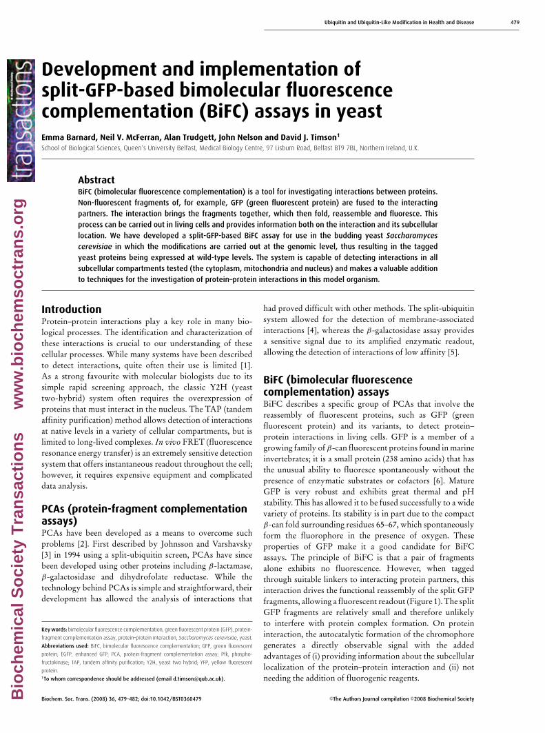
Ubiquitin and Ubiquitin-Like Modification in Health and Disease 479
Development and implementation ofsplit-GFP-based bimolecular fluorescencecomplementation (BiFC) assays in yeastEmma Barnard, Neil V. McFerran, Alan Trudgett, John Nelson and David J. Timson1
School of Biological Sciences, Queen’s University Belfast, Medical Biology Centre, 97 Lisburn Road, Belfast BT9 7BL, Northern Ireland, U.K.
AbstractBiFC (bimolecular fluorescence complementation) is a tool for investigating interactions between proteins.Non-fluorescent fragments of, for example, GFP (green fluorescent protein) are fused to the interactingpartners. The interaction brings the fragments together, which then fold, reassemble and fluoresce. Thisprocess can be carried out in living cells and provides information both on the interaction and its subcellularlocation. We have developed a split-GFP-based BiFC assay for use in the budding yeast Saccharomycescerevisiae in which the modifications are carried out at the genomic level, thus resulting in the taggedyeast proteins being expressed at wild-type levels. The system is capable of detecting interactions in allsubcellular compartments tested (the cytoplasm, mitochondria and nucleus) and makes a valuable additionto techniques for the investigation of protein–protein interactions in this model organism.
IntroductionProtein–protein interactions play a key role in many bio-logical processes. The identification and characterization ofthese interactions is crucial to our understanding of thesecellular processes. While many systems have been describedto detect interactions, quite often their use is limited [1].As a strong favourite with molecular biologists due to itssimple rapid screening approach, the classic Y2H (yeasttwo-hybrid) system often requires the overexpression ofproteins that must interact in the nucleus. The TAP (tandemaffinity purification) method allows detection of interactionsat native levels in a variety of cellular compartments, but islimited to long-lived complexes. In vivo FRET (fluorescenceresonance energy transfer) is an extremely sensitive detectionsystem that offers instantaneous readout throughout the cell;however, it requires expensive equipment and complicateddata analysis.
PCAs (protein-fragment complementationassays)PCAs have been developed as a means to overcome suchproblems [2]. First described by Johnsson and Varshavsky[3] in 1994 using a split-ubiquitin screen, PCAs have sincebeen developed using other proteins including β-lactamase,β-galactosidase and dihydrofolate reductase. While thetechnology behind PCAs is simple and straightforward, theirdevelopment has allowed the analysis of interactions that
Key words: bimolecular fluorescence complementation, green fluorescent protein (GFP), protein-
fragment complementation assay, protein–protein interaction, Saccharomyces cerevisiae, yeast.
Abbreviations used: BiFC, bimolecular fluorescence complementation; GFP, green fluorescent
protein; EGFP, enhanced GFP; PCA, protein-fragment complementation assay; Pfk, phospho-
fructokinase; TAP, tandem affinity purification; Y2H, yeast two hybrid; YFP, yellow fluorescent
protein.1To whom correspondence should be addressed (email [email protected]).
had proved difficult with other methods. The split-ubiquitinsystem allowed for the detection of membrane-associatedinteractions [4], whereas the β-galactosidase assay providesa sensitive signal due to its amplified enzymatic readout,allowing the detection of interactions of low affinity [5].
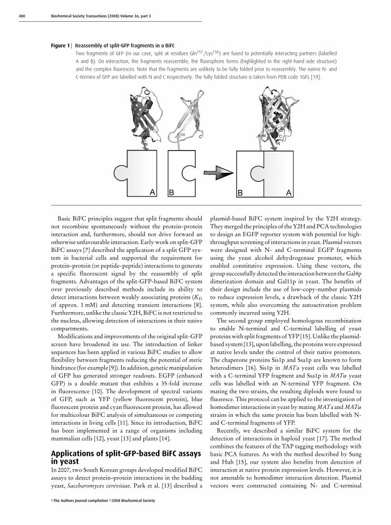
BiFC (bimolecular fluorescencecomplementation) assaysBiFC describes a specific group of PCAs that involve thereassembly of fluorescent proteins, such as GFP (greenfluorescent protein) and its variants, to detect protein–protein interactions in living cells. GFP is a member of agrowing family of β-can fluorescent proteins found in marineinvertebrates; it is a small protein (238 amino acids) that hasthe unusual ability to fluoresce spontaneously without thepresence of enzymatic substrates or cofactors [6]. MatureGFP is very robust and exhibits great thermal and pHstability. This has allowed it to be fused successfully to a widevariety of proteins. Its stability is in part due to the compactβ-can fold surrounding residues 65–67, which spontaneouslyform the fluorophore in the presence of oxygen. Theseproperties of GFP make it a good candidate for BiFCassays. The principle of BiFC is that a pair of fragmentsalone exhibits no fluorescence. However, when taggedthrough suitable linkers to interacting protein partners, thisinteraction drives the functional reassembly of the split GFPfragments, allowing a fluorescent readout (Figure 1). The splitGFP fragments are relatively small and therefore unlikelyto interfere with protein complex formation. On proteininteraction, the autocatalytic formation of the chromophoregenerates a directly observable signal with the addedadvantages of (i) providing information about the subcellularlocalization of the protein–protein interaction and (ii) notneeding the addition of fluorogenic reagents.
Biochem. Soc. Trans. (2008) 36, 479–482; doi:10.1042/BST0360479 C©The Authors Journal compilation C©2008 Biochemical SocietyBio
chem
ical
So
ciet
y T
ran
sact
ion
s
ww
w.b
ioch
emso
ctra
ns.
org
480 Biochemical Society Transactions (2008) Volume 36, part 3
Figure 1 Reassembly of split-GFP fragments in a BiFC
Two fragments of GFP (in our case, split at residues Gln157/Lys158) are fused to potentially interacting partners (labelled
A and B). On interaction, the fragments reassemble, the fluorophore forms (highlighted in the right-hand side structure)
and the complex fluoresces. Note that the fragments are unlikely to be fully folded prior to reassembly. The native N- and
C-termini of GFP are labelled with N and C respectively. The fully folded structure is taken from PDB code 1GFL [19].
Basic BiFC principles suggest that split fragments shouldnot recombine spontaneously without the protein–proteininteraction and, furthermore, should not drive forward anotherwise unfavourable interaction. Early work on split-GFPBiFC assays [7] described the application of a split GFP sys-tem in bacterial cells and supported the requirement forprotein–protein (or peptide–peptide) interactions to generatea specific fluorescent signal by the reassembly of splitfragments. Advantages of the split-GFP-based BiFC systemover previously described methods include its ability todetect interactions between weakly associating proteins (KD
of approx. 1 mM) and detecting transient interactions [8].Furthermore, unlike the classic Y2H, BiFC is not restricted tothe nucleus, allowing detection of interactions in their nativecompartments.
Modifications and improvements of the original split-GFPscreen have broadened its use. The introduction of linkersequences has been applied in various BiFC studies to allowflexibility between fragments reducing the potential of sterichindrance (for example [9]). In addition, genetic manipulationof GFP has generated stronger readouts. EGFP (enhancedGFP) is a double mutant that exhibits a 35-fold increasein fluorescence [10]. The development of spectral variantsof GFP, such as YFP (yellow fluorescent protein), bluefluorescent protein and cyan fluorescent protein, has allowedfor multicolour BiFC analysis of simultaneous or competinginteractions in living cells [11]. Since its introduction, BiFChas been implemented in a range of organisms includingmammalian cells [12], yeast [13] and plants [14].
Applications of split-GFP-based BiFC assaysin yeastIn 2007, two South Korean groups developed modified BiFCassays to detect protein–protein interactions in the buddingyeast, Saccharomyces cerevisiae. Park et al. [13] described a
plasmid-based BiFC system inspired by the Y2H strategy.They merged the principles of the Y2H and PCA technologiesto design an EGFP reporter system with potential for high-throughput screening of interactions in yeast. Plasmid vectorswere designed with N- and C-terminal EGFP fragmentsusing the yeast alcohol dehydrogenase promoter, whichenabled constitutive expression. Using these vectors, thegroup successfully detected the interaction between the Gal4pdimerization domain and Gal11p in yeast. The benefits oftheir design include the use of low-copy-number plasmidsto reduce expression levels, a drawback of the classic Y2Hsystem, while also overcoming the autoactivation problemcommonly incurred using Y2H.
The second group employed homologous recombinationto enable N-terminal and C-terminal labelling of yeastproteins with split fragments of YFP [15]. Unlike the plasmid-based system [13], upon labelling, the proteins were expressedat native levels under the control of their native promoters.The chaperone proteins Sis1p and Ssa1p are known to formheterodimers [16]. Sis1p in MATa yeast cells was labelledwith a C-terminal YFP fragment and Ssa1p in MATα yeastcells was labelled with an N-terminal YFP fragment. Onmating the two strains, the resulting diploids were found tofluoresce. This protocol can be applied to the investigation ofhomodimer interactions in yeast by mating MATa and MATα
strains in which the same protein has been labelled with N-and C-terminal fragments of YFP.
Recently, we described a similar BiFC system for thedetection of interactions in haploid yeast [17]. The methodcombines the features of the TAP tagging methodology withbasic PCA features. As with the method described by Sungand Huh [15], our system also benefits from detection ofinteraction at native protein expression levels. However, it isnot amenable to homodimer interaction detection. Plasmidvectors were constructed containing N- and C-terminal
C©The Authors Journal compilation C©2008 Biochemical Society
Ubiquitin and Ubiquitin-Like Modification in Health and Disease 481
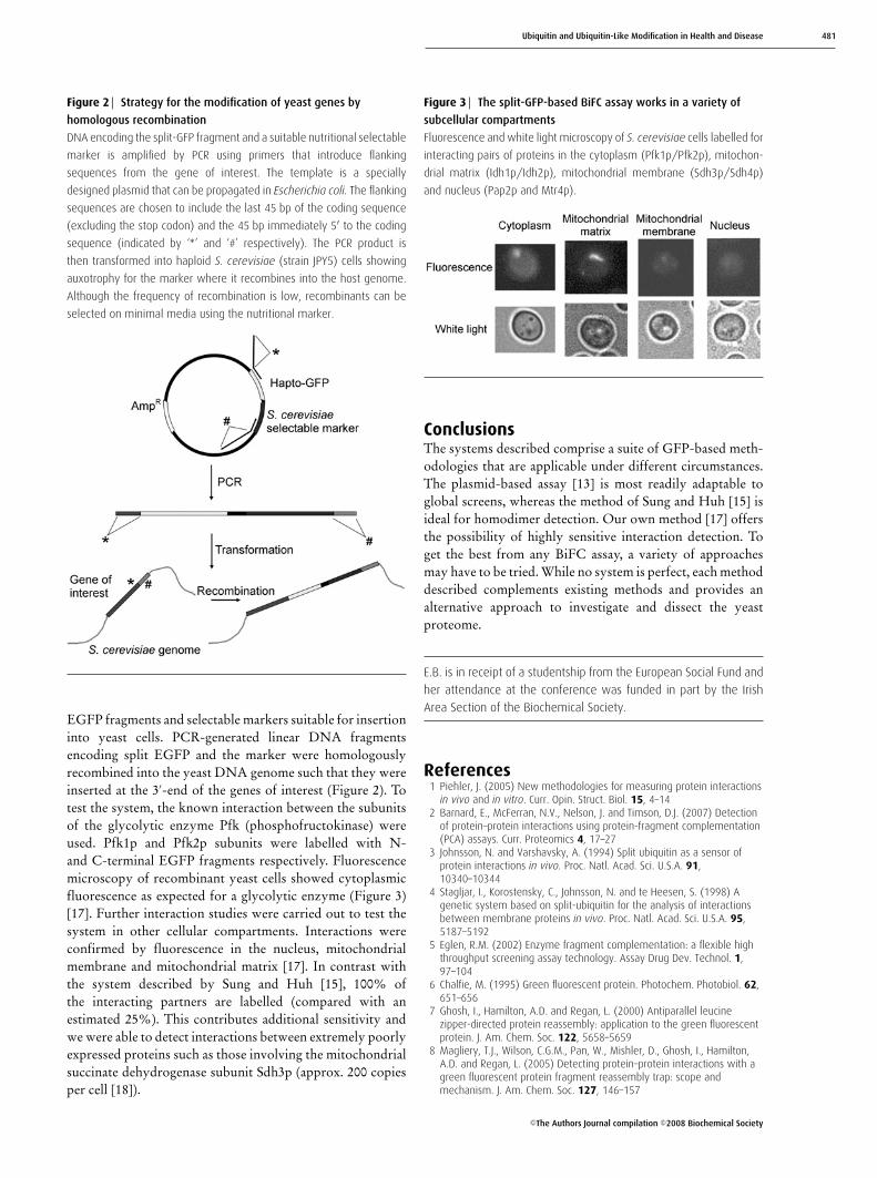
Figure 2 Strategy for the modification of yeast genes by
homologous recombination
DNA encoding the split-GFP fragment and a suitable nutritional selectable
marker is amplified by PCR using primers that introduce flanking
sequences from the gene of interest. The template is a specially
designed plasmid that can be propagated in Escherichia coli. The flanking
sequences are chosen to include the last 45 bp of the coding sequence
(excluding the stop codon) and the 45 bp immediately 5′ to the coding
sequence (indicated by ‘∗’ and ‘#’ respectively). The PCR product is
then transformed into haploid S. cerevisiae (strain JPY5) cells showing
auxotrophy for the marker where it recombines into the host genome.
Although the frequency of recombination is low, recombinants can be
selected on minimal media using the nutritional marker.
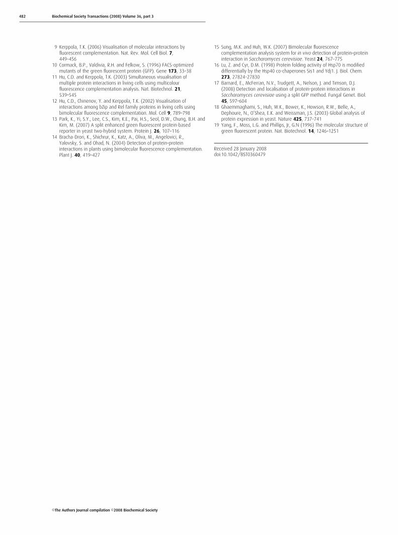
EGFP fragments and selectable markers suitable for insertioninto yeast cells. PCR-generated linear DNA fragmentsencoding split EGFP and the marker were homologouslyrecombined into the yeast DNA genome such that they wereinserted at the 3′-end of the genes of interest (Figure 2). Totest the system, the known interaction between the subunitsof the glycolytic enzyme Pfk (phosphofructokinase) wereused. Pfk1p and Pfk2p subunits were labelled with N-and C-terminal EGFP fragments respectively. Fluorescencemicroscopy of recombinant yeast cells showed cytoplasmicfluorescence as expected for a glycolytic enzyme (Figure 3)[17]. Further interaction studies were carried out to test thesystem in other cellular compartments. Interactions wereconfirmed by fluorescence in the nucleus, mitochondrialmembrane and mitochondrial matrix [17]. In contrast withthe system described by Sung and Huh [15], 100% ofthe interacting partners are labelled (compared with anestimated 25%). This contributes additional sensitivity andwe were able to detect interactions between extremely poorlyexpressed proteins such as those involving the mitochondrialsuccinate dehydrogenase subunit Sdh3p (approx. 200 copiesper cell [18]).
Figure 3 The split-GFP-based BiFC assay works in a variety of
subcellular compartments
Fluorescence and white light microscopy of S. cerevisiae cells labelled for
interacting pairs of proteins in the cytoplasm (Pfk1p/Pfk2p), mitochon-
drial matrix (Idh1p/Idh2p), mitochondrial membrane (Sdh3p/Sdh4p)
and nucleus (Pap2p and Mtr4p).
ConclusionsThe systems described comprise a suite of GFP-based meth-odologies that are applicable under different circumstances.The plasmid-based assay [13] is most readily adaptable toglobal screens, whereas the method of Sung and Huh [15] isideal for homodimer detection. Our own method [17] offersthe possibility of highly sensitive interaction detection. Toget the best from any BiFC assay, a variety of approachesmay have to be tried. While no system is perfect, each methoddescribed complements existing methods and provides analternative approach to investigate and dissect the yeastproteome.
E.B. is in receipt of a studentship from the European Social Fund and
her attendance at the conference was funded in part by the Irish
Area Section of the Biochemical Society.
References1 Piehler, J. (2005) New methodologies for measuring protein interactions
in vivo and in vitro. Curr. Opin. Struct. Biol. 15, 4–142 Barnard, E., McFerran, N.V., Nelson, J. and Timson, D.J. (2007) Detection
of protein–protein interactions using protein-fragment complementation(PCA) assays. Curr. Proteomics 4, 17–27
3 Johnsson, N. and Varshavsky, A. (1994) Split ubiquitin as a sensor ofprotein interactions in vivo. Proc. Natl. Acad. Sci. U.S.A. 91,10340–10344
4 Stagljar, I., Korostensky, C., Johnsson, N. and te Heesen, S. (1998) Agenetic system based on split-ubiquitin for the analysis of interactionsbetween membrane proteins in vivo. Proc. Natl. Acad. Sci. U.S.A. 95,5187–5192
5 Eglen, R.M. (2002) Enzyme fragment complementation: a flexible highthroughput screening assay technology. Assay Drug Dev. Technol. 1,97–104
6 Chalfie, M. (1995) Green fluorescent protein. Photochem. Photobiol. 62,651–656
7 Ghosh, I., Hamilton, A.D. and Regan, L. (2000) Antiparallel leucinezipper-directed protein reassembly: application to the green fluorescentprotein. J. Am. Chem. Soc. 122, 5658–5659
8 Magliery, T.J., Wilson, C.G.M., Pan, W., Mishler, D., Ghosh, I., Hamilton,A.D. and Regan, L. (2005) Detecting protein–protein interactions with agreen fluorescent protein fragment reassembly trap: scope andmechanism. J. Am. Chem. Soc. 127, 146–157
C©The Authors Journal compilation C©2008 Biochemical Society
482 Biochemical Society Transactions (2008) Volume 36, part 3
9 Kerppola, T.K. (2006) Visualisation of molecular interactions byfluorescent complementation. Nat. Rev. Mol. Cell Biol. 7,449–456
10 Cormack, B.P., Valdivia, R.H. and Felkow, S. (1996) FACS-optimizedmutants of the green fluorescent protein (GFP). Gene 173, 33–38
11 Hu, C.D. and Kerppola, T.K. (2003) Simultaneous visualisation ofmultiple protein interactions in living cells using multicolourfluorescence complementation analysis. Nat. Biotechnol. 21,539–545
12 Hu, C.D., Chinenov, Y. and Kerppola, T.K. (2002) Visualisation ofinteractions among bZip and Rel family proteins in living cells usingbimolecular fluorescence complementation. Mol. Cell 9, 789–798
13 Park, K., Yi, S.Y., Lee, C.S., Kim, K.E., Pai, H.S., Seol, D.W., Chung, B.H. andKim, M. (2007) A split enhanced green fluorescent protein-basedreporter in yeast two-hybrid system. Protein J. 26, 107–116
14 Bracha-Drori, K., Shichrur, K., Katz, A., Oliva, M., Angelovici, R.,Yalovsky, S. and Ohad, N. (2004) Detection of protein–proteininteractions in plants using bimolecular fluorescence complementation.Plant J. 40, 419–427
15 Sung, M.K. and Huh, W.K. (2007) Bimolecular fluorescencecomplementation analysis system for in vivo detection of protein–proteininteraction in Saccharomyces cerevisiae. Yeast 24, 767–775
16 Lu, Z. and Cyr, D.M. (1998) Protein folding activity of Hsp70 is modifieddifferentially by the Hsp40 co-chaperones Sis1 and Ydj1. J. Biol. Chem.273, 27824–27830
17 Barnard, E., McFerran, N.V., Trudgett, A., Nelson, J. and Timson, D.J.(2008) Detection and localisation of protein–protein interactions inSaccharomyces cerevisiae using a split GFP method. Fungal Genet. Biol.45, 597–604
18 Ghaemmaghami, S., Huh, W.K., Bower, K., Howson, R.W., Belle, A.,Dephoure, N., O’Shea, E.K. and Weissman, J.S. (2003) Global analysis ofprotein expression in yeast. Nature 425, 737–741
19 Yang, F., Moss, L.G. and Phillips, Jr, G.N (1996) The molecular structure ofgreen fluorescent protein. Nat. Biotechnol. 14, 1246–1251
Received 28 January 2008doi:10.1042/BST0360479
C©The Authors Journal compilation C©2008 Biochemical Society