Embed Size (px)
Citation preview

Development in Zebrafish, a Genetic Approach
Didier Stainier University of California

5/2/05
Zebrafish geneticsDidier Stainier, UCSF Biochemistry
Juquehy, Brazil

Saccharomyces cerevisiae
Drosophila melanogaster
Caenorhabditis elegans
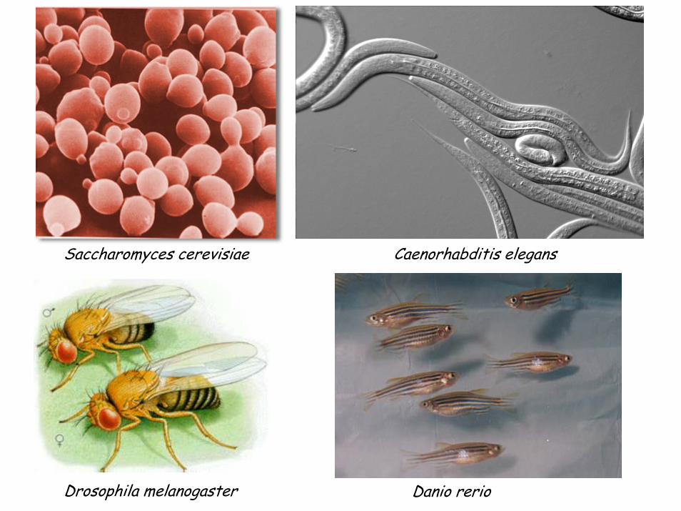
Saccharomyces cerevisiae
Drosophila melanogaster
Caenorhabditis elegans
Danio rerio

GENETIC SCREENS

What can one screen for?
- developmental processes
- physiological processes
- behavior
- other

external fertilization

some keydevelopmentalstages

Zebrafish embryonic development
QuickTime™ and aAnimation decompressor
are needed to see this picture.
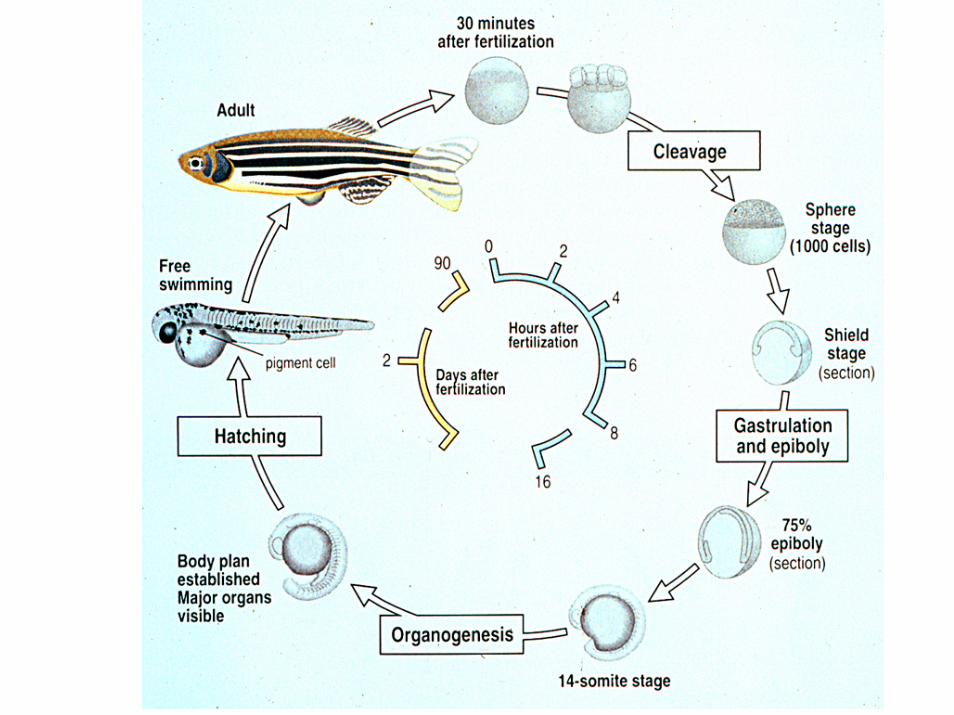

large number of progeny

screening based onmorphological traits
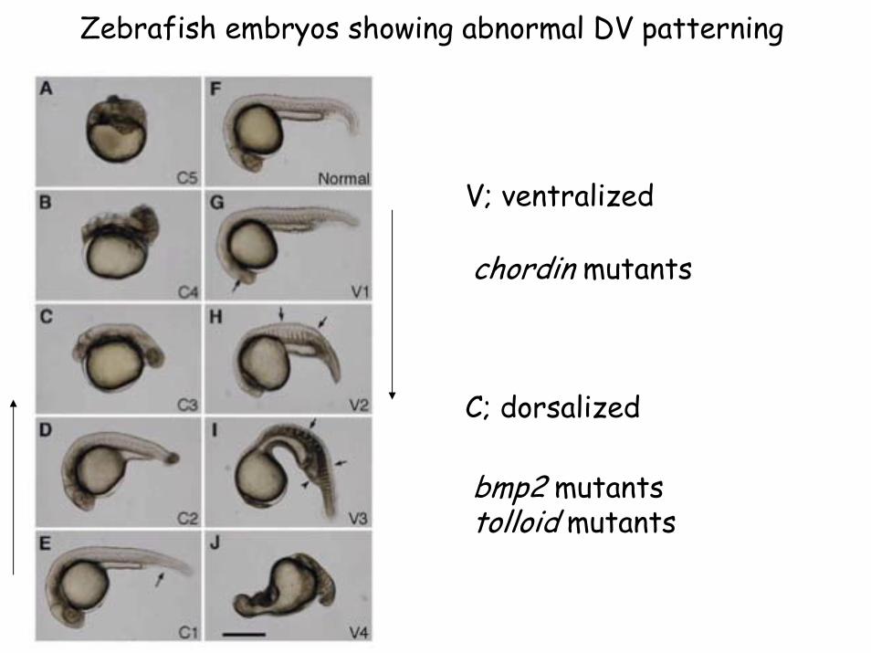
Zebrafish embryos showing abnormal DV patterning
V; ventralized
C; dorsalized
chordin mutants
bmp2 mutantstolloid mutants

QuickTime™ and aVideo decompressor
are needed to see this picture.
QuickTime™ and aVideo decompressor
are needed to see this picture.
Cardiac valve formation

QuickTime™ and aCinepak decompressor
are needed to see this picture.
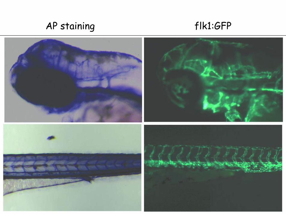
AP staining flk1:GFP

Liver
Pancreas
Intestinalbulb
Swimbladder
Ventral View
52 hpf gutGFP line

QuickTime™ and a Motion JPEG A decompressor are needed to see this picture.
Duc Dong

QuickTime™ and aMotion JPEG A decompressor
are needed to see this picture.
Ryan Anderson
lfabp-dsRedelastase-GFPinsulin-dsRed
L
P
80 hpf

forward genetic screens in zebrafish
- large number of tanks (diploid screens)
- 3-4 scientists
- 18-24 months
- ENU
- insertional mutagenesis (retroviral vectors)

What can you do with your mutants?
- clone the gene, look at its expression pattern
- analyze cell-autonomy
- gain-of-function experiments

4
fj87b02.x1=angiotensinogen
on fugu scaffold 000344
~cfk
2*
135L3.Sp6.2(has fl81g12.y1 in it
= TFIID on fuguscaffold 003167) 135L3.T7=
zfishC-a1020b08.p1c(R).3
6*
unp1669=sau22-10-3' =
Rab1 GTPase 3'=fb53a02.x1 on
no fugu scaffold
2
4 cR
12 cR13 cR
37G24
z9289 z10582
163H6
1018=919+99
fk37a09.y1.2=Rab1 5'
***
**
*
25H6
163H5
135L3
101N13
166B10202M22
4D23
80C1
244O10
188E2
76N8
8A9
210D22210F22
21A8
zeh0631(coding)=
non-cardiac,non-skeletal
actin
0 2
202M22-T7
1
202M22-Sp6
Going from mutation to gene
- maps: genetic,physical, RH
- genome sequence inprogress
- fugu, tetraodon(synteny)
- 6-12 months (average)


sox17 late gastrula
Mutations that affect early endoderm formation in zebrafish
- one-eyed pinhead (oep)- casanova (cas)- bonnie and clyde (bon)- faust (fau)
all endodermal cellsmissing
90% of endodermal cells missing
60% of endodermal cells missing

sox17 first implicatedin endoderm formation inthe frog Xenopus laevis
Alexander et al., 1999
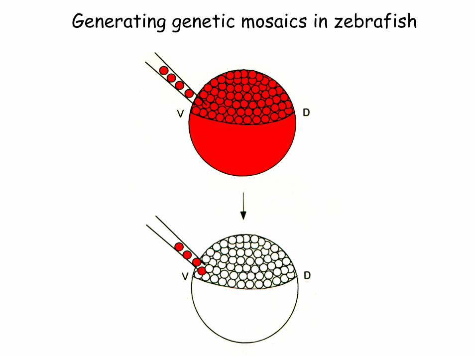
Generating genetic mosaics in zebrafish

Results of the cell transplantation experiments
- casanova mutant cells never form endoderm(either in wild-type or mutant embryos)
- wild-type cells can form endoderm in casanovamutant embryos
The endoderm phenotype in casanova mutants is cell-autonomous, i.e. casanova functions within theendodermal lineage
Alexander et al., 1999

sox17 late gastrula
Proteins that affect early endoderm formation in zebrafish
- Oep (Nodal co-receptor. Schier; Whitman)- Cas (Sox-related transcription factor)- Bon (Mix-type HD transcription factor)- Fau (Gata5: Zinc-finger transcription factor)

How are Oep, Cas/Sox-relatedBon/Mix-type Fau/Gata5
positioned relative to each other in the pathway leading to endoderm formation?
?
?
?
sox17
sox17
A. B.

Alexander et al., 1999

oep
bon/mix-type
cas/sox-related
sox17(endoderm)
Endoderm formation in zebrafish

Reiter et al., 2001

Reiter et al., 2001

sox17 late gastrula
A possible model of early endoderm formation in zebrafish
Nodal signaling
Bon/Mix-type
Cas
sox17
Fau/Gata5 X


Analyzing the function of casby gain-of-function (i.e. misexpression)
experiments
cas mRNA andGFP mRNA
16-cell stage

Kikuchi et al., 2001
sox17
gsc
GFP GFP
gsc
Ectopic cas expression can transfatemesoderm into endoderm

Kikuchi et al., 2001

What can you do with your mutants?
- clone the gene, look at its expression pattern
- analyze cell-autonomy
- gain-of-function experiments
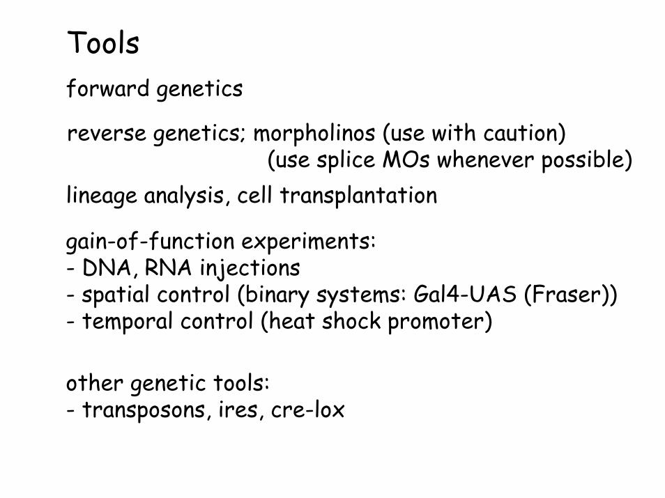
Tools
gain-of-function experiments:- DNA, RNA injections- spatial control (binary systems: Gal4-UAS (Fraser))- temporal control (heat shock promoter)
other genetic tools:- transposons, ires, cre-lox
forward genetics
lineage analysis, cell transplantation
reverse genetics; morpholinos (use with caution)(use splice MOs whenever possible)
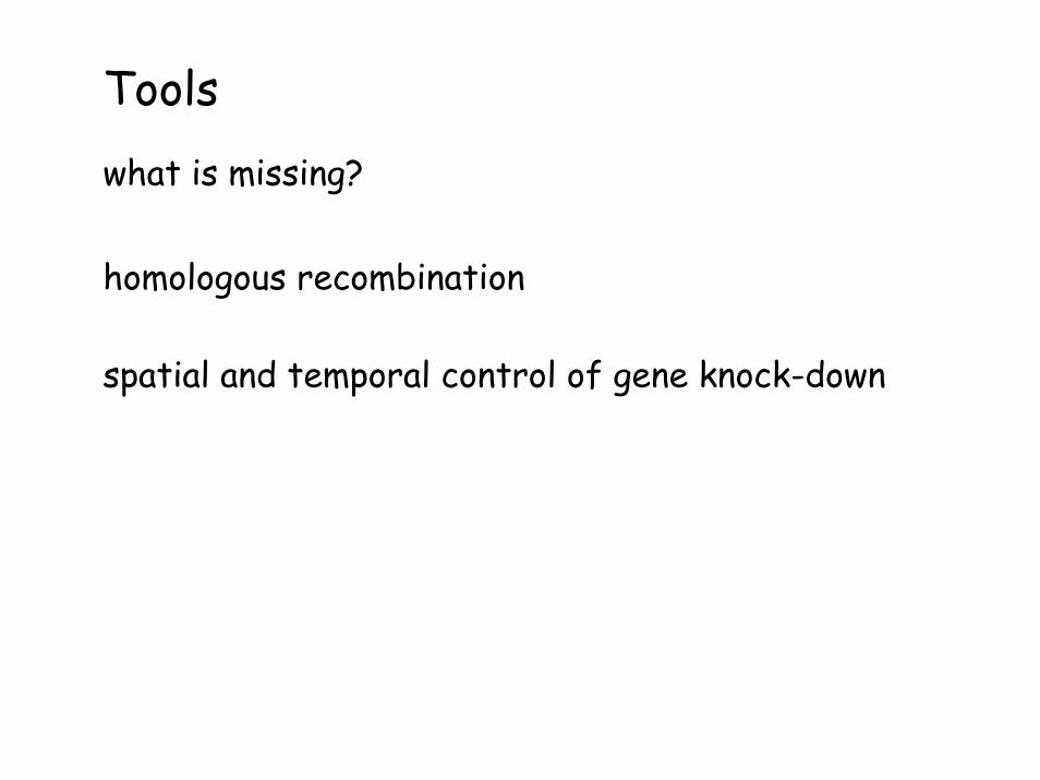
Toolswhat is missing?
homologous recombination
spatial and temporal control of gene knock-down
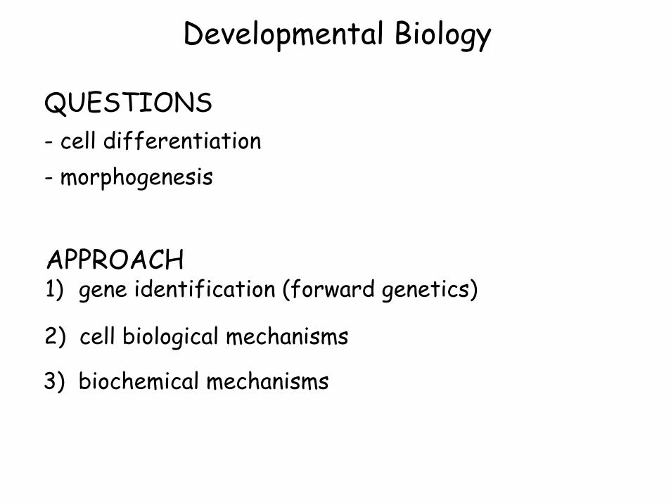
Developmental Biology
QUESTIONS- cell differentiation- morphogenesis
APPROACH1) gene identification (forward genetics)
2) cell biological mechanisms
3) biochemical mechanisms

1. embryology2. forward genetics3. cell biology

precardiac mesoderm morphogenesisin zebrafish
13 somite15.5 hpf
17 somite17.5 hpf
20 somite19 hpf
dorsal views
wild-type
cardia bifidamutants

precardiac mesoderm morphogenesisin zebrafish
13 somite15.5 hpf
17 somite17.5 hpf
20 somite19 hpf
endoderm
pre-endocardial cells
pre-myocardial cells
transverse sections
13 somite15.5 hpf
17 somite17.5 hpf
20 somite19 hpf

20μm
16-somite
18-somite
20-somitecmlc2-GFPHJ Tsai
Myocardial migration
20-somite
12-somite
Endocardial migration
flk1-GFP
16-somite
20-somite

Endodermal GFP line
Wt10S
β-cateninher5::gfp 10-somite Nick Osborne

CARDIA BIFIDA MUTATIONS1) AFFECTING MYOCARDIAL DIFFERENTIATION
- one-eyed pinhead (oep) (CFC protein)- faust (fau) (Gata5)- hands off (han) (Dhand/Hand2)
2) AFFECTING HEART CELL MIGRATIONA) VIA AFFECTING ENDODERM FORMATION
- one-eyed pinhead (oep) (CFC protein)- casanova (cas) (Sox32)- bonnie and clyde (bon) (Mix-type HD)- faust (fau) (Gata5)
B) VIA SOME OTHER MECHANISM- natter (fibronectin)- miles apart (mil) (S1P receptor) - two-of-hearts (toh)

How does Fibronectin regulate myocardial migration?
- chick: Fn appears to be distributed in a gradient towards the midline (K. Linask)
Model: haptotaxis (moving towards areas of greater adhesiveness)
- mouse: Fn mutants can show cardia bifida (R. Hynes)

16-somite 16-somite
20-somite 20-somite
Fnβ-catenincmlc2::GFP
Fnβ-catenincmlc2::GFP
Lamininβ-catenincmlc2::GFP
Lamininβ-catenincmlc2::GFP
nat20-somite
nat20-somite
wt wt
wt wt
Lamininβ-catenincmlc2::GFP
Fnβ-catenincmlc2::GFP
Lack of Fn deposition in nat -/- embryos
Le Trinh

aPKCsβ-catenincmlc2::GFP
Myocardial precursors form maturing epithelia during the migration stages
16-somite
18-somite
20-somite

wt
Fibronectin is required for proper junction formation in the myocardial precursors
nat nat
wtwt 20-somite
20-somite
aPKCsβ-catenincmlc2::GFP
Le Trinh

aPKCsβ-catenincmlc2::GFP
Myocardial precursors form maturing epithelia during the migration stages
20-somite
genetic requirements for myocardial polarization- Fibronectin (Trinh and Stainier, Dev Cell, 2004)
- Dhand/Hand2 (Trinh, Yelon and Stainier, Current Biology, 2005)

cell biological mechanisms
cell polarity, cell shape, cell migration
intracellular trafficking, signaling, organelle biogenesis
IMAGING

QuickTime™ and aAnimation decompressor
are needed to see this picture.
A novel transgenic line to visualize Ca++ flux
Neil Chi


Calcium Green/Optical Mapping
Sehnert et al., 2001 Nature Genetics
Explanted 48 hpf hearts (wt and sih -/-) exposed to calcium green

gCAMP/Optical Mapping
Nakai et al., 2001 NBT/Imoto Lab
Kd~190nMF/F’~4
F/F’di-4ANNEPES 1-2/100calcium green 14FCIP FRET 1-2/10gCAMP 4
generation of a transgenic line(gCAMP expressed in myocardial cells) (Neil Chi)

G-CAMP/Optical Mapping
RawRT
Processed1/3 RT
QuickTime™ and aAnimation decompressor
are needed to see this picture.
QuickTime™ and aAnimation decompressor
are needed to see this picture.
All embryos were treated with 2,3 BDM, an ATP myofibrillar inhibitor, in order to arrest cardiac contraction. Images were obtained with a CCD camera at a 20-30ms/frame capture rate. 40x objective used.
24 hpf
Neil Chi

G-CAMP/Optical Mapping
RawRT
Processed1/3 RT
RawRT QuickTime™ and a
Animation decompressorare needed to see this picture.
QuickTime™ and aAnimation decompressor
are needed to see this picture.
Processed1/3 RT
QuickTime™ and aAnimation decompressor
are needed to see this picture.
QuickTime™ and aAnimation decompressor
are needed to see this picture.
48 hpf
5 dpf
Neil Chi

QuickTime™ and aAnimation decompressor
are needed to see this picture.
Neil Chi
embryos were co-injected with ctnt2 and connexin MOs
connexin mutants display aberrant calcium waves

Neil Chi, M.D., Ph.D.(cardiac function)
Robin Shaw, M.D., Ph.D.Lily Jan
ACKNOWLEDGEMENTS
Le TrinhFn, Hand2
Debbie YelonSkirball
Endoderm formation*Jon Alexander*Jeremy Reiter*Yutaka Kikuchi, Ph.D.Pia Aanstad, Ph.D.
heart formationheart function

UCSFSan Francisco
vasculogenesis
QuickTime™ and aAnimation decompressor
are needed to see this picture.
endocardialcushionformation
cardiac function