Embed Size (px)
Citation preview
DEVELOPMENT OF A BIOSENSOR BASED ON AMINE OXIDASE FROM
Cicer arietinum FOR THE DETECTION OF BIOGENIC AMINES
(PEMBANGUNAN BIOSENSOR BERDASARKAN AMINE OXIDASE
DARIPADA Cicer arietinum BAGI MENENTUKAN BIOGENIC AMINES)
AZILA ABD. AZIZ
RESEARCH VOTE NO:
74194
Bioprocess Engineering Department
Faculty of Chemical and Natural Resources Engineering
Universiti Teknologi Malaysia
2007

ii
ACKNOWLEDGEMENT
I would like to express my appreciation to all those who have assisted me,
whether directly or indirectly, in conducting this research. There are too many names
to list down. However, special thanks goes to the research assistant, Mohd. Hafiz
Kassim for his dedications and tireless efforts in making this research work a success.
iii
ABSTRACT
Biogenic amines such as histamine, cadaverine and putrescine have been
confirmed as useful chemical indicators to estimate bacterial spoilage of foods,
particularly fish and fish products, cheese, meat and fermented foods. Histamine is
toxic at high intakes, while cadaverine and putrescine potentiate the effects of
histamine. The regulated level of histamine is 200 mg/kg (200 ppm). For biogenic
amines biosensor, the basic principle is the action of diamine oxidase (DAO) that
catalyzes the oxidative deamination of primary amines to the corresponding
aldehydes, hydrogen peroxide and ammonia. Biogenic amines concentration can be
measured by monitoring either the decrease in oxygen or the increase of hydrogen
peroxide concentration. Recently, it has been found that DAO from pea seedlings
shows higher activity compared to commercial porcine kidney diamine oxidase
(PKAO). For that reason, in this research, DAO from Cicer arietinum (chick pea)
seedlings will be used to develop the biogenic amines biosensor. Amine oxidases
from chick pea (CPAO) was successfully purified following three chromatographic
steps, giving a specific activity of 12.7, 11.7 and 0.45 U/mg with putrescine,
cadaverine and histamine as substrate, respectively. The molecular mass of the
CPAO was 73 kDa, determined by SDS-PAGE. Immobilization of PKAO in cross-
linked poly (vinyl alcohol) (PVA) has been performed, and PVA concentration of
10% and cross-linking ratio (CR) of 0.06 were found to be the optimum parameters
for CPAO immobilization. Immobilization of partially purified CPAO has also been
done. Result showed that at equal enzyme loading, CPAO-PVA membranes gave
higher apparent activities compared to commercial PKAO-PVA membranes.
iv
ABSTRAK
Biogenic amines seperti histamin, kadaverin dan putresin telah disahkan
sebagai penunjuk kimia yang sangat berguna untuk menganggarkan kerosakan
makanan hasil tindakan bakteria, khususnya ikan dan produk ikan, keju, daging dan
makanan yang diperam. Histamin adalah toksik dalam kadar pengambilan yang
tinggi, manakala kadaverin dan putresin merangsang kesan-kesan histamin. Had
yang dibenarkan bagi histamin ialah 200 mg/kg (200 ppm). Bagi biosensor biogenic
amines, prinsip asas ialah tindakan diamine oxidase (DAO) yang memangkinkan
tindakbalas deaminasi oksidatif terhadap amina utama kepada aldehid yang
bersepadanan, hidrogen peroksida dan ammonia. Kepekatan biogenic amines boleh
ditentukan dengan memantau samada tahap pengurangan kepekatan oksigen atau
pertambahan kepekatan hidrogen peroksida. Terkini, DAO dari sumber bebenih
kekacang menunjukkan aktiviti yang lebih tinggi berbanding DAO komersil dari
sumber buah pinggang khinzir (PKAO). Atas sebab itu, dalam penyelidikan ini,
DAO dari bebenih Cicer arietinum (kacang kuda) akan digunakan untuk
membangunkan biosensor biogenic amines. Amine oxidase dari bebenih kacang kuda
(CPAO) telah ditulenkan melalui tiga peringkat kromatografi, memberikan nilai
aktiviti spesifik 12.7, 11.7 dan 0.45 U/mg dengan masing-masing putresin, kadaverin
dan histamin sebagai substrat. Berat molekul bagi CPAO ialah 73 kDa, ditentukan
melalui SDS-PAGE. Immobilisasi PKAO di dalam poly (vinyl alkohol) (PVA) yang
disambung-silang telah dilakukan, dan kepekatan PVA 10% dan nisbah
penyambungan-silang (CR) 0.06 di dapati merupakan parameter optima bagi
immobilisasi CPAO. Immobilisasi terhadap CPAO separa tulen juga telah dilakukan.
Keputusan yang diperolehi menunjukkan, pada muatan enzim yang sama, membran
CPAO-PVA memberikan aktiviti bandingan yang lebih tinggi berbanding membran
PKAO-PVA.
v
TABLE OF CONTENTS
CHAPTER TITLE PAGE
TITLE PAGE i
ACKNOWLEDGEMENT ii
ABSTRACT iii
ABSTRAK iv
TABLE OF CONTENTS v
LIST OF TABLES ix
LIST OF FIGURES x
LIST OF SYMBOLS xii
1 INTRODUCTION
1.1 Introduction 1
1.2 Research Objectives 3
1.3 Research Scopes 4
2 LITERATURE REVIEW
2.1 Biogenic Amines 5
2.2 Diamines 7
2.3 Histamine Poisoning 8
2.4 Amines in Plants 9
2.5 Diamine Oxidase 11
2.6 Chickpeas (Cicer arietinum) 14
2.6.1 Chickpea Varieties 15
2.6.2 Chickpea Nutrition and Uses 16
2.6.3 History of Cultivation 17
vi
2.7 Introduction of Biosensor 18
2.7.1 Chemical Sensor and Biosensor 20
2.7.2 Electrochemical Biosensor 22
2.7.3 Electrochemical Detection 23
2.7.3.1 Amperometry 23
2.7.3.2 Potentiometry 23
2.8 The First Biosensor 24
2.9 Fundamental of Amperometric Biosensor 26
2.10 Histamine Biosensor 27
2.11 Enzyme Immobilization 30
2.11.1 Poly (vinyl alcohol) (PVA) 33
2.11.2 Freeze-thawed PVA 35
2.11.3 Immobilization Methods in
Constructing Histamine Biosensor
36
3 METHODOLOGY
3.1 Purification of Pea Seedling Amine Oxidase 38
3.1.1 Enzyme Purification 38
3.1.2 Ammonium Sulphate Precipitation 40
3.1.2.1 Methods 40
3.1.3 Dialysis 41
3.1.3.1 Methods 41
3.1.4 Enzyme Activity Determination 42
3.1.4.1 Chemicals 42
3.1.4.2 Methods 43
3.1.5 Protein Concentration Determination –
Bicinchoninic Acid (BCA) Assay
44
3.1.5.1 Chemicals 44
3.1.5.2 Methods 45
3.1.6 Sodium Dodecyl Sulfate-
Polyacrylamide Gel Electrophoresis
(SDS-PAGE)
45
vii
3.1.6.1 Equipment 46
3.1.6.2 Chemicals 46
3.1.6.3 Pouring a Gel 46
3.1.6.4 Preparing and Loading
Samples
49
3.1.6.5 Running a Gel 50
3.1.6.6 Staining a Gel with
Coomassie Blue
50
3.1.6.7 Silver Staining a Gel 51
3.2 Immobilization of Diamine Oxidase
(Cross-linking Method)
52
3.2.1 Chemicals 52
3.2.1.1 Working Solutions 53
3.2.1.2 Membrane Preparation 53
4 RESULTS AND DISCUSSION
4.1 Purification of Pea Seedling Amine Oxidase 55
4.1.1 Sample Preparation 56
4.1.2 Anion Exchange Chromatography
(DEAE-Cellulose)
57
4.1.3 Hydroxyapatite Chromatography (Bio-
Gel HTP)
59
4.1.4 Size-Exclusion Chromatography
(Sephacryl S-300 HR)
60
4.1.5 Sodium Dodecyl Sulfate-
Polyacrylamide Gel Electrophoresis
(SDS-PAGE)
64
4.2 Immobilization of Diamine Oxidase
(Cross-linking Method)
65

viii
5 SUMMARY AND RECOMMENDATION
5.1 Summary 69
5.2 Recommendations 70
REFERENCES 72
ix
LIST OF TABLES
TABLE TITLE PAGE
2.1 Scientific classification of chickpeas. 15
2.2 Types of receptors used in biosensors and
electrochemical measurement techniques.
21
2.3 The different types of electrochemical transducer. 22
4.1 The purification of chick pea diamine oxidase (CPAO) 62
4.2 Specific activity and purification fold of chick pea
diamine oxidase (CPAO).
63
x
LIST OF FIGURES
FIGURE TITLE PAGE
2.1 Decarboxylation of certain amino acids to produce
biogenic amines.
6
2.2 Conversion of histidine to histamine by histidine
decarboxylase (HDC).
8
2.3 The fruit and seeds of the chickpeas (Cicer arietinum). 14
2.4 A schematic drawing for a typical biosensor. The
specific chemical target (analyte) is recognized by the
biological element, creating a stimulus to the detecting
transducer from which a reproducible signal is
measured.
19
2.5 A Clark oxygen electrode. 25
2.6 A glucose biosensor based on Clark oxygen electrode. 26
2.7 Oxidative deamination of histamine to
imidazoleacetaldehyde, hydrogen peroxide and
ammonia by diamine oxidase.
27
2.8 The basic principle of the electron transfer during
measurements in amperometric bi-enzyme system.
29
3.1 The three-electrode arrangement of the potentiostat
equipment.
55
4.1 Hydrogen peroxides (H2O2) standard curve. 57
xi
4.2 Protein concentration standard curve (BSA standard
curve).
57
4.3 Protein elution profile for the DEAE-Cellulose
chromatography.
58
4.4 Protein elution profile for the hydroxyapatite (Bio-Gel
HTP) chromatography.
60
4.5 Protein elution profile for Sephacryl S-300 HR
chromatography.
61
4.6 SDS-PAGE of CPAO 64
4.7 SDS-PAGE of CPAO 65
4.8 PKAO-PVA membranes apparent activity at 10% PVA
concentration with the cross-linking ratio varied.
66
4.9 PKAO-PVA membranes apparent activity at cross-
linking ratio of 0.06 with the PVA concentration varied.
67
4.10 Comparison of CPAO-PVA and PKAO-PVA
membranes apparent activity at an equal enzyme
loading.
68
xii
LIST OF SYMBOLS
SYMBOL MEANING
α Alpha
A280 Absorbance at 280 nm
A450 Absorbance at 450 nm
A562 Absorbance at 562 nm
β Beta
BCA Bicinchoninic acid
BSA Bovine serum albumin
CE Counter electrode
CPAO Chick pea seedling amine oxidase
CR Cross-linking ratio
DAO Diamine oxidase
DEAE Diethyl amino ethyl
EPR Electron paramagnetic resonance
GOD Glucose oxidase
HDC Histidine decarboxylase
HMT Histamine-N-methyl transferase
HRP Horseradish peroxidase
HPLC High Performance Liquid Chromatography
kat Katal; SI unit of enzyme activity
kDa Kilo Dalton
xiii
LC Liquid Chromatography
MW Molecular weight
PEG Poly(ethylene glycol)
PKAO Porcine kidney amine oxidase
PPB Potassium phosphate buffer
ppm Part per million
PSAO Pea seedling amine oxidase
PVA Poly (vinyl alcohol)
RE Reference electrode
SDS-PAGE Sodium dodecylsulphate-polyacrylamide gel electrophoresis
TEMED (N,N,N’,N’-tetramethylene-ethylenediamine
U Enzyme activity unit; 1 unit (U) is the amount of enzyme that
catalyses the conversion of 1 micromole of substrate per minute
under defined conditions.
WE Working electrode
CHAPTER 1
INTRODUCTION
1.1 Introduction
Histamine poisoning, also known as ‘scombroid fish poisoning’ is an illness
that results from eating spoiled fish because of inadequate refrigeration or preservation
after it is being caught. It is most commonly reported with fish from Scombridae and
Scomberesocidae families. One of the toxic agents implicated in scombroid
poisoning is a chemical called histamine. For years, studies had been conducted to
find the best method to detect and determine the level of histamine in food. This can
avoid people from consuming spoiled food, instead of giving them treatment after
being poisoned.
Traditionally, histamine has been measured by derivatization with fluorescent
reagents followed by chromatographic separation (e.g. HPLC in most cases). As the
HPLC analysis of biogenic amines is tedious with regard to sample clean-up prior to the
analysis and it requires trained personnel in combination with quite expensive
equipment, further analysis methods have been described such as capillary
electrophoresis, immunochemical methods such as ELISA, and even some gas
chromatographic methods, thin-layer liquid chromatography (LC), reversed phase
2
LC and LC with pre-column, post column and on-column derivatization techniques
(Tombelli and Mascini, 1998; Lange and Wittmann, 2001). To reduce the time
needed for analysis and to offer a rapid screening method for industrial food quality
testing, some enzymatic methods and several enzyme sensors have been described
so far by previous researchers (Chemnitius et al., 1992; Esti et al., 1998; Loughran
et al., 1995; Wimmerova et al., 1999). Biosensor applications, in general, exhibit
various advantages such as allowing a more rapid analysis with less sample treatment
being required (Lange, J. and Wittmann, C., 2001).
In this work, an enzyme-based histamine amperometric biosensor will be
developed. In developing a biosensor for detecting and determining the content of
histamine in fish and its product, the basic principles used are the same as a glucose
biosensor, except it needs to have a layer of diamine oxidase membrane instead of
glucose oxidase. Extensive studies in constructing histamine biosensor have been
conducted by lots of researcher before, which majority of them used diamine oxidase
from porcine kidney as the immobilized enzyme. However, recently some researchers
such as Tombelli et al., (1998), Niculescu et al., (2001), Wimmerova et al., (1999)
and Frebort et al., (2000) have found that diamine oxidase extracted and purified
from pea seedling shows higher activity compared to the commercial diamine
oxidase from porcine. Consequently, the sensitivity of sensor was improved. An
additional reason for using the enzyme is because since Malaysia is an Islamic country,
it is better to avoid the usage of components or substances from pigs.
For that reason, in this research, diamine oxidase from pea seedlings will be
used. Since pea seedling diamine oxidase is not available in the market, it has to be
purified. The type of pea that will be used for enzyme purification is Cicer
arietinum (chick pea) (Tombelli et al., 1998).
3
The purified chick pea diamine oxidase (CPAO) will be entrapped in cross-
linked poly (vinyl alcohol) (PVA) membrane. PVA is a non-toxic water-soluble
synthetic material that has good film forming properties, resulting in tough
membranes. PVA can also stabilize the activity of various enzymes such as
horseradish peroxidase. The stabilization effect is achieved through the inhibition of
the formation of non-functional conformations due to the extensive hydrogen
bonding between the H-atoms of the alcohol groups in PVA and the O-atoms of the
carbohydrate groups in diamine oxidase. These properties make PVA an appropriate
matrix for diamine oxidase immobilization. Glutaraldehyde, a bifunctional agent that
can react with organic hydroxyl groups and lysine amino acid residues in the
enzyme, will be used as the cross-linking agent. The cross-linking process overcomes
the loss of enzyme activity due to diffusional loss, which is a prevalent problem for
enzyme immobilized in physical entrapment.
1.2 Research Objectives
The objectives of this research are:
1. To purify diamine oxidase from Cicer arietinum seedlings (chick pea) as an
alternative source of enzyme to porcine kidney diamine oxidase.
2. To develop highly active and stable chick pea seedling diamine oxidase
(CPAO) membranes for histamine biosensor.
4
1.3 Research Scopes
The scopes of this research are:
1. Purification of chick pea seedling diamine oxidase (CPAO).
2. Comparison of the enzyme activity and stability between chick pea seedling
diamine oxidase (CPAO) and the commercial porcine kidney diamine oxidase
(PKAO).
3. Development of chick pea seedling diamine oxidase membranes which are
highly active, stable and have long operational life using appropriate
immobilization methods.
CHAPTER 2
LITERATURE REVIEW
2.1 Biogenic Amines
Biogenic amines are low molecular-weight organics compounds which are
derived from the corresponding amino acids when the carboxylic acid is removed by
enzymatic reactions. They are termed biogenic amines because they are formed in
raw food by bacterial action. Biogenic amines may present in various foods,
particularly fish and fish products, cheese, meat and fermented foods (Eerola et al.,
1993). During storage and processing, if foods are mishandled, certain protein within
the foods might break down to free amino acids, which may also be naturally present
within the food. If the food is contaminated with bacteria containing decarboxylase
enzymes, these free amino acids undergo decarboxylation to produce biogenic
amines. For example, lysine is decarboxylated to produce cadaverine, histidine is
decarboxylated to produce histamine, while glutamine, agmatine and arginine is
decarboxylated to produce putrescine (Halasz et al., 1994) (Figure 2.1).
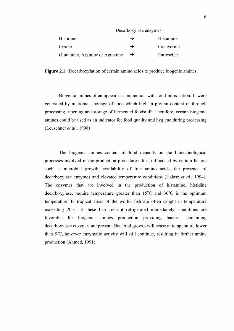
6
Decarboxylase enzymes
Histidine Histamine
Lysine Cadaverine
Glutamine, Arginine or Agmatine Putrescine
Figure 2.1 : Decarboxylation of certain amino acids to produce biogenic amines.
Biogenic amines often appear in conjunction with food intoxication. It were
generated by microbial spoilage of food which high in protein content or through
processing, ripening and storage of fermented foodstuff. Therefore, certain biogenic
amines could be used as an indicator for food quality and hygiene during processing
(Leuschner et al., 1998)
The biogenic amines content of food depends on the biotechnological
processes involved in the production procedures. It is influenced by certain factors
such as microbial growth, availability of free amino acids, the presence of
decarboxylase enzymes and elevated temperature conditions (Halasz et al., 1994).
The enzymes that are involved in the production of histamine, histidine
decarboxylase, require temperature greater than 15oC and 30oC is the optimum
temperature. In tropical areas of the world, fish are often caught in temperature
exceeding 20°C. If these fish are not refrigerated immediately, conditions are
favorable for biogenic amines production providing bacteria containing
decarboxylase enzymes are present. Bacterial growth will cease at temperature lower
than 5oC, however enzymatic activity will still continue, resulting in further amine
production (Ahmed, 1991).
7
2.2 Diamines
Many biogenic amines have been studied in scientific literature, however
diamines such as histamine, putrescine and cadaverine are often documented in
clinical studies with histamine being linked to food poisoning and putrescine and
cadaverine potentiating the toxicity of histamine (Public Health Division, 2002).
Histamine, cadaverine and putrescine have been confirmed as useful chemical
indicators to estimate bacterial spoilage. Consumption of high level of histamine can
lead to scombrotoxicosis while the presence of other biogenic amines is described to
potentiate the effects. The significance of histamine is well known. Person being
highly sensitive to histamine often develops pseudoallergic symptoms shortly after
ingestion. For healthy individuals, the putrescine or cadaverine is not considered to
be toxic. In general, dietary polyamines at levels normally present in food are
nontoxic, while biogenic amines, particularly histamine is toxic at high intakes. The
Food Standards Code stated that the regulated level for histamine is 200 mg/kg (200
ppm). Histamine itself is not destroyed by cooking, freezing, smoking, curing and
canning (Lange and Wittmann, 2001). This is the same histamine that causes
problems for some people when high levels are produced in cheese and red wine.
Histamine has an important role in human metabolism, such as the release
of stomach acid. In small doses it has little effect, but in larger doses it has toxic
effects. The intestinal tract of humans contains the enzymes diamine oxidase
(DAO) and histamine-N-methyl transferase (HMT) which convert histamine to
harmless degradation products. Putrescine and cadaverine can inhibit these
enzymic reactions and are therefore potentiators of histamine toxicity. The
presence of low levels of histamine, in the diet normally has no toxic effect as
humans do not absorb histamine efficiently from the gastrointestinal tract. If a
high level of histamine is present in the diet, then the capacity of DAO and HMT
to detoxify histamine will be limited and histamine will enter into the bloodstream
resulting in histamine poisoning (Taylor, 1986).
8
2.3 Histamine Poisoning
Histamine poisoning, also known as ‘scombroid fish poisoning’, histamine
overdose, pseudo allergic fish poisoning or mahi-mahi flush is among the most
common toxicities related to fish ingestion. The poisoning directly relates to
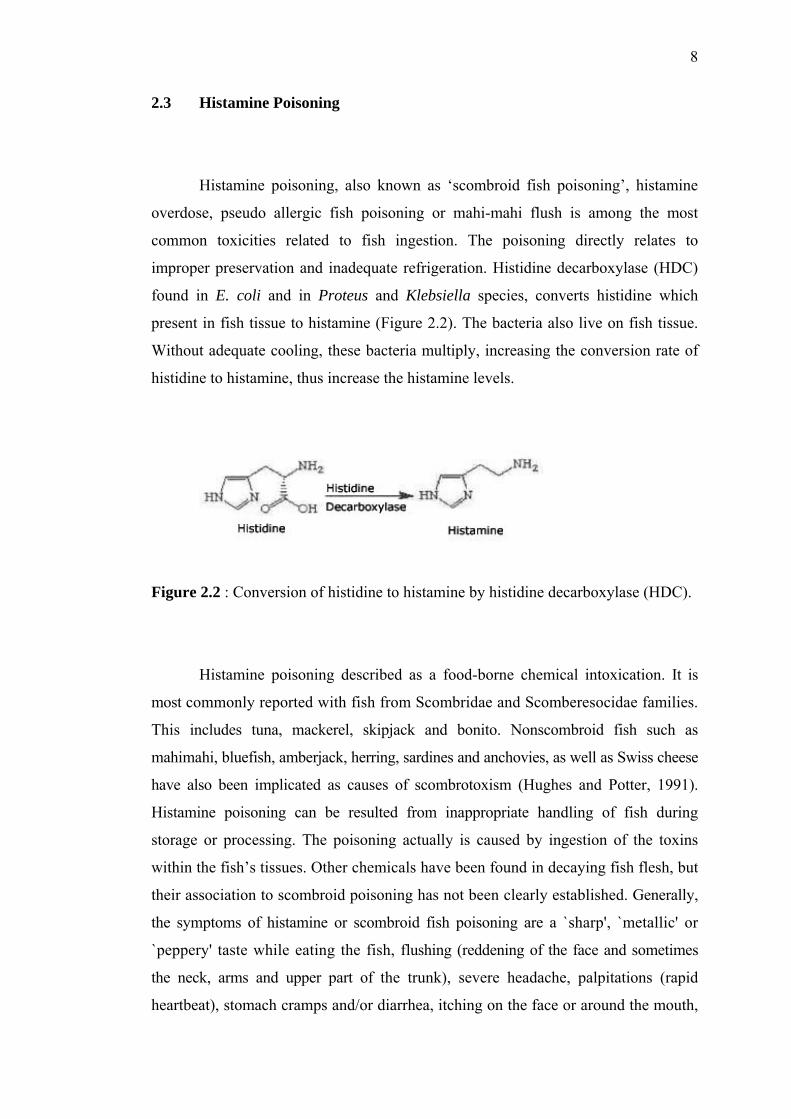
improper preservation and inadequate refrigeration. Histidine decarboxylase (HDC)
found in E. coli and in Proteus and Klebsiella species, converts histidine which
present in fish tissue to histamine (Figure 2.2). The bacteria also live on fish tissue.
Without adequate cooling, these bacteria multiply, increasing the conversion rate of
histidine to histamine, thus increase the histamine levels.
Figure 2.2 : Conversion of histidine to histamine by histidine decarboxylase (HDC).
Histamine poisoning described as a food-borne chemical intoxication. It is
most commonly reported with fish from Scombridae and Scomberesocidae families.
This includes tuna, mackerel, skipjack and bonito. Nonscombroid fish such as
mahimahi, bluefish, amberjack, herring, sardines and anchovies, as well as Swiss cheese
have also been implicated as causes of scombrotoxism (Hughes and Potter, 1991).
Histamine poisoning can be resulted from inappropriate handling of fish during
storage or processing. The poisoning actually is caused by ingestion of the toxins
within the fish’s tissues. Other chemicals have been found in decaying fish flesh, but
their association to scombroid poisoning has not been clearly established. Generally,
the symptoms of histamine or scombroid fish poisoning are a `sharp', `metallic' or
`peppery' taste while eating the fish, flushing (reddening of the face and sometimes
the neck, arms and upper part of the trunk), severe headache, palpitations (rapid
heartbeat), stomach cramps and/or diarrhea, itching on the face or around the mouth,
9
a burning sensation in the throat or dryness of the mouth, difficulty in swallowing
and/or breathing, muscle weakness and nausea (Wu et al., 1997; Bardocz, 1995;
Hughes and Potter, 1991).
The symptoms listed above usually appear within one hour after eating
decayed fish but the onset can range from a few minutes to several hours. It is often last
for 8 to 12 hours, after which most persons recover rapidly. The treatments usually
given to patients are drugs containing antihistamines, which have been shown to help in
many cases. However, in more serious cases, prompt medical attention may be required.
The toxic levels for histamine are estimated at 200-500 mg/kg (200-500 ppm)
(Noltkamper, 2002) while the recent regulated level is 200 mg/kg (200 ppm).
Regulatory limits for histamine content in fish vary with countries. In the United States
of America, 200 ppm denotes mishandling of fish while 500 ppm levels indicate
`hazard action level'. In Germany and Sweden, 200 ppm histamine in fish results in
the rejection of a consignment (Mohd Nasir Azudin and Nazamid Saari, 1988).
2.4 Amines in Plants
Plant amines can be considered simply as the products of decarboxylation
of amino acids, formed by the reaction:
RCH(NH2)CO2H RCH2NH2 + CO2
The most widespread plant amines are conveniently divided into three groups;
aliphatic monoamines, aliphatic polyamines and aromatic amines. Aliphatic amines
are volatile compounds (e.g. methylamine (CH3NH2) to n-hexylamine
(CH3(CH2)5NH2). They are widely distributed in higher plants and fungi and when
10
present in any concentration, have unpleasant fish-like smell. They function in
flowers (e.g. in the cow parsley, Heracleum sphondvlium) as insect attractants by
simulating the smell of carrion.
By contrast to the monoamines, diamines and other polyamines are less
volatile, although they still possess offensive odors. Widespread polyamines include
putrescine NH2(CH2)4NH2, agmatine NH2(CH2)4NHC(=NH)NH2, spermidine
NH2(CH2)3NH(CH2)4NH2 and spermine NH2(CH2)3NH(CH2)4NH(CH2)3NH2. There
are several others of more limited occurrence such as cadaverine NH2(CH2)5NH2.
Polyamines are of topical research interest because of their growth stimulating
activity in relation to their effect on ribosomal RNA.
The best known aromatic amine from plants is probably mescaline, the active
principle of the flowering heads (peyote) of the cactus, Lophophora williamsii, a
powerful natural hallucinogenic compound. Indeed, many of the known aromatic
amines are physiologically active and for this reason they are sometimes classified
with the alkaloids. Three substances which are very important in animal physiology
are noradrenaline, histamine and serotonin; all three occur in plants, noradrenaline
for example being present in the banana and the potato.
All the amines so far mentioned are primary amines, have the general
formula RNH2. Secondary amines, general formula R2NH and tertiary amines, R3N,
are known in plants but they are not very common. A typical secondary amine found
in plants is dimethylamine, a typical tertiary amine is hordenine N-
dimethyltyramine, which is the principal `alkaloid' of barley (Harborne, 1973).
11
2.5 Diamine Oxidase
Diamine oxidase (DAO, EC 1.4.3.6) [amine: O2 oxidoreductase
(deaminating)] is a member of the class of copper-containing amine oxidases and
catalyzes the oxidative deamination of histamine and other biogenic amines. Other
names are diamino oxhydrase, histaminase, histamine deaminase, histamine
oxidase, amine oxidase, amine oxygen oxidoreductase, Cu-amine oxidase,
monoamine oxidase and others. Diamine oxidase was originally characterized as
the enzyme degrading histamine and was therefore earlier called histaminase. It is
characterized by possessing the active-site cofactor topa quinine, formed post-
translationally by modification of a conserved tyrosine residue. Although diamine
oxidase appears to play an important role in histamine catabolism, the enzymes
efficiently converts many diamines besides histamine and is expressed in many
tissues, suggests that it might have additional function. These ubiquitous soluble
enzymes catalyze the oxidative deamination of primary amines to form the
corresponding aldehydes, ammonia and hydrogen peroxide (Wilflingseder and
Schwelberger, 2000).
RCH2NH2 + H2O + O2 ⎯⎯→⎯DAO RCHO + NH3 + H2O2
Measurements of the oxygen consumption or the hydrogen peroxide production are
commonly used for assays of the enzyme activity (Wimmerova and Macholan,
1999).
These Cu-amine oxidase enzymes have been found in several
microorganisms such as bacteria and fungi, various plants and animals (Wimmerova
and Macholan, 1999). Analyses of genes and cDNAs encoding copper amine
oxidases revealed that all members of this enzyme family have homologous
sequences with several absolutely conserved amino acid residues. The conserved
residues appear to be important for the overall protein structure and for the catalytic
function and include the tyrosine that is converted to topa quinone, three histidine
residues that bind to the copper ion and an aspartic acid residue important for
12
substrate conversion (Wilflingseder and Schwelberger, 2000). Plant diamine
oxidases are of widespread occurrence in Leguminaceae such as Cicer arietinum
(chick pea), Lathyrus sativus (grass pea) and Vigna radiata (mungbean)
(Choundhary et al., 1999).
Disclosure of topa quinine as the organic cofactor (Janes et al., 1990, 1992)
and determination of amino acid sequences of some amine oxidases (Rossi et al.,
1992; Mu et al., 1994; Tipping and McPherson, 1995) brought new aspects into the
study of the molecular and structural properties of these enzymes. For the formation
of topa quinone derived from the specific tyrosyl residue in the premature protein, a
self-oxidation mechanism catalyzed by cupric ions has been proposed (Cai and
Klinman, 1994; Matsuzaki et al., 1994). Copper ion and quinone cofactor mediate
the catalytic reaction following a ping-pong mechanism (Hartman and Klinman,
1991). Electron paramagnetic resonance (EPR) studies have shown the occurrence of
a Cu(I)/topa-semiquinone state as an intermediate in the catalytic cycle at the
substrate reduced enzyme (Dooley et al., 1991; Turowski et al., 1993)
Diamine oxidase was first purified by Mann (1955) from pea seedlings. There
after, the enzyme has been purified to homogeneity and characterized from different
plant sources. Pea seedling amine oxidase (PSAO) has wide substrate specificity,
oxidizing preferably natural diamines and polyamines (Kenten and Mann, 1952).
However, the commercially available diamine oxidase is from porcine kidney. This
porcine diamine oxidase has a low specific activity, even when purified, in contrast
to the plant homogenous enzyme. PSAO has already been used in amperometric
biosensors for the assay of biogenic amines and for the assay of some
pseudosubstrates and drugs (Wimmerova and Macholan, 1999).
The crystal structure of amine oxidase from E. coli has been determined in
both an active and inactive form (Parson et al., 1995). Each subunit of the
mushroom-shaped dimer comprises of four domains: a large C-terminal β-sandwich
domain, which contains the active site and provides the dimer interface, and three
13
smaller peripheral α/β domains. The active sites are buried in the protein and lay
some 35 Å apart, connected by a pair of β-hairpin arms. Copper binds directly to topa
quinone in the inactive form, but not in the active form.
Recently, the structure of amine oxidase from pea seedlings (Pisum sativum)
has been reported. The protein structure is quite similar to that of the enzyme from E.
coli, and the study provided additional information on the arrangement and catalytic
mechanism of the active site (Kumar et al., 1996). Plant copper-containing amine
oxidases have been mostly isolated from seedlings of members of the Fabaceae,
particularly of pea and lentil (Medda et al., 1995). Two copper-containing amine
oxidases from plants of the genus Lathyrus have also been characterized. The
enzyme from Lathyrus sativus was first reported by Suresh et al., (1976). Substrate
specificity and sensitivity of this enzyme to several inhibitors were rather similar to
the oxidases from lentil and pea (Medda et al., 1995).
Some of the molecular properties of this enzyme have been determined using
improved purification method which gave a homogeneous enzyme (Padiglia et al.,
1991). This enzyme is a homodimer of 70 to 90 kDa subunits, each containing a
single copper ion and a covalently bound cofactor formed by the post-translational
modification of the catalytic tyrosyl residue to 2,4,5-trihydroxyphenylalanine
quinone (TPQ). The molecular mass of 150 kDa determined by gel permeation
chromatography and of 72 kDa found by sodium dodecylsulphate-polyacrylamide
gel electrophoresis (SDS-PAGE) confirmed the enzyme dimeric structure. Similar
properties were found for the amine oxidase from seedlings of Lathyrus cicera,
which was purified using the same procedure (Cogoni et al., 1989).
In mammalian tissues, Cu-amine oxidases enzymes prefer either monoamines
or diamines as substrates. Mammalian DAO is a homodimeric glycoprotein with
subunits of a relative molecular weight of approximately 100,000 linked by disulfide
bonds. DAO primary structures are highly conserved in mammals. It posses a
classical signal peptide and is N-glycosylated indicating that the protein enters the
14
secretory pathway. It is soluble enzyme mainly found inside cells. In porcine kidney
and intestine, it is localized in vesicular structures in proximity to the plasma
membrane. Recent crystallographic studies of Cu-amine oxidases from
microorganisms and plants have extremely large contributed to the understanding of
the enzymology and structural organization of these proteins (Wilflingseder and
Schwelberger, 2000).
2.6 Chickpeas (Cicer arietinum)
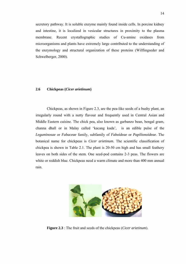
Chickpeas, as shown in Figure 2.3, are the pea-like seeds of a bushy plant, an
irregularly round with a nutty flavour and frequently used in Central Asian and
Middle Eastern cuisine. The chick pea, also known as garbanzo bean, bengal gram,
channa dhall or in Malay called ‘kacang kuda’, is an edible pulse of the
Leguminosae or Fabaceae family, subfamily of Faboideae or Papilionoideae. The
botanical name for chickpeas is Cicer arietinum. The scientific classification of
chickpea is shown in Table 2.1. The plant is 20-50 cm high and has small feathery
leaves on both sides of the stem. One seed-pod contains 2-3 peas. The flowers are
white or reddish blue. Chickpeas need a warm climate and more than 400 mm annual
rain.
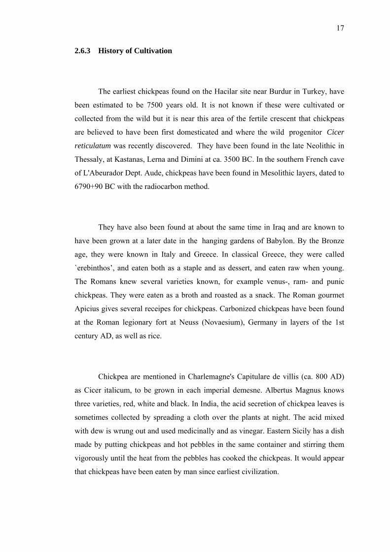
Figure 2.3 : The fruit and seeds of the chickpeas (Cicer arietinum).
15
Table 2.1 : Scientific classification of chickpeas.
Kingdom : Plantae
Division : Magnoliophyta
Class : Magnoliopsida
Order : Fabales
Family : Fabaceae
Subfamily : Faboideae
Genus : Cicer
Species : arietinum
2.6.1 Chickpea Varieties
Several dozen distinct chickpea varieties are cultivated. There are two groups
of chickpeas, European and Asian, distinguished by seed size, shape and colour.
They also have different growth requirements and end uses. The European varieties,
also known as `kabuli’ types, are large (typically around 15 mm diameter) and more
rounded or brain-shaped types which are most commonly pale yellow in color. There
were several other varieties, with black and reddish seeds that are rarely grown
today. The black variety was mainly used as fodder. Asian varieties, also known as
`desi’ types, are smaller (typically 5-8 mm) and angular seeds, which are dark brown
in color.
The chickpea is not known in a wild state. The kabuli types are commonly
found throughout southern Europe, Western Asia, the Nile Valley, North Africa and
South America, whereas the desi types are mostly found in the Indian subcontinent,
Iran, Ethiopia and parts of central America. They are available mainly dried whole or
split. In parts of the world where chickpeas are grown they are frequently sold as the
whole green plant from which the seeds are consumed fresh as a snack or the whole
16
plant can be placed in a fire and the parched seeds eaten as a snack. They are also
available tinned whole or as a puree. Chickpea flour is also available in some
countries.
2.6.2 Chickpea Nutrition and Uses
Chickpeas are an excellent source of carbohydrates and proteins, which
constitute about 80% of the total dry seed weight. Dried chickpeas contain about
20% protein. The bulk of the seed is made up of carbohydrates (61%) and 5% fat.
Crude fiber is mostly located within the seed coat. They are relatively rich in lecithin,
potassium, phosphorus, calcium, folate and vitamin C, and also have small quantities
of vitamins A and B. 100 g of chickpeas can supply about 350 calories.
Chickpeas can be eaten in salads, cooked in stews and ground into a flour
called `gram flour’ (also known as besan, garbanzo bean flour or in Malay `tepung
kacang kuda’, and usually used in Indian cuisine). They can also be ground and
shaped in balls and fried as `falafel’, cooked and ground into a paste called hummus,
or roasted and spiced and eaten as a snack. The plant can also be used as a green
vegetable. Dried chickpeas can be kept almost indefinitely. Tinned chickpeas last
well for up to 5 years. Once cooked, the chickpeas can be stored covered for several
days in the fridge.
17
2.6.3 History of Cultivation
The earliest chickpeas found on the Hacilar site near Burdur in Turkey, have
been estimated to be 7500 years old. It is not known if these were cultivated or
collected from the wild but it is near this area of the fertile crescent that chickpeas
are believed to have been first domesticated and where the wild progenitor Cicer
reticulatum was recently discovered. They have been found in the late Neolithic in
Thessaly, at Kastanas, Lerna and Dimini at ca. 3500 BC. In the southern French cave
of L'Abeurador Dept. Aude, chickpeas have been found in Mesolithic layers, dated to
6790+90 BC with the radiocarbon method.
They have also been found at about the same time in Iraq and are known to
have been grown at a later date in the hanging gardens of Babylon. By the Bronze
age, they were known in Italy and Greece. In classical Greece, they were called
`erebinthos’, and eaten both as a staple and as dessert, and eaten raw when young.
The Romans knew several varieties known, for example venus-, ram- and punic
chickpeas. They were eaten as a broth and roasted as a snack. The Roman gourmet
Apicius gives several receipes for chickpeas. Carbonized chickpeas have been found
at the Roman legionary fort at Neuss (Novaesium), Germany in layers of the 1st
century AD, as well as rice.
Chickpea are mentioned in Charlemagne's Capitulare de villis (ca. 800 AD)
as Cicer italicum, to be grown in each imperial demesne. Albertus Magnus knows
three varieties, red, white and black. In India, the acid secretion of chickpea leaves is
sometimes collected by spreading a cloth over the plants at night. The acid mixed
with dew is wrung out and used medicinally and as vinegar. Eastern Sicily has a dish
made by putting chickpeas and hot pebbles in the same container and stirring them
vigorously until the heat from the pebbles has cooked the chickpeas. It would appear
that chickpeas have been eaten by man since earliest civilization.
18
2.7 Introduction of Biosensor
A biosensor may be broadly defined as any measuring device that contains a
biological element (Buerk, 1993) or a device incorporating a biological sensing
element connected to a transducer (Eggins, 1996). It is also frequently described as a
'reagentless' system, or it is more correct to say that the reagents are already part of
the reaction chamber and do not therefore have to be added by user, most likely be
concerned with immobilized reagent (Hall, 1991). The biological element involved
might be tissue, microorganisms, organelles, cell receptors, enzymes, antibodies or
nucleic acids (Rogers and Gerlach, 1996). The sensing element which responds to
the substance being measured is biological in nature. It has to be connected to a
transducer of some sort so that a visually observable response occurs. A transducer
converts an observed change, physically or chemically into a measurable signal,
usually an electronic signal whose magnitude is proportional to the concentration of
a specific chemical or set of chemicals. It is an apparently alien marriage of two
contrasting disciplines which combines the specificity and sensitivity of biological
systems with the computing power of the microprocessor. Biosensors are generally
concerned with sensing and measuring particular chemicals which need not be
biological components themselves, although sometimes they are. They are referred
as the substrate, although the more general term analyte is often used. (Eggins,
1996). A schematic diagram drawing for a generalized biosensor is shown in Figure
2.4.
19
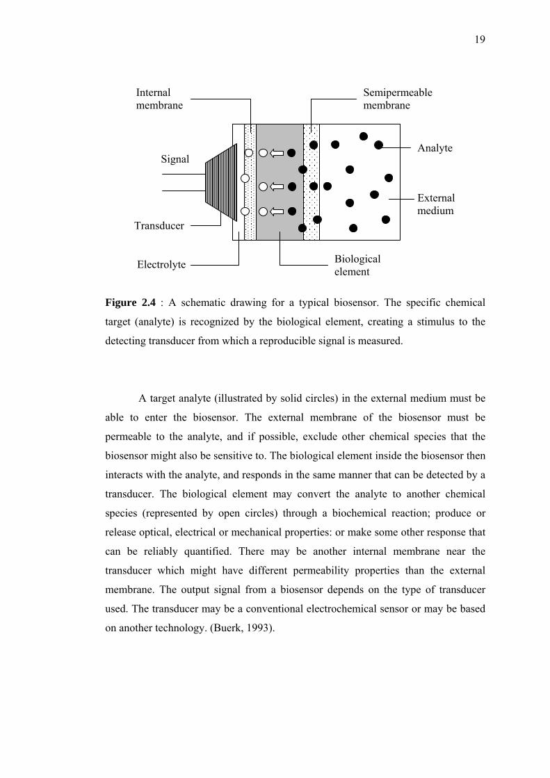
Figure 2.4 : A schematic drawing for a typical biosensor. The specific chemical
target (analyte) is recognized by the biological element, creating a stimulus to the
detecting transducer from which a reproducible signal is measured.
A target analyte (illustrated by solid circles) in the external medium must be
able to enter the biosensor. The external membrane of the biosensor must be
permeable to the analyte, and if possible, exclude other chemical species that the
biosensor might also be sensitive to. The biological element inside the biosensor then
interacts with the analyte, and responds in the same manner that can be detected by a
transducer. The biological element may convert the analyte to another chemical
species (represented by open circles) through a biochemical reaction; produce or
release optical, electrical or mechanical properties: or make some other response that
can be reliably quantified. There may be another internal membrane near the
transducer which might have different permeability properties than the external
membrane. The output signal from a biosensor depends on the type of transducer
used. The transducer may be a conventional electrochemical sensor or may be based
on another technology. (Buerk, 1993).
Internal membrane
Biological element
Semipermeable membrane
Signal
Transducer
Electrolyte
Analyte
External medium
20
2.7.1 Chemical Sensor and Biosensor
A chemical sensor is a device that transforms chemical information, ranging
from the concentration of a specific sample component to total composition analysis,
into analytically useful signal. Chemical sensor usually contains two basic
components connected in series that is a chemical recognition system (receptor) and
a physicochemical transducer. Biosensors are chemicals sensors in which the
recognition system utilizes a biochemical mechanism.
The main purpose of the recognition system is to provide the sensor with a
high degree of selectivity for the analyte to be measured. The biological recognition
system translates information from the biochemical domain, usually an analyte
concentration, into a chemical or physical output signal with a defined sensitivity.
While all biosensors are more or less selective (non-specific) for a particular analyte,
some are, by design and construction, only class-specific, since they use class
enzymes such as phenolic compound biosensors or whole cells used to measure
biological oxygen demand. Because in sensing system presents in living organisms
or systems, such as olfaction and taste, the actual recognition is performed by a cell
receptor, the word ‘bioreceptor’ is often be used for recognition system of a
biosensor.
The transducer is a part of the biosensor serves to transfer the signal from the
output domain of the recognition system to, mostly the electrical domain. A
transducer provides bidirectional signal transfer (non-electrical to electrical). The
transducer part of sensor is also called a detector or electrode, but the term transducer
is often used to avoid confusion. Table 2.2 shows the types of receptors used in
electrochemical and biosensors measurement techniques. Indicated in italic types are
the biological receptors, which part of the electrochemical biosensors.
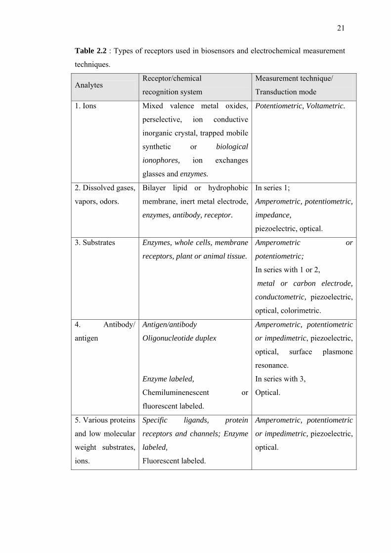
21
Table 2.2 : Types of receptors used in biosensors and electrochemical measurement
techniques.
Analytes Receptor/chemical
recognition system
Measurement technique/
Transduction mode
1. Ions Mixed valence metal oxides,
perselective, ion conductive
inorganic crystal, trapped mobile
synthetic or biological
ionophores, ion exchanges
glasses and enzymes.
Potentiometric, Voltametric.
2. Dissolved gases,
vapors, odors.
Bilayer lipid or hydrophobic
membrane, inert metal electrode,
enzymes, antibody, receptor.
In series 1;
Amperometric, potentiometric,
impedance,
piezoelectric, optical.
3. Substrates Enzymes, whole cells, membrane
receptors, plant or animal tissue.
Amperometric or
potentiometric;
In series with 1 or 2,
metal or carbon electrode,
conductometric, piezoelectric,
optical, colorimetric.
4. Antibody/
antigen
Antigen/antibody
Oligonucleotide duplex
Enzyme labeled,
Chemiluminenescent or
fluorescent labeled.
Amperometric, potentiometric
or impedimetric, piezoelectric,
optical, surface plasmone
resonance.
In series with 3,
Optical.
5. Various proteins
and low molecular
weight substrates,
ions.
Specific ligands, protein
receptors and channels; Enzyme
labeled,
Fluorescent labeled.
Amperometric, potentiometric
or impedimetric, piezoelectric,
optical.
22
Besides quantification of the above mentioned analytes, biosensors are also
used for detection and quantification of micro-organisms, receptors are bacteria,
yeast or oligonucleotide probes coupled to electrochemical, piezoelectric, optical or
calorimetric transducers.
2.7.2 Electrochemical Biosensor
A biosensor with an electrochemical transducer is called an electrochemical
biosensor. It is an integrated receptor-transducer device, which is a capable of
providing selective quantitative or semi-quantitative analytical information using a
biological recognition element. A biosensor can be used to monitor either biological
or non-biological matrices. Non-electrochemical transducers are used within
biosensors, these includes piezoelectric, calorimetric (thermistor) and optical (planar
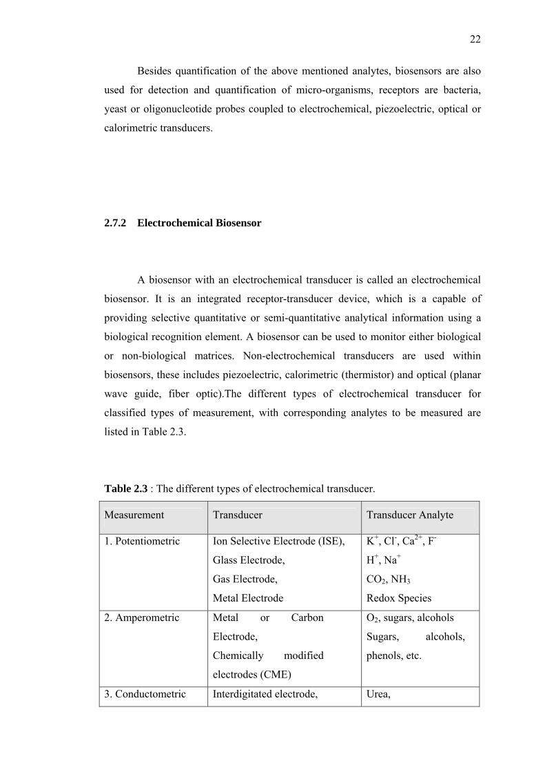
wave guide, fiber optic).The different types of electrochemical transducer for
classified types of measurement, with corresponding analytes to be measured are
listed in Table 2.3.
Table 2.3 : The different types of electrochemical transducer.
Measurement Transducer Transducer Analyte
1. Potentiometric Ion Selective Electrode (ISE),
Glass Electrode,
Gas Electrode,
Metal Electrode
K+, Cl-, Ca2+, F-
H+, Na+
CO2, NH3
Redox Species
2. Amperometric Metal or Carbon
Electrode,
Chemically modified
electrodes (CME)
O2, sugars, alcohols
Sugars, alcohols,
phenols, etc.
3. Conductometric Interdigitated electrode, Urea,
23
Metal electrode. oligonucleotides,
charged species.
4. Ion charge or
field effect
Ion-sensitive field-effect
transistor, Enzyme FET
H+, K+
2.7.3 Electrochemical Detection
2.7.3.1 Amperometry
Amperometry is based on the measurement of the current resulting from the
electrochemical oxidation or reduction of an electroactive species. The resulting
current is directly correlated to the bulk concentration of the electroactive species or
its production or consumption rate within the biocatalytic layer.
2.7.3.2 Potentiometry
Potentiometric measurements involves the determination of the potential
difference between either indicator and a reference electrode or two reference
electrodes separated by a permiselective membrane, when there is no significant
current flowing between them. The most common potentiometric devices are pH
electrodes, several other ion (F-, I-, CN-, Na+, K+) or gas (CO2, NH3) selective
electrodes are also available.
24
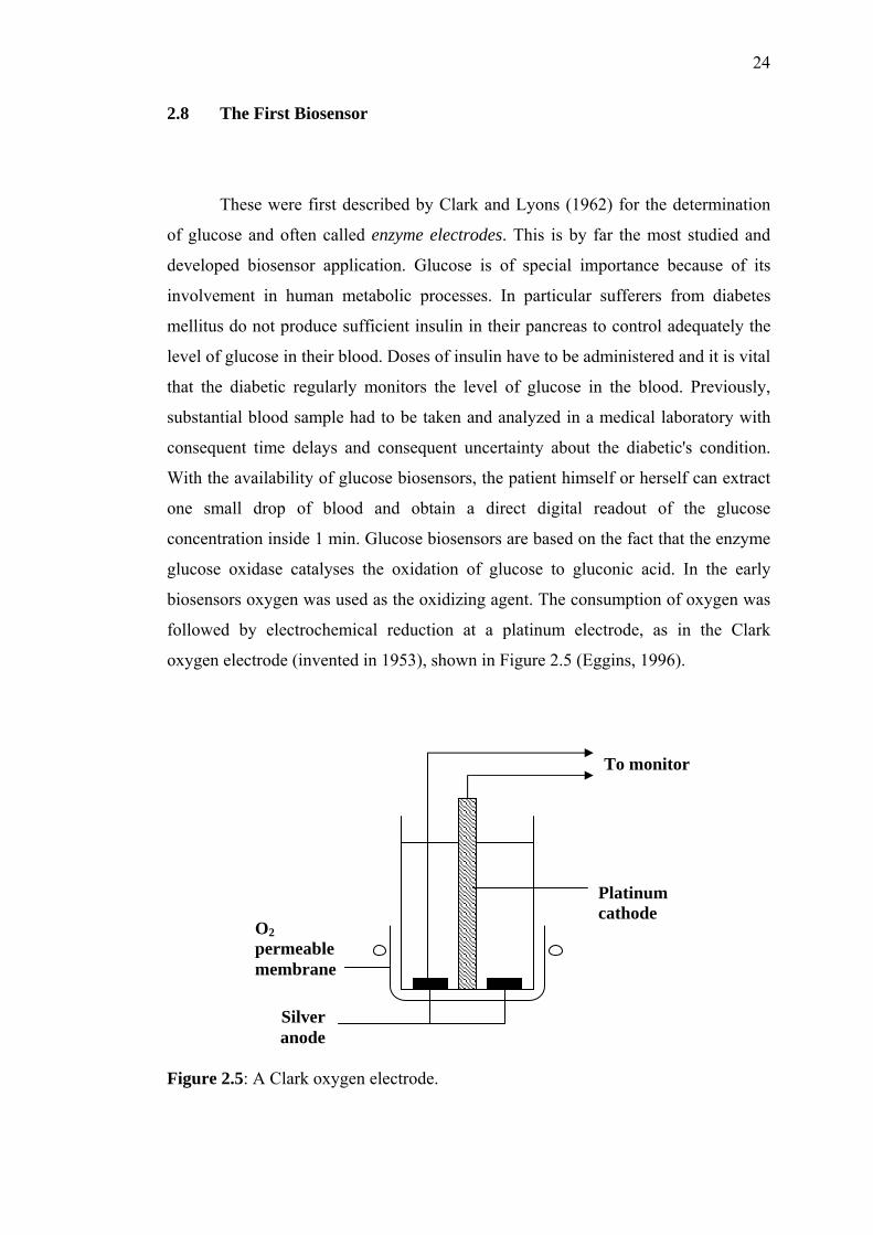
2.8 The First Biosensor
These were first described by Clark and Lyons (1962) for the determination
of glucose and often called enzyme electrodes. This is by far the most studied and
developed biosensor application. Glucose is of special importance because of its
involvement in human metabolic processes. In particular sufferers from diabetes
mellitus do not produce sufficient insulin in their pancreas to control adequately the
level of glucose in their blood. Doses of insulin have to be administered and it is vital
that the diabetic regularly monitors the level of glucose in the blood. Previously,
substantial blood sample had to be taken and analyzed in a medical laboratory with
consequent time delays and consequent uncertainty about the diabetic's condition.
With the availability of glucose biosensors, the patient himself or herself can extract
one small drop of blood and obtain a direct digital readout of the glucose
concentration inside 1 min. Glucose biosensors are based on the fact that the enzyme
glucose oxidase catalyses the oxidation of glucose to gluconic acid. In the early
biosensors oxygen was used as the oxidizing agent. The consumption of oxygen was
followed by electrochemical reduction at a platinum electrode, as in the Clark
oxygen electrode (invented in 1953), shown in Figure 2.5 (Eggins, 1996).
Figure 2.5: A Clark oxygen electrode.
Platinum cathode
Silver anode
O2 permeable membrane
To monitor
25
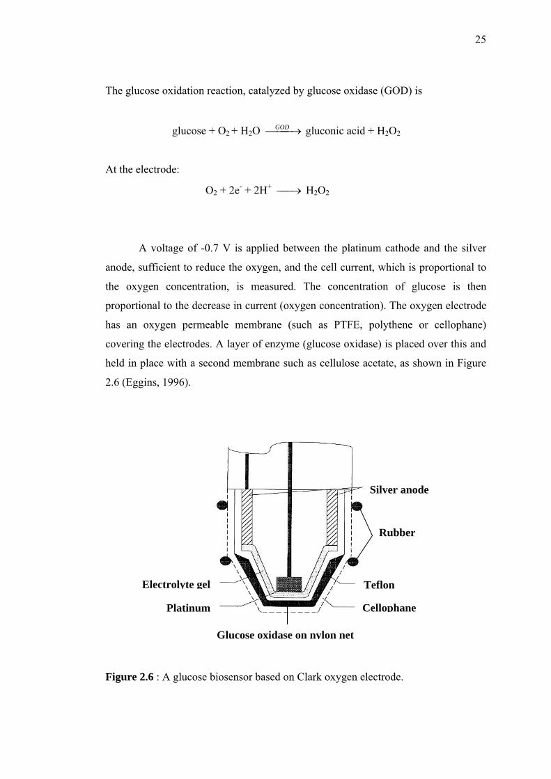
The glucose oxidation reaction, catalyzed by glucose oxidase (GOD) is
glucose + O2 + H2O ⎯⎯ →⎯GOD gluconic acid + H2O2
At the electrode:
O2 + 2e- + 2H+ ⎯→⎯ H2O2
A voltage of -0.7 V is applied between the platinum cathode and the silver
anode, sufficient to reduce the oxygen, and the cell current, which is proportional to
the oxygen concentration, is measured. The concentration of glucose is then
proportional to the decrease in current (oxygen concentration). The oxygen electrode
has an oxygen permeable membrane (such as PTFE, polythene or cellophane)
covering the electrodes. A layer of enzyme (glucose oxidase) is placed over this and
held in place with a second membrane such as cellulose acetate, as shown in Figure
2.6 (Eggins, 1996).
Figure 2.6 : A glucose biosensor based on Clark oxygen electrode.
Rubber
Platinum
Teflon
Cellophane
Electrolyte gel
Silver anode
Glucose oxidase on nylon net
26
2.9 Fundamental of Amperometric Biosensor
Amperometric measurements can be measured when a defined potential is
applied at a working electrode with respect to the reference electrode. This result
current that can be related to the concentration of an electroactive substance in the
solution. At low current densities, it is sufficient for most of elementary
electrochemical setup to have two electrodes, which are working electrode (WE) and
reference electrode (RE). However, at higher current densities, the potential of
reference electrode may change with current, so it is not possible to obtain
reproducible determination of analyte. To avoid this problem, a third electrode is
required (three-electrode setup) which is known as counter electrode (CE). For the
three-electrode setup, current is measured between working and counter electrodes,
while potential is measured based on reference electrode. The measured current is
directly related to the rate of the overall process in the electrochemical cell. (Wagner,
and Guilbault, 1994).
2.10 Histamine Biosensor
In histamine biosensor, the basic principles are the same as the principles of the
glucose biosensor. The basic underlying chemistry is the action of diamine oxidase
(DAO) that catalyzed the oxidative deamination of histamine to
imidazoleacetaldehyde, hydrogen peroxide (H2O2) and ammonia (NH3). The reaction
involved is shown in Figure 2.7 (Niculescu et al., 2001).
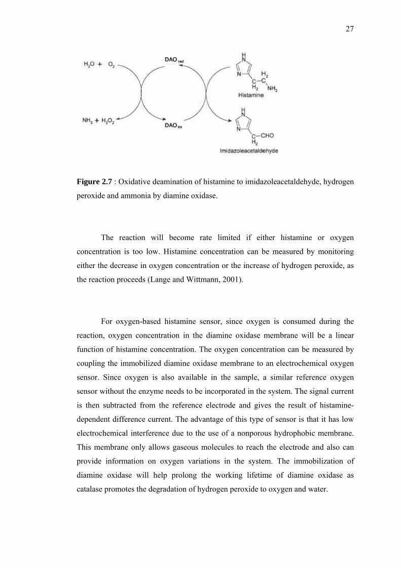
27
Figure 2.7 : Oxidative deamination of histamine to imidazoleacetaldehyde, hydrogen
peroxide and ammonia by diamine oxidase.
The reaction will become rate limited if either histamine or oxygen
concentration is too low. Histamine concentration can be measured by monitoring
either the decrease in oxygen concentration or the increase of hydrogen peroxide, as
the reaction proceeds (Lange and Wittmann, 2001).
For oxygen-based histamine sensor, since oxygen is consumed during the
reaction, oxygen concentration in the diamine oxidase membrane will be a linear
function of histamine concentration. The oxygen concentration can be measured by
coupling the immobilized diamine oxidase membrane to an electrochemical oxygen
sensor. Since oxygen is also available in the sample, a similar reference oxygen
sensor without the enzyme needs to be incorporated in the system. The signal current
is then subtracted from the reference electrode and gives the result of histamine-
dependent difference current. The advantage of this type of sensor is that it has low
electrochemical interference due to the use of a nonporous hydrophobic membrane.
This membrane only allows gaseous molecules to reach the electrode and also can
provide information on oxygen variations in the system. The immobilization of
diamine oxidase will help prolong the working lifetime of diamine oxidase as
catalase promotes the degradation of hydrogen peroxide to oxygen and water.
28
The hydrogen peroxide-based histamine sensor has found wide application in
the development of such a sensor, especially an implantable version, due to its simple
sensor configuration that facilitates ease of miniaturization. Unlike oxygen, hydrogen
peroxide is not present in the sample to be analyzed. This make no differential setup
needed. However, it suffers from an intrinsic problem, the interference from small
endogenous analytes, which may be electro-active at the detection potential of
hydrogen peroxide which is quite high (Azila Abdul Aziz, 2001).
Those two types of sensors mentioned above are known as the first
generation amperometric biosensors. The second generation of histamine biosensors
makes use of mediators to shuttle electrons from the enzyme to the electrode, instead
of oxygen, which are reversible, had appropriate oxidation potentials and whose
concentration could be controlled. If the system is oxygen, the biosensor will become
insensitive to histamine, thus will only respond to changes in oxygen concentration.
As oxygen remains in the system, the mediator must be able to compete effectively
for the electrons (Azila Abdul Aziz, 2001).
The use of mediator in determining the content of histamine and other
biogenic amines has been studied by Tombelli and Mascini (1998), compared to a
single enzyme sensor and a flow system based on hydrogen peroxide generated by
enzymatic reaction. A bi-enzyme FIA system with amperometric detection was used
based on the following enzyme reactions, with ferrocene carboxylic acid (Fc-COOH)
as the mediator facilitating the electron transfer between the electrode and
horseradish peroxidase (HRP). Pea seedling amine oxidase (PSAO) catalyses the
oxidation of the amine and subsequently the co-substrate, molecular oxygen, is
reduced to hydrogen peroxide. Hydrogen peroxide is then expends in the following
peroxidase catalyzed reaction using Fc-COOH as the mediator. The amperometric
signal is monitored reductively at the electrode. Figure 2.7 illustrated the basic
principle of the electron transfer during measurements (Wimmerova and Macholan,
1999).
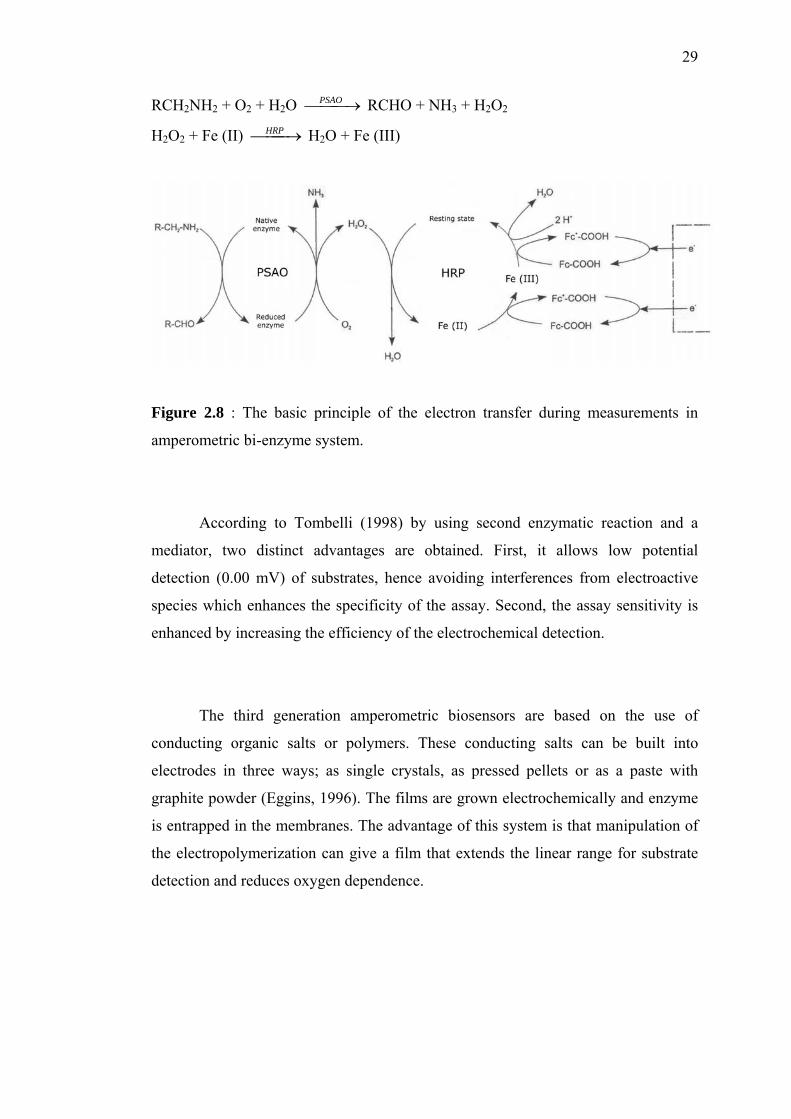
29
RCH2NH2 + O2 + H2O ⎯⎯ →⎯PSAO RCHO + NH3 + H2O2
H2O2 + Fe (II) ⎯⎯→⎯HRP H2O + Fe (III)
Figure 2.8 : The basic principle of the electron transfer during measurements in
amperometric bi-enzyme system.
According to Tombelli (1998) by using second enzymatic reaction and a
mediator, two distinct advantages are obtained. First, it allows low potential
detection (0.00 mV) of substrates, hence avoiding interferences from electroactive
species which enhances the specificity of the assay. Second, the assay sensitivity is
enhanced by increasing the efficiency of the electrochemical detection.
The third generation amperometric biosensors are based on the use of
conducting organic salts or polymers. These conducting salts can be built into
electrodes in three ways; as single crystals, as pressed pellets or as a paste with
graphite powder (Eggins, 1996). The films are grown electrochemically and enzyme
is entrapped in the membranes. The advantage of this system is that manipulation of
the electropolymerization can give a film that extends the linear range for substrate
detection and reduces oxygen dependence.
30
2.11 Enzyme Immobilization
Enzymes are biocatalytically active entities upon which the metabolisms of
all living organisms are based. Enzymes are usually proteins of high molecular
weight (15,000<MW<several million Daltons) that act as catalyst. Enzymes are
specific, versatile and very effective biological catalyst resulting in much higher
reaction rates compared to chemically catalyzed reaction under ambient condition.
They speedup biochemical reactions by lowering the energy of activation, without
themselves appearing in the reaction products. The catalytic actions of enzymes
involve their ability to alter the distribution of charges on the compound to be
converted, thus bringing about a lowering of the energy of activation. Furthermore,
they are highly specific, thus side reactions can be avoided by employing enzymatic
breakdown.
Enzymes are proteins that are constructed from chains of amino acids.
Different types of amino acid links have a different shapes and properties. As an
unfolded chain, the enzyme has no catalytic activity. Only the folded structure forms
the catalytic or active site, which is brought about by their tertiary and quaternary
(oligometric enzyme) structures. However, this folded structure will generally be
held together by non-covalent interactions, unlike the covalent bonds that hold the
amino acid links together. These consist of interactions such as ionic bridges,
hydrogen bonds, hydrophobic and hydrophilic interaction (Trevor Palmer, 1985).
The molecular structure of enzymes that is essential for their catalytic activity is
liable to be destroyed under conditions such as high temperature, high or low pH,
with presence of organic solvents, or even conditions suitable for catalysis. The
recovery of active enzymes from spent reaction mixtures is another problem when
free or non-immobilized enzymes are utilized.
A biocatalyst is termed ‘immobilized’ if its mobility has been restricted by
chemical or physical means. This limitation of mobility may be achieved by widely
different methods. For example, by trapping in the network of a polymer matrix or
31
by membrane confinement. An essential criterion for defining a system as
immobilized is that human interference has to be involved. A variety of
immobilization methods have been used in the development of a successful
biosensors. Immobilized enzyme preparations may be more effective since they are
recoverable and possibly more stable than free enzymes. Moreover, being in their
native environment, operational denaturation of the enzymes can be minimized.
Enzymes are largely used as biocatalysts in chemical, pharmaceutical and
food industries, and as specific ligands in clinical and chemical analysis. Since the
recovery and the reusability of the free enzyme are limited, immobilization of
enzyme has attracted the attention. Immobilized enzymes have the advantages of
using in batch and continuous systems, removing easily from the reaction medium
and providing the facility of controlled production. However, immobilized enzyme
systems do also have limitations, such as loss activity due to the immobilization
technique and decrease in the apparent activity due to mass transfer resistance for the
substrate and product. An optimal support material should provide large surface area
per unit volume (or mass) of the carrier for effective immobilization of the desired
amount of enzyme and should allow substrate and product transport with the least
diffusional resistance and also should be easily available and non toxic. The
immobilization technique should also lead high immobilization and activity yields
(Bulmus et al., 1998).
In order to construct a viable biosensor, the enzyme used has to be properly
attached to the transducer. There are five regular methods in enzyme immobilization,
as follows (Eggins, 1996).
i. Adsorption
This is the simplest and involves minimal preparation. However, the bonding
is weak and this method is only suitable for exploratory work over a short
time-span.
32
ii. Microencapsulation.
This was the method used in the early biosensors. The biomaterial is held in
place behind a membrane, giving close contact between the biomaterial and
the transducer. It is adaptable. It does not interfere with the reliability of the
enzyme. It limits contamination and biodegradation. It is stable towards
changes in temperature, pH, ionic strength and chemical composition. It can
be permeable to some materials, e.g. small molecules, gas molecules and
electrons.
iii. Entrapment
The biomaterial is mixed with monomer solution, which is then polymerized
to a gel, trapping the biomaterial. Unfortunately, this can cause barriers to the
diffusion of substrate, thus slowing the reaction. It can also result in loss of
bioactivity through pores in the gel. This can be counteracted by crosslinking.
The most commonly used gel is polyacrylamide, although starch gels, nylon
and silastic gels have been used. Conducting polymers such as polypyrroles
are particularly useful with electrodes.
iv. Cross-linking
In this method, the biomaterial is chemically bonded to solid supports or to
another supporting material such as a gel. Bifunctional reagents such as
glutaraldehyde are used. Again, there is some diffusion limitation and there
can be damage to the biomaterial. Also, the mechanical strength is poor. It is
a useful method to stabilize adsorbed biomaterials.
v. Covalent bonding
This involves a carefully designed bond between a functional group in the
biomaterial to the support matrix. Some functional groups which are not
essential for the catalytic activity of an enzyme can be covalently bonded to
the support matrix (transducer or membrane). This method uses nucleophilic
groups for coupling such as COOH, OH, C6H4OH , SH and amidazole.
33
Overall, the lifetime of the biosensor is greatly enhanced by proper
immobilization. Typical lifetimes for the same biosensor, in which different methods
of immobilization are used, are as follows:
Adsorption 1 day
Membrane entrapment 1 week
Physical entrapment 3 - 4 weeks
Covalent entrapment 4 - 14 months
2.11.1 Poly (vinyl alcohol) (PVA)
Poly (vinyl alcohol) is manufactured by the hydrolysis of polyvinyl acetate.
Even after a prolonged hydrolysis, PVA generally retains around 1 to 2 mole percent
of acetate groups. The amount of residual acetate groups affects the physical and
chemical properties of PVA, as they are hydrophobic relative to the hydroxyl groups.
The residual acetate groups can interfere with inter-molecular and intra-molecular
forces such as hydrogen bonding. Highly hydrolyzed PVA has strong hydrogen
bonds within and between molecules.
PVA is used as a basic material for a variety of biomedical applications
because of their inherent non-toxicity, non-carcinogenicity, good biocompatibility
and desirable physical properties, such as elastic nature, good film forming property,
high degree of swelling in aqueous solutions, and their water content matches that of
biological tissue (M. Masuda, 1992). PVA gives a hydrophilic tough membrane
which can stabilize the activity of various enzymes, including diamine oxidase
(DAO) and horseradish peroxidase (HRP), through the inhibition of the formation of
non-functional conformations due to the extensive hydrogen bonding between the H
atoms of alcohol groups in PVA and the O atoms of the carbohydrate groups in
DAO. Moreover, PVA is water-soluble polymer that readily reacts with different
34
cross-linking to form gel. However, owing to strong internal hydrogen bond, it only
goes into solution at higher temperature, around 90°C. Aqueous solutions of PVA
exhibit non-Newtonian behavior at room temperature.
PVA can be cross-linked chemically or physically to form a hydrogel. The
polymer can be cross-linked chemically by any bifunctional agent that can react with
organic hydroxyl groups. Some of the various chemicals that can cross-link PVA are
glutaraldehyde, formaldehyde, maleic acid and boric acid. Cross-linking can also be
achieved physically using ultraviolet (UV) light in the presence of photo-sensitizers,
by electron beam or by y-radiation. Another physical method of cross-linking PVA is
through freeze-thaw cycles, where physical bonds are formed.
Cross-linking causes PVA to be insoluble in water. Furthermore, by
controlling the cross-link density of this material, variety of transport properties can
be obtained. In addition, poly (vinyl alcohol) is also considered biocompatible.
Protein adsorption onto cross-linked PVA has been shown to be negligible, thus the
potential of minimizing fibrotic capsule development around implantable PVA
exists. Hence, due to its promised biocompatibility, ease of manipulation and
hydrophilicity, PVA has been used extensively in biomedical applications. In the
biomedical area, PVA has found use in hernia treatment, in artificial heart valve
replacement and as a drug carrier in controlled drug release system, among others.
In dense hydrogels, diffusion of solutes is determined by the cross-linking
density of the network, or mesh size and the degrees of swelling. The `pores' are the
open spaces between the cross-link points and are not fixed. Different theories have
been proposed to describe solute diffusion in hydrogels. Polymer chains are
hypothesized to hinder solute diffusion through one or a combination of these
methods: by physically obstructing the passage of solute, by increasing the
hydrodynamic drag on the solute molecule or by reducing the available free volume
for the solute (Azila Abdul Aziz, 2001).
35
2.11.2 Freeze-thawed PVA
Physically cross-linked PVA through freeze-thawing method may be a good
choice for immobilizing the enzymes. It can minimize the problems associate with
chemically cross-linked PVA, which maintaining the good properties of PVA. The
exposition of aqueous PVA solution to several freeze-thawing cycles leads to
reinforced gels owing to a densification of the macromolecular structure, which is a
function of the cycling time and temperature (Christie M. Hassa and Nikolas A.
Peppas, 2000). After the freeze-thawing process, fine crystallites are formed due to
the slow heat treatment. The chains are physically cross-linked by semi-permanent
entanglements, molecular associations or crystalline (Lucio Doretti et al., 1997).
Formations of crystallites serve as physical cross-links to render the material
insoluble in water.
Some characteristics of the physical cross-linked PVA gels include high
degree of swelling in water, a rubbery and elastic nature, and a high mechanical
strength because the mechanical load can be distributed along the crystallites of the
three-dimensional structure.
The freeze-thawing method as characterized by the absence of chemical
cross-linking agents that could compromise is biocompatibility or physical agent,
such as gamma radiation that could deactivate the biological substrates, due to the
damage caused mostly by the indirect effect of water radiolysis. Generally, physical
entrapment of enzyme molecules in polymeric membranes is one of the most
advantages method because it is rapid, simple and the retained activity is high (Lucio
Doretti et al., 1998).
36
2.11.3 Immobilization Methods in Constructing Histamine Biosensor
Several immobilization methods have been used and investigated in order to
improve the sensitivity of biosensors. Lange et al. (2001) were firstly used 20% of
transglutaminase solution for immobilization of diamine oxidase, plasma amine
oxidase and tyramine oxidase. The immobilization technique improved the
sensitivity of sensor, but unfortunately, when lots of transglutaminase was finished,
difficulties occurred with the regular quality (sensor reproducibility was low).
Therefore, they investigated the conventional immobilization method, which was
based on glutaraldehyde-albumin cross-linking. In their research, they compared the
results obtained (to determine biogenic amines: specifically histamine and tyramine)
between enzyme sensor array and high performance liquid chromatography (HPLC).
From the results, it can be concluded that by using enzyme sensor array, less time
was required to conduct experiments and was not tedious as when handling HPLC,
but still the reproducibility, data validity, detection limit and so on were still poor.
Glutaraldehyde was always chosen as the cross-linking agent to entrapped
diamine oxidase. Tombelli and Mascini (1998) used glutaraldehyde solution on
cellulose acetate membrane to immobilize diamine oxidase on a platinum electrode.
It was also helped Bouvrette et al. to develop their membranes. Poly (vinyl alcohol)
is a non-toxic water-soluble synthetic material that has good film forming properties,
resulting in tough membranes. Glutaraldehyde, a bifunctional agent that can react
with organic hydroxyl groups, was used as the cross-linking agent. Glutaraldehyde
can also react with the lysine amino acid residues in the enzyme.
The cross-linking process overcomes the loss of enzyme activity due to
diffusional loss, which is a prevalent problem for enzyme immobilized in physical
entrapment. PVA can stabilize the activity of various enzymes such as horseradish
peroxidase. The stabilization effect is achieved through the inhibition of the
formation of non-functional conformations due to the extensive hydrogen bonding
between the H-atoms of the alcohol groups in PVA and the O-atoms of the

37
carbohydrate groups in diamine oxidase. These properties make PVA an appropriate
matrix for diamine oxidase immobilization.
CHAPTER 3
METHODOLOGY
3.1 Purification of Pea Seedling Amine Oxidase
The purification of pea seedling amine oxidase will be done according to the
method given by Sebela et al., (1998) with some modifications. The type of pea that
will be used is Cicer arietinum (chick pea).
3.1.1 Enzyme Purification
The chick pea seeds were soaked in distilled water for 12 hr, transferred onto
wet cotton/tissue paper and germinated in the dark for 7 days at room temperature.
The whole seedlings were homogenized in Waring blender with 3 volumes (w/v) of
chilled 0.1 M potassium phosphate buffer (PPB) pH 7.0 for about 10 min. The crude
homogenate was filtered through a nylon or cotton mesh cloth, and centrifuged at
10,000 x g for 30 min using refrigerated centrifuge. The precipitate was discarded
and the supernatant was treated with 30% ammonium sulfate, (NH4)2SO4, stirred for
39
30 minutes at 4oC and centrifuged at 10,000 x g for 30 min using refrigerated
centrifuge. The precipitate was discarded and the supernatant obtained was further
treated with 70% ammonium sulfate, stirred for 30 minutes at 4oC and centrifuged at
10,000 x g for 30 min using refrigerated centrifuge. The precipitate was then
collected, resuspended in 2 volumes (v/v) of chilled 0.1 M PPB (pH 7.0) and
dialysed overnight against the same buffer at 4oC. The dialysate was rapidly heated
to 55-58oC and kept at 60oC for 5 min with constant stirring. The solution was cooled
to 4oC on an ice-water bath, and centrifuged at 10,000 x g for 30 min using
refrigerated centrifuge. The insoluble material was discarded. The supernatant was
dialysed overnight against 20 mM PPB (pH 7.0) at 4oC.
The dialysate was then loaded onto a DEAE-Cellulose (Sigma) column (2.5 x
20 cm) equilibrated with 20 mM PPB (pH 7.0) until the eluate showed no further
absorbance at 280 nm (A280). During loading and washing with buffer, the
eluate/fractions that shows the amine oxidase activity were pooled. The enzyme
solution was then applied directly to a Hydroxyapatite (Bio-Gel HTP, BioRad)
column (2.5 x 20 cm) equilibrated with 20 mM PPB (pH 7.0). The column was
washed with the same buffer until the eluate showed no further A280. The amine
oxidases were eluted with 0.2 M PPB (pH 7.0). Fractions with the highest amine
oxidase activity were pooled and dialysed overnight against 20 mM PPB (pH 7.0) at
4oC. The dialysate was then concentrated using ultrafiltration. Finally, the enzyme
was submitted to size-exclusion chromatography on a Sephacryl S-300 HR (Sigma)
column (1.5 x 100 cm) equilibrated and eluted with 20 mM PPB (pH 7.0). Fractions
with the highest enzymatic activity were pooled and concentrated by ultrafiltration.
Absorption spectra of the purified enzymes were recorded on a spectrophotometer.
40
3.1.2 Ammonium Sulfate Precipitation
When high concentrations of salt are present, proteins tend to aggregate and
precipitate out of solution. This technique is referred to as “salting out”. Since
different proteins precipitate at different salt concentrations, salting out is often used
during protein purification. Ammonium sulfate is the salt of choice because it
combines many useful features such as salting out effectiveness, pH versatility, high
solubility, low heat of solution and low price. Ammonium sulfate concentrations are
generally expressed in percent saturation. A simple equation for calculation of m
grams of ammonium sulfate needed to make an x% solution starting from xo% for 1
L solution at 0oC is m = 515(x – xo) / (100 – 0.27x). Since most proteins will
precipitate at 55% saturation, a good value for obtaining maximum protein
precipitation is 85%. For 100 ml solution containing no ammonium sulfate at the
start, the following protocol is recommended (Bollag, 1996).
3.1.2.1 Methods
A beaker containing protein solution was placed in a cooling bath on top of a
magnetic stir plate. This can be accomplished by placing the beaker within another
beaker containing ice-water slurry. While agitating gently on a magnetic stirrer, 56.8
g ammonium sulfate was slowly added. Salt was added very slowly as final
saturation is approached. This step should be completed within 5-10 min. After all
salt has been added, solution was continually stirred for 10-30 min. Solution was
then centrifuged at 10,000 x g for 10 min or at 3,000 x g for 30 min. Supernatant was
discarded and the precipitate was resuspended in 1-2 pellet volumes of buffer. Any
insoluble material remained was probably denatured protein and should be removed
by centrifugation. Ammonium sulfate can be removed by dialysis, ultrafiltration or a
desalting column.
41
3.1.3 Dialysis
Dialysis is typically used for changing the buffering solution of a protein, but
it can also be used as a method for concentrating protein solutions if carried out in a
vacuum or hygroscopic environment (e.g. poly(ethylene glycol) (PEG), Sephadex).
The protein solution is contained within a membrane whose pore size prevents the
protein from escaping and which permits solute exchange with either air at reduced
pressure or a surrounding solution (Bollag, 1996).
3.1.3.1 Methods
Dialysis tubing was boiled in 10 mM sodium bicarbonate (NaHCO3)/1 mM
EDTA for at least 30 min to remove chemical contaminants from the manufacturing
process. Following the boiling step, the tubing was then washed extensively in
distilled water (for storage, tubing was stored in 1 mM EDTA at 4oC to prevent
microbial contamination). Two tight knots were made at one end of the tubing.
Protein solution was delivered into the dialysis tubing using a pipette or funnel. The
other end of the tubing was tied with a double knot and the closed dialysis sack was
placed in more than 10 volumes of dialysis buffer. The buffer should be gently
stirred with magnetic stirrer bar to improve solute exchange. Equilibrium occurs after
several hours of dialysis, and the dialysis buffer was changed several times (If
dialyzing against a buffer with a lower salt or organic solvent concentration than the
dialysate, be aware that osmotic forces will cause water to influx into the dialysis
sack. Allow space for volume increases to avoid the risk of the membrane bursting).
42
3.1.4 Enzyme Activity Determination
The enzyme activity is most frequently expressed in terms of units (U) such
that one unit is the amount of enzyme that catalyses the conversion of 1 micromole
of substrate per minute under defined conditions. In some cases, the unit is too large
and the activity can be more conveniently expressed in terms of nmol/min or
pmol/min. The SI unit of enzyme activity is the katal (kat) which represents the
transformation of 1 mole of substrate per second. This unit is big and more
manageable figures are obtained by expressing activities in microkatals (µkat),
nanokatals (nkat) or pikokatals (pkat).1 U = µkat/60 = 16.67 nkat. The purity of an
enzyme is expressed in terms of the specific activity, which is the number of enzyme
units (U) per milligram of protein.
Free and immobilized diamine oxidase activity was measured using a coupled
reaction with horseradish peroxidase (HRP) and chromogen solution, with
putrescine, cadaverine and histamine as the substrate, respectively. The activity was
determined from standard calibration curve using commercial diamine oxidase
(Sigma).
3.1.4.1 Chemicals
An o-dianisidine tablet (10 mg) (Sigma) was dissolved in 1 ml of distilled
water to produce 1% (w/v) of o-dianisidine solution. Chromogen solution was
prepared by diluting 0.1 ml of 1% o-dianisidine solution in 12 ml of potassium
phosphate buffer (pH 7.4); 2 mg of horseradish peroxidase (HRP) (Sigma) was
weight and dissolved in 1 ml of distilled water. The solution was further diluted to
produce 200 µg/ml of HRP solution; 46.025 mg of histamine (Sigma) was dissolved
in 5 ml of distilled water to produce 0.05 M of histamine solution; 4 M hydrochloric
acid (HCl).
43
3.1.4.2 Methods
1.25 ml of chromogen solution, 150 µl of 0.05 M histamine and 50 µl of 200
µg/ml HRP was mixed into a clean test tube. The test tube was duplicated. The
solution was preincubated in a waterbath at 37oC for several minutes for temperature
equilibrium. 50 µl of a sample (with the unknown amount of diamine oxidase) was
pipetted into the test tube and the solution was stirred using vortexer. Reaction was
allowed to proceed at 37oC for exactly 10 min. The reaction was stopped by adding
150 µl of 4 M HCl and the final solution was stirred using vortexer. The amount of
color formed was measured using spectrophotometer by reading the absorbance at
450 nm (A450).
The diamine oxidase activity in the sample was determined from a calibration
curve. One unit (1 U) of activity causes the oxidation of 1 micromole of o-dianisidine
per minute under defined conditions (at 37oC and pH 7.4 ). To construct a calibration
curve, 50 µl of sample (with the unknown amount of diamine oxidase) of the
previous step was replaced with 50 µl of known amount/unit of standard diamine
oxidase. The total unit of standard diamine oxidase was varied between 50 – 500
mU. For immobilized diamine oxidase, 50 µl of sample was replaced with enzyme
membrane. After exactly 10 min of reaction, the membrane was removed from the
solution and 150 µl of 4 M HCl was added and stirred using vortexer to stop the
reaction.
44
3.1.5 Protein Concentration Determination – Bicinchoninic Acid (BCA) Assay
Bicinchoninic acid (BCA) assay has become the preferred method for
quantifying protein in many laboratories. It is a recently developed variation of
Lowry assay. The reaction is simpler to perform and has fewer interfering substances
than Lowry assay. Besides, other advantages are it involve single reagent and the end
product is stable. Range of sensitivity is 10 – 1200 µg/ml for standard assay and 0.5
– 10 µg/ml for microassay (Bollag, 1996).
3.1.5.1 Chemicals
Reagent A (1 L); 10 g BCA (1%) – bicinchoninic acid, 20 g Na2CO3 · H2O
(2%) – sodium carbonate, 1.6 g Na2C4H4O6 · 2H2O (0.16%) – sodium tartrate, 4 g
NaOH (0.4%) – sodium hydroxide and 9.5 g NaHCO3 (0.95%) – sodium bicarbonate
was added with distilled water to 1 L. NaOH or solid NaHCO3 was added to adjust
pH to 11.25 if needed. Solution was stable for at least 12 months at room
temperature, and commercially available (Pierce).
Reagent B (50 ml); 2 g CuSO4 · 5H2O (4%) – copper sulfate was added with
distilled water to 50 ml. Solution was stable for at least 12 months at room
temperature, and commercially available (Pierce).
Standard Working Reagent (SWR); 50 volumes Reagent A was mixed with 1
volume Reagent B. Solution was stable for 1 week.
45
3.1.5.2 Methods
Bovine serum albumin (BSA) (Pierce) at a concentration of 1 mg/ml was
used as protein standard. For the calibration curve, the concentration of BSA was
varied from 0.1 – 1 mg/ml. 1 volume of sample was mixed with 20 volumes of SWR
(e.g. 100 µl sample, 2 ml SWR). Solution was incubated either at room temperature
for 2 hours or at 37oC for 30 min. Solution was cooled to room temperature if it was
incubated at 37oC. Absorbance at 562 nm (A562) was read (Bollag, 1996).
3.1.6 Sodium Dodecyl Sulfate-Polyacrylamide Gel Electrophoresis (SDS-
PAGE)
SDS-PAGE is a low-cost, reproducible and rapid method for quantifying,
comparing and characterizing proteins under denaturing conditions. This method
separates proteins based primarily on their molecular weight (Laemmli, 1970). SDS
binds to hydrophobic portions of a protein, disrupting its folded structure and
allowing it to exist stably in solution in an extended conformation. As a result, the
length of the SDS-protein complex is proportional to its molecular weight. The ease
of execution and wide application of SDS-PAGE have made it an important
analytical technique in many fields of research. Sensitivity of staining; Coomassie
Blue (0.1 – 1 µg per band) (Smith, 1984) and Silver Staining (2 – 10 ng per band)
(Giulian et al., 1983). Optimal resolution ranges (acrylamide percentage – separating
resolution); 15% gel – 15 to 45 kDa, 12.5% gel – 15 to 60 kDa, 10% gel – 18 to 75
kDa, 7.5% gel – 30 to 120 kDa, and 5% gel – 60 to 212 kDa (Hames, 1981). The
experimental procedures and reagents in this method have been calculated for use
with mini-gel system (Bollag, 1996).
46
3.1.6.1 Equipment
Mini-Gel apparatus, Bio-Rad Mini-Protean II apparatus was used. Minigel
systems were highly recommended due to the savings in material and time, and also
because they provide high resolution protein separation.
3.1.6.2 Chemicals
Acrylamide (electrophoresis grade), bis-acrylamide (N,N’-
methylenebisacrylamide), Tris (2-hydroxymethyl-2-methyl-1,3-propanediol), SDS
(sodium dodecyl sulfate), TEMED (N,N,N’,N’-tetramethylene-ethylenediamine),
ammonium persulfate, 2-mercaptoethanol, glycerol, bromophenol blue, glycine,
hydrochloric acid (HCl), dithiothreitol (DDT), standard protein molecular weight
marker (Promega), Coomassie blue R-250, methanol, glacial acetic acid, silver
nitrate (AgNO3), sodium hydroxide, 14.8 M (30%) ammonium hydroxide (NH4OH),
citric acid, 38% formaldehyde, Kodak rapid fix and Kodak hypo clearing agent were
used as received.
3.1.6.3 Pouring a Gel
Stock Solutions; (1) 2 M tris-HCl (pH 8.8) (100 ml); 24.2 g of Tris base was
weighed and distilled water was added to 50 ml. Concentrated HCl was slowly added
to pH 8.8 (about 4 ml). pH will increase as solution was allowed to cool to room
temperature. Distilled water was added to a total volume of 100 ml. (2) 1 M Tris-HCl
(pH 6.8) (100 ml); 12.1 g of Tris base was weighed and distilled water was added to
50 ml. Concentrated HCl was slowly added to pH 6.8 (about 8 ml). pH will increase
as solution was allowed to cool to room temperature. Distilled water was added to a
total volume of 100 ml. (3) 10% SDS (w/v) (100 ml); 10 g of SDS was weighed and
distilled water was added to a total volume of 100 ml. Solution can be stored at room
47
temperature. (4) 50% glycerol; 50 ml of 100% glycerol was poured and added with
50 ml of distilled water. (5) 1% bromophenol blue (w/v) (10 ml); 100 mg of
bromophenol blue was weighed and distilled water was added to 10 ml. Solution was
stirred until bromophenol blue became completely dissolved.
Working Solutions; (1) Solution A (Acrylamide Stock Solution, 30% (w/v)
acrylamide, 0.8% (w/v) bis-acrylamide, 100 ml); 29.2 g of acrylamide and 0.8 g of
bis-acrylamide were mixed. Distilled water was added to a total volume of 100 ml
and stirred. Solution can be stored for months in the refrigerator. (Caution:
Unpolymerized acrylamide is a skin irritant and a neurotoxin. Always handle with
gloves and work under hood and acrylamide solution was kept covered with Parafilm
until completely dissolved). (2) Solution B (4x Separating Gel Buffer, 100 ml); 75 ml
2 M Tris-HCl (pH 8.8), 4 ml 10% SDS and 21 ml distilled water were mixed and
stirred. Solution can be stored for months in the refrigerator. (3) Solution C (4x
Stacking Gel Buffer, 100 ml); 50 ml 1 M Tris-HCl (pH6.8), 4 ml 10% SDS and 46
ml distilled water were mixed and stirred. Solution can be stored for months in the
refrigerator. (4) 10% Ammonium persulfate (5 ml); 0.5 g ammonium persulfate was
added with 5 ml of distilled water. Solution can be stored for months in a capped
tube in the refrigerator. (5) Electrophoresis Buffer (1 L); 3 g Tris, 14.4 g glycine and
1 g SDS (0.1%) was added with distilled water to 1 L. pH should be approximately
8.3. Solution can be stored for months in the refrigerator. The solution is stable
indefinitely at room temperature. (6) 5x Sample Buffer (10 ml); 0.6 ml 1 M Tris-HCl
(pH 6.8), 5 ml 50% glycerol, 2 ml 10% SDS, 0.5 ml 2-mercaptoethanol, 1 ml 1%
bromophenol blue and 0.9 ml distilled water were mixed and stirred. The solution is
stable for weeks in the refrigerator or for months at -20oC.
Amount of Working Solutions to Use; Volumes necessary for pouring gels of
different thickness (for two 6 x 8 cm gels).
Gel thickness Separating Stacking
0.5 mm 5.6 ml 1.4 ml
0.75 mm 8.4 ml 2.1 ml
1.0 mm 11.2 ml 2.8 ml
1.5 mm 16.8 ml 4.2 ml
48
Calculation for x% Separating Gel.
Solution A x/3 ml
Solution B 2.5 ml
Distilled water (7.5 – x/3) ml
Ammonium persulfate 50 µl
TEMED 5 µl
Total Volume 10 ml
Pouring the Separating Gel: Gel sandwich was assembled according to the
manufacturer’s instruction. For Mini-Gel apparatus, make sure that the bottom of
both gel plates and spacers are perfectly flushed against a flat surface before
tightening clamp assembly. A slight misalignment will result in a leak. Solution A, B
and distilled water were combined in a small Erlenmeyer flask or test tube. Plastics
gloves should be worn all times because acrylamide (Solution A) is a neurotoxin.
Ammonium sulfate and TEMED were added and the solution was mixed by swirling
gently (excessive aeration will interfere with polymerization). Work rapidly at this
point because polymerization will be under way. The solution was carefully
introduced into the gel sandwich using pipette so that it descends along a spacer. This
minimizes the possibility of air bubbles becoming trapped within the gel. When the
appropriate amount of separating gel solution has been added (in the case of the
Mini-Gel, about 1.5 cm from top of front plate or 0.5 cm below level where teeth of
comb will reach), about 1 – 5 mm of distilled water was gently layered on top of the
separating gel solution. This keeps the gel surface plat. Gel was allowed to
polymerize (30 – 60 min). When the gel has polymerized, a distinct interface will
appear between the separating gel and the water.
Pouring the Stacking Gel (Example of Standard Stacking Gel Preparation for
Two 5% Stacking Gels (for two 60 x 80 x 0.75 mm gels): 2.3 ml distilled water, 0.67
ml Solution A, 1.0 ml Solution C, 30 µl ammonium persulfate and 5 µl TEMED was
needed. Distilled water covering the separating gel was poured off. The small
droplets remaining will not disturb the stacking gel. Solution A, C and distilled water
49
were combined in a small Erlenmeyer flask or test tube. Ammonium sulfate and
TEMED were added and solution was mixed by swirling gently. Stacking gel
solution was pipetted onto separating gel until solution reaches top of front plate.
Comb was carefully inserted into gel sandwich until bottom of teeth reach top of
front plate. Be sure no bubbles are trapped on ends of teeth. Tilting the comb at a
slight angle is helpful for insertion without trapping air bubbles. Stacking gel was
allowed to polymerize (about 30 min). After stacking gel has polymerized, comb was
carefully removed, making sure not to tear the well ears. Gel was placed into
electrophoresis chamber. If using the Mini-Gel system, both gels were attached to
electrode assembly before inserting into electrophoresis tank. Electrophoresis buffer
was added to inner and outer reservoir, making sure that both the top and the bottom
of the gel are immersed in buffer. Wells were checked for trapped air bubbles and
damaged well ears. Distorted well ears can be repositioned using Hamilton syringe.
Air bubbles clinging to bottom of gel should be removed to insure even current flow.
It is useful to rinse wells with electrophoresis buffer prior to loading in order to
remove unpolymerized acrylamide and any contaminants.
3.1.6.4 Preparing and Loading Samples
Capacity per Well (Mini-Gel System).
Gel Thickness 5 Wells 10 Wells 15 Wells
0.5 mm 45 µl 16 µl 9 µl
0.75 mm 68 µl 24 µl 14 µl
1.0 mm 90 µl 32 µl 18 µl
1.5 mm 135 µl 48 µl 27 µl
Protein sample and buffer were combined in an eppendorf tube in a 4:1
volume ratio (i.e. 20 µl sample + 5 µl sample buffer). Protein solution was heated at
100oC for 2 – 10 min. Solution was spin down for 1 second in microfuge (longer if
large quantities of debris are present). Sample solution was introduced into well
50
using Hamilton syringe or disposable gel loading tip. Protein solution was layered on
the bottom of the well and the syringe tip was raised as dye level rises. Be careful to
avoid introducing air bubbles as this may allow some of sample to be carried to
adjacent well. Syringe was rinsed thoroughly with electrode buffer or distilled water
before loading different samples. A standard protein molecular weight marker
(Promega) was included in one or both outside wells.
3.1.6.5 Running a Gel
Electrode plugs were attached to proper electrodes. Current should flow
towards the anode. Power supply was turned on to 200 V (constant voltage; current
will be about 100 mA at start and 60 mA at end of electrophoresis for two 0.75 mm
gels; 110 mA at start and 80 mA at end of electrophoresis for two 1.5 mm gels). The
dye front should migrate to 1 – 5 mm from the bottom of the gel in 30 – 40 min for
two 0.75 mm gels or 40 – 50 min for two 1.5 mm gels. Power supply was turned off
and electrode plugs were removed from electrodes. Gel plates were removed from
electrode assembly. A spacer was carefully removed. The spacer was inserted in one
corner between the plates, and the gel plates were gently pried apart. The gel will
stick to one of the plates.
3.1.6.6 Staining a Gel with Coomassie Blue
Working Solutions: (1) Coomassie Gel Stain (1 L); 1.0 g Coomassie Blue R-
250, 450 ml methanol, 450 ml distilled water and 100 ml glacial acetic acid were
mixed and stirred. (2) Coomassie Gel Destain (1 L); 100 ml methanol, 100 ml glacial
acetic acid and 800 ml distilled water were mixed and stirred.
51
Staining Procedure: Gloves must be worn all the times to prevent transfer of
fingerprints to the gel. Gel was picked up and transferred into a small container
containing a small amount of Coomassie Gel Stain solution (20 ml is sufficient), or
glass plate was gently agitated in stain solution until the gel separated from plate.
Container was agitated on slow rotary shaker (5 – 10 min for 0.75 mm gels, 10 – 20
min for 1.5 mm gels). Container was covered with lid or plastic wrap during staining
and destaining. Stain solution was poured out (can be reused several times), and the
gel was rinsed with a few changes of distilled water. The water was discarded.
Coomassie Gel Destain solution (about 50 ml) was added into the container. Strong
bands were visible immediately on a light box, and the gel was largely destained
within an hour. To destain completely, destain solution was changed and agitated
overnight. If Coomassie staining was not sensitive enough, the gel can be rinsed and
then silver stained.
3.1.6.7 Silver Staining a Gel
Working Solutions: (1) 0.36% NaOH, 1% citric acid (can be stored for
several weeks), 50% methanol/10% acetic acid and 1% acetic acid. (2) Solution A:
0.8 g silver nitrate in 4 ml distilled water; Solution B: 21 ml 0.36% NaOH mixed
with 1.4 ml 14.8 M (30%) ammonium hydroxide; Solution C: Solution A was added
to Solution B dropwise with constant vigorous stirring, allowing brown precipitate to
clear. Distilled water was added to 100 ml. Solution must be used within 10 min;
Solution D: 0.5 ml 1% citric acid was mixed with 50 µl 38% formaldehyde. Distilled
water was added to 100 ml. Solution must be fresh.
Staining Procedure: The gel was picked up and transferred to a small
container by wearing gloves. The gel was soaked in 50% methanol/10% acetic acid
for at least 1 hour with 2 – 3 changes of solution. The gel was then rinsed with water
for 30 min with at least 3 changes. Solution A, B and C was prepared. The gel was
removed to a clean container and stained in Solution C for 15 min with gentle
agitation. Solution D was prepared. The gel was removed to a clean container and
52
developed by washing gel in Solution D. Bands should appear in less than 10 min or
else Solution D must be changed. Reaction should be stopped if a pale yellow
background appears. Development was stopped by rinsing gel in 1% acetic acid. The
gel was washed in distilled water for at least 1 hour with at least 3 changes of water.
If protein was too dark, the gel was stained with Kodak Rapid Fix or Kodak Unifix.
Destain was stopped with Kodak hypo clearing agent such as Orbit. The gel then
washed in 50% methanol/10% acetic acid. The gel was stored in distilled water or
dried.
3.2 Immobilization of Diamine Oxidase (Cross-linking Method)
Diamine oxidase (DAO) membrane was prepared by entrapment and cross-
linking the enzyme in a poly (vinyl alcohol) (PVA) membrane cross-linked with
glutaraldehyde. The cross-linking ratio (CR) is defined as moles of glutaraldehyde
per moles of PVA repeat unit (CR = moles glutaraldehyde/moles PVA). The PVA
concentration and the cross-linking ratio was varied between 5 – 15% for PVA
concentration and 0.02 – 0.12 for cross-linking ratio to determine the optimum
parameter for enzyme immobilization.
3.2.1 Chemicals
Poly (vinyl alcohol) (PVA), diamine oxidase (Sigma, 0.16 U/mg), 10% (v/v)
acetic acid, 50% (v/v) methanol, 10% (v/v) sulfuric acid and 1.2% (w/v)
glutaraldehyde were used as received.
53
3.2.1.1 Working Solutions
DAO Solution: 200 mg of DAO was dissolved in 1 ml of distilled water to
produced 200 mg/ml of DAO solution (32 U/ml). PVA Solution: To prepare 10%
(w/v) of PVA solution, 5 g of PVA powder was mixed with 50 ml of distilled water.
The solution was heated up to approximately 90oC and stirred to make the PVA easy
to dissolve. Cross-linking Solution: 30 µl 10% PVA was mixed with 18 µl 10%
acetic acid (buffer), 12 µl 50% methanol (quencher), and 6 µl 10% sulfuric acid
(catalyst) in an eppendorf tube in a volume ratio of 5:3:2:1 respectively. The solution
was then quickly mixed with an appropriate amount of 1.2% glutaraldehyde (e.g.
34.1 µl for cross-linking ratio of 0.06) and stirred using vortexer. Solution must be
used immediately before it polymerized.
3.2.1.2 Membrane Preparation
In another eppendorf tube, 90 µl of the cross-linking solution was mixed and
stirred with 15 µl of DAO solution in a volume ratio of 6:1. 75 µl of the mixture
solution was pipetted on a glass slide, air dried for 1 hour and covered with another
glass slide to prevent the membrane from contracting. Membrane thickness was
controlled using aluminium spacer tape (e.g. average thickness = 0.39 mm,
membrane average diameter = 8 mm). Both glass slides were clamped together and
left for 24 hours at ± 25oC. The glass slides was then soaked and unclamped in 35 ml
potassium phosphate buffer pH 7.0 for a prescribed amount of time. The cross-linked
PVA-DAO membrane was gently stripped out from the glass slide. The washing
buffer was collected and analyzed for enzyme activity assay purpose (enzyme
leakage). The PVA-DAO membrane was soaked in 5 ml of the same buffer in a
universal bottle at 4oC. The membrane was then transferred to another universal
bottle containing 5 ml of the same buffer for every 24 hours. Each buffer was
collected and analyzed for enzyme activity (enzyme leakage). After the membrane

54
was stable and no further enzyme leakage, the membrane was tested every 3 or 5
days to determine its apparent enzyme activity.
CHAPTER 4
RESULTS AND DISCUSSION
Recently, some researchers such as Tombelli et al., (1998), Niculescu et al.,
(2001), Wimmerova et al., (1999) and Frebort et al., (2000) have found that
diamine oxidase (DAO) extracted and purified from pea seedling shows higher
activity than the commercial DAO from porcine kidney. Pea seedling DAO has wide
substrate specificity, oxidizing preferably natural diamines and polyamines. This can
improve the sensitivity of the biosensor.
4.1 Purification of Pea Seedling Amine Oxidase
In this work, DAO from pea seedling will be used to develop a biogenic
amines biosensor. However, since pea seedling DAO is not commercially available, it
has to be extracted and purified. The type of pea used in this work is Cicer
arietinum (chick pea). The purification methods followed the methods given by
Merek Sebela et al., (1998) with some modifications. The chick pea diamine oxidase
(CPAO) was purified using ammonium sulfate precipitation and dialysis, followed
56
by DEAE-Cellulose, hydroxyapatite (Bio-Gel HTP) and Sephacryl S-300 HR
chromatography.
4.1.1 Sample Preparation
Seedlings of chick pea were germinated in the dark for 7 days. Maximum
amine oxidase activity was usually found to be present between 5 – 7 days of
germination and declined thereafter. After the homogenization of the germinated
seedlings (30 g, minus roots) in 3 weight volumes (v/w) of 0.1 M potassium
phosphate buffer pH 7.0, the crude extract was fractionated with 30% ammonium
sulfate to remove the suspended solid and some contaminating proteins. The amine
oxidases were precipitated completely with 70% ammonium sulfate. The precipitate
was then re-dissolved in 3 volumes of 0.1 M potassium phosphate buffer pH 7.0, and
dialyzed overnight against 20 mM potassium phosphate buffer pH 7.0 at 4oC to
remove salt and low molecular weight contaminating proteins. During the heat
treatment process, after a short heating at 60oC for 5 minutes, the total protein
content decreased significantly. The CPAO were further purified by DEAE-
Cellulose, Bio-Gel HTP and Sephacryl S-300 HR chromatography.
CPAO activity was measured colorimetrically using a coupled reaction with
horseradish peroxidase (HRP) and chromogen solution utilizing o-dianisidine as the
dye. Figure 4.1 shows the hydrogen peroxides standard curve, which was used to
measure the CPAO activity. Protein concentration was determined according to
bicinchoninic acid (BCA) assay with BSA as a standard. Figure 4.2 shows the BSA
standard curve used to determine the protein concentration.
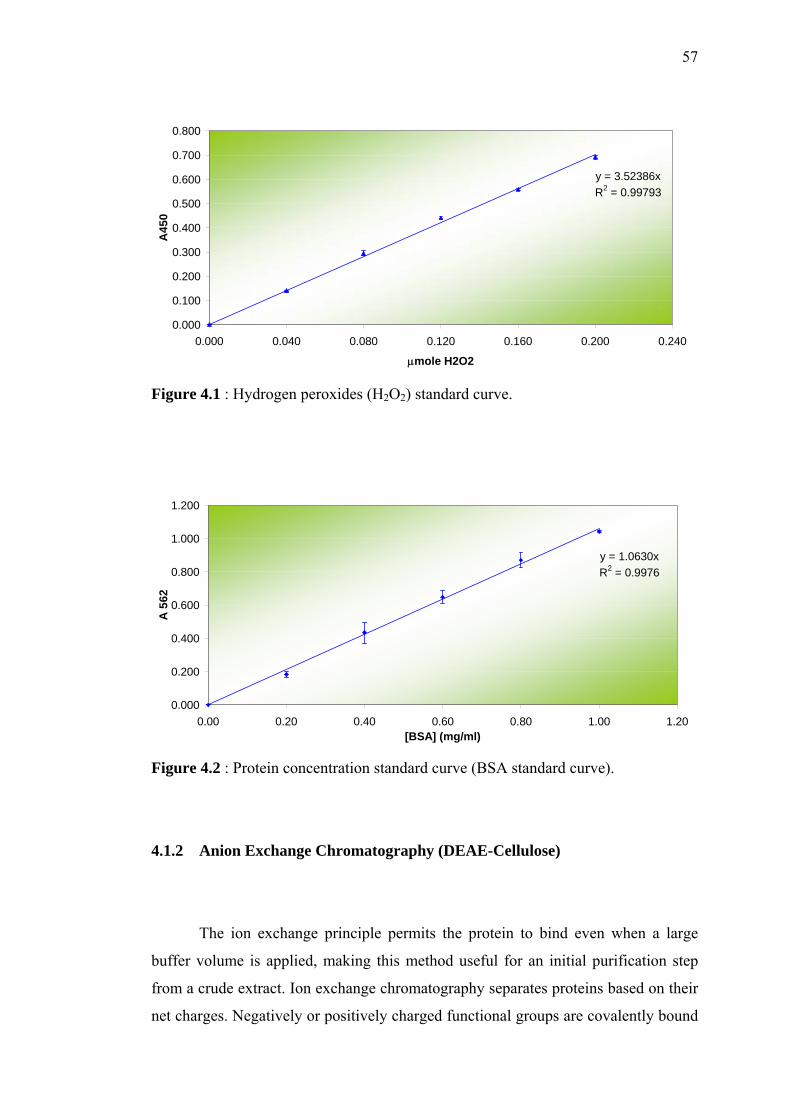
57
y = 3.52386xR2 = 0.99793
0.000
0.100
0.200
0.300
0.400
0.500
0.600
0.700
0.800
0.000 0.040 0.080 0.120 0.160 0.200 0.240
µmole H2O2
A45
0
Figure 4.1 : Hydrogen peroxides (H2O2) standard curve.
y = 1.0630xR2 = 0.9976
0.000
0.200
0.400
0.600
0.800
1.000
1.200
0.00 0.20 0.40 0.60 0.80 1.00 1.20[BSA] (mg/ml)
A 5
62
Figure 4.2 : Protein concentration standard curve (BSA standard curve).
4.1.2 Anion Exchange Chromatography (DEAE-Cellulose)
The ion exchange principle permits the protein to bind even when a large
buffer volume is applied, making this method useful for an initial purification step
from a crude extract. Ion exchange chromatography separates proteins based on their
net charges. Negatively or positively charged functional groups are covalently bound
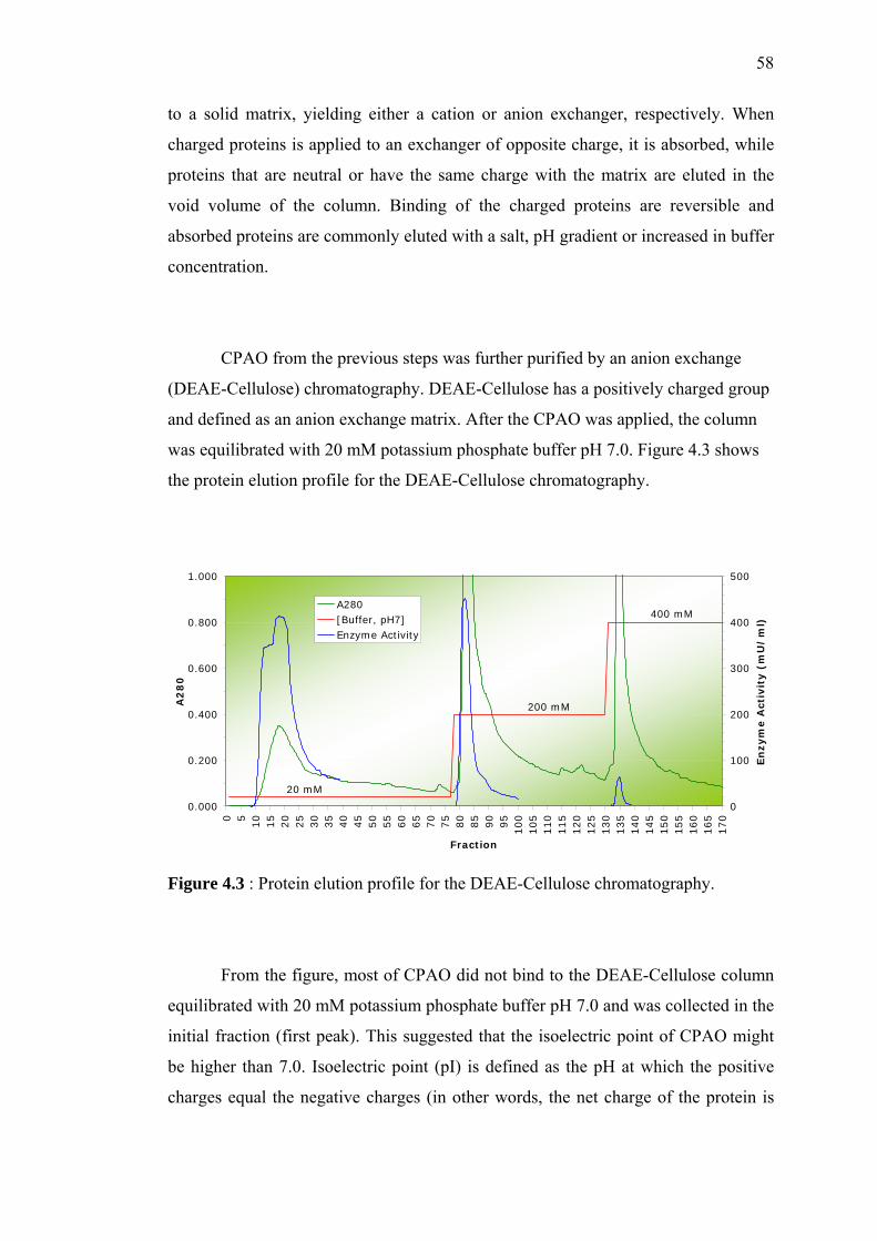
58
to a solid matrix, yielding either a cation or anion exchanger, respectively. When
charged proteins is applied to an exchanger of opposite charge, it is absorbed, while
proteins that are neutral or have the same charge with the matrix are eluted in the
void volume of the column. Binding of the charged proteins are reversible and
absorbed proteins are commonly eluted with a salt, pH gradient or increased in buffer
concentration.
CPAO from the previous steps was further purified by an anion exchange
(DEAE-Cellulose) chromatography. DEAE-Cellulose has a positively charged group
and defined as an anion exchange matrix. After the CPAO was applied, the column
was equilibrated with 20 mM potassium phosphate buffer pH 7.0. Figure 4.3 shows
the protein elution profile for the DEAE-Cellulose chromatography.
Figure 4.3 : Protein elution profile for the DEAE-Cellulose chromatography.
From the figure, most of CPAO did not bind to the DEAE-Cellulose column
equilibrated with 20 mM potassium phosphate buffer pH 7.0 and was collected in the
initial fraction (first peak). This suggested that the isoelectric point of CPAO might
be higher than 7.0. Isoelectric point (pI) is defined as the pH at which the positive
charges equal the negative charges (in other words, the net charge of the protein is
0.000
0.200
0.400
0.600
0.800
1.000
0 510
15
20
25
30
35
40
45
50
55
60
65
70
75
80
85
90
95
100
105
110
115
120
125
130
135
140
145
150
155
160
165
170
Fraction
A2
80
0
100
200
300
400
500
En
zym
e A
ctiv
ity (
mU
/m
l)
A280[Buffer, pH7]Enzyme Activity
20 mM
200 mM
400 mM
59
zero). At a pH above its pI, the protein of interest will be negatively charged, and at a
pH below its pI, the protein will be positively charged. However, some CPAO were
also present in the second and third peak, after the column was eluted with the 200
mM and 400 mM potassium phosphate buffer of the same pH, respectively. Its total
activity is lower than in the initial fraction/peak. Therefore, only the active fractions
from the first peak were pooled and prepared for further purification.
4.1.3 Hydroxyapatite Chromatography (Bio-Gel HTP)
Hydroxyapatite, (Ca5(PO4)3OH)2 is a crystalline form of calcium phosphate
that is widely used in preparative biochemistry, having proven itself a unique tool for
the fractionation and purification of monoclonal antibodies and other proteins,
enzymes and nucleic acids. Hydroxyapatite is useful for preparative work in column
or batch modes, and for quantitative analysis of proteins or nucleic acids.
Hydroxyapatite chromatography can be utilized at any stage in a process from initial
capture to final polishing. Hydroxyapatite has unique separation properties and
unparalleled selectivity and resolution. It often separates proteins that appear to be
homogeneous by other chromatographic and electrophoretic techniques.
Hydroxyapatite has unique selectivity. Since molecular separation on
hydroxyapatite is not primarily dependent on molecular weight, molecular size,
charge density or isoelectric point, hydroxyapatite chromatography is a valuable
complement to other separations techniques. Hydroxyapatite has a high capacity for
nucleic acids and proteins. Its surface area is about 50 m2. Non-specific adsorption of
hydrophobic substances is minimized by the inorganic crystalline matrix of
hydroxyapatite. Hydroxyapatite displays negligible adsorptive capacity for low
molecular weight substances such as mononucleotides, salts and amino acids.
Hydroxyapatite is chemically and thermally stable. It is compatible with wide range
60
of aqueous and organic solvents and can be sanitized in sodium hydroxide. It has a
pH tolerance greater than 5.5 and is autoclavable.
The active fractions from the DEAE-Cellulose column was loaded directly
onto the hydroxyapatite column, equilibrated with 20 mM potassium phosphate
buffer pH 7.0 and the CPAO was eluted with 200 mM potassium phosphate buffer
pH 7.0. The active fractions were pooled, dialyzed overnight against 20 mM
potassium phosphate buffer pH 7.0 and concentrated using ultrafiltration. Figure 4.4
shows the protein elution profile for the hydroxyapatite (Bio-Gel HTP)
chromatography.
Figure 4.4 : Protein elution profile for the hydroxyapatite (Bio-Gel HTP)
chromatography.
4.1.4 Size-Exclusion Chromatography (Sephacryl S-300 HR)
The remaining impurities, mainly proteins of lower molecular weight, were
removed in the final polishing step using size-exclusion (Sephacryl S-300 HR)
chromatography. The protein elution profile for the Sephacryl S-300 HR
0.000
0.100
0.200
0.300
0.400
0.500
0.600
0.700
0.800
0 5
10
15
20
25
30
35
40
45
50
55
60
65
70
75
80
85
90
Fraction
A2
80
0
100
200
300
400
500
600
700
800
En
zym
e A
ctiv
ity (
mU
/m
l)
A280
[Buffer, pH7]
Enzyme Activity
20 mM
200 mM
61
chromatography is shown in Figure 4.5. The overall purification results of chick pea
diamine oxidase are shown in Table 4.1 and 4.2.
The final preparation of CPAO had a specific activity of 12,702 mU/mg
(putrescine as a substrate), 11,695 mU/mg (cadaverine as a substrate) and 452
mU/mg (histamine as substrate). The final purification fold was 53.4, 66.1 and 35.0-
fold for putrescine, cadaverine and histamine as substrate, respectively. However, the
final recovery (yield) of CPAO for each substrates was very low, less than 20% of
the total CPAO activity in the crude extract.
Figure 4.5 : Protein elution profile for Sephacryl S-300 HR chromatography
0.000
0.010
0.020
0.030
0.040
0.050
0.060
0 5
10
15
20
25
30
35
40
45
50
55
60
65
Fraction
A2
80
0
100
200
300
400
500
600
En
zym
e A
ctiv
ity
(m
U/
ml)
A280
[Buffer, pH7]
Enzyme Activity
20 mM
62
Table 4.1 : The purification of chick pea diamine oxidase (CPAO).
CPAO Activity (mU/ml) Total CPAO Activity (mU) Total CPAO Recovery (%) Steps
Volume
(ml) Putrescine Cadaverine Histamine Putrescine Cadaverine Histamine Putrescine Cadaverine Histamine
Crude Extract 100.0 3,315 2,468 180 331,467 246,816 18,003 100% 100% 100%
Ammonium sulfate
Precipitation (30%),
Supernatant
98.0 3,081 2,393 180 301,970 234,481 17,643 91% 95% 98%
Ammonium sulfate
Precipitation (70%),
Precipitate, Dialysis
40.0 6,802 4,803 331 272,090 192,110 13,241 82% 78% 74%
Heat Treatment, Dialysis 42.0 4,648 3,540 247 195,196 148,665 10,366 59% 60% 58%
Anion Exchange
Chromatography
(DEAE-Cellulose)
38.0 2,606 1,727 75 99,040 65,642 2,858 30% 27% 16%
Hydroxyapatite
Chromatography
(Bio-Gel HTP)
6.2 10,689 10,411 438 66,271 64,550 2,718 20% 26% 15%
Size-Exclusion
Chromatography
(Sephacryl S-300 HR)
18.3 2,426 2,234 86 44,373 40,857 1,577 13% 17% 9%
Note : 1000 mU = 16.67 nkat
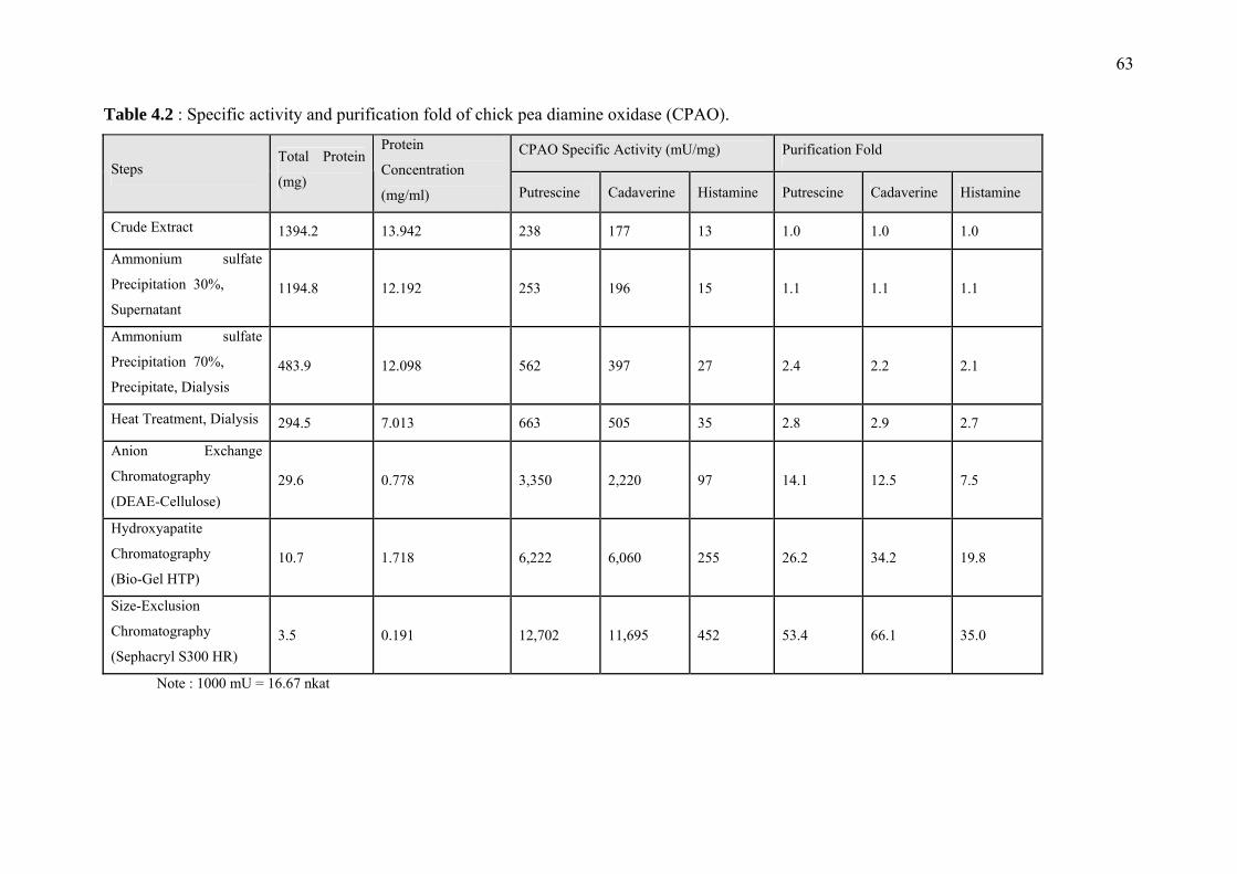
63
Table 4.2 : Specific activity and purification fold of chick pea diamine oxidase (CPAO).
CPAO Specific Activity (mU/mg) Purification Fold Steps
Total Protein
(mg)
Protein
Concentration
(mg/ml) Putrescine Cadaverine Histamine Putrescine Cadaverine Histamine
Crude Extract 1394.2 13.942 238 177 13 1.0 1.0 1.0
Ammonium sulfate
Precipitation 30%,
Supernatant 1194.8 12.192 253 196 15 1.1 1.1 1.1
Ammonium sulfate
Precipitation 70%,
Precipitate, Dialysis 483.9 12.098 562 397 27 2.4 2.2 2.1
Heat Treatment, Dialysis 294.5 7.013 663 505 35 2.8 2.9 2.7
Anion Exchange
Chromatography
(DEAE-Cellulose) 29.6 0.778 3,350 2,220 97 14.1 12.5 7.5
Hydroxyapatite
Chromatography
(Bio-Gel HTP) 10.7 1.718 6,222 6,060 255 26.2 34.2 19.8
Size-Exclusion
Chromatography
(Sephacryl S300 HR) 3.5 0.191 12,702 11,695 452 53.4 66.1 35.0
Note : 1000 mU = 16.67 nkat
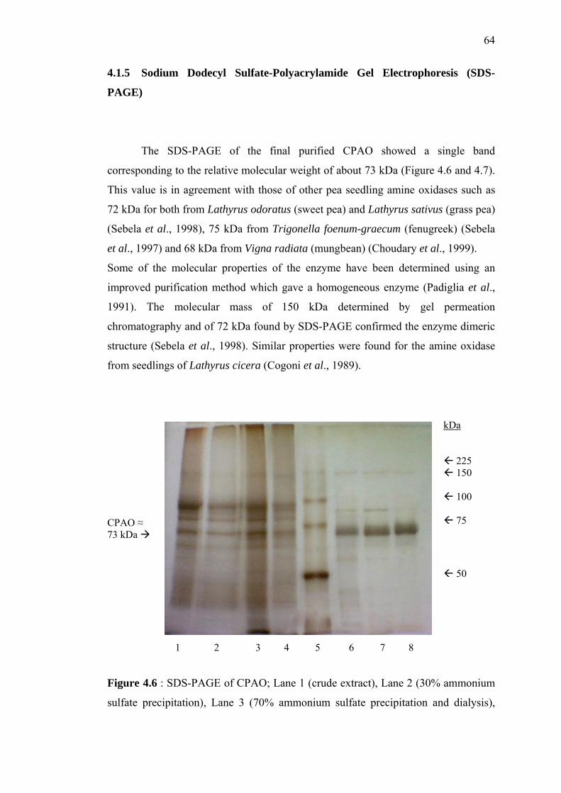
64
4.1.5 Sodium Dodecyl Sulfate-Polyacrylamide Gel Electrophoresis (SDS-
PAGE)
The SDS-PAGE of the final purified CPAO showed a single band
corresponding to the relative molecular weight of about 73 kDa (Figure 4.6 and 4.7).
This value is in agreement with those of other pea seedling amine oxidases such as
72 kDa for both from Lathyrus odoratus (sweet pea) and Lathyrus sativus (grass pea)
(Sebela et al., 1998), 75 kDa from Trigonella foenum-graecum (fenugreek) (Sebela
et al., 1997) and 68 kDa from Vigna radiata (mungbean) (Choudary et al., 1999).
Some of the molecular properties of the enzyme have been determined using an
improved purification method which gave a homogeneous enzyme (Padiglia et al.,
1991). The molecular mass of 150 kDa determined by gel permeation
chromatography and of 72 kDa found by SDS-PAGE confirmed the enzyme dimeric
structure (Sebela et al., 1998). Similar properties were found for the amine oxidase
from seedlings of Lathyrus cicera (Cogoni et al., 1989).
Figure 4.6 : SDS-PAGE of CPAO; Lane 1 (crude extract), Lane 2 (30% ammonium
sulfate precipitation), Lane 3 (70% ammonium sulfate precipitation and dialysis),
CPAO ≈ 73 kDa
kDa
225 150
100
75
50
1 2 3 4 5 6 7 8
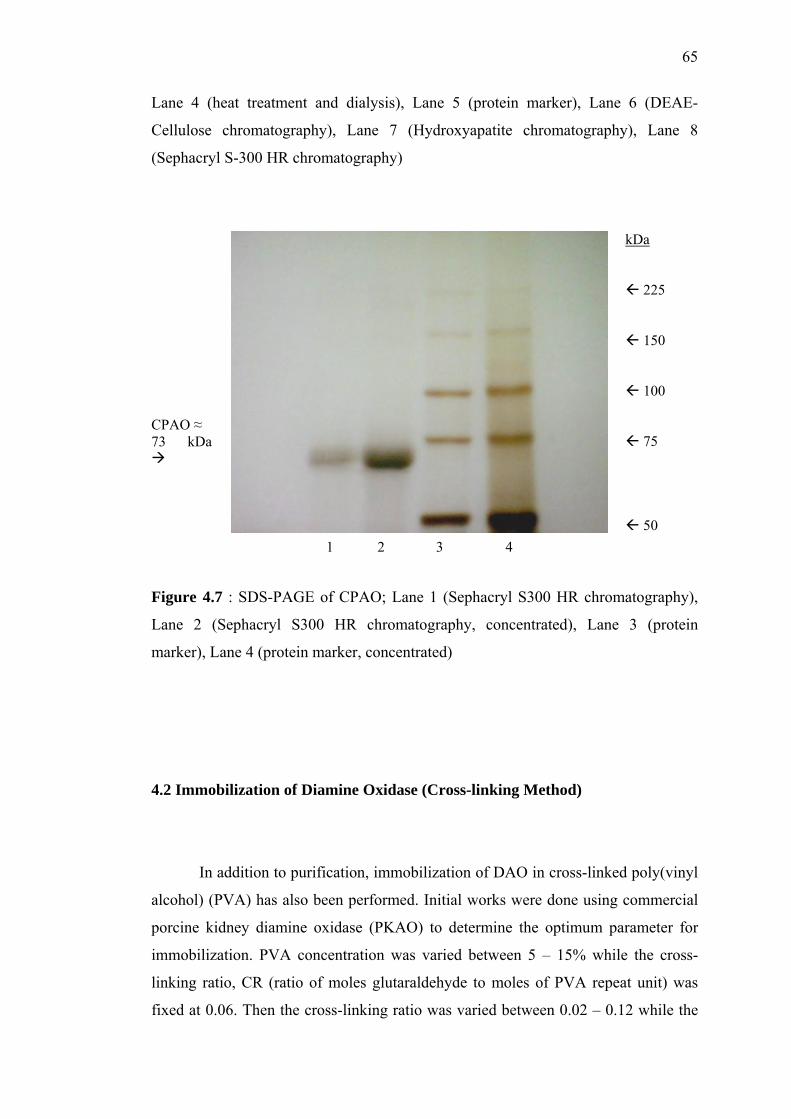
65
Lane 4 (heat treatment and dialysis), Lane 5 (protein marker), Lane 6 (DEAE-
Cellulose chromatography), Lane 7 (Hydroxyapatite chromatography), Lane 8
(Sephacryl S-300 HR chromatography)
CPAO ≈ 73 kDa
kDa
225
150
100
75
50 1 2 3 4
Figure 4.7 : SDS-PAGE of CPAO; Lane 1 (Sephacryl S300 HR chromatography),
Lane 2 (Sephacryl S300 HR chromatography, concentrated), Lane 3 (protein
marker), Lane 4 (protein marker, concentrated)
4.2 Immobilization of Diamine Oxidase (Cross-linking Method)
In addition to purification, immobilization of DAO in cross-linked poly(vinyl
alcohol) (PVA) has also been performed. Initial works were done using commercial
porcine kidney diamine oxidase (PKAO) to determine the optimum parameter for
immobilization. PVA concentration was varied between 5 – 15% while the cross-
linking ratio, CR (ratio of moles glutaraldehyde to moles of PVA repeat unit) was
fixed at 0.06. Then the cross-linking ratio was varied between 0.02 – 0.12 while the
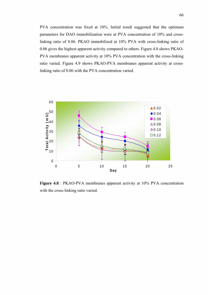
66
PVA concentration was fixed at 10%. Initial result suggested that the optimum
parameters for DAO immobilization were at PVA concentration of 10% and cross-
linking ratio of 0.06. PKAO immobilized at 10% PVA with cross-linking ratio of
0.06 gives the highest apparent activity compared to others. Figure 4.8 shows PKAO-
PVA membranes apparent activity at 10% PVA concentration with the cross-linking
ratio varied. Figure 4.9 shows PKAO-PVA membranes apparent activity at cross-
linking ratio of 0.06 with the PVA concentration varied.
Figure 4.8 : PKAO-PVA membranes apparent activity at 10% PVA concentration
with the cross-linking ratio varied.
0
10
20
30
40
50
60
0 5 10 15 20 25Day
To
tal
Act
ivit
y (
mU
)
0.020.040.060.080.100.12
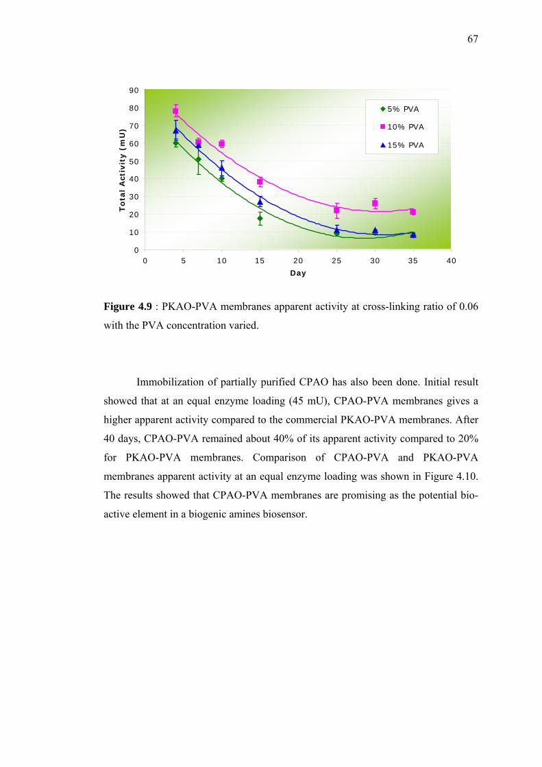
67
Figure 4.9 : PKAO-PVA membranes apparent activity at cross-linking ratio of 0.06
with the PVA concentration varied.
Immobilization of partially purified CPAO has also been done. Initial result
showed that at an equal enzyme loading (45 mU), CPAO-PVA membranes gives a
higher apparent activity compared to the commercial PKAO-PVA membranes. After
40 days, CPAO-PVA remained about 40% of its apparent activity compared to 20%
for PKAO-PVA membranes. Comparison of CPAO-PVA and PKAO-PVA
membranes apparent activity at an equal enzyme loading was shown in Figure 4.10.
The results showed that CPAO-PVA membranes are promising as the potential bio-
active element in a biogenic amines biosensor.
0
10
20
30
40
50
60
70
80
90
0 5 10 15 20 25 30 35 40
Day
To
tal
Act
ivit
y (
mU
)
5% PVA
10% PVA
15% PVA
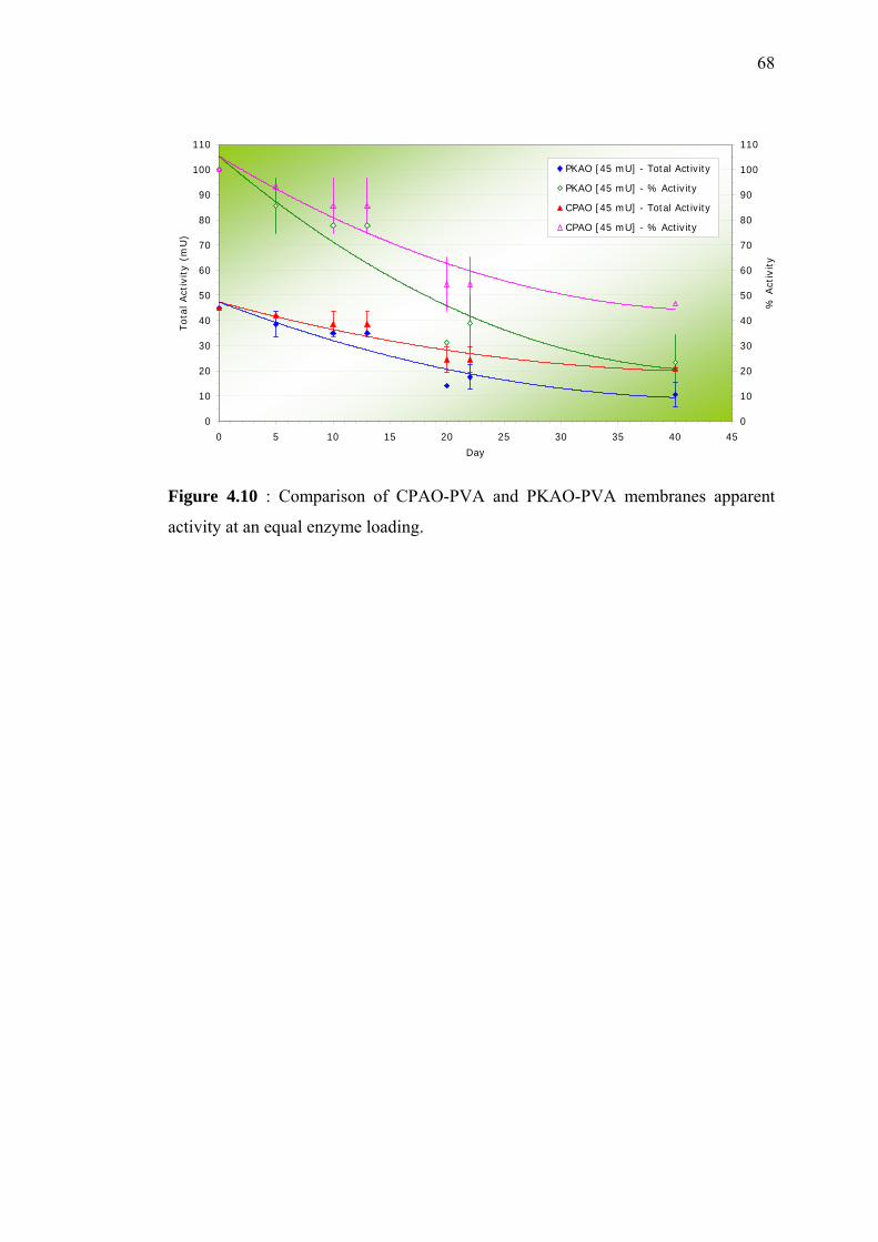
68
Figure 4.10 : Comparison of CPAO-PVA and PKAO-PVA membranes apparent
activity at an equal enzyme loading.
0
10
20
30
40
50
60
70
80
90
100
110
0 5 10 15 20 25 30 35 40 45
Day
Tot
al Act
ivity
(mU
)
0
10
20
30
40
50
60
70
80
90
100
110
% A
ctiv
ity
PKAO [45 mU] - Total Activity
PKAO [45 mU] - % Activity
CPAO [45 mU] - Total Activity
CPAO [45 mU] - % Activity
69
CHAPTER 5
SUMMARY AND RECOMMENDATION
5.1 Summary
Pure amine oxidase has been successfully isolated from the Cicer arietinum
seedlings, following three chromatographic steps. The molecular weight of the
purified chick pea amine oxidase (CPAO) was 73 kDa, as shown by a single band
determined by SDS-PAGE. The pure CPAO gave a specific activity of 12.7, 11.7 and
0.45 U/mg with putrescine, cadaverine and histamine as substrate, respectively. The
purification fold was about 53-fold for putrescine as a substrate, 66-fold for
cadaverine and 35-fold for histamine. Although the pure CPAO has been
successfully obtained, the total recoveries of the enzymes were quite low, which
were about 13%, 17% and 9% with putrescine, cadaverine and histamine as
substrate, respectively. The substrate selectivity relative to putrescine (assuming
100% for putrescine) was 92% for cadaverine and 4% for histamine.
The optimum parameters for CPAO immobilization were PVA concentration
of 10% and cross-linking ratio of 0.06, determined earlier by the immobilization of
PKAO in cross-linked poly (vinyl alcohol) (PVA). Result for immobilized partially
purified CPAO showed that at an equal enzyme loading, CPAO-PVA membranes
70
gave higher apparent activities compared to the commercial PKAO-PVA
membranes. CPAO-PVA membranes retained about 40% of its apparent activity
compared to 20% for PKAO-PVA membranes, after 40 days of storage.
5.2 Recommendations
Although pure CPAO had been successfully isolated from the chick pea
seedlings, however enzyme recovery was still very low. The type of anion exchange
chromatography used in this research resulted in a significant loss of the total
enzymes (recovery of only about 30-50%, while typical recovery is 70-90%). Thus,
it is suggested that other type of anion exchange matrix available in the market is
used. Other types of chromatography system can also be used, such as absorption
chromatography.
The success of an enzymatic biosensor partly depends on the enzyme
immobilization technique. Poor technique will result in significant loss of enzyme
activity and short operational life. Since the reusability and the recovery of the free
enzymes are limited, immobilization of enzymes is very attractive. The advantages of
immobilized enzymes are that they can be used either in batch or continuous
systems, they can be removed easily from the reaction medium and they can be used
for the controlled production. Immobilized enzyme systems do also have limitations,
such as loss of activity due to the immobilization technique and decrease in the
apparent activity due to mass transfer resistance towards the substrate and product.
An optimal support material should provide large surface area per unit volume (or
mass) of the carrier for effective immobilization of the desired amount of enzyme
and should allow substrate and product transport with the least diffusional resistance.
The immobilization technique should also lead to high immobilization and activity
yields.
71
In this research, CPAO was immobilized by entrapment and cross-linking of
the enzyme in a poly (vinyl alcohol) (PVA) membrane, cross-linked with
glutaraldehyde. The disadvantages of using the cross-linking approach are there is
some diffusion limitation and there can be damage to the biomaterial. Diffusion of
solutes is determined by the cross-linking density of the membrane, or mesh size and
the degrees of swelling. The `pores' are the open spaces between the cross-link points
and are not fixed. Alternatively, a physically cross-linked PVA through freeze-
thawing method may be a good choice for immobilizing the enzymes. It can
minimize the problems associate with chemically cross-linked PVA while
maintaining the good properties of PVA. The exposition of aqueous PVA solution to
several freeze-thawing cycles leads to reinforced gels. Fine crystallites are formed
after the freeze-thawing process, due to the slow heat treatment.
Addition of protein stabilizing agent, such as bovine serum albumin (BSA)
into the PVA mixture for immobilization might be very helpful to increase the
membranes performance. With the existence of BSA, less enzymes leak out from the
membranes, thus more enzymes can be retained inside the membranes. An
appropriate amount of BSA must be calculated, because too much BSA will not
necessarily correlate well with enzymes activity. Too much BSA might block the
active sites of the enzymes, thus it becomes hard for the substrate to diffuse into the
membranes, resulting in low reaction rates. The thickness of the membranes must be
well controlled. The thickness of the membranes affects the diffusion rate and
permeability. The thicker the membranes, the longer the response time is. So, the
possibility of casting thinner enzymes membranes that can still retain the sensitivity
to the substrate should be examined.
REFERENCES
Angeline, S., Di Lisi, F. and Federico, R. (1985). Immunoaffinity Purification and
Characterization of Diamine Oxidase from Cicer. Phytochemistry. 24. 2511-
2513.
Azila Abdul Aziz (2001). Amperometric Glucose Biosensor: Systematic Material
Selection and Quantitative Analysis of Performance. The John Hopkins
University: Thesis of Doctor of Philosophy.
Bardocz, S. (1995). Trends in Food Science and Technology. 6. 341.
Bollag, D. M. (1996). Protein Method. 2nd Edition. United States of America: John
Wiley & Sons Inc.
Buerk, D. G. (1993). Biosensor: Theory and Application. First Edition. United State
of America: Technomic Publishing Company, Inc.
Bulmus, V., Kesenci, K. and Piskin, E. (1998). Ply(EGDMA/AAm) Copolymer
Beads: A Novel Carrier for Enzyme Immobilization. Reactive Functional
Polymers. 38. 1-9.
Cai, D. and Klinman, J. P. (1994). Evidence for a Self-catalytic Mechanism of 2,4,5-
trihydroxyphenylalanine Quinone Biogenesis in Yeast Copper Amine Oxidase.
J. Biol. Chem. 269. 32039-32042.
73
Choudhary, A., Singh, I. and Sigh, R. P. (1999). A thermostable Diamine Oxidase
from Vigna radiata Seedlings. Phytochemistry. 52. 1-5.
Cogoni, A., Farci, R., Medda, R., Rinaldi, A. and Floris, G. (1989). Amine Oxidase
from Lathyrus cicera and Phaseolus vulgari: Purification and Properties. Prep.
Biochem. 19. 95-112.
Dooley, D. M., McGuirl, M., Brown, D. E., Turowski, P., McIntire, W. S. and
Knowles, P. F. (1991). A Cu(I)-semiquinone State in Substrate-reduced Amine
Oxidases. Nature. 349. 262-264.
Eggins, B. R. (1996). Biosensors: An Introduction. First Edition. New york, United
States of America: John Wiley & Sons Ltd and B.G. Teubner.
Frebort, I., Skoupa, L. and Pec, P. (2000). Amine Oxidase-Based Flow Biosensor
for the Assessment of Fish Freshness. Food Control. 11. 13-18.
Harborne, J. B. (1973). Phytochemical Methods: A Guide to Modern Techniques of
Plant Analysis. Singapore: Chapman and Hall Ltd. 177-178.
Hughes, J. M. and Potter, M. E. (1991). Scombroid-fish Poisoning: From
Pathogenesis to Prevention. 324(11). 766-8.
Janes, S. M., Mu, D., Wemmer, D., Smith, A. J., Kaur, S., Maltby, D., Burlingame,
A. L. and Klinman, J. P. (1990). A New Redox Cofactor in Eukaryotic
Enzymes: 6-Hydroxydopa at the Active Site of Bovine Serum Amine Oxidase.
Science. 248. 981-987.
Janes, S. M., Palcic, M. M., Scaman, C. H., Smith, A. J., Brown, D. E., Dooley, D.
M., Mure, M. and Klinman, J. P. (1992). Identification of Topa Quinone and Its
Consensus Sequence in Copper Amine Oxidases. Biochemistry. 31. 12147-
12154.
Kumar, V., Dooley, D. M., Freeman, H. C., Guss, J. M., Harvey, I., McGuirl, M. A.,
Wilce, M. C. J. and Zubak, V. M. (1996). Crystal Structure of a Eukaryotic (Pea
74
Seedling) Copper-containing Amine Oxidase at 2.2 Å Resolution. Structure. 4.
943-955.
Lange, J. and Wittmann, C. (2001). Enzyme Sensor Array for the Determination of
Biogenic Amines in Food Samples. Analytical and Bioanalytical Chemistry.
Leuschner, R.G., Heidel, M. and Hammes, W. P. (1998). International Journal of
Food Microbiology. 39.
Matsuzaki, R., Suzuki, S., Yamaguchi, K., Fukui, T. and Tanizawa, K. (1995).
Spectroscopic Studies on the Mechanism of the Topa Quinone Generation in
Bacterial Monamine Oxidase. Biochemistry. 34. 4524-4530.
Medda, R., Padiglia, A. and Floris, G. (1995). Plant Copper amine Oxidases.
Phytochemistry. 39. 1-9.
Mohd Nasir Azudin and Nazamid Saari (1988). Histamine in fish Products in
Malaysia. Proc. Sem. Adv. Food Res. Malaysia – II. Malaysia: Universiti
Pertanian Malaysia.
Mu, D., Medzihradsky, K. F., Adams, G. W., Mayer, P., Hines, W. M., Burlingame,
A. J., Smith, A. J., Cai, D. and Klinman, J. P. (1994). Primary Structure for
Mammalian Cellular and serum copper Amine Oxidase. J. Biol, Chem. 296.
9926-9932.
Niculescu, M., Nistor, C., Ruzgas, t., Frebort, I., Sebela, M., Pec, P. and Csoregi, e.
(2001). Detection of Histamine and Other Biogenic Amines Using Biosensors
Based on Amine Oxidase. Inflamm. res. 50. S146-S148.
Padiglia, A., Cogoni, A. and Floris, G. (1991). Characterization of Amine Oxidases
from Pisum, Lens, Lathyrus and Cicer. Phytochemistry. 30. 3895-3897.
Parsons, M. R., Convery, M. A., Wilmot, C. M., Yadav, K. D. S., Blakeley, V.,
Corner, A. S., Philips, S. E. V., McPherson, M. J. and Knowels, P. F. (1995).
75
Crystal Structure of a Quinoenzyme: Copper Amine Oxidase of E. coli at 2Å
Resolution. Structure. 3. 1171-1184.
Public Health Division (2002). Investigation of Biogenic Amines in fish and Fish
Products. First Edition. State of Victoria: Victorian Government Department of
Human Services.
Suresh, M. R., Ramakrishna, S. and Adiga, P. R. (1976). Diamine Oxidase of
Lathyrus sativus Seddlings. Phytochemistry. 15. 483-485.
Rossi, A., Petruzelli, R. and Finazzi-Agro, A. (1992). cDNA Derived Amino Acid
Sequence of Lentil Seedlings Amine Oxidase. FEBS Lett. 301. 253-257.
Tipping, A. J. and McPherson, M. J. (1995). Cloning and Molecular Analysis of the
Pea Seedling Copper Amine Oxidase. J. Biol. Chem. 270. 16939-16946.
Tombelli, S. and Mascini, M. (1998). Electrochemical Biosensors for Biogenic
Amines: A Comparison between Different Approaches. Analytica Chimica Acta.
358. 277-284.
Turowski, P. N., McGuirl, M. A., and Dooley, D. M. (1993). Intra Molecular
Electron Transfer Rate between Actice-site Copper and Topa Quinone in Pea
Seedling Amine Oxidase. J. Biol. Chem. 268. 17680-17682.
Wagner, G. and Guilbault, G. (1994). Food Biosensor Analysis. First Edition. New
York.
Wilflingseder, D. and Schwelberger, G. (2000). High Efficient Purification of
Porcine Diamine Oxidase. Journal of Chromatography. 737. 161-166.
Wimmerova. M. and Macholan, L. (1999) Sensitive Amperometric Biosensor for the
Determination of Biogenic and Synthetic Amines Using Pea Seedlings Amine
Oxidase: A Novel Approach for Enzyme Immobilization. Biosensor and
Bioelectronics. 14. 695-702.

76
Wu, M. L., Yang, C. C., Yang, G. Y., Ger, J. and Deng, J. F. (1997). Vet Human
Toxicol. 39. 236.