Embed Size (px)
Citation preview

Development of a covalent site-specific antibody labeling strategy by the use of
photoactivable Z domains
Anna Konrad
Royal Institute of Technology
School of Biotechnology
Stockholm 2012

© Anna Konrad Stockholm 2012
Royal Institute of Technology School of Biotechnology AlbaNova University Center SE-106 91 StockholmSweden
Printed by E-print Oxtorgsgatan 9-11 SE-111 57 Stockholm Sweden
ISBN 978-91-7501-329-9 TRITA-BIO Report 2012:9 ISSN 1654-2312

Anna Konrad (2012): Development of a covalent site-specific antibody labeling strategy by the use of photoactivable Z domains. School of Biotechnology, Royal Institute of Technology (KTH), Sweden ISBN 978-91-7501-329-9
Abstract
The joining of two molecular functions or the strategy of adding functions to proteins has been
tremendously important for the development of proteins as tools in research and clinic.
Depending on the intended application, there are a wide variety of functions that can be added
to a proteins. In clinical applications drugs are a commonly conjugated to antibodies and in
research adding reporter groups such as biotin, enzymes or fluorophores is a routine procedure.
The chemistries and methods most often used suffer from drawbacks such as lack of stringency,
which could lead to undesired effects on the protein. Many site-specific methods of labeling of
antibodies require modification or insertion of handles in the antibody recombinantly, before
labeling can be performed.
The core of this thesis is the development of a strategy for covalent specific labeling of
antibodies by exploiting the site specific binding of the Z domain to Protein A. Photoreactive Z-
domains were produced by solid phase peptide synthesis, which provides the opportunity to
insert a photoreactive amino acid and a reporter biotin at specific positions in the domain. The
inherited binding to the Fc-part of the antibody in combination with the incorporated
photoreactive amino acid, BPA, is used for site-specific interaction, and thereafter, covalent
coupling to the antibody. The exposure with the appropriate wavelength of light enables the
formation a covalent linkage between the Z domain and the antibody. The biotinylated
photoactivable domains were subsequently used to site-specifically label a number of different
types of antibodies, polyclonal rabbit IgG, monoclonal human IgG1 and monoclonal mouse
IgG2a, and thereafter the antibodies was employed in a variation of applications. The
photolabeling procedure of antibodies by the use of photoactivable Z domains has proven to be
successful and could serve as a valuable tool in several applications.
Keywords: antibody labeling, site-specific, covalent, SPPS, Z domain , BPA © Anna Konrad

ListofPublications
This thesis is based upon the following three papers, which are included in the appendix.
I. Anna Konrad, Amelie Eriksson Karlström and Sophia Hober.
Covalent Immunoglobulin labeling through a photoactivable synthetic Z domain.
Bioconjug Chem. 2011 Dec 21;22(12):2395-403.
II. Sandra Andersson, Anna Konrad, Nikhil Ashok, Fredrik Pontén, Sophia Hober and Anna
Asplund.
Antibodies biotinylated using a synthetic Z-domain provide stringent in situ protein
detection. Manuscript.
III. Anna Konrad, Josefin Anfelt, Sandra Andersson, Amelie Eriksson Karlström and Sophia
Hober. Optimization of an antibody labeling strategy through the use of a photoactivable
synthetic Z domains. Manuscript.

RelatedPublications
Vernet E, Konrad A, Lundberg E, Nygren PA, Gräslund T. Affinity-based entrapment of the
HER2 receptor in the endoplasmic reticulum using an affibody molecule. J Immunol Methods.
2008 Sep 30;338(1-2):1-6
Tegel H, Steen J, Konrad A, Nikdin H, Pettersson K, Stenvall M, Tourle S, Wrethagen U, Xu L,
Yderland L, Uhlén M, Hober S, Ottosson J. High-throughput protein production--lessons from
scaling up from 10 to 288 recombinant proteins per week. Biotechnol J. 2009 Jan;4(1):51-7.

Contents ABBREVIATIONS......................................................................................................................... 1
INTRODUCTION........................................................................................................................... 3
ANTIBODIES ................................................................................................................................ 7 STRUCTURE ................................................................................................................................ 7 POLYCLONAL, MONOCLONAL OR RECOMBINANT ANTIBODIES ........................................................ 9
PROTEIN A................................................................................................................................. 11 Z DOMAIN .................................................................................................................................. 12
MODIFICATION OF PROTEINS................................................................................................. 15 INCORPORATION OF UNNATURAL AMINO ACIDS INTO PROTEINS .................................................... 16 BENZOYLPHENYLALANINE – BPA – A PHOTO REACTIVE AMINO ACID............................................. 18
PRESENT INVESTIGATION ...................................................................................................... 21 COVALENT IMMUNOGLOBULIN LABELING THROUGH A PHOTOACTIVABLE SYNTHETIC Z
DOMAIN (PAPER I) ..................................................................................................................... 23 ANTIBODIES BIOTINYLATED USING A SYNTHETIC Z-DOMAIN PROVIDE STRINGENT IN SITU
PROTEIN DETECTION (PAPER II) ................................................................................................. 24 OPTIMIZATION OF AN ANTIBODY LABELING STRATEGY THROUGH THE USE OF
PHOTOACTIVABLE SYNTHETIC Z DOMAINS (PAPER III).................................................................. 25 CONCLUDING REMARKS ............................................................................................................. 26
ACKNOWLEDGEMENTS........................................................................................................... 27
REFERENCES............................................................................................................................ 31


1
Abbreviations
ADC – antibody drug conjugate
Boc – t-butyloxycarbonyl
BP – benzophenone
BPA – benzoylphenylalanine
CDR – complementarity determining region
DNA – deoxyribonucleic acid
E. coli – Escherichia coli
Fab – fragment antigen binding
Fc – fragment crystallizable
Fmoc – fluorenylmethyloxycarbonyl
HSA – human serum albumin
IgG – immunoglobulin G
IHC – immunohistochemistry
mRNA – messenger ribonucleic acid
scFv – single chain fragment variable
SPA – staphylococcal protein A
SPPS – solid phase peptide synthesis
SPR – surface plasmon resonance
tRNA – transfer ribonucleic acid

2

3
Introduction
Everyday around the world a vast number of researchers and hospital personnel strive to
answer questions regarding disease with the help of different methods. The dependence and
need for reliable tools in research and medicine are great and a considerable contribution has
been made through the technical revolution within the field of biotechnology. Any progress made
within method development increases the prospect of understanding disease and biology better
and serves the common good.
Timelines describing the milestones of Modern Biotechnology often start with the term
biotechnology being coined by the Hungarian engineer Karl Ereky in 1917, defining it as ”all
lines of work by which products are produced from raw material with the aid of living things” (1).
However it was in the 1960s that a microbiologist Carl Göran Hedén introduced the term in a
sense that is regarded as modern biotechnology, meaning that it stands on the ground of the
scientific fields of microbiology, biochemistry and chemical engineering (1). In a review from
2002 Rita Colwell reflects over the history of biotechnology and the time after the
deoxyribonucleic acid (DNA) structure being solved in the 1950s: “The field of biotechnology has
progressed from the world of science fiction to the world of science present”, meaning that
during the later part of the twentieth century the field progressed tremendously (2). In fact in the
mid 1970s the research community felt that the technical advances were faster than the ability to
make ethical considerations regarding what the technique could lead to. Therefore they decided
to temporarily put all genetic engineering on hold. This voluntary moratorium was held until
laboratory guidelines were established at the Asilomar Conference in 1975 (3, 4). A few
highlights in the history of biotechnology are discoveries that today are taken for granted and
used in laboratories everyday worldwide, a few of these will be mentioned in short.
The understanding of genetic material and structure of the DNA molecule was a significant step
in history. Avery and coworkers suggested that DNA was the factor responsible for making
bacteria transform (5) and, inspired by Rosalind Franklin (6, 7), Watson and Crick published the

4
description of the helical structure of the DNA-molecule (8). Gaining knowledge of genetic
material led to the discovery of diseases that are result of a mutation at the genetic level, sickle
cell anemia being one example, which was shown by Pauling in 1949 (9). Further discoveries in
the field, for example the replication process of DNA (10-12) and the isolation of DNA modifying
enzymes, such as DNA polymerase and restriction enzymes (13-15), made it possible to
perform genetic engineering. This was indeed important for Boyer and Cohen, who
demonstrated the first recombinant DNA transformed into bacteria in 1973 (16). This resulted in
the recombinant production of human insulin helping people around the world that suffer from
diabetes (17). Finally, in the 1980s the polymerase chain reaction (PCR) was developed, that
allowed for exponential amplification of DNA (18) and made genetic cloning into routine work.
Two events in biotechnological history that paved the way for the use of immunoglobulins, or
antibodies, as biotherapeutics and research reagents is the developed technique of producing
monoclonal antibodies by Köhler and Milstein, for which they were rewarded the Nobel prize in
1984 (19), and also the discovery of the gene rearrangement of antibodies described by
Tonegawa (20). The combination of many of the above mentioned techniques with selection
systems, e.g phage display (21), made it possible to create and select human antibodies or
fragments thereof in vitro (22, 23) making them able to recognize targets relevant from a clinical
or a research perspective.
In the early 1990s we entered the “age of omics”, when the techniques of DNA sequencing were
enough developed to render the Human Genome Project possible. During 2000 a draft of the
human DNA sequence was declared finished (24, 25). The knowledge about the genome set the
stage for exploration of the human proteome. Where, when and how much of the human
proteins are expressed in our bodies are tremendously important, since proteins are involved in
every process in our cells, i.e. from constructing the cell to its function. Several initiative have
been started to investigate human proteins and some used affinity reagents (26, 27). During
2003 in Sweden, a large-scale project was initiated for the mapping of the human proteome by
using antibodies to visualize expression and localization of the proteins. A valuable source of
reagents to almost all human proteins has been created as well as a publically available Human
Protein Atlas on the web (www.proteinatlas.org) (28-30).
5
Today an exceptional amount of antibodies has been produced worldwide, both as therapeutics
and as reagents in research and diagnostics. Up to date approximately 168 000 reviewed
antibodies to human targets can be found which is covering 84% of all human genes (31). The
success of the use of antibodies as affinity reagents in clinic and research depends on the ability
of the antibody to bind its target selectively and thereby being a reliable reporter system. The
work this thesis rests upon is the field of labeling of antibodies and the focus has been
exploration of possibilities of a new site-specific and covalent labeling system.

6

7
Antibodies
Antibodies are proteins that are part of the adaptive immune system where their significant
function is recognition of foreign molecules. The amazing trait of antibodies was employed
unintentionally as early as in the 1890s when passive immunization first came in use and serum
from animals actively immunized with diphteria toxin was exploited as antitoxins (32). In 1948
Astrid Fagreus could show that white blood cells, or more specifically plasma cells a kind of
mature B-cells, were the cells producing antibodies in the body (33). As previously mentioned
antibodies form a group of molecules with enormous impact in the area of biotechnology and
speculations on its promise in medicine and trends in the development of tools based on
antibodies are featured in many articles (34-37).
Structure
The antibodies are constituted of four peptide chains, two longer identical heavy chains, and two
shorter identical light chains (Figure 1). These four chains order themselves by the help of
disulfide bonds and non-covalent interactions in a way that gives the antibody a characteristic Y-
shape. The two “arms” on the Y corresponds to one light chain and a part of one heavy chain,
forming the fragment antigen binding (Fab) and the rest of the two heavy chains interact forming
the fragment crystallizable (Fc), the “stem” of the Y (38, 39). The cleavage of the antibody into
fragments with the protease papain divides the antibody into distinct parts with different
functions; the Fab fragment mediates the antigen binding whilst the Fc part mainly conducts
effector functions such as binding and activating the complement cascade in the immune
response (39, 40). Furthermore, the light and heavy chains have a constant region, composed of
the Fc- and a part of the Fab-fragment, and a variable region, which is positioned at the tip of the
Fab fragment (38). The diversity in the variable region that is responsible for binding to other
molecules, the so-called molecular recognition of the antibody, is located in the complementarity
determining regions (CDR’s), which present immense variety given the limitation at gene level.
The immune system’s ability to produce a pool of antibodies with high variety and thus
increasing the probability to defend us against antigens originating from invading
microorganisms is essential. Nature has solved this by varying the length and composition of the

8
CDRs in the antigen-binding site through shuffling and recombination of gene segments of B-
cells (38). Two other occurrences that contribute to the variation found in the antigen-binding site
of antibodies are somatic hypermutation, which introduces point mutations in the loops in the
CDRs and affinity maturation, which introduces single mutations in a later stage of B-cell
maturation (38). Immunoglobulins are divided into different subclasses/isotypes depending on
the functional properties of the antibody and structure of the Fc-part of the antibody, IgG, IgA,
IgD, IgE, IgM. The subtype most commonly found in sera is IgG (38).
Figure 1.The structure of an IgG-molecule (antibody) and single chain fragment variable (scFV)
represented in a schematic illustration. An antibody is stabilized by several disulphide bonds and it
consists of two identical heavy chains (C, in turquoise) and two identical light chains (L, in purple). In the
variable domains (V) the complementarity determining regions (CDRs) can be found which are
responsible for the antigen binding. The heavy chain includes three constant domains (CH1-3) and the
variable chain (VL) includes one. An scFv constitutes of the variable domains of an antibody, which are
genetically joined by a peptide linker.

9
Fragments of antibodies, Fab-fragments and single chain fragments (ScFv) being two examples,
are frequently used in different applications today (39). However, the use of antibodies as a
whole molecule are central to the work this work. Therefore a short description of three main
production routes of obtaining antibodies will be described.
Polyclonal,MonoclonalorRecombinantAntibodies
In order to acquire polyclonal antibodies animals are immunized with an antigen and a pool of
antibodies recognizing the antigen can be obtained. The pool typically contains antibodies that
recognize distinct epitopes and that bind to different parts of the antigen because the antibodies
come from different B-cells. An advantage with polyclonal production of antibodies is that the
process is rather uncomplicated and less expensive than monoclonal production. However, a
disadvantage is that the source of antibodies is not renewable, as another immunization with the
same antigen will not give the same pool of antibodies (28). The ability of a polyclonal antibody
pool of recognizing several epitopes could be beneficial for some applications were an apparent
risk of a single epitope to be inaccessible could be found, such as immunohistochemistry (28,
41). Affinity purification, employing the antigen as ligand to retrieve the target-specific antibodies
from the polyclonal sera, so called monospecific antibodies (28) is beneficial, although the use of
Protein A or Protein G as generic, immunoglobulin-binding ligands is a widely applied strategy
for obtaining a pure antibody sample (42).
As mentioned, a great achievement in the history of biotechnology was the creation of
hybridomas. By fusing mouse myeloma cells with antibody producing B-cells from mice
immunized with an antigen, immortality of the antibody producing cell and unlimited production
of identical antibodies were achieved (19). The feature of the monoclonal antibody binding a
single epitope is an advantage in applications as immunotherapy and diagnostics where
minimizing the risk of crossreactivity is important (43). However, when antibodies have been
developed for biotherapeutics, issues regarding the rodent origin has been raised since they
may evoke an immune response when administered to humans (44). This has given strong
incentives for driving the development of antibody production to a recombinant route. Isolation of
cDNA (complementery DNA) in combination with genetic engineering has given progress such
as; chimerization, where the variable domains and antigen binding are of mouse origin whilst the

10
constant regions are of human origin (45, 46), or humanization processes, where only the CDRs
originate from the mouse clone and are grafted upon a human framework giving almost a
complete human antibody (47). Moreover, efforts have been made to provide expression
systems for production of antibodies or antibody fragments produced in non-mammalian cells for
instance Escherichia coli (E. coli) (48, 49), and through the employment of synthetic libraries and
selection systems antibodies and fragments can thereof be isolated (39, 50).

11
ProteinA
Several pathogenic strains of bacteria have evolved a strategy to evade the host immune
response by expressing cell wall proteins with the ability to bind host proteins (51). Various
streptococcal and staphylococcal strains, as well as other strains, have been found with these
features (52, 53).
The first cell wall-exposed protein to be characterized was staphylococcal Protein A (SPA) and
in 1966 Forsgren and Sjöquist could show that the interaction between SPA and antibodies was
not a specific antigen-antibody binding event, which was the belief at the time. They showed that
the interaction of SPA and the antibody was non-immune in the sense that it took place in other
parts than the binding site of the antibody (54). The sequencing and the expression of the gene
led to the insight that the protein was a single polypeptide chain, composed of five homologous
domains that share the IgG-binding feature (Figure 2) (55). The domains were denoted E, D, A,
B and C each composed of approximately of 58 amino acids (55). Further studies of SPA
revealed other distinct parts of the protein; in the N-terminal part of the protein a signal
sequence was found and at the C-terminal part there is a region that is responsible for binding to
the bacteria’s cell wall (55, 56).
In 1981 Deisenhofer published the result from an X-ray crystallography study of the B domain
binding the Fc part of a human antibody, and it showed that helix 1 and helix 2 of the B domain
were structured in an antiparallel fashion and responsible for binding to the CH2 and CH3
domains of Fc (57). In this first study the third helix seemed unstructured but NMR studies of the
B domain binding an Fc fragment confirm a three-helical bundle structure also in the binding
state (58, 59). There are eleven proposed amino acids of the IgG-binding domain that take part
in binding to IgG and the characteristics of the binding are mainly hydrophobic (57-60). In
addition, the IgG-binding domains of SPA exhibit binding to the Fab regions of immunoglobulin
and the interface is not overlapping with the site for the Fc binding. The disclosure of the crystal
structure of the complex with the D domain binding to the Fab region of human IgM showed that

12
residues of helices 2 and 3 were involved in the interaction. Also, it was shown that the
interaction with the variable heavy chain of the antibody was mainly of polar character (61).
The ability to bind IgG has given SPA expansive use as a tool in biotechnology; it is frequently
used in many different applications whereof affinity purification of IgG molecules is the most
prevalent. Nevertheless, the detection and quantification of antibodies on cell surfaces and
depletion of IgG in serum samples are other applications were the ability to bind the Fc part
antibodies is exploited (42, 62).
Zdomain
The combination of the small size and the ability to fold and retain the IgG binding feature, that
the domains of SPA possess, makes them suited for use in many applications (42). The
sequence of the B domain exhibits the attribute of being the consensus sequence of the IgG-
binding domains found in SPA (55), and it was also the most studied domain. Therefore the B
domain was a natural choice for the creation of an engineered variant, the Z domain. Two
positions in the amino acid sequence were altered to obtain the Z domain; to facilitate cloning an
AccI site was introduced in the N-terminal of the domain resulting in a replacement of an alanine
residue by valine. Also, for chemical stabilization of the protein a glycine-to-alanine substitution
was performed, which also resulted in loss of binding to the Fab region (61, 63, 64). Jendeberg
and colleagues investigated the structure of the Z domain and when comparing it to the B
domain they found that the Z domain displayed an unaltered binding towards the hinge region of
the Fc-part of immunoglobulin. Moreover, it retains the structure of the B-domain, a three-helical
bundle with the helices placed in an antiparallel fashion (65, 66). Furthermore a comparison of
the kinetics of the B- and Z-domains interacting with Fc has been done and they were found to
be almost identical (51, 67).
13
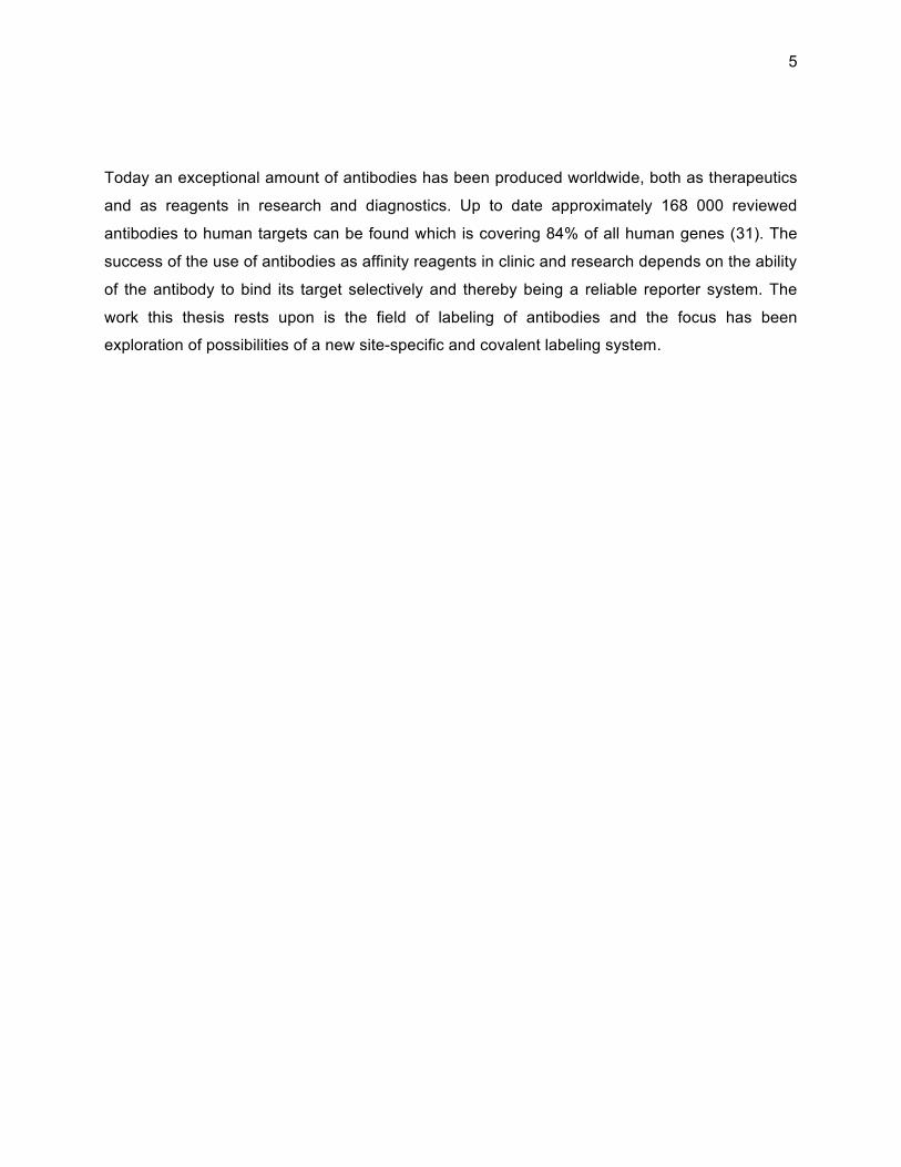
Figure 2. A schematic illustration of staphylococcal protein A. Five homologous domains A-E shares the
immunoglobulin-binding feature. S is a signal peptide and the X and M domains are cell wall anchoring
parts. The three-helical bundle that constitutes the Z domain was engineered from the B domain.
Like SPA the Z domain has been used for protein purification, and the domain has also been
use as a fusion tag for purification of recombinant target proteins, by IgG capture (68). Another
application that the Z domain has been used for is site-directed capture or immobilization of
antibodies on cells or beads (69, 70). Efforts have been made to further improve and engineer
the Z domain which as an example has resulted in an alkali stabilized variant that is used
commercially as ligand in a resin for purification of antibodies or Fc-fused proteins (71;
MabSelect SuRe, GE Healthcare). The Z domain has also been the subject of the construction
of library with the intention to change the specificity from Fc to other molecules, i.e. creation of
so-called affinity ligands. In the library 13 positions in helix 1 and helix 2 in the domain are
randomized to create a novel interaction surface (72).
S DE A B C X M
Z
SPA
Immunoglobulin binding domains

14
As mentioned, among the advantages of the Z domain is the fact that it is small (6.7 kDa), and it
has a stable three-dimensional structure and also the capacity to refold (42). These features are
beneficial when performing chemical peptide synthesis, which the Z domain has proven suited
for, and thereby render the introduction of synthetic groups possible, extending the usability of
the domain (73).
15
Modificationofproteins
In the beginning of the year of 1990 a new journal saw the light, Bioconjugate Chemistry. In the
editorial of the first issue Claude Meares stated that the rationale for the new journal was that
bioconjugate chemistry is a growing field with rapid development and has a potential for
scientific breakthrough (74), and indeed the need for reliable conjugation strategies are still
found both in research and clinical applications. The term bioconjugate chemistry is described
as the joining of two different molecular functions chemically or biologically and the importance
of the strategy to add functions to proteins has been tremendous in the development of proteins
as tools in research and clinic. There are numerous functions that proteins can be provided with;
groups that either increase or decrease the immunogenicity of proteins, drugs, reporter groups,
enzymes and fluorescent groups, are a few examples. In a review in the first issue of
Bioconjugate Chemistry the history of chemical modifications of proteins are discussed and how
protein modification from the past were sprung out from organic chemistry with methods like
acetylation, iodination, deamination and reaction with formaldehyde (75). Today there are plenty
of commercially available kits and reagents that serve different purposes, but they predominantly
conjugate functional molecules to specific side chains in the protein.
The use of a specific kind of reactive group in the protein is the most common strategy when
modifying proteins and the predominant choice is to target the primary amines, preferably the ε-
amino group situated on lysine (76). Lysine is an abundant amino acid in proteins and it is a
good nucleophile over pH 8, which provides efficiency to the reaction. However, since a reactive
lysine can appear anywhere within the amino acid sequence the risk of influencing the proteins
active site is quite substantial. The thiol group in cysteine is also considered as highly reactive
and has the favorable ability to react at neutral pH, but cysteines are rather uncommon and
when they do appear in proteins they are often forming disulphide bridges and the reduction of
these might influence the stability of the protein. Nevertheless targeting a naturally occurring or
inserted cysteine provides a specificity to the modification of the protein that is beneficial in
some applications (77). Carboxylic acids that can be found in the side chains of amino acids

16
aspartic acid and glutamic acid can occasionally be subject to conjugations of reporter groups
although they display relatively low reactivity in water which is considered a disadvantage (76).
The introduction of specific reactive groups into the protein for conjugation of reporter groups
can be done on a genetic level and/or by the use of enzymes, and several elegant methods in
this field have been developed. Intein-mediated ligation is a method were the protein to be
modified is expressed recombinantly in fusion with an intein-chitin binding domain and during the
binding of the construct to chitin a cleavage can be performed and a peptide is ligated to the
protein (78). The holoenzyme synthetase BirA covalently attaches a biotin to the ε-amino group
on a specific lysine found in a substrate when enzyme and substrate are expressed in E. coli
and a site-specific modification can be obtained (79, 80). The Sortase family of enzymes found
in several gram positive bacteria are enzymes that covalently attach proteins with a specific
motif to cell wall proteins containing a poly-glycine peptide sequence and the system has been
exploited as a method for in vitro modification of recombinant proteins such as biotinylation of
scFv and PEGylation of proteins containing the motif for the enzyme (81, 82).
In the development of antibody-drug conjugates (ADC) as a therapeutic agent the general idea
is to utilize the antibody as targeting probe and the drug as payload to carry the therapeutic
effect. For this strategy to be successful the conjugation method used must be reliable since it is
vital that the drug stays bound to the antibody, minimizing risk for unspecific tissue damage. It
has been shown that the linker and the conjugation site influences the in vivo stability and
efficacy of ADCs (83, 84), which presents a strong incentive for the continuation of progress in
the field of covalent specific modification of proteins and antibodies.
Incorporationofunnaturalaminoacidsintoproteins
The capability to insert another amino acid than the 20 natural amino acids is most certainly a
powerful tool, thus the option to incorporate different functions or handles into the protein
appears. The possibility to introduce an orthogonal reactivity means that specificity in
conjugation can be obtained. There are two main routes to incorporate unnatural amino acids
into the amino acid sequence of proteins, recombinantly or through peptide synthesis.

17
In the cell when a protein is being synthesized the translation machinery reads the messenger
ribonucleic acid (mRNA) and the appropriate transfer ribonucleic acid (tRNA) charged with a
specific amino acid binds the corresponding triplet codon to insert the amino acid into the
growing peptide chain. The enzyme aminoacyl-tRNA synthetase is responsible for charging the
tRNA with the corresponding amino acid. This natural, protein production system in the cell was
exploited in the lab of Peter Schultz at the end of the 1980s and a new method was developed
(85). The employment of site-directed mutagenesis for the introduction of an amber stop codon
in combination with a corresponding chemically amino-acylated tRNA were proven to site-
specific introduce unnatural amino acids into proteins (85). In the beginning of the century
further steps were taken in the ambition of expanding the genetic code and progress was made
by genetical engineering of tRNAs to make them orthogonal to the bacteria’s own tRNA but still
specific for one, in this case, unnatural amino acid (86-88). A system for selection and
identification of aminoacyl-tRNA mutants specific for unnatural amino acids of choice has made
it possible to site-specific introduce functional groups into recombinant proteins to a high level of
efficiency and usability (89). Proteins, especially antibodies and fragments thereof, have been
modified through the use of orthogonal aminoacyl-tRNA synthetase/tRNA pairs (90, 91) and
approximately 70 unnatural amino acids has been added to proteins by the translational
machinery of E. coli, yeast and mammalian cells (92). Of special interest for the work presented
in this thesis, is the introduction of photoreactive groups such as benzoylphenylalanine and
azidophenylalanine that has successfully been incorporated into recombinant proteins produced
in E.coli (93, 94).
The ability to perform chemical synthesis and create shorter peptides developed during the first
half of the twentieth century (95, 96), but a true scientific revolution occurred when Bruce
Merrifield introduced a solid-phase method to synthesize peptides (97, 98). Until then, the
synthesis of peptides was performed in solution and even though at the time a biologically active
protein like oxytocin (99) was successfully produced there were disadvantages with poor
solubility, slow reaction rates and purification from byproducts being the main issues. Another
improvement Merrifield was responsible for was the use of t-butyloxycarbonyl (Boc) as a
temporary protecting group and as a result the synthesis could be performed under milder
reaction conditions. In 1964 Merrifield published the synthesis of the peptide bradykinin and

18
proved the increase of efficiency using the new chemistry (95, 100). Even milder reaction
conditions could be obtained by the introduction of the fluorenylmethyloxycarbonyl (Fmoc) group
and the development of Fmoc chemistry, which is today considered to be the main choice of
chemistry in solid phase peptide synthesis (SPPS) (95, 101, 102).
During the process of solid phase peptide synthesis the amino acid sequence grows from the C-
terminal to the N-terminal by one amino acid at the time. The synthesis starts with the first amino
acid being coupled to the solid support, which usually is a polystyrene resin. Every cycle in the
process contains the step of first cleavage of the protecting group of the amine situated on the
growing chain, then an activated amino acid, where a good leaving group has been placed on
the carboxyl group of the amino acid, is allowed to react with the amino group creating a peptide
bond. As a final step the protecting groups of the side chains and the release of the peptide from
the resin are performed in one step.
In the field of SPPS the development of different orthogonal protection strategies of side chains
gives a great benefit since it provides the opportunity to incorporate functional groups in a
specific manner. When using Fmoc as temporary protecting group, which is base-labile, the side
chains are protected with groups that are cleaved off with strong acid and a common orthogonal
protecting group that can be used on amines is the weakly acid-labile 4-methyltrityl (Mtt) group.
Other examples of groups that provide orthogonality in Fmoc chemistry are the allyloxycarbonyl
(Allloc) group, which is cleaved by palladium catalysis and the thiol-labile dithiasuccinoyl (Dts)
group (103-105).
Benzoylphenylalanine–BPA–aphotoreactiveaminoacid
Photoactivable probes have mostly been used in the field of mapping protein-ligand binding and
drug-target identification and they can be found in quite a variety and a number of these are
commercially available. The photo reactive groups mainly function through two routes. Either
they form a new covalent bond, photoaffinity labeling, upon irradiation or a special bond is
cleaved, photodeprotection (106). The strategy when performing protein-protein interaction
studies is to produce variants of a protein with a photolabile amino acid incorporated at different
19
positions. Followed by allowing the protein to bind its interaction partner and when exposed to
light of appropriate wavelength a covalent bond might be created and different methods will be
applied to analyze the interaction (107).
Benzophenone (BP) is a photophore that is one of the most commonly used photoactivable
probes since it is considered to be efficient, stable and can be handled in ambient light (108).
Another important feature related to BP is that its activation requires wavelengths of 350-360 nm
(109), which is above the wavelength regarded as damaging for proteins. Moreover it primarily
reacts with C-H bounds rather than water, which also is a great advantage (110). The structure
of BP is regarded bulky and to better serve its purpose it is preferably used in a hydrophobic
interaction site. However upon irradiation, if no weak C-H bound is able to react with the
activated triplet state of the benzophenone group it will relax to its ground state (109, 110). As
part of the synthetic amino acid benzoylphenylalanine (BPA), BP can be incorporated in a
peptide during solid phase peptide synthesis (108, 111).

20
21
PresentInvestigation
The work presented in this thesis focuses on the development of a strategy for covalent and
specific labeling of antibodies. Furthermore, the analysis of the method developed and the
investigation of the labeled antibodies in several applications have been performed. A
photoreactive reagent was developed and produced using solid phase peptide synthesis and
subsequently used to label different variants of antibodies. The thesis rests upon three papers,
in paper I the development of the technology, the production of the reagent and the specific
labeling of antibodies are presented. In paper II the functionality of the photolabeled antibodies
are compared with antibodies covalently labeled by another method. The antibodies were used
as a detection tool for proteins and to visualize protein targets in tissue using
immunohistochemistry. In order to investigate the possibility to obtain a higher signal and a
higher sensitivity in the use of the photolabeled antibodies, different variants of the photoreactive
reagents are compared and discussed in paper III.
The modification of proteins and covalent conjugation of other molecules covalently to proteins
are a frequent operation in biotechnology and most often the incentive is to add a feature to be
exploited in a particular application. The method and the chemistry utilized to enable the actual
conjugation ought to be efficient and leave the binding site of the protein unaffected. The most
commonly used method to couple reporter groups to proteins is to address the ε-amino group
situated on lysines in the protein by the use of NHS-esters (76). This method is indeed efficient,
but the absence of control of level and location of the conjugation could lead to an undesired
influence of the proteins active site since a protein often contain several lysines. Site-specific
covalent attachment of reporter groups to antibodies is important and there are many
applications both in the development of reagents for research and therapeutics for clinic where
this is highly desired. Antibodies are often modified by labeling with different parts of reporter
systems, such as fluorophores, biotin, haptens or enzymes, which makes them useful tools in
research and diagnostics, and conjugated with drugs for use in therapy. The greatest advantage
when employing a site-specific conjugation is the high likelihood for the binding site to be
preserved.

22
The method that has been developed in this thesis, aims for site-specific labeling of antibodies
by exploiting a modified Z domain incorporated with a photoreactive group. The Z domain has
been produced using SPPS that enables the incorporation of BPA as a synthetic amino acid in
specific position within the domain. The BPA should preferably be located in the vicinity of the
interaction site with the antibody. The strategy that has been established in this thesis makes
use of the Z domains natural binding to the Fc-part of the antibody. This inherited activity is
combined with the crosslinking features of the incorporated BPA and by exposure of light of a
defined wavelength a covalent linkage between the Z domain and the antibody will be formed
(Figure 3). Furthermore, taking advantage of orthogonal strategy, reporter groups can be
inserted at side chains of specific amino acids. In the first study a biotin was introduced at the C-
terminus of the Z domain. This reporter group was utilized in a number of different detection
methods. In the third study the reagent was extended with an additional biotin. By the use of
biotinylated photoreactive Z domains, several types of antibodies, polyclonal rabbit IgG,
monoclonal human IgG1 and monoclonal mouse IgG2a, have been site-specifically labeled and
analyzed.
Figure 3. An illustration of the developed strategy for covalent site-specific labeling of antibodies through
the use of a photoactivable Z domain. A Z domain inserted with a BPA molecule (blue round shape) and a
biotin binding the Fc domain of an antibody.

23
CovalentImmunoglobulinLabelingthroughaPhotoactivableSyntheticZdomain(PaperI) A strategy for site-specific labeling of antibodies was developed and a production route for a
photoreactive reagent is presented in paper I. Previously Jung and colleagues has reported the
utilization of an IgG-binding protein with an incorporated photoreactive group for attachment to
antibodies. By recombinant production of a C2 domain from streptococcal Protein G, modified
with two incorporated cysteines, a covalently attachment of a BP-molecule by maleimide
chemistry was made. Thereby, a photoreactive procedure could be used for covalent
modification of antibodies. It was demonstrated that this approach could be used for directed
antibody immobilization onto a surface (112). Our approach for covalent modification of
antibodies was to, by SPPS, produce two different Z domains with the photoactivable amino acid
BPA incorporated at position 5 in one variant and position 18 in the other variant. The variant
with BPA in position 5 displayed similar affinity to human IgG as the parental molecule, proving
that the insertion of the bulky group of BPA in the binding site of the Z domain had limited effect
on the affinity. The insertion of the amino acid BPA at position 18 in the Z domain proved to
have a large negative effect on the binding of the domain to human IgG since the affinity of that
variant was considerably lower than that of the wildtype domain. It was shown that the variant
with BPA in position 5 could successfully covalently couple to antibodies. However, the variant
with BPA in position 18 could not obtain any covalent attachment between the domain and the
antibody. Furthermore in paper I, it was shown that antibodies of various types and origin were
covalently and site-specifically labeled with the biotinylated Z domain. The photolabeled
antibodies were analyzed using the bead array Luminex system (113, 114). Two different set-
ups were used, both with antigen attached to beads but also with a sandwich set-up were
capture antibodies were coupled to the beads and incubated with antigen and finally detection
were made with photo-coupled detection antibodies and fluorescent streptavidin. Furthermore,
Western blot was successfully employed and show that the biotinylated photolabeled antibodies
could be used for detection in this application. Moreover, this technology was used to prove that
the covalent attachment is site-specific and occurs at the Fc part of the antibody when the
biotinylated Z domain with BPA is used. Further evidence on the specificity of the photolabeling
procedure was shown when samples containing IgG and human serum albumin were analyzed

24
using Western blot and Luminex. In these experiments it was shown that no biotinylation of
albumin occured. Surface Plasmon Resonance (SPR) technology was used to prove that the
procedure of photolabeling of the antibodies does not impair the binding of the corresponding
antigen. Hence, the conclusion of this is that the strategy for site-specific covalent labeling of
antibodies presented in paper I was successfully performed and tested in different applications.
AntibodiesbiotinylatedusingasyntheticZ‐domainprovidestringent in situ proteindetection(PaperII)
Protein detection in situ is of importance since the location of a protein in a cell is essential for
understanding its function. Immunohistochemistry and immunofluorescence microscopy are
methods that can be applied to establish protein location. Both methods rely on the use of
antibodies and their specificity and therefore it is imperative that the antibody or the reporter
system exclusively conducts specific binding events. For the detection of a binding antibody, a
secondary antibody, directed towards the primary antibody is often used. This is a concept that
works efficiently, but it would be of great advantage to reduce the number of experimental steps
by omitting the secondary antibody. This could be accomplished by directly link the primary
antibody to the reporter group. There are different techniques available for covalent labeling of
antibodies. Hence, we decided to compare the labeling technology using the photoactivable Z
domain with another commercially available technology, Lightning Link. In this project the
antibodies were applied in antibody-based protein profiling using immunohistochemistry. In total
13 antibodies were used in the comparison and labeled with the different systems. The obtained
staining patterns from tissue sections were compared to unconjugated antibodies, using a
secondary antibody for detection. It was found that the staining pattern from all 13 antibodies
labeled with the photoactivable Z domain were in concordance with the pattern from
unconjugated antibodies. As a comparison 3 out of 13 antibodies labeled with Lightning Link
produced staining patterns similar to those of unconjugated antibodies. Tissue microarrays with
18 formalin paraffin-embedded human tissues were used in the staining procedure with the
differently biotinylated antibodies. In conclusion, the method of antibody labeling of the
photoactivable Z domain proved to be stringent and specific.

25
Optimization of an antibody labeling strategy through the use ofphotoactivablesyntheticZdomains(PaperIII)
The method of labeling antibodies by the use of a photoactivable Z domain was further
developed. Antibodies biotinylated using the photoactivable Z domain has proven to be
successful in many different applications. However, in some applications question marks were
raised regarding the number of biotins and also the availability of the biotin for binding of
streptavidin influencing the sensitivity of the method. Therefore, two new variants of the
photoactivable Z domain were produced using SPPS to exploit the opportunity to insert the
photoreactive amino acid, BPA, in position 5 and biotin groups in a specific manner. First variant
was made with a biotin coupled to position 58 but with a PEG-linker in between the lysine in the
domain and the biotin. The second variant, compared to the first variant, had an additional biotin
with linker incorporated in position 39, where a serine had been exchange with lysine, i.e. one
biotin in each end of the three-helix bundle domain. The new variants were compared with the
original photoreactive reagent, the Z domain with one biotin coupled to a lysine in position 58..
The biotins in these three variants were used to immobilize the Z domains on a Neutravidin chip
and the affinities to antibodies were analyzed using surface plasmon resonance (SPR). Different
types of IgG was flown over the surface and the conclusion were made that the variant with two
biotins had a higher apparent affinity than the variants with only one biotin incorporated into
them when immobilized to a surface. The three variants were photo-coupled to several types of
antibodies and analyzed using the Luminex system utilizing beads that had been coupled with
corresponding antigen and the detection were made with fluorescently labeled streptavidin. The
result implied that antibodies coupled with the variant with two biotins provided a 100 % higher
signal in Luminex. However, the PEG linker that was incorporated between the protein domain
and the biotin does not affect the signal noticeably. Hence, the PEG-linker does not influence
the availability of the biotin. Moreover, the labeled antibodies were used in
immunohistochemistry and a stronger and more evident staining pattern was obtained with the
variant containing two biotins than when using a single biotin. Hence, a Z-variant with two biotins
gives a higher sensitivity and provides a higher signal compared to Z-version with a single biotin
in the application analyzed.

26
Concludingremarks
The modification of proteins, in particular antibodies, is increasingly important since the
exploration of proteins and antibodies as tools in research, diagnostics and therapeutics expand.
The functionalities that are most often of interest to combine with the protein or antibody depend
on the intended use of the system. Fluorophores, enzymes, haptens and drugs are all examples
of regular functionalities added to proteins or antibodies. The chemistry most often used could
suffer from lack of specificity of the labeling and thereby risk of influencing the binding site. To
increase the specificity of labeling a cysteine could be employed. However, there may be a
drawback, in vivo instability and the release of the conjugate as a consequence (84). There is a
need for new conjugations strategies since in some cases the methods normally applied do not
meet the requirements for antibodies used as tools or drugs.
The development and investigation of the strategy for covalent site-specific labeling of
antibodies using photoactivable Z domains is the foundation for this licentiate thesis. The SPPS-
produced photo-reactive reagents have been used to covalently biotinylate several types of
antibodies. The first study was done with a photoactivable Z domain containing one biotin and
the photolabeled antibodies was proved to operate successfully in a number of applications. The
metod of labeling antibodies by the use of the photoactivable Z domain was compared to a
commercially available kit for biotinylation of antibodies in the application of protein profiling, by
immunohistochemistry. It was found that the staining by the Z-labeled antibodies was in
concordance with the control staining and the performance of the system was satisfactory. To
further enhance the system the Z domain was equipped with a second biotin group. Indeed
antibodies coupled with the Z domain containing two biotins showed a higher signal when
binding its antigen on beads and also a stronger staining in IHC. The photoactivable Z domains
have proven to covalently and specifically label antibodies successfully and shown to be a
valuable tool for many biotechnological applications, in research, diagnostics and also potentially
for use in therapeutics.

27
Acknowledgements
Det är mer än en handfull gånger under årens lopp som jag skänkt denna delen av
avhandlingen en tanke eftersom jag tillhör den känslosamma (sentimentala) sorten. Och nu när
jag väl ska nämna er som har gjort detta arbete möjligt, är ni så många, och det är jag verkligen
tacksam för. Tack till alla er som jag inte nämner men ni är många som ler i korridoren, hjälper
till i labbet eller vid kopiatorn eller helt enkelt delar en trevlig stund vid lunchbordet.
Min handledare Sophia, det har varit otroligt lärorikt att jobba i din grupp och jag har fått
chansen att utveckla så mycket under denna tiden. Jag är riktigt stolt över det arbete vi gjort.
Men ibland vet jag inte om jag ska önska dig ett lugnare arbetsliv eller en snabbare cykel.
Amelie, som bihandledare har du varit närvarande och hjälpt mig mycket, inte minst med alla
knepiga syntes-grejer.
ProNova Vinn Excellence center for Protein Technology för finansiering.
I ProNova vill jag tacka Amelie och Per-Åke som styrt upp hela centret men också alla övriga
medarbetare inom affinity-gruppen, Feifan, Peter, Mahya, Afshin och Anna P. Jag vill också
rikta ett stort tack till de företagsrepresentanter som deltagit i ZBPA-projektet som jag haft
kontakt med; Niklas Ahlborg (Mabtech), Mats Inganäs (Gyros), Åke Danielsson (GE), Bo
Jansson (Bioinvent), Simon Fredriksson och Mats Gullberg (O-link) och Henrik Johannesson (Atlas Antibodies). Jag har verkligen värdesatt att få ta del av ert yrkeskunnande.
Jag vill tacka övriga PIs, och Stefan för att det alltid känns som om du lyssnar och stöttar.
Hjältar lite mer bakom kulisserna som styr upp administrationen Inger, Kristina, Mona och
Christelle. Emma Ö för att du gjort livet i labbet så mycket enklare med grym ordning i
diskrummet och avlastning av inköp.
Jag är verkligen tacksam för den granskning och hjälpt jag fått med avhandlingen; Tobbe,
Andreas, Amelie och Basia.

28
Mina riktigt grymma ex-jobbare, Anna P, Josefin och Sara, jag gillar att ni alla stannade kvar,
för jag låtsas att jag har en liten del i det. Tack för era insatser i projekten. Anna Torell Holm för
det lilla gästspelet i C2-projektet.
Tack Tobbe, för att jag fick göra ett fantastiskt roligt ex-jobb i din grupp. John, Johan N och
Magdalena för ovärderlig hjälp med SPR. Ett stort tack för all hjälp med Luminex, Jochen, Maja
och Ulrika. Gustav som kört och pratat mycket MS med mig.
Sandra, Nikhil, Anna A och Fredrik för ett lyckat samarbete. Tack för de fina bilderna Sandra.
Tack till alla som jag delat grupptillhörighet med och haft både vetenskapliga och andra roliga
diskussioner med och mycket skojiga aktiviteter, Sophias grupp; Cilla, Tove A, Johanna,
Hanna, Karin, Margareta, Johan, ToveBo, Sara, Cajsa, Jenny OT, Henrik W, Micke och
Amelies grupp; Anna P, Joel, Peter, Daniel, Nima och Kristina.
Jenny OT, du anställde mig i Proteinfabriken, tack för det förtroendet. Tack till Hanna och
Johanna, det var otroligt roligt att vara foing med er ”på den gamla goda tiden” . Tack också till
Lanlan, Pawel, Asif, Ulla, Kattis, Jens, Hero, Lasse, Anna B, Louise och övriga Profabbare.
Ett tack också till Mathias och alla andra i HPA för årliga träffar nära och långt bort och alla
roliga människor som gör den fantastiska atlasen. Ett speciellt tack till immunotech för generös
hjälp med WB och till alla i molbio som accepterat mina intrång i gruppen med jämnmod.
Tack till alla er jag delat skrivrum med och som verkligen gjort stor skillnad i vardagen, Emma,
Tove A, Cecilia, Pawel, Mårten, Julia, Johan, Charlotte, Magdalena, Burcu, Chi, Filippa,
Andreas och nu på slutet, nya kompisar som jag inte riktigt hunnit lära känna, Aman, Ali och
Pavan.
Det är mycket och många som jag kommer att minnas med glädje från livet på plan 3, alla
trevliga reskamrater under flertalet konferenser som i soliga Portugal; Basia, Anna P, Johan N.
Joel, Amelie, Sophia med sina fina pojkar Marcus och Jonathan. Det stora tjej-gänget i
Boston; Nina, Basia, Camilla, Anna P, Lisa, Johanna, Hanna L och i underbara Bologna;
Tove A, ToveBo och Sophia. Slutligen John och Stefan i San Diego när vi hängde med
Andreas, Caroline och Elin. Stor kram till dig Sverker, det var kul att starta upp
Socialförvaltingen med dig och tack till alla som fixat/fixar aktiviteter för andra på avdelningen.
Smarta och underbara Basia, lika knäpp som jag, tack för all hjälp med avhandlingen och alla
skratt. Alla som druckit fredagsöl med DN-kryss, och sköna öl-snubben, Peter, vi har handlat öl

29
och labbat tillsammans, det var grymt. Härliga och viktiga små stunder som kaffe, lunch, frukost
och/eller AW; Nina, Emma, Julia, Basia, Helena, Cilla, Maja, Johanna, Tove A och flera
andra fina personer.
Mina kära Kittel-tjejer, Emma, Nina, Julia, Tove och Torun, den årliga skid-resan till Kittel är
helig för mig. Det en sådan grym höjdpunkt på året och förbaskat roligt. Nån dag ska jag fanimej
klara mig hyfsat utanför pisten också.
Lena du kommer som en frisk fläkt och delar generöst med dig av dina upplevelser, resor och
om stort och smått. Helt underbart, jag är uppfylld flera dagar efteråt och är tacksam för att du
kom “med i gänget” även om det var lite sent, som i sjuan typ. Självklart jag hänger med på en
träningsresa framöver.
Ulla och Jenny, det känns som om jag känt er hela livet. Verkligen magiskt, tack för att ni finns
och för luncher, stöd och framförallt alla skratt. Ni vill mig så väl och jag önskar er allt gott som
finns på jorden.
Beda, du är som en syster och med dig delar jag i princip hela min barndom och ungdom och
utan dig hade jag aldrig kommit i tid en enda gång under högstadiet. Tack för alla fina minnen
från Österlen och i mormors trädgård med våra familjer.
Mina nära vänner och Kajsas gudföräldrar varma, fina Monki, och påhittiga, långa Rillo. Tack
för alla resor, helger på Rådmansö och allt vi delat genom många, många år. Ni finns alltid där
och ni är fantastiska gudföräldrar, världens bästa.
Familjen Frank-Bille, Petra, Magnus & Robert, er trio är så oerhört viktig får vår trio. Inte för mitt
liv hade jag kunnat föreställa allt vi nu delat när vi träffades på föräldrautbildningen. Minnen på
flera nivåer, resor och skidåkning med alla de äventyren, men framförallt alla middagar och
samtal i våra respektive kök under årens lopp. Jag ser framemot att dela minst lika många
äventyr till i livet.
Till härliga och fina kusin Noak med familj, det är verkligen bra att ni finns och tack Noak för att
du livar upp oss lite när vi ses. Tack också till min svärmor Ann-Mari, jag känner så ofta hur du
hejar på mig, tack för ditt stöd och till Peter, Anni, Anders, Martin, Christina, Perka, Linnea
och Henrik som accepterat mig så gott det går trots ”svår” dialekt.

30
Min kära, älskade mamma du har klarat av så mycket och kämpat så hårt, jag hade önskat att
du hade fått bli mer belönad. Det är inspirerande att tänka på hur förbaskat duktig du var på ditt
jobb och hur du jonglerade allt långt innan termen “livspussel” var uppfunnen. Tack för att du
alltid hjälpt till när vi behövt det.
Min mentor, Eva-Britt, du har väglett och coachat mig till ett fantastiskt liv, ibland har det känts
som du räddat livet på mig, tack.
Kajsa och Staffan, ni betyder allt. Kajsa min smarta, vackra, roliga och älskade unge, jag kan
inte tänka mig livet utan att få vara din mamma. Staffan min älskling, jag är så tacksam för att vi
har levt ihop i 19 år och mer än nånsin känner jag hur mycket vi båda vill fortsätta leva ihop. Jag
älskar er.
31
References
(1) Bud, R. (1989) Janus-Faced Biotechnology - an Historical-Perspective. Trends in
Biotechnology 7, 230-233.
(2) Colwell, R. R. (2002) Fulfilling the promise of biotechnology. Biotechnol Adv 20, 215-28.
(3) Berg, P., Baltimore, D., Brenner, S., Roblin, R. O., and Singer, M. F. (1975) Summary
statement of the Asilomar conference on recombinant DNA molecules. Proc Natl Acad
Sci U S A 72, 1981-4.
(4) Berg, P., and Singer, M. F. (1995) The recombinant DNA controversy: twenty years later.
Proc Natl Acad Sci U S A 92, 9011-3.
(5) Avery, O. T., Macleod, C. M., and McCarty, M. (1944) Studies on the Chemical Nature of
the Substance Inducing Transformation of Pneumococcal Types : Induction of
Transformation by a Desoxyribonucleic Acid Fraction Isolated from Pneumococcus Type
Iii. J Exp Med 79, 137-58.
(6) Zallen, D. T. (2003) Despite Franklin's work, Wilkins earned his Nobel. Nature 425, 15.
(7) Maddox, B. (2003) The double helix and the 'wronged heroine'. Nature 421, 407-8.
(8) Watson, J. D., and Crick, F. H. (1953) Molecular structure of nucleic acids; a structure for
deoxyribose nucleic acid. Nature 171, 737-8.
(9) Itano, H. A., and Pauling, L. (1949) A rapid diagnostic test for sickle cell anemia. Blood 4,
66-8.
(10) Lehman, I. R., Bessman, M. J., Simms, E. S., and Kornberg, A. (1958) Enzymatic
synthesis of deoxyribonucleic acid. I. Preparation of substrates and partial purification of
an enzyme from Escherichia coli. J Biol Chem 233, 163-70.

32
(11) Bessman, M. J., Lehman, I. R., Simms, E. S., and Kornberg, A. (1958) Enzymatic
synthesis of deoxyribonucleic acid. II. General properties of the reaction. J Biol Chem
233, 171-7.
(12) Kresge, N., Simoni, R. D., and Hill, R. L. (2009) 100 years of biochemistry and molecular
biology. The decade-long pursuit of a reconstituted yeast transcription system: the work
of Roger D. Kornberg. J Biol Chem 284, e18-20.
(13) Smith, H. O., and Wilcox, K. W. (1970) A restriction enzyme from Hemophilus influenzae.
I. Purification and general properties. J Mol Biol 51, 379-91.
(14) Danna, K., and Nathans, D. (1971) Specific cleavage of simian virus 40 DNA by
restriction endonuclease of Hemophilus influenzae. Proc Natl Acad Sci U S A 68, 2913-7.
(15) Roberts, R. J. (2005) How restriction enzymes became the workhorses of molecular
biology. Proc Natl Acad Sci U S A 102, 5905-8.
(16) Cohen, S. N., Chang, A. C., Boyer, H. W., and Helling, R. B. (1973) Construction of
biologically functional bacterial plasmids in vitro. Proc Natl Acad Sci U S A 70, 3240-4.
(17) Crea, R., Kraszewski, A., Hirose, T., and Itakura, K. (1978) Chemical synthesis of genes
for human insulin. Proc Natl Acad Sci U S A 75, 5765-9.
(18) Bartlett, J. M., and Stirling, D. (2003) A short history of the polymerase chain reaction.
Methods Mol Biol 226, 3-6.
(19) Kohler, G., and Milstein, C. (1975) Continuous cultures of fused cells secreting antibody
of predefined specificity. Nature 256, 495-7.
(20) Hozumi, N., and Tonegawa, S. (1976) Evidence for somatic rearrangement of
immunoglobulin genes coding for variable and constant regions. Proc Natl Acad Sci U S
A 73, 3628-32.
(21) Smith, G. P. (1985) Filamentous Fusion Phage - Novel Expression Vectors That Display
Cloned Antigens on the Virion Surface. Science 228, 1315-1317.
(22) Marks, J. D., Griffiths, A. D., Malmqvist, M., Clackson, T. P., Bye, J. M., and Winter, G.
(1992) By-passing immunization: building high affinity human antibodies by chain
shuffling. Biotechnology (N Y) 10, 779-83.
33
(23) Johnson, K. S., and Chiswell, D. J. (1993) Human-Antibody Engineering. Current Opinion
in Structural Biology 3, 564-571.
(24) Lander, E. S., Linton, L. M., Birren, B., Nusbaum, C., Zody, M. C., Baldwin, J., Devon, K.,
Dewar, K., Doyle, M., FitzHugh, W., Funke, R., Gage, D., Harris, K., Heaford, A.,
Howland, J., Kann, L., Lehoczky, J., LeVine, R., McEwan, P., McKernan, K., Meldrim, J.,
Mesirov, J. P., Miranda, C., Morris, W., Naylor, J., Raymond, C., Rosetti, M., Santos, R.,
Sheridan, A., Sougnez, C., Stange-Thomann, N., Stojanovic, N., Subramanian, A.,
Wyman, D., Rogers, J., Sulston, J., Ainscough, R., Beck, S., Bentley, D., Burton, J.,
Clee, C., Carter, N., Coulson, A., Deadman, R., Deloukas, P., Dunham, A., Dunham, I.,
Durbin, R., French, L., Grafham, D., Gregory, S., Hubbard, T., Humphray, S., Hunt, A.,
Jones, M., Lloyd, C., McMurray, A., Matthews, L., Mercer, S., Milne, S., Mullikin, J. C.,
Mungall, A., Plumb, R., Ross, M., Shownkeen, R., Sims, S., Waterston, R. H., Wilson, R.
K., Hillier, L. W., McPherson, J. D., Marra, M. A., Mardis, E. R., Fulton, L. A., Chinwalla,
A. T., Pepin, K. H., Gish, W. R., Chissoe, S. L., Wendl, M. C., Delehaunty, K. D., Miner,
T. L., Delehaunty, A., Kramer, J. B., Cook, L. L., Fulton, R. S., Johnson, D. L., Minx, P.
J., Clifton, S. W., Hawkins, T., Branscomb, E., Predki, P., Richardson, P., Wenning, S.,
Slezak, T., Doggett, N., Cheng, J. F., Olsen, A., Lucas, S., Elkin, C., Uberbacher, E.,
Frazier, M., et al. (2001) Initial sequencing and analysis of the human genome. Nature
409, 860-921.
(25) Venter, J. C., Adams, M. D., Myers, E. W., Li, P. W., Mural, R. J., Sutton, G. G., Smith,
H. O., Yandell, M., Evans, C. A., Holt, R. A., Gocayne, J. D., Amanatides, P., Ballew, R.
M., Huson, D. H., Wortman, J. R., Zhang, Q., Kodira, C. D., Zheng, X. H., Chen, L.,
Skupski, M., Subramanian, G., Thomas, P. D., Zhang, J., Gabor Miklos, G. L., Nelson,
C., Broder, S., Clark, A. G., Nadeau, J., McKusick, V. A., Zinder, N., Levine, A. J.,
Roberts, R. J., Simon, M., Slayman, C., Hunkapiller, M., Bolanos, R., Delcher, A., Dew,
I., Fasulo, D., Flanigan, M., Florea, L., Halpern, A., Hannenhalli, S., Kravitz, S., Levy, S.,
Mobarry, C., Reinert, K., Remington, K., Abu-Threideh, J., Beasley, E., Biddick, K.,
Bonazzi, V., Brandon, R., Cargill, M., Chandramouliswaran, I., Charlab, R., Chaturvedi,
K., Deng, Z., Di Francesco, V., Dunn, P., Eilbeck, K., Evangelista, C., Gabrielian, A. E.,
Gan, W., Ge, W., Gong, F., Gu, Z., Guan, P., Heiman, T. J., Higgins, M. E., Ji, R. R., Ke,
Z., Ketchum, K. A., Lai, Z., Lei, Y., Li, Z., Li, J., Liang, Y., Lin, X., Lu, F., Merkulov, G. V.,
Milshina, N., Moore, H. M., Naik, A. K., Narayan, V. A., Neelam, B., Nusskern, D., Rusch,

34
D. B., Salzberg, S., Shao, W., Shue, B., Sun, J., Wang, Z., Wang, A., Wang, X., Wang,
J., Wei, M., Wides, R., Xiao, C., Yan, C., et al. (2001) The sequence of the human
genome. Science 291, 1304-51.
(26) Taussig, M. J., Stoevesandt, O., Borrebaeck, C. A. K., Bradbury, A. R., Cahill, D.,
Cambillau, C., de Daruvar, A., Dubel, S., Eichler, J., Frank, R., Gibson, T. J., Gloriam, D.,
Gold, L., Herberg, F. W., Hermjakob, H., Hoheisel, J. D., Joos, T. O., Kallioniemi, O.,
Koegl, M., Konthur, Z., Korn, B., Kremmer, E., Krobitsch, S., Landegren, U., van der
Maarel, S., McCafferty, J., Muyldermans, S., Nygren, P. A., Palcy, S., Pluckthun, A.,
Polic, B., Przybylski, M., Saviranta, P., Sawyer, A., Sherman, D. J., Skerra, A., Templin,
M., Ueffing, M., and Uhlen, M. (2007) ProteomeBinders: planning a European resource
of affinity reagents for analysis of the human proteome (vol 4, pg 13, 2007). Nature
Methods 4, 126-126.
(27) Mersmann, M., Meier, D., Mersmann, J., Helmsing, S., Nilsson, P., Graslund, S., Colwill,
K., Hust, M., Dubel, S., and Consortium, S. G. (2010) Towards proteome scale antibody
selections using phage display. New Biotechnology 27, 118-128.
(28) Uhlen, M., and Ponten, F. (2005) Antibody-based proteomics for human tissue profiling.
Molecular & Cellular Proteomics 4, 384-393.
(29) Ponten, F., Kampf, C., Wester, K., Andersson, A., Bjorling, E., and Uhlen, M. (2005)
Antibody-based proteomics for human tissue profiling; the Swedish Human Proteome
Resource project (HPR). Molecular & Cellular Proteomics 4, S65-S65.
(30) Nilsson, P., Paavilainen, L., Larsson, K., Odling, J., Sundberg, M., Andersson, A. C.,
Kampf, C., Persson, A., Szigyarto, C. A. K., Ottosson, J., Bjorling, E., Hober, S.,
Wernerus, H., Wester, K., Ponten, F., and Uhlen, M. (2005) Towards a human proteome
atlas: High-throughput generation of mono-specific antibodies for tissue profiling.
Proteomics 5, 4327-4337.
(31) Antibodypedia. (2011), Human Antibody Initiative and Nature Publishing Group.
(32) Llewelyn, M. B., Hawkins, R. E., and Russell, S. J. (1992) Discovery of antibodies. BMJ
305, 1269-72.

35
(33) Silverstein, A. M. (2004) Labeled antigens and antibodies: the evolution of magic
markers and magic bullets. Nat Immunol 5, 1211-7.
(34) Carter, P. J. (2006) Potent antibody therapeutics by design. Nat Rev Immunol 6, 343-57.
(35) Hale, G. (2006) Therapeutic antibodies--delivering the promise? Adv Drug Deliv Rev 58,
633-9.
(36) Nelson, A. L., and Reichert, J. M. (2009) Development trends for therapeutic antibody
fragments. Nat Biotechnol 27, 331-7.
(37) Golay, J., and Introna, M. Mechanism of action of therapeutic monoclonal antibodies:
Promises and pitfalls of in vitro and in vivo assays. Arch Biochem Biophys.
(38) Goldsby, A. R. K., J.T.; Osborne, A.B. (2007) Immunlogy, 6th ed., W.H. Freeman and
Company.
(39) Holliger, P., and Hudson, P. J. (2005) Engineered antibody fragments and the rise of
single domains. Nature Biotechnology 23, 1126-1136.
(40) Torres, M., and Casadevall, A. (2008) The immunoglobulin constant region contributes to
affinity and specificity. Trends in Immunology 29, 91-97.
(41) Uhlen, M., Bjorling, E., Agaton, C., Szigyarto, C. A., Amini, B., Andersen, E., Andersson,
A. C., Angelidou, P., Asplund, A., Asplund, C., Berglund, L., Bergstrom, K., Brumer, H.,
Cerjan, D., Ekstrom, M., Elobeid, A., Eriksson, C., Fagerberg, L., Falk, R., Fall, J.,
Forsberg, M., Bjorklund, M. G., Gumbel, K., Halimi, A., Hallin, I., Hamsten, C., Hansson,
M., Hedhammar, M., Hercules, G., Kampf, C., Larsson, K., Lindskog, M., Lodewyckx, W.,
Lund, J., Lundeberg, J., Magnusson, K., Malm, E., Nilsson, P., Odling, J., Oksvold, P.,
Olsson, I., Oster, E., Ottosson, J., Paavilainen, L., Persson, A., Rimini, R., Rockberg, J.,
Runeson, M., Sivertsson, A., Skollermo, A., Steen, J., Stenvall, M., Sterky, F.,
Stromberg, S., Sundberg, M., Tegel, H., Tourle, S., Wahlund, E., Walden, A., Wan, J.,
Wernerus, H., Westberg, J., Wester, K., Wrethagen, U., Xu, L. L., Hober, S., and Ponten,
F. (2005) A human protein atlas for normal and cancer tissues based on antibody
proteomics. Mol Cell Proteomics 4, 1920-32.
(42) Stahl, S., and Nygren, P. A. (1997) The use of gene fusions to protein A and protein G in
immunology and biotechnology. Pathologie Biologie 45, 66-76.

36
(43) Borrebaeck, C. A. (2000) Antibodies in diagnostics - from immunoassays to protein
chips. Immunol Today 21, 379-82.
(44) Little, M., Kipriyanov, S. M., Le Gall, F., and Moldenhauer, G. (2000) Of mice and men:
hybridoma and recombinant antibodies. Immunology Today 21, 364-370.
(45) Boulianne, G. L., Hozumi, N., and Shulman, M. J. (1984) Production of Functional
Chimaeric Mouse Human-Antibody. Nature 312, 643-646.
(46) Morrison, S. L., Johnson, M. J., Herzenberg, L. A., and Oi, V. T. (1984) Chimeric Human-
Antibody Molecules - Mouse Antigen-Binding Domains with Human Constant Region
Domains. Proceedings of the National Academy of Sciences of the United States of
America-Biological Sciences 81, 6851-6855.
(47) Jones, P. T., Dear, P. H., Foote, J., Neuberger, M. S., and Winter, G. (1986) Replacing
the Complementarity-Determining Regions in a Human-Antibody with Those from a
Mouse. Nature 321, 522-525.
(48) Better, M., Chang, C. P., Robinson, R. R., and Horwitz, A. H. (1988) Escherichia-Coli
Secretion of an Active Chimeric Antibody Fragment. Science 240, 1041-1043.
(49) Skerra, A., and Pluckthun, A. (1988) Assembly of a Functional Immunoglobulin-Fv
Fragment in Escherichia-Coli. Science 240, 1038-1041.
(50) Wark, K. L., and Hudson, P. J. (2006) Latest technologies for the enhancement of
antibody affinity. Advanced Drug Delivery Reviews 58, 657-670.
(51) Starovasnik, M. A., Skelton, N. J., OConnell, M. P., Kelley, R. F., Reilly, D., and
Fairbrother, W. J. (1996) Solution structure of the E-domain of staphylococcal protein A.
Biochemistry 35, 15558-15569.
(52) Achari, A., Hale, S. P., Howard, A. J., Clore, G. M., Gronenborn, A. M., Hardman, K. D.,
and Whitlow, M. (1992) 1.67-Angstrom X-Ray Structure of the B2 Immunoglobulin-
Binding Domain of Streptococcal Protein-G and Comparison to the Nmr Structure of the
B1 Domain. Biochemistry 31, 10449-10457.

37
(53) Kronvall, G., and Jonsson, K. (1999) Receptins: a novel term for an expanding spectrum
of natural and engineered microbial proteins with binding properties for mammalian
proteins. J Mol Recognit 12, 38-44.
(54) Forsgren, A., and Sjoquist, J. (1966) "Protein A" from S. aureus. I. Pseudo-immune
reaction with human gamma-globulin. J Immunol 97, 822-7.
(55) Uhlen, M., Guss, B., Nilsson, B., Gatenbeck, S., Philipson, L., and Lindberg, M. (1984)
Complete sequence of the staphylococcal gene encoding protein A. A gene evolved
through multiple duplications. J Biol Chem 259, 1695-702.
(56) Schneewind, O., Fowler, A., and Faull, K. F. (1995) Structure of the Cell-Wall Anchor of
Surface-Proteins in Staphylococcus-Aureus. Science 268, 103-106.
(57) Deisenhofer, J. (1981) Crystallographic Refinement and Atomic Models of a Human Fc
Fragment and Its Complex with Fragment-B of Protein-a from Staphylococcus-Aureus at
2.9-a and 2.8-a Resolution. Biochemistry 20, 2361-2370.
(58) Gouda, H., Torigoe, H., Saito, A., Sato, M., Arata, Y., and Shimada, I. (1992) 3-
Dimensional Solution Structure of the B-Domain of Staphylococcal Protein-a -
Comparisons of the Solution and Crystal-Structures. Biochemistry 31, 9665-9672.
(59) Gouda, H., Shiraishi, M., Takahashi, H., Kato, K., Torigoe, H., Arata, Y., and Shimada, I.
(1998) NMR study of the interaction between the B domain of staphylococcal protein A
and the Fc portion of immunoglobulin G. Biochemistry 37, 129-136.
(60) Moks, T., Abrahmsen, L., Nilsson, B., Hellman, U., Sjoquist, J., and Uhlen, M. (1986)
Staphylococcal Protein-a Consists of 5 Igg-Binding Domains. European Journal of
Biochemistry 156, 637-643.
(61) Graille, M., Stura, E. A., Corper, A. L., Sutton, B. J., Taussig, M. J., Charbonnier, J. B.,
and Silverman, G. J. (2000) Crystal structure of a Staphylococcus aureus protein A
domain complexed with the Fab fragment of a human IgM antibody: Structural basis for
recognition of B-cell receptors and superantigen activity. Proceedings of the National
Academy of Sciences of the United States of America 97, 5399-5404.
(62) Langone, J. J. (1982) Applications of Immobilized Protein-a in Immunochemical
Techniques. Journal of Immunological Methods 55, 277-296.

38
(63) Nilsson, B., Moks, T., Jansson, B., Abrahmsen, L., Elmblad, A., Holmgren, E.,
Henrichson, C., Jones, T. A., and Uhlen, M. (1987) A Synthetic Igg-Binding Domain
Based on Staphylococcal Protein-A. Protein Engineering 1, 107-113.
(64) Jansson, B., Uhlen, M., and Nygren, P. A. (1998) All individual domains of
staphylococcal protein A show Fab binding. Fems Immunology and Medical Microbiology
20, 69-78.
(65) Jendeberg, L., Tashiro, M., Tejero, R., Lyons, B. A., Uhlen, M., Montelione, G. T., and
Nilsson, B. (1996) The mechanism of binding staphylococcal protein A to immunoglobin
G does not involve helix unwinding. Biochemistry 35, 22-31.
(66) Tashiro, M., Tejero, R., Zimmerman, D. E., Celda, B., Nilsson, B., and Montelione, G. T.
(1997) High-resolution solution NMR structure of the Z domain of staphylococcal protein
A. Journal of Molecular Biology 272, 573-590.
(67) Jendeberg, L., Persson, B., Andersson, R., Karlsson, R., Uhlen, M., and Nilsson, B.
(1995) Kinetic-Analysis of the Interaction between Protein-a Domain Variants and Human
Fc Using Plasmon Resonance Detection. Journal of Molecular Recognition 8, 270-278.
(68) Uhlen, M., Forsberg, G., Moks, T., Hartmanis, M., and Nilsson, B. (1992) Fusion proteins
in biotechnology. Curr Opin Biotechnol 3, 363-9.
(69) Nakamura, Y., Shibasaki, S., Ueda, M., Tanaka, A., Fukuda, H., and Kondo, A. (2001)
Development of novel whole-cell immunoadsorbents by yeast surface display of the IgG-
binding domain. Appl Microbiol Biotechnol 57, 500-5.
(70) Mazzucchelli, S., Colombo, M., De Palma, C., Salvade, A., Verderio, P., Coghi, M. D.,
Clementi, E., Tortora, P., Corsi, F., and Prosperi, D. Single-domain protein A-engineered
magnetic nanoparticles: toward a universal strategy to site-specific labeling of antibodies
for targeted detection of tumor cells. ACS Nano 4, 5693-702.
(71) Linhult, M., Gulich, S., Graslund, T., Simon, A., Karlsson, M., Sjoberg, A., Nord, K., and
Hober, S. (2004) Improving the tolerance of a protein a analogue to repeated alkaline
exposures using a bypass mutagenesis approach. Proteins 55, 407-16.
39
(72) Nord, K., Gunneriusson, E., Ringdahl, J., Stahl, S., Uhlen, M., and Nygren, P. A. (1997)
Binding proteins selected from combinatorial libraries of an alpha-helical bacterial
receptor domain. Nat Biotechnol 15, 772-7.
(73) Engfeldt, T., Renberg, B., Brumer, H., Nygren, P. A., and Karlstrom, A. E. (2005)
Chemical synthesis of triple-labelled three-helix bundle binding proteins for specific
fluorescent detection of unlabelled protein. Chembiochem 6, 1043-1050.
(74) Meares, C. (1990) Editorial - Introduction to Bioconjugate Chemistry. Bioconjugate
Chemistry 1, 1-1.
(75) Means, G. E., and Feeney, R. E. (1990) Chemical Modifications of Proteins: History and
Applications. Bioconjugate Chemistry 1, 2-12.
(76) Brinkley, M. (1992) A brief survey of methods for preparing protein conjugates with dyes,
haptens and crosslinking reagents. Bioconjugate Chemistry 3, 2-13.
(77) Kim, Y. G., Ho, S. O., Gassman, N. R., Korlann, Y., Landorf, E. V., Collart, F. R., and
Weiss, S. (2008) Efficient site-specific Labeling of proteins via cysteines. Bioconjugate
Chemistry 19, 786-791.
(78) Ghosh, I., Considine, N., Maunus, E., Sun, L., Zhang, A. H., Buswell, J., Evans, T. C.,
and Xu, M. Q. (2011) Site-Specific Protein Labeling by Intein-Mediated Protein Ligation.
Heterologous Gene Expression in E Coli: Methods and Protocols 705, 87-107.
(79) Tsao, K. L., DeBarbieri, B., Michel, H., and Waugh, D. S. (1996) A versatile plasmid
expression vector for the production of biotinylated proteins by site-specific, enzymatic
modification in Escherichia coli. Gene 169, 59-64.
(80) Cronan, J. E. (1990) Biotination of Proteins Invivo - a Posttranslational Modification to
Label, Purify, and Study Proteins. Journal of Biological Chemistry 265, 10327-10333.
(81) Popp, M. W., Dougan, S. K., Chuang, T. Y., Spooner, E., and Ploegh, H. L. (2011)
Sortase-catalyzed transformations that improve the properties of cytokines. Proceedings
of the National Academy of Sciences of the United States of America 108, 3169-3174.
(82) Madej, M. P., Coia, G., Williams, C. C., Caine, J. M., Pearce, L. A., Attwood, R., Bartone,
N. A., Dolezal, O., Nisbet, R. M., Nuttall, S. D., and Adams, T. E. Engineering of an anti-

40
epidermal growth factor receptor antibody to single chain format and labeling by sortase
A-mediated protein ligation. Biotechnol Bioeng.
(83) Phillips, G. D. L., Li, G. M., Dugger, D. L., Crocker, L. M., Parsons, K. L., Mai, E., Blattler,
W. A., Lambert, J. M., Chari, R. V. J., Lutz, R. J., Wong, W. L. T., Jacobson, F. S.,
Koeppen, H., Schwall, R. H., Kenkare-Mitra, S. R., Spencer, S. D., and Sliwkowski, M. X.
(2008) Targeting HER2-Positive Breast Cancer with Trastuzumab-DM1, an Antibody-
Cytotoxic Drug Conjugate. Cancer Research 68, 9280-9290.
(84) Shen, B. Q., Xu, K. Y., Liu, L. N., Raab, H., Bhakta, S., Kenrick, M., Parsons-Reponte, K.
L., Tien, J., Yu, S. F., Mai, E., Li, D. W., Tibbitts, J., Baudys, J., Saadi, O. M., Scales, S.
J., McDonald, P. J., Hass, P. E., Eigenbrot, C., Nguyen, T., Solis, W. A., Fuji, R. N.,
Flagella, K. M., Patel, D., Spencer, S. D., Khawlil, L. A., Ebens, A., Wong, W. L.,
Vandlen, R., Kaur, S., Sliwkowski, M. X., Scheller, R. H., Polakis, P., and Junutula, J. R.
(2012) Conjugation site modulates the in vivo stability and therapeutic activity of
antibody-drug conjugates. Nature Biotechnology 30, 184-189.
(85) Noren, C. J., Anthonycahill, S. J., Griffith, M. C., and Schultz, P. G. (1989) A General-
Method for Site-Specific Incorporation of Unnatural Amino-Acids into Proteins. Science
244, 182-188.
(86) Liu, D. R., and Schultz, P. G. (1999) Progress toward the evolution of an organism with
an expanded genetic code. Proceedings of the National Academy of Sciences of the
United States of America 96, 4780-4785.
(87) Pastrnak, M., Magliery, T. J., and Schultz, P. G. (2000) A new orthogonal suppressor
tRNA/aminoacyl-tRNA synthetase pair for evolving an organism with an expanded
genetic code. Helvetica Chimica Acta 83, 2277-2286.
(88) Wang, L., Magliery, T. J., Liu, D. R., and Schultz, P. G. (2000) A new functional
suppressor tRNA/aminoacyl-tRNA synthetase pair for the in vivo incorporation of
unnatural amino acids into proteins. Journal of the American Chemical Society 122,
5010-5011.
(89) Melancon, C. E., and Schultz, P. G. (2009) One plasmid selection system for the rapid
evolution of aminoacyl-tRNA synthetases. Bioorganic & Medicinal Chemistry Letters 19,
3845-3847.

41
(90) Liu, C. C., Choe, H., Farzan, M., Smider, V. V., and Schultz, P. G. (2009) Mutagenesis
and Evolution of Sulfated Antibodies Using an Expanded Genetic Code. Biochemistry 48,
8891-8898.
(91) Hutchins, B. M., Kazane, S. A., Staflin, K., Forsyth, J. S., Felding-Habermann, B.,
Schultz, P. G., and Smider, V. V. (2011) Site-Specific Coupling and Sterically Controlled
Formation of Multimeric Antibody Fab Fragments with Unnatural Amino Acids. Journal of
Molecular Biology 406, 595-603.
(92) Liu, C. C., and Schultz, P. G. (2010) Adding New Chemistries to the Genetic Code.
Annual Review of Biochemistry, Vol 79 79, 413-444.
(93) Chin, J. W., Santoro, S. W., Martin, A. B., King, D. S., Wang, L., and Schultz, P. G.
(2002) Addition of p-azido-L-phenylaianine to the genetic code of Escherichia coli.
Journal of the American Chemical Society 124, 9026-9027.
(94) Lee, H. S., Dimla, R. D., and Schultz, P. G. (2009) Protein-DNA photo-crosslinking with a
genetically encoded benzophenone-containing amino acid. Bioorganic & Medicinal
Chemistry Letters 19, 5222-5224.
(95) Sheppard, R. (2003) The fluorenylmethoxycarbonyl group in solid phase synthesis.
Journal of Peptide Science 9, 545-552.
(96) Coin, I., Beyermann, M., and Bienert, M. (2007) Solid-phase peptide synthesis: from
standard procedures to the synthesis of difficult sequences. Nature Protocols 2, 3247-
3256.
(97) Merrifield, R. B. (1963) Solid Phase Peptide Synthesis. I. The Synthesis of a
Tetrapeptide. Journal of the American Chemical Society 85, 2149-2154.
(98) Marshall, G. R. (2003) Solid-phase synthesis: A paradigm shift. Journal of Peptide
Science 9, 534-544.
(99) Vigneaud, V. d., Ressler, C., Swan, C. J. M., Roberts, C. W., Katsoyannis, P. G., and
Gordon, S. (1953) The synthesis of an octapeptide amide with the hormonal activity of
oxytocin. Journal of the American Chemical Society 75, 4879-4880.

42
(100) Merrifield, R. B. (1964) Solid Phase Peptide Synthesis. II. The Synthesis of Bradykinin.
Journal of the American Chemical Society 86, 304-305.
(101) Carpino, L. A., and Han, G. Y. (1972) 9-Fluorenylmethoxycarbonyl amino-protecting
group. The Journal of Organic Chemistry 37, 3404-3409.
(102) Atherton, E., Fox, H., Harkiss, D., Logan, C. J., Sheppard, R. C., and Williams, B. J.
(1978) Mild Procedure for Solid-Phase Peptide-Synthesis - Use of
"Fluorenylmethoxycarbonylamino-Acids. Journal of the Chemical Society-Chemical
Communications, 537-539.
(103) Matysiak, S., Boldicke, T., Tegge, W., and Frank, R. (1998) Evaluation of
monomethoxytrityl and dimethoxytrityl as orthogonal amino protecting groups in fmoc
solid phase peptide synthesis. Tetrahedron Letters 39, 1733-1734.
(104) Albericio, F. (2000) Orthogonal protecting groups for N alpha-amino and C-terminal
carboxyl functions in solid-phase peptide synthesis. Biopolymers 55, 123-139.
(105) Isidro-Llobet, A., Alvarez, M., and Albericio, F. (2009) Amino Acid-Protecting Groups.
Chemical Reviews 109, 2455-2504.
(106) Dorman, G., and Prestwich, G. D. (2000) Using photolabile ligands in drug discovery and
development. Trends in Biotechnology 18, 64-77.
(107) Kawamura, A., Hindi, S., Mihai, D. M., James, L., and Aminova, O. (2008) Binding is not
enough: Flexibility is needed for photocrosslinking of Lck kinase by benzophenone
photoligands. Bioorganic & Medicinal Chemistry 16, 8824-8829.
(108) Wittelsberger, A., Mierke, D. F., and Rosenblatt, M. (2008) Mapping ligand-receptor
interfaces: Approaching the resolution limit of benzophenone-based photoaffinity
scanning. Chemical Biology & Drug Design 71, 380-383.
(109) Dorman, G., and Prestwich, G. D. (1994) Benzophenone Photophores in Biochemistry.
Biochemistry 33, 5661-5673.
(110) Weber, P. J. A., and BeckSickinger, A. G. (1997) Comparison of the photochemical
behavior of four different photoactivatable probes. Journal of Peptide Research 49, 375-
383.

43
(111) Kauer, J. C., Ericksonviitanen, S., Wolfe, H. R., and Degrado, W. F. (1986) Para-
Benzoyl-L-Phenylalanine, a New Photoreactive Amino-Acid - Photolabeling of
Calmodulin with a Synthetic Calmodulin-Binding Peptide. Journal of Biological Chemistry
261, 695-700.
(112) Jung, Y. W., Lee, J. M., Kim, J. W., Yoon, J. W., Cho, H. M., and Chung, B. H. (2009)
Photoactivable Antibody Binding Protein: Site-Selective and Covalent Coupling of
Antibody. Analytical Chemistry 81, 936-942.
(113) Fulton, R. J., McDade, R. L., Smith, P. L., Kienker, L. J., and Kettman, J. R. (1997)
Advanced multiplexed analysis with the FlowMetrix(TM) system. Clinical Chemistry 43,
1749-1756.
(114) Schwenk, J. M., Lindberg, J., Sundberg, M., Uhlen, M., and Nilsson, P. (2007)
Determination of binding specificities in highly multiplexed bead-based assays for
antibody proteomics. Molecular & Cellular Proteomics 6, 125-132.


![fi-adrenergic receptor:Rapid purification and covalent labeling by … · Soluble receptor ac-tivity was then assayed byusing [3H]dihydroalprenolol at sat-uratingconcentrations (16nM)orl"I-labeledcyanopindolol](https://img.pdfslide.net/doc/110x75/6102140ef30760121100276b/fi-adrenergic-receptorrapid-purification-and-covalent-labeling-by-soluble-receptor.jpg)


![Chapter 6fpm/immunology/documents/Ch-06.pdfChapter 6 Antigen-Antibody Interactions: ... Immunofluorescence 1 2 3 Immunogold labeling. ... Ch-06.ppt [Compatibility Mode]](https://img.pdfslide.net/doc/110x75/5aa58fc27f8b9ac8748d4ab0/chapter-6-fpmimmunologydocumentsch-06pdfchapter-6-antigen-antibody-interactions.jpg)