Embed Size (px)
Citation preview
DISEASES OF AQUATIC ORGANISMSDis Aquat Org
Vol. 55: 109–115, 2003 Published July 8
INTRODUCTION
Vibriosis is one of the most important and the oldestrecognized fish disease in marine aquaculture world-wide. The causative agent, Listonella anguillarum, wasinitially isolated by Canestrini (1893). Since the firstidentification, vibriosis has been described in anadro-mous and catadromous species (Toranzo & Barja 1990,Austin & Austin 1999, Pedersen et al. 1999a) and isreported to produce disease in more than 48 fishspecies (Austin & Austin 1999).
Among the 23 different O-serogroups described forListonella anguillarum (Pedersen et al. 1999b), onlySerogroups O1, O2 and O3 are important as thecausative agent of mortalities in farmed and feralfishes, with the remaining serogroups considered tocomprise mainly environmental strains isolated fromwater and sediment (Sorensen & Larsen 1986, Austinet al. 1995).
Diagnosis of vibriosis is mainly based on the study ofthe phenotypical traits of the isolated bacteria followedby serological confirmation of the identification.Immunological assays such as dot-blot (Cipriano et al.1985) and ELISA (Adone et al. 1996) have also been
developed for the detection of bacteria in fish tissues.However, the use of these methods has been limited bytheir poor detection levels and the existence of cross-reactions with the close relative Vibrio ordalii species.Thus, there is a need for fast, sensitive and specificmolecular methods to detect Listonella anguillarum infield samples.
Hirono et al. (1996), cloned a hemolysin gene ofListonella anguillarum, and used the sequence todesign a DNA probe. However, this hemolysin gene isnot present in all the L. anguillarum isolates, and itsuse as a diagnosis tool does not guarantee the detec-tion of all isolates of this fish pathogen.
Sequencing of the 5S rRNA and 16S rRNA genesequences in Listonella anguillarum (MacDonell &Colwell 1985, Valle et al. 1990) and in other vibrio spe-cies (Kita-Tsukamoto et al. 1993, Ruimy et al. 1994) hasfacilitated the design of oligonucleotide probes. Rehn-stam et al. (1989) and Ito et al. (1995) developed probesfor the identification of L. anguillarum by RNA-DNAcolony hybridization targeted to the 16S rRNA and 5SrRNA gene sequences, respectively. Later, Martínez-Picado et al. (1996) published a method for detection ofL. anguillarum based on the combined use of the selec-
© Inter-Research 2003 · www.int-res.com
Development of a PCR-based method for thedetection of Listonella anguillarum in fish
tissues and blood samples
Santiago F. Gonzalez*, Carlos R. Osorio, Ysabel Santos
Department of Microbiology and Parasitology, Faculty of Biology, University of Santiago de Compostela, 15782 Santiago de Compostela, Spain
ABSTRACT: A PCR assay for detection and identification of the fish pathogen Listonella anguillarumwas developed. Primers amplifying a 519 bp internal fragment of the L. anguillarum rpoN gene,which codes for the factor σ54, were utilized. The detection limit of the PCR using L. anguillarum purecultures was approximately 1 to 10 bacterial cells per reaction. For tissue or blood samples of infectedturbot Scophthalmus maximus, the detection limit was 10 to 100 L. anguillarum cells per reaction,which corresponds to 2 × 10³ to 2 × 10⁴ cells g–1 fish tissue. Our results suggest that this PCR protocolis a sensitive and specific molecular method for the detection of the fish pathogen L. anguillarum.
KEY WORDS: PCR · rpoN gene · Listonella anguillarum · Detection
Resale or republication not permitted without written consent of the publisher
*Email: [email protected]
Dis Aquat Org 55: 109–115, 2003
tive Vibrio anguillarum medium (VAM: Alsina et al.1994) and a nonradioactively labelled oligodeoxynu-cleotide sequence complementary to 16S rRNA(Martínez-Picado et al. 1994). Although these nucleicacid hybridization techniques are useful for L. anguil-larum identification, they are hampered by the needfor previous isolation of the pathogen. The same diffi-culty in the detection of other bacterial species wassolved by PCR-based methods (Hiney & Smith 1998).There are many DNA target sequences that can beused for amplification in the PCR reaction. Sequencingof the 16S rRNA gene in virtually all the bacterial spe-cies described to date has made this gene one of themost important tools in the design of PCR-based diag-nostic methods (Hiney & Smith 1998, Osorio et al. 1999,Osorio & Toranzo 2002). However, extensive databasecomparisons demonstrate that differences in the 16Sgene sequence between L. anguillarum and closelyrelated species are insufficient to warrant the design ofspecies-specific PCR primers using that gene as a tar-get. Among the currently available L. anguillarumgene sequences in the EMBL database, we selectedrpoN gene coding for the cellular sigma factor σ54 as aPCR target. As a housekeeping gene, rpoN is expectedto be present in all L. anguillarum isolates. Moreover,regions of high sequence variability are found in thisgene compared to the homologous genes in other Lis-tonella and Vibrio species. Therefore, the aim of thepresent study was to design 2 specific primers for L.anguillarum based on the rpoN gene sequence andvalidate a PCR method for the identification of thispathogen in different kinds of samples.
MATERIALS AND METHODS
Bacterial strains. A total of 57 strains from 22 bacte-rial species were used in the study. The geographicalorigin and host species are given in Table 1. The iden-tity of bacterial strains was confirmed, when necessary,by standard biochemical procedures (Santos et al.1993) or by slide agglutination assays (Toranzo et al.1987).
Bacteria were obtained from the National Collectionof Industrial and Marine Bacteria (NCIMB, Aberdeen,Scotland), American Type Culture Collection (ATCC,Manassas, Virginia, USA), laboratories in differentcountries and our own collection. The strains were rou-tinely cultured on tryptic soy agar (TSA; Difco), sup-plemented with 1% (w/v) NaCl at 22°C for 24 to 48 h.Stock cultures of the strains were stored at –80°C intryptic soy broth (TSC, Difco) supplemented with 15%(v/v) glycerol.
Primer design. The rpoN gene of Listonella anguil-larum, which codes for the cellular factor σ54, was
chosen for primer design. The rpoN sequence of aSeroype O1 strain was recently cloned and sequencedby O’Toole et al. (1997). The sequence (listed in theEMBL database under Accession No. U86585) wascompared with other rpoN gene sequences describedfor the genus Vibrio using the MegaAlign program(DNAstar by Lasergene). On the basis of the align-ment, 2 variable regions were chosen and a set ofprimers was designed. A forward primer, rpoN-ang5’(5’-GTTC-ATAGCATCAATGAGGAG-3’, Positions 181to 201, in the L. anguillarum rpoN gene), and a reverseprimer, rpoN-ang3’ (5’-GAGCAGACAATATGTTG-GATG-3’, Positions 693 to 714 in the L. anguillarumrpoN gene) were developed. These primers flank a 519bp internal fragment of the rpoN gene.
Preparation of samples from pure cultures, infectedtissues and blood. Samples of DNA were obtainedfrom pure bacterial cultures or tissues and blood fromhealthy or infected turbot Scophthalmus maximus.Bacterial cell suspensions were adjusted to contain 109
cells ml–1 (McFarland Scale 4) in sterile phosphate-buffered saline, PBS (8 g l–1 NaCl; 0.3 g l–1 ClK; 0.73 gl–1 PO4HNa2; 0.2 g l–1 PO4H2K), and serially dilutedfrom 1 × 10⁹ to 1 × 101 cells ml–1. Samples of spleen andkidney from turbot free of vibriosis were homogenizedwith PBS to achieve 50% (w/v) suspension. Aliquots offish tissue homogenate were seeded with an equal vol-ume of the bacterial suspension. The final dilution ofthe tissue was 25% (w/v). Blood samples from healthyturbot were collected by vein puncture using aheparinized syringe and mixed with an equal volume(0.5 ml) of the bacterial suspensions. Samples of spleenand kidney from turbot suffering vibriosis, as con-firmed by isolation and conventional identification ofListonella anguillarum, were also taken. These sam-ples were homogenized with PBS by the proceduredescribed above. In all cases CFU were verified byplating dilutions onto TSA-1 and counting the bacterialcolonies produced.
DNA extraction. DNA was extracted using 2 differ-ent commercial systems: InstaGene matrix (BioRad) forthe pure bacterial cultures, and Dynabeads DNADIRECT™ (Dynal) for fish tissue homogenates andblood samples. In all cases, DNA purification wasperformed as recommended by the manufacturer.
Polymerase chain reaction (PCR). For the amplifica-tion, the Ready-to-go™ PCR beads (Amersham Phar-macia Biotech) system was used. Each bead containsall necessary reagents for the PCR reactions, exceptspecific primers and template; 150 pmol of primers and0.5 µl of DNA extracted from bacterial suspensions,fish tissue homogenates and blood were added.
Samples were subjected to 30 amplification cycles ina DNA thermal cycler (Mastercycler personal: Eppen-dorf). In order to optimize the thermal conditions for
110
Gonzalez et al.: PCR detection of Listonella anguillarum 111
Organism Strain Origin
Listonella anguillarum ATCC 43305 (O1) Onchorhynchus mykiss, DenmarkR-82 (O1) Scophthalmus maximus, Spain96-F (O1) Morone saxatilis, USA775 (O1) Oncorhynchus kisuch, USATM-14 (O1) O. mykiss, SpainNCIMB 571 (O1) O. mykiss, JapanRG-111 (O2α) S. maximus, SpainATCC 14181 (O2α) Salmo trutta, USAATCC 43306 (O2α) Gadus morhua, Denmark43-F (O2β) Dicentrarchus labrax, USARV-22 (O2β) S. maximus, SpainATCC 43307 (O3A) O. mykiss, Denmark13 A5 (O3A) Seawater, SpainPT 493 (O3A) Plecoglosus altivelis, Japan11008 (O3A) Sparus aurata, FranceET-208 (O3B) Anguilla japonica, JapanATCC 43308 (O4) G. morhua, DenmarkATCC 43310 (O6) G. morhua, DenmarkATCC 43311 (O7) A. anguilla, DenmarkATCC 43312 (O8) G. morhua, DenmarkATCC 43313 (O9) G. morhua, DenmarkATCC 43314 (O10) G. morhua, Denmark
Listonella pelagia ATCC 25916 Seawater, SpainRG-165 S. maximus, SpainST-11 Salmo salar, SpainNF-182 O. mykiss, SpainRPM-87.1 S. maximus, SpainRPM-58.1 S. maximus, SpainNCIMB 1900 Seawater, USA
Vibrio splendidus ATCC 33125 Marine fishATCC 25914 Seawater, USARG 52 S. maximus, Spain
Vibrio ordalii NCIMB 2167 O. kisutch., USA
Vibrio tubiashii EX 1 Crassostrea gigas, Spain
Vibrio fischeri ATCC 7744 Unknown
Vibrio harveyi ATCC 14126 Talorchestia sp., USA
Vibrio vulnificus ATCC 29307 Human
Vibrio proteolyticus ATCC 15338 Limnoria tripunctata
Vibrio aestuarianus ATCC 35048 Oyster, USA
Vibrio natriegens ATCC 14048 Salt-marsh mud, USA
Vibrio metschnikovii ATCC 11170 Unknown
Vibrio nereis ATCC 25917 Seawater, USA
Vibrio campbellii ATCC 25920 Seawater
Photobacterium phosphoreum ATCC 11040 Unknown
Photobacterium leiognathi ATCC 25521 Leiognathus equula, Thailand
Photobacterium histaminum JCM 8968 Labracoglassa argentiventris, Japan
Photobacterium damselae ssp. damselae RG-91 S. maximus, SpainRM-71 S. maximus, Spain
Photobacterium damselae ssp. piscicida DI-21 S. aurata, SpainATCC-17911 M. americanus, USA
Aeromonas hydrophila 80A1 O. mykiss, SpainB-32 O. mykiss, SpainB-35 O. mykiss, Spain
Aeromonas caviae 1.25 Human, USA
Aeromonas salmonicida ssp. salmonicida ATCC 35658 S. salar, USAMT- 004 S. salar, ScotlandSEG-9.1 S. salar, Spain
Table 1. Bacterial species and strains used in this study. ATCC: American Type Culture Collection; NCIMB: National Collection of Marine and Industrial Bacteria; JCM: Japan Collection of Type Cultures
Dis Aquat Org 55: 109–115, 2003
specific detection of Listonella anguillarum, differentannealing temperatures, from 55 to 65°C, were tested.In the PCR program a first denaturation step at 95°Cfor 3 min was included, followed by 30 amplificationcycles consisting of 95°C for 1 min, annealing ofprimers at 55 to 65°C for 1 min, and 72°C for 40 s. Afinal extension step of 5 min at 72°C was also includedin the PCR program. In all the PCR experiments, DNAfrom pure cultures of L. anguillarum was included as apositive control, whereas non-inoculated tissues, DNAfrom other bacterial species and PBS were used asnegative controls.
Analysis of PCR products. PCR products were ana-lyzed by horizontal 1% (w/v) agarose gel elec-trophoresis (100 V, 1 h) in TAE 1X (Tris 0.04 M, EDTA0.001 M, pH 8) electrophoresis buffer, to which 0.5 µgml–1 of ethidium bromide was added. A 1 kb DNAladder (Bio-Rad) was included as a molecular weightmarker. After electrophoretic separation, PCRproducts were visualized using a UV transilluminator(Fotodyne) and photographed using a polaroid MP-4camera.
RESULTS
Experimental validation of primers
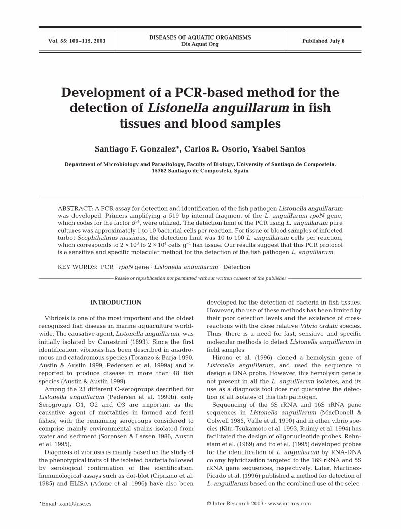
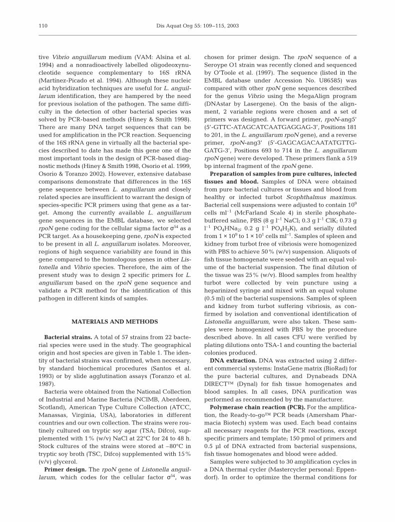
The specificity of the primers rpoN-ang5’ and rpoN-ang3’ was tested with 22 pure cultures of Listonellaanguillarum and 35 pure cultures of other bacterialspecies (Table 1). PCR reactions were performed atdifferent annealing temperatures ranging from 55 to65°C. At 58 and 62°C, all 22 L. anguillarum strainsproduced a unique and clear PCR band of theexpected 519 bp length. However, at 58°C this ampli-fication band was also produced with Vibrio ordalii(NCIMB 2167). This undesirable reaction was elimi-nated using the annealing temperature of 62°C. Whenthe other non-L. anguillarum bacterial strains inTable 1 were tested with the same primers andannealing temperatures, no amplification occurred.The results of PCR analysis performed at annealingtemperatures of 62 and 58°C with L. anguillarum andother bacterial species tested are shown in Figs. 1 & 2,respectively.
112
Fig. 1. Specific PCR productsobtained with pure cultures of7 Listonella anguillarum strainsand other related species at anannealing temperature of 62°C.Lane M: 1 kb molecular weightmarker; Lane 1: PCR negativecontrol; Lanes 2 to 5: L. anguil-larum 775 (Serotype O1), ATCC43305 (Serotype O1), ATCC43306 (Serotype O2α) and RV-22 (Serotype O2β), respectively;Lane 6: Vibrio ordalii: Lane 7: V.splendidus; Lanes 8 to 10: L. an-guillarum ATCC 43307 (Sero-type O3A), ATCC 43308 (Sero-type O4) and ATCC 43310
(Serotype O6), respectively
Fig. 2. PCR products obtainedwith pure cultures of Listonellaanguillarum strains and otherbacterial species at an annea-ling temperature of 58°C. LaneM: 1 kb molecular weightmarker; Lane 1: Vibrio ordalii;Lane 2: Aeromonas caviae;Lane 3: A. salmonicida; Lane 4:A. hydrophila; Lane 5: L. pela-gia; Lane 6: L. anguillarum TM-14 (Serotype O1); Lane 7: V.splendidus; Lanes 8 and 9: L.anguillarum ATCC 43305(Serotype O1) and ATCC 43306(Serotype O2α), respectively;Lane 10: PCR-negative control
519 bp
M 1 2 3 4 5 6 7 8 9 10
519 bp
M 1 2 3 4 5 6 7 8 9 10
Gonzalez et al.: PCR detection of Listonella anguillarum
Sensitivity of PCR system
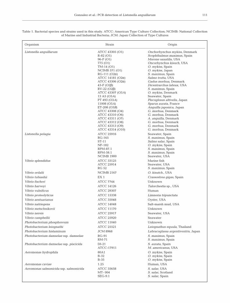
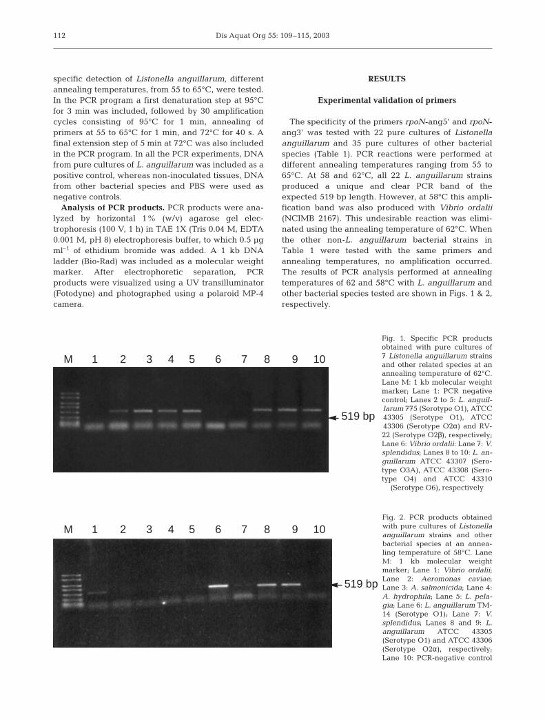
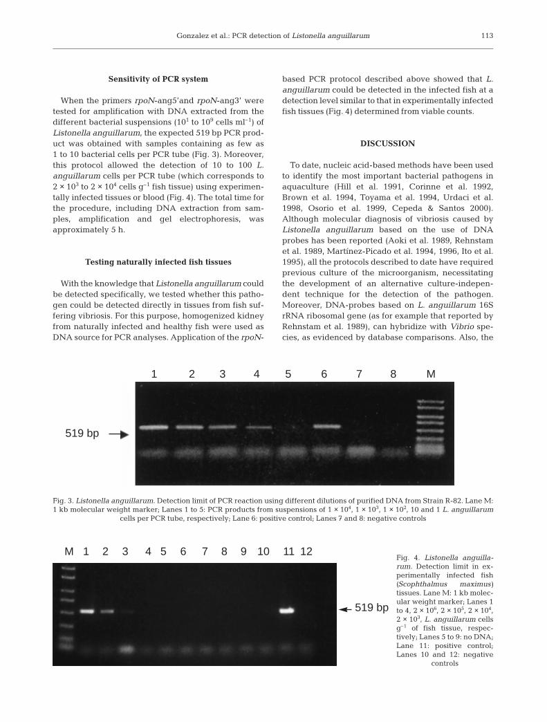
When the primers rpoN-ang5’and rpoN-ang3’ weretested for amplification with DNA extracted from thedifferent bacterial suspensions (101 to 109 cells ml–1) ofListonella anguillarum, the expected 519 bp PCR prod-uct was obtained with samples containing as few as1 to 10 bacterial cells per PCR tube (Fig. 3). Moreover,this protocol allowed the detection of 10 to 100 L.anguillarum cells per PCR tube (which corresponds to2 × 103 to 2 × 10⁴ cells g–1 fish tissue) using experimen-tally infected tissues or blood (Fig. 4). The total time forthe procedure, including DNA extraction from sam-ples, amplification and gel electrophoresis, wasapproximately 5 h.
Testing naturally infected fish tissues
With the knowledge that Listonella anguillarum couldbe detected specifically, we tested whether this patho-gen could be detected directly in tissues from fish suf-fering vibriosis. For this purpose, homogenized kidneyfrom naturally infected and healthy fish were used asDNA source for PCR analyses. Application of the rpoN-
based PCR protocol described above showed that L.anguillarum could be detected in the infected fish at adetection level similar to that in experimentally infectedfish tissues (Fig. 4) determined from viable counts.
DISCUSSION
To date, nucleic acid-based methods have been usedto identify the most important bacterial pathogens inaquaculture (Hill et al. 1991, Corinne et al. 1992,Brown et al. 1994, Toyama et al. 1994, Urdaci et al.1998, Osorio et al. 1999, Cepeda & Santos 2000).Although molecular diagnosis of vibriosis caused byListonella anguillarum based on the use of DNAprobes has been reported (Aoki et al. 1989, Rehnstamet al. 1989, Martínez-Picado et al. 1994, 1996, Ito et al.1995), all the protocols described to date have requiredprevious culture of the microorganism, necessitatingthe development of an alternative culture-indepen-dent technique for the detection of the pathogen.Moreover, DNA-probes based on L. anguillarum 16SrRNA ribosomal gene (as for example that reported byRehnstam et al. 1989), can hybridize with Vibrio spe-cies, as evidenced by database comparisons. Also, the
113
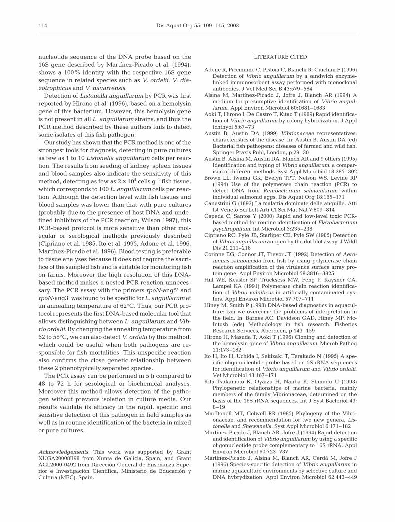
Fig. 3. Listonella anguillarum. Detection limit of PCR reaction using different dilutions of purified DNA from Strain R-82. Lane M:1 kb molecular weight marker; Lanes 1 to 5: PCR products from suspensions of 1 × 10⁴, 1 × 10³, 1 × 10², 10 and 1 L. anguillarum
cells per PCR tube, respectively; Lane 6: positive control; Lanes 7 and 8: negative controls
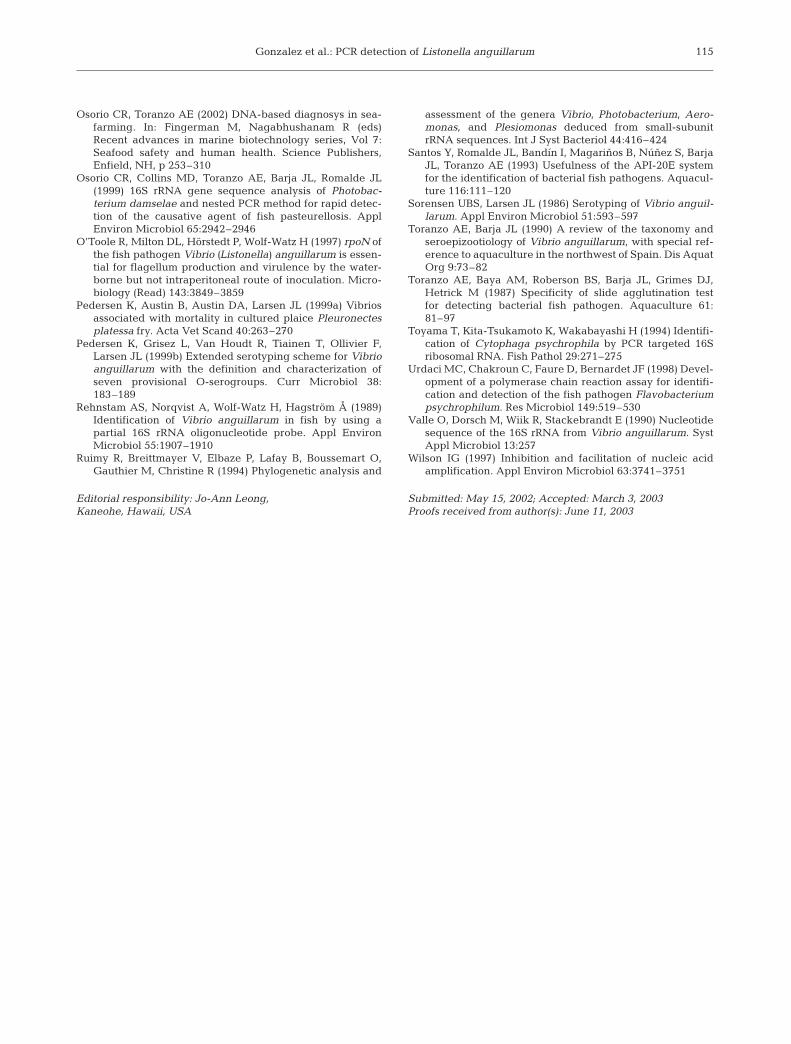
Fig. 4. Listonella anguilla-rum. Detection limit in ex-perimentally infected fish(Scophthalmus maximus)tissues. Lane M: 1 kb molec-ular weight marker; Lanes 1to 4, 2 × 10⁶, 2 × 10⁵, 2 × 104,2 × 103, L. anguillarum cellsg–1 of fish tissue, respec-tively; Lanes 5 to 9: no DNA;Lane 11: positive control;Lanes 10 and 12: negative
controls
1 2 3 4 5 6 7 8 M
519 bp
M 1 2 3 4 5 6 7 8 9 10 11 12
519 bp
Dis Aquat Org 55: 109–115, 2003
nucleotide sequence of the DNA probe based on the16S gene described by Martínez-Picado et al. (1994),shows a 100% identity with the respective 16S genesequence in related species such as V. ordalii, V. dia-zotrophicus and V. navarrensis.
Detection of Listonella anguillarum by PCR was firstreported by Hirono et al. (1996), based on a hemolysingene of this bacterium. However, this hemolysin geneis not present in all L. anguillarum strains, and thus thePCR method described by these authors fails to detectsome isolates of this fish pathogen.
Our study has shown that the PCR method is one of thestrongest tools for diagnosis, detecting in pure culturesas few as 1 to 10 Listonella anguillarum cells per reac-tion. The results from seeding of kidney, spleen tissuesand blood samples also indicate the sensitivity of thismethod, detecting as few as 2 × 10⁴ cells g–1 fish tissue,which corresponds to 100 L. anguillarum cells per reac-tion. Although the detection level with fish tissues andblood samples was lower than that with pure cultures(probably due to the presence of host DNA and unde-fined inhibitors of the PCR reaction; Wilson 1997), thisPCR-based protocol is more sensitive than other mol-ecular or serological methods previously described(Cipriano et al. 1985, Ito et al. 1995, Adone et al. 1996,Martínez-Picado et al. 1996). Blood testing is preferableto tissue analyses because it does not require the sacri-fice of the sampled fish and is suitable for monitoring fishon farms. Moreover the high resolution of this DNA-based method makes a nested PCR reaction unneces-sary. The PCR assay with the primers rpoN-ang5’ andrpoN-ang3’ was found to be specific for L. anguillarum atan annealing temperature of 62°C. Thus, our PCR pro-tocol represents the first DNA-based molecular tool thatallows distinguishing between L. anguillarum and Vib-rio ordalii. By changing the annealing temperature from62 to 58°C, we can also detect V. ordalii by this method,which could be useful when both pathogens are re-sponsible for fish mortalities. This unspecific reactionalso confirms the close genetic relationship betweenthese 2 phenotypically separated species.
The PCR assay can be performed in 5 h compared to48 to 72 h for serological or biochemical analyses.Moreover this method allows detection of the patho-gen without previous isolation in culture media. Ourresults validate its efficacy in the rapid, specific andsensitive detection of this pathogen in field samples aswell as in routine identification of the bacteria in mixedor pure cultures.
Acknowledgements. This work was supported by GrantXUGA20008B98 from Xunta de Galicia, Spain, and GrantAGL2000-0492 from Dirección General de Enseñanza Supe-rior e Investigación Científica, Ministerio de Educación yCultura (MEC), Spain.
LITERATURE CITED
Adone R, Piccininno C, Pistoia C, Bianchi R, Ciuchini F (1996)Detection of Vibrio anguillarum by a sandwich enzyme-linked immunosorbent assay performed with monoclonalantibodies. J Vet Med Ser B 43:579–584
Alsina M, Martínez-Picado J, Jofre J, Blanch AR (1994) Amedium for presumptive identification of Vibrio anguil-larum. Appl Environ Microbiol 60:1681–1683
Aoki T, Hirono I, De Castro T, Kitao T (1989) Rapid identifica-tion of Vibrio anguillarum by colony hybridization. J ApplIchthyol 5:67–73
Austin B, Austin DA (1999) Vibrionaceae representatives:characteristics of the disease. In: Austin B, Austin DA (ed)Bacterial fish pathogens: diseases of farmed and wild fish.Springer Praxis Publ, London, p 29–30
Austin B, Alsina M, Austin DA, Blanch AR and 9 others (1995)Identification and typing of Vibrio anguillarum: a compar-ison of different methods. Syst Appl Microbiol 18:285–302
Brown LL, Iwama GK, Evelyn TPT, Nelson WS, Levine RP(1994) Use of the polymerase chain reaction (PCR) todetect DNA from Renibacterium salmonilarum withinindividual salmonid eggs. Dis Aquat Org 18:165–171
Canestrini G (1893) La malattia dominate delle anguille. AttiIst Veneto Sci Lett Arti Cl Sci Mat Nat 7:809–814
Cepeda C, Santos Y (2000) Rapid and low-level toxic PCR-based method for routine identification of Flavobacteriumpsychrophilum. Int Microbiol 3:235–238
Cipriano RC, Pyle JB, Starliper CE, Pyle SW (1985) Detectionof Vibrio anguillarum antigen by the dot blot assay. J WildlDis 21:211–218
Corinne EG, Connor JT, Trevor JT (1992) Detection of Aero-monas salmonicida from fish by using polymerase chainreaction amplification of the virulence surface array pro-tein gene. Appl Environ Microbiol 58:3816–3825
Hill WE, Keasler SP, Trucksess MW, Feng P, Kaysner CA,Lampel KA (1991) Polymerase chain reaction identifica-tion of Vibrio vulnificus in artificially contaminated oys-ters. Appl Environ Microbiol 57:707–711
Hiney M, Smith P (1998) DNA-based diagnostics in aquacul-ture: can we overcome the problems of interpretation inthe field. In: Barnes AC, Davidson GAD, Hiney MP, Mc-Intosh (eds) Methodology in fish research. FisheriesResearch Services, Aberdeen, p 143–159
Hirono H, Masuda T, Aoki T (1996) Cloning and detection ofthe hemolysin gene of Vibrio anguillarum. Microb Pathog21:173–182
Ito H, Ito H, Uchida I, Sekizaki T, Terakado N (1995) A spe-cific oligonucleotide probe based on 5S rRNA sequencesfor identification of Vibrio anguillarum and Vibrio ordalii.Vet Microbiol 43:167–171
Kita-Tsukamoto K, Oyaizu H, Nanba K, Shimidu U (1993)Phylogenetic relationships of marine bacteria, mainlymembers of the family Vibrionaceae, determined on thebasis of the 16S rRNA sequences. Int J Syst Bacteriol 43:8–19
MacDonell MT, Colwell RR (1985) Phylogeny of the Vibri-onaceae, and recommendation for two new genera, Lis-tonella and Shewanella. Syst Appl Microbiol 6:171–182
Martínez-Picado J, Blanch AR, Jofre J (1994) Rapid detectionand identification of Vibrio anguillarum by using a specificoligonucleotide probe complementary to 16S rRNA. ApplEnviron Microbiol 60:723–737
Martínez-Picado J, Alsina M, Blanch AR, Cerdá M, Jofre J(1996) Species-specific detection of Vibrio anguillarum inmarine aquaculture environments by selective culture andDNA hybrydization. Appl Environ Microbiol 62:443–449
114
Gonzalez et al.: PCR detection of Listonella anguillarum
Osorio CR, Toranzo AE (2002) DNA-based diagnosys in sea-farming. In: Fingerman M, Nagabhushanam R (eds)Recent advances in marine biotechnology series, Vol 7:Seafood safety and human health. Science Publishers,Enfield, NH, p 253–310
Osorio CR, Collins MD, Toranzo AE, Barja JL, Romalde JL(1999) 16S rRNA gene sequence analysis of Photobac-terium damselae and nested PCR method for rapid detec-tion of the causative agent of fish pasteurellosis. ApplEnviron Microbiol 65:2942–2946
O’Toole R, Milton DL, Hörstedt P, Wolf-Watz H (1997) rpoN ofthe fish pathogen Vibrio (Listonella) anguillarum is essen-tial for flagellum production and virulence by the water-borne but not intraperitoneal route of inoculation. Micro-biology (Read) 143:3849–3859
Pedersen K, Austin B, Austin DA, Larsen JL (1999a) Vibriosassociated with mortality in cultured plaice Pleuronectesplatessa fry. Acta Vet Scand 40:263–270
Pedersen K, Grisez L, Van Houdt R, Tiainen T, Ollivier F,Larsen JL (1999b) Extended serotyping scheme for Vibrioanguillarum with the definition and characterization ofseven provisional O-serogroups. Curr Microbiol 38:183–189
Rehnstam AS, Norqvist A, Wolf-Watz H, Hagström Å (1989)Identification of Vibrio anguillarum in fish by using apartial 16S rRNA oligonucleotide probe. Appl EnvironMicrobiol 55:1907–1910
Ruimy R, Breittmayer V, Elbaze P, Lafay B, Boussemart O,Gauthier M, Christine R (1994) Phylogenetic analysis and
assessment of the genera Vibrio, Photobacterium, Aero-monas, and Plesiomonas deduced from small-subunitrRNA sequences. Int J Syst Bacteriol 44:416–424
Santos Y, Romalde JL, Bandín I, Magariños B, Núñez S, BarjaJL, Toranzo AE (1993) Usefulness of the API-20E systemfor the identification of bacterial fish pathogens. Aquacul-ture 116:111–120
Sorensen UBS, Larsen JL (1986) Serotyping of Vibrio anguil-larum. Appl Environ Microbiol 51:593–597
Toranzo AE, Barja JL (1990) A review of the taxonomy andseroepizootiology of Vibrio anguillarum, with special ref-erence to aquaculture in the northwest of Spain. Dis AquatOrg 9:73–82
Toranzo AE, Baya AM, Roberson BS, Barja JL, Grimes DJ,Hetrick M (1987) Specificity of slide agglutination testfor detecting bacterial fish pathogen. Aquaculture 61:81–97
Toyama T, Kita-Tsukamoto K, Wakabayashi H (1994) Identifi-cation of Cytophaga psychrophila by PCR targeted 16Sribosomal RNA. Fish Pathol 29:271–275
Urdaci MC, Chakroun C, Faure D, Bernardet JF (1998) Devel-opment of a polymerase chain reaction assay for identifi-cation and detection of the fish pathogen Flavobacteriumpsychrophilum. Res Microbiol 149:519–530
Valle O, Dorsch M, Wiik R, Stackebrandt E (1990) Nucleotidesequence of the 16S rRNA from Vibrio anguillarum. SystAppl Microbiol 13:257
Wilson IG (1997) Inhibition and facilitation of nucleic acidamplification. Appl Environ Microbiol 63:3741–3751
115
Editorial responsibility: Jo-Ann Leong, Kaneohe, Hawaii, USA
Submitted: May 15, 2002; Accepted: March 3, 2003Proofs received from author(s): June 11, 2003