Embed Size (px)
Citation preview
DEVELOPMENT OF A ROBUST AND IMPROVED SYSTEM FOR STUDYING INTERACTIONS BETWEEN CCL20 AND CCR6 USING BOTH RECOMBINANT AND
CHEMICALLY SYNTHESIZED RHESUS MACAQUE CHEMOKINES
by
Cynthia René Klamar
BS, Biology, University of Pittsburgh, 2003
Submitted to the Graduate Faculty of
Infectious Diseases and Microbiology
Graduate School of Public Health in partial fulfillment
of the requirements for the degree of
Master of Science
University of Pittsburgh
2010
ii
UNIVERSITY OF PITTSBURGH
Graduate School of Public Health
This thesis was presented
by
Cynthia René Klamar
It was defended on
July 21, 2010
and approved by
Velpandi Ayyavoo, Ph.D Associate Professor
Department of Infectious Diseases and Microbiology Graduate School of Public Health, University of Pittsburgh
Michael Murphey-Corb, Ph.D Professor
Department of Microbiology and Molecular Genetics, School of Medicine Department of Infectious Diseases and Microbiology
Graduate School of Public Health, University of Pittsburgh
Thesis Advisor: Todd Reinhart, Sc.D Professor
Department of Infectious Diseases and Microbiology Graduate School of Public Health, University of Pittsburgh
iii
Copyright © by Cynthia René Klamar
2010
iv
The chemokine CCL20 is thought to be an integral part of the communication between
the innate and adaptive arms of the immune system, due to expression of the cognate receptor,
CCR6, on immature dendritic cells and on memory T cells and B cells. Interest in this particular
chemokine/chemokine receptor interaction has grown over time and more recently due to roles in
SIV infection, mucosal immunology, and vaccinology. The need to further study the
CCL20/CCR6 interactions is bolstered by our laboratory’s previous findings of increased
expression of CCL20 in acutely SIV infected lymph nodes and the increased expression of
CCL20 in response to PAMPs in cells of lymphatic vessels. This thesis aims to develop and
improve a system for studying the interaction between CCL20 and CCR6. I have found that the
recombinant expression system utilized to obtain macaque chemokines provided highly pure
fusion proteins. However, cleavage of the fusion protein into macaque CCL20 has been
inefficient. Rhesus macaque CCL20 chemically synthesized using regioselective cyclization was
highly biologically active using the chemotaxis assay and stable cell lines expressing CCR6.
Chemotactic inhibition studies identified five compounds that inhibited CCL20-induced
chemotaxis. The surfactant, GML, did not inhibit CCL20-induced migration. The anti-
inflammatory botanicals, EGCG and gallotannin, both inhibited CCL20-driven migration at high
concentrations. The three CCR6 extracellular loop mimetic peptides also partially inhibited
CCL20-induced migration at high concentrations. In conclusion, I have utilized both a
DEVELOPMENT OF A ROBUST AND IMPROVED SYSTEM FOR STUDYING INTERACTIONS BETWEEN CCL20 AND CCR6 USING BOTH RECOMBINANT
AND CHEMICALLY SYNTHESIZED RHESUS MACAQUE CHEMOKINES
Cynthia René Klamar, M.S.
University of Pittsburgh, 2010
v
recombinant protein expression system and regioselective cyclization peptide synthesis to obtain
bioactive, nonhuman primate chemokines. I have also successfully developed an in vitro system
to study CCL20-induced migration, and have identified a number of botanical and biochemical
elements that inhibit CCL20-induced migration. The public health significance of this study is
related to the fact that vaccine efficacy may be affected by anti-inflammatory compounds that
inhibit CCL20-mediated chemotaxis. Another way in which public health could be affected by
this study is in using the anti-inflammatory compounds studied to treat chronic inflammatory
conditions in which the pathology of the disease is related to up-regulation of CCL20 and CCR6.
vi
TABLE OF CONTENTS
1.0 Introduction ......................................................................................................................... 1
1.1 Chemokines and Chemokine Receptors ............................................................................. 1
1.1.1 Chemokine CCL20: Expression and Other Properties ................................................ 2
1.1.2 Chemokine Receptor CCR6: Expression and Other Properties .................................. 3
1.1.3 HIV and CCL20 / CCR6 ............................................................................................. 4
1.2 Recombinant Protein Purification ....................................................................................... 6
1.2.1 Origami B DE3 and Rosettagami B DE3 .................................................................... 7
1.2.2 SUMO Fusion Protein Expression .............................................................................. 8
1.3 Peptide Synthesis ................................................................................................................ 8
2.0 Statement of the Problem .................................................................................................. 10
2.1 Specific Aim 1: To Develop a System to Generate Rhesus Macaque CCL20 ................. 11
2.2 Specific Aim 2: To Develop and Apply a Functional Assay for Testing the Bioactivity of
Recombinant and Synthetic Macaque CCL20 .................................................................. 11
3.0 Materials and Methods ...................................................................................................... 13
3.1 Generation of rhesus macaque chemokine ....................................................................... 13
3.2 Application of chemotaxis functional assay for in vitro system development ................. 16
4.0 Results: Recombinant Protein Expression ........................................................................ 20
4.1 Expression and purification of a SUMO-CCL20 fusion protein. ..................................... 20
4.1.1 Cleavage of the SUMO-CCL20 fusion protein ......................................................... 24
4.2 Strategies to increase efficiency of cleavage .................................................................... 26
4.2.1 Multi-parameter cleavage experimental design ........................................................ 27
vii
4.2.1.1 Differences i n buf fer, q uantity o f pr otease or pr otein, t ime a nd t emperature do not
increase the efficiency of cleavage of SUMO-CCL20. ................................................. 29
4.2.2 Rosettagami and Origami B DE3 strain of E. coli .................................................... 35
4.2.2.1 Differences i n S UMO-CCL20 s equence a t pos ition 32 do not a ffect O rigami B D E3
expression or cleavage of protein. ................................................................................. 37
4.2.2.2 Expression and pur ification of SUMO-CCL20 isolates are d ifferent in Rosettagami B
DE3 cells. ....................................................................................................................... 39
4.3 Peptide synthesis ............................................................................................................... 41
4.4 Overall Protein Expression System Summary .................................................................. 41
5.0 Results: A pplication o f a F unctional C hemotaxis A ssay to D evelop f or V itro S ystem
Development ..................................................................................................................... 43
5.1 Primary Cells .................................................................................................................... 43
5.2 Regioselectively cyclized synthetic rhesus CCL20 is highly bioactive............................ 46
5.2.1 Stable Cell Line Development .................................................................................. 48
5.2.2 A new approach for obtaining highly functional CCR6 expressing cells ................. 49
5.3 Inhibiting the CCL20/CCR6 Interaction ........................................................................... 51
5.3.1 GML does not inhibit CCL20-mediated chemotaxis ................................................ 51
5.3.2 CCR6 receptor peptide mimetics ECL-2 inhibit CCL20-induced migration ............ 53
5.3.3 Anti-Inflammatory Botanical Inhibitors .................................................................... 57
5.3.3.1 EGCG Inhibition of CCL20-mediated Chemotaxis ....................................................... 57
5.3.3.2 Gallotannin Inhibition of CCL20-mediated Chemotaxis ............................................... 58
5.4 Overall Functional Application Summary ........................................................................ 59
6.0 Discussion ......................................................................................................................... 61
viii
6.1 Protein Expression ............................................................................................................ 61
6.2 Application of a functional chemotaxis assay for in vitro system development .............. 64
6.3 Conclusion ........................................................................................................................ 66
6.3.1 Next Steps and Future Studies ................................................................................... 66
6.3.2 Public Health Revelance of this Study ...................................................................... 69
BIBLIOGRAPHY ......................................................................................................................... 72
ix
LIST OF FIGURES
Figure 1. Amino acid differences of rhesus macaque CCL20. .................................................... 21
Figure 2. SUMO-CCL20 Fusion protein purification and cleavage. ........................................... 22
Figure 3. SUMO-CCL20 is cleaved inefficiently, although SUMO protease is highly efficient at
cleaving control SUMO fusions............................................................................................ 25
Figure 4. Multi-parameter cleavage analysis. .............................................................................. 28
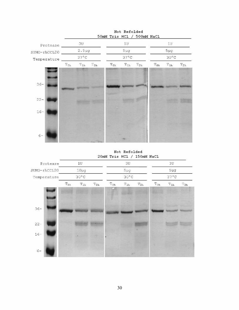
Figure 5. Changing four different variables did not increase cleavage efficiency of the 'not
refolded' SUMO-CCL20 fusion. ........................................................................................... 31
Figure 6. Varying temperature and incubation time did not increase the efficiency of cleavage of
SUMO-CCL20 fusion. .......................................................................................................... 33
Figure 7. Isolation of clones of the plasmids p.SUMO.CCL20.1 and p.SUMO.CCL20.2. ......... 36
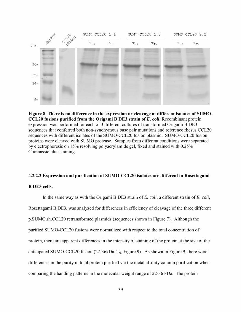
Figure 8. There is no difference in the expression or cleavage of different isolates of SUMO-
CCL20 fusions purified from the Origami B DE3 strain of E. coli. ..................................... 39
Figure 9. Rosettagami B DE3 cells appear to differ in the expression and cleavage of different
isolates of SUMO-CCL20 fusions.. ...................................................................................... 40
Figure 10. CCR6 is expressed on both B- and T-lymphocytes. ................................................... 44
Figure 11. Normal macaque PBMCs do not respond to synthetic macaque CCL20. .................. 45
Figure 12. Peak migration of transiently transfected L1.2 cells is towards 10nM synthetic
macaque CCL20.................................................................................................................... 47
Figure 13. Only pre-treated stable CCR6 L1.2 cells express CCR6 and migrate towards
synthetic macaque CCL20. ................................................................................................... 50
Figure 14. GML does not inhibit CCL20-mediated chemotaxis. ................................................ 53
Figure 15. Design of the second extracellular loop of CCR6 (ECL-2).. ...................................... 54
Figure 16. R6 ECL-2 (receptor peptide mimetics) inhibit CCL20-induced migration. ............... 57
Figure 17. EGCG inhibits CCL20-mediated migration. .............................................................. 58
Figure 18. Gallotannin inhibits CCL20-mediated migration. ...................................................... 59
x
ACKNOWLEDGEMENT
I would like to thank my advisor and mentor Dr. Reinhart, first and foremost. He has been the
best boss, mentor and teacher I’ve ever had. His energy, drive, focus and overall dedication to
teaching us good science has made me strive to plan the best experiment I can almost every time
and to be waiting on pins and needles for the results. He has been very influential in making me
the curious scientist I now am. I would additionally like to thank Yu-jen Tung who developed
and tweaked the protein expression protocol and who taught me some of my first graduate bench
work. I would like to thank my past lab mates, Yu-jen Tung, Dr. Sudheer Raviuiri, Dr. Yongjon
Sui, Dr. Amarendra Pegu, Kristi Gaus, and Charis Tjoeng, for all of the support and rousing
scientific discussions. In my opinion, I am part of probably one of the best labs, and would like
to thank our current lab members who are colleagues and friends to me for all their support,
technical and emotional, Beth Junecko, Carissa Flores, Dr. Shulin Qin, Stella Berendam and
Chris Bowen. I would like to acknowledge and thank Dr. Rinaldo’s Flow lab for all of the
support in training and educating me in the world of flow cytometry, Luann Borowski, Kim
Stojka, Edwin and Jill. Finally, I greatly appreciate all of the support and encouragement I have
received throughout this journey from my family and friends.
1
1.0 Introduction
The complexity of the immune system provides the body with a method to fight pathogen
associated challenges. The immune response to inflammation caused by the invading pathogens
develops as infected or injured cells in the local environment produce signals to aid the local
entry of specialized immune cells. A gradient of small proteins, or chemokines, drives the
homing of the immune cells.
1.1 Chemokines and Chemokine Receptors
Chemokines are small, structurally related chemo-attractant cytokines, in the size range
of 8-10kDa, that are involved in a wide range of biological functions, from hematopoeisis to
organogenesis3, 43. The expression of some chemokines is required to direct the architecture of
developing lymphoid tissue42, 43. A third functional activity of chemokines is to aid in the
activation of a specific adaptive response, either a TH1 or TH2 immune response45, 46, 47.
Chemokine biology is important in immunology and infectious disease in that in some
pathogenic viral and bacterial infections, as well as cancers and autoimmune diseases, dys-
regulation of chemokines and expression of chemokine receptors can contribute to the
pathogenesis of the disease9, 16, 44. One of the main functions of chemokines is to induce the
trafficking of leukocytes into sites of inflammation3, 12.
Chemokines are defined and functionally grouped in at least two ways. First they are
classified by the sequence of the amino acids near the amino-terminus of the protein, i.e. by the
proximity and location of the amino-terminal conserved cysteines that form the disulfide bridges
in the protein, specifically they are CC, CXC, CX3Cand XC chemokines12, 13. The conserved
cysteines of CC chemokines are directly next to each other. The cysteines of CXC and CX3C
2
chemokines have as the name implies some other amino acid present in the sequence before the
next conserved cysteine. XC chemokines have only one conserved cysteine in the amino-
terminal region of the protein sequence12, 13.
Chemokines are also defined functionally by their expression profile as present during
either homeostasis or inflammation3, 12. Homeostatic chemokines are expressed under normal
physiological conditions and aid in the development of secondary microstructures within
lymphoid tissue19, 42, 43. For example, CCL21 is a homeostatic chemokine that is highly
expressed in the paracortical region of peripheral lymph nodes41, 45. This expression aids in the
cross talk, maturation, and antigen education of naïve T cells and mature dendritic cells (DCs)
which both express the receptor, CCR741, 42, 56, 57. Another homeostatic chemokine CXCL13 is
expressed mainly in the germinal centers of lymph nodes, aiding in the development and
maturation of B cells41. An additional function of a homeostatic chemokine is to draw immature
dendritic cells (DCs) into the tissue, so that these local DCs can continually sample the local
environment and be poised to respond to pathogen-associated inflammation44, 47.
Inflammatory chemokines are up-regulated via the NFκB pathway during pathogen
challenge, in response to increased IFNγ expression9, 44. The chemokine CXCL10 is a prime
example of an inflammatory chemokine, due to findings that upon SIV infection, as IFNγ goes
up, this chemokine is also highly expressed6, 44.
1.1.1 Chemokine CCL20: Expression and Other Properties
The chemokine CCL20 or MIP-3α, (macrophage inflammatory protein) is a CC
chemokine13, with unique properties in that in some local tissue environments CCL20 is thought
to have properties of both the homeostatic and inflammatory chemokines19. As expected for
other inflammatory chemokines, CCL20 is secreted by macrophages in response to inflammation
3
and has been shown to be under the control of the NFκB inflammation response pathway26, 32. In
the skin, epithelial cells secrete CCL20 at low constitutive levels under normal conditions; on the
other hand, with a pathogen challenge or chronic inflammation as in atopic dermatitis, CCL20 is
up-regulated32. Additionally, the expression of CCL20 in the skin has been related to the
homeostatic influx and maintenance of Langerhan cell (LC) precursors56, 57. Both properties of
homeostatic and inflammatory expression have been shown in the gut, where the follicular
associated epithelial dome in Peyer’s patches constitutively expresses CCL2033, 34. The
importance of a homeostatic interaction of CCL20 with its receptor, CCR6, in the development
of gut-associated lymph tissue (GALT) is indicated by the fact that CCR6-/- mice have reduced
numbers and cellularity of Peyers’ patches36. CCL20 is also up-regulated due to inflammation in
the gut, as in SIV infection6, or to chronic inflammation as in inflammatory bowel disorder or
Crohns’ disease27. In the peripheral lymph node, CCL20 is shown to be an inflammatory
chemokine, where the constitutive level of CCL20 expression is increased during acute SIV
infection6. Another indicator of the inflammatory properties of CCL20 is that lymphatic
endothelial cells increase CCL20 expression with IL-1β or LPS treatment via TLR signaling62.
In the female genital tract, there is evidence of both homeostatic and inflammatory chemokine
properties2, 7. Recent vaccine adjuvant studies of the female genital tract define a successful
adjuvant or immunogen by increases in CCL20 expression7. For the above reasons, immune
function of CCL20 is unique depending on the local tissue environment.
1.1.2 Chemokine Receptor CCR6: Expression and Other Properties
The receptor for CCL20 is CCR6 19. Adaptive immune cells including memory T cells,
mononuclear cells, B cells, and immature dendritic cells (DCs) express CCR6 32. CCL20 and
CCR6 are thought to be integral parts of the communication between the innate and adaptive
4
arms of the immune system because of the expression of CCR6 on immature DCs and memory T
cells19, 37. Local response to inflammation up-regulates CCL20 expression56. This and other
signals bring the immature DCs towards the focus of increased CCL20 expression where they
become mature and activated, following antigen uptake and processing 56. DCs mature losing
some level of CCR6 expression and gaining CCR7 expression, which drives the mature DCs to
home to the lymph node where they interact with naïve T cells3. Memory T cells may migrate
into the inflamed environment via some of the same signals, and as the DC mature and present
antigen to the memory T cells, they can begin to display effector immunological properties and
contribute to resolution of the pathogen challenge and local inflammation28.
1.1.3 HIV and CCL20 / CCR6
The most recognized chemokine receptors in SIV or HIV immunology are CCR5 and
CXCR4, which are used by gp120 protein for viral entry into CD4 positive cells38, 39. One
commonly studied aspect of HIV immunology is the reduction of immune cells associated with
the gut-associated lymphoid tissue (GALT) during acute infection15, 20. It has been shown that
the CD4 T cells in the GALT are targeted by virus infection and these numbers do not rebound to
the original percentages even with HAART treatment20, 21. The chemokine/chemokine receptor
pair of CCL20 and CCR6 become important considerations during SIV or HIV infection for a
few reasons. One reason is the CCL20/CCR6 pair are thought to be influential in regulating
immune responses in both regions affected by sexual transmission of HIV, the vagina and the
gut, where the primary immune response is mounted15, 20, 21, 50. Another reason is that DCs
(which express CCR6 and respond to CCL20) have been shown to be some of the first targets of
HIV replication, in addition to serving as ‘trojan horses’ for virus dissemination21, 48.
5
CCL20 has been shown to be up-regulated in both the gut (colon) and lymph node during
acute SIV infection6. This up-regulation is congruent during other lentiviral infections such as
by human T-cell leukemia virus type 1 (HTLV) where there is up-regulation of CCL20
expression9. Further indication of a dysfunctional relationship of CCL20 and HIV is that HIV
proteins reduce chemotaxis of B cells to CCL20, which are a cell type that expresses CCR61.
Ghosh, et al, further identifies anti-HIV properties of CCL20 where CCL20 directly inhibited
HIV replication55. In the female macaque vagina, treatment with GML (glycerol monolaurate), a
biochemical surfactant, inhibited SIV replication10. Reported in the same study, CCL20
expression in the vagina was increased with SIV infection and that upon observing inhibition of
SIV replication with GML treatment, CCL20 expression was reduced as well 10. Finally, another
CCR6 mediated anti-HIV activity was observed by Lafferty, et al49. The authors found that
signaling through the CCR6 receptor up-regulated the expression of a naturally occurring anti-
HIV molecule, APOBEC3G49.
CCR6 and CCL20 are associated with mucosal tissue, both the female genital tract and
gut lymphoid tissue, including Peyers patches59, 28, 33, 35. Although the pathways and functional
roles have not been well developed yet, there may be great importance to the CCR6/CCL20
interaction early during HIV infection. It may be that upon sexual transmission, the epithelial
layers of the female genital tract or anal/rectal junction is damaged. This epithelial damage, as
well as virus-infected cells, free virus and other proteins and factors present in mucus or
ejaculate perpetuates a local inflammatory response, and the down-stream up-regulation of
CCL20. The increased levels of CCL20 as well as other inflammatory cytokines and adhesion
signals increase the number of immature DCs brought into the environment, which can serve as
‘trojan horses’ for virus dissemination or as target cells21, 48. While primary up-regulation of
6
CCL20 is likely not the only answer to HIV immune evasion and persistence, there is a
substantial body of evidence suggesting that consideration of the CCR6/CCL20 interactions
during SIV/HIV may be lucrative in developing successful HIV vaccines or therapeutics.
Models that will improve our understanding of the CCR6 and CCL20 pathways and
interactions in health and disease, including macaque models, need to be developed and
improved. Since most reagents commercially available are of human specificity, an efficient
protocol for expression and purification of non-human primate chemokines must be developed to
accommodate future studies. In particular, there are few if any reagents for the study of
nonhuman primate chemokines, including CCL20. Therefore, the primary goal of these studies
was to generate macaque CCL20 and develop a system to characterize of the CCR6: CCL20
interaction.
1.2 Recombinant Protein Purification
Recombinant DNA technology has developed exponentially as the use of genes and
proteins in vaccines and therapeutics has developed with modern medicine. Recombinant protein
expression is a method for obtaining large quantities of protein that has long been utilized
commercially75, 83. The systems that have been used are both eukaryotic and prokaryotic, viral
vector or plasmid based systems each having advantages and disadvantages in the production,
yield and safety of the desired product75, 83.
One of the first prokaryotic systems used to produce commercial recombinant proteins is
the gram-negative bacteria E. coli75. Advantages to expression in E. coli are that cultures are
grown easily to a high density in simple nutrition media and that a large body of knowledge
about recombinant expression in E. coli has been gathered to enhance production of proteins75.
In genetically un-manipulated E. coli, the reduced state of the cytosol inhibits di-sulfide bond
7
formation and endogenous enzymes rarely mark the protein for proteolysis75. Other protein
modifications, such as glycosylation, do not occur in E. coli75.
Recombinant protein expression in E. coli has been manipulated by knowledge of
bacterial cellular metabolism pathways and antibiotic resistant plasmids to enhance protein
production75. Plasmids are mutated to contain antibiotic resistance, a strong promoter, and the
lacz gene linked to the recombinant gene83. A relatively universal manipulation utilizing these
factors is that cells that have taken up plasmids encoding antibiotic resistance genes also acquire
the control of the T7 promoter and the lacz gene and the recombinant protein is expressed upon
the addition of IPTG (isopropyl β-D-1-thiogalactopyranoside)75, 83. Other manipulations of
recombinant protein expression in E. coli include transforming the cells with plasmids encoding
genes that enhance production of a certain type of protein. In the end, based on the type of
recombinant protein desired, a wide variety of E. coli competent cells are available and are
chosen based on the protein translational properties that will ease the production of the
recombinant protein.
1.2.1 Origami B DE3 and Rosettagami B DE3
The strains of E. coli utilized in the expression and purification protocol here are the
Origami B DE3 and Rosettagami B DE3 (Invitrogen) cells, based on properties that would
enhance the production of a recombinant non-human primate chemokine. Both strains are
derived from a mutant cell line, DE3 lysogens, that express protein in response to IPTG under
the control of T7 promoter and lacz metabolic pathway. They both also express mutant plasmids
that allow for the formation of disulfide bonds in the cytosol of the E. coli and more efficient
protein refolding in vivo72.
8
Recombinant mammalian protein expression in E. coli is made more difficult by the
rarity of certain tRNAs in E. coli5. Low expression of tRNAs that complement the codon push
the cell to mis-incorporate an amino acid and therefore potentially change the structure and
function of the resultant recombinant protein5. To combat this problem, in addition to the other
plasmids contained in the Origami B DE3 cell, Rosettagami B DE3 cells encode the rare tRNAs
that are lacking in the E. coli but are necessary for proper mammalian translation of protein5. In
conclusion, we used strains of E. coli that were purported to enhance the expression of our
desired recombinant proteins, chemokines.
1.2.2 SUMO Fusion Protein Expression
A proper protein expression technology is also used to express recombinant proteins of
interest. The SUMO (Small Ubiquitin-Like Modifier) plasmid expression system (Life Sensors)
provides specificity during purification of the recombinant proteins, as both the fusion protein
and the protease contain 6-X-His tags allowing purification of these elements, simply and to a
high degree, by metal affinity column purification. Another advantage of the SUMO expression
system is that the exact n-terminal residue of the recombinant protein can be defined (Life
Sensors). The SUMO protease is highly specific in that the tertiary structure of the fusion
protein is the only substrate for the enzyme cleavage reaction70, 71, 79. In most cases, specificity
of cleavage results in un-altered recombinant protein, in this study, rhesus macaque CCL20.
1.3 Peptide Synthesis
Peptides can be synthesized using organic chemistry protocols, but it is a more expensive
biochemical technique used to obtain large quantities of small proteins81, 82. There are two major
methods by which peptides are synthesized by coupling the N-terminus of an amino acid to the
9
C-terminus of another, beginning with the C-terminal amino acid in the peptide and ending at the
n-terminal residue82.
Liquid phase peptide synthesis is the classical method for chemically producing
peptides81. The other method for synthesizing peptides is called solid phase synthesis, where the
c-terminal amino acid is bound to insoluble beads or resin82. The side chains of the amino acid
can be protected by tboc (using trifluoroacetic acid) or fmoc (using 9-fluorenylmethylcarbonyl)
chemistry81, 82. The difference between each is that upon building the peptide either a neutral
(tboc) or positively charged amine group (fmoc) is exposed to the next amino acids c-terminus82.
For the synthesis of our rhesus macaque CCL20, regioselective cyclization was used. By this
solid phase peptide synthesis, sulfides are protected from disulfide bond formation which occurs
regularly in protein synthesis by thiol protecting groups81, 82. Additionally, in this type of
synthesis, as the amino acid chain is synthesized, sulfides can be revealed (or de-protected) and
the disulfide bond formation can be directed82.
10
2.0 Statement of the Problem
Understanding the contribution of chemokines and their receptors to the complexity of
the immune system is important to modeling the progression of immunologic and infectious
diseases. CCL20 is constitutively and over expressed during homeostatic and inflammatory
conditions, respectively. The cognate receptor CCR6 is unique in that it is one of few non-
promiscuous chemokine receptors12. The only other known ligands for CCR6 are a family of
host anti-microbial peptides, the β-defensins8, 32. Inflammatory expression and response to
CCL20 may provide a link between the innate and adaptive immune systems. Response of
immature dendritic cells to CCL20 via CCR6 signaling makes study of CCL20 as a vaccine
adjuvant a likely course. Expression of CCL20 (vagina2, 7, 10, 59, likely the anal/rectal junction,
colon19, and GALT19 ) in tissue compartments exposed to or serve as primary sites of virus
dissemination HIV during sexual transmission also lends credence to study of CCR6/CCL20 in
HIV vaccines and mucosal immunity. We have shown that CCL20 is expressed by lymphatic
endothelial cells at the afferent face of LNs and this is upregulated during the early phase of SIV
infection48, 62. The recently revealed anti-HIV activity of CCL20 and intracellular ligand-driven
signaling through CCR6 provides additional evidence that the CCR6/CCL20 interaction is
important in understanding innate immune properties that combat HIV infection49, 55. These
findings have provided impetus for study of this chemokine/chemokine receptor interaction in
vaccine adjuvant studies, mucosal immunology and HIV or SIV infection. The overall
objective of this study was to develop a robust and improved system for studying the
interaction between CCL20 and CCR6 using both recombinant and chemically synthesized
nonhuman primate chemokines. In this study, generating macaque CCL20 provides a method
11
to study the efficacy of using CCL20 as a vaccine adjuvant in future studies. Identification of
inhibitory compounds of CCL20-mediated chemotaxis may provide possible treatments for
chronic inflammatory diseases to which some of the disease pathology is associated with up-
regulated CCL20 expression (atopic dermatitis32, Crohn’s disease27, etc.).
2.1 Specific Aim 1: To Develop a System to Generate Rhesus Macaque CCL20
Expression of chemokines in E. coli is a well-established method for obtaining large
quantities of recombinant proteins. However, there are few, if any, commercially available
macaque chemokines for in vitro and in vivo studies of monkey models of immunology and
disease. Recombinant expression of proteins can be a flexible and cost effective way to obtain a
wide variety of molecules in large quantities. In addition, such a strategy readily allows
mutagenesis as a relatively simple way to obtain different forms of proteins for study.
Chemical synthesis of peptides is an alternative method for obtaining highly pure proteins
for experimental application. Additionally, regioselectively cyclized peptide synthesis can
provide a method to produce highly pure proteins with directed di-sulfide bonding, which is
thought to be required for proper tertiary structure formation of chemokines and which could
contribute to more robust bioactivity of the resultant molecule. I have established systems
whereby non-human primate chemokines were obtained by recombinant expression and whereby
chemically synthesized non-human primate chemokines were found to be bioactive.
2.2 Specific Aim 2: To Develop and Apply a Functional Assay for Testing the
Bioactivity of Recombinant and Synthetic Macaque CCL20
The importance of studying the interaction between CCL20 and CCR6 is defined by
vaccine adjuvant studies, DC biology, and mucosal immunology. Due to the expression of
multiple chemokine receptors on most cell types, studying specific chemokine and chemokine
12
receptor interaction is difficult with primary cells. A robust functional assay is important for
studying the activity any specific chemokine. Therefore, I established a robust chemotaxis assay
with the L1.2 murine pre-B cell line transfected both transiently and stably to express human
CCR6. These stable CCR6 cells allowed me to examine specifically the outcomes of some
CCL20 and CCR6 interactions.
13
3.0 Materials and Methods
3.1 Generation of rhesus macaque chemokine
To utilize recombinant protein expression for producing a macaque chemokine, the E.
coli strains Origami B DE3 and Rosettagami B DE3 cells (Invitrogen) were transformed with
one µl ampicillin (amp) resistant plasmid, p.SUMO.rh.CCL20 plasmid (Basu, et al84, YT/TAR
2006). After heat shock (100°C in a water bath) the cells were rested on ice for 5 minutes. The
cells were incubated for one hour at 37°C with vigorous shaking. Positive transformants were
selected by plating transformations on LB (Luria Bertani) / amp (100µg/ml) plates containing
kanamycin (15µg/ml) and tetracycline (12.5µg/ml) for Origami B DE3 and kanamycin,
tetracycline, and chloramphenicol (34µg/ml) for Rosettagami B DE3. Plates were incubated for
24-36 hours at 37°C. A seed culture was prepared by inoculating one colony into 12-200 ml of
LB media supplemented with the appropriate drugs or antibiotics for each E. coli strain as
described above and grown overnight at 37°C with vigorous shaking. The seed culture was then
added at a 1:10 dilution to 1L pre-warmed LB media supplemented with the appropriate drug or
antibiotics and cultured until the optical density of the culture reached an A600 of 0.4. IPTG
(isopropyl β-D-1-thiogalactopyranoside) was then added to the culture to a final concentration of
200µM to induce protein expression and the cells were further cultured at 37°C with vigorous
shaking for 3 hours.
To isolate the induced SUMO-CCL20 fusion protein from the culture of bacteria, the
culture was lysed with 15 ml Bugbuster protein extraction reagent (Novagen) supplemented with
15µl Benzonase (Novagen), and 67µg/ml PMSF (phenyl methyl sulphonyl fluoride). The lysate
14
was pelleted to separate soluble and insoluble material. The insoluble inclusion bodies were
solubilized to release aggregated SUMO-CCL20 fusion protein in 50mM HEPES-NaOH pH7.5,
6M guanidine HCl, 25mM DTT (diothiothreitol). The SUMO-CCL20 fusion protein was
purified by metal affinity column binding with TALON spin columns (Clontech), according to
the manufacturer’s recommendations.
To prepare the SUMO-CCL20 fusion protein for cleavage and the recombinant CCL20
protein for HPLC purification, the sample was randomly refolded at a ratio of one to five
volumes with refolding buffer containing 50mM HEPES pH7.5, 200mM NaCl, 1mM DTT, 1M
NDSB201 (3-(1-Pyridino)-1-propane sulfonate). NDSB201 acts to facilitate the renaturation of
proteins in addition to preventing protein aggregation. The refolded SUMO fusion protein
preparation was concentrated and further purified by size by centrifugation in the Amicon Ultra-
4 30kDa cut-off centrifugal filter device (Millipore, # UFC803024). The purified fusion protein
preparation was then dialyzed to remove high concentrations of chelating and reducing agents by
overnight dialysis in 1L 50mM Tris-HCl, 500mM NaCl. The dialyzed SUMO fusion protein
preparation was subsequently cleaved of the SUMO fusion tag by addition of SUMO protease
(Life Sensors) and incubation at 37°C for from one up to three hours or overnight. Both the
SUMO fusion protein and SUMO protease were purified from the cleavage reaction by metal
affinity column binding by the same method as above (Talon Spin columns [Clontech]). At least
1mg of the SUMO-CCL20 fusion protein was sent for HPLC purification.
To examine the CCL20 product throughout the expression and purification steps the
bacterial culture and protein preparations were sampled after performing different steps of
expression and were analyzed by SDS-PAGE and immunoblotting. All samples were prepared
for SDS-PAGE analysis by boiling for 5 minutes with an equal volume of 2X SDS-PAGE buffer.
15
Samples were then separated on 12 or 15% resolving polyacrylamide gels by electrophoresis at
85V for 30 minutes at 4ºC followed by 125V for 1 hour at 4ºC. For Coomassie brilliant blue
staining, gels were then fixed with 10% acetic acid / 25% isopropanol with shaking for 15
minutes at room temperature, stained with shaking with 0.25% Coomassie blue stain overnight at
room temperature, de-stained with 5% methanol / 7.5% acetic acid, and subsequently dried
encased in cellulose for analysis. Immunoblotting was performed by transferring proteins to
PVDF membranes and use of the Pierce Fast Western Blot Kit (Pierce, cat 35050). PVDF
membrane was wetted in 100% methanol for 15 sec, then in water for 2 minutes, and finally
equilibrated in Tris / Glycine transfer buffer until use. The polyacrylamide gel was also
equilibrated in transfer buffer for 15 minutes. A gel sandwich was prepared with fiber pads,
filter paper, PVDF membrane and polyacrylamide gel, all soaked in transfer buffer. A wet
transfer was performed with the Bio-Rad Mini-Protean Cell II transfer apparatus at 4ºC for one
hour at 100V 350mA. The membrane was then dried for two hours at RT, and subsequently
blocked in 2% milk for 30 minutes with shaking at RT. Either anti-CCL20 (human, R&D
Systems, AF360 or rhesus, ProSci, PAS 13809) or anti-HIS (RGS•His HRP conjugate (Qiagen,
34450) antibodies were diluted to the manufacturers’ suggested concentrations in Pierce Fast
Western Blot antibody diluent. As required, a secondary antibody (mouse anti-goat, Rockland,
Inc., 105-3102) was utilized to detect the anti-CCL20 staining. The membrane was washed five
times and subsequently incubated with detection reagent, then exposed to film for 30 seconds.
The film was then developed and analyzed.
Peptide synthesis using regioselective cyclization was performed by providing the
University of Pittsburgh Peptide Facility with rhesus macaque CCL20 sequence (70 amino acids,
mature form). Two milligrams (lyophilized) of a test batch was received for biological activity
16
testing. The protein was resuspended in nuclease free water and quantified by BCA protein
assay (Pierce, cat# 23227). After biological activity was confirmed via a chemotaxis assay, the
peptide facility continued with HPLC purification of the remaining batch (35 milligrams). Pure
(99%) lyophilized rhesus macaque CCL20 was resuspended in nuclease free water, quantitated
by BCA protein assay and used in the functional assays of this study.
3.2 Application of chemotaxis functional assay for in vitro system development
To develop a functional assay to analyze CCL20-mediated chemotaxis, transient
transfections were performed following methods developed by Fox et al. L1.2 murine pre-B
cells were transfected by electroporation (300V, 975µF, Bio-Rad Gene Pulser II) with plasmid
encoding the human CCR6 receptor (pcDNA3.1 human CCR6 from UMR cDNA resource
center). Transfected cells were cultured in L1.2 media (Hepes modified RPMI (Sigma), 10%
FBS, 1mM non-essential amino acids, L-glutamine, penicillin/streptomycin, sodium pyruvate
and 0.5mM β-mercaptoethanol) overnight in the presence of 5mM sodium butyrate.
To perform chemotaxis, cells were prepared for chemotaxis by centrifugation and
resuspension in chemotaxis media (0.1% BSA RPMI). Concentrations (1nM-1µM) of
synthesized regioselectively cyclized rhesus (University of Pittsburgh Peptide Facility) and
commercial human CCL20 (Peprotech) were diluted in chemotaxis media. The bottom reservoir
of chemotaxis plates (Neuroprobe, 5um pore size) were blocked against non-specific chemokine
adherence with 1% BSA RPMI for 30 minutes at room temperature. The chemokine dilutions
were then loaded in the aspirated wells and the membrane placed over the plate. Cells (20ul)
were loaded on to the membrane at 200,000 cells/20ul volume. The plate was incubated at
37°C/5%CO2 for 3 hours. After incubation, the plate was scraped to remove cells off the top of
the membrane and cells that had migrated to the lower wells of the chemotaxis were counted on
17
a hemocytometer. Data analyses of chemotactic migration were performed using GraphPad
Prism software (GraphPad Software, Inc., San Diego, CA).
To generate stable cell lines for continued CCL20-mediated chemotaxis analyses,
transfected L1.2 cells were selected in geneticin supplemented L1.2 media to develop stable cell
lines to study subsequent preparations of macaque CCL20. A separate culture of transiently
transfected cells was grown in L1.2 media overnight. Cultures were centrifuged and
resuspended in L1.2 media supplemented with 1mg/ml of geneticin (Gibco) at a cell
concentration of 20,000/150ul. Cells were plated in 96 well plates and cultured until foci of
growing cells were evident in multiple wells. Wells with cells growing were chosen randomly
and expanded in geneticin supplemented L1.2 media. Primary clones were moved forward based
upon their migration to the optimal concentration of synthetic rhesus CCL20 (10nM). Secondary
clones were selected from cultures that migrated potently to chemokine were cloned by diluting
the culture to one cell/450ul in geneticin supplemented L1.2 media. Wells with cells growing
were expanded, chemotactic ability was confirmed, and 12 stable cell clones were saved for
further CCL20-mediated chemotaxis analysis.
To characterize and correlate receptor expression with functional chemotactic ability on
both the transiently transfected and the stable cell line, flow cytometry was performed on
parental, transiently transfected and stable CCR6 expressing cell clones. Cells that had been
prepared for chemotaxis were washed and stained in blocking buffer (1X PBS with 2%
BSA/200µM sodium azide) with PE-labeled anti-human CCR6 antibody (BD Biosciences,
(Clone 11A9, 551773)) at 4°C for 30 minutes. Samples were washed and fixed by resuspension
in 0.25% paraformaldehyde/block buffer. Data were acquired on a BD Canto using FACS Diva
18
Software. Data were subsequently analyzed with FlowJo software (Tree Star, Inc., Ashland,
OR).
To examine the cell populations that express CCR6 in blood, primary rhesus macaque
PBMCs were stained with antibodies PBMCs were then stained with a 6 color panel (BD
Biosciences, Anti-CD3 Pe-Cy7 (Clone SP34.2, 557749), Anti-CD4 PerCP (Clone L200,
550631), Anti-CD20 FITC (Clone 2H7, 556632), Anti-CCR6 PE (Clone 11A9 551773), Anti-
CD8 APC-Cy7 (Clone RPA-T8, 557760) and Anti-CD69 APC (Clone FN50, 555533). Data was
aqcuired on the BD Canto, and analyzed with Flow Jo software (Tree Star, Inc., Ashland, OR).
To analyze the effect of botanicals, receptor peptide mimetics, and the biochemical
surfactant on CCL20-mediated chemotaxis, chemotaxis was performed with stable CCR6 cell
clones in the presence of the candidate inhibitors. To identify the effect of the botanicals or
receptor peptide mimetic on CCL20-mediated chemotaxis, 10nM rhesus CCL20 was diluted with
increasing concentrations of each. Migration towards the optimal concentration of synthetic
rhesus CCL20 was considered the control for chemotactic activity, percent migration was
calculated by dividing samples containing the botanicals or receptor peptide mimetic by the
positive control (10nM synthetic rhesus CCL20) and setting the migration towards 10nM
synthetic rhesus CCL20 at 100%. Three R6-ECL2s (described in Figure 15) were synthesized
by the University of Pittsburgh Peptide facility. All R6-ECL2s were resuspended in nuclease free
water and quantitated by BCA protein assay. A titration of one nM to one µM of each R6-ECL2
was added to 10nM rhesus CCL20. EGCG (Sigma-Aldrich, catalog #E4143) and gallotannin
(Sigma-Aldrich, catalog #403040) were also water-soluble and treated in the same way as the
R6-ECL2s. Glycerol monolaurate or Monomuls 90-L 12 (Code 56150COG, batch GR72954301,
Cognis corporation, Cincinatti, OH) was not water-soluble and was resuspended in 100% ethanol
19
at 10 mg/ml. The final concentration of ethanol was 0.025% in the 50µg/ml preparation. All
samples were normalized to 0.025% ethanol to negate any inhibitory effects that may have been
attributed to ethanol. Chemokine (10nM) or cells were mixed with varying concentrations of
GML and at lower concentrations, ethanol was also added to control for ethanol in every sample.
Statistical analysis was performed by the paired t-test. Values of * p<0.05, ** p<0.01,
*** p<0.001 were found to be significant.
20
4.0 Results: Recombinant Protein Expression
4.1 Expression and purification of a SUMO-CCL20 fusion protein.
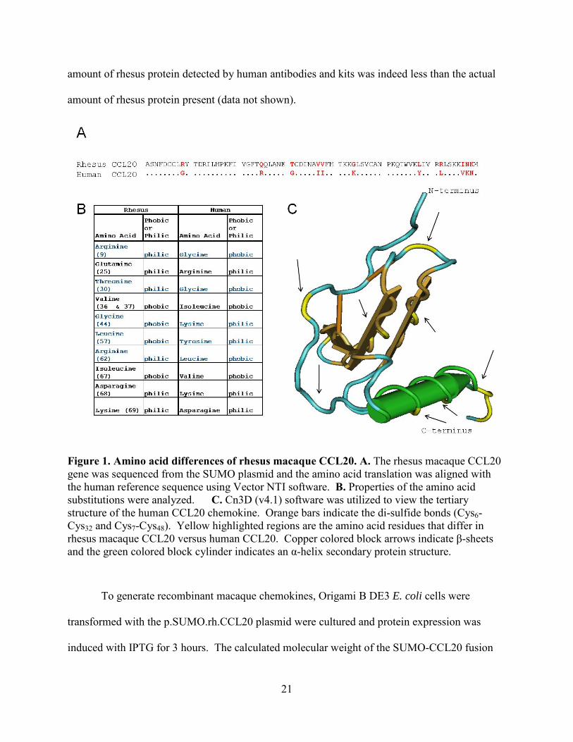
To identify and analyze the sequence differences between human and rhesus macaque
CCL20, p.SUMO.rh.CCL20 was sent to the University of Pittsburgh Sequencing Facility after
isolation of the plasmid using the Wizard Plus Miniprep Kit (Promega). Both the reference
human CCL20 (NM 004591) and the rhesus macaque CCL20 nucleotide sequence were
translated using Vector NTI software. Rhesus macaque CCL20 protein shared 85% identity to
the human CCL20 protein (Figure 1A). To analyze the location of the differences in amino acid
as it related to the structure of the protein, human CCL20 protein sequence was analyzed with
Cn3d software. Secondary protein structures of an α-helix are marked by a green cylinder and
β-sheets are marked by a gold bar (Figure 1C). The amino acid differences in the rhesus
sequence as compared to the human sequence are marked in yellow (Figure 1C). Analysis of the
location of the differences in the rhesus CCL20 revealed that changes occurred over the entire
sequence of the protein (Figure 1A and 1C). Further analysis revealed that some of the amino
acid differences in the rhesus macaque CCL20 protein differ in their hydrophobic or hydrophilic
nature, highlighted in blue in Figure 1B. These analyses provided evidence of the differences
between human and rhesus CCL20 that may cause mis-interpretation of results when using
human tests (ELISA, Immunoblotting, IHC, and FC) to detect rhesus chemokine. Specifically,
antibodies raised against a human antigen may cover a sequence that exhibits amino acid
differences in the rhesus sequence. The binding affinity of the antibodies to antigen may be
reduced by these amino acid changes. I have observed by ELISA and immunoblotting that the
21
amount of rhesus protein detected by human antibodies and kits was indeed less than the actual
amount of rhesus protein present (data not shown).
To generate recombinant macaque chemokines, Origami B DE3 E. coli cells were
transformed with the p.SUMO.rh.CCL20 plasmid were cultured and protein expression was
induced with IPTG for 3 hours. The calculated molecular weight of the SUMO-CCL20 fusion
Figure 1. Amino acid differences of rhesus macaque CCL20. A. The rhesus macaque CCL20 gene was sequenced from the SUMO plasmid and the amino acid translation was aligned with the human reference sequence using Vector NTI software. B. Properties of the amino acid substitutions were analyzed. C. Cn3D (v4.1) software was utilized to view the tertiary structure of the human CCL20 chemokine. Orange bars indicate the di-sulfide bonds (Cys6-Cys32 and Cys7-Cys48). Yellow highlighted regions are the amino acid residues that differ in rhesus macaque CCL20 versus human CCL20. Copper colored block arrows indicate β-sheets and the green colored block cylinder indicates an α-helix secondary protein structure.
22
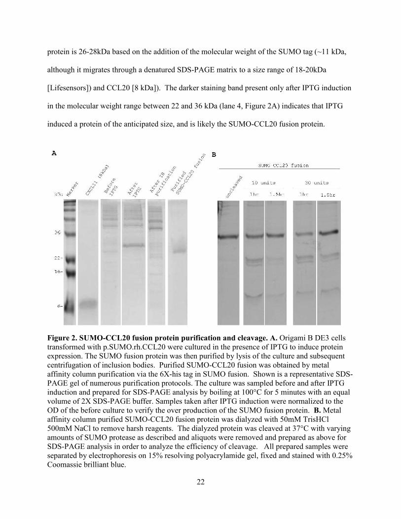
protein is 26-28kDa based on the addition of the molecular weight of the SUMO tag (~11 kDa,
although it migrates through a denatured SDS-PAGE matrix to a size range of 18-20kDa
[Lifesensors]) and CCL20 [8 kDa]). The darker staining band present only after IPTG induction
in the molecular weight range between 22 and 36 kDa (lane 4, Figure 2A) indicates that IPTG
induced a protein of the anticipated size, and is likely the SUMO-CCL20 fusion protein.
Figure 2. SUMO-CCL20 fusion protein purification and cleavage. A. Origami B DE3 cells transformed with p.SUMO.rh.CCL20 were cultured in the presence of IPTG to induce protein expression. The SUMO fusion protein was then purified by lysis of the culture and subsequent centrifugation of inclusion bodies. Purified SUMO-CCL20 fusion was obtained by metal affinity column purification via the 6X-his tag in SUMO fusion. Shown is a representative SDS-PAGE gel of numerous purification protocols. The culture was sampled before and after IPTG induction and prepared for SDS-PAGE analysis by boiling at 100°C for 5 minutes with an equal volume of 2X SDS-PAGE buffer. Samples taken after IPTG induction were normalized to the OD of the before culture to verify the over production of the SUMO fusion protein. B. Metal affinity column purified SUMO-CCL20 fusion protein was dialyzed with 50mM TrisHCl 500mM NaCl to remove harsh reagents. The dialyzed protein was cleaved at 37°C with varying amounts of SUMO protease as described and aliquots were removed and prepared as above for SDS-PAGE analysis in order to analyze the efficiency of cleavage. All prepared samples were separated by electrophoresis on 15% resolving polyacrylamide gel, fixed and stained with 0.25% Coomassie brilliant blue.
23
Some inducible or over produced proteins in E. coli aggregate in insoluble inclusion
bodies (IB)17, 65. Based on this limitation, further steps need to be taken to purify recombinant
proteins, if they are insoluble. To purify the induced protein and determine whether the SUMO–
CCL20 fusion protein was soluble or insoluble, the E. coli culture was pelleted and lysed.
Centrifugation separated soluble and insoluble material including inclusion bodies. The
insoluble pellet was then solubilized with reducing and chaotropic agents to disaggregate protein
by disrupting and protecting free charged atoms from inter- and intra- molecular bonding. After
another round of centrifugation, the supernatant containing solubilized inclusion body proteins
was analyzed for the presence of the SUMO-CCL20 fusion protein. The lack of a 26-28kDa
band present in the soluble fraction after cell lysis, shown in lane 5 [After IB Purification],
Figure 2A indicated that the induced SUMO-CCL20 fusion protein was located nearly
exclusively in inclusion bodies.
The SUMO fusion tag contains a hexahistidine (6X-His) tag that allows the SUMO-
CCL20 fusion protein to be purified from cell lysate via metal affinity column purification, by
binding to nickel or cobalt ions in the column (Life Sensors). In lane 6 [Purified SUMO-CCL20
fusion] Figure 2A, only one protein of size 26-28kDa was abundant, which confirmed that only
the 6X-His tagged SUMO-CCL20 fusion protein was purified by metal affinity column binding.
In summary, sequence differences between the human and rhesus CCL20 warrant the
production of non-human primate chemokines to use as reagents in monkey models studying
immunology and infectious disease. The recombinant protein expression system using SUMO
fusion technology to express rhesus macaque CCL20 in the Origami B DE3 E. coli strain
produced a protein of the anticipated size of SUMO-CCL20 fusion upon IPTG induction. The
induced protein was aggregated in inclusion bodies, but was purified by centrifugation and
24
solubilization. The solubilized SUMO-CCL20 fusion protein was abundant after metal affinity
column purification, indicating that a 6X-His tag protein was purified. The success of the
expression and purification of the SUMO-CCL20 fusion means that this system is a good
candidate protocol for obtaining recombinant SUMO-rhesus chemokines.
4.1.1 Cleavage of the SUMO-CCL20 fusion protein
To obtain bioactive chemokine from recombinant protein expression using the SUMO
fusion technology, the chemokine (CCL20) must likely be cleaved from the fusion protein. The
SUMO fusion system is ideal for obtaining recombinant proteins because the SUMO protease
recognizes only the tertiary structure of the SUMO tag in the protein rather than recognizing
cryptic or inappropriate target sequences for cleavage. Additionally, the SUMO protease was
mutated to contain a 6X His tag and can therefore be removed from preparations via the same
metal affinity column purification (Talon Spin Columns [Clontech]). These facts facilitate the
resultant desired recombinant chemokine from being cleaved inside the protein and that after a
second round of metal affinity column purification, all that should flow through is the final
cleaved recombinant chemokine.
Depicted in Figure 2B, is the pattern of a typical cleavage reaction with the SUMO-
CCL20 fusion protein. The same amount of purified SUMO-CCL20 fusion protein was cleaved
with SUMO protease, 10 or 30 units, and the reaction was incubated for 1.5 hours at 37°C. The
appearance of two lower molecular weight proteins in lanes 2 through 5 of Figure 2B showed
that the SUMO-CCL20 fusion protein was indeed cleaved by the SUMO protease. Comparison
of the reduced intensity of staining of the substrate (the largest staining protein band in lanes 2
25
through 5) to the input substrate (lane 1), revealed that after 1 hour, the SUMO protease was only
able to cleave about 10-30% of the SUMO-CCL20 fusion protein. Additionally, there appeared
to be little difference in the efficiency of the cleavage reactions, even upon increasing SUMO
protease 10-30 times the recommended amount (1U protease cleaves 100µg [Lifesensors]).
Increasing the time of the reaction by a short time also did not appear to have an effect on
increasing the efficiency of the cleavage reaction (lanes 3 and 5, Fig. 2B).
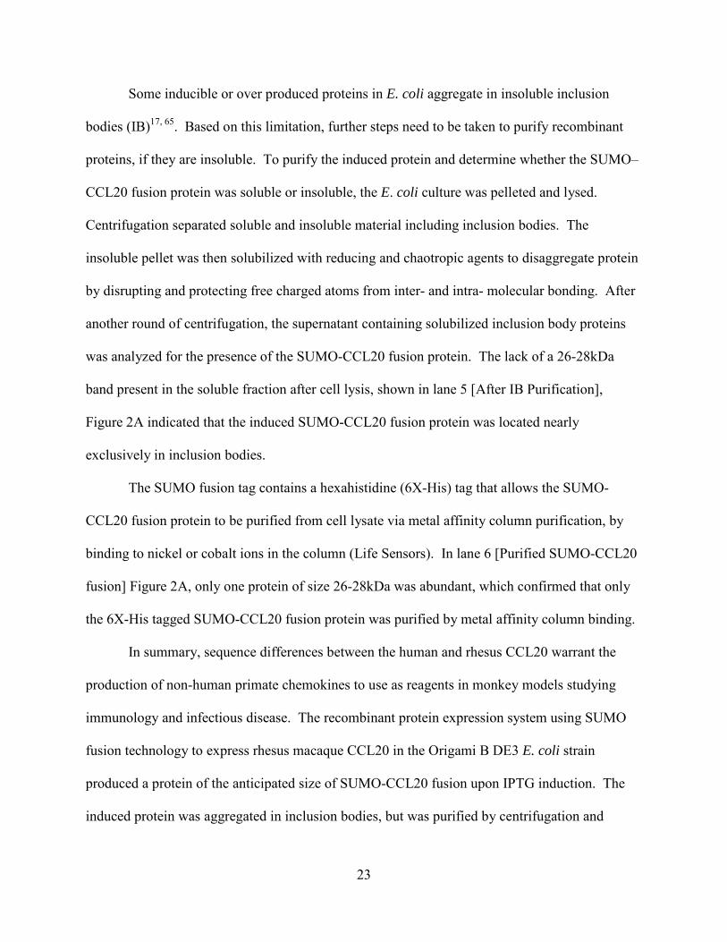
Figure 3. SUMO-CCL20 is cleaved inefficiently, although SUMO protease is highly efficient at cleaving control SUMO fusions. 150ug/ml of purified SUMO-CCL20 fusion protein was cleaved with 10 units of SUMO protease at 30˚C for 1 hour. As a control for protease activity, 50ug/ml of SUMO-met-GFP was cleaved with 3 units of protease under the same conditions. Both cleavage reaction samples were then stored at 4˚C overnight to allow for complete cleavage of fusion proteins. The first panel shows the MW marker, SUMO protease control (26kDa) and synthetic CCL20 (8kDa). Samples were prepared for SDS-PAGE analysis by boiling at 100°C for 5 minutes with an equal volume of 2X SDS-PAGE buffer. All prepared samples were separated by electrophoresis on 15% resolving polyacrylamide gel, fixed and stained with 0.25% Coomassie brilliant blue.
26
One of the first steps taken toward optimizing the cleavage of SUMO-CCL20 fusion was
to verify the ability of the SUMO protease to cleave SUMO fusions. To clarify this, the SUMO-
CCL20 fusion and the control SUMO-met-GFP fusion (LifeSensors) were cleaved with SUMO
protease at 30°C for 1 hour and then at 4°C overnight. The second panel of Figure 3 confirms in
an independent experiment, as previously described in Figure 2B, there was inefficient cleavage
of the SUMO-CCL20 fusion. Cleavage of the SUMO-CCL20 fusion was not completed even
with overnight incubation with SUMO protease. However, the control fusion SUMO-met-GFP
was cleaved nearly completely after only one hour, which verified that the SUMO protease was
active and efficient. Complete cleavage of the control SUMO fusion protein was achieved with
incubation overnight, proving that as described by the manufacturer, SUMO protease is also
active at 4°C.
Since there appeared to be problems with cleavage of the SUMO-CCL20 fusion protein
(Figure 2B and 3), it was decided that a strategy to improve cleavage efficiency was necessary.
Development of a multi-parameter experiment may solve the problem of identifying optimal
cleavage conditions. In the next section, I will describe my efforts at increasing the efficiency of
cleavage of the SUMO-CCL20 fusion.
4.2 Strategies to increase efficiency of cleavage
After demonstrating that the SUMO protease was active (Figure 3) and that the SUMO-
CCL20 fusion protein was not efficiently cleaved (Figure 2B and Figure 3), I reasoned that there
were many possibilities for observing inefficient cleavage of the SUMO-CCL20 fusion protein.
I developed a multi-parameter experiment to optimize the efficiency of cleavage of the SUMO-
CCL20 fusion.
27
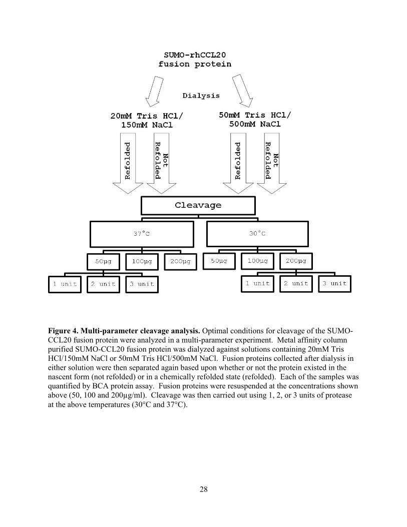
4.2.1 Multi-parameter cleavage experimental design
Optimization of the cleavage reaction was required for obtaining cleaved recombinant
chemokine prior to HPLC purification and use in bioassays. A multi-parameter experiment was
performed with the goal of increasing efficiency of cleavage of the SUMO-CCL20 fusion. To
work towards identifying parameters that would increase cleavage of the SUMO-CCL20 fusion,
the parameters analyzed were the use of differing dialysis buffers, refolding techniques and
temperature or concentration dependent variables. The specific parameters analyzed were the
time at which the protein was refolded, the dialysis reagents and concentrations of cleavage
reaction reagents and the temperature at which the cleavage reaction was performed. A flow
chart, shown in Figure 4, depicts the variables analyzed in the multi-parameter experiment.
28
Figure 4. Multi-parameter cleavage analysis. Optimal conditions for cleavage of the SUMO-CCL20 fusion protein were analyzed in a multi-parameter experiment. Metal affinity column purified SUMO-CCL20 fusion protein was dialyzed against solutions containing 20mM Tris HCl/150mM NaCl or 50mM Tris HCl/500mM NaCl. Fusion proteins collected after dialysis in either solution were then separated again based upon whether or not the protein existed in the nascent form (not refolded) or in a chemically refolded state (refolded). Each of the samples was quantified by BCA protein assay. Fusion proteins were resuspended at the concentrations shown above (50, 100 and 200µg/ml). Cleavage was then carried out using 1, 2, or 3 units of protease at the above temperatures (30°C and 37°C).
29
4.2.1.1 Differences in buffer, quantity of protease or protein, time and temperature do not
increase the efficiency of cleavage of SUMO-CCL20.
To analyze the effects of varied conditions of the multi-parameter optimization
experiment, SUMO-CCL20 fusion protein was dialyzed against one of two different neutral salt
buffers. The protein was then placed under conditions thought to facilitate refolding or allowed
to remain in the form after dialysis. Cleavage reactions containing differing amounts of substrate
ranging from 50-200µg/ml were carried out at different temperatures with different amounts of
protease. SDS-PAGE with Coomassie brilliant blue staining was performed to analyze the
results of the different cleavage reactions.
To analyze the difference in dialysis buffers on the cleavage of not-refolded SUMO-
CCL20 fusion, different amounts of protease were added to varying amounts of SUMO-CCL20
fusion. The cleavage reaction was incubated at either 30°C or 37°C for 1 (1h) or 2 hours (2h) to
identify possible temperature or time dependent differences in efficiency of cleavage. In
contrary to the manufacturer’s reference that one unit of SUMO protease cleaves 100µg of
SUMO fusion protein (Lifesensors); all gels shown below in Figure 5 revealed that none of the
conditions examined lead to efficient cleavage of the SUMO-CCL20 fusion.
30
31
Under the conditions analyzed, where 3U (or 3 units of SUMO protease) was the largest
amount of protease added to the cleavage reaction, and 50µg (2.5µg) total SUMO-CCL20 fusion
was the least amount of substrate added to the cleavage reaction, increasing the amount of
protease did not increase the extent of cleavage the SUMO-CCL20 fusion protein, first gel panel,
Figure 5. Additionally, the middle panel and far right panel of the lower gel, where 3 units of
protease was used to cleave 100µg (5µg) of SUMO-CCL20 fusion revealed that increasing the
amount of protease did not increase the efficiency of cleavage. All other gel panels lend
additional support to the overall conclusion of increasing the amount of protease does not
increase the efficiency of cleavage by a significant degree.
Comparison of the top and bottom 2 right gels (1U, 5µg, 30ºC and 37ºC with dialysis
buffer containing 50mM TrisHCl and 3U, 5µg, 30ºC and 37ºC with dialysis buffer containing
20mM TrisHCl) indicates that temperatures of 30°C vs. 37°C yielded similar, incomplete levels
of cleavage. Based on the decreased intensity of staining of the SUMO-CCL20 fusion protein
(lane T0h of each gel) compared to the staining in lane T1h of each gel and also given the
appearance of 3 smaller proteins (2 bands at ~22kDa and 1 very light band at ~8kDa), the
percentage of SUMO-CCL20 fusion that was cleaved was about 20-30%. Additionally, there
was little to no increased detection of cleavage products after another hour of cleavage (lane T2h
of each gel).
Figure 5. Changing four different variables did not increase cleavage efficiency of the 'not refolded' SUMO-CCL20 fusion. SUMO-CCL20 fusion protein was quantified by BCA assay to achieve the final concentrations in the cleavage reaction as shown. Metal affinity column purified SUMO-CCL20 fusion protein that was not chemically refolded prior to cleavage was aliquotted into 50µg/ml (2.5μg), 100µg/ml (5μg), 200µg/ml (10μg) total protein concentrations, cleaved with varying quantities of SUMO protease and the reaction was allowed to continue for 1 hour at either 30° or 37°C. Samples were prepared for SDS-PAGE analysis by boiling at 100°C for 5 minutes with an equal volume of 2X SDS-PAGE buffer. All prepared samples were separated by electrophoresis on 15% resolving polyacrylamide gel, fixed and stained with 0.25% Coomassie brilliant blue.
32
In summary, the multi-parameter experiment showed that the SUMO protease did cleave
the SUMO-CCL20 fusion. However, the cleavage of the SUMO-CCL20 fusion protein was not
efficient. The composition of the dialysis buffer in which the protein was equilibrated did not
appear to have a significant effect on the efficiency of cleavage. Therefore, it was decided that
the 50mM TrisHCl dialysis buffer would be sufficient to use in the protocol. Neither
temperature analyzed (30ºC or 37ºC) provided any improved cleavage. No ratio of substrate to
protease yielded better cleavage than the others, however, based on data from the multi-
parameter experiment, Figures 2B and 3, as well as additional references70, 71, 73, it was decided
that a more concentrated substrate provide additional opportunity for diagnosing the efficiency of
a cleavage reaction. Overall, the multi-parameter experiment was useful in narrowing down the
variables to analyze to increase efficiency of cleavage of the SUMO-CCL20 fusion.
33
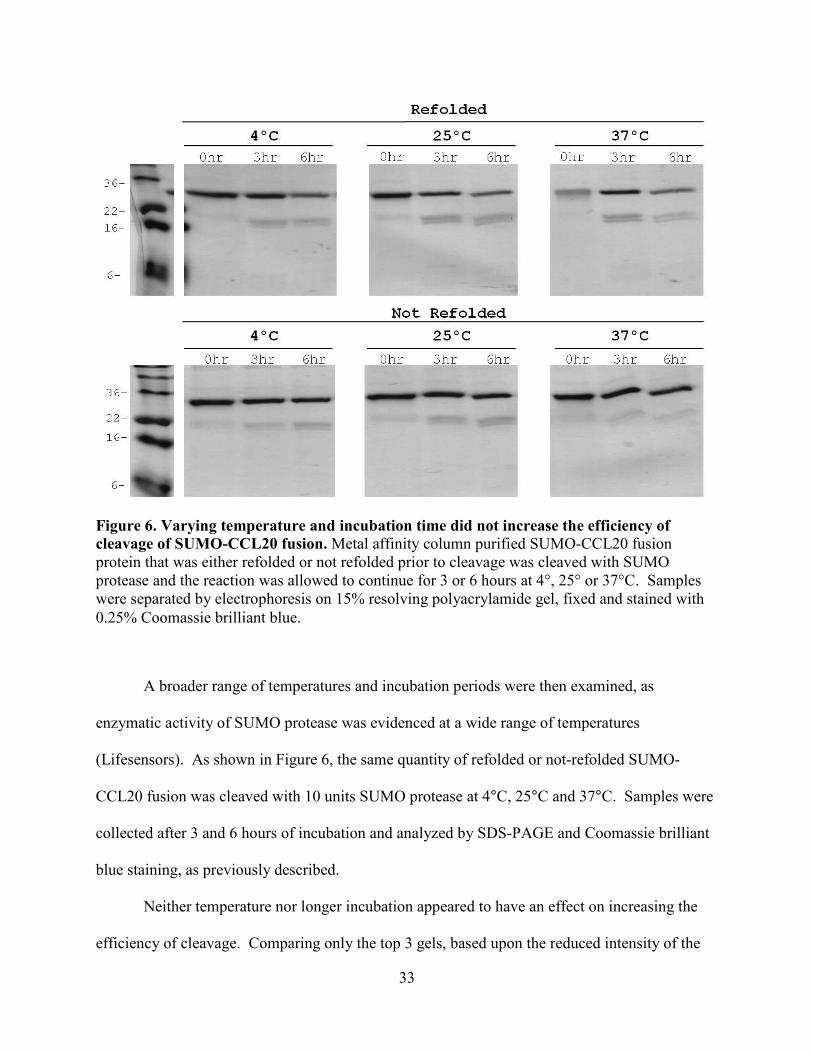
A broader range of temperatures and incubation periods were then examined, as
enzymatic activity of SUMO protease was evidenced at a wide range of temperatures
(Lifesensors). As shown in Figure 6, the same quantity of refolded or not-refolded SUMO-
CCL20 fusion was cleaved with 10 units SUMO protease at 4°C, 25°C and 37°C. Samples were
collected after 3 and 6 hours of incubation and analyzed by SDS-PAGE and Coomassie brilliant
blue staining, as previously described.
Neither temperature nor longer incubation appeared to have an effect on increasing the
efficiency of cleavage. Comparing only the top 3 gels, based upon the reduced intensity of the
Figure 6. Varying temperature and incubation time did not increase the efficiency of cleavage of SUMO-CCL20 fusion. Metal affinity column purified SUMO-CCL20 fusion protein that was either refolded or not refolded prior to cleavage was cleaved with SUMO protease and the reaction was allowed to continue for 3 or 6 hours at 4°, 25° or 37°C. Samples were separated by electrophoresis on 15% resolving polyacrylamide gel, fixed and stained with 0.25% Coomassie brilliant blue.
34
un-cleaved SUMO-CCL20 fusion protein between 22-36kDa and the appearance of staining of
products of the cleavage reaction, bands staining between 16-22kDa (SUMO fusion tag) and
only slightly visible band at ~8kDa (CCL20), efficiency of cleavage was approximately 30% at
most. The additional three hours of incubation did not increase cleavage, as seen by the band
pattern in lane '6hr' in each of the same three top gels. The same conclusion can be drawn by
results shown in the bottom 3 gels.
There does not appear to be a difference in cleavage of either the chemically refolded or
not-refolded SUMO-CCL20 fusion, since the ratios of input substrate to products are all about
the same or a significant amount of SUMO-CCL20 fusion protein appears to be left un-cleaved
after the endpoint of the reaction.
Based on the continued findings of inefficient cleavage, it was possible that something
inherent to the SUMO-CCL20 fusion prevented it from being cleaved by SUMO protease. It
was then hypothesized based on a study done by Calderone, et al, that because the CCL20
sequence is mammalian there may be mis-incorporated amino acids that could affect the tertiary
structure of the resultant protein5, which could potentially affecting the ability of the SUMO
protease to cleave the SUMO-CCL20 fusion protein.
Two approaches were utilized to address these questions. First, a SUMO fusion protein
that previously had been successfully cleaved in our laboratory, SUMO-CXCL11, was analyzed
for the capacity of the SUMO protease to cleave that fusion protein. It does appear that SUMO-
CXCL11 was cleaved more efficiently (data not shown). This, as well as the success of previous
experiments by others that showed almost complete cleavage of the SUMO-CXCL11 fusion
protein (data not shown, Yu-jen Tung), suggests that the specific chemokine attached to the
SUMO fusion tag affects the efficiency of cleavage. The efficient cleavage of the SUMO-
35
CXCL11 fusion protein suggests that the cleavage of SUMO fusions may be better suited for
some SUMO chemokine fusion proteins and not others.
The method utilized to resolve the other postulate that due to the expression of the fusion
protein in E. coli there may be mis-incorporated amino acids conferring a different tertiary
structure that prohibits the SUMO protease from cleaving the fusion protein was the use of
another strain of E. coli (Rosettagami B DE3). This was done to examine whether synthesis of
the protein in this strain, which has been modified to express mammalian tRNAs rare in E. coli,
increased the efficiency of cleavage of the SUMO-CCL20 fusion protein.
4.2.2 Rosettagami and Origami B DE3 strain of E. coli
Calderone et al5. showed that E. coli used to express fusion proteins off mammalian
cDNA sequences could mis-incorporate a lysine residue for an arginine residue, when arginine is
coded for by ‘AGA’ 5. Rhesus CCL20 does not contain any ‘AGA’ codons (data not shown).
The SUMO fusion tag however, contains four ‘AGA’ codon arginine residues (data not shown).
As postulated previously, mis-incorporation of lysines for these arginine residues might be a
factor in the poor efficiency of cleavage. There may be different intramolecular interactions due
to the amino acid substitution that would affect the structure of the fusion protein causing the
SUMO protease to be unable to recognize and cleave the fusion protein appropriately.
To address this we utilized the Rosettagami B DE3 strain of E. coli because of its
expression of tRNAs that are rarely seen in E. coli. The p.SUMO.rh.CCL20 plasmid was
transformed into Rosettagami B DE3 cells and subsequently sequenced. Analysis of the
sequences revealed that the p.SUMO.rh.CCL20 plasmid aligned with the reference rhesus
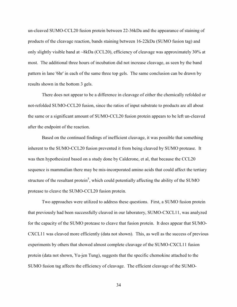
CCL20 sequence except for one nucleotide substitution (Figure 7). There was a non-
synonymous ‘T’ to a ‘G’ nucleotide substitution, causing the cysteine at position 32 to be
36
changed to a tryptophan (see Figure 1A for the reference rhesus CCL20 protein sequence).
Although the SUMO CCL20 expression plasmid had been isolated and sequenced after passing
through multiple E. coli strains (DH5 alphas and Origami B DE3) the output sequence had no
nucleotide substitutions. We then sub-cloned and sequenced five p.SUMO.rh.CCL20 plasmids
from the Rosettagami B DE3 strain of E. coli. Each of the sequences of the five isolates of
p.SUMO.rh.CCL20 showed the same tryptophan substitution at position 32; leading to a
conclusion that it was possible that the E. coli strain the plasmid was isolated from was affecting
the sequence output.
Since we had not observed this nucleotide substitution before in other E. coli strains, and
to analyze whether the two original clones of the p.SUMO.rh.CCL20 plasmids had evidence of
this same base pair change, all past sequence trace files of the p.SUMO.rh.CCL20 plasmid were
then analyzed (data not shown). This revealed that in the clone 1 of p.SUMO.rh.CCL20 plasmid
used in all protein expression, there were two peaks present at the base pair in question (Figure
7). Results of double peaks seen in a sequencing trace file could mean at least two things. It
Figure 7. Isolation of clones of the plasmids p.SUMO.CCL20.1 and p.SUMO.CCL20.2. The p.SUMO.CCL20.1 plasmid preparation was shown to have a mixed population of plasmids, with either a ‘T’ or a ‘G’ at the base pair in question. The p.SUMO.CCL20.2 plasmid preparation did not have evidence of a mixed population of plasmids.

37
could mean that the gene that was sequenced and used for cloning had a point mutation or that
there was a mixed population of plasmids in clone 1 of the p.SUMO.rh.CCL20 plasmid. The
other original clone (2) of the p.SUMO.rh.CCL20 plasmid showed no evidence of this ambiguity
at the base pair in question (Figure 7).
To identify a sub-clone of the p.SUMO.rh.CCL20 (clone 1) plasmid preparation that did
not show evidence of two peaks in the sequence trace file. Plasmid p.SUMO.rh.CCL20 (clone 1)
was re-transformed into the DH5α E. coli strain, and individual colonies were sequenced. Out of
the five clones (named simply 1.1, 1.2, 1.3, 1.4. 1.5), only plasmid p.SUMO.rh.CCL20.1.1 was
revealed to have ambiguity at the base in question (Figure 7 and data not shown). Five clones
sequenced from plasmid p.SUMO.rh.CCL20 (original clone 2) all aligned with 100% identity to
the reference rhesus CCL20 sequence.
After sequencing of five retransformed clones for original clones 1 and 2 of
p.SUMO.rh.CCL20, revealed that only one retransformed clone named p. SUMO.CCL20.1.1
showed the base-pair change which translated to a tryptophan substitution at position 32 (from
cysteine) in the protein, it was hypothesized that the translation of pSUMO.rh.CCL20.1.1 C32 to
T32 might be the fusion protein left uncleaved. To address this hypothesis, it was assumed that it
would be possible to see differences in the cleavage of the sub-clones that translated either
SUMO-CCL20 fusion with the cognate cysteine32 or mutated tryptophan32.
4.2.2.1 Differences in SUMO-CCL20 sequence at position 32 do not affect Origami B DE3
expression or cleavage of protein.
Because of the differences in the rhesus macaque CCL20 sequence in the plasmid that
was used in the protein expression system, we sought to determine if these sequence differences
had an effect on the efficiency of the cleavage of the SUMO-CCL20 protein. Three different
38
sub-clones of the SUMO-CCL20 fusion protein were sequence verified and transformed into the
Origami B DE3 E. coli strain. Protein was induced with the addition of IPTG; the protein was
purified from the inclusion bodies by centrifugation and metal affinity column purification, as
previously described. Purified protein was normalized to total protein concentration and cleaved
with SUMO protease. There were few differences in the amounts of protein purified (data not
shown). As shown in Figure 8, cleavage of the purified SUMO-CCL20 fusion protein was
inefficient (Figure 8). Only 25-50% cleavage of the SUMO-CCL20 fusion protein was achieved,
as depicted by the reduced staining of the SUMO-fusion protein and the appearance of staining
of two bands lower in molecular weight. With cleavage of the SUMO-CCL20 fusion protein, the
SUMO fusion tag of the protein stains in the area of about 22kDa, and rhesus CCL20 stains in
the area of 8kDa. The low molecular weight protein marker of synthetic rhesus CCL20 shown in
lane 2 (CCL20 (8kDa)) confirms that upon cleavage of the SUMO-CCL20 fusion protein a
protein of similar molecular weight appears and is assumed to be the recombinant CCL20.
Differences in cleavage of the retransformed SUMO-CCL20 fusion proteins could not be
attributed to a reduced activity of the SUMO protease as the protease was added at the same time
in each reaction and the control fusion protein, SUMO-met-GFP, was cleaved within one hour
(Figure 9, SUMO-met-GFP cleavage).
39
4.2.2.2 Expression and purification of SUMO-CCL20 isolates are different in Rosettagami
B DE3 cells.
In the same way as with the Origami B DE3 strain of E. coli, a different strain of E. coli,
Rosettagami B DE3, was analyzed for differences in efficiency of cleavage of the three different
p.SUMO.rh.CCL20 retransformed plasmids (sequences shown in Figure 7). Although the
purified SUMO-CCL20 fusions were normalized with respect to the total concentration of
protein, there are apparent differences in the intensity of staining of the protein at the size of the
anticipated SUMO-CCL20 fusion (22-36kDa, T0, Figure 9). As shown in Figure 9, there were
differences in the purity in total protein purified via the metal affinity column purification when
comparing the banding patterns in the molecular weight range of 22-36 kDa. The protein
Figure 8. There is no difference in the expression or cleavage of different isolates of SUMO-CCL20 fusions purified from the Origami B DE3 strain of E. coli. Recombinant protein expression was performed for each of 3 different cultures of transformed Origami B DE3 sequences that conferred both non-synonymous base pair mutations and reference rhesus CCL20 sequences with different isolates of the SUMO-CCL20 fusion plasmid. SUMO-CCL20 fusion proteins were cleaved with SUMO protease. Samples from different conditions were separated by electrophoresis on 15% resolving polyacrylamide gel, fixed and stained with 0.25% Coomassie blue staining.
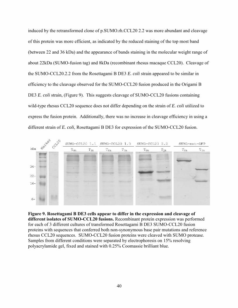
40
induced by the retransformed clone of p.SUMO.rh.CCL20 2.2 was more abundant and cleavage
of this protein was more efficient, as indicated by the reduced staining of the top most band
(between 22 and 36 kDa) and the appearance of bands staining in the molecular weight range of
about 22kDa (SUMO-fusion tag) and 8kDa (recombinant rhesus macaque CCL20). Cleavage of
the SUMO-CCL20.2.2 from the Rosettagami B DE3 E. coli strain appeared to be similar in
efficiency to the cleavage observed for the SUMO-CCL20 fusion produced in the Origami B
DE3 E. coli strain, (Figure 9). This suggests cleavage of SUMO-CCL20 fusions containing
wild-type rhesus CCL20 sequence does not differ depending on the strain of E. coli utilized to
express the fusion protein. Additionally, there was no increase in cleavage efficiency in using a
different strain of E. coli, Rosettagami B DE3 for expression of the SUMO-CCL20 fusion.
Figure 9. Rosettagami B DE3 cells appear to differ in the expression and cleavage of different isolates of SUMO-CCL20 fusions. Recombinant protein expression was performed for each of 3 different cultures of transformed Rosettagami B DE3 SUMO-CCL20 fusion proteins with sequences that conferred both non-synonymous base pair mutations and reference rhesus CCL20 sequences. SUMO-CCL20 fusion proteins were cleaved with SUMO protease. Samples from different conditions were separated by electrophoresis on 15% resolving polyacrylamide gel, fixed and stained with 0.25% Coomassie brilliant blue.
41
4.3 Peptide synthesis
As there were continued problems with obtaining recombinant protein for HPLC
purification and subsequent biological activity testing, rhesus macaque CCL20 was synthesized
using regioselective cyclization. Mature signal peptide cleaved (amino acids 1-70, Figure 1A)
rhesus macaque CCL20 sequence was forwarded to the University of Pittsburgh Peptide Facility.
The peptide was synthesized using solid phase peptide synthesis with regioselective cyclization,
where a peptide is synthesized from the c-terminal amino acid up to the n-terminal amino acid,
with thiol groups protecting side groups of cysteines to prevent disulfide bond formation,
intermediately. When the second cysteine amino acid of a disulfide bond is added to the growing
peptide, the cysteines are de-protected and allowed to form the intended disulfide bond. For
CCL20, disulfide bonds are formed between Cys6-Cys32 and Cys7-Cys48. CCL20 was then
purified by HPLC and analyzed with NMR. Pure lyophilized peptide was resuspended in
nuclease free water and quantitated by BCA protein assay. This protein was then used in the
second aim of this study to analyze the CCL20/CCR6 interaction.
4.4 Overall Protein Expression System Summary
SUMO-CCL20 fusion was expressed upon IPTG addition and was found to be
aggregated in inclusion bodies. The protein was purified and detected by the 6X His-tag in the
SUMO-tag. Only 30-50% efficiency of cleavage of the SUMO-CCL20 fusion was achieved
overall. Chemically refolding or not refolding in different dialysis buffers did not increase
efficiency of cleavage. Varying temperature and changing the ratios of SUMO-CCL20 fusion:
SUMO protease did not increase efficiency of cleavage. There was no difference in the cleavage
of the SUMO-CCL20 in two different E. coli strains, Origami B DE3 or Rosettagami B DE3s.
Repeated purifications of SUMO-CCL20 fusion from up to 12 L of culture did not yield enough
42
SUMO-CCL20 fusion or cleaved recombinant rhesus CCL20 for bioactivity testing. To
generate non-human primate chemokine, rhesus CCL20 was synthesized with regioselective
cyclization by Univ. Pitt. Peptide Facility for bioactivity testing.
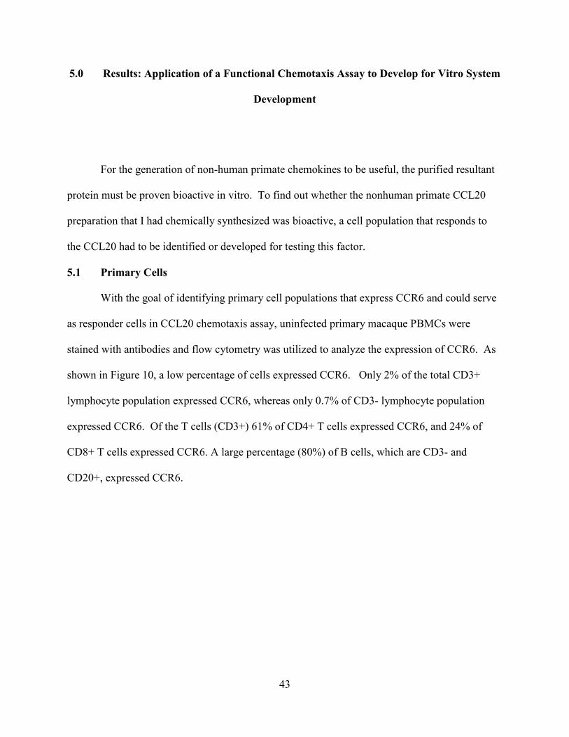
43
5.0 Results: Application of a Functional Chemotaxis Assay to Develop for Vitro System
Development
For the generation of non-human primate chemokines to be useful, the purified resultant
protein must be proven bioactive in vitro. To find out whether the nonhuman primate CCL20
preparation that I had chemically synthesized was bioactive, a cell population that responds to
the CCL20 had to be identified or developed for testing this factor.
5.1 Primary Cells
With the goal of identifying primary cell populations that express CCR6 and could serve
as responder cells in CCL20 chemotaxis assay, uninfected primary macaque PBMCs were
stained with antibodies and flow cytometry was utilized to analyze the expression of CCR6. As
shown in Figure 10, a low percentage of cells expressed CCR6. Only 2% of the total CD3+
lymphocyte population expressed CCR6, whereas only 0.7% of CD3- lymphocyte population
expressed CCR6. Of the T cells (CD3+) 61% of CD4+ T cells expressed CCR6, and 24% of
CD8+ T cells expressed CCR6. A large percentage (80%) of B cells, which are CD3- and
CD20+, expressed CCR6.

44
To determine whether uninfected primary PBMCs would be a good population of cells to
test the bioactivity of the regioselectively cyclized rhesus macaque CCL20 (abbreviated
synthetic rhesus CCL20), the isolated PBMCs were placed in a functional chemotaxis assay. To
determine whether the small population of PBMCs that express CCR6 (see Figure 10) were able
to migrate toward to the synthetic rhesus CCL20, freshly isolated normal rhesus PBMCs were
placed in a chemotaxis assay with a titration of synthetic rhesus CCL20, as well as 2 other
chemokines, CXCL11 and CCL21. After incubation at 37ºC with 5% CO2 for 1.5 hours, cells
that had migrated through the membrane toward the chemokine were counted on a
hemocytometer.
Figure 10. CCR6 is expressed on both B- and T-lymphocytes. Flow cytometry was utilized to analyze the expression of CCR6 on B and T cells by isolating primary rhesus macaque PBMCs. PBMCs were then stained with a 6 color panel (Anti-CD3 Pe-Cy7, Anti-CD4 PerCP, Anti-CD20 FITC, Anti-CCR6 PE, Anti-CD8 APC-Cy7 and Anti-CD69 APC). Data was aqcuired on the BD Canto, and analyzed with Flow Jo software.

45
As shown in Figure 11, the cells were able to migrate towards certain chemokines,
CXCL11 and CCL21, with the highest number of cells migrating towards CCL21. These results
are consistent with expression (data not shown), in that, 60-80% of lymphocytes express CCR7,
the receptor for CCL21, and 30-60% of lymphocytes express CXCR3, the receptor for CXCL11.
No cells migrated towards the increasing concentrations of CCL20 (Figure 11), suggesting that
either the synthetic rhesus CCL20 was not bioactive, or the receptor, although expressed, was
biologically inactive, or that the population tested did not have sufficient numbers of cells that
expressed CCR6.
Given the finding that primary PBMCs do not migrate towards CCL20 despite expression
of the receptor on both T- and B-lymphocytes, a cell line that expressed CCR6 had to be
developed to test whether the rhesus macaque CCL20 produced the regioselectively cyclized
synthesis was bioactive. The cell line should be one that proliferates quickly, are easily
Figure 11. Normal macaque PBMCs do not respond to synthetic macaque CCL20. Freshly isolated normal rhesus macaque PBMCs were placed in a chemotaxis assay. A titration of synthetic rhesus macaque CCL20 was prepared in chemotaxis media. Human CXCL11 and CCL21 were prepared in the same manner and used as positive controls for migration. 200,000 cells (PBMCs) were loaded on the top of the membrane, and the plate was incubated for 1.5 hours at 37°C and 5% CO2.
46
transfectible and do not respond to many human chemokine receptor ligands, including CCL20.
Murine L1.2 pre-B cells were found to be a cell line that possessed these properties and were
therefore utilized in developing a CCR6 cell line63.
5.2 Regioselectively cyclized synthetic rhesus CCL20 is highly bioactive
As there were continued problems with obtaining recombinant chemokine for in vitro
study, rhesus macaque CCL20 was synthesized by regioselective cyclization, described in
material and methods and above (section 4.3). To examine the bioactivity of rhesus macaque
CCL20 synthesized by regioselectively cyclization, L1.2 murine pre-B cells were transfected
with a mammalian expression vector, pcDNA3.1 containing the cDNA sequence for human
CCR6. After transfection, the cultures were treated with 5mM sodium butyrate overnight to
increase expression of the receptor. To determine the expression profiles, cells were then
isolated, washed and stained with CCR6-specific antibody. Data were acquired with either the
Coulter XL or the BD Canto flow cytometers. CCR6 expression data were further analyzed with
FlowJo software. To determine the functionality of the CCR6 receptor in transiently tranfected
L1.2 cells, the cells were also placed in a traditional chemotaxis assay. A titration of both human
and synthetic rhesus macaque CCL20 was prepared was and after three hours incubation at 37°C
in 5% CO2, cells that had migrated into the bottom well were then counted on a hemocytometer
and total migrated cells was calculated.
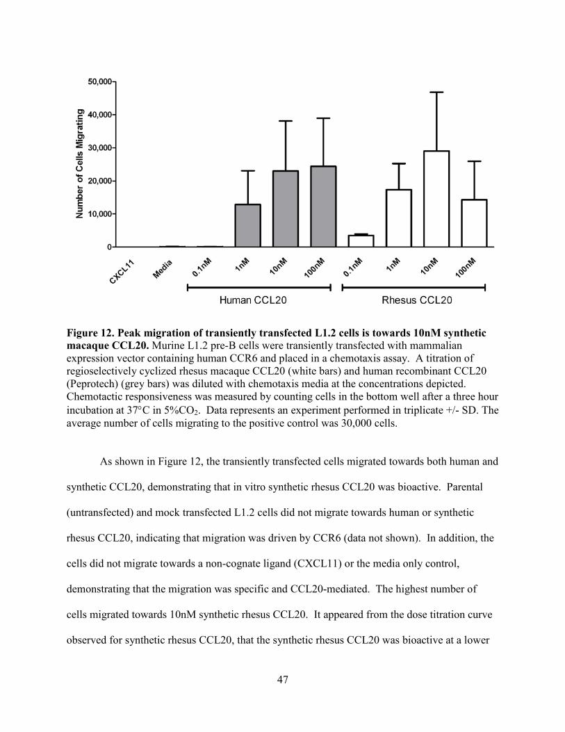
47
As shown in Figure 12, the transiently transfected cells migrated towards both human and
synthetic CCL20, demonstrating that in vitro synthetic rhesus CCL20 was bioactive. Parental
(untransfected) and mock transfected L1.2 cells did not migrate towards human or synthetic
rhesus CCL20, indicating that migration was driven by CCR6 (data not shown). In addition, the
cells did not migrate towards a non-cognate ligand (CXCL11) or the media only control,
demonstrating that the migration was specific and CCL20-mediated. The highest number of
cells migrated towards 10nM synthetic rhesus CCL20. It appeared from the dose titration curve
observed for synthetic rhesus CCL20, that the synthetic rhesus CCL20 was bioactive at a lower
Figure 12. Peak migration of transiently transfected L1.2 cells is towards 10nM synthetic macaque CCL20. Murine L1.2 pre-B cells were transiently transfected with mammalian expression vector containing human CCR6 and placed in a chemotaxis assay. A titration of regioselectively cyclized rhesus macaque CCL20 (white bars) and human recombinant CCL20 (Peprotech) (grey bars) was diluted with chemotaxis media at the concentrations depicted. Chemotactic responsiveness was measured by counting cells in the bottom well after a three hour incubation at 37°C in 5%CO2. Data represents an experiment performed in triplicate +/- SD. The average number of cells migrating to the positive control was 30,000 cells.
48
concentration than the recombinant human CCL20, since a lower concentration induced more
cells to migrate (compare 10nM of human and synthetic rhesus CCL20).
5.2.1 Stable Cell Line Development
There was variability in the transfection efficiency with the CCR6 expression plasmid,
with efficiency varying from 8-60% of the input cells (data not shown). With a transient
transfection that is not 100% efficient, it is likely not all the cells loaded on the top of the
membrane in chemotaxis express CCR6 or respond chemotactically to CCL20. In addition, to
complete the study of inhibitory compounds on CCL20-mediated-chemotaxis, it is necessary to
control these other variables (such as transfection efficiency) with a stable cell line so as not to
attribute inhibition to these other factors.
To obtain a more efficient and controlled population of cells to test CCL20-mediated
chemotaxis and chemotactic inhibition, stable cell lines were developed. CCR6 transfected L1.2
cells were cultured for 48 hours at 37°C in 5% CO2 then resuspended with 1mg/ml G418
(geneticin). The cells were then aliquotted into a 96-well plate and cultured in the presence of
G418 until cell foci were apparent, typically 5-7 days. These primary clones were cultured,
treated with sodium butyrate and placed in a CCL20-mediated chemotaxis assay to measure their
chemotactic responses to CCL20. Reserved cultures of primary clones that migrated in high
numbers towards synthetic rhesus CCL20 were further cultured, single cell cloned and confirmed
for CCL20-mediated chemotaxis. To this end, 12 stable cell clones were identified and moved
ahead for further characterization and analysis of CCL20-mediated chemotaxis and inhibition
studies.
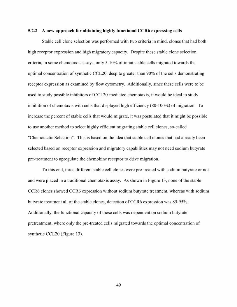
49
5.2.2 A new approach for obtaining highly functional CCR6 expressing cells
Stable cell clone selection was performed with two criteria in mind, clones that had both
high receptor expression and high migratory capacity. Despite these stable clone selection
criteria, in some chemotaxis assays, only 5-10% of input stable cells migrated towards the
optimal concentration of synthetic CCL20, despite greater than 90% of the cells demonstrating
receptor expression as examined by flow cytometry. Additionally, since these cells were to be
used to study possible inhibitors of CCL20-mediated chemotaxis, it would be ideal to study
inhibition of chemotaxis with cells that displayed high efficiency (80-100%) of migration. To
increase the percent of stable cells that would migrate, it was postulated that it might be possible
to use another method to select highly efficient migrating stable cell clones, so-called
"Chemotactic Selection". This is based on the idea that stable cell clones that had already been
selected based on receptor expression and migratory capabilities may not need sodium butyrate
pre-treatment to upregulate the chemokine receptor to drive migration.
To this end, three different stable cell clones were pre-treated with sodium butyrate or not
and were placed in a traditional chemotaxis assay. As shown in Figure 13, none of the stable
CCR6 clones showed CCR6 expression without sodium butyrate treatment, whereas with sodium
butyrate treatment all of the stable clones, detection of CCR6 expression was 85-95%.
Additionally, the functional capacity of these cells was dependent on sodium butyrate
pretreatment, where only the pre-treated cells migrated towards the optimal concentration of
synthetic CCL20 (Figure 13).
50
In summary, a low percentage of uninfected primary macaque PBMCs expressed CCR6
but did not respond functionally by chemotaxing towards synthetic rhesus CCL20. Transient
transfection yielded only 8-60% efficiency as analyzed by flow cytometry, and only 5-20%
migration of input cells as analyzed by chemotaxis. Twelve (12) stable cell lines were
Figure 13. Only pre-treated stable CCR6 L1.2 cells express CCR6 and migrate towards synthetic macaque CCL20. Stable CCR6 L1.2 pre-B cells were treated (+) or not (-) with 5mM sodium butyrate overnight. Cells were then stained with anti-human CCR6-PE (BD biosciences) and flow cytometry data was acquired on the BD Canto. Cells were also placed in a chemotaxis assay. Regioselectively cyclized rhesus macaque CCL20 was diluted with chemotaxis media to 10nM. Chemotactic responsiveness was measured by counting cells in the bottom well after a 3 hour incubation at 37°C/5%CO2 and normalized to the positive control (rhesus CCL20). Data represents an experiment performed in triplicate +/- SD.
51
established to increase CCR6 expression and functional response to CCL20. Even with 85-95%
CCR6 expression, only 10-40% of input cells migrated towards synthetic rhesus CCL20. A
chemotactic selection strategy of the stable cell clones failed to yield 80-100% migration of input
cells, but revealed that pre-treatment with sodium butyrate was required to observe a positive
result of CCR6 expression and CCL20-induced migration.
5.3 Inhibiting the CCL20/CCR6 Interaction
Modifying the CCL20/CCR6 may prove to be important in studying mucosal
immunology, HIV transmission and vaccinology, so it follows that identifying compounds that
do inhibit CCL20-mediated chemotaxis is important. To study the effects of a biochemical
surfactant, anti-inflammatory botanicals, and receptor peptide mimetics on CCL20-mediated
chemotaxis, the stable cell lines were utilized.
5.3.1 GML does not inhibit CCL20-mediated chemotaxis
GML (glycerol monolaurate) is an FDA approved fatty acid surfactant used recently to
inhibit SIV replication. GML has been shown to have anti-microbial and virucidal properties
without cytotoxic effects on normal macaque vaginal flora 76, 77. Based on a study showing that
inhibition of SIV-induced CCL20 up-regulation observed with GML treatment in the female
macaque vagina 10, we sought to analyze whether CCL20/CCR6 chemokine/chemokine receptor
pair was affected by GML. We used cell clones stably expressing CCR6 L1.2 cells to test this.
GML at increasing concentrations was added to the chemokine, the cells, or both. Cytotoxicity
of the cells was tested separately, and 50µg/ml GML was about 30% cytotoxic when mixed with
the cells (data not shown).
A significant variability was observed among the triplicate repeated experiments, and
may have been due to the slight cytotoxic effects of GML observed at higher concentrations.
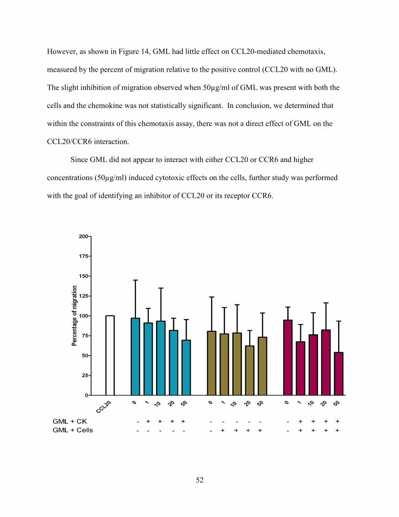
52
However, as shown in Figure 14, GML had little effect on CCL20-mediated chemotaxis,
measured by the percent of migration relative to the positive control (CCL20 with no GML).
The slight inhibition of migration observed when 50µg/ml of GML was present with both the
cells and the chemokine was not statistically significant. In conclusion, we determined that
within the constraints of this chemotaxis assay, there was not a direct effect of GML on the
CCL20/CCR6 interaction.
Since GML did not appear to interact with either CCL20 or CCR6 and higher
concentrations (50µg/ml) induced cytotoxic effects on the cells, further study was performed
with the goal of identifying an inhibitor of CCL20 or its receptor CCR6.
53
5.3.2 CCR6 receptor peptide mimetics ECL-2 inhibit CCL20-induced migration
Our laboratory has shown that the extracellular loops of chemokine receptors, so called
receptor peptide mimetics or ECL-X, where X refers to the number of the sequential
extracellular loop, can bind directly to chemokine64 and inhibit chemokine-induced migration.
The method of using the second extracellular loop to affect outcomes of signaling through
chemokine receptors has also been used to inhibit HIV entry by using the second extracellular
loop of CCR5 85. Interest in this property of the ECL-X was applied here to CCL20-induced
migration. To this end, the second extracellular loop, or ECL-2, of CCR6 (R6 ECL-2) was
synthesized by the University of Pittsburgh Peptide Facility. Due to the possibility that the
cognate rhesus R6 ECL-2 CYS (containing a cysteine residue) could form disulfide bridges in
solution, two other R6 ECL-2s were also synthesized. Amino acid substitutions for the cysteine
residue were decided upon based on weight and isoelectric point, striving for comparable
behaviors in solution and similar overall molecular weights. To this end, alanine or threonine
were substituted for the cysteine during peptide synthesis producing R6 ECL-2 ALA and R6
ECL-2 THR. A graphic representation of the location of the extracellular loop with respect to
the structure of a G protein coupled receptor and the location of the amino substitutions used is
shown in Figure 15.
Figure 14. GML does not inhibit CCL20-mediated chemotaxis. Stable CCR6 L1.2 cells were prepared as for chemotaxis, with overnight pre-treatment with sodium butyrate. 10nM synthetic rhesus CCL20 was prepared and increasing concentrations (µg/ml) of GML were added to the chemokine or to the cells. After a three hour incubation at 37°C in 5% CO2, cells that had migrated through the membrane were counted on a hemocytometer and percent migration total was calculated as the total number of cells that had migrated towards the positive control, 10nM CCL20. Data represents 3 independent repeats, +/- SD.
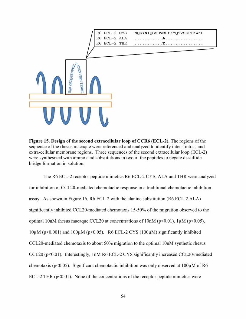
54
The R6 ECL-2 receptor peptide mimetics R6 ECL-2 CYS, ALA and THR were analyzed
for inhibition of CCL20-mediated chemotactic response in a traditional chemotactic inhibition
assay. As shown in Figure 16, R6 ECL-2 with the alanine substitution (R6 ECL-2 ALA)
significantly inhibited CCL20-mediated chemotaxis 15-50% of the migration observed to the
optimal 10nM rhesus macaque CCL20 at concentrations of 10nM (p<0.01), 1µM (p<0.05),
10µM (p<0.001) and 100µM (p<0.05). R6 ECL-2 CYS (100µM) significantly inhibited
CCL20-mediated chemotaxis to about 50% migration to the optimal 10nM synthetic rhesus
CCL20 (p<0.01). Interestingly, 1nM R6 ECL-2 CYS significantly increased CCL20-mediated
chemotaxis (p<0.05). Significant chemotactic inhibition was only observed at 100µM of R6
ECL-2 THR (p<0.01). None of the concentrations of the receptor peptide mimetics were
Figure 15. Design of the second extracellular loop of CCR6 (ECL-2). The regions of the sequence of the rhesus macaque were referenced and analyzed to identify inter-, intra-, and extra-cellular membrane regions. Three sequences of the second extracellular loop (ECL-2) were synthesized with amino acid substitutions in two of the peptides to negate di-sulfide bridge formation in solution.

55
cytotoxic (data not shown), so it can be concluded that the inhibition observed was due to an
interaction of R6 ECL-2 with either CCL20 or CCR6.
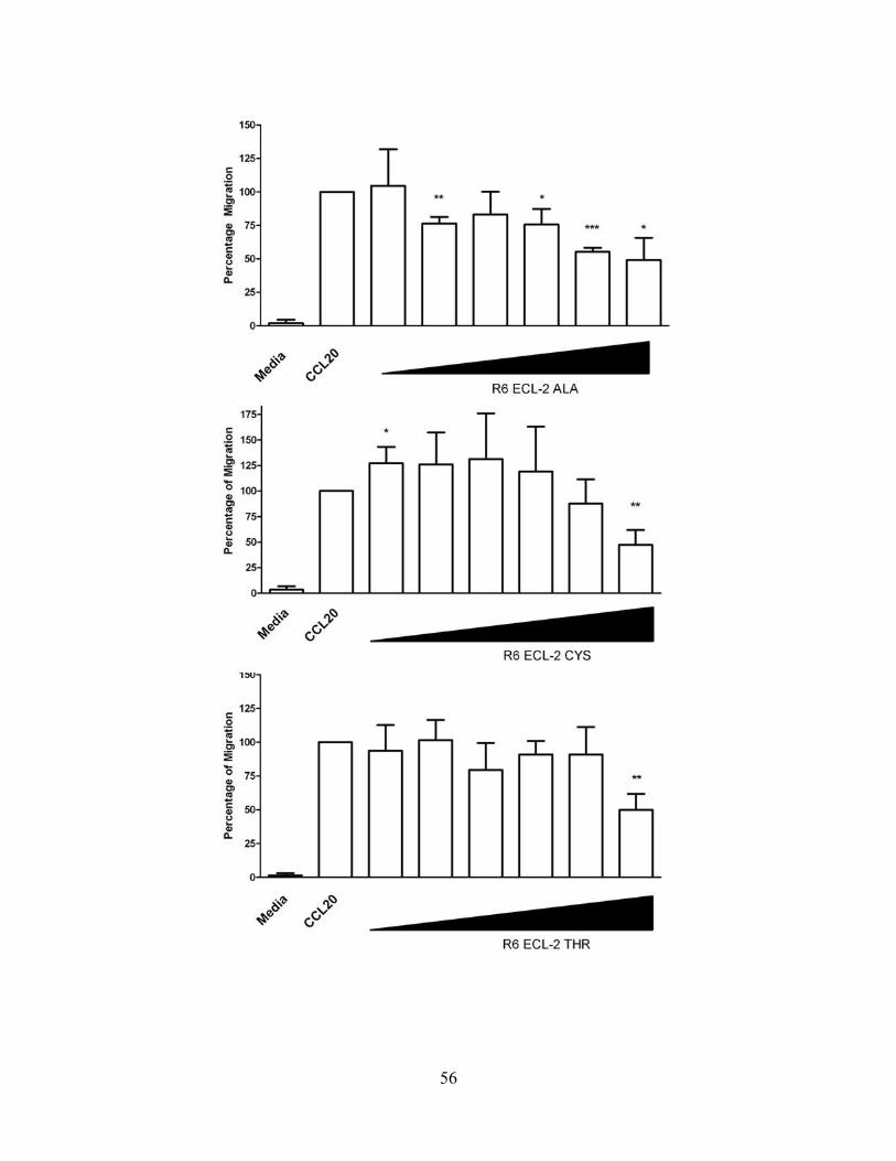
56
57
5.3.3 Anti-Inflammatory Botanical Inhibitors
Botanical elements have long been studied for their medicinal and biochemical
properties. Our laboratory has recently been interested in a polyphenol compound found in
green tea, epigallocatechin gallate, EGCG, due to its anti-inflammatory properties52.
Additionally, our laboratory has observed that low concentrations of EGCG can inhibit
chemotaxis of CXCR3+ cells 64. Biacore binding assays revealed that EGCG bound CXCR3
ligands with high affinity (ref.64 and Qin, et al, in preparation). Our laboratory has observed
another anti-inflammatory property of EGCG in a mouse model of asthma, where there were a
reduced number of T cells and airway inflammation with EGCG treatment (Qin et al, in
preparation). For these reasons, EGCG seemed an excellent candidate inhibitor that may affect
CCL20-mediated chemtoaxis.
5.3.3.1 EGCG Inhibition of CCL20-mediated Chemotaxis
To determine whether EGCG inhibited CCL20-mediated chemotaxis as was observed by
our laboratory for CXCR3 ligands, stable L1.2 human CCR6 cells were placed in a chemotactic
inhibition assay with increasing concentrations of EGCG. A significant (10-20%) inhibition of
CCL20-mediated chemotaxis was observed at concentrations of 1nM and 10µM EGCG (Figure
18, p<0.01). At the 100µM EGCG concentration, 75% inhibition was observed (p<0.001).
EGCG induced inhibition of CCL20 chemotaxis was at a much higher concentration than we
have previously observed with EGCG inhibited CXCR3 mediated chemotaxis (Tjoeng, 2009).
Figure 16. R6 ECL-2 (receptor peptide mimetics) inhibit CCL20-induced migration. Stable L1.2 human CCR6 cells were placed in a chemotaxis inhibition assay. 10nM synthetic rhesus CCL20 served as a positive control for migration and was mixed with 10 fold increasing concentrations (range of 1nM to 100µM) of the 3 receptor peptide mimetics, R6 ECL-2 ALA, R6 ECL-2 CYS and R6 ECL-2 THR. Data represents 3 repeat experiments +/- SD. Paired t-test, * p<0.05, ** p<0.01, *** p<0.001.
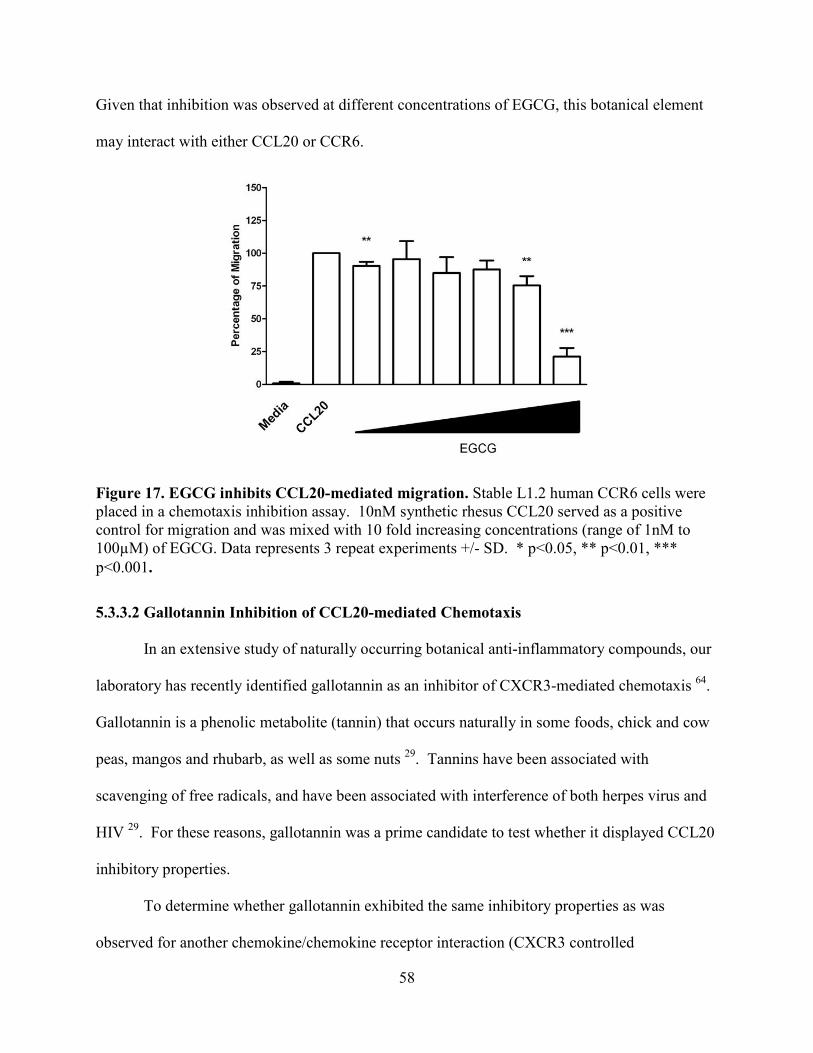
58
Given that inhibition was observed at different concentrations of EGCG, this botanical element
may interact with either CCL20 or CCR6.
5.3.3.2 Gallotannin Inhibition of CCL20-mediated Chemotaxis
In an extensive study of naturally occurring botanical anti-inflammatory compounds, our
laboratory has recently identified gallotannin as an inhibitor of CXCR3-mediated chemotaxis 64.
Gallotannin is a phenolic metabolite (tannin) that occurs naturally in some foods, chick and cow
peas, mangos and rhubarb, as well as some nuts 29. Tannins have been associated with
scavenging of free radicals, and have been associated with interference of both herpes virus and
HIV 29. For these reasons, gallotannin was a prime candidate to test whether it displayed CCL20
inhibitory properties.
To determine whether gallotannin exhibited the same inhibitory properties as was
observed for another chemokine/chemokine receptor interaction (CXCR3 controlled
Figure 17. EGCG inhibits CCL20-mediated migration. Stable L1.2 human CCR6 cells were placed in a chemotaxis inhibition assay. 10nM synthetic rhesus CCL20 served as a positive control for migration and was mixed with 10 fold increasing concentrations (range of 1nM to 100µM) of EGCG. Data represents 3 repeat experiments +/- SD. * p<0.05, ** p<0.01, *** p<0.001.
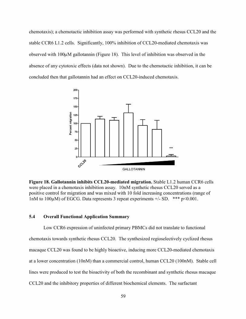
59
chemotaxis); a chemotactic inhibition assay was performed with synthetic rhesus CCL20 and the
stable CCR6 L1.2 cells. Significantly, 100% inhibition of CCL20-mediated chemotaxis was
observed with 100µM gallotannin (Figure 18). This level of inhibition was observed in the
absence of any cytotoxic effects (data not shown). Due to the chemotactic inhibition, it can be
concluded then that gallotannin had an effect on CCL20-induced chemotaxis.
5.4 Overall Functional Application Summary
Low CCR6 expression of uninfected primary PBMCs did not translate to functional
chemotaxis towards synthetic rhesus CCL20. The synthesized regioselectively cyclized rhesus
macaque CCL20 was found to be highly bioactive, inducing more CCL20-mediated chemotaxis
at a lower concentration (10nM) than a commercial control, human CCL20 (100nM). Stable cell
lines were produced to test the bioactivity of both the recombinant and synthetic rhesus macaque
CCL20 and the inhibitory properties of different biochemical elements. The surfactant
Figure 18. Gallotannin inhibits CCL20-mediated migration. Stable L1.2 human CCR6 cells were placed in a chemotaxis inhibition assay. 10nM synthetic rhesus CCL20 served as a positive control for migration and was mixed with 10 fold increasing concentrations (range of 1nM to 100µM) of EGCG. Data represents 3 repeat experiments +/- SD. *** p<0.001.
60
compound that has been linked with reduced SIV replication in the macaque vagina10 where
CCL20 is expressed, GML, was not found to affect migration towards CCL20. The receptor
peptide mimetic, R6 ECL-2 peptides, R6 ECL-2 ALA, CYS and THR were all found to inhibit
CCL20-mediated chemotaxis significantly at a concentration of 100µM. R6 ECL-2 ALA was
also found to significantly inhibit CCL20-mediated chemotaxis at 10nM, 1µM and 10µM. n R6
ECL-2 CYS significantly enhanced CCL20-mediated chemotaxis at 1nM. The anti-
inflammatory botanical EGCG inhibited chemotaxis at 1nM, 10µM and 100µM, with almost
complete inhibition observed at 100µM. Another botanical anti-inflammatory, gallotannin,
significantly inhibited chemotaxis to 100% at 100µM. Inhibitory concentrations of all anti-
inflammatory elements that inhibited CCL20-mediated chemotaxis were all higher than has been
observed previously in our laboratory.
61
6.0 Discussion
Chemokines are important factors in immunology in that they exhibit multiple functional
roles, including lymph organogenesis, polarization of immune responses and leukocyte
trafficking. Non-human primate chemokines are unavailable for use in studies modeling
immunological responses, infectious disease and pathogenesis. My project provided our
laboratory with two methods by which to obtain non human primate chemokines to use in vitro
and in vivo. Additionally, I improved our understanding of the affect of our repertoire of anti-
inflammatory compounds, in addition to studying new compounds to study (GML), on the
CCL20/CCR6 interaction.
6.1 Protein Expression
Both the Origami B DE3 and Rosettagami B DE3 strains of E. coli were utilized due to
their ability to over-express proteins via IPTG addition and plasmids encoding a gene that aided
in the di-sulfide bond formation in the cytosol that may disallow over-expressed proteins to
aggregate in inclusion bodies. The SUMO fusion technology was utilized due to the ease of
purification of the SUMO-fusion proteins and the specificity of the SUMO protease. I found that
both strains of E. coli expressed the protein upon IPTG induction. However, the resultant
SUMO-CCL20 fusion protein aggregated in inclusion bodies.
Aggregation may have affected the ability to extract and purify abundant SUMO-CCL20
fusion proteins as large scale batch (up to 12L) protein expression did not yield sufficiently high
quantities of SUMO-CCL20 fusion protein, since after HPLC purification of the yield of 1.7
milligrams of SUMO-CCL20, 10µg of SUMO-CCL20 fusion was available for additional
cleavage testing and biological activity testing. Factors affecting the yield of SUMO-CCL20 are
62
potentially that all inter-molecular bonding of aggregated proteins might not be completely
dispersed and some of this protein may not be a proper substrate for metal affinity column
purification. Evidence to this fact is that it has been shown that once proteins have aggregated
in proteins, these same proteins could have a tendency to aggregate in solution despite reducing
and chaotropic agents that should disrupt inter-molecular aggregation.
Reasons for the aggregation of SUMO-CCL20 in inclusion bodies are as follows. It may
be that SUMO-fusion proteins have a propensity to aggregate upon over-production. It may also
be that the sequence and structure of the chemokine-here CCL20-contribute to the formation of
inclusion bodies. Disulfide bonding in the cytosol has long been identified as a difficulty in E.
coli due to the reduced state of the cytosol 75. This may be a reason why inclusion bodies are still
formed despite the expression of a thioredoxin reductase and glutathione reductase, two factors
that increase the formation of disulfide bonds in the cytosol 72. Additionally, it has been shown
that proteins that are cytotoxic to E. coli are aggregated in inclusion bodies 72, 74. CCL20 has
been proven to have anti-microbial properties which may contribute to its aggregation in
inclusion bodies upon overproduction23.
The SUMO fusion technology was successful in providing relatively low quantities of
pure protein. However, when the time came to remove the SUMO-fusion tag by cleavage with
the SUMO protease, the cleavage of the SUMO-CCL20 fusion protein was only about 30%
efficient and precluded the ability to obtain enough recombinant protein for bioactivity analysis.
The main issue with the recombinant protein expression system here was the poor yield
of the desired, final protein. Cleavage of the SUMO-CCL20 protein, despite efforts at
optimizing the yield and cleavage of the protein, remained at an efficiency of only 30-50%. The
multi-parameter experiment (Figure 4) did not reveal conditions optimal for cleavage. None of
63
the reaction conditions showed complete cleavage of the SUMO-CCL20 fusion protein (Figure 5
and 6), despite the protease being shown to be active (Figure 3). Use of two different strains of
E. coli did not change the efficiency of cleavage (Figure 8 and 9).
It may be that the cleavage site in the SUMO-CCL20 fusion protein is unavailable for the
protease. Because the recognition 'site' of the SUMO protease is the tertiary structure of SUMO
tag, the fusion protein requires refolding through chemical agents and dialysis in a neutral buffer,
so that both the chemokine and fusion tag of the protein have the best chance to be in the proper
tertiary structure(s) to allow efficient cleavage. Refolding of the fusion protein could make the
cleavage site of the fusion protein unable to be recognized by the SUMO protease, in that the
tertiary structure of the SUMO-CCL20 fusion would be disrupted as the chemokine may fold
over the SUMO protease recognition sequence. Additionally, or even in conjunction, there
might be different co-existent entities of partially folded fusion proteins, misfolded fusion
proteins and properly folded fusion proteins that influence the level of cleavage that can be
detected. It may be that the SUMO-CCL20 fusion protein that is evidenced as being cleaved
(Fig 2B and Figure 3) may be only the properly folded fusion protein and other mis-folded
SUMO-CCL20 fusions are left uncleaved.
Inefficient cleavage may be the result of SUMO protease may be more active in cleaving
the SUMO-CCL20 fusion protein at a different concentration or temperature than was examined
in the multi-parameter analysis. It may also be that despite using Rosettagami B DE3 cells to
control the incorporation of rare tRNAs, there were mis-incorporations of amino acids in the
SUMO tag protein versus mis-incorporation in the rhesus CCL20 protein since, as stated in
section 4.2.2, SUMO tag contains four 'AGA' targets for mis-incorporation5. Mis-incorporation
64
could again cause disrupted proper tertiary structure of SUMO tag protein hindering its
recognition by SUMO protease.
Despite the problems with cleavage of SUMO-CCL20 fusion, a bioactive form of rhesus
macaque CCL20 was chemically synthesized. Regioselective cyclization peptide synthesis
provided a method to obtain a large batch (35mgs) of highly pure protein measured by HPLC. In
vitro studies revealed the rhesus macaque CCL20 was highly bioactive.
The assessment of the recombinant expression protocol utilizing the SUMO technology is
that further optimization and study may reveal the steps by which large quantities of a
recombinant rhesus macaque CCL20 can be purified and used in vitro or in vivo as a therapeutic
agent or vaccine adjuvant. Although peptide synthesis provided a method by which highly pure
bioactive protein was produced, mutation or amino acid substitution analysis of the protein
sequence to identify important binding sequences of recombinant chemokine would be much
more expensive and therefore limited than utilizing the recombinant expression system. Both
recombinant expression and chemical synthesis of a rhesus macaque chemokine is definitely
needed because very few if any macaque chemokines are available commercially.
6.2 Application of a functional chemotaxis assay for in vitro system development
A cell line expressing stable CCR6 was produced to study the bioactivity of both the
recombinant and synthesized rhesus macaque CCL20. Enough SUMO-CCL20 fusion or cleaved
recombinant rhesus CCL20 was not produced, and therefore bioactivity of the recombinant
proteins was not measured. Rhesus macaque CCL20 was generated for bioactivity testing by
peptide synthesis using regioselective cyclization. The synthetic rhesus macaque CCL20, in
contrast, was shown to be highly bioactive in a chemotaxis assay. Multiple compounds were
analyzed for inhibition of CCL20-mediated chemotaxis. The biochemical anti-microbial
65
surfactant, GML, did not have a direct effect on CCL20-mediated chemotaxis. Second
extracellular receptor peptide mimetics R6 ECL-2 ALA, R6 ECL-2 CYS and R6 ECL-2 THR
were shown to inhibit chemotaxis at high concentrations and were determined to have some
effect on CCL20-mediated chemotaxis. Both anti-inflammaotry botanicals, EGCG and
Gallotannin, analyzed potently inhibited chemotaxis at high concentrations.
In vivo studies of the response of immune cells to intradermal injections with synthetic
rhesus CCL20 did not show that an inflammatory response to the chemokine. After further
analysis, it may be that the concentration of CCL20 administered may have been at a
concentration too high to elicit an appropriate localized immune response. CCL20 may have
been marked for degradation due to the over-abundance by the 'chemokine sink' receptors which
act to sequester over abundant chemokine25. Another possible explanation is that the time at
which samples were taken for analysis was too soon to observe the effects of the CCL20 skin
injections.
The importance of the development of a cell line stably expressing CCR6 is that the
specific response to CCL20 and how that response may be affected by potential inhibitors can be
studied. For example, on primary cells isolated from the blood or other tissues, there are a
variety of chemokine receptors expressed on any cell type3. While the varied chemokine
expression is no doubt immunologically indispensable, study of the influence of an individual
chemokine response (i.e. CCL20/CCR6) would be difficult to interpret and define functionally.
Therefore defining the specific effect of any inhibitory element on the CCL20/CCR6 interaction
is warranted in chemokine/chemokine receptor in vitro studies.
66
6.3 Conclusion
In conclusion, the utilization of the recombinant protein expression system to produce
chemokines can sufficiently produce pure SUMO-CCL20 fusion proteins, but the quantity of
culture needed to produce enough SUMO-CCL20 fusion or recombinant CCL20 for bioactivity
testing was not feasible. Additionally, the most efficient cleavage of the SUMO-CCL20 fusion
protein achieved was about 30-50% cleavage.
In addition, the CCR6 stable cell lines were highly efficient at responding to synthetic
rhesus CCL20 and represents a good tool for performing CCL20 chemotaxis inhibition. Using
this assay, I determined that the CCR6/CCL20 interaction was modulated by high concentrations
of several compounds, R6 ECL-2 peptides and the botanical elements, EGCG and gallotannin.
The model of chemotactic inhibition induced by EGCG and gallotannin has been
described elsewhere64. Briefly, the high number of hydroxyl groups on the gallate groups of
both EGCG and gallotannin may mimic glycosaminoglycans (GAGs)60 and bind to GAG binding
domains on the chemokine, in this case, CCL20. The fact that only high concentrations of both
botanicals inhibited chemotaxis may mean that gag binding domains within rhesus CCL20 are
resistant to binding of EGCG or gallotannin gallate groups.
6.3.1 Next Steps and Future Studies
In taking these studies to the next level, the following studies will be important to
execute. The expression and purification protocol needs further optimization and adaptation to
be useful for any future in vitro or in vivo studies with recombinant rhesus macaque CCL20.
Also, the bioactivity of the SUMO-CCL20 and the recombinant CCL20 needs to be tested. To
optimize the yield of SUMO-CCL20, the plasmid containing both the fusion protein tag and
rhesus CCL20 could be codon optimized for E.coli expression. This may help to determine
67
whether the problems of cleavage could be attributed to the mis-incorporations of amino acids in
both the SUMO fusion protein tag and rhesus CCL20. Expression of the protein could be carried
out at different temperatures to analyze whether the solubility of the fusion protein could be
increased. A soluble protein may be cleaved more efficiently and this would increase the yield
of recombinant rhesus CCL20 from the expression system.
The stable CCR6 cell line could be utilized to test the effects other anti-inflammatory
compounds, such as curcumin, other green tea polyphenols, EGC, ECG, and EC, on CCL20-
mediated chemotaxis. To further confirm the model of inhibition of CCL20-mediated
chemotaxis via gallate groups acting as GAG binding domains, the GAG binding domain of
CCL20 could be mutated, as suggested in Tjoeng, 200964.
In vitro dendritic cell studies could be performed to analyze whether addition of CCL20
and HIV immunogens would enhance antigen processing and subsequent T cell priming and
direction in vitro. Specifically, a prime and boost strategy of vaccination using CCL20 as an
adjuvant for HIV immunogens could be performed to identify whether CCL20 is in fact a
candidate adjuvant for vaccine studies.
Since constitutive CCL20 expression has been shown in the vagina and colon, but likely
is also present at the anal/rectal junction, and since it has been suggested that chemokine receptor
expression is or may be involved in the directing of immune cells to different tissue
environments, it could be tested whether using CCL20 as a vaccine adjuvant also directed
memory T cells to the correct tissue compartment (i.e. vagina, anal/rectal junction, or colon) for
fighting initial HIV/SIV challenge. The synthetic rhesus CCL20 could be used as a vaccine
adjuvant in mucosal delivery (vaginal or anal/rectal) of highly expressed SIV or HIV
immunogens to study whether the delivery of CCL20 with SIV/HIV immunogens boosts the
68
initial immune response to sexual transmission of SIV/HIV in that tissue compartment.
Specifically, another prime and boost vaccination strategy, but where both the immunogen
(HIV/SIV proteins) and adjuvant (CCL20) were applied to the mucosal interface where initial
challenge occurs in transmission (vagina, anal/rectal junction, or colon). If CCL20 were found
to be a candidate adjuvant, stronger memory T cell responses would be observed with the use of
CCL20 and immunogen versus immunogen alone upon HIV/SIV challenge.
Since inhibition of CCL20-mediated chemotaxis was observed, any one of the
compounds could be used in vivo to study the effects of the compound on vaginal SIV
transmission, similar to the study done by Li, et al with GML10. Specifically, the question would
be whether the mucosal delivery of anti-inflammatory compounds inhibits SIV/HIV
transmission. Although, this may seem contradictory to a desired immunological outcome of a
fast and strong response to the invading pathogen (HIV), it may be that despite chronic immune
activation it appears that the virus evades the immune system, turning the favor to a slower
inflammatory response may give the immune system the foothold it needs to overcome virus
replication.
Other immunological diseases that may be studied based on this study are those in which
at least some of the pathology of the disease is associated with up-regulation of CCL20
expression. Examples of diseases to consider with respect to CCL20 expression are chronic
inflammatory diseases, such as some skin diseases, i.e. atopic dermatitis and psoriasis, or gut
associated diseases, inflammatory bowel disease (IBD) and Crohn’s disease27 and some
autoimmune diseases, such as colorectal cancer. If the up-regulation of CCL20 expression
causes tissue destruction, application of the anti-inflammatory compounds that inhibited CCL20
migration may be useful as treatments of these diseases. For the skin diseases, the inhibitory
69
dose of EGCG or gallotannin could be applied in a cream to psoriatic lesions to test whether the
treatment reduced the size and longevity (i.e. how long until the lesion disappears) of the lesion.
Use of the anti-inflammatory compounds for gut associated diseases (IBD, Crohn’s disease)
would be a little more complicated as the metabolism of EGCG and gallotannin may break down
or alter the structures (gallate groups) that inhibit CCL20-mediated chemotaxis. A way in which
the delivery to more internal tissue compartments of these anti-inflammatory compounds could
be made easier may be through something like biodegradable microspheres. Microspheres are
similar to micelles, in that the desired compound is encased in polylactic acid (PLA)11. As the
microsphere degrades, increasing levels of the desired compound are released, in a so-called
time-release fashion. Biodegradable microspheres have been used to time deliver insulin in a
mouse model of diabetes11. The time-released insulin did appear to aid in leveling the blood
glucose levels11. Additionally, while working out their own protocol, the University of
Pittsburgh Peptide Facility (UPPF) encased our rhesus macaque CCL20 in PLA microspheres.
Although the protocol was not optimized by UPPF, this at least gives a possible avenue for
studying the time-release drug delivery hypothesis for anti-inflammatory botanicals. In the same
fashion, biodegradable microspheres could be utilized to deliver EGCG or gallotannin to the gut
essentially unperturbed by metabolic and enzymatic processing or breakdown. It could then be
analyzed whether these treatments produced desirable prognosis or outcomes of the disease, i.e.
reduction of the chronic inflammation of the gut in IBD or Crohn’s disease.
6.3.2 Public Health Revelance of this Study
The public health relevance of studying ways to produce recombinant bioactive non-
human primate chemokines is that studies of human immunity and treatment for diseases are
sometimes modeled in monkeys. In monkey models of infections and immunologic diseases,
70
most reagents used are of human specificity. The species difference may alter the immune
response generated or the ability to detect the antigen, thereby skewing the interpretation of the
results, which then translate to poor modeling of human immunity and disease. The use of
species specific reagents will provide more confidence in the results and interpretations of non-
human primate studies.
Prevention is a main public health consideration and vaccination is one way prevention of
transmissible diseases is achieved. If an anti-inflammatory compound affects the immune
response to a vaccine, the efficacy and longevity of the vaccine may then be affected. One
reason studying how CCL20/CCR6 interaction is modulated by anti-inflammatory compounds is
that to mount an efficient immune response to vaccinations, a main player is immature DCs
which express CCR6. Other factors affecting the efficacy of vaccination are the immunogen and
the inflammatory immune response that is mounted to a vaccination. Therefore, identifying anti-
inflammatory compounds that potentially cause a stinted inflammatory response to the vaccine
and inhibit the influx of CCR6+ immature DCs is important in developing efficacious vaccines.
An additional reason studying the CCR6/CCL20 interaction is important to public health
is in relation to the HIV epidemic. The ultimate goal to eradicate the world-wide HIV-1
epidemic will require a vaccine to or treatment of HIV that either prevents HIV transmission, or
cures the prolonged immune activation and also inhibits virus replication. Recent HIV studies
have linked CCR6 and CCL20 with both enhanced and reduced HIV replication49, 51, 53, 54, 55 .
The simple fact that a chemokine/chemokine receptor pair has potentially host-beneficial
properties is reason to consider the pair in transmission and treatment of HIV. The
CCR6/CCL20 interaction may be important in developing ‘a site of entry’ mucosal vaccine to
HIV because of CCL20 expression in the mucosal tissues involved by sexual transmission of
71
HIV and CCL20’s role in maintaining and recruiting immature DCs within the tissue. Another
indication that CCR6/CCL20 should be considered to combat HIV transmission and replication
is that a naturally occurring anti-HIV molecule, APOBEC3G, is up-regulated by CCR6
signaling, thereby protecting the cell expressing APOBEC3G from HIV replication49. These
findings indicate that CCR6/CCL20 may be important in combating HIV transmission and
persistence, and therefore important to consider in developing methods to treat and prevent HIV.
72
BIBLIOGRAPHY
1. Badr G, Borhis G, Treton D, Moog C, Garraud O, Richard Y: HIV type 1 glycoprotein 120 inhibits Human B Cell Chemotaxis to CXC Chemokine Ligand (CCL) 20, and CCL21. J Immunol. 2005 Jul 1; 175(1):302-10.
2. Berlier W, Cremel M, Hamzeh H, Lévy R, Lucht F, Bourlet T, Pozzetto B, Delézay O: Seminal plasma promotes the attraction of Langerhans cells via the secretion of CCL20 by vaginal epithelial cells: involvement in the sexual transmission of HIV. Hum Reprod. 2006 May; 21(5):1135-42.
3. Bromley SK, Mempel TR, Luster AD: Orchestrating the orchestrators: chemokines in control of T cell traffic. Nat Immunol. 2008 Sep; 9(9):970-80.
4. Butt TR, Edavettal SC, Hall JP, Mattern M: SUMO fusion technology for difficult-to-express proteins. Protein Expr Purif. 2005 Sep;43(1):1-9.
5. Calderone TL, Stevens RD, Oas TG: High-level misincorporation of lysine for arginine at AGA codons in a fusion protein expressed in Escherichia coli. J Mol Biol. 1996 Oct 4; 262(4):407-12.
6. Choi YK, Whelton KM, Mlechick B, Murphey-Corb MA, Reinhart TA: Productive infection of dendritic cells by simian immunodeficiency virus in macaque intestinal tissues. J Pathol. 2003 Dec; 201(4):616-28.
7. Cremel M, Hamzeh-Cognasse H, Genin C, Delézay O: Female genital tract immunization: evaluation of candidate immunoadjuvants on epithelial cell secretion of CCL20 and dendritic/Langerhans cell maturation. Vaccine. 2006 Jul 17; 24(29-30):5744-54.
8. Hoover DM, Boulegue C, Yang D, Oppenheim JJ, Tucker K, Lu W, Lubkowski J: The structure of human macrophage inflammatory protein-3alpha /CCL20. Linking antimicrobial and CC chemokine receptor-6-binding activities with human beta- defensins. J Biol Chem. 2002 Oct 4; 277(40):37647-54.
9. Imaizumi Y, Sugita S, Yamamoto K, Imanishi D, Kohno T, Tomonaga M, Matsuyama T: Human T cell leukemia virus type-I Tax activates human macrophage inflammatory protein-3 alpha/CCL20 gene transcription via the NF-kappa B pathway. Int Immunol. 2002 Feb; 14(2):147-55.
10. Li Q, Estes JD, Schlievert PM, Duan L, Brosnahan AJ, Southern PJ, Reilly CS, Peterson ML, Schultz-Darken N, Brunner KG, Nephew KR, Pambuccian S, Lifson JD, Carlis JV, Haase AT: Glycerol monolaurate prevents mucosal SIV transmission. Nature. 2009 Mar 4.
73
11. Mathiowitz E, Jacob JS, Jong YS, Carino GP, Chickering DE, Chaturvedi P, Santos CA,
Vijayaraghavan K, Montgomery S, Bassett M, Morrell C :Biologically erodable microspheres as potential oral drug delivery systems. Nature. 1997 Mar 27; 386(6623):410-4.
12. Murphy PM, Baggiolini M, Charo IF, Hébert CA, Horuk R, Matsushima K, Miller LH, Oppenheim JJ, Power CA: International union of pharmacology. XXII. Nomenclature for chemokine receptors. Pharmacol Rev. 2000 Mar; 52(1):145-76.
13. Rajagopalan L, Rajarathnam K: Structural basis of chemokine receptor function--a model for
binding affinity and ligand selectivity. Biosci Rep. 2006 Oct; 26(5):325-39.
14. Sabel MS, Skitzki J, Stoolman L, Egilmez NK, Mathiowitz E, Bailey N, Chang WJ, Chang AE: Intratumoral IL-12 and TNF-alpha-loaded microspheres lead to regression of breast cancer and systemic antitumor immunity. Ann Surg Oncol. 2004 Feb; 11(2):147-56.
15. Schneider T, Jahn HU, Schmidt W, Riecken EO, Zeitz M, Ullrich R: Loss of CD4 T lymphocytes in patients infected with human immunodeficiency virus type 1 is more pronounced in the duodenal mucosa than in the peripheral blood. Berlin Diarrhea/Wasting Syndrome Study Group. Gut. 1995 Oct, 37(4): 524-9.
16. Sierro F, Dubois B, Coste A, Kaiserlian D, Kraehenbuhl JP, Sirard JC: Flagellin stimulation of intestinal epithelial cells triggers CCL20-mediated migration of dendritic cells. Proc Natl Acad Sci U S A. 2001 Nov 20; 98(24):13722-7.
17. Singh SM, Panda AK: Solubilization and refolding of bacterial inclusion body proteins. J Biosci Bioeng. 2005 Apr; 99(4):303-10.
18. Singh SP, Zhang HH, Foley JF, Hedrick MN, Farber JM. Human T cells that are able to produce IL-17 express the chemokine receptor CCR6. J Immunol. 2008 Jan 1; 180(1):214-21.
19. Williams, IR: CCR6 and CCL20: partners in intestinal immunity and lymphorganogenesis. Ann N Y Acad Sci. 2006 Aug; 1072:52-61.
20. Veazey RS, DeMaria M, Chalifoux LV, Shvetz DE, Pauley DR, Knight HL, Rosenzweig M, Johnson RP, Desrosiers RC, Lackner AA: Gastrointestinal tract as a major site of CD4+ T cell depletion and viral replication in SIV infection. Science. 1998 Apr 17; 280(5362):427-31.
21. Veazey RS, Lackner AA: HIV swiftly guts the immune system. Nat Med. 2005 May; 11(5):469-70.
74
22. Wu YY, Tsai HF, Lin WC, Hsu PI, Shun CT, Wu MS, Hsu PN: Upregulation of CCL20 and recruitment of CCR6+ gastric infiltrating lymphocytes in Helicobacter pylori gastritis. Infect Immun. 2007 Sep; 75(9):4357-63.
23. Yang D, Chen Q, Hoover DM, Staley P, Tucker KD, Lubkowski J, Oppenheim JJ: Many chemokines including CCL20/MIP-3alpha display antimicrobial activity. J Leukoc Biol. 2003 Sep; 74(3):448-55.
24. Zuo X, Mattern MR, Tan R, Li S, Hall J, Sterner DE, Shoo J, Tran H, Lim P, Sarafianos SG, Kazi L, Navas-Martin S, Weiss SR, Butt TR: Expression and purification of SARS coronavirus proteins using SUMO-fusions. Protein Expr Purif. 2005 Jul; 42(1):100-10.
25. Graham GJ, McKimmie CS: Chemokine scavenging by D6: a movable feast? Trends Immunol. 2006 Aug;27(8):381-6.
26. Fujiie S, Hieshima K, Izawa D, Nakayama T, Fujisawa R, Ohyanagi H, Yoshie O.
:Proinflammatory cytokines induce liver and activation-regulated chemokine/macrophage inflammatory protein-3alpha/CCL20 in mucosal epithelial cell through NF-kappaB. Int Immunol. 2001 Oct;13(10):1255-63. Erratum in: Int Immunol 2001 Nov;13(11):1443.
27. Gullberg E, Söderholm JD: Peyer's patches and M cells as potential sites of the inflammatory
onset in Crohn's disease. Ann N Y Acad Sci. 2006 Aug;1072:218-32. 28. Dieu MC, Vanbervliet B, Vicari A, Bridon JM, Oldham E, Aït-Yahia S, Brière F, Zlotnik A,
Lebecque S, Caux C: Selective recruitment of immature and mature dendritic cells by distinct chemokines expressed in different anatomic sites. J Exp Med. 1998 Jul 20;188(2):373-86.
29. Serrano J, Puupponen-Pimiä R, Dauer A, Aura AM, Saura-Calixto F. Tannins: current knowledge of food sources, intake, bioavailability and biological effects. Mol Nutr Food Res. 2009 Sep;53 Suppl 2:S310-29.
30. Kunkel EJ, Campbell DJ, Butcher EC. Chemokines in lymphocyte trafficking and intestinal immunity.Microcirculation. 2003 Jun;10(3-4):313-23.
31. Shaykhiev R, Bals R: Interactions between epithelial cells and leukocytes in immunity
andtissue homeostasis. J Leukoc Biol. 2007 Jul;82(1):1-15.
32. Schutyser E, Struyf S, Van Damme J. The CC chemokine CCL20 and its receptor CCR6. Cytokine Growth Factor Rev. 2003 Oct;14(5):409-26.
33. Iwasaki A, Kelsall BL. Localization of distinct Peyer's patch dendritic cell subsets and their
recruitment by chemokines macrophage inflammatory protein (MIP)-3alpha, MIP-3beta, and secondary lymphoid organ chemokine. J Exp Med. 2000 Apr 17;191(8):1381-94.
75
34. Müller CA, Autenrieth IB, Peschel A. Innate defenses of the intestinal epithelial barrier. Cell Mol Life Sci. 2005 Jun;62(12):1297-307.
35. Vongsa RA, Zimmerman NP, Dwinell MB. CCR6 regulation of the actin cytoskeleton
orchestrates human beta defensin-2- and CCL20-mediated restitution of colonic epithelial cells. J Biol Chem. 2009 Apr 10;284(15):10034-45
36. McDonald KG, McDonough JS, Wang C, Kucharzik T, Williams IR, Newberry RD. CC
chemokine receptor 6 expression by B lymphocytes is essential for the development of isolated lymphoid follicles. Am J Pathol. 2007 Apr;170(4):1229-40.
37. Yang D, Chertov O, Bykovskaia SN, Chen Q, Buffo MJ, Shogan J, Anderson M, Schröder
JM,Wang JM, Howard OM, Oppenheim JJ. Beta-defensins: linking innate and adaptive immunity through dendritic and T cell CCR6. Science. 1999 Oct 15;286(5439):525-8.
38. Zhang Z, Schuler T, Zupancic M, Wietgrefe S, Staskus KA, Reimann KA, Reinhart TA, Rogan M, Cavert W, Miller CJ, Veazey RS, Notermans D, Little S, Danner SA, Richman DD, Havlir D, Wong J, Jordan HL, Schacker TW, Racz P, Tenner-Racz K, Letvin NL, Wolinsky S, Haase AT. Sexual transmission and propagation of SIV and HIV in resting and activated CD4+ T cells. Science. 1999 Nov 12;286(5443):1353-7. Erratum in: Science 1999 Dec 17;286(5448):2273.
39. Hladik F, McElrath MJ. Setting the stage: host invasion by HIV. Nat Rev Immunol. 2008
Jun;8(6):447-57. 40. Lehner T, Anton PA. Mucosal immunity and vaccination against HIV.
AIDS. 2002;16 Suppl 4:S125-32.
41. Ebert LM, Schaerli P, Moser B. Chemokine-mediated control of T cell traffic in lymphoid and peripheral tissues. Mol Immunol. 2005 May;42(7):799-809.
42. Sallusto F, Mackay CR. Chemoattractants and their receptors in homeostasis and
inflammation. Curr Opin Immunol. 2004 Dec;16(6):724-31. 43. Le Y, Zhou Y, Iribarren P, Wang J. Chemokines and chemokine receptors: their manifold
roles in homeostasis and disease. Cell Mol Immunol. 2004 Apr;1(2):95-104.
44. Glass WG, Rosenberg HF, Murphy PM. Chemokine regulation of inflammation during acute viral infection. Curr Opin Allergy Clin Immunol. 2003 Dec;3(6):467-73.
45. Olson TS, Ley K. Chemokines and chemokine receptors in leukocyte trafficking. Am J
Physiol Regul Integr Comp Physiol. 2002 Jul;283(1):R7-28.
46. Mora JR, von Andrian UH. T-cell homing specificity and plasticity: new concepts and future challenges. Trends Immunol. 2006 May;27(5):235-43.
76
47. Viola A, Contento RL, Molon B. T cells and their partners: The chemokine dating agency. Trends Immunol. 2006 Sep;27(9):421-7.
48. Choi YK, Fallert BA, Murphey-Corb MA, Reinhart TA. Simian immunodeficiency virus
dramatically alters expression of homeostatic chemokines and dendritic cell markers during infection in vivo. Blood. 2003 Mar 1;101(5):1684-91.
49. Lafferty MK, Sun L, DeMasi L, Lu W, Garzino-Demo A. CCR6 ligands inhibit HIV by inducing APOBEC3G. Blood. 2010 Feb 25;115(8):1564-71.
50. Hu J, Gardner MB, Miller CJ. Simian immunodeficiency virus rapidly penetrates the cervicovaginal mucosa after intravaginal inoculation and infects intraepithelial dendritic cells. J Virol. 2000 Jul;74(13):6087-95.
51. El Hed A, Khaitan A , Kozhaya L, Manel N, Daskalakis D, Borkowsky W, Valentine F, Littman DR, Unutmaz D. Susceptibility of human Th17 cells to human immunodeficiency virus and their perturbation during infection. J Infect Dis. 2010 Mar 15;201(6):843-54.
52. Kidd PM. Bioavailability and activity of phytosome complexes from botanical polyphenols: the silymarin, curcumin, green tea, and grape seed extracts. Altern Med Rev. 2009 Sep;14(3):226-46.
53. Gosselin A, Monteiro P, Chomont N, Diaz-Griffero F, Said EA, Fonseca S, Wacleche V, El Far M, Boulassel MR, Routy JP, Sekaly RP, Ancuta P. Peripheral blood CCR4+CCR6+ and CXCR3+CCR6+CD4+ T cells are highly permissive to HIV-1 infection. J Immunol. 2010 Feb 1;184(3):1604-16.
54. Lécureuil C, Combadière B, Mazoyer E, Bonduelle O, Samri A, Autran B, Debré P, Combadière C. Trapping and apoptosis of novel subsets of memory T lymphocytes expressing CCR6 in the spleen of HIV-infected patients. Blood. 2007 May 1;109(9):3649-57.
55. Ghosh M, Shen Z, Schaefer TM, Fahey JV, Gupta P, Wira CR. CCL20/MIP3alpha is a novel
anti-HIV-1 molecule of the human female reproductive tract. Am J Reprod Immunol. 2009 Jul;62(1):60-71.
56. Dieu-Nosjean MC, Massacrier C, Homey B, Vanbervliet B, Pin JJ, Vicari A, Lebecque S, Dezutter-Dambuyant C, Schmitt D, Zlotnik A, Caux C. Macrophage inflammatory protein 3alpha is expressed at inflamed epithelial surfaces and is the most potent chemokine known in attracting Langerhans cell precursors. J Exp Med. 2000 Sep 4;192(5):705-18.
57. Charbonnier AS, Kohrgruber N, Kriehuber E, Stingl G, Rot A, Maurer D. Macrophage
inflammatory protein 3alpha is involved in the constitutive trafficking of epidermal langerhans cells. J Exp Med. 1999 Dec 20;190(12):1755-68.
77
58. Berlier W, Cremel M, Hamzeh H, Lévy R, Lucht F, Bourlet T, Pozzetto B, Delézay O.
Seminal plasma promotes the attraction of Langerhans cells via the secretion of CCL20 by vaginal epithelial cells: involvement in the sexual transmission of HIV. Hum Reprod. 2006 May;21(5):1135-42.
59. Cremel M, Berlier W, Hamzeh H, Cognasse F, Lawrence P, Genin C, Bernengo JC, Lambert C, Dieu-Nosjean MC, Delézay O. Characterization of CCL20 secretion by human epithelial vaginal cells: involvement in Langerhans cell precursor attraction. J Leukoc Biol. 2005 Jul;78(1):158-66.
60. Handel TM, Johnson Z, Crown SE, Lau EK, Proudfoot AE. Regulation of protein function
by glycosaminoglycans--as exemplified by chemokines. Annu Rev Biochem. 2005;74:385-410.
61. Wen H, Hogaboam CM, Lukacs NW, Cook DN, Lira SA, Kunkel SL. The chemokine
receptor CCR6 is an important component of the innate immune response. Eur J Immunol. 2007 Sep;37(9):2487-98.
62. Pegu A, Flynn JL, Reinhart TA. Afferent and efferent interfaces of lymph nodes are
distinguished by expression of lymphatic endothelial markers and chemokines. Lymphatic Research and Biology. 2007 5(2): 91-103.
63. Fox JM, Najarro P, Smith GL, Struyf S, Proost P, Pease JE. Structure/function relationships
of CCR8 agonists and antagonists. Amino-terminal extension of CCL1 by a single amino acid generates a partial agonist. J Biol Chem. 2006 Dec 1;281(48):36652-61.
64. Tjoeng, Charis. Development of a Murine Model to Study Inhibitors of CXCR3-Ligand
Interactions. Master’s Thesis. http//:etd.library.pitt.edu etd-08312009-200542. 2009. 65. Carrió MM, Villaverde A. Protein aggregation as bacterial inclusion bodies is reversible.
FEBS Lett. 2001 Jan 26;489(1):29-33. 66. Veldkamp CT, Peterson FC, Hayes PL, Mattmiller JE, Haugner JC 3rd, de la Cruz N,
Volkman BF. On-column refolding of recombinant chemokines for NMR studies and biological assays. Protein Expr Purif. 2007 Mar;52(1):202-9.
67. Lu H, Yu M, Sun Y, Mao W, Wang Q, Wu M, Han W. Expression and purification of
bioactive high-purity mouse monokine induced by IFN-γ in Escherichia coli. Protein Expr Purif. 2007 Sep;55(1):132-8.
68. Baneyx, F. Recombinant protein expression in Ecscherichia coli. Curr Opin Biotechnol. 1999 Oct;10(5):411-21.
69. Kim KI, Baek SH, Chung CH. Versatile protein tag, SUMO: Its enzymology and biological
function. J Cell Physiol. 2002 Jun;191(3):257-68.
78
70. Zuo X, Li S, Hall J, Mattern MR, Tran H, Shoo J, Tan R, Weiss SR, Butt TR. Enhanced
expression and purification of membrane proteins by SUMO fusion in Escherichia coli. J Struct Funct Genomics. 2005;6(2-3):103-11.
71. Malakov MP, Mattern MR, Malakhova OA, Drinker M, Weeks SD, Butt TR. SUMO
fusions and SUMO-specific protease for efficient expression and purification of proteins. J Struct Funct Genomics. 2004;5(1-2):75-86.
72. Prinz WA, Aslund F, Holmgren A, Beckwith J. The role of thioredoxin and glutaredoxin
pathways in reducing protein disulfide bonds in the Escherichia coli cytoplasm. J Biol Chem. 1997 Jun 20;272(25):15661-7
73. Zuo X, Mattern MR, Tan R, Li S, Hall J, Sterner DE, Shoo J, Tran H, Lim P,
Sarafianos SG, Kazi L, Navas-Martin S, Weiss SR, Butt TR. Expression and purification of SARS coronavirus proteins using SUMO-fusions. Protein Expr Purif. 2005 Jul;42(1):100-10.
74. Singh SM, Panda AK. Solubilization and refolding of bacterial inclusion body proteins.
J Biosci Bioeng. 2005 Apr;99(4):303-10. 75. Palomares LA, Estrada-Mondaca S, Ramirez OT. Production of recombinant proteins.
From editors: P. Balbas and A. Lorence. Recombinant Gene Expression: Reviews and Protocols, Second Edition. Methods in Molecular Biology vol. 267: pp 15-51. 2004, Humana Press, Inc. Totowa, NJ.
76. Hilmarsson, H, Traustason, BS, Krismundsdottir, T, Thormar, H. Virucidal activities of medium- and long-chain fatty alcohols and lipids against respiratory syncytial virus and parainfluenza virus type 2: comparison at different pH levels. Arch Virol. 2007;152(12):2225-36
77. Thormar H, Isaacs CE, Brown HR, Barshatzky MR, Pessolano T. Inactivation of
enveloped viruses and killing of cells by fatty acids and monoglycerides. Antimicrob Agents Chemother. 1987 Jan;31(1):27-31.
78. Schlievert PM, Strandberg KL, Brosnahan AJ, Peterson ML, Pambuccian SE, Nephew
KR, Brunner KG, Schultz-Darken NJ, Haase AT. Glycerol monolaurate does not alter rhesus macaque (macaca mulatta) vaginal lactobacilli and is safe for chronic use. Antimicrob Agents Chemother. 2008 Dec;52(12):4448-54
79. Melchoir, F. SUMO-nonclassical ubiquitin. Annu Rev Cell Dev Biol. 2000;16:591-626.
79
80. Clark-Lewis I, Schumacher C, Baggioloini M, Moser B. Structure-activity relationships of interleukin-8 determined using chemically synthesized analogs.Critical role of NH2-terminal residues and evidence for uncoupling of neutrophils chemotaxis, exocytosis and receptor binding activities. J Biol Chem. 1991 Dec 5;266(34):23128-34.
81. Amblard M, Fehrentz JA, Martinez J, Subra G. Fundamentals of modern peptide
synthesis. From editor: J. Howl. Peptide Synthesis and Applications. Methods in Molecular Biology vol: 298. pp. 3-24. 2005, Humana Press, Inc. Totowa, NJ.
82. Peptide Synthesis. Wikipedia, Wikimedia Foundation, Inc. 2001. Date of access: 9June
2010. http://en.wikipedia.org/wiki/Peptide_synthesis 83. Tuan, R. Recombinant gene expression protocols. Overview of experimental strategies for
the expression of recombinant genes. From Editor: R. Tuan. Recombinant gene expression protocols. Methods in Molecular Biology, vol 62. pp 3-8. 1997, Humana Press, Inc. Totowa, NJ.
84. Basu S, Schaefer TM, Ghosh M, Fuller CL, Reinhart TA: Molecular cloning and sequencing
of 25 different rhesus macaque chemokine cDNAs reveals evolutionary conservation among C, CC, CXC, AND CX3C families of chemokines. Cytokine. 2002 May 7;18(3):140-8.
85. Maeda K, Das D, Yin PD, Tsuchiya K, Ogata-Aoki H, Nakata H, Norman RB, Hackney LA, Takaoka Y, Mitsuya H.: Involvement of the second extracellular loop and transmembrane residues of CCR5 in inhibitor binding and HIV-1 fusion: insights into the mechanism of allosteric inhibition. J Mol Biol. 2008 Sep 12;381(4):956-74.