Embed Size (px)
Citation preview
Supplementary Information for
Development of a traceable linker containing a thiol-responsive amino acid for the
enrichment and selective labelling of target proteins
Jun Yamamoto,a Masaya Denda,
a Nami Maeda,
a Miku Kita,
a Chiaki Komiya,
a Tomohiro Tanaka,
b
Wataru Nomura,b Hirokazu Tamamura,
b Youichi Sato,
a Aiko Yamauchi,
a Akira Shigenaga*
a,c and
Akira Otaka*a
a Institute of Health Biosciences and Graduate School of Pharmaceutical Sciences, The University of Tokushima,
Shomachi, Tokushima 770-8505, Japan. b Institute of Biomaterials and Bioengineering, Tokyo Medical and Dental University, Chiyoda-ku, Tokyo 101-0062,
Japan. c JST, PRESTO, 4-1-8 Honcho, Kawaguchi, Saitama 332-0012, Japan.
E-mail:
[email protected] (A. Shigenaga)
[email protected] (A. Otaka)
Electronic Supplementary Material (ESI) for Organic .This journal is © The Royal Society of Chemistry 2014
S1
Table of Contents
Scheme S1………………………………………………………………………….........S2
Fig. S1…………………………………………………………………………………... S3
Fig. S2…………………………………………………………………………………... S3
Fig. S3…………………………………………………………………………………... S4
Experimental Section
General Methods………………………………………………………...………...……. S5
Synthesis of Chiral Thiol-responsive Amino Acid Derivative S4………………...……. S5
Preparation of Traceable Linkers 5, 6 and 7, and negative control 15…..……..…….… S6
Model Reactions Using Alkynylated Peptide 8…………………..…………………….. S7
Preparation of Alkynylated Enolase……...….....………………………………………. S8
Introduction of Traceable Linker onto Alkynylated Enolase……………………..…….. S8
Adsorption on Streptavidin Beads, Linker Cleavage,
and Labelling of Enolase Conjugate……………….. S9
Examination of Orthogonality of Aminooxy Group on Enolase……………………….. S10
Enrichment and Selective Labelling of Enolase in Protein Mixture………………….. S10
References………………………………………………………………………………. S11
NMR spectra of S2–S4……...………………………………………………………….. S12
S2
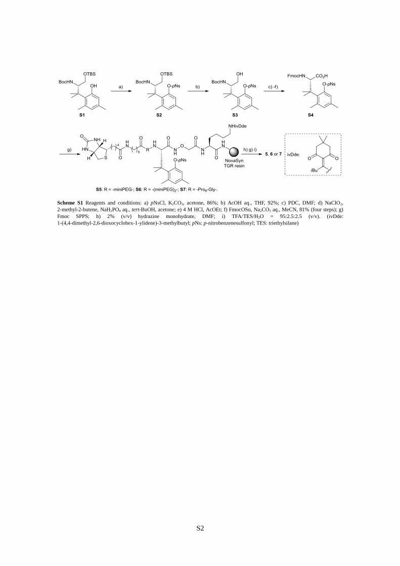
Scheme S1 Reagents and conditions: a) pNsCl, K2CO3, acetone, 86%; b) AcOH aq., THF, 92%; c) PDC, DMF; d) NaClO2,
2-methyl-2-butene, NaH2PO4 aq., tert-BuOH, acetone; e) 4 M HCl, AcOEt; f) FmocOSu, Na2CO3 aq., MeCN, 81% (four steps); g)
Fmoc SPPS; h) 2% (v/v) hydrazine monohydrate, DMF; i) TFA/TES/H2O = 95:2.5:2.5 (v/v). (ivDde:
1-(4,4-dimethyl-2,6-dioxocyclohex-1-ylidene)-3-methylbutyl; pNs: p-nitrobenzenesulfonyl; TES: triethylsilane)

S3
Fig. S1 HPLC monitoring of the reactions as shown in Scheme 1. A) Before the click chemistry (without CuSO4 and Na ascorbate).
B) After the click chemistry (reaction time = 1 h). C) After the linker cleavage (reaction time < 5 min). D) After the linker cleavage
(reaction time = 24 h). E) After treatment with o-bromobenzaldehyde and aniline. Analytical HPLC conditions: linear gradient of
0.1% (v/v) TFA/MeCN in 0.1% (v/v) TFA aq., 5 to 90% over 30 min for A and B, 10 to 50% over 30 min for C, D, and E. *Peaks
observed when 2-mercaptoethanol was incubated in Na phosphate buffer with DMSO. **Derivative of pNs group.
Fig. S2 Effect of concentration of 2-mercaptoethanol on the thiol-induced linker cleavage. Enolase-traceable linker 5 conjugate was
prepared and adsorbed on streptavidin beads as similar to that described in footnote of Fig. 4. The obtained beads were reacted with
2-mercaptoethanol (10 or 100 mM) and NP40 (1% (v/v)) in Na phosphate buffer (10 mM, pH 7.8) at 37 °C for 24 h. All proteins
were visualized by silver staining. [a] Eluted proteins by the thiol-treatment. After centrifugation, the obtained supernatant was
analyzed. [b] Proteins remaining on streptavidin beads after the thiol treatment. The beads after centrifugation followed by removal
of the supernatant was suspended in SDS-PAGE sample loading buffer, and the resulting mixture was heated at 100 °C for 5 min.
After centrifugation, the supernatant was analyzed.
S4
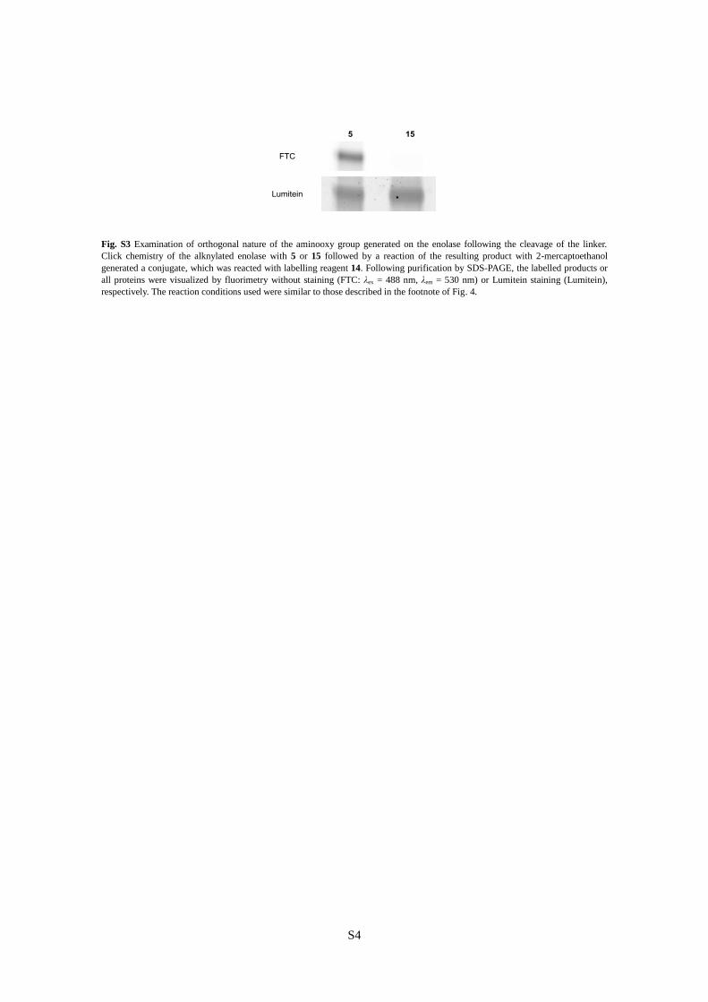
Fig. S3 Examination of orthogonal nature of the aminooxy group generated on the enolase following the cleavage of the linker.
Click chemistry of the alknylated enolase with 5 or 15 followed by a reaction of the resulting product with 2-mercaptoethanol
generated a conjugate, which was reacted with labelling reagent 14. Following purification by SDS-PAGE, the labelled products or
all proteins were visualized by fluorimetry without staining (FTC: λex = 488 nm, λem = 530 nm) or Lumitein staining (Lumitein),
respectively. The reaction conditions used were similar to those described in the footnote of Fig. 4.
S5
General Methods
All reactions of small molecules were carried out under a positive pressure of argon. For column
chromatography, silica gel (KANTO KAGAKU N-60) was employed. Mass spectra were recorded
on a Waters MICROMASS® LCT PREMIER
TM (ESI-TOF) or a Bruker Esquire2000T (ESI-Ion
Trap). NMR spectra were recorded using a Bruker AV400N. For HPLC separations, a Cosmosil
5C18-AR-II analytical column (Nacalai Tesque, 4.6 × 250 mm, flow rate 1.0 mL/min) or a Cosmosil
5C18-AR-II preparative column (Nacalai Tesque, 20 × 250 mm, flow rate 10.0 mL/min) was
employed, and eluting products were detected by UV at 220 nm. For HPLC elution, linear gradient
of 0.1% TFA (v/v) in MeCN in 0.1% (v/v) TFA in H2O over 30 min was used. Optical rotations were
measured using a JASCO P-2200 polarimeter (concentration in g/100 mL). ECL signals from the
western blot analysis were detected using a LAS-4000mini (Fujifilm). A Molecular Imager FX Pro
and a Quantity One 1-D Analysis Software (Bio-Rad Laboratories) were employed for fluorescence
gel images and its analyses, respectively.
Synthesis of Chiral Thiol-responsive Amino Acid Derivative S4
Starting from chiral intermediate S1,S1
thiol-responsive amino acid derivative S4 was prepared
according to the literature.S2
1H NMR spectra were identical to those reported previously.
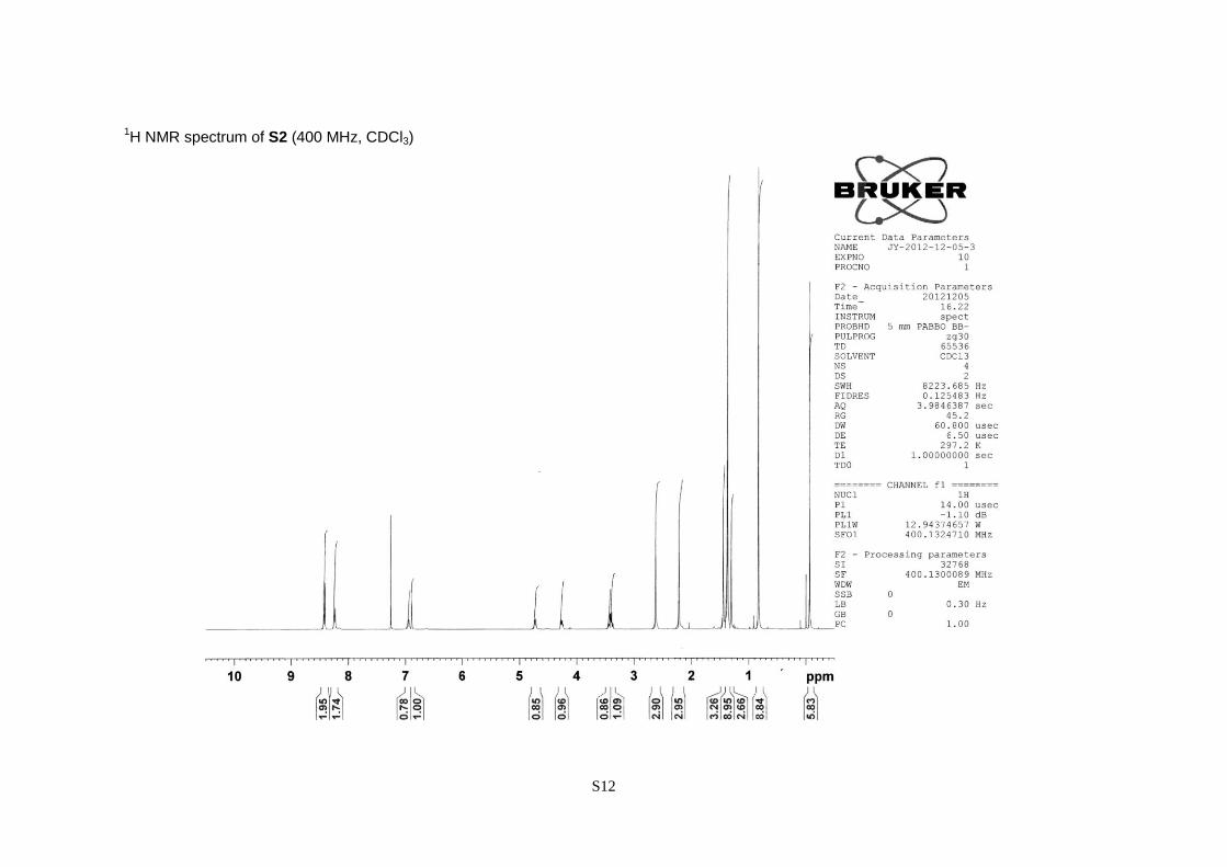
(S)-2-tert-Butoxycarbonylamino-3,3-dimethyl-3-[2,4-dimethyl-6-(nitrobenzene-4-sulfonyloxy)-
phenyl] propanol tert-butyldimethylsilyl ether (S2).
A pale yellow oil; 86% yield; [α]20
D –35.6 (c 1.22, CHCl3); 1H NMR (CDCl3, 400 MHz) = –0.07
(6H, s), 0.83 (9H, s), 1.30 (3H, s), 1.37 (9H, s), 1.44 (3H, s), 2.21 (3H, s), 2.62 (3H, s), 3.38 (1H, dd,
J = 10.8 and 4.7 Hz), 3.43 (1H, dd, J = 10.8 and 4.5 Hz), 4.26 (1H, ddd, J = 9.8, 4.7 and 4.5 Hz),
4.73 (1H, d, J = 9.8 Hz), 6.89 (1H, s), 6.94 (1H, s), 8.24 (2H, d, J = 8.7 Hz), 8.42 (2H, d, J = 8.7
Hz).
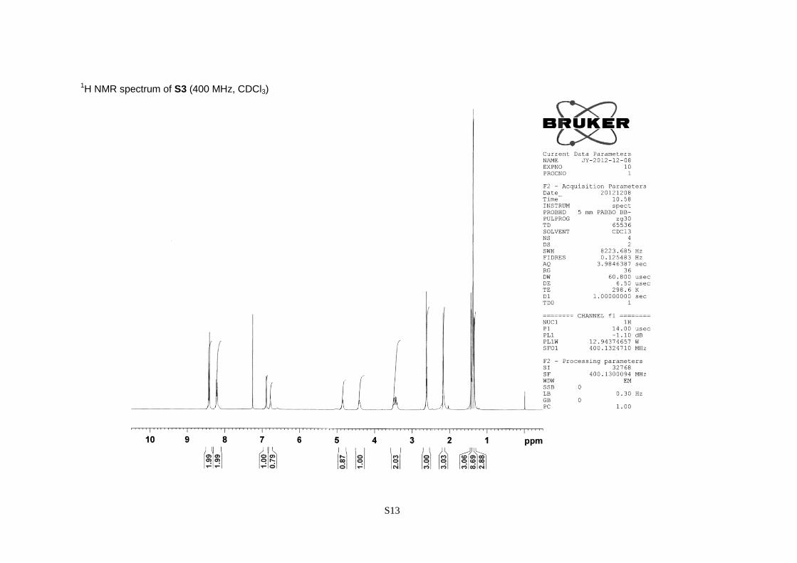
(S)-2-tert-Butoxycarbonylamino-3,3-dimethyl-3-[2,4-dimethyl-6-(nitrobenzene-4-sulfonyloxy)-
phenyl]propanol (S3).
A white powder; 92% yield; [α]20
D –38.3 (c 1.13, CHCl3); 1H NMR (CDCl3, 400 MHz) = 1.33 (3H,
s), 1.37 (9H, s), 1.42 (3H, s), 2.17 (3H, s), 2.61 (3H, s), 3.42 (1H, dd, J = 11.2 and 7.6 Hz), 3.49 (1H,
dd, J = 11.2 and 2.6 Hz), 4.40 (1H, m), 4.85 (1H, d, J = 9.0 Hz), 6.78 (1H, s), 6.89 (1H, s), 8.22 (2H,
d, J = 8.8 Hz), 8.42 (2H, d, J = 8.8 Hz).
S6
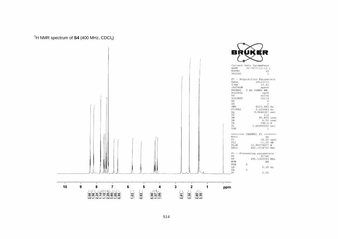
(S)-3,3-Dimethyl-3-[2,4-dimethyl-6-(nitrobenzene-4-sulfonyloxy)phenyl]-2-(9-fluorenylmethyl-
carbonylamino)propionic acid (S4).
A pale yellow powder; 81% yield over four steps; [α]20
D –19.6 (c 1.02, CHCl3); 1
H NMR (CDCl3,
400 MHz) = 1.53 (3H, s), 1.56 (3H, s), 2.14 (3H, s), 2.64 (3H, s), 4.16 (1H, t, J = 7.2 Hz), 4.27 (1H,
dd, J = 10.5 and 7.2 Hz), 4.35 (1H, dd, J = 10.5 and 7.2 Hz), 5.20 (1H, d, J = 9.0 Hz), 5.73 (1H, d, J
= 9.0 Hz), 6.66 (1H, s), 6.91 (1H, s), 7.23–7.31 (2H, m), 7.39 (2H, t, J = 7.5 Hz), 7.49 (1H, d, J = 7.5
Hz), 7.56 (1H, d, J = 7.5 Hz), 7.75 (2H, d, J = 7.5 Hz), 8.18 (2H, d, J = 8.6 Hz), 8.40 (2H, d, J = 8.8
Hz).
Preparation of Traceable Linkers 5, 6 and 7, and negative control 15
General Procedure: The peptides were synthesized using Fmoc-based solid phase peptide synthesis
(Fmoc SPPS). Building blocks were coupled on NovaSyn TGR resin (0.22 mmol amine/g). Reagents
and solvents are listed below. All coupling reactions were performed for 2 h.
building block reagents solvent
S4 (2 eq.) HATU (1.9 eq.), DIPEA (1.9 eq.) DMF
(+)-biotin (5 eq.) DIC (5 eq.), HOBt·H2O (5 eq.) DMSO/DMF = 1/1 (v/v)
N3(CH2)4CO2HS3
(5 eq.) DIC (5.3 eq.), Oxyma PureS4
(5 eq.) DMF
Others (3 eq.) S5
DIC (3.2 eq.), Oxima Pure (3 eq.) DMF
Abbreviations. DIC: N,N’-diisopropylcarbodiimide; DIPEA: N,N-diisopropylethylamine; HATU: 1-[bis(dimethylamino)methylene]-
1H-1,2,3-triazolo[4,5-b]pyridinium 3-oxid hexafluorophosphate; HOBt: 1-hydroxybenzotriazole; Oxyma pure: ethyl
cyanoglyoxylate-2-oxime.
For removal of an ivDde (1-(4,4-dimethyl-2,6-dioxocyclohex-1-ylidene)-3-methylbutyl) group, the
peptide resin was treated with 2% (v/v) hydrazine hydrate in DMF (twice for 2 h followed by once
overnight). Following to completion of the peptide elongation, the resin was subjected to global
deprotection using TFA/triethylsilane (TES)/H2O (95:2.5:2.5 (v/v)) for 2 h at room temperature.
After the resin was filtered off, cooled diethyl ether was added to the filtrate. The resulting
precipitate was collected by centrifugation, washed with diethyl ether, and then purified by a
preparative HPLC. When an amount of the precipitate had not been sufficient, the filtrate after the
global deprotection was concentrated using N2 flow and neutralized by addition of NaHCO3. Then it
was purified by a preparative HPLC without the precipitation step.
5: A white lyophilized powder; 13% yield; Analytical HPLC conditions: 10 to 90%. Retention time =
18.8 min; Preparative HPLC conditions: 37 to 47%; LRMS (ESI-Ion Trap) m/z calcd for [M + H]+
1377.6, found 1378.0.
S7
6: A white lyophilized powder; 13% yield; Analytical HPLC conditions: 10 to 90%. Retention time =
18.6 min; Preparative HPLC conditions: 35 to 45%; LRMS (ESI-TOF) m/z calcd for [M + 2H]2+
761.9, found 761.7.
7: A white lyophilized powder; 14% yield; Analytical HPLC conditions: 10 to 90%. Retention time =
18.0 min; Preparative HPLC conditions: 35 to 45%; LRMS (ESI-TOF) m/z calcd for [M + 2H]2+
936.5, found 936.2.
15: A white lyophilized powder; 33% yield; Analytical HPLC conditions: 10 to 90%. Retention time
= 13.2 min; Preparative HPLC conditions: 22 to 32%; LRMS (ESI-Ion Trap) m/z calcd for [M + H]+
1136.6, found 1137.1.
Model Reactions Using Alkynylated Peptide 8
Preparation of Model Peptide 8: The peptide was synthesized on Nova Syn TGR resin (0.22 mmol
amine/g) using Fmoc SPPS. Fmoc protected amino acids (3 eq.) were coupled at room temperature
for 2 h by using DIC (3.2 eq.) and Oxyma Pure (3 eq.) in DMF. After treatment with TFA/TES/H2O
(95:2.5:2.5 (v/v)) for 1.5 h at room temperature, the resin was filtered off and cooled diethyl ether
was added to the filtrate. The resulting precipitate was collected by centrifugation, washed with
diethyl ether, and then purified by a preparative HPLC.
8: A white lyophilized powder; 67% yield; Analytical HPLC conditions: 5 to 30%. Retention time =
16.7 min; Preparative HPLC conditions: 15 to 25%; LRMS (ESI-TOF) m/z calcd for [M + H]+ 849.5,
found 849.2.
Click Chemistry: Traceable linker 5 in DMSO (6.0 mM, 66.6 μL, final concn. 0.20 mM), peptide 8
in PBS (1.25 mM, 400 μL, final concn. 0.25 mM), TBTA in 20% (v/v) DMSO/tert-BuOH (1.7 mM,
118 μL, final concn. 0.10 mM), CuSO4 in water (50 mM, 40.0 μL, final concn. 1.0 mM), sodium
ascorbate in water (25 mM, 40.0 μL, final concn. 0.50 mM), and PBS (416 μL) were added to 1.00
mL of water. After 1 h of the reaction at room temperature, reaction mixture was injected into a
preparative HPLC to yield conjugate 9.
9: A white lyophilized powder; 56% yield; Analytical HPLC conditions: 5 to 90%. Retention time =
16.7 min; Preparative HPLC conditions: 30 to 39%; LRMS (ESI-TOF) m/z calcd for [M + 3H]3+
742.7, found 742.6.
S8
Thiol-induced cleavage: To sodium phosphate buffer (10 mM, pH 7.8, 137 μL) were added
conjugate 9 in DMSO (6.0 mM, 2.37 μL, final concn. 0.10 mM), 2-mercaptoethanol (0.99 μL, final
concn. 100 mM), and NP40 (1.42 μL, final concn. 1% (v/v)). After incubation at 37 °C for 24 h
under argon, completion of cleavage of the linker was confirmed using HPLC and the products were
characterized by MS analyses.
10: Analytical HPLC conditions: 10 to 50%. Retention time = 11.9 min; LRMS (ESI-TOF) m/z calcd
for [M + 2H]2+
669.4, found 669.3.
11: Analytical HPLC conditions: 10 to 50%. Retention time = 28.2 min; LRMS (ESI-TOF) m/z calcd
for [M + H]+ 704.4, found 704.2.
Labelling with o-bromobenzaldehyde: To the reaction mixture after the linker cleavage (72.0 μL)
were added 10 mM sodium phosphate buffer (pH 7.8, 634 μL), o-bromobenzaldehyde in DMSO (10
mM, 7.20 μL, final concn. 0.10 mM), and aniline (6.56 μL, final concn. 100 mM). The reaction was
performed at room temperature for 1 h, and labelled product 12 was characterized using MS
analysis.
12: Analytical HPLC conditions: 10 to 50%. Retention time = 19.8 min; LRMS (ESI-TOF) m/z calcd
for [M + 2H]2+
752.3 (79
Br deriv.) and 753.3 (81
Br deriv.), found 752.2 and 753.2.
Preparation of Alkynylated Enolase
Starting from a commercially available enolase (7.5 mg), the alkynylated enolase was prepared
according to the literature.S6
After the reaction, unreacted small molecules were removed by dialysis
(Slide-A-Lyzer®
G2 Dialysis Cassette, Thermo SCIENTIFIC) with PBS instead of gel filtration.
Volume of the resulting solution was adjusted to 1.8 to 2.0 mL, and it was used as 4.2 g/L to 3.8 g/L
solution of the alkynylated enolase in PBS.
Introduction of Traceable Linker onto Alkynylated Enolase
Click Chemistry: To a mixture of PBS (550 μL) and water (447 μL) were added the alkynylated
enolase in PBS (4.2 g/L, 180 μL, final concn. 0.50 g/L), traceable linker 5, 6, 7, or negative control
15 in DMSO (6 mM, 25.0 μL, final concn. 0.10 mM), TBTA in 20% (v/v) DMSO/tert-BuOH (1.7
mM, 88.0 μL, final concn. 0.10 mM), CuSO4 aq. (50 mM, 30.0 μL, final concn. 1.0 mM), sodium
ascorbate aq. (25 mM, 30.0 μL, final concn. 0.50 mM), and SDS aq. (10% (w/v), 150 μL, final
concn. 1% (w/v)). After the reaction at room temperature for 1 h, small molecules were removed by
S9
dialysis (Slide-A-Lyzer® G2 Dialysis Cassette, Thermo SCIENTIFIC) with PBS.
SDS-PAGE: After addition of 5 × non-reducing SDS-PAGE sample loading buffer (5 × SDS-PAGE
sample loading buffer without 2-mercaptoethanol) followed by heating at 100 °C for 5 min, the
reaction mixture was analyzed using SDS-PAGE in 12% polyacrylamide gels. For the
chemiluminescence imaging of the biotinylated proteins, the proteins were transferred to Amersham
Hybond-P PVDF Membrane (GE Healthcare) and detected with a streptavidin-horseradish
peroxidase conjugate (SAv-HRP, GE Healthcare) and ECL plus Western Blotting Detection System
(GE Healthcare). For fluorescence imaging of all proteins, proteins in a gel were stained with
LumiteinTM
Protein Gel Stain (Nacalai Tesque).
Adsorption on Streptavidin Beads, Linker Cleavage, and Labelling of Enolase Conjugate
Adsorption on Streptavidin Beads: After the click chemistry, Pierce® Streptavidin UltraLink
®
Resin (35 μL, Thermo SCIENTIFIC) was added to the reaction mixture containing ca. 100 μg
enolase and its derivatives. After the adsorption at room temperature for 24 h, the resulting resin was
washed with PBS five times and it was subjected to subsequent reactions.
Linker Cleavage: To the resulting streptavidin beads was added a cleavage cocktail consisting of
2-mercaptoethanol (1.40 μL, final concn. 100 mM), NP40 (2.00 μL, final concn. 1% (v/v)), and
sodium phosphate buffer (197 μL, 10 mM, pH 7.8). The reaction was conducted at 37 °C for 24 h
under N2. After centrifugation of the resulting mixture (2000 rpm, 2 min), supernatant was collected
and the precipitate was suspended in 100 μL PBS. The suspension was subjected to centrifugation
(2000 rpm, 2 min) again and the obtained supernatant was combined with the first one.
Labelling: To the obtained supernatant were added fluorophore 14S7
(final concn. 0.10 mM) and
aniline (final concn. 100 mM), and the mixture was stirred at room temperature for 24 h. After
concentration using Amicon® Ultra-0.5, Ultracel-10 Membrane, 10 kDa (Merk Millipore) (14,000 ×
g, 15 min), addition of PBS and the concentration was repeated four times to remove the excess of
the non-reacted fluorophore. Then the obtained mixture was subjected to the SDS-PAGE as
mentioned above. In this case, 5 × SDS-PAGE sample loading buffer was used instead of the
non-reducing SDS-PAGE sample loading buffer. The labelled enolase was detected by fluorescence
imaging (λex = 488 nm, λem = 530 nm) without staining or the use of a western blot analysis.
Elution of Proteins Remaining on Streptavidin Beads After Thiol-induced Cleavage: The resin
obtained after the linker cleavage as mentioned above was suspended in 2 × SDS-PAGE sample
S10
loading buffer (25 μL) and water (25 μL), and the mixture was heated at 100 °C for 5 min. After
centrifugation as mentioned in the section “Linker Cleavage”, the combined supernatant was
concentrated and analyzed using SDS-PAGE as similar to those described in section “Labelling”.
Examination of Orthogonality of Aminooxy Group on Enolase
After click chemistry of the alkynylated enolase with the linkers followed by treatment with
2-mercaptoethanol, the product was treated with fluorophore 14. The obtained mixture was
analyzed using SDS-PAGE followed by fluorimetric detection. Reaction conditions as similar to that
described in section “Introduction of Traceable Linker onto Alkynylated Enolase” and “Adsorption
on Streptavidin Beads, Linker Cleavage, and Labelling of Enolase Conjugate” were employed.
Enrichment and Selective Labelling of Enolase in Protein Mixture
As a protein mixture, solution of the alkynylated enolase, bovine serum albumin (BSA), and
ovalbumin (1/1/1 (w/v)) was used.
Click Chemistry: The alkynylated enolase in PBS (3.9 g/L, 127 μL, final concn. 0.50 g/L), BSA in
PBS (3.3 g/L, 150 μL, final concn. 0.50 g/L), ovalbumin in PBS (3.3 g/L, 150 μL, final concn. 0.50
g/L), the traceable linker in DMSO (6.0 mM, 17 μL, final concn. 0.10 mM), TBTA in 20% (v/v)
DMSO/tert-BuOH (1.7 mM, 59 μL, final concn. 0.10 mM), CuSO4 aq. (50 mM, 20 μL, final concn.
1.0 mM), sodium ascorbate aq. (25 mM, 20 μL, final concn. 0.50 mM), SDS aq. (10% (w/v), 100 μL,
final concn. 1% (w/v)), PBS (100 μL), and water (257 μL) were mixed. Following to reaction at
room temperature for 1 h, the resulting solution was dialyzed using Slide-A-Lyzer® G2 Dialysis
Cassette (Thermo SCIENTIFIC) with PBS.
Adsorption on Streptavidin Beads, Cleavage of Linker, and Labelling: It was performed as
similar to that described in a section “Adsorption on Streptavidin Beads, Linker Cleavage, and
Labelling of Enolase Conjugate”.
Enrichment Without Use of Thiol-induced Linker Cleavage: After the adsorption of the protein
mixture on streptavidin beads, the proteins on the beads were eluted and analyzed as mentioned in a
section “Elution of Proteins Remaining on Streptavidin Beads After Thiol-induced Cleavage”.
S11
References
S1 A. Shigenaga, J. Yamamoto, N. Nishioka and A. Otaka, Tetrahedron, 2010, 66, 7367–7372.
S2 A. Shigenaga, J. Yamamoto, H. Hirakawa, K. Ogura, N. Maeda, K. Morishita and A. Otaka,
Tetrahedron Lett., 2010, 51, 2525–2528.
S3 W. Shi, S. Dolai, S. Averick, S. S. Fernando, J. A. Saltos, W. L’Amoreaux, P. Banerjee and
K. Raja, Bioconjugate Chem., 2009, 20, 1595–1601.
S4 R. Subiros-Funosas, S. N. Khattab, L. Nieto-Rodriguez, A. El-Faham and F. Albericio,
Aldrichimica Acta, 2013, 46, 21–40.
S5 Preparation of FmocNHOCH2CO2H: L. Cipolla, M. Rescigno, A. Leone, F. Peri, B. L.
Ferla and F. Nicotra, Bioorg. Med. Chem., 2002, 10, 1639–1646.
S6 K. D. Park, R. Liu and H. Kohn, Chem. Biol., 2009, 16, 763–772.
S7 E. Trevisiol, E. Defrancq, J. Lhomme, A. Laayoun and P. Cros, Eur. J. Org. Chem., 2000,
211–217.
S12
1H NMR spectrum of S2 (400 MHz, CDCl3)
S13
1H NMR spectrum of S3 (400 MHz, CDCl3)
S14
1H NMR spectrum of S4 (400 MHz, CDCl3)