Embed Size (px)
Citation preview

Development of an Effective Polarizable Bond Method forBiomolecular SimulationXudong Xiao,†,‡ Tong Zhu,† Chang G. Ji,*,†,‡,§ and John Z. H. Zhang*,†,§
†State Key Laboratory of Precision Spectroscopy, Department of Physics, Institute of Theoretical and Computational Science, EastChina Normal University, Shanghai 200062, China‡Institutes for Advanced Interdisciplinary Research, East China Normal University, Shanghai 200062, China§NYU-ECNU Center for Computational Chemistry at NYU Shanghai, Shanghai 200062, China
*S Supporting Information
ABSTRACT: An effective polarizable bond (EPB) model has beendeveloped for computer simulation of proteins. In this partial polarizableapproach, all polar groups of amino acids are treated as polarizable, and therelevant polarizable parameters were determined by fitting to quantumcalculated electrostatic properties of these polar groups. Extensive numericaltests on a diverse set of proteins (including 1IEP, 1MWE, 1NLJ, 4COX,1PGB, 1K4C, 1MHN, 1UBQ, 1IGD) showed that this EPB model is robustin MD simulation and can correctly describe the structure and dynamics ofproteins (both soluble and membrane proteins). Comparison of thecomputed hydrogen bond properties and dynamics of proteins withexperimental data and with results obtained from the nonpolarizable forcefield clearly demonstrated that EPB can produce results in much betteragreement with experiment. The averaged deviation of the simulatedbackbone N−H order parameter of the B3 immunoglobobulin-binding domain of streptococcal protein G from experimentalobservation is 0.0811 and 0.0332 for Amber99SB and EPB, respectively. This new model inherited the effective character of theclassic force field and the fluctuating feature of previous polarizable models. Different from other polarizable models, thepolarization cost energy is implicitly included in the present method. As a result, the present method avoids the problem of overpolarization and is numerically stable and efficient for dynamics simulation. Finally, compared to the traditional fixed AMBERcharge model, the present method only adds about 5% additional computational time and is therefore highly efficient for practicalapplications.
I. INTRODUCTION
Extensive applications of molecular dynamics (MD) simulationto biomolecular systems rely on the use of an empiricalmolecular force field in which the interaction energy ofbiomolecules is represented as a function of atomiccoordinates.1−4 As a result, the correctness of the simulationresult depends fundamentally on the accuracy of the force fieldemployed in the simulation. Although great advances have beenmade in computational prediction of biomolecular properties,further improvement of force fields, especially by includingproper polarization, is much needed.5,6 A major challenge in thepresent force field development is how to accurately describeelectrostatic interaction in biomolecular systems, in particular,how to properly include the polarization effect in MDsimulation. Traditional fixed charge force field models areunable to adapt to environmental changes and therefore lackthe polarization effect.1,3
For more than 30 years, many attempts have been made toexplicitly include polarization effects in molecular model-ing.7−18 To date, there are several general models that includethe polarization effect in the force field such as the fluctuatingcharge model,10−12 Drude oscillator,7−9 induced dipole,13−17
electronic polarization via quantum mechanical treatment, ormixed QM/MM.18 Reviews and accounts on the similarity anddifference among these approaches are available.19−21
Electronic polarization results from redistribution ofelectrons when a molecule is placed in an electric field. Thereare two opposing energetic effects that occur during thepolarization process. On one hand, polarization will enhancethe interaction energy between the molecule and the externalelectric field in order to lower the energy of the system. On theother hand, the internal energy of the molecule will rise as aresult of distortion of the electron charge distribution of themolecule. For example, when a polar molecule is placed in anelectric field, these two opposing energetic effects will counterbalance each other, and the molecular charge distribution willdistort to a polarized state to establish a new systemequilibrium (minimum energy state). On the basis of thissimple physical picture, “equilibrium between polarization costenergy up and interaction energy down”, we developed a new
Received: August 13, 2013Revised: November 19, 2013Published: November 19, 2013
Article
pubs.acs.org/JPCB
© 2013 American Chemical Society 14885 dx.doi.org/10.1021/jp4080866 | J. Phys. Chem. B 2013, 117, 14885−14893

fluctuating charge model for protein backbone polar groups. Inthis approach, the fluctuating charges are the sum of permanent(or reference) and induced charges that are determined by thedifference of electrostatic potentials at different atoms. Inparticular, charges are only allowed to fluctuate between twooppositely charged atoms of the polar bond in this model. Thispolarizable model was applied to study site-specific backbonehydrogen bond energies in protein, and the theoreticalpredictions agreed very well with experimental measurement.22
In the current work, we extend this polarizable model to allpolar groups of amino acids and develop a general “effectivepolarizable bond” model (EPB) for biomolecular simulation. Inthe present EPB model, energy cost during polarization isembraced into electrostatic interaction, and effective atomiccharges fluctuate according to the local environment.The rest of this article is organized as follows. Section II
contains a detailed description of this method and tables ofparameters. In Section III, the EPB model is applied to calculatethe charge distribution of some proteins, and some results arepresented. Conclusions are given in Section IV.
II. THEORETICAL METHODEffective Polarizable Bond. When a gas phase molecule is
placed in an electric field such as in a polar solvent, the electrondistribution of the molecule becomes distorted. The energyneeded to distort the electron distribution of the gas phasemolecule is called the distortion energy or polarization cost.The quantitative relationship between the polarization cost andthe polarization state of certain chemical groups can bedetermined through electronic structure calculation.The Schrodinger equation, which governs the state of the
molecule’s electrons in gas phase and in an electric field, isdetermined by quantum mechanical equations:
Η Ψ = ΨE0 0 0 0 (1)
and
Η + Η′ Ψ = ΨE( )0 (2)
where H0 is the Hamiltonian of the molecule itself and H′ is theHamiltonian representing the interaction between the moleculeand external electric field. The polarization cost energy of apolar group due to distortion of its electron distribution in thepresence of an external electric field is defined by
Δ = ⟨Ψ|Η |Ψ⟩ − ⟨Ψ |Η |Ψ ⟩−Ep tcos 0 0 0 0 (3)
Figure 1 shows the relationship between polarization costand the change of the dipole moment of the −SH and −OHgroup. The data in Figure 1 can be reasonably well-fit into aquadratic relation:
μ μΔ = −−E k( )p tcos liquid gas2
(4)
1/k represents polarizability of the relevant polar group. Forexample, Figure 1 shows that the −SH group is morepolarizable than the −OH group.Now, consider the transfer of a polar group −SH from gas
phase into liquid phase, the energy of the system can be writtenas
μ μ
= +
= − + Φ + Φ
E E E
k q q[ ( ) ] [ ]
self ele
liquid gas2
S S H H (5)
where qS and qH are, respectively, the ESP (electrostaticpotential) charges of the S and H atoms of the −SH group. TheΦS and ΦH are electrostatic potentials at S and H atoms,respectively. In the present approach, the polarization can betreated as charge transfer between atoms of a polar group. If theamount of charge transfer from atom S to atom H is Δq, as inthe −SH group, the final partial charges of the atoms are givenby
= + Δq q qS Sgas
(6)
= − Δq q qH Hgas
(7)
where qSgas and qH
gas are, respectively, the atomic charges of Sand H atoms in gas phase (or reference charges). Thus, thechange of dipole moment of the −SH group due to polarization(from gas phase to solvent) is given by
μ μΔμ = − = Δ ·q dliquid gas SH (8)
where dSH is the bond length of the S−H bond. Then, eq 5 canbe rewritten as
= Δ · + + Δ Φ + − Δ ΦE k q d q q q q( ) ( ) ( )SH2
Sgas
S Hgas
H
(9)
It should be noted that in the above approach, the direction ofthe induced dipole is along the direction of the polar bond.Thus, the approach is appropriate for treating polar groups witha single polar bond (or linearized polar bond).Equation 9 gives the change of electrostatic energy of the
molecule (the −SH group) in solvent. The equilibrium isreached by minimizing this energy with respect to variation ofΔq
∂∂Δ
=Eq
0(10)
from which we obtain the charge transfer along the S−H bondunder a given electrostatic potential,
Δ =Φ − Φ
qd k
( )2H S
SH2
(11)
Just as partial atomic charges can be introduced to describeelectrostatic interaction, the effective charge concept can also
Figure 1. Polarization cost energy as a function of the change of thedipole moment. The black points represent data of the OH groupwhich is fitted to a quadratic curve colored by green. The red pointsrepresent data of the SH group which is fitted to a quadratic curvecolored by blue. For each group, 15 000 different configurationssampled from MD simulation were used in quantum mechanicalcalculation.
The Journal of Physical Chemistry B Article
dx.doi.org/10.1021/jp4080866 | J. Phys. Chem. B 2013, 117, 14885−1489314886
be introduced in the fluctuating charge model. It is thusconvenient to express the polarization cost term in eq 5 in aform of electrostatic interaction. For the SH group, eq 5 can berewritten as:
= +
= Δ · + + Δ Φ + − Δ Φ
= Φ + Φ
−
∼ ∼
E E E
k q d q q q q
q q
( ) [( ) ( ) ]
self ele
S H2
Sgas
S Hgas
H
S S H H
(12)
where∼qS and
∼qHare, respectively, effective fluctuating charges
(EFQ) of S and H atoms. Combining eq 11 with eq 12, weobtain
= Δ · + + Δ Φ + − Δ Φ
= Δ · Δ + + Δ Φ + − Δ Φ
= Δ ·Φ − Φ
+ + Δ Φ
+ − Δ Φ
= Δ · Φ − Φ + + Δ Φ + − Δ
Φ
= + Δ Φ + − Δ Φ
= Φ + Φ
−
−
−
∼ ∼
⎜ ⎟ ⎜ ⎟
⎛⎝⎜
⎞⎠⎟
⎡⎣⎢⎛⎝
⎞⎠
⎛⎝
⎞⎠
⎤⎦⎥
E k q d q q q q
q qkd q q q q
qd k
kd q q
q q
q q q q q
q q q q
q q
( ) [( ) ( ) ]
( ) [( ) ( ) ]
( )2
[( )
( ) ]
12
( ) [( ) ( )
]
12
12
S H2
Sgas
S Hgas
H
S H2
Sgas
S Hgas
H
H S
SH2 S H
2Sgas
S
Hgas
H
H S Sgas
S Hgas
H
Sgas
S Hgas
H
S S H H
The effective fluctuating charges can be defined as:
= + Δ∼q q q/2S S
gas(13)
= − Δ∼q q q/2H H
gas(14)
which is essentially the result of linear response theory.23
The EFQ are directly used in MD simulation to correctlydescribe electrostatic interactions with polarization. The use ofEFQ gives more balanced electrostatic interaction because itincludes the effect of distortion or polarization cost in bothenergy and force. Electrostatic potential of an atom isdetermined by the charges of external potential and all otheratoms except those in the same residue.15
Parameterization of the Fluctuating Charge Model.Since the energy expression in eq 5 or 9 is quite general, we canprefit the specific polarization parameters, such as referenceatomic charges and polarizability k of all polar groups in aminoacids. While previous work only treated polarization ofbackbone polar groups,22 the current work extends fluctuatingcharge treatment to all polar groups including those of proteinside chains, which is necessary for accurately modelingpolarization of protein systems.Fitting Protocol. Equations 3 and 4 can be used to
determine the polarizable parameter k by quantum chemistrycalculation for a given polar group such as the −SH bond.However, we notice that the polarizable parameter k is relatedto a linear polar bond that only have two atoms, thus we cannotdirectly parametrize k for polar groups that have more than twoatoms such guanidine which has nine atoms. Here a new
approach is employed in which ESP (electrostatic potential)charge is used to fit the polarizable parameter k.First of all, we need to select appropriate molecular models
(Figures S1−S11). Here, for the −SH bond, we choose CH3SHbecause −CH3 is a nonpolar group and simple enough. Also itbest mimics the −CH2− group which usually links to polargroups in the side chain of amino acids.To model under external electric field, the CH3SH was
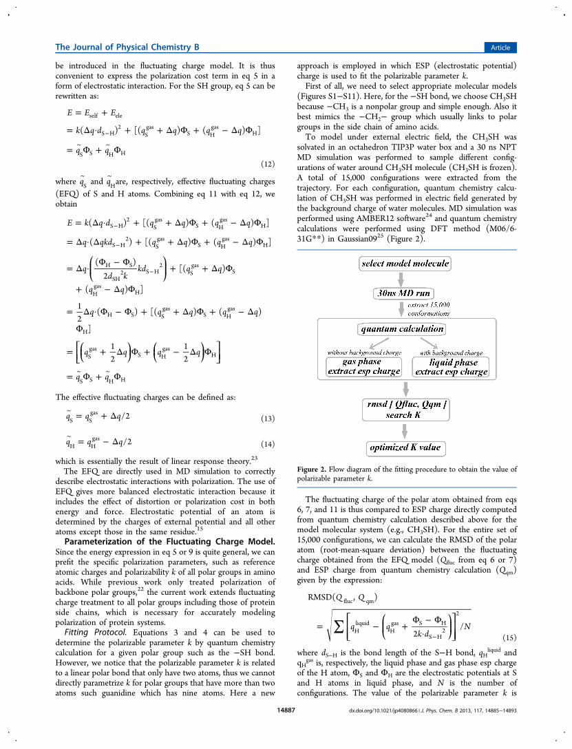
solvated in an octahedron TIP3P water box and a 30 ns NPTMD simulation was performed to sample different config-urations of water around CH3SH molecule (CH3SH is frozen).A total of 15,000 configurations were extracted from thetrajectory. For each configuration, quantum chemistry calcu-lation of CH3SH was performed in electric field generated bythe background charge of water molecules. MD simulation wasperformed using AMBER12 software24 and quantum chemistrycalculations were performed using DFT method (M06/6-31G**) in Gaussian0925 (Figure 2).
The fluctuating charge of the polar atom obtained from eqs6, 7, and 11 is thus compared to ESP charge directly computedfrom quantum chemistry calculation described above for themodel molecular system (e.g., CH3SH). For the entire set of15,000 configurations, we can calculate the RMSD of the polaratom (root-mean-square deviation) between the fluctuatingcharge obtained from the EFQ model (Qfluc from eq 6 or 7)and ESP charge from quantum chemistry calculation (Qqm)given by the expression:
∑= − +Φ − Φ
· −
⎡⎣⎢⎢
⎛⎝⎜
⎞⎠⎟⎤⎦⎥⎥
Q Q
q qk d
N
RMSD( , )
2/
fluc qm
Hliquid
Hgas S H
S H2
2
(15)
where dS−H is the bond length of the S−H bond, qHliquid and
qHgas is, respectively, the liquid phase and gas phase esp charge
of the H atom, ΦS and ΦH are the electrostatic potentials at Sand H atoms in liquid phase, and N is the number ofconfigurations. The value of the polarizable parameter k is
Figure 2. Flow diagram of the fitting procedure to obtain the value ofpolarizable parameter k.
The Journal of Physical Chemistry B Article
dx.doi.org/10.1021/jp4080866 | J. Phys. Chem. B 2013, 117, 14885−1489314887
determined by minimizing RMSD[Qfluc,Qqm].26 We also fitted
some parameters by using “gas phase interactions withelectrostatic probes”, and the results did not change.For the more complicated polar groups, the fitting procedure
was exactly the same as the −SH group described above, exceptthat they were split into more fragments, each with a singlepolar bond.List of Parameters. The list of fitted parameters of side chain
polar groups in polar and charged amino acids including SER,THR, CYS, CYM, TYR, ASN, GLN, ASP, GLU, LYS, ARG,HIS, as well as the refined parameters of backbone polargroups, CO and N−H are given in Table 1. The parameters
are divided into two classes, one is the set of gas phase chargesand the other is the value of polarizable parameter k. The bondlength used in the calculation and fitting is also given in theTable for reference purpose.As mentioned above, for some complicated polar groups,
they were split into several fragments. Each fragment is a polarbond containing two connected atoms, similar to simple polargroups (e.g., −SH) so the fitting approach is almost the same.There are exceptions such as guanidine for which the charge ofthe buried C atom is not allowed to fluctuate because the ESP
charge was poor for buried atoms. In addition, for the sidechain of unprotonated histidine, the atoms located in the twospecial polar bonds are allowed to fluctuate because they can beimportant in forming side chain H bonds. For backbone polargroups −CO and −NH, the N-methyl acetamide (NMA) isused as the model molecule to fit their parameters. All theparameters are listed in Table 1.
III. APPLICATIONS TO REALISTIC SYSTEMSSeveral systems were studied here using the newly developedEPB model to investigate polarization effects in proteins. Allinitial structures were taken from the protein data bank. Mostof the MD simulations were performed using the force fields ofAMBER99SB27 and AMBER99SB mixed with the EPB model.The modified version of AMBER12 package was used as acomputational tool. Electrostatic potential on each atom wassaved in the simulation for calculation of the fluctuating charge.In the AMBER12 package, the electrostatic potential is
calculated with the particle−mesh Ewald (PME) method. Inthe PME method, calculation of infinite summation ofcontribution from surrounding atoms is transformed from aconditionally convergent series into an absolutely convergentone by the introduction of a set of Gaussian screening chargesadded to and subtracted from the charge distribution. Theelectrostatic potential is broken into four parts in PME routinesof AMBER12: (1) The “direct coulomb contribution”,accumulated in the short_ene_dip subroutine in short_ene.f.(2) The “reciprocal space contribution”, accumulated in thegrad_sum_dipolerc subroutine in ew_dipole_recip.f. (3) The“adjustment and self corrections”, accumulated in thenb_adjust_dipole subroutine of ew_force.f. (4) The “(1,4)corrections”, accumulated in do_14_dipole of extra_pts.f.Changes to PME routines of AMBER12 were made in thecode paths to save electrostatic potential. Fluctuating chargeswere calculated and updated in the main routine (runmd.f)according to the electrostatic potential on each atom.The soluble proteins were embedded in a truncated
octahedral TIP3P28 water box, and counterions were addedto neutralize the whole system. The membrane protein wasembedded into 108 POPC lipid molecules, and a size of 68 ×70 × 96 box was used, the rest of which were filled by 8815tip3p waters and 0.2 M KCl. The LIPID11 force field29 wasadopted for POPC molecule. Periodic boundary conditions andPME methods30 were adopted to calculate the long-rangeelectrostatic interactions. A cutoff of 10 Å was used for short-range interactions. Each system was relaxed in 10 000 stepswith constraints on protein, followed by full minimizationwithout any constraints. The system was then subjected to a150 ps restrained MD simulation in NVT ensemble withprotein atoms constrained at a force constant of 500 KJ/mol sothat the solvent and ions can be properly positioned around theprotein. Another 150 ps NPT simulation was run for adjustingthe density of the system. Temperature was regulated usingLangevin dynamics with the collision frequency setting to 2ps−1. All the MD simulations used a time step of 1 fs, and all thecovalent bonds involving hydrogen atoms were constrainedwith the SHAKE algorithm.31
Range of Fluctuating Charge. Fluctuation of Charges inSide Chains. Partial charges on a particular residue aredetermined by their specific conformation and chemicalenvironment due to other residues of the protein and solvents.For a specific amino acid, its polarization state when buriedinside a protein can be very different from when it is located on
Table 1. Fitted EPB Parameters of the Side Chain andBackbone Polar Groupsa
RES bond q0 k d
ASP Cγ−Oδ 0.7406 −0.7720 3.28 1.26ASH Cγ−Oδ 0.6412 −0.6326 2.97 1.21
Oδ−Hδ −0.5365 0.4558 7.46 0.97GLU Cδ−Oε 0.7258 −0.7790 3.28 1.26GLH Cδ−Oε 0.6850 −0.5887 2.97 1.21
Oε−Hε −0.5990 0.4120 7.46 0.96SER Oγ−Hγ −0.6301 0.4030 9.77 0.96THR Oγ−Hγ −0.6689 0.4030 9.77 0.96TYR Oη−Hη −0.5617 0.4030 8.41 0.96CYS Sγ−Hγ −0.3296 0.2110 3.52 1.33CYM Cβ−Sγ −0.2000 −0.9257 1.73 1.81ASN Cγ−Oδ 0.7680 −0.6481 3.28 1.23
Nδ−Hδ −0.9681 0.4441 7.84 1.01GLN Cδ−Oε 0.7513 −0.6648 3.28 1.23
Nε−Hε −0.9735 0.4415 7.84 1.01LYS Nζ−Hζ −0.3778 0.3375 15.2 1.01ARG Nε−Hε −0.4797 0.2958 8.75 1.01
Nη−Hη −0.8549 0.4439 7.14 1.01HID Nδ−Hδ −0.3620 0.3458 8.76 1.01
Cε−Nε 0.1610 −0.5280 3.27 1.31HIE Nδ−Cε −0.5210 0.1413 2.73 1.32
Nε−Hε −0.2840 0.3384 9.70 1.01HIP Nδ−Hδ1 0.4050 −0.1697 8.86 1.01
Cε−Hε1 0.2374 0.0137 7.62 1.08Nε−Hε2 0.3520 −0.1336 10.8 1.01Cδ−Hδ2 0.2807 −0.1631 8.40 1.08
BB-I C−O 0.5714 −0.5420 3.28 1.23N−H −0.4478 0.3040 9.19 1.01
BB-II C−O 0.4967 −0.5420 3.28 1.23N−H −0.5267 0.3040 9.19 1.01
BB-III C−O 0.6867 −0.5420 3.28 1.23N−H −0.3772 0.3040 9.19 1.01
aThe k is in kcal/mol·debye−2 and distance d in Å. BB- I, II, and IIIrepresent backbone polar groups for neutral, negative, and positiveamino acids, respectively.
The Journal of Physical Chemistry B Article
dx.doi.org/10.1021/jp4080866 | J. Phys. Chem. B 2013, 117, 14885−1489314888

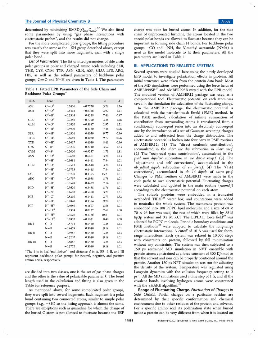
a protein surface (Table S1). To illustrate this feature, fourproteins (PDB codes: 1IEP, 1MWE, 1NLJ, 4COX) werestudied. In the present calculation, a 3.5 ns NPT simulation isperformed followed by a 1.5 ns production run for all foursystems. The histogram of charge distribution for some sidechain atoms are listed in Figure 3. We find that charges on the
same type of amino acid can differ significantly from each other.For example, the value of atomic charge ranges from −0.64 to−0.58 for OH@TYR, from −0.70 to −0.64 for OG@SER, andfrom −0.75 to −0.68 for OD@ASN in those four proteins westudied.Fluctuation of Charges in Backbone. Figure 4 shows the
average charges of the backbone hydrogen and oxygen for the
B1 binding domain of protein G32 (PDB: 1PGB) as a functionof position along the main chain from N to C terminus. Theerror bars measure the fluctuation of atomic charges during MDsimulation (up to 10 ns here). The fluctuation behavior of theatomic partial charge has been investigated by other fluctuatingcharge models and QM/MM study. In the case of backbone
oxygen atoms, the charges in CHARMM fluctuating chargeforce field33 ranged from −0.8e to −0.9e, while the QM/MMstudy on crambin (PDB:1CRN) by Yang and co-workers34
showed a range from −0.55e to −0.65e. For comparison, therange from the present EPB model is from −0.56e to −0.66e,which is in excellent good agreement with QM/MM study.In recent years, with the increasing number of membrane
protein crystal structures being solved,35−38 the study ofmembrane protein becomes more and more popular. Sincemembrane proteins often span different regions with verydifferent electrostatic environments, standard fixed charge forcefields are not accurate to model different microenvironments ofmembrane protein effectively. So, the EPB model can be veryeffective to study membrane proteins.The KcsA K+ channel(PDBID:1K4C) is a tetramer
composed of four identical subunits and features the TVGYGsequence motif, which constructs the selectivity filter at an axisof the protein near the extracellular side.35 KcsA provides veryspecial microenvironments to test charges: the part whichclosely interacts with the lipid molecules is thought to behydrophobic, and the channel part, which can conductpotassium ions selectively, is thought to be polar. Thefluctuation of backbone oxygen partial charges in KcsA isplotted in Figure 5, and only one subunit’s charges are shown,as the microenvironments of the four subunits are nearlyidentical.
Charges of carbonyl oxygen of the five filter residues rangefrom −0.597 to −0.620, which is near the average value of allbackbone carbonyl oxygen, implying that filter polar groups arenot overly polarized compared to others. However, we foundthat charges on residues before and after the filter are relativelyhigher than others (see the two diamonds in Figure 5), whichmeans that these backbone polar groups are less polarized thanothers. Detailed analysis of the dynamic structures of KcsA inMD simulation showed that the backbone CO groups of theseresidues are located in a hydrophobic environment without anyhydrogen bonding partners near them. Statistical analysis oflarge amounts of protein structures showed that 95.4% of thebackbone NH and CO of the completely buried peptide groupsare hydrogen bonded.39 Since a large amount of desolvationenergy is needed to bury polar groups in the inner region ofprotein during the folding process, most of those polar groups
Figure 3. Partial charge distributions (percentage) for six types of sidechain atoms. All side chains from four proteins (PDB code:1IEP,1MWE,1NLJ,4COX) were counted.
Figure 4. Fluctuation of backbone hydrogen and oxygen charges in1PGB. Error bars represent the RMS fluctuation of charges during theentire MD simulation.
Figure 5. Fluctuation of backbone oxygen charges in KcsA membraneprotein. The five circles in the middle denote the TVGYG sequencemotif, and the two diamonds are the residues besides the TVGYGsequence motif.
The Journal of Physical Chemistry B Article
dx.doi.org/10.1021/jp4080866 | J. Phys. Chem. B 2013, 117, 14885−1489314889

prefer to form hydrogen bonds or salt bridges. The “unpaired”polar groups are in dynamically hot regions of proteins, andthey prefer to find hydrogen bond partners. Residues beforeand after the TVGYG motif are dynamically unstable and canaccelerate the ion passing process by destabilizing the filter inKcsA.40 The dynamic character of those residues is importantfor protein to achieve its function (Figure 6).
Conformation Changes in Proteins. Hydrogen bondsthat are buried in the hydrophobic environment are relativelymore stable than those on the protein surface which wereexposed to solvents due to competition from water molecules.The dynamic stability of the hydrogen bond between NH@LYS4 and CO@LYS50 in 1PGB was examined here. Theevolution of hydrogen bond length during MD simulationplotted in Figure 7 indicates that this hydrogen bond is brokenin the 4 to 7 ns period. Detailed structural comparison aroundLYS4 before and after the breaking of the hydrogen bond was
carried out, and Figure 8 shows that a water molecule wascrowded into the hydrogen bond during MD simulation. Thewater molecule serves as a bridge between LYS4 and LYS50.
It is informative to investigate how polarization state waschanged in such a short conformational process. Figure 7b(bottom) plots charge fluctuation of the LYS50s carbonyloxygen. Before the breaking of the hydrogen bond, the averagecharge of oxygen was steady at about −0.62e. After the breakingof this hydrogen bond, the oxygen atom is more negativelycharged (−0.64e) because it formed a water bridge, as shown inFigure 8B. After 7 ns, the lys50−lys4 hydrogen bond wasformed again, and the average oxygen charge returned to thevalue around −0.62e.Proteins in solvent may exist as an ensemble of multiple
conformational states. Large scale conformational change isimportant for protein to achieve its biological function,including enzyme catalytic cycle and protein−ligand binding.Electrostatic environment may change during the conforma-tional change. The preset EPB model can effectively capture thepolarization changes associated with these conformationalchanges and thus make MD simulation more accurate andefficient.
Distribution of H−O Hydrogen Bond Distance. Thehydrogen bond is a critical element in modulating the protein’sthree-dimensional structures. Electrostatic interaction is themain contributor of hydrogen bond interaction. To testwhether the new charge model was suitable in describing theintraprotein hydrogen bond, hydrogen bond stability in MDsimulation was analyzed for three soluble proteins: the SMNTudor domain (PDBID:1MHN), the B1 immunoglobulin-binding domain of protein G(PDBID: 1PGB), and ubiquitin-(PDBID:1UBQ).Distribution of the average length of all hydrogen bonds for
each snapshot from the MD was pictured in Figure 9. As shownin Figure 9, for all three proteins, the vertical lines indicateaverage length of H−O bonds in the crystal structure, themaximum of the distributions under our EPB model werelocated at 1.93, 1.98, and 1.96, respectively; in contrast, underthe pure AMBER99SB force field those values were located at2.01, 2.08, and 2.07, respectively. This result indicates that thehydrogen bond structure was well-preserved in MD simulationwhen using effective fluctuating charge. The hydrogen bondlength deviates a lot from the crystal structure when using
Figure 6. Selectivity filter of KcsA. Only two subunits are depicted forclarity. Blue spheres represent potassium ions, the red spheresrepresent water oxygen atoms, and the surrounding red sticksrepresent carbonyl oxygen atoms which are coordinated to potassiumions.
Figure 7. Distance between Lys50 backbone oxygen and Lys4backbone hydrogen (top) and fluctuating charges on Lys50 backboneoxygen plotted as a function of simulation time (bottom). Averagevalues are shown in red lines.
Figure 8. Structure of 1PGB around the Lys50−Lys4 hydrogen bondregion. (A) Represents the state of forming a hydrogen bond, and (B)represents the state of forming a water bridge.
The Journal of Physical Chemistry B Article
dx.doi.org/10.1021/jp4080866 | J. Phys. Chem. B 2013, 117, 14885−1489314890

Amber99SB charge. Our findings suggest that polarization isimportant in stabilizing the protein’s hydrogen bond.Comparison of Simulated Protein Dynamics with
NMR Experiments. Since NMR relaxation experimentsprovide direct observation of dynamic behavior of proteins,validation of the force fields can be accomplished bycomparison of protein motions derived from MD simulationand NMR experiments.27,41 The B3 immunoglobobulin-bind-ing domain of streptococcal protein G (GB3), which isextensively studied by both NMR experiments41 and computersimulations,42was used in our study. Here, we compare N−Hbond order parameters of GB3 derived, respectively, fromNMR experiment and MD simulations.For this application, crystal structure of GB3 (PDB ID:
1IGD) is used in our simulation. Pretreatment of the structureis done according to NMR experiments as described in ref 43.After heating and equilibration, another 2 ns equilibration run isdone at 297 K (NPT). A total of 15 structures are extractedfrom this 2 ns run, and they are used as the starting structuresfor the following 10 ns production runs (NVE). The simulationis done by using EPB and AMBER charge, respectively. Thegeneralized order parameters are calculated from MDsimulation as an ensemble average according to43
ς μ μ= < · − >S /2 3( ) 1i j2 2
where μi and μj are the particular N−H bond vector scaled tounit magnitude at time moments of i and j, which are ensembleaveraged. The order parameters are scaled by ς = 0.89 sinceSHAKE is used to constrain chemical bonds involvinghydrogen.44
Comparison of the simulated order parameters and theexperimental results is shown in Figure 10. Each of thesecomputed results is averaged from 15 production runs. Thecorrelations between experimental and calculated orderparameters are 0.83 and 0.86 for AMBER99SB charge andEPB charge, respectively. Since protein structure did notchange much during simulation, the calculated orderparameters all correlate well with the experimental values.From Figure 10, we can see that the EPB performs much betterin reproducing dynamic behavior of the protein as measured bythe NMR relaxation experiment. The averaged deviation of δS2
is 0.0811 and 0.0332 for Amber99SB and EPB, respectively.The EPB gives the order parameters that are in much betteragreement with the NMR experiment. A similar phenomenonwas observed by Palmer et al;42 that is, many order parametersderived from simulation with the Amber99SB, Amber03, andOPLS-AA force field all gave much lower values than thosefrom the NMR experiments. The lower values of orderparameters indicate overflexibility of the protein and demon-
strate that the standard force field may “allow too muchmotion”.43 Without polarization, the conformational ensemblesampled from MD simulation may deviate substantially fromthe real native state. Our simulation and previous study suggestthat systematic errors exist in MD simulation with a traditionalfixed charge model due to the lack of a polarization effect.These results present strong evidence that it is important to
include polarization effect in modeling protein’s structure anddynamics. Including polarization is critical to get creditablepredictions from molecular modeling. The EPB properlydescribes the polarization effect in proteins dynamically. TheEPB model keeps the “effective charge” character of theclassical force fields and provides a good correction to thecurrent force field for MD simulation of protein throughintroducing “fluctuating” character.
IV.. CONCLUSIONSWe presented a partial polarizable EPB model for exploringprotein dynamics in MD simulation. The parameters werederived from large scale quantum mechanical calculation ofmodel molecules under different electrostatic environments. Inthis EPB model, the polarization cost or distortion energy inpolarization is effectively included by using effective fluctuatingatomic charges. This feature differs from other polarizablemodels. In previous polarizable models,7−18 although polar-ization cost is explicitly included in energy function, the effect isnot included in the calculation of force during MDsimulation.7−18 In reality, the polarization cost prevents twooppositely charged groups, such as NH and CO, fromapproaching too close to each other.We have employed our EPB model to simulate different
systems, both soluble protein and membrane protein. Proteinstructure and dynamics were better described in the simulationusing the fluctuating charge than using traditional AMBER99SBcharge. We did not add too much computational complexityduring simulation because gathering the electrostatic potentialon each atom does not cost much time. Thus, compared to thetraditional fixed AMBER charge model, our new method onlyadds about 5% additional CPU time.Our new charge method can also be used in Monte Carlo
simulations. Like in the Car−Parrinello molecular dynamicsapproach, the effective fluctuating charges can be treated asadditional degrees of freedom with a specific temperature Tefq inthe Monte Carlo simulation.45 The movement of charges can
Figure 9. Distribution of H−O bond distances in MD simulationunder our EFQ model and AMBER99SB for three proteins.Experimental values are indicted by XRD.
Figure 10. Comparison of NMR spin relaxation order parameterscalculated from simulations under EFQ and Amber99SB withexperimental measurements.
The Journal of Physical Chemistry B Article
dx.doi.org/10.1021/jp4080866 | J. Phys. Chem. B 2013, 117, 14885−1489314891
be accepted/rejected using the standard metropolis acceptancerule with Tefq.It should be noted that all other parameters, except the
charge, were obtained from the Amber99SB force field. Furtherwork on optimizing the initial charges and Lennard−Jonesparameters is ongoing in our lab to further improve the EPBmethod.
■ ASSOCIATED CONTENT*S Supporting InformationPartial charges on the protein-protein binding interface uponbinding calculated for the Barnase−barstar complex by ourEFQ model and molecular models of pertinent compounds.This material is available free of charge via the Internet athttp://pubs.acs.org.
■ AUTHOR INFORMATIONCorresponding Authors*E-mail: [email protected] (C.G.J.).*E-mail: [email protected] (J.Z.H.Z.).NotesThe authors declare no competing financial interest.
■ ACKNOWLEDGMENTSWe thank the National Natural Science Foundation of China(Grants No. 21003048, 10974054, and 20933002). C.G.J. isalso supported by the “Fundamental Research Funds for theCentral Universities” and the Open Research Fund of the StateKey Laboratory of Precision spectroscopy, East China NormalUniversity. C.G.J. thanks Dr. John D. Chodera for his generoushelp on modifying the Amber source code. We also thank theComputer Center of ECNU for providing us computationaltime.
■ REFERENCES(1) Cornell, W. D.; Cieplak, P.; Bayly, C. I.; et al. A secondgeneration force field for the simulation of proteins, nucleic acids, andorganic molecules. J. Am. Chem. Soc. 1995, 117, 5179−5197.(2) MacKerell, A. D.; Bashford, D.; Bellott, M.; et al. All-atomempirical potential for molecular modeling and dynamics studies ofproteins. J. Phys. Chem. B 1998, 102, 3586−3616.(3) Jorgensen, W. L.; Maxwell, D. S.; TiradoRives, J. Developmentand testing of the OPLS all-atom force field on conformationalenergetics and properties of organic liquids. J. Am. Chem. Soc. 1996,118, 11225−11236.(4) Schuler, L. D.; Daura, X.; Van Gunsteren, W. F. An improvedGROMOS96 force field for aliphatic hydrocarbons in the condensedphase. J. Comput. Chem. 2001, 22, 1205−1218.(5) Freddolino, P. L.; Harrison, C. B.; Liu, Y. X. Challenges inprotein-folding simulations. Nat. Phys 2010, 6, 751−758.(6) Tong, Y.; Mei, Y.; Li, Y. L.; et al. Electrostatic polarization makesa substantial contribution to the free energy of avidin−biotin binding.J. Am. Chem. Soc. 2010, 132, 5137−5142.(7) Kunz, A. P. E.; van Gunsteren, W. F. Development of a nonlinearclassical polarization model for liquid water and aqueous solutions:COS/D. J. Phys. Chem. A 2009, 113, 11570−11579.(8) Lamoureux, G.; MacKerell, A. D.; Roux, B. A simple polarizablemodel of water based on classical Drude oscillators. J. Chem. Phys.2003, 119, 5185−5197.(9) Lamoureux, G.; Harder, E.; Vorobyov, I. V.; et al. A polarizablemodel of water for molecular dynamics simulations of biomolecules.Chem. Phys. Lett. 2006, 418, 245−249.(10) Patel, S.; Brooks, C. L. CHARMM fluctuating charge force fieldfor proteins: I parameterization and application to bulk organic liquidsimulations. J. Comput. Chem. 2004, 25, 1−15.
(11) Kaminski, G. A.; Stern, H. A.; Berne, B. J.; et al. Development ofa polarizable force field for proteins via ab initio quantum chemistry:first generation model and gas phase tests. J. Comput. Chem. 2002, 23,1515−1531.(12) Kaminski, G. A.; Stern, H. A.; Berne, B. J.; et al. Development ofan accurate and robust polarizable molecular mechanics force fieldfrom ab initio quantum chemistry. J. Phys. Chem. A 2004, 108, 621−627.(13) Cieplak, P.; Caldwell, J.; Kollman, P. Molecular mechanicalmodels for organic and biological systems going beyond the atomcentered two body additive approximation: Aqueous solution freeenergies of methanol and N-methyl acetamide, nucleic acid base, andamide hydrogen bonding and chloroform/water partition coefficientsof the nucleic acid bases. J. Comput. Chem. 2001, 22, 1048−1057.(14) Wang, Z. X.; Zhang, W.; Wu, C.; et al. Strike a balance:optimization of backbone torsion parameters of AMBER polarizableforce field for simulations of proteins and peptides. J. Comput. Chem.2006, 27, 781−790.(15) Jorgensen, W. L.; Jensen, K. P.; Alexandrova, A. N. Polarizationeffects for hydrogen-bonded complexes of substituted phenols withwater and chloride ion. J. Chem. Theory Comput. 2007, 3, 1987−1992.(16) Wang, J. M.; Cieplak, P.; Li, J.; et al. Development of polarizablemodels for molecular mechanical calculations I: parameterization ofatomic polarizability. J. Phys. Chem. B 2011, 115, 3091−3099.(17) Ponder, J. W.; Wu, C. J.; Ren, P. Y.; et al. Current Status of theAMOEBA polarizable force field. J. Phys. Chem. B 2010, 114, 2549−2564.(18) Friesner, R. A. Modeling polarization in proteins and protein−ligand complexes: methods and preliminary results. Adv. Protein Chem.2006, 72, 79−104.(19) Cieplak, P.; Dupradeau, F. Y.; Duan, Y.; et al. Polarization effectsin molecular mechanical force fields. J. Phys.:Condens. Matter 2009, 21,333102.(20) Yu, H. B.; van Gunsteren, W. F. Accounting for polarization inmolecular simulation. Comput. Phys. Commun. 2005, 172, 69−85.(21) Lopes, P. E. M.; Roux, B.; MacKerell, A. D. Molecular modelingand dynamics studies with explicit inclusion of electronic polarizability:theory and applications. Theor. Chem. Acc. 2009, 124, 11−28.(22) Ji, C. G.; Xiao, X. D.; Zhang, J. Z. H. Studying the effect of site-specific hydrophobicity and polarization on hydrogen bond energy ofprotein using a polarizable method. J. Chem. Theory Comput. 2012, 8,2157−2164.(23) Li, Y.; Ji, C. G.; Xu, W. X.; et al. Dynamical stability andassembly cooperativity of beta-sheet amyloid oligomerseffect ofpolarization. J. Phys. Chem. B 2012, 116, 13368−13373.(24) Case, D. A.; Darden, T. A.; Cheatham, T. E. et al. AMBER 12;University of California: San Francisco, 2012.(25) Frisch, M. J.; Trucks, G. W.; Schlegel, H. B.; et al.Gaussian 09,revision B.01; Gaussian, Inc.: Wallingford, CT, 2009(26) Rick, S. W.; Berne, B. J. Dynamical fluctuating charge forcefields: the aqueous solvation of amides. J. Am. Chem. Soc. 1996, 118,672−679.(27) Hornak, V.; Abel, R.; Okur, A.; et al. Comparison of multipleamber force fields and development of improved protein backboneparameters. Proteins 2006, 65, 712−725.(28) Mahoney, M. W.; Jorgensen, W. L. A five-site model for liquidwater and the reproduction of the density anomaly by rigid,nonpolarizable potential functions. J. Chem. Phys. 2000, 112, 8910−8922.(29) Skjevik, A. A.; Madej, B. D.; Walker, R. C.; Tiegen, K. LIPID11:a modular framework for lipid simulations using amber. J. Phys. Chem.B 2012, 116, 11124−11136.(30) Darden, T.; York, D.; Pedersen, L. Particle mesh Ewald: anN.log(N) method for Ewald sums in large systems. J. Chem. Phys.1993, 98, 10089−10092.(31) Andersen, H. C. RATTLE: a “velocity” version of the SHAKEalgorithm for molecular dynamics calculations. J. Comput. Phys. 1983,52, 24−34.
The Journal of Physical Chemistry B Article
dx.doi.org/10.1021/jp4080866 | J. Phys. Chem. B 2013, 117, 14885−1489314892
(32) Gallagher, T.; Alexander, P.; Bryan, P.; et al. Two crystalstructures of the B1 immunoglobulin-binding domain of streptococcalprotein G and comparison with NMR. Biochemistry 1994, 33, 4721−4729.(33) Patel, S.; Mackerell, A. D.; Brooks, C. L. CHARMM fluctuatingcharge force field for proteins: II protein/solvent properties frommolecular dynamics simulations using a nonadditive electrostaticmodel. J. Comput. Phys. 2004, 25, 1504−1514.(34) Liu, H. Y.; Elstner, M.; Kaxiras, E.; et al. Quantum mechanicssimulation of protein dynamics on long timescale. Proteins 2001, 44,484−489.(35) Zhou, Y. F.; Morais-Cabral, J. H.; Kaufman, A.; et al. Chemistryof ion coordination and hydration revealed by a K+ channel-Fabcomplex at 2.0 angstrom resolution. Nature 2001, 414, 43−48.(36) Long, S. B.; Campbell, E. B.; MacKinnon, R. Crystal structure ofa mammalian voltage-dependent Shaker family K+ channel. Science2005, 309, 897−903.(37) Long, S. B.; Tao, X.; Campbell, E. B.; et al. Atomic structure of avoltage-dependent K+ channel in a lipid membrane-like environment.Nature 2007, 450, 376−373.(38) ter Haar, E.; Koth, C. M.; Abdul-Manan, N.; et al. Crystalstructure of the ectodomain complex of the CGRP receptor, a class-BGPCR, reveals the site of drug antagonism. Structure 2010, 18, 1083−1093.(39) Schell, D.; Tsai, J.; Scholtz, J. M.; et al. Hydrogen bondingincreases packing density in the protein interior. Proteins 2006, 63,278−282.(40) Noskov, S. Y.; Berneche, S.; Roux, B. Control of ion selectivityin potassium channels by electrostatic and dynamic properties ofcarbonyl ligands. Nature 2004, 431, 830−834.(41) Hall, J. B.; Fushman, D. Variability of the N-15 chemicalshielding tensors in the B3 domain of protein G from N-15 relaxationmeasurements at several fields. Implications for backbone orderparameters. J. Am. Chem. Soc. 2006, 128, 7855−7870.(42) Trbovic, N.; Kim, B.; Friesner, R. A.; et al. Structural analysis ofprotein dynamics by MD simulations and NMR spin-relaxation.Proteins 2008, 71, 684−694.(43) Case, D. A. Molecular dynamics and NMR spin relaxation inproteins. Acc. Chem. Res. 2002, 35, 325−331.(44) Case, D. A. Calculations of NMR dipolar coupling strengths inmodel peptides. J. Biomol. NMR 1999, 15, 95−102.(45) Martin, M. G.; Chen, B.; Siepmann, J. I. A novel Monte Carloalgorithm for polarizable force fields: application to a fluctuatingcharge model for water. J. Chem. Phys. 1998, 108, 3383−3385.
The Journal of Physical Chemistry B Article
dx.doi.org/10.1021/jp4080866 | J. Phys. Chem. B 2013, 117, 14885−1489314893