Embed Size (px)
Citation preview

ORIGINAL ARTICLE
Development of cycad ovules and seeds. 2. Histologicaland ultrastructural aspects of ontogeny of the embryoin Encephalartos natalensis (Zamiaceae)
Wynston Ray Woodenberg & Patricia Berjak &
N. W. Pammenter & Jill M. Farrant
Received: 18 September 2013 /Accepted: 30 October 2013# Springer-Verlag Wien 2013
Summary Development of the embryo of Encephalartosnatalensis from a rudimentary meristematic structure approx-imately 700 μm in length extends over 6 months after the seedis shed from the strobilus. Throughout its development, theembryo remains attached to a long suspensor. Differentiationof the shoot meristem flanked by two cotyledonary protuber-ances occurs over the first 2 months, during which peripheraltannin channels become apparent. Tannins, apparently elabo-rated by the endoplasmic reticulum, first accumulate in thelarge central vacuole and ultimately fill the channel. By thefourth month of development, the root meristem is apparentand procambial tissue forming discrete vascular bundles canbe discerned in the elongating cotyledons. Between 4 and6 months, mucilage ducts differentiate; after 6 months, whenthe seed becomes germinable, the embryo is characterised bycotyledons far longer than the axis. Shoot and root meristemcells remain ultrastructurally similar throughout embryo on-togeny, containing small vacuoles, many well-differentiatedmitochondria and endoplasmic reticulum (ER) profiles, abun-dant polysomes, plastids containing small starch deposits andGolgi bodies. Unusually, however, Golgi bodies are infre-quent in other cells including those elaborating mucilage
which is accumulated in distended ER and apparently secretedinto the duct lumen directly by ER-derived vesicles. The non-meristematic cells accumulate massive starch deposits to theexclusion of any protein bodies and only very sparse lipid,features which are considered in terms of the prolonged periodof embryo development and the high atmospheric oxygencontent of the Carboniferous Period, when cycads are sug-gested to have originated.
Keywords Cycad .Encephalartos natalensis .
Embryogenesis . Histology .Microscopy . Recalcitrant seed .
Ultrastructure
Introduction
Cycads (class Cycadopsida, family Zamiaceae), which arecone-bearing gymnosperms with large compound leaves, arethe most primitive spermatophytes in the world today (Brenneret al. 2003). Although the present-day cycads are a mereremnant of a family that once dominated the earth’s vegetationin the Mesozoic Era, ∼200 mya (Bhatnagar and Moitra 1996;Pooley 1993), their antiquity renders them a significant roleplayer in our understanding of the evolution of morphologicalcharacteristics in plants (Brenner et al. 2003).
Only 11 % of the 36 species of Encephalartos in SouthAfrica are categorised as being of least concern (National RedList of South African Plants 2009), most other species beingnoted in that document as near threatened, vulnerable, endan-gered, critically endangered or extinct in the wild. However,surprisingly little research has been undertaken on cycad seedsin spite of the pressing need for the conservation of these
Handling Editor: Alexander Schulz
W. R. Woodenberg (*) : P. Berjak :N. W. Pammenter :J. M. FarrantSchool of Life Sciences, University of KwaZulu-Natal(Westville Campus), Durban 4001, South Africae-mail: [email protected]
J. M. FarrantDepartment of Molecular and Cell Biology, University of CapeTown, Rondebosch 7701, South Africa
ProtoplasmaDOI 10.1007/s00709-013-0582-z

plants (Vorster 1995). Cycad embryology has received littleconsideration from morphologists and systematists since themajor studies by Chamberlain (1935); however, a deficiencyof interest cannot be blamed for lack of research. The reasonsfor the dearth of research on cycad seeds are, in particular, thelack of availability of suitable research material in sufficientquantities for experimental purposes (Dehgan and Schutzman1989; Stevenson 1990) and the severe protective regulationsthat govern the acquisition of cycad seeds (Schlegel 1991),amongst others (Woodenberg et al. 2007, 2009).
Despite—or because of—the challenges that have delayedcycad seed research to date, considerable investigation is re-quired for the preservation of cycad germplasm and, ultimate-ly, the restoration of cycad populations by means of improvedpropagation. Fundamental to the improvement of propagationusing seeds is a thorough understanding of embryogenesis incycads. Hence, the present investigation was undertaken,which aims to make a contribution to the body of knowledgeon cycad embryo ontogeny, using Encephalartos natalensisDyer and Verdoorn (a near threatened species, National RedList of South African Plants 2009) as a case study.
From published work (reviewed by Bhatnagar and Moitra1996; Biswas and Johri 1997), it has become apparent thatthere are similarities between the embryo ontogeny of cycadsand Ginkgo—especially during early embryogenesis. Hence,these two plant groups form what is referred to as the ‘Cycadand Ginkgo type’ of gymnosperm embryo development.Whilst the reader is referred to the aforementioned reviewsfor detailed accounts of embryogenesis in other gymno-sperms, the Cycad and Ginkgo type of embryo developmentis described as follows.
The zygote nucleus divides in situ followed by many freenuclear divisions. The nuclei disperse evenly throughout theentire pro-embryo, and on some occasions, evanescent wallsappear to develop during the free nuclear period (Chamberlain1910; Favre-Ducharte 1956). In later stages, whilst the freenuclei are distributed evenly in Ginkgo , they become concen-trated at the base of the pro-embryo in cycads. The upper partof the cycad pro-embryo contains considerably fewer nuclei ina thin cytoplasm—considered to be an ‘advanced’ trend overpro-embryos with evenly distributed nuclei (cf. conifers; seeDogra 1992). Subsequently, nuclei at the base of the pro-embryo divide, whilst those in the upper part display signsof degeneration (Bryan 1952).
By the time walls are formed, there are approximately 256free nuclei in Ginkgo (Singh 1978), 512 in Cycas circinalis(Rao 1963) and 512 or 1,024 in Dioon (Chamberlain 1910).The resultant cells fill the entire pro-embryo in Ginkgo ; how-ever, in cycads, newly formed cells are found only in the lowerpart of the pro-embryo (Biswas and Johri 1997). InEncephalartos friderici -guiliemi , Encephalartos villosusand Macrozamia spiralis , segmentation of almost the wholeegg cell gives rise to the formation of a primary pro-embryo
having a dense, active basal area (Sedgwick 1924; Brough andTaylor 1940). In Macrozamia reidlei , formation of the wallsoccurs throughout the pro-embryo apart from a small region inthe centre characterised by free nuclei (Baird 1939). This areadisintegrates later, giving rise to a central cavity. In C.circinalis , cellularisation is restricted to the peripheral andbasal regions (Swamy 1948), whilst in Stangeria sp., Zamiaspp. and Bowenia spp., pro-embryo cells form only basally(Bryan 1952; Chamberlain 1916; Lawson 1926), as in coni-fers (Dogra 1992).
Once walls have been formed, the basal cells divide andfunction as primary embryo cells, whilst the cells of the upperregion elongate to give rise to a massive suspensor. The regiondesignated the primary embryo comprises compact, dense,actively dividing uniform cells concentrated at the tip(Biswas and Johri 1997). This forms the meristematic cellregion of the embryo, the derivatives of which contribute tothe elongating suspensor that pushes the mass of embryo cellsdeep into the centre of the megagametophyte (Singh and Johri1972). However, there appears to be no well-defined suspen-sor in Ginkgo apart from a micropylar region of elongatedcells (Biswas and Johri 1997).
In Zamia and Cycas (and probably in other cycad genera),cells of the outermost layer of the embryo elongate slightly togive rise to a noticeable cap around the group of meristematiccells, which probably occurs in other cycad genera as well(Bryan 1952; Maheshwari 1960). The cap cells persist for awhile; however, they ultimately degenerate and do not add tothe anatomy of the mature embryo (Bryan 1952). A fewyoung embryos may be found in some seeds, which usuallyarise when more than one zygote is formed (simple/polyzygotic/archegonial polyembryony) [Singh and Johri1972]. Cleavage polyembryony (several embryos derivedfrom a single zygote) has not been found to occur in thecycads (Biswas and Johri 1997). As a contrasting example,in the conifer, Araucaria angustifolia , both types of polyem-bryony occur (with monozygotic predominating), but gener-ally only one embryo survives (Agapito-Tenfen et al. 2011).Generally in cycads, the mature embryo is dicotyledonous, butthree cotyledons have been found on some occasions (Biswasand Johri 1997).
From the studies outlined above, it is evident that most ofthe descriptive work on cycad embryogenesis thus far hascentred on the early stages, i.e. development of the pro-embryo. Presently, many questions pertaining to the histologyand ultrastructure of cycad embryos in particular remain un-answered. In this regard, the present investigation seeks toprovide an in-depth microscopical account of some aspects ofthe embryo ontogeny of E. natalensis . Since development ofthe embryo of this species is largely a post-shedding phenom-enon, one of the major objectives of the present investigationwas to observe the morphological and anatomical aspects ofembryogenesis from seed shed to 6 months after shedding—
Woodenberg et al.

when the seeds were readily able to germinate. To this end,light microscopy, histochemistry and transmission electronmicroscopy were employed to monitor the development ofcells and tissues.
Materials and methods
Plant material
As a mandatory requirement, application was made and apermit granted for scientific research on a species, E.natalensis , listed as threatened or protected in terms of theNational Environmental Management: Biodiversity Act ofSouth Africa. Male and female E. natalensis plants used wereaccessed on the Howard College Campus (29°52′7.28″ S,30°58′49.52″ E) of the University of KwaZulu-Natal inDurban. Since it is very easy to mistake futile ovules forfertilised seeds in E. natalensis (Woodenberg et al. 2009),and because natural pollination often yields low actual seednumbers in garden settings where male and female cones arefar apart (personal observation), hand pollination was under-taken during the period when male E. natalensis plants wereshedding pollen (i.e. May–June). To this end, a few scales fromthe top of the female cone were removed using a scalpel bladeso that a part the axis of the cone was exposed; freshly collecteddry pollen was then blown into the opening created in thefemale cones using a drinking straw. This procedure wasperformed at 3-day intervals as long as fresh pollen was beingshed. Following hand pollination, seeds were collected inDecember of the same year from E. natalensis plants, uponthe disintegration of the female cone. The seeds were preparedby removal of the sarcotesta with a sharp scalpel blade andrinsing with water before surface decontamination by soakingin a 2.5% solution of sodium hypochlorite (NaOCl) for 10min.The seeds were then rinsed three times with distilled water,blotted dry, dusted with Benlate® (benzimidazole, 500 g kg−1),placed in brown paper bags and stored at 16 °C until required.Embryos were removed from longitudinally sectioned newlyshed seeds and monthly from stored seeds for morphologicalexamination and tissue processing. The observations incorpo-rated in the present contribution derive from embryos excisedat seed shed and from seeds stored for 2, 4 and 6 months.
Imaging of the morphology of intact embryos
Images of the external appearance of whole intact embryosviewed with a Nikon AZ100 stereo microscope were capturedusing NIS Elements D 3.0 imaging software over the 0- to 4-month developmental stages, whilst mature embryos(6 months after seed shed) were photographed using aCanon EOS 350D digital camera equipped with a Canon EF100 mm f/2.8 Macro USM lens.
Tissue processing for microscopy
Processing for light microscopy
For histology, dissected embryo segments (approx. 125 mm3)were fixed in 4 % paraformaldehyde buffered at pH 7.2 with0.1M phosphate buffer. The segments were then rinsed brieflyin the buffer and dehydrated in a graded ethanol series dilutedwith phosphate-buffered saline (PBS), after which they wereinfiltrated under vacuum in a 37 °C oven with Steedman’s(1960) wax. Sections (12 μm thick) were cut using anAmerican Optical 8209 rotary microtome, mounted onHaupt’s adhesive-coated slides (Jensen 1962) and dewaxedas described below before being subjected to histologicalstaining. Toluidine blue, as a 1 % solution (pH unknown),made up as follows—60 ml 1 % sodium bicarbonate, 40 mlglycerol, 1 g toluidine blue—was used for routine staining.
Histochemistry
Sections were dewaxed first by placing the slides on a hot trayuntil the waxmelted and then briefly exposing them to xylene.The presence of protein in air-dried sections was tested for bystaining with eosin dye (BDH Chemicals, UK) according toJames and Tas (1984) or mercuric bromophenol blue (MBB;BDH Chemicals) after Mazia et al. (1953), whilst lipids werevisualised histochemically using Sudan Black B (Sigma-Aldrich, Germany) according to McManus (1946). The appli-cation of these methods (excluding MBB) to cycad tissues isfully described in Woodenberg et al. (2010). For MBB stain-ing, dewaxed sections were treated with mercuricbromophenol blue for 15 min, followed by 20 min in 0.5 %acetic acid and brief immersion in distilled water. The sectionsmounted in distilled water were then viewed immediately withthe light microscope. Carbohydrate histochemistry was per-formed by treating dewaxed sections with Lugol’s solution(Jensen 1962) for the detection of starch, which stained darkbrown/black. In all cases, sections were compared with thosenot subjected to specific histochemical staining.
Tannins were detected with vanillin–HCl according toPizzolato and Lillie (1973). Dewaxed sections were immersedfor 5 min in a solution containing 10 % (w /v ) vanillin (CarloErtia, Rome) dissolved in a mixture of 100 % ethanol andconcentrated HCl of equal proportions. Sections weremounted in the same reagent and viewed immediately withthe microscope. Tannins stained red.
Ducts, lined by epithelial cells were checked for the pres-ence of mucilage using a modified (addition of toluidine blue)version of Mace and Howell’s (1974) protocol for the stainingof mucins. Sections were dewaxed using xylene and treatedwith a 5 % solution of tannic acid in water for 10 min,followed by rinsing briefly in distilled water for approx.15 s, after which the sections were flooded with 0.08 M
Embryogenesis in the cycad, Encephalartos natalensis
(2 %) ferric chloride for 1 min, washed briefly in distilledwater, stained with 1 % toluidine blue, rinsed again with waterand viewed microscopically. Mucilage stained pale blue.
Because the regular Mace and Howell (1974) protocol didnot yield the characteristic pink stain in the mucilage ducts,another histochemical test was carried out to ascertain thepresence of acidic polysaccharides, like mucilage. Dewaxedsections were treated for 10 min with aqueous Ruthenium reddiluted 1:5,000. Excess stain was rinsed off with water and thesections viewed. Mucilage stained pink.
Tissue processing for TEM
Small cubes (1–2 mm3) were excised from embryos using asharp scalpel blade and subjected to the following infiltrationand embedding protocol.
Samples were fixed overnight in 2.5 % glutaraldehydebuffered at pH 7.2 with 0.1 M phosphate buffer containing1 % caffeine, washed in 0.1 M phosphate buffer, post-fixed in0.5 % phosphate-buffered osmium tetroxide for 2 h andwashed again in 0.1 M phosphate buffer before dehydration.Samples were dehydrated in a graded ethanol series (25–75%,2×10 min each) and left in 75 % ethanol overnight. Thesamples were then block-stained with a saturated solution ofuranyl acetate in 75 % ethanol for 45 min and dehydratedfurther in 100 % ethanol, followed by 2×10 min changes inpropylene oxide. The dehydrated samples were then placed ina 1:1 mixture of propylene oxide/epoxy resin (Spurr 1969),rotated on a vertical turntable overnight at room temperature,placed in full resin (Spurr 1969) for another 24 h and thereafterembedded in fresh resin for polymerisation at 80 °C for 8 h.
Microtomy and microscopy
The resin-embedded samples were sectioned using a Reichert-Jung Ultracut E microtome. Sections (1 μm thick) werestained with 1 % toluidine blue and viewed with a NikonEclipse 80i light microscope equipped with NIS Elements FPackage imaging software. Ultrathin sections were cut forTEM analysis and stained with lead citrate (Reynolds 1963)for 10 min. A maximum of five sections were viewed on eachgrid using a Jeol JEM 1010 transmission electron microscopeand iTEM Soft Imaging System GmbH imaging software.The following stages were sectioned and viewed: at seed shedand 2, 4 and 6 months after this stage (n =6).
Results and discussion
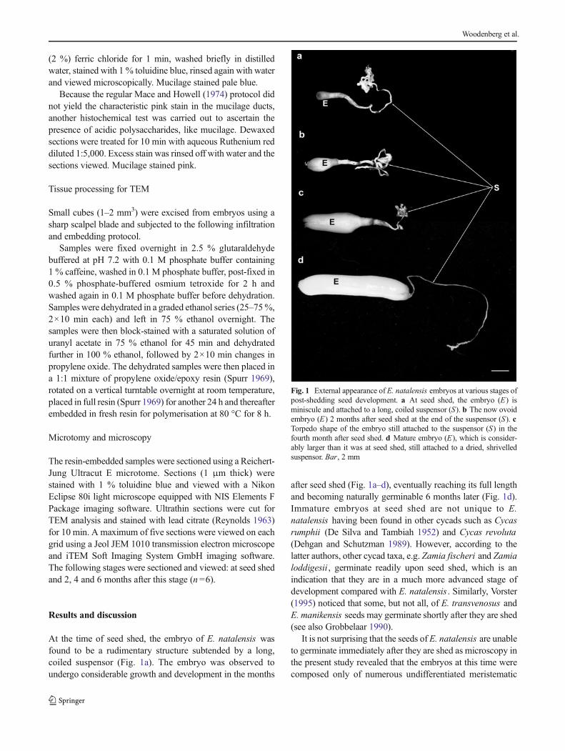
At the time of seed shed, the embryo of E. natalensis wasfound to be a rudimentary structure subtended by a long,coiled suspensor (Fig. 1a). The embryo was observed toundergo considerable growth and development in the months
after seed shed (Fig. 1a–d), eventually reaching its full lengthand becoming naturally germinable 6 months later (Fig. 1d).Immature embryos at seed shed are not unique to E.natalensis having been found in other cycads such as Cycasrumphii (De Silva and Tambiah 1952) and Cycas revoluta(Dehgan and Schutzman 1989). However, according to thelatter authors, other cycad taxa, e.g. Zamia fischeri and Zamialoddigesii , germinate readily upon seed shed, which is anindication that they are in a much more advanced stage ofdevelopment compared with E. natalensis . Similarly, Vorster(1995) noticed that some, but not all, of E. transvenosus andE. manikensis seeds may germinate shortly after they are shed(see also Grobbelaar 1990).
It is not surprising that the seeds of E. natalensis are unableto germinate immediately after they are shed as microscopy inthe present study revealed that the embryos at this time werecomposed only of numerous undifferentiated meristematic
Fig. 1 External appearance of E. natalensis embryos at various stages ofpost-shedding seed development. a At seed shed, the embryo (E) isminiscule and attached to a long, coiled suspensor (S). b The now ovoidembryo (E) 2 months after seed shed at the end of the suspensor (S). cTorpedo shape of the embryo still attached to the suspensor (S) in thefourth month after seed shed. d Mature embryo (E), which is consider-ably larger than it was at seed shed, still attached to a dried, shrivelledsuspensor. Bar, 2 mm
Woodenberg et al.
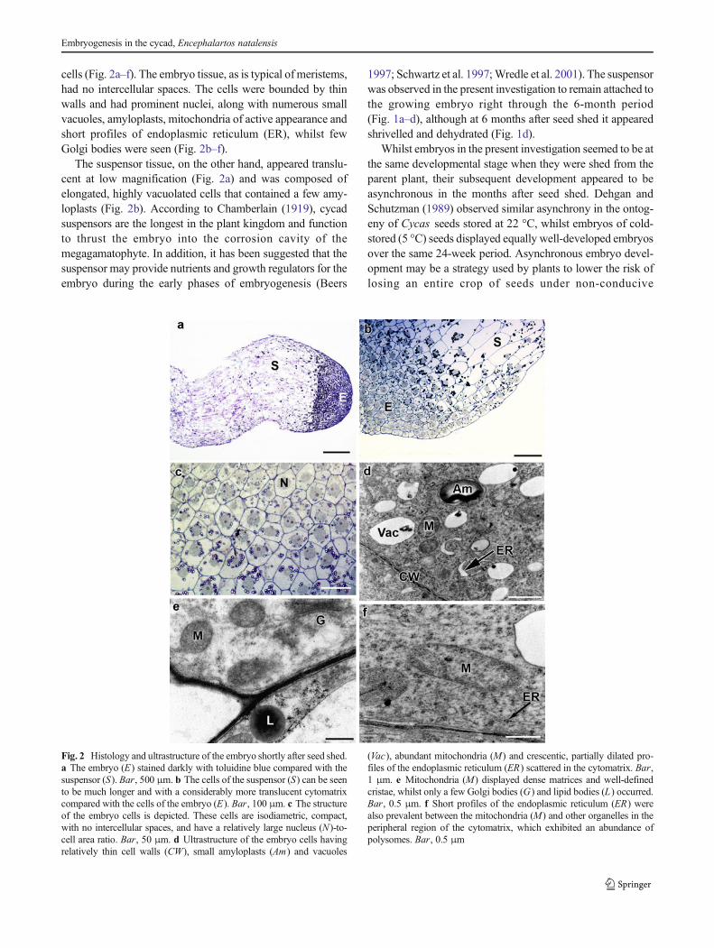
cells (Fig. 2a–f). The embryo tissue, as is typical of meristems,had no intercellular spaces. The cells were bounded by thinwalls and had prominent nuclei, along with numerous smallvacuoles, amyloplasts, mitochondria of active appearance andshort profiles of endoplasmic reticulum (ER), whilst fewGolgi bodies were seen (Fig. 2b–f).
The suspensor tissue, on the other hand, appeared translu-cent at low magnification (Fig. 2a) and was composed ofelongated, highly vacuolated cells that contained a few amy-loplasts (Fig. 2b). According to Chamberlain (1919), cycadsuspensors are the longest in the plant kingdom and functionto thrust the embryo into the corrosion cavity of themegagamatophyte. In addition, it has been suggested that thesuspensor may provide nutrients and growth regulators for theembryo during the early phases of embryogenesis (Beers
1997; Schwartz et al. 1997;Wredle et al. 2001). The suspensorwas observed in the present investigation to remain attached tothe growing embryo right through the 6-month period(Fig. 1a–d), although at 6 months after seed shed it appearedshrivelled and dehydrated (Fig. 1d).
Whilst embryos in the present investigation seemed to be atthe same developmental stage when they were shed from theparent plant, their subsequent development appeared to beasynchronous in the months after seed shed. Dehgan andSchutzman (1989) observed similar asynchrony in the ontog-eny of Cycas seeds stored at 22 °C, whilst embryos of cold-stored (5 °C) seeds displayed equally well-developed embryosover the same 24-week period. Asynchronous embryo devel-opment may be a strategy used by plants to lower the risk oflosing an entire crop of seeds under non-conducive
Fig. 2 Histology and ultrastructure of the embryo shortly after seed shed.a The embryo (E) stained darkly with toluidine blue compared with thesuspensor (S). Bar, 500 μm. b The cells of the suspensor (S) can be seento be much longer and with a considerably more translucent cytomatrixcompared with the cells of the embryo (E). Bar, 100 μm. c The structureof the embryo cells is depicted. These cells are isodiametric, compact,with no intercellular spaces, and have a relatively large nucleus (N)-to-cell area ratio. Bar, 50 μm. d Ultrastructure of the embryo cells havingrelatively thin cell walls (CW), small amyloplasts (Am) and vacuoles
(Vac), abundant mitochondria (M) and crescentic, partially dilated pro-files of the endoplasmic reticulum (ER) scattered in the cytomatrix. Bar,1 μm. e Mitochondria (M) displayed dense matrices and well-definedcristae, whilst only a few Golgi bodies (G) and lipid bodies (L) occurred.Bar, 0.5 μm. f Short profiles of the endoplasmic reticulum (ER) werealso prevalent between the mitochondria (M) and other organelles in theperipheral region of the cytomatrix, which exhibited an abundance ofpolysomes. Bar, 0.5 μm
Embryogenesis in the cycad, Encephalartos natalensis

environmental conditions at the time when some seeds areready to germinate. Given the asynchronous nature of embryodevelopment in the current species, micrographs currently pre-sented are of the most representative embryos at each stage.
The embryos of the next stage observed in this study(i.e. 2 months after seed shed) had undergone a measure ofmorphological, histological and cellular differentiation(Fig. 3a–f). The acropetal region observed at the earlier stagehad remained meristematic, constituting the shoot meristemand two flanking cotyledonary protuberances (Fig. 3a). Thesedeveloping cotyledons appeared to have originated from twogroups of cells, one on either side of the shoot meristem, asdescribed previously for Ginkgo biloba (Lyon 1904), and, asSaxton (1910) suggested, is probably the case for all
gymnosperms. As illustrated by Chamberlain (1919), differ-entiation of the cotyledonary protuberances of cycads seemsto occur as follows: The rapid growth of the undifferentiatedembryo becomes somewhat retarded acropetally, whilstgrowth of the region around it is accelerated. This causes whatthe author describes as a depression surrounded by a ring ofcotyledonary tissue. The cotyledonary ring is not fully com-plete and consists of two equally crescentic parts that nearlytouch each other at their ends (Chamberlain 1919).
In the current study of embryos, 2 months after seed shed,whilst the cotyledonary protuberances were a major feature, afew tannin channels were evident near the periphery of theembryo growing in the direction of its longitudinal plane(Fig. 3a–c). The structures presently termed tannin channels
Fig. 3 Some histological and ultrastructural aspects of the embryo at2 months after seed shed are shown. a Two cotyledons (C), which areseen in the early stages of their development, flank the shoot meristem(SM) of the embryo, whilst the cells of the suspensor (S) show no visiblechange compared with the earlier stage. A few tannin channels (TC) werenow apparent near the periphery of the embryo. Bar, 500 μm. b Thetannin channels (TC) appeared to develop parallel with the longitudinalplane of the embryo, whilst the procambium (PC), appearing as bands ofelongated meristematic cells, was also now apparent. Bar, 100 μm. cTannin channel (TC ) in cross-section in which the contents appear
condensed peripherally, whilst non-tanniniferous cells display darklystained nuclei (N ) and large vacuoles (Vac ). Bar , 50 μm. d Thecytomatrix between the vacuoles (Vac) typically displayed long profilesof endoplasmic reticulum (ER), small amyloplasts (Am), mitochondria(M) and occasional lipid bodies (L) near the cell wall (CW). Bar, 0.5 μm.e Occasional Golgi bodies (G ) were seen in the periphery of thecytomatrix where other organelles like the mitochondrion (M , illustrated)were found. Bar, 0.5 μm. f Mitochondria (M) had relatively densematrices and clearly defined cristae, and clusters of polysomes wereapparent. Bar, 0.5 μm
Woodenberg et al.

have been seen in previous studies on cycad germplasm, andwhilst they have been referred to as tannin cells inmost reports(Dorety 1909, 1919; Sanchez-Tinoco and Engelman 2004;Saxton 1910), they have also been called canals (Saxton1910), tannin idioblasts and gold cells (Vovides 1991;Vovides et al. 1993, respectively).
The tannins in the current investigation appeared amor-phous in wax-embedded embryo material (e.g. Fig. 3a, b),but were sometimes condensed or perhaps precipitated inmaterial embedded in resin (e.g. Fig. 3c). Sanchez-Tinocoand Engelman (2004) have also observed two manifestationsof tannin in the seed coat of another cycad, Ceratozamiamexicana . This may be an indication of two types of tanninsor other phenolic compounds, or it may indicate poor pene-tration into the tannins by resin. From the present study, thelatter explanation is considered to be more likely as the dualappearance of the tannins was consistent with the embeddingmedium used.
Non-tanniniferous embryo cells at 2 months after seed shedcontained large vacuoles, as illustrated in Fig. 3c–e, whilstrelatively small amyloplasts, few discrete lipid bodies andGolgi bodies, mitochondria with dense matrices and shortprofiles of ER were common features of the cytomatrix(Fig. 3d, e), but generally few Golgi bodies were observed.Mitochondria exhibited well-developed cristae, and relativelydense matrices and clusters of polysomes were consistentfeatures (Fig. 3f). These intracellular features are congruentwith the ongoing metabolism that accompanies embryogene-sis. The cell walls appeared more substantial than the previousstage, and nascent intercellular spaces (not illustrated) weresometimes observed, indicating that the embryowas no longercomposed of meristematic cells only.
In the early stages of channel formation, the tannin ap-peared to be contained within vacuoles in cells also showingthe occurrence of amyloplasts and nuclei (Fig. 4a).Subsequently, the channels elongated considerably (Fig. 4b,d), although there was no evidence of prior coenocyte forma-tion, as was observed by Zobel (1985) in shoots of Sambucusracemosa , an angiospermous species. The contents of thetannin channels were amber/brown in unstained sections(see Fig. 12a, e–g) and light to very dark blue in sectionsstained with toluidine blue (Fig. 4c, d). Transmission electronmicroscopy confirmed tannin accumulations within vacuoles(Fig. 4e–h) following internalisation of vesicles containingsmall condensations (Fig. 4g). There was evidence of thevesicles being ER-derived (Fig. 4f), suggesting the origin ofthe tannins to be from the endoplasmic reticulum. This is inagreement with views that tannins originate in the ER prior tobeing accumulated intravacuolarly (Zobel 1985; Rao 1988;Evert 2006).
The embryo at the next stage, i.e. 4 months after seed shed(Figs. 5, 6 and 7), consisted of a shoot and root meristem thatappeared to be separated by a short hypocotyl region (Fig. 5a).
When viewed in cross-section, the root meristem displayedradiating rows of cells (Fig. 5b), which suggested that newmeristematic cells in this tissue are formed mostly by anticli-nal mitoses from centripetal initials. The root meristem ap-pears to be endogenous in the E. natalensis embryo (as it is inDioon spinulosum ; Dorety 1919) since it is surrounded bydifferentiated cells that are interspersed with tannin channels(Fig. 5a, b). Those differentiated cells had considerably larger,multifaceted amyloplasts (Fig. 5c, d) than seen in the previousstages. Similar amyloplasts have been observed previously inthe megagametophyte of E. natalensis , where they were insuch abundance that other organelles and the nuclei appearedexceedingly compressed (Woodenberg et al. 2010). The dif-ferentiated cells were highly vacuolated (Fig. 5d–f), whichfurther compressed the ground cytomatrix in which longprofiles of ER, the occasional lipid bodies (Fig. 5e) andmitochondria (Fig. 5f) occurred. A striking feature of thesecells also was the abundance of polysomes (Fig. 5e, f), con-tributing further to the dense appearance of the cytomatrix.
The region of differentiated cells between the root meri-stem and suspensor has been observed previously in othercycad embryos and has been called either the coleorhiza(Chamberlain 1910, 1919; Dehgan and Schutzman 1989;Hooft 1970) or root cap (Chavez et al. 1995; Saxton 1910).Regardless of the nomenclature, this tissue reportedly acts as ahard sheath that protects the root meristem from injury whenthe germinating embryo penetrates the woody sclerotesta(Chamberlain 1919; Sporne 1965).
Whilst some cellular differentiation was seen in the hypo-cotyl region of the embryo 4 months after the seeds had beenshed, the cells of the root and shoot meristems remainedundifferentiated (Fig. 6a–d) and appeared essentially similarto the cells of the embryo at seed shed, i.e. small, compactcells each containing a prominent nucleus (Fig. 6c, d), manysmall vacuoles and plastids showing only relatively smallstarch deposits (Fig. 6d). Profiles of ER and a few lipid bodieswere seen (Fig. 6e). There were numerous mitochondria(Fig. 6f), but only occasional Golgi bodies (Fig. 6g). One ofthe most striking features of the meristem cells was the fre-quency of both cytomatrical and membrane-associated poly-somes (Fig. 6h), attesting to intensive protein synthesis.Although the ultrastructure of the cells of both the root andshoot meristems remained essentially consistent across thedevelopmental stages of the embryos, the overall size of thesemeristems had increased as the embryos developed (Fig. 6a;cf. Fig. 3a). In the case of the meristematic region of the shoot,it is probable that the cellular proliferation was partially di-rected towards the considerable growth and development ofthe cotyledons (Fig. 6a), which ultimately became far longerthan the axis of the embryo itself.
Whilst the cotyledons were observed to be separate fromeach other (Fig. 5a) at this stage of ontogeny, in some sections,they could be seen to be in close contact, at least laterally
Embryogenesis in the cycad, Encephalartos natalensis

(Fig. 7a). In cross-section, there appeared to be some sevenvascular bundles (Fig. 7a) forming an arc in the central regionof each cotyledon. Longitudinal or tangential sections re-vealed that one or two tracheids had now differentiated
centripetally in the developing vascular tissue (Fig. 7b, c).These tracheids, when viewed in cross-section, had muchthicker walls than the surrounding cells and displayed clearlumina (Fig. 7c), suggesting that they may be potentially
Fig. 4 Optical and electron micrographs demonstrating tannin channels.a Cross-section showing the early stages of tannin channel (TC) forma-tion with the tannins apparently contained in vacuoles. Amyloplasts (Am)and nuclei (N) can also be resolved. Bar, 50 μm. b Longitudinal sectionof a resin-embedded embryo showing a tannin channel (TC) duringdevelopment. This channel appears considerably longer than the sur-rounding cells and contains darkly staining deposits and a few amylo-plasts. Bar, 50 μm. c Tannin channels (TC) in a cross-section of wax-embedded embryo tissue at a later stage of development, displayingrelatively uniform tannin content filling the entire channel. Bar, 50 μm.d A tannin channel sectioned longitudinally showing tannin drawn away
from the walls, whilst no organelles can be discerned. Bar, 50 μm. eEarly stages of tannin channel formation showing tannin (T) accumula-tions in the tonoplast and small electron-opaque deposits (asterisk) be-tween the plasmalemma (PM ) and cell wall (CW). Bar, 0.5 μm. fNascent vesicles apparently originating from the endoplasmic reticulum(ER , arrowed) and incorporating small electron-opaque deposits can beseen between the tannin vacuole (TV) and plasmalemma. Bar, 1 μm. gER-derived vesicles bearing small electron-opaque deposits (asterisk)appeared to be internalised by the tannin vacuole (TV). Bar, 1 μm. h Alarge central tannin vacuole, the predominant organelle in a developingtannin channel, is illustrated. Bar, 5 μm
Woodenberg et al.

functional at this stage in the transport of water contributing tocotyledon expansion during intra-seminal embryo ontogeny.Presently, however, this is only conjectural as the tracheidsthat develop in this stage may become fully operational onlyduring and after germination and seedling establishment.
Whilst tracheids could be unequivocally identified in thecotyledons at this stage, other than the procambium, themajority of the cotyledonary cells were similar to the differ-entiated cells of the hypocotyl region, with a notable abun-dance of amyloplasts (Fig. 7c, d), polysomes (Fig. 7d) andwell-developed mitochondria (inset, Fig. 7d). The starch de-posited in these amyloplasts, and in those of the axis cells, hadto have been built up from hydrolysed starch reserves of themegagametophyte (Woodenberg et al. 2010) since all contactwith the female strobilus was lost very early in embryo on-togeny. The starch was progressively accumulated seemingly
without dimunition in the developing embryo. It is probablethat it functions as the carbohydrate reserve during germina-tion and seedling establishment rather than during in ovulogrowth of the embryo, which is suggestively sustained bycontinuous importation from the megagametophyte. Whilstthe content of the few discrete lipid bodies found in theembryo cells may also be utilised at germination, proteinbodies such as those which were common in the megagame-tophyte cells of this species (Woodenberg et al. 2010) wereentirely absent from the cells of the embryo (see later) despitethe frequency of polysomes (Fig. 7d).
The predominance of starch in the mature embryo is inagreement with observations on Dioon edule (Chamberlain1910) and whole seeds of Ginkgo (Singh et al. 2008).However, it is in marked contrast to most other gymnospermspecies, e.g. conifers, which typically accumulate lipid as the
Fig. 5 Histology and ultrastructure of the embryo 4 months after seedshed. a Longitudinal section demonstrating the root meristem (RM) andshoot meristem (SM), the latter flanked by the two cotyledons (C) whichare longer than at the previous stage. The procambium (PC) and tanninchannels (TC) can be seen in each cotyledon. Bar, 500 μm. b Cross-section of the embryo depicting the root meristem (RM) with radiatingrows of meristematic cells. Bar, 200 μm. c Tannin channels (TC )appeared to have condensed, poorly preserved contents in cross-sectionin resin-embedded material, whilst the surrounding cells can be seen to be
dominated by amyloplasts. Bar, 50 μm. d The non-meristematic cellswere extensively vacuolated (Vac) and typified by large amyloplasts(Am) each containing a considerable amount of starch, whilst otherorganelles and the nucleus (N) occurred in the dense cytomatrix. Bar,5 μm. e A few lipid bodies (L) as well as numerous long and shortprofiles of the endoplasmic reticulum (ER) were apparent in the densecytomatrix; Bar, 0.5 μm. f Mitochondria (M) were a common feature ofthe cytomatrix, its density being ascribed to the occurrence of manypolysomes (arrowheads). Bar, 20 μm
Embryogenesis in the cycad, Encephalartos natalensis

primary storage reserve (Ching 1966; Gifford 1988;Krasowski and Owens 1993). However, according to Haines(1983), some Araucaria species, viz. A. angustifolia ,A. hunsteinii , A. araucana , accumulate mostly carbohydrates,whilst carbohydrates constitute the total food storage
component of A. bidwillii seeds. Apart from the situation inAraucaria , the preponderance of starch as a major storageproduct in the embryo appears to be a comparatively primitivetrait, with lipid- and protein-dominant embryos representingthe more advanced condition amongst the gymnosperms. In
Fig. 6 Structure and ultrastructure of the meristematic regions of theembryo 4 months after seed shed. a Longitudinal section of the rootmeristem (RM) and shoot meristem (SM), which appeared to be separatedfrom each other by a region of larger vacuolated cells. Bar, 200 μm. bThe root meristem (RM) in longitudinal section displayed a gentle arc(asterisk) of meristematic cells between its centre and coleorhiza region(Co). Bar, 200 μm. c A resin-embedded shoot apex sectioned longitu-dinally illustrating the meristem, in which the peripheral tunica (T)overlies the corpus (C). Bar, 50 μm. d Cells of both root and shootmeristems displayed a similar ultrastructure that seemed unchanged fromthe previous stage, typified by a relatively large nucleus (N), numerous
small vacuoles (Vac) and plastids (P) containing only small starch de-posits. Bar, 2 μm. e As is typical for meristems, no intercellular spaceswere evident at cell wall (CW) junctions, whilst a few relatively darklystaining lipid bodies and short profiles of the endoplasmic reticulum (ER)were seen. Bar, 5 μm. f Mitochondria (M) were numerous and showedwell-defined cristae and dense matrices. Bar, 0.5 μm. g Golgi bodies (G)also occurred, particularly near the cell periphery, whilst plastids (P)displayed varying numbers of relatively small electron-translucent starchinclusions and scattered plastoglobuli (Pg ). Bar , 0.5 μm. h Thecytomatrix was typified by the occurrence of polysomes, many of whichwere clearly ER-associated (arrows). Bar, 0.2 μm
Woodenberg et al.

terms of the basal evolutionary position of the cycads ingymnosperm phylogeny, it could be suggested that starchwas the first reserve to be accumulated generally in embryosof seed plants of ancient lineage. The accumulation ofmassivestarch deposits initially in the gametophyte of E. natalensis(Woodenberg et al. 2010) with subsequent sucrose transportinto, and starch synthesis in, the developing embryo is bio-chemically far less complicated than would be a system in-volving lipids (M. Black, King’s College, London, personalcommunication).
Furthermore, the predominance of starch as opposed to lipidas the major storage reserve in the E. natalensis embryo is notsurprising given the relatively long period (approx. 12 months)of embryo development from fertilisation to germination.Lipids are known to be vulnerable to oxidation (McDonald2004), which may lead to a loss of viability during theprotracted development of the embryo in the current species.This may have been especially important considering thatcycads are suggested to have originated in the CarboniferousPeriod (Schwendemann et al. 2009), when atmospheric oxygenconcentrations may have been as high as 35 % (Berner andCanfield 1989). A further salient feature may be that seeds ofE.natalensis and other species (Woodenberg et al. 2007) andthose of A. angustifolia (Farrant et al. 1989), A. hunsteinii(Pritchard and Prendergast 1986), A. araucana (RoyalBotanic Gardens 2008) and A. bidwillii (Del Zoppo et al.
1998) are desiccation-sensitive, thus requiring them to remainhydrated throughout development to germination. It is sug-gested that lipid would be more prone to oxidative degradationin metabolically active tissue, which remains hydrated forrelatively prolonged periods, than in desiccation-tolerant(orthodox) seed tissues of, e.g. most of the conifers.
At 6 months after seed shed, the root meristem region of theembryo had increased in overall size (Fig. 8a, b). A conspic-uous feature of the embryo at this stage was the developmentof the mucilage ducts (Fig. 8c). These ducts were lined byepithelial cells, and whilst the one demonstrated in Fig. 8cappears to have retained its contents, the lumina of mostappeared clear (illustrated later). The evidence that these mu-cilage ducts were lined by intact cells suggested that they mayhave been formed schizogenously, i.e. by separation of paren-chyma cells from each other (Esau 1953; Evert 2006).According to these authors, after a few divisions, the cells thatseparate from each other come to line the duct, effectivelyforming an epithelium. Duct-lining epithelial cells have beendescribed as producing hydrophilic mucilaginous polysaccha-rides intracellularly before transferring the secretion into theduct (Romberger 1993); however, in E. natalensis embryos, itis suggested that they elaborate glycoproteins/mucopolysaccharides, as has been suggested for fruit tissueofMangifera indica (Joel and Fahn 1980). The epithelial cellsin the current study were characterised ultrastructurally by
Fig. 7 Some aspects of the histology and ultrastructure of the cotyledonsat 4 months after seed shed are demonstrated. a Cross-section showingthe orientation of vascular bundles (arrows) in each of the two cotyle-dons. Bar, 500 μm. b A tangential section through a vascular bundlewhich appears to be made up primarily of procambial tissue with one ortwo tracheids (arrow) apparent. Tannin channels (TC) were locatedperipherally. Bar , 200 μm. c At higher magnification, occasional
tracheids (arrow) can be seen in the developing vascular bundle. Thesolitary tracheid has a typically thickened wall compared with the sur-rounding cells, in which amyloplasts (Am) can clearly be resolved. Bar,50 μm. d Cells contained numerous relatively large amyloplasts (Am)and vacuoles (Vac), many polysomes, a few lipid bodies (L) and mito-chondria (M) with well-defined cristae (inset; bar, 0.5 μm). Bar, 1 μm
Embryogenesis in the cycad, Encephalartos natalensis

Fig. 8 Structure and ultrastructure of the hypocotyl region of theembryo 6 months after the seeds were shed. a The root meristem(RM ) in longitudinal section which stained densely with toluidineblue appears very broad in relation to the width of the embryo. Bar ,500 μm. b The hypocotyl in cross-section demonstrating the centralposition of the root meristem (RM ). Bar , 500 μm. c Mucilage ducts(MD ) lined by a layer of epithelial cells were observed for the firsttime at this stage of development. These ducts were positioned be-tween the meristem and tannin channel (TC ) regions of the hypo-cotyls. Bar , 50 μm. d Ultrastructure of mucilage duct epithelial cellsdisplayed many variously distended lengths of endoplasmic reticulum(ER ) amongst relatively small amyloplasts (Am ). Bar , 1 μm. e
Numerous vesicles (Ves ) were also a common feature of thecytomatrix and were sometimes observed to join with the plasmalem-ma (asterisk ), whilst undistended (arrowheads ) and a grosslydistended profile of the endoplasmic reticulum (ER ) are also depicted.The surface of cell walls (CW ) facing the lumen (Lu ) displayed small,dark protrusions, as illustrated. Bar , 1 μm. f The hypocotyl regionother than the root meristem had relatively large intercellular spaces(asterisk ), whilst the cells, bounded by relatively thin walls (CW ),contained numerous large vacuoles (Vac ) and abundant amyloplasts(Am ) and mitochondria (M ). Bar , 10 μm. g The cytomatrix alsocontained abundant profiles of the endoplasmic reticulum (ER ) andpolysomes. Bar , 0.5 μm
Woodenberg et al.

numerous profiles of variously distended ER (Fig. 8d, e) thatappeared to give rise to many vesicles (Fig. 8e). Some of thosevesicles appeared to fuse with the plasmalemma (Fig. 8e) andare suggested to be involved in the secretion of mucopolysac-charides into the lumen of the mucilage duct. Since Golgibodies were observed extremely infrequently in the epithelialcells, it appeared that the endoplasmic reticulum is the intra-cellular system involved in the manufacture and secretion ofmucopolysaccharides in the current species. The apparentnon-involvement of Golgi bodies in mucilage secretion indeveloping E. natalensis embryos was an unexpected feature;however, it is in keeping with observations on earlycellularisation of the megagametophyte in this species, whereER-derived vesicles were found to elaborate and export cellwall material without the intermediary involvement of Golgibodies, which were rarely observed (Woodenberg et al. 2010).Core glycosylation of nascent polypeptides/proteins is an ER-mediated process, and it could be argued that this processsuffices in the production of mucopolysaccharide mucilagein embryos of E. natalensis . Golgi bypass in secretory path-ways has been described and debated for animal tissues(Grieve and Rabouille 2011) and described for maturingpumpkin seeds (Hara-Nishimura et al. 1998) and storageprotein in rice endosperm (Takahashi et al. 2005). Althoughunusual, Golgi bypass in plants seems likely (Hawes 2005),but is not uncontestable (Tian et al. 2013). However, to ourknowledge, no such studies have been carried out on seeds ofplants of ancient lineage, as represented by the cycads.
According to the review by Bhatnagar and Moitra (1996),mucilage ducts in cycads may act as water reservoirs, as axerophytic characteristic. At this stage also, 6 months after theE. natalesis seeds were shed, intercellular spaces had devel-oped in the hypocotyl tissue (Fig. 8f), the cells of which werecharacterised by many amyloplasts and large vacuoles, mito-chondria, ER and numerous polysomes (Fig. 8f, g).
Another conspicuous feature of the embryo 6 months afterseed shed was the appearance of developing leaf primordia(Fig. 9a, b). Whilst the shoot meristem appeared generallysimilar to that of the earlier developmental stage, leafprimordia that were not seen previously could now bediscerned in some cases as slight bulges and in others assubstantial structures, associated basolaterally with the shootmeristem in longitudinal and cross-sections (Fig. 9a, b, re-spectively). The development of these primordia coincidedwith the time the seeds were able to germinate readily(Woodenberg et al. 2009), i.e. when the embryos could beconsidered to be mature.
Apart from the developing leaf primordia, the cells of theshoot meristem (Fig. 9c, d) appeared unchanged from thatseen at the 4-month developmental stage, with the clear
appearance of the tunica and corpus regions (Fig. 9c; cf.Fig. 6c). Some indications of the plane of division of thesecells could be seen (Fig. 9c). Whilst most of the tunica cells ofthe shoot meristem seemed to undergo periclinal divisions,some cells in this external layer were observed to divideanticlinally (Fig. 9c). Both anticlinal and periclinal planes ofdivision were observed in the sub-apical cells of the corpus(Fig. 9c).
Cells of the root and shoot meristems were essentiallysimilar at 6 months after seed shed (Fig. 9d) compared withthe previous stages of ontogeny. The cells were dominated bylarge nuclei, whilst plastids containing only a little starch andsmall vacuoles were common and occasional lipid bodiesoccurred (Fig. 9d). Mitochondria were prominent (Fig. 9e)and Golgi bodies were far more common in the cells of bothroot and shoot meristems (Fig. 9f) than in cells of all otherregions of the embryonic axes where their occurrence wassparse. As in the earlier developmental stages, polysomeswere abundant (Fig. 9f).
The most striking feature of the embryos in the sixth monthafter seed shed was the differentiation which had occurred inthe cotyledons (Fig. 10a–d). Numerous mucilage ducts linedby cells which stained pink with toluidine blue and havingtranslucent contents had formed in the central region of eachcotyledon (Fig. 10a, b), whilst the primary vascular tissue wasalso prominent towards the adaxial surface of the cotyledon incross-section (Fig. 10a). This arrangement contrasts with thatobserved in Microcycas calocoma (Dorety 1909), where cot-yledonary traces were found to alternate with mucilage ducts.Each of the primary vascular bundles observed in the presentstudy was surrounded by a few tannin channels (Fig. 10a, b),whilst more of these structures were abundant in the peripheralregions of each cotyledon (Fig. 10a).
More tracheids also appeared to have differentiated in theprimary vascular tissue when compared with the previousstage (Fig. 10c), although this vascular tissue was still domi-nated by procambial cells (Fig. 10d). This indicated thatalthough the embryo was at a stage of ontogeny consideredto be mature, the vascular tissue appeared still to be in animmature state overall and may well be equipped to be fullyfunctional only later on in development, i.e. during germina-tion and subsequent stages of seedling growth.
The cotyledons were seen in the earlier stages to develop asdiscrete structures (ref. Figs. 5a, 6a and 7a). However, in themature embryos, they were tightly adpressed, as demonstratedby the close association of the adjacent adaxial epidermallayers of the two cotyledons (Fig. 10e). Such closely associ-ated cotyledons have also been seen in studies on other cycadembryos (Dorety 1909, 1919; Saxton 1910;Webb et al. 1984),and it was suggested by these authors that the cotyledons were
Embryogenesis in the cycad, Encephalartos natalensis

fused. According to Dorety (1909), for example, when thecotyledons of M. calocoma were examined microscopically,no trace could be found of the characteristic seam expected ifthe adaxial epidermal layers had become adpressed. Althoughcotyledon fusion may appear to have occurred in the cycadspecies investigated by these authors, the seam alluded to byDorety (1909) was evident in the present investigation on E.natalensis embryos. In this regard, Monnier and Norstog(1986) found that whilst the cotyledons of Zamia sp. remainedclosely adpressed in situ, they grew separately when embryoswere cultured in vitro. These authors proposed that the coty-ledons of that species may be closely associated in the seedbecause of the pressure exerted on them by the surroundingmegagametophyte tissue. This is probably also true for E.natalensis as bisected seeds showed the embryo enlarging ina tight space provided by the corrosion cavity in the centre ofthe comparatively large and fairly rigid megagametophytetissue (Woodenberg et al. 2009).
Cotyledons of mature embryos examined in the presentstudy had tannin channels with contents that consistentlydisplayed oblique striations when longitudinal sections wereviewed (Fig. 10f). Whilst these may be artefacts of sectioning,they occurred repeatedly in a similar pattern, which may be an
indication of an underlying ridging of the channel wall. On afew occasions where the embryo had appeared to be injuredbefore fixation, tannin from the surrounding channel(s)seemed to infiltrate the wound (Fig. 10g). This is suggestiveof the manner in which the tannins may function in theprevention of pathogen entry upon wounding (Wood 1967;Goodman et al. 1967), and/or provision of antioxidant activity(Esau 1953), as wounding is known to be associated with asurge of reactive oxygen species (Bolwell 1999). The positionrevealed by the present investigation of tannin channels nearthe periphery and vital tissues of the embryo, viz. the vasculartissue and shoot meristem, is suggested to provide defenceagainst external threats including pathogen invasion and phys-ical trauma. The cotyledonary ground tissue in which inter-cellular spaces were evident comprised starch-containing,vacuolated, rounded parenchymatous cells (Fig. 10h).
In the current study, the embryo was found to produce onlytwo cotyledons. This situation has been observed in someother cycads, e.g. M. calocoma (Dorety 1909), Zamia spp.(Monnier and Norstog 1984) and Encephalartos friderici -guilielmi (Saxton 1910), whilst Dorety (1919) found that D.spinulosum could have from two to four cotyledons (althoughthe number was commonly 2). In addition, Dorety (1908)
Fig. 9 Histological andultrastructural situation of the shootand root meristems 6 months post-shedding. a Longitudinal section ofthe root meristem (RM) and shootmeristem (SM) with what appearsto be the initiation of a leafprimordium (arrow); .Bar,500 μm. b Shoot meristem incross-section (arrow) in relation toa well-developed leaf primordium(asterisk). Bar, 200 μm. cIsodiametric cells of the shootmeristem with newly divided cells(arrowed) in the tunica (T) andcorpus (C) are apparent. Bar,50 μm. d The ultrastructure wasconsistent with previous stages,with meristem cells showing arelatively large nucleus (N) withnumerous plastids (P) containinglittle accumulated starch, vacuoles(Vac), lipid bodies (L) and densemitochondria (M) in the cytomatrixthat was bound by a relatively thincell wall (CW). Bar, 1 μm. e Thecytomatrix contained mitochondria(M) with dense matrices and well-defined cristae. Bar, 0.5 μm. fGolgi bodies (G) were also seen inthe cytomatrix, as were numerouspolysomes. Bar, 0.5 μm
Woodenberg et al.

demonstrated that whilst Ceratozamia species may appear tohave one cotyledon, the second cotyledon is in fact present,but its growth is substantially inhibited in that genus.
Aside from vascular tissue, resin ducts and tannin chan-nels, the cells of the cotyledons in the current study weredominated by large amyloplasts containing substantialamounts of starch, vacuoles, a considerable number ofmitochondria, the limited occurrence of lipid bodies(Fig. 11a, b) and only occasional Golgi bodies. Together,the vacuoles and bulky amyloplasts compressed the groundcytomatrix and other organelles mostly against the cellperiphery (Fig. 11b). The compression of the polysome-rich ground cytomatrix was so great that although the
mitochondria and ER (Fig. 11c, d) could be resolved, theeffect was almost one of negative staining. Whilst thegeneral intracellular situation of the cotyledonary groundtissue was essentially similar to that in the mature megaga-metophyte cells of E. natalensis , what was remarkablydifferent was the absence of the dense protein bodies whichwere prevalent in the latter tissue (Woodenberg et al. 2010).The absence of similar substantial protein deposits in theembryo tissues was confirmed by the lack of specific stain-ing histochemically (see below).
In the current study, mucilage ducts were positively iden-tified by staining with a modified version of the protocol ofMace and Howell (1974) for the location of mucins. Initial
Fig. 10 Histology of thecotyledons 6 months after seedshed. a Cross-section displayingthe position of numerous mucilageducts (arrows) in the central regionof the cotyledon, whilst threevascular bundles (V) and manyblue-staining tannin channels canbe seen. Bar, 200 μm. b Arelatively largemucilage duct (MD)with a translucent cavity lined byepithelial cells and developingvascular tissue each surrounded bya few tannin channels (arrows).Bar, 100 μm. c Avascular bundlein cross-section displaying a smallcluster of thick-walled tracheids(arrows) rather than the solitarystructures seen 4 months after seedshed. Bar, 50 μm. d Theprocambium (PC) was seen inlongitudinal sections to becomposed of relatively elongated,thin-walled cells, the cytomatrix ofwhich stained mauve to pink withtoluidine blue. Bar, 100 μm. e Thetwo cotyledons were closelyadpressed in some places (asterisk).Bar, 50 μm. f Tannin channels(TC) near the peripheral region ofthe cotyledons displayed striations,as illustrated in this longitudinalsection. Bar, 100 μm. g Tannins(T) presumably from tanninchannels appeared to have seepedinto a wound that was presumablycreated before fixation. Bar,100 μm. h Cross-section ofnumerous cells that were packedwith amyloplasts and were roundedwith concomitant development ofintercellular spaces (arrows). Bar,50 μm
Embryogenesis in the cycad, Encephalartos natalensis

results using the standard protocol outlined by these authorsyielded no staining in the schizogenously formed ducts; how-ever, indications of their contents were obtained serendipi-tously when toluidine blue was applied to sections alreadystained using the Mace and Howell (1974) method (Fig. 12a).The ducts displayed pale blue staining when this was done,indicating that they might not be empty. Staining of thesestructures was also evident when Ruthenium red wasemployed for the detection of acidic polysaccharides typicalof mucilage (Fig. 12b), where the epithelial cells stained redand the duct contents appeared a light pink. The constituentssuch as amyloplasts and vacuoles of other cells remainedunstained. These two results seem to complement one anotherand strongly suggest that the structures investigated are, infact, mucilage ducts.
When vanillin–HCl was applied, positive bright redstaining indicated the presence of tannins in discrete loca-tions (Fig. 12c), serving to confirm that these are, in alllikelihood, the tannin channels. The amyloplasts, whichconstituted a large proportion of the contents of the major-ity of cells, tested positive for starch with the use of Lugol’sstain (Fig. 12d). As indicated by transmission electronmicroscopy, lipid was confirmed to be sparsely present inthe cells as a few very small, discrete entities when SudanBlack B was used (Fig. 12e); however, the use of that stainalso indicated the presence of a layer of lipid, probably wax,covering the exterior of the embryo (Fig. 12f). Effectively,
this would constitute a cuticle and be a further adaptation(additional to the relatively impermeable sclerotesta) toretard the loss of water from these embryos, which havebeen shown to be desiccation-sensitive (Woodenberg et al.2007, 2009).
Protein histochemistry in the present investigationyielded an unexpected interesting result. According toWoodenberg et al. (2010), many of the vacuoles of themegagametophyte cells of E. natalensis displayed consid-erable amounts of a relatively dense, granular material thatstained positively for protein when eosin dye was used.When the same stain was utilised in the current study, thevacuoles did not stain positively (Fig. 10g, h), and nosimilar dense, granular material was observed with theTEM or light microscope. However, some staining forprotein did occur, but confined to the nuclei, when mercu-ric bromophenol blue was utilised (Fig. 12h). The embryoappears, therefore, not to accumulate any significantamount of reserve protein as protein bodies compared withthe megagametophyte, but it suggestively obtains the re-quired amino acids directly from the protein reserves of themegagametophyte tissue. From the prevalence of poly-somes throughout embryogenesis, there can be no doubtthat active protein synthesis is ongoing. However, thispresumably is directed towards the synthesis of structuralproteins of, e.g. membranes and the cytoskeleton, as wellas of enzymes.
Fig. 11 Ultrastructure of the cotyledonary cells 6 months after seed shed.a Cells were dominated by abundant relatively large amyloplasts (Am)and vacuoles (Vac). Bar, 5 μm. b Ultrastructure of one of those cellsdemonstrating the size of the abundant starch-filled amyloplasts (Am)compared with the mitochondria (M), shown compressed in the periph-eral cytomatrix and the few lipid bodies (L) that were present in the cells.
Bar, 20 μm. c The mitochondria (M) that were present had well-definedcristae and patchily dense matrices. Bar, 0.2 μm. d Relatively shortprofiles of the endoplasmic reticulum (ER) were also prevalent in someareas of the dense cytomatrix, which displayed an abundance of poly-somes as seen in other stages of development. Bar, 0.2 μm
Woodenberg et al.

Concluding remarks
The present investigation has revealed some important aspectsof the post-shedding development of the E. natalensis embryo
that further the understanding of the recalcitrant post-sheddingbehaviour of these seeds (Woodenberg et al. 2009). Seeds ofthis species are shed from the parent plant when the embryo isrudimentary and decidedly physiologically immature; its
Fig. 12 Histochemistry of secondary metabolites and storage productsaccumulated in the mature embryo of E. natalensis (toluidine blue wasused only on the section in ‘a’). a Contents of mucilage ducts (arrow)appeared faintly blue when toluidine blue was applied to the sections afterstaining for mucins with the tannic acid/ferric chloride protocol of Maceand Howell (1974), whilst the non-stained, orange/brown colouration oftannin channels is also evident; Bar, 50 μm. b The contents of mucilageducts stained pale pink (arrow) with the epithelial cells staining a deeperpink to red when Ruthenium red was used for the detection of acidicpolysaccharides, whilst tannin channels stained darkly; Bar, 50 μm. cThe contents of tannin channels stained red (arrow) with vanillin-HClwhilst the rest of the tissue did not stain, indicating the localised presence
of tannins; Bar, 200 μm. d Amyloplasts in most of the tissue stainedblack when Lugol’s solution, which localises carbohydrate (by reactionwith potassium iodide/iodine; Jensen 1962) was employed, whilst vascu-lar tissue (V) did not display similar staining; Bar, 200 μm. e Scattered,small, discrete bodies of lipid were observed to stain darkly (arrows) withSudan Black B; Bar, 50 μm. f The use of Sudan Black B also yielded athin film of darkly staining substance, presumed to be cuticular wax, onthe external surface of the epidermis (arrows); Bar, 50 μm. g There wasno staining which would have indicated the presence of protein bodieswhen Eosin dye was used; Bar, 50 μm. h A similar, negative result wasobtained when mercuric bromophenol blue was used to localise protein,although the nuclei stained prominently; Bar, 50 μm
Embryogenesis in the cycad, Encephalartos natalensis

development has been shown to be a continuous process afterseed shed that ultimately culminates in germination.Histochemistry in this study showed that the major storageproduct accumulated in the embryo is starch, which is accu-mulated abundantly in the many amyloplasts of non-meristematic cells, whilst lipid is accumulated in only minorquantities and reserve protein accumulation is negligible. Thisinformation, along with that revealed about the megagameto-phyte (Woodenberg et al. 2010), may be important for thefuture design of biotechnological protocols for the in vitrogrowth of immatureE. natalensis embryos that were observedin the present study at the time of seed shed. Immatureembryos of this species may provide a suitable explant forthe cryopreservation of this species, the seeds of which arecurrently unstorable in the long term.
The current investigation has also increased the under-standing of the secondary metabolites accumulated in theembryo of this species, particularly mucilage and tannin.Mucilage ducts were found to be a common feature of matureembryos in this species, which, along with the possession of atough sclerotesta external to the seed and a waxy layer on thesurface of the embryo, may contribute to water retention in themonths after seed shed (Woodenberg et al. 2009). Thesecharacteristics are suggested to contribute to the relative lon-gevity (1–2 years) in storage under conditions precludingdehydration compared with recalcitrant seeds of other species,which is generally a matter of days to months (Pammenteret al. 1994).
Apart frommucilage, the other major secondary metabolitefound in the embryo of the current species was tannin, accu-mulated in tannin channels. These may be analogous to theresin canals of most conifers, gum canals ofWelwitschia , andlaticifers of Gnetum and some angiosperm species (Esau1953; Romberger 1993). The purported antimicrobial andwound-healing properties of tannins may also account forthe use of parts of cycads in traditional medicine in SouthAfrica (e.g. Osborne et al. 1994), and according to Vashishta(1995), the ‘resin’ obtained from C. rumphii is apparentlyapplied to malignant tumours. The use of tannins from cycadsfor medicinal purposes therefore warrants further investiga-tion and, if verified, would justify research on in vitro culturefor their production as further depletion of the naturally oc-curring plants cannot be countenanced.
Further study is also encouraged on the early stages of wallformation in the embryo of E. natalensis . As Woodenberget al. (2010) demonstrated, initial cellularisation of themegagametophe of this species seemed to occur without theinvolvement of Golgi bodies. Similarly, very fewGolgi bodieswere evident in the early embryo in the present investigation.It will therefore be intriguing to see whether or not a similarmode of primary cell wall development occurs during theinitial cellularisation of the E. natalensis embryo. This wouldentail seed collection immediately and, shortly after,
fertilisation (the dynamics of which would require to beascertained) and adaptation of preparative technology formicroscopy of the minute nascent structures.
Acknowledgments We wish to thank Vishal Bharuth, Phillip Christo-pher and Nelisha Murugan (the Microscopy and Microanalysis Unit,University of KwaZulu-Natal, Westville Campus), as well as Dr JamesWesley-Smith (CSIR, Pretoria) for technical assistance with the micros-copy and Professors Ashley Nicholas (UKZN, Durban), Michael Black(King’s College, London) and Françoise Corbineau (Université Pierre etMarie Curie-Paris 6) for helpful discussions. This work was supported bythe National Research Foundation (NRF) of South Africa.
References
Agapito-Tenfen SZ, Steiner N, Guerra MP, Nodari RO (2011) Patterns ofpolyembryony and frequency of surviving multiple embryos of theBrazillian pine, Araucaria angustifolia. Aust J Bot 59:749–755
Baird AM (1939)A contribution to the life history ofMacrozamia reidlei .J R Soc WAust 25:153–175
Beers EP (1997) Programmed cell death during plant growth and devel-opment. Cell Death Differ 4:649–661
Berner RA, Canfield DE (1989) A new model for atmospheric oxygenover Phanerozoic time. Am J Sci 289:333–361
Bhatnagar SP, Moitra A (1996) Gymnosperms. New Age InternationalLimited, New Delhi
Biswas C, Johri BM (1997) The gymnosperms. Springer, New DelhiBolwell GP (1999) Role of active oxygen species and NO in plant
defence responses. Curr Opin Plant Biol 2:287–294Brenner DE, Stevenson W, Twigg RW (2003) Cycads: evolutionary
innovations and the role of plant-derived neurotoxins. Trends PlantSci 8:446–452
Brough P, Taylor MH (1940) An investigation of the lifecycle ofMacrozamia spiralis Miq. P Linn Soc 65:494–524
Bryan GS (1952) The cellular embryo of Zamia and its cap cells. Am JBot 39:433–443
Chamberlain CJ (1910) Fertilisation and embryogeny in Dioon edule .Bot Gaz 50:415–429
Chamberlain CJ (1916) Stangeria paradoxa . Bot Gaz 61:353–372Chamberlain CJ (1919) The living cycads. Chicago University Press,
ChicagoChamberlain CJ (1935) Gymnosperms: structure and evolution. Chicago
University Press, ChicagoChavez VM, Litz RE, Marquez J (1995) Histology of somatic embryo-
genesis of the cycad Ceratozamia Mexicana var. Robusta (Miq.)Dyer. Plant Sci 108:191–200
Ching TM (1966) Compositional changes of Douglas fir seeds duringgermination. Plant Physiol 41:1313–1319
De Silva BLT, Tambiah MS (1952) A contribution to the life history ofCycas rumphii Miq. Ceylon J Sci 12:223–249
Dehgan B, Schutzman B (1989) Embryo development and germinationof Cycas seeds. J Am Soc Hortic Sci 114:125–129
Del Zoppo M, Galleschi L, Saviozzi F (1998) Long term storage ofAraucaria bidwillii Hook. seeds. Seed Sci Technol 26:267–270
Dogra PD (1992) Embryogeny of primitive gymnosperms Ginkgo andcycads—pro-embryo—basal plan and evolutionary trends.Phytomorphology 42:157–184
Dorety HA (1908) The embryo of Ceratozamia : a physiological study.Bot Gaz 45:412–416
Dorety HA (1909) Vascular anatomy of the seedling of Microcycascalocoma. Bot Gaz 47:139–147
Woodenberg et al.

Dorety HA (1919) Embryo and seedling of Dioon spinulosum. Bot Gaz67:251–257
Esau K (1953) Plant Anatomy, 2nd edn. Wiley, New YorkEvert RF (2006) Esau’s plant anatomy, 3rd edn. Wiley-Interscience,
HobokenFarrant JM, Pammenter NW, Berjak P (1989) Germination-associated
events and the desiccation-sensitivity of recalcitrant seeds—a studyon three unrelated species. Planta 178:189–198
Favre-Ducharte M (1956) Contribution a l’etude de la reproduction chezGinkgo biloba. Rev Cyt Biol Veg 17:1–218
Gifford DJ (1988) An electrophoretic analysis of the seed proteins fromPinusmonticola and eight other species of pine. Can J Bot 66:1808–1812
Goodman RN, Kiraly Z, Zaitlin M (1967) The biochemistry and physi-ology of infectious disease. D. van Nostrand Co. Inc., Princetown
Grieve AG, Rabouille C (2011) Golgi bypass: skirting around theheart of classical secretion. Cold Spring Harb Perspect Biol3:a005298
Grobbelaar N (1990) Encephalartos embryos. Encephalartos 24:23Haines RJ (1983) Seed development in Araucaria Juss. Aust J Bot 31:
255–267Hara-Nishimura I, Shiada T, Hatano K, Takeuchi Y, Nishimura M (1998)
Transport of storage protein to protein storage vacuoles is mediatedby large precursor-accumulating vesicles. Plant Cell 10:825–836
Hawes C (2005) Cell biology of the plant Golgi apparatus. New Phytol165:29–44
Hooft J (1970) Zamia from seed. Carolina Tips 33:21–22James J, Tas J (1984) Histochemical protein staining methods. Oxford
University Press, New YorkJensen WA (1962) Botanical histochemistry. WH Freeman, LondonJoel DM, Fahn A (1980) Ultrastructure of the resin ducts of Mangifera
indica L. (Anacardiaceae). 3. Secretion of the protein–polysaccha-ride mucilage in fruits. Annu Bot Lond 46:785–790
Krasowski MJ, Owens JN (1993) Ultrastructural and histochemicalpostfertilisation megagametophyte and zygotic embryo develop-ment of white spruce (Picea glauca) emphasizing the depositionof seed storage products. Can J Bot 71:98–112
Lawson AA (1926) A contribution to the life history of Bowenia . TranslR Soc Edinb 54:357–394
Lyon HL (1904) The embryogeny of Ginkgo . Minn Bot Stud 3:275–290Mace ME, Howell CR (1974) Histochemistry and identification of con-
densed tannin precursor in roots and cotton seedlings. Can J Bot 52:2423–2426
Maheshwari P (1960) Morphology and embryology of Cycas. MScthesis, University of Delhi
Mazia D, Brewer PA, Alfert TM (1953) The cytochemical staining andmeasurement of protein with mercuric bromophenol blue. Biol Bull104:57–67
McDonald MB (2004) Orthodox seed deterioration and its repair. In:Benech-Arnold RL, Sánchez RA (eds) Handbook of seed physiol-ogy: applications to agriculture. The Haworth Press, New York, pp273–304
McManus JFA (1946) Demonstration of certain fatty substances in par-affin sections. J Pathol Bacteriol 58:93–95
Monnier M, Norstog K (1984) Developmental aspects of immatureZamia embryos cultured in vitro. Z Pflanzenphysiol 113:105–115
Monnier M, Norstog K (1986) Effect of in ovulo period on the differen-tiation and regulation of immature embryos of Zamia culturedin vitro. J Exp Bot 37:1633–1642
National Red List of South African Plants (2009) SANBI, Pretoria, SouthAfrica
Osborne R, Grove A, Oh P, Mabry TJ, Ng J, Seawright AA (1994) Themagical and medical usage of Stangeria eriopus in South Africa. JEthnopharmacol 43:67–72
Pammenter NW, Berjak P, Farrant JM, SmithMT, Ross G (1994)Why dostored hydrated recalcitrant seeds die? Seed Sci Res 4:187–191
Pizzolato TD, Lillie RD (1973) Mayer’s tannic acid–ferric chloride stainfor mucins. J Histochem Cytochem 21:56–64
Pooley E (1993) Trees of Natal, Zululand and Transkei. Natal FloraPublications Trust, Durban, South Africa
Pritchard HW, Prendergast FG (1986) Effects of desiccation and cryo-preservation on the in vitro viability of embryos of the recalcitrantseed species Araucaria hunsteinii K. Schum. J Exp Bot 37:1388–1397
Rao LN (1963) Life history of Cycas circinalis L. Part 2. Fertilisation,embryogeny and germination of the seed. J Indian Bot Soc 42:319–332
Rao KS (1988) Fine structural details of tannin accumulations in non-dividing cambial cells. Ann Bot—Lond 62:575–581
Reynolds ES (1963) The use of lead citrate at high pH as an electron-opaque stain in electron microscopy. J Cell Biol 17:208–211
Romberger JA (1993) Plant structure: function and development: a trea-tise on anatomy and vegetative development, with special referenceto woody plants. Springer, Berlin
Royal Botanic Gardens Kew (2008) Seed Information Database (SID),version 7.1. http://data.kew.sid (May 2008). Accessed 16 September2013
Sanchez-Tinoco MY, Engelman E (2004) Seed coat anatomy ofCeratozamia mexicana (Cycadales). Bot Rev 70:24–38
Saxton WT (1910) The development of the embryo of Encephalartos .Bot Gaz 49:13–18
Schlegel H (1991) Domestic growing of cycads from seed to houseplantsize. Encephalartos 26:2–25
Schwartz BW, Vernon DM, Meinke DW (1997) Development ofthe suspensor: differentiation, communication, and pro-grammed cell death during plant embryogenesis. In:Larkins BA, Vasil IK (eds) Cellular and molecular biologyof plant seed development. Kluwer Academic, Dordrecht, pp53–72
Schwendemann AB, Taylor TN, Taylor EL (2009) Pollen of the Triassiccycad Delemaya spinulosa and implications on cycad evolution.Rev Palaentol Palynol 156:98–103
Sedgwick PJ (1924) Life history of Encephalartos . Bot Gaz 77:300–310Singh H (1978) Embryology of gymnosperms. Gebrüder Borntraeger,
BerlinSingh H, Johri BM (1972) Development of gymnosperm seeds. In:
Kozlowski TT (ed) Seed biology, vol. 1. Importance, development,and germination. Academic, New York, pp 21–75
Singh B, Kaur P, Gopichand SRD, Ahuja PS (2008) Biology and chem-istry of Ginkgo biloba. Fitoterapia 79:401–418
Sporne KR (1965) The morphology of gymnosperms. Hutchinson,London
Spurr AR (1969) A low-viscosity epoxy resin embedding medium forelectron microscopy. J Ultrastruct Res 26:31–43
Steedman HF (1960) Tropical ester wax. Q J Microsc Sci 101:463–464Stevenson DW (1990) Chigua , a new genus in the Zamiaceae with
comments on its biogeographic significance. Mem N Y Bot Gard57:169–172
Swamy BGL (1948) Contributions to the life history of a Cycas fromMysore (India). Am J Bot 35:77–88
Takahashi H, Saito Y, Kitagawa T, Morita S, Masumura T, Tanaka K(2005) A novel vesicle derived directly from endoplasmic reticulumis involved in the transport of vacuolar storage proteins in riceendosperm. Plant Cell Physiol 46:245–249
Tian L, Dai LL, Yin ZJ, Fukuda M, Kumamaru T, Dong XB, Xu XP, QuLQ (2013) Small GTPase Sar1 is crucial for proglutelin and α-globulin export from the endoplasmic reticulum in rice endosperm.J Exp Bot 64:2831–2845
Vashishta PC (1995) Gymnosperms: botany for degree students, vol. 5.Chand S and Company LTD, New Delhi
Vorster P (1995) Aspects of the reproduction of cycads. 1. Pollinatingmechanisms and the association of Amorphocerus (Curculionidae)
Embryogenesis in the cycad, Encephalartos natalensis
with Encephalartos . In: Vorster P (ed) Proceedings of the thirdinternational conference on cycad biology. Cycad Society of SouthAfrica, Stellenbosch, pp 367–389
Vovides AP (1991) Cone idioblasts of eleven cycad genera: morphology,distribution, and significance. Bot Gaz 152:91–99
Vovides AP, Norstog KJ, Fawcett PKS, Duncan MW, Nash RJ,Molsen DV (1993) Histological changes during maturation inmale and female cones of the cycad Zamia furfuracea andtheir significance in relation to pollination biology. Bot J LinnSoc 111:241–252
Webb DT, Nevarez M, De Jesus S (1984) Further in vitro studies of light-induced root nodulation in the cycadales. Environ Exp Bot24:37–44
Wood RKS (1967) Physiological plant pathology. Botanical MonographsNo. 6. Blackwell Scientific Publications, Oxford
Woodenberg WR, Erdey D, Pammenter NW, Berjak P (2007) Post-shedding seed behaviour of selected Encephalartos species.
Abstracts from the 5th International Workshop on DesiccationTolerance and Sensitivity of Seeds and Vegetative Plant Tissues. SAfr J Bot 73:496
Woodenberg WR, Pammenter NW, Berjak P (2009) Some aspects ofmegagametophyte development and post-shedding seed behaviourof Encephalartos natalensis (Zamiaceae). Master’s thesis,University of KwaZulu-Natal, Durban, South Africa
Woodenberg WR, Berjak P, Pammenter NW (2010) Development ofcycad ovules and seeds. 1. Implication of the ER in primarycellularisation of the megagametophyte inEncephalartos natalensisDyer and Verdoorn. Plant Growth Regul 62:265–278
Wredle U, Walles B, Hakman I (2001) DNA fragmentation and nucleardegradation during programmed cell death in the suspensor andendosperm of Vicia faba. Int J Plant Sci 162:1053–1063
Zobel AM (1985) Ontogenesis of tannin coenocytes in Sambucusracemosa L. I. Development of the coenocytes from mononucleatetannin cells. Ann Bot Lond 55:765–773
Woodenberg et al.