Embed Size (px)
Citation preview


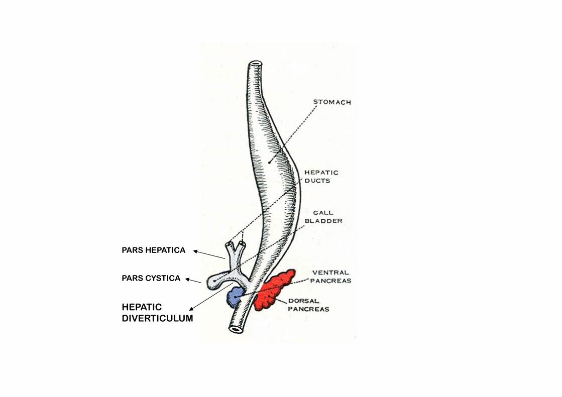
PARS HEPATICA
PARS CYSTICA
HEPATIC DIVERTICULUM
D
D
D
VPBDPB
DPB
DPBVPB
VPB
Factors causing physiological hernia:1. Rapid growth & large size of liver which occupies a
large area of abdominal cavity.
2. Small size of abdominal cavity because the lumbarsegment is not yet formed.
3. Mid gut is growing at a fast pace.
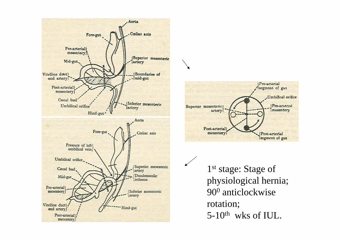
1st stage: Stage of physiological hernia; 900 anticlockwise rotation; 5-10th wks of IUL.
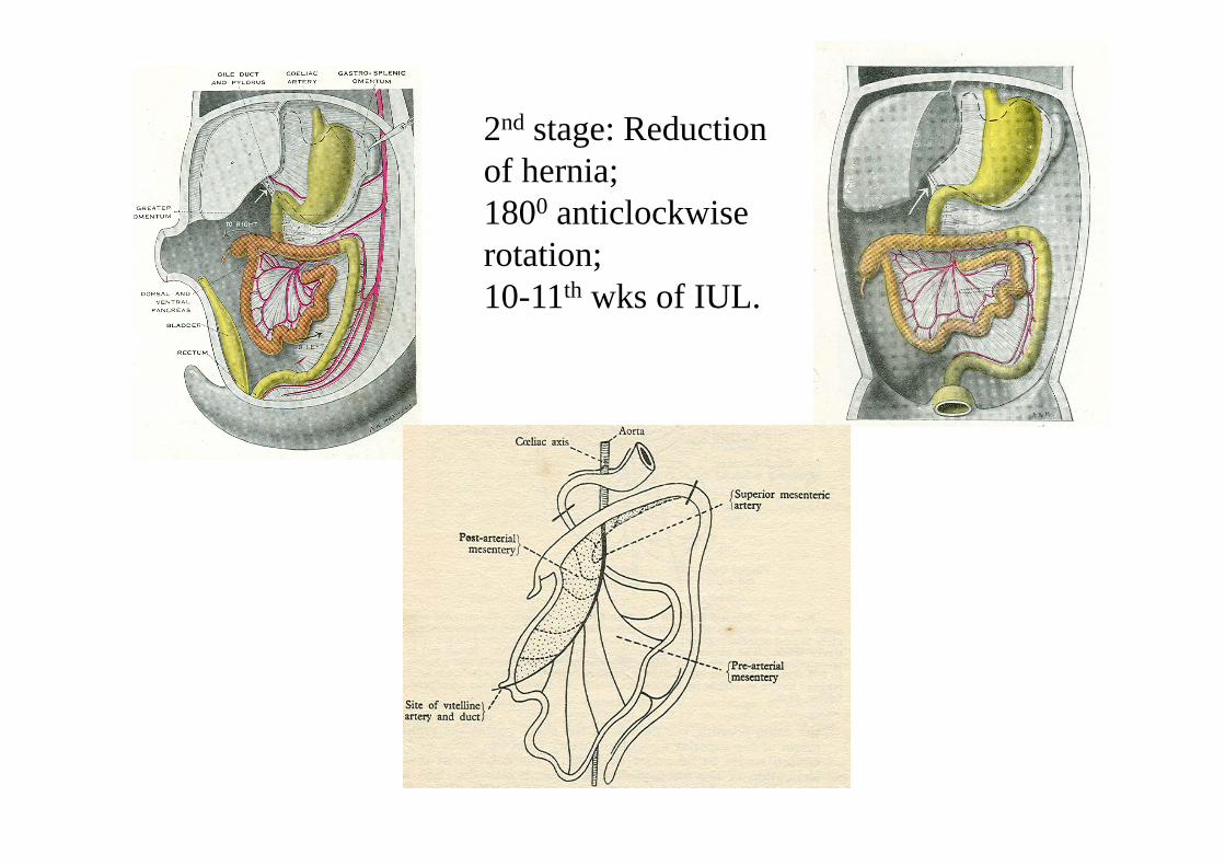
2nd stage: Reduction of hernia; 1800 anticlockwise rotation; 10-11th wks of IUL.
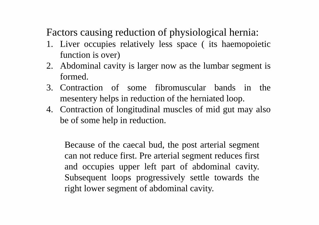
Factors causing reduction of physiological hernia:1. Liver occupies relatively less space ( its haemopoietic
function is over)2. Abdominal cavity is larger now as the lumbar segment is
formed.3. Contraction of some fibromuscular bands in the
mesentery helps in reduction of the herniated loop.4. Contraction of longitudinal muscles of mid gut may also
be of some help in reduction.
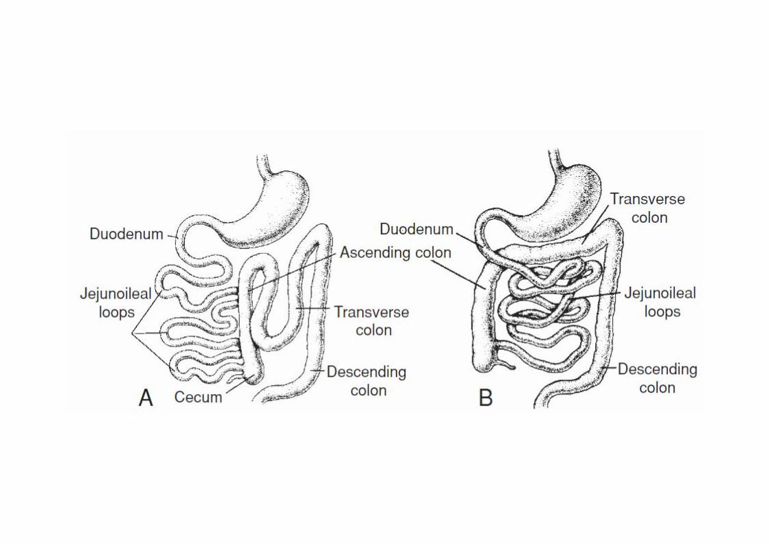
Because of the caecal bud, the post arterial segmentcan not reduce first. Pre arterial segment reduces firstand occupies upper left part of abdominal cavity.Subsequent loops progressively settle towards theright lower segment of abdominal cavity.
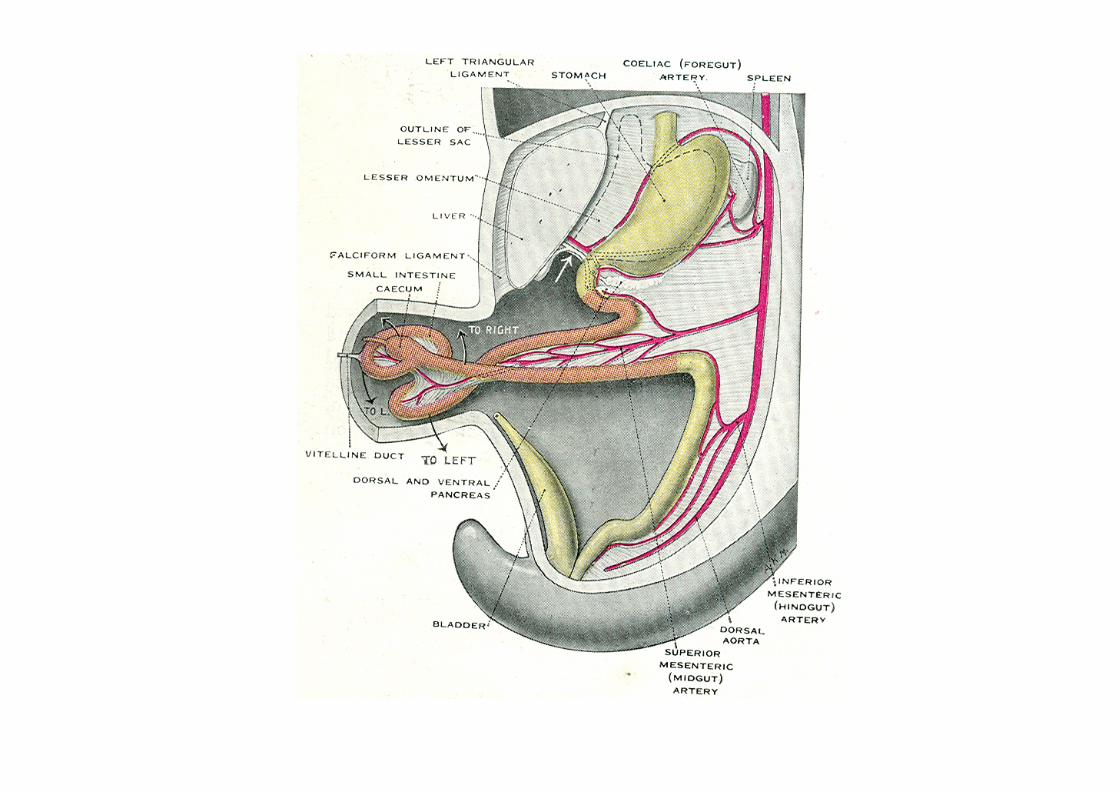
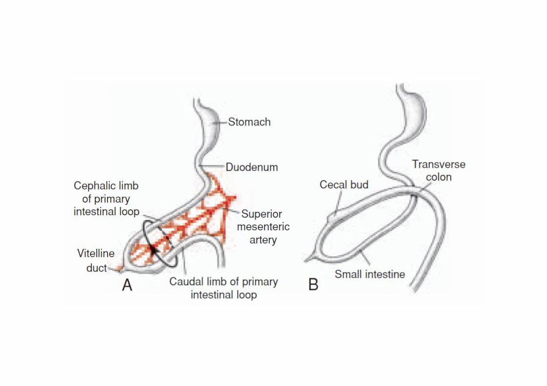
Rotation of midgut
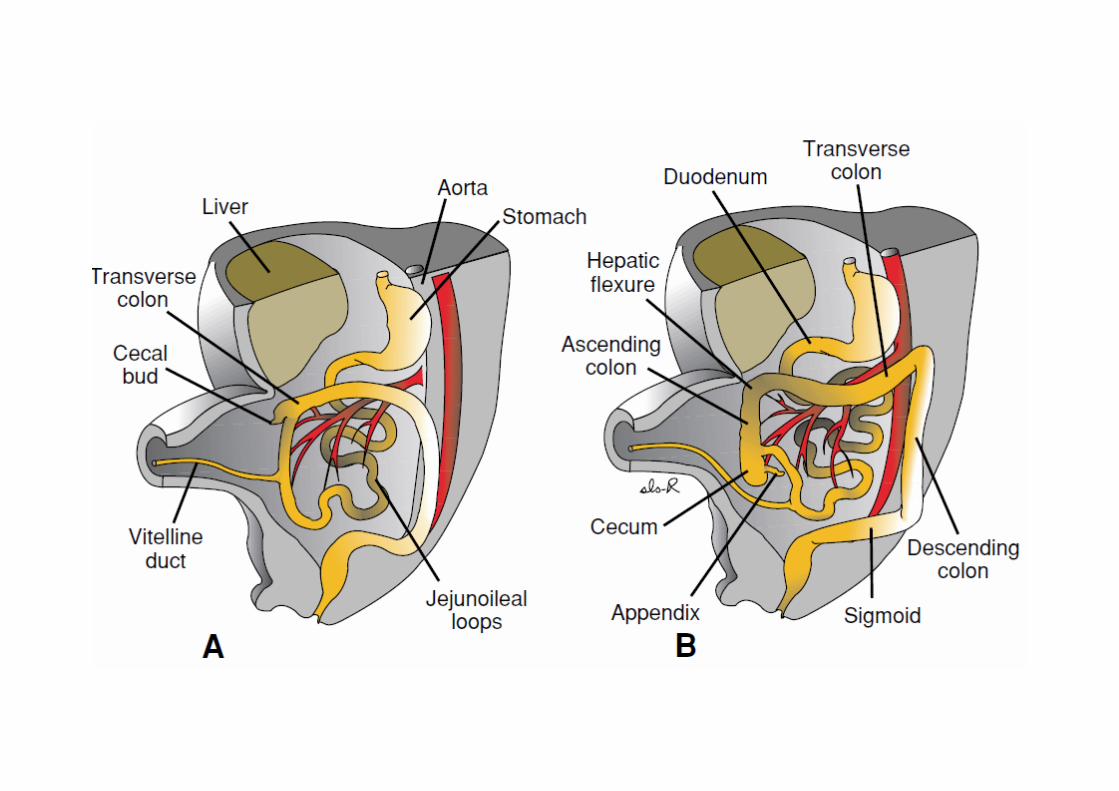
• Ist Stage: Herniation. Prearterial and postarterial segments. Caecal bud
• IInd stage: Reduction. Duodenum crosses behind artery; transverse colon in front; caecum in the right side; intestine from left upper to right lower segments of abdomen.
• IIIrd stage: Fixation. Caecum reaches right illiac fossa. Zygosis occurs in some parts. Mesentry becomes adherent to post. Abdominal wall. Transverse mesocolon.
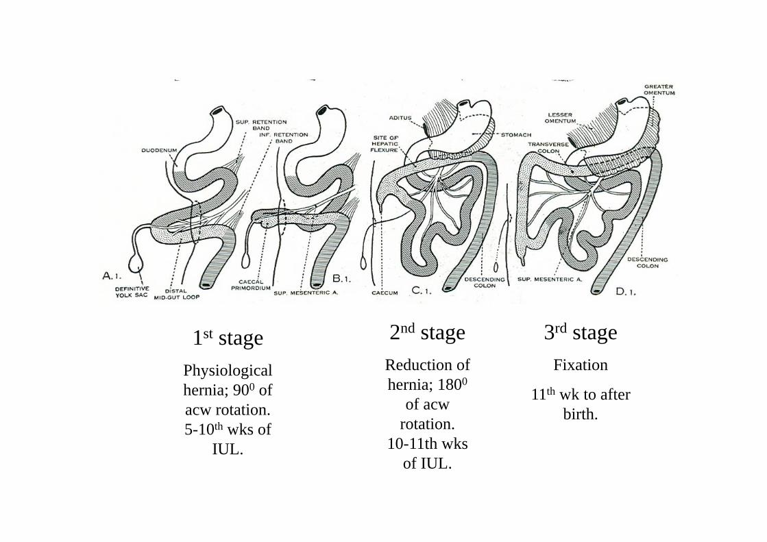
1st stagePhysiological hernia; 900 of acw rotation. 5-10th wks of
IUL.
2nd stageReduction of hernia; 1800
of acw rotation.
10-11th wks of IUL.
3rd stageFixation
11th wk to after birth.
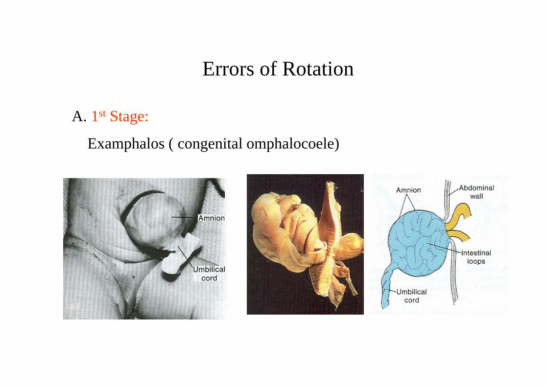
Errors of Rotation
A. 1st Stage:
Examphalos ( congenital omphalocoele)
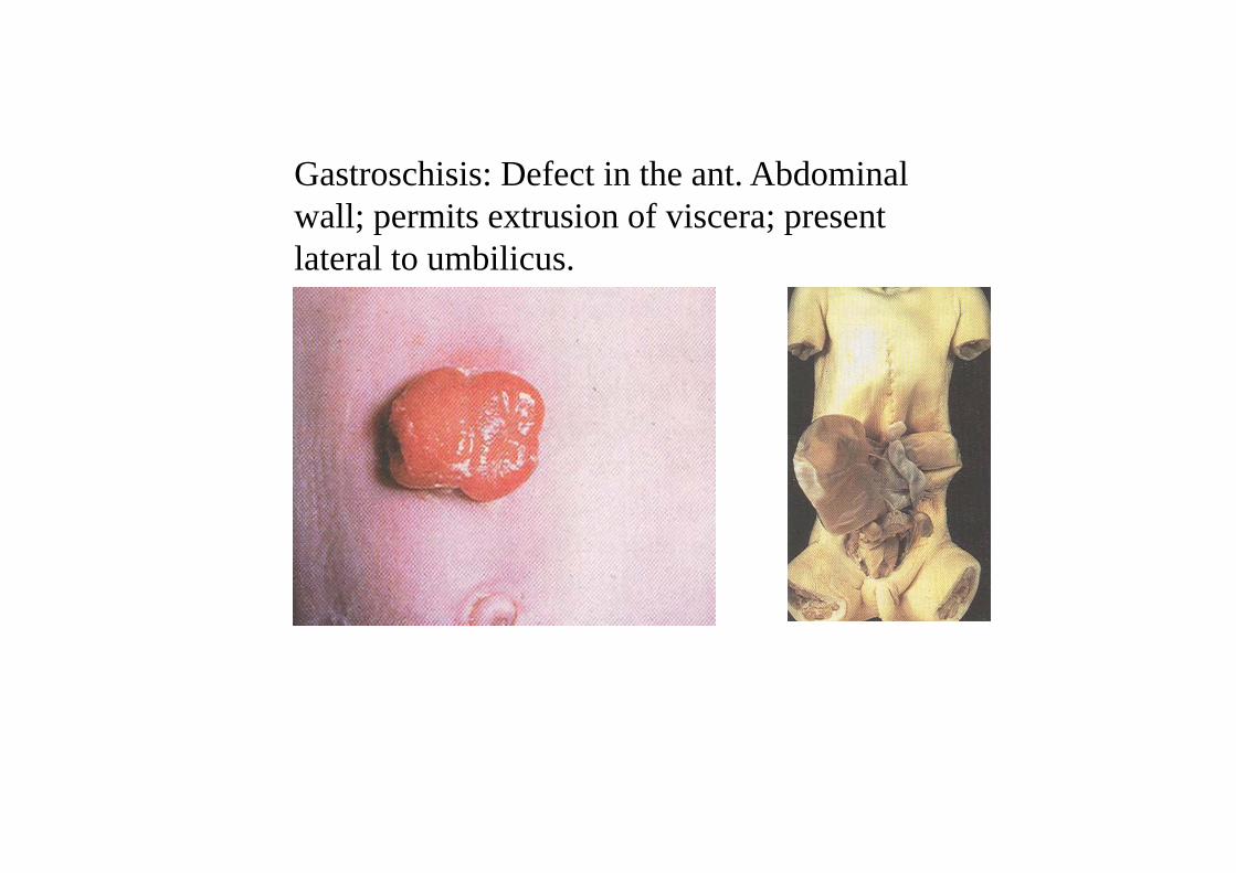
Gastroschisis: Defect in the ant. Abdominal wall; permits extrusion of viscera; present lateral to umbilicus.
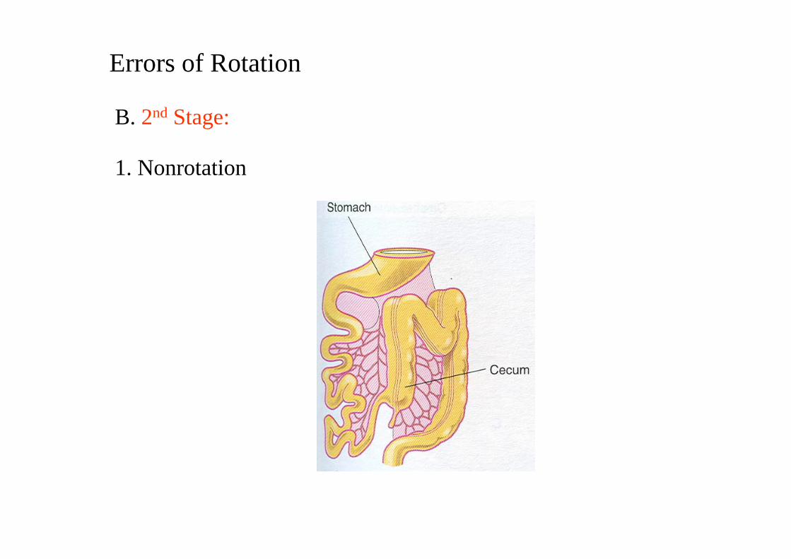
Errors of Rotation
B. 2nd Stage:
1. Nonrotation
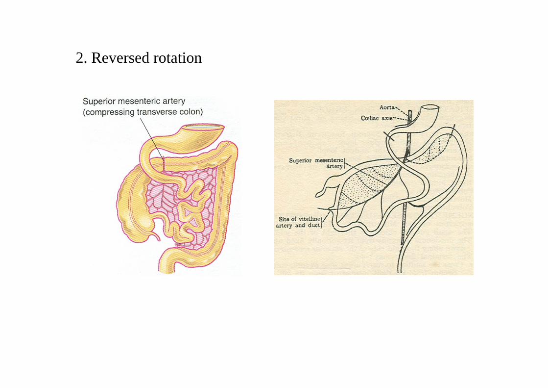
2. Reversed rotation
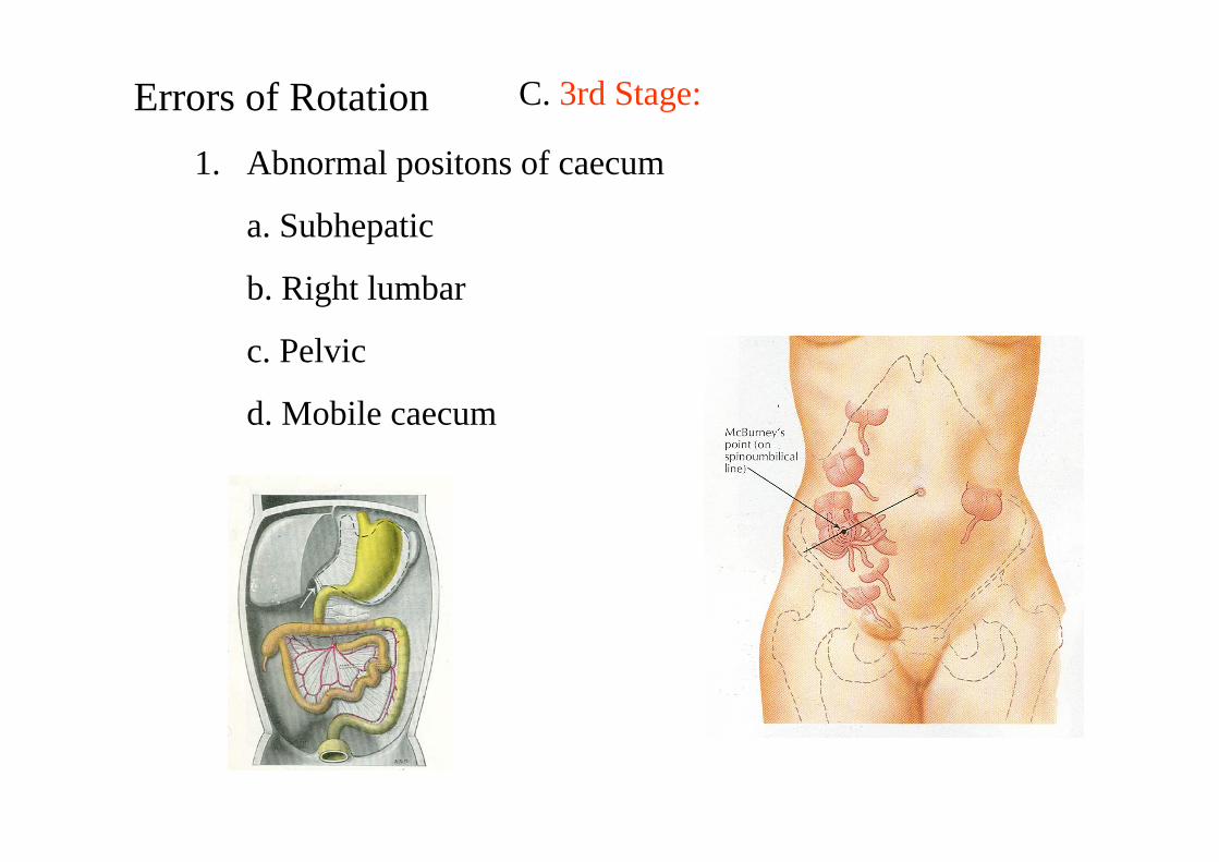
Errors of Rotation1. Abnormal positons of caecum
a. Subhepatic
b. Right lumbar
c. Pelvic
d. Mobile caecum
C. 3rd Stage:
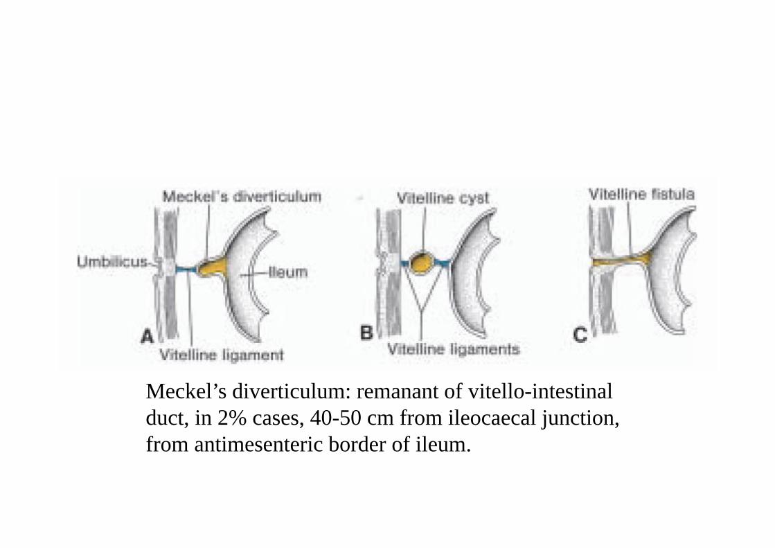
Meckel’s diverticulum: remanant of vitello-intestinal duct, in 2% cases, 40-50 cm from ileocaecal junction, from antimesenteric border of ileum.
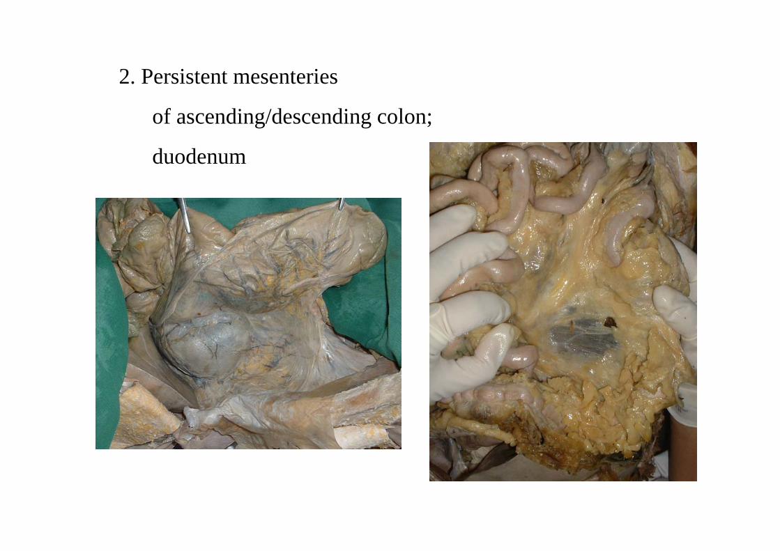
2. Persistent mesenteries
of ascending/descending colon;
duodenum
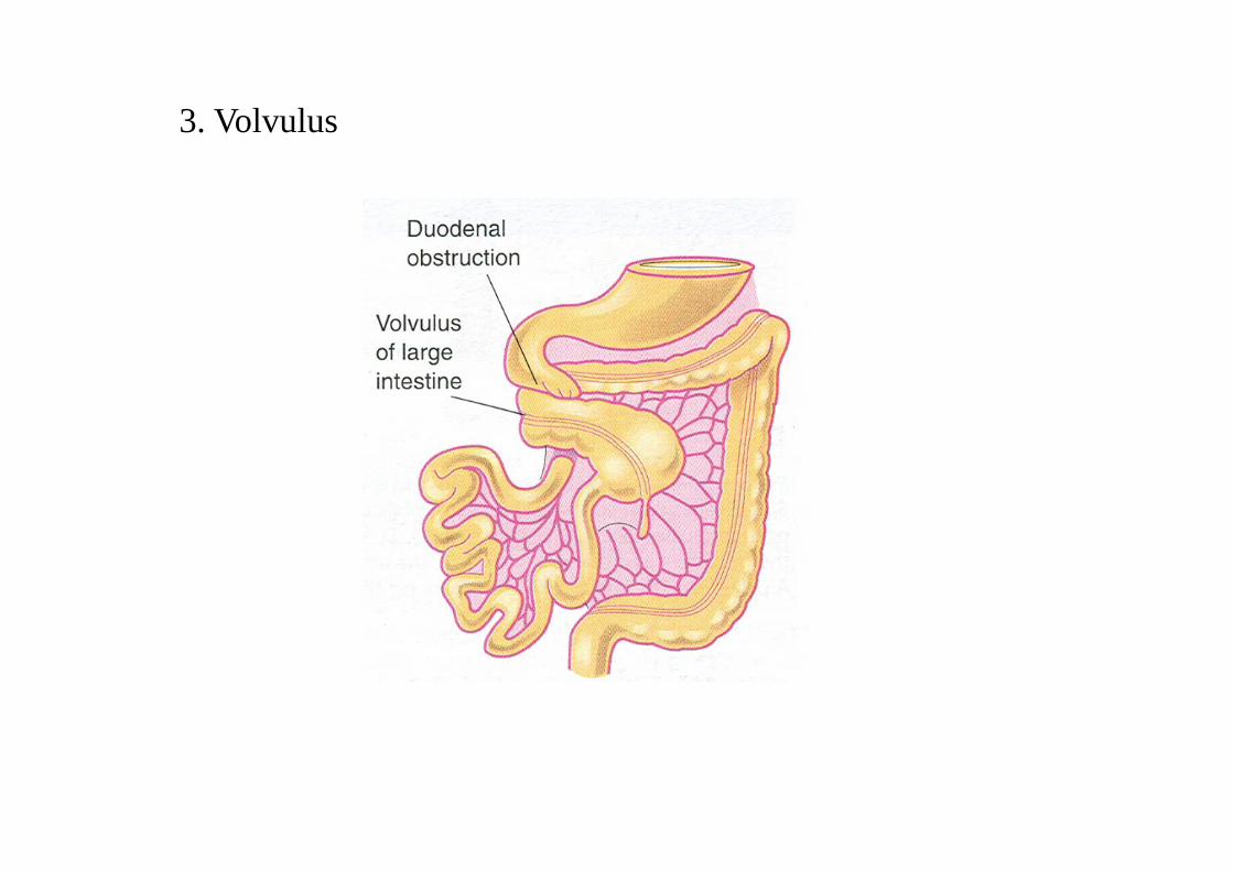
3. Volvulus
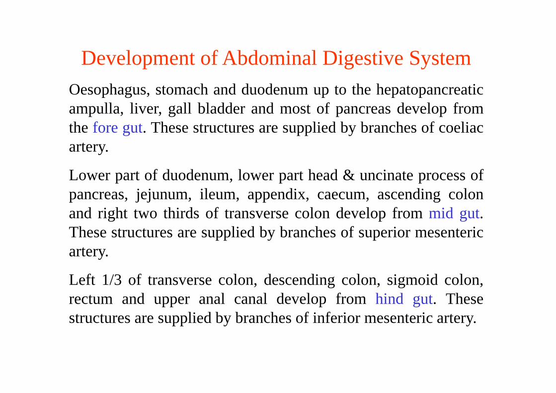
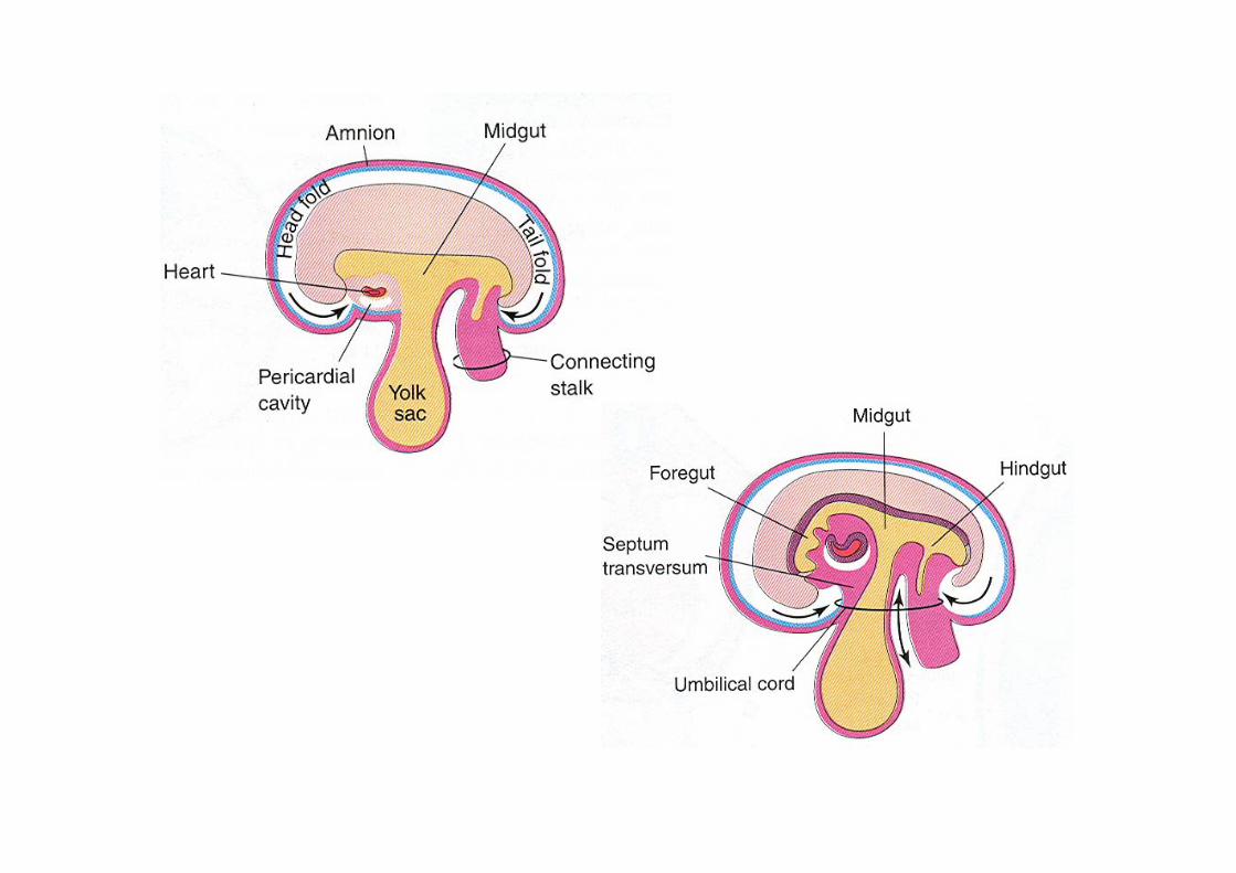
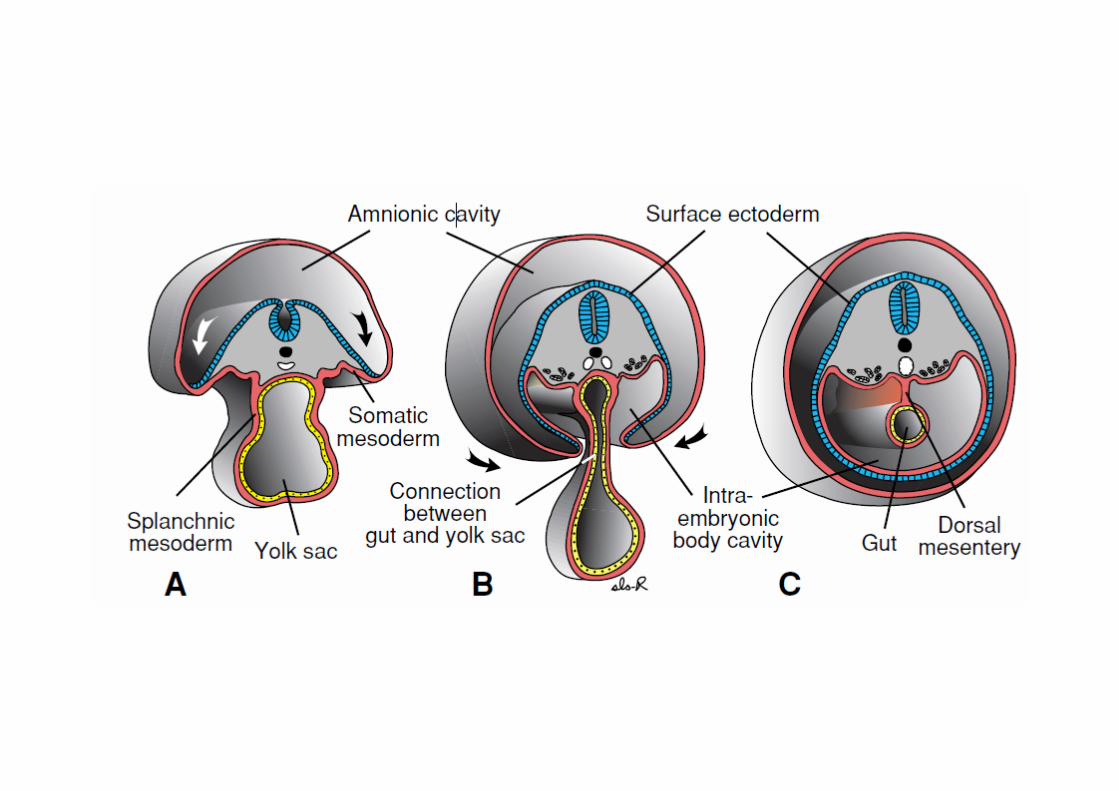
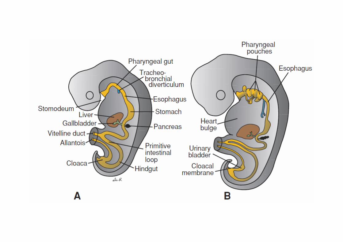
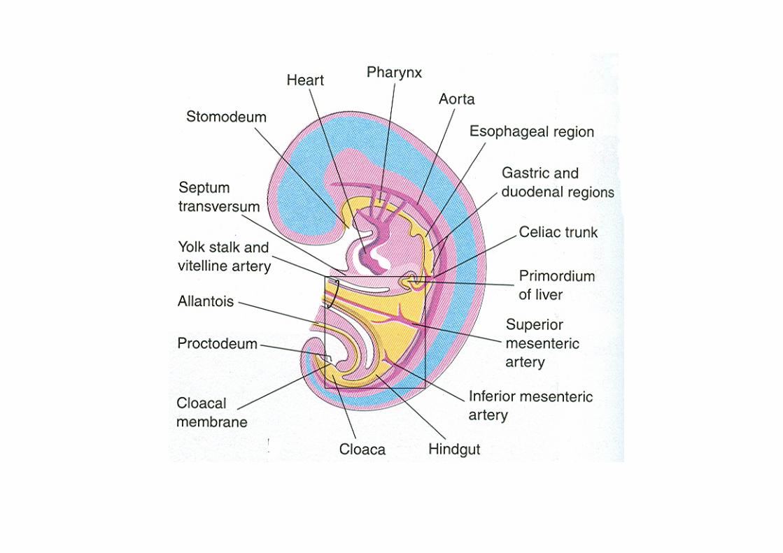
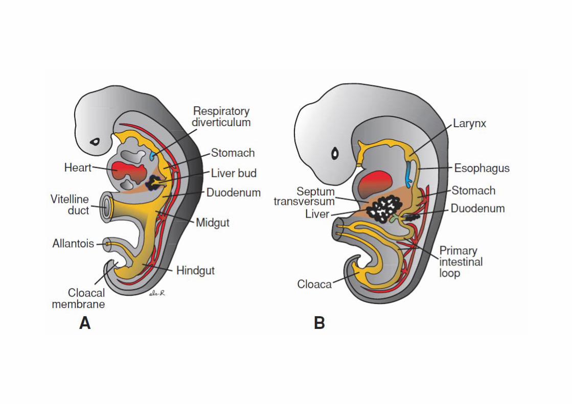
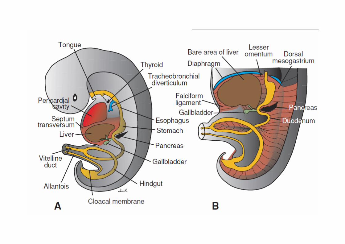
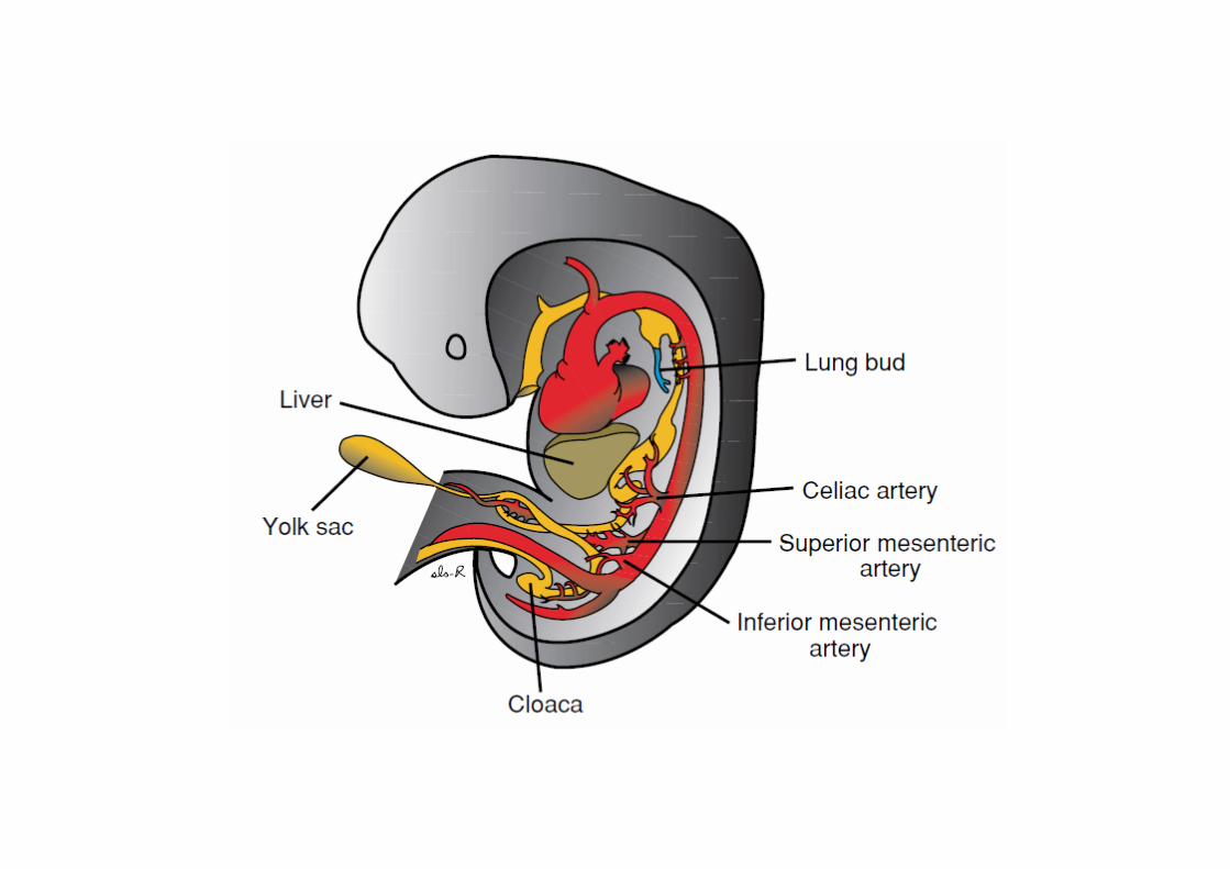
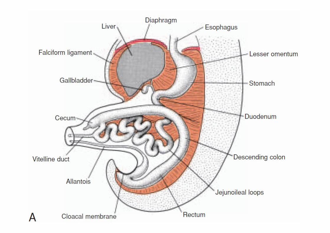
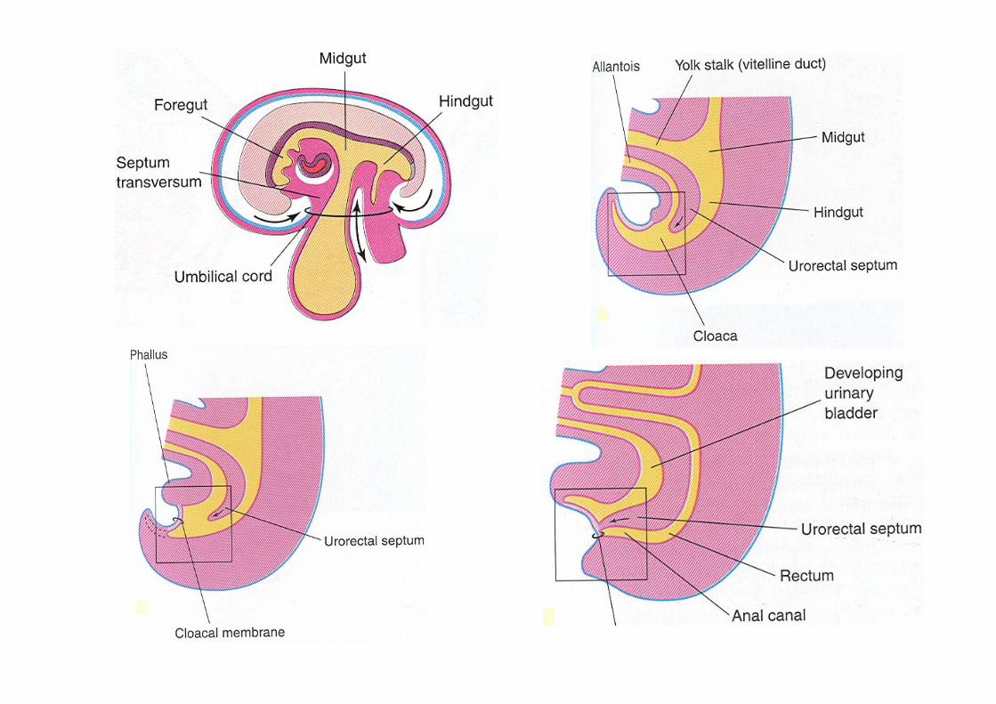
Development of Abdominal Digestive SystemOesophagus, stomach and duodenum up to the hepatopancreaticampulla, liver, gall bladder and most of pancreas develop fromthe fore gut. These structures are supplied by branches of coeliacartery.
Lower part of duodenum, lower part head & uncinate process ofpancreas, jejunum, ileum, appendix, caecum, ascending colonand right two thirds of transverse colon develop from mid gut.These structures are supplied by branches of superior mesentericartery.
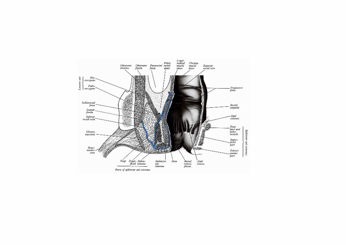
Left 1/3 of transverse colon, descending colon, sigmoid colon,rectum and upper anal canal develop from hind gut. Thesestructures are supplied by branches of inferior mesenteric artery.
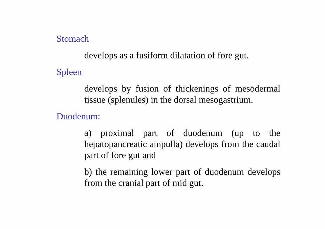
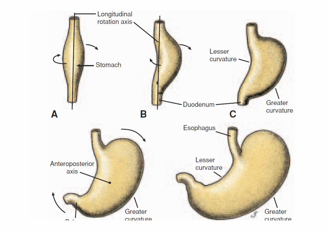
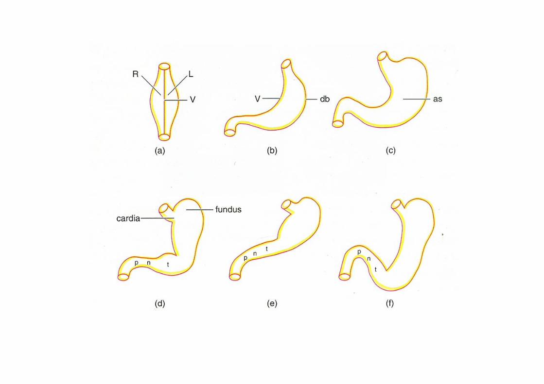
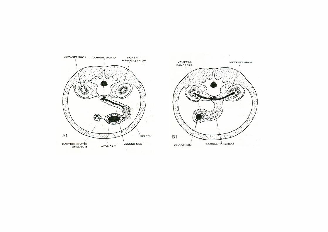
Stomach
develops as a fusiform dilatation of fore gut.
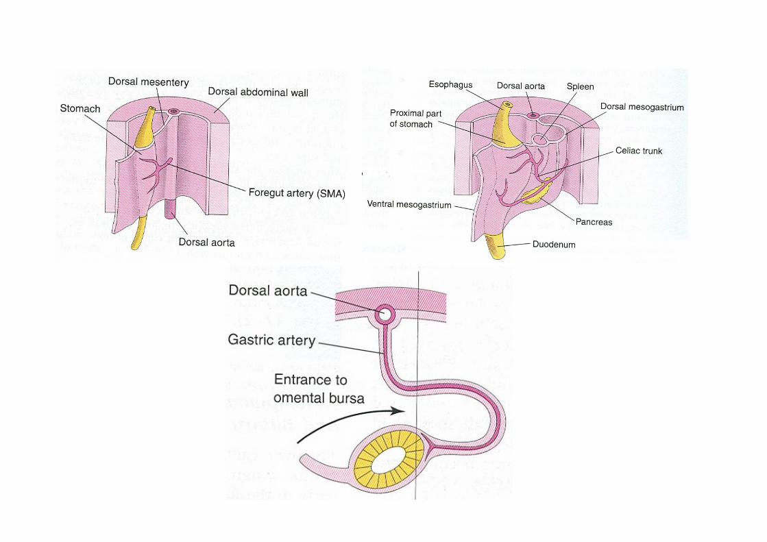
Spleen
develops by fusion of thickenings of mesodermaltissue (splenules) in the dorsal mesogastrium.
Duodenum:
a) proximal part of duodenum (up to thehepatopancreatic ampulla) develops from the caudalpart of fore gut and
b) the remaining lower part of duodenum developsfrom the cranial part of mid gut.
Caecum
develops from a conical caecal bud which arises from the post arterial segment of mid gut.
The apical part of the caecal bud forms the appendix.
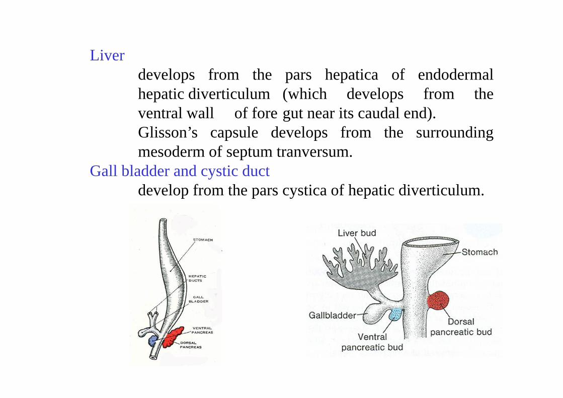
Liverdevelops from the pars hepatica of endodermalhepatic diverticulum (which develops from theventral wall of fore gut near its caudal end).Glisson’s capsule develops from the surroundingmesoderm of septum tranversum.
Gall bladder and cystic ductdevelop from the pars cystica of hepatic diverticulum.
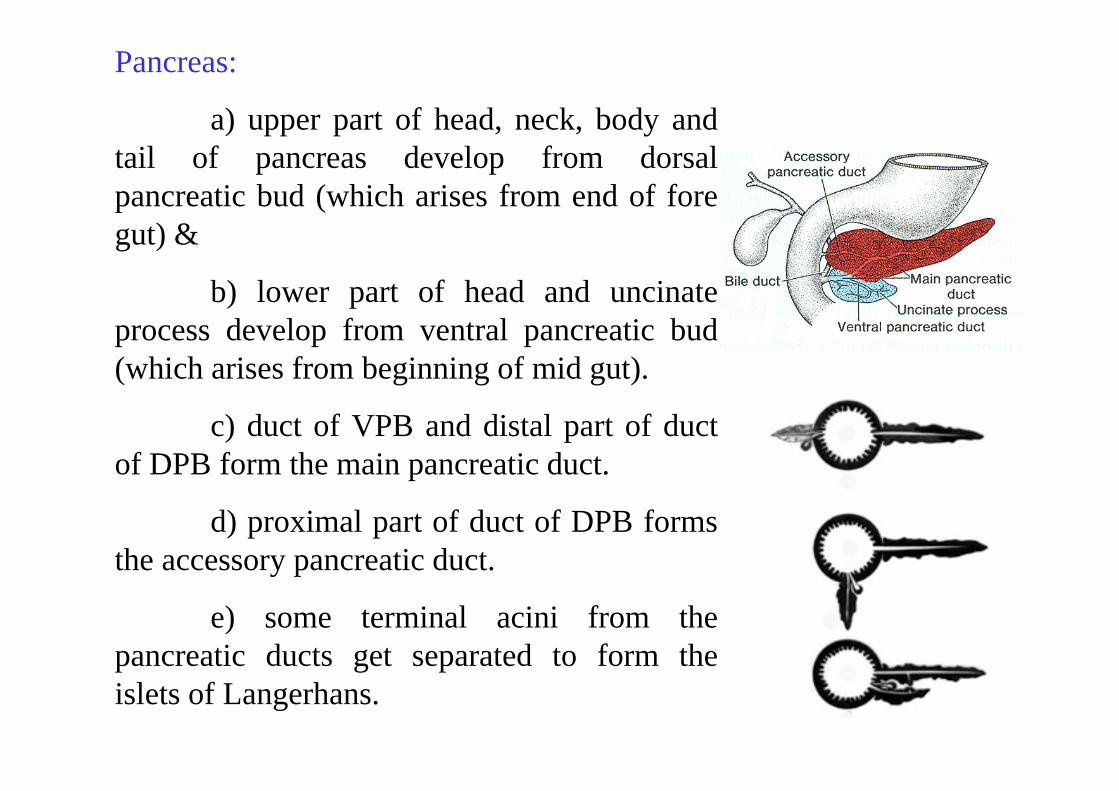
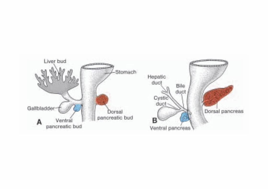
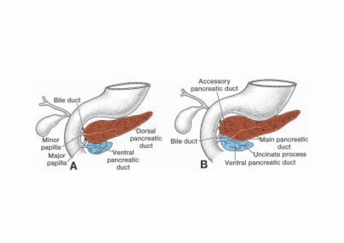
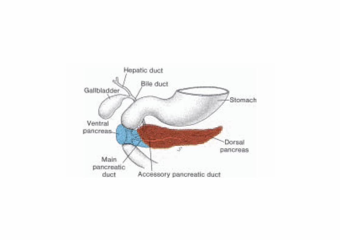
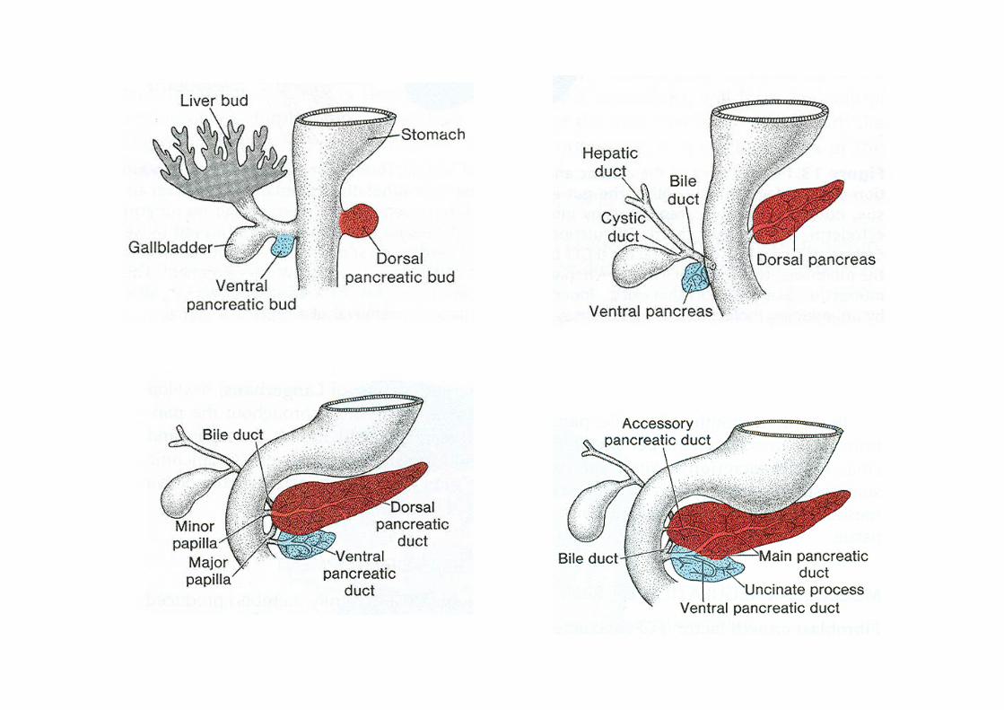
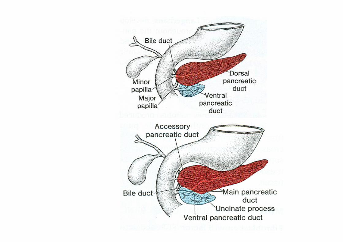
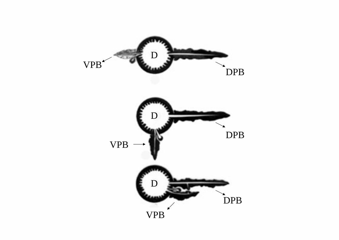
Pancreas:
a) upper part of head, neck, body andtail of pancreas develop from dorsalpancreatic bud (which arises from end of foregut) &
b) lower part of head and uncinateprocess develop from ventral pancreatic bud(which arises from beginning of mid gut).
c) duct of VPB and distal part of ductof DPB form the main pancreatic duct.
d) proximal part of duct of DPB formsthe accessory pancreatic duct.
e) some terminal acini from thepancreatic ducts get separated to form theislets of Langerhans.
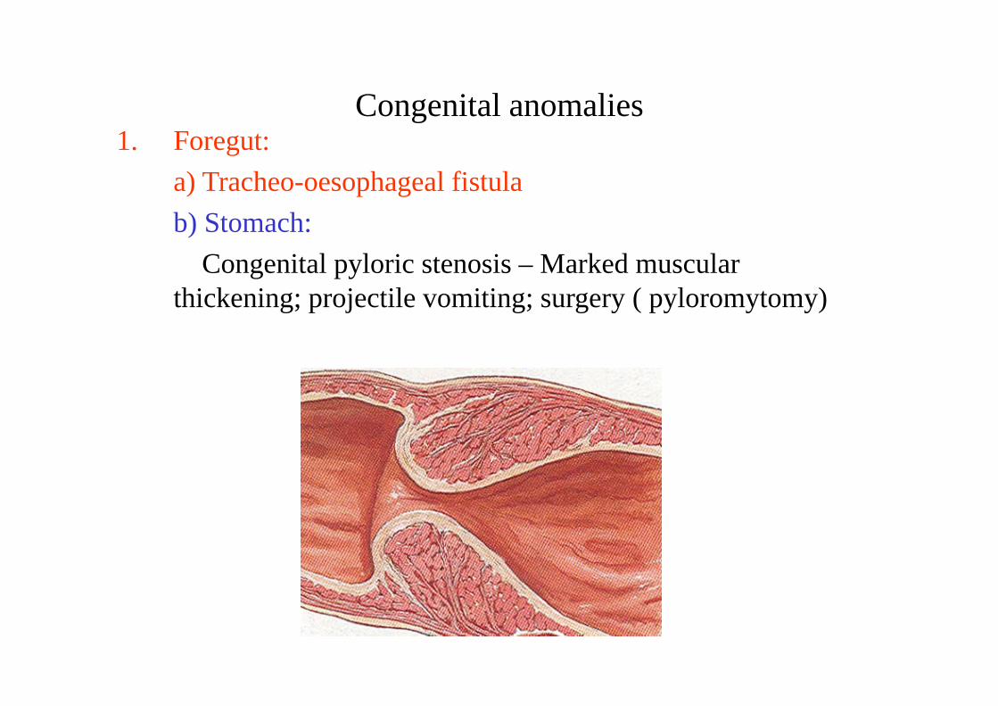
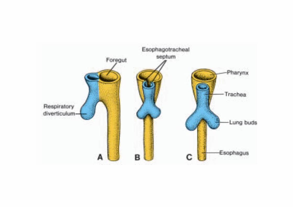
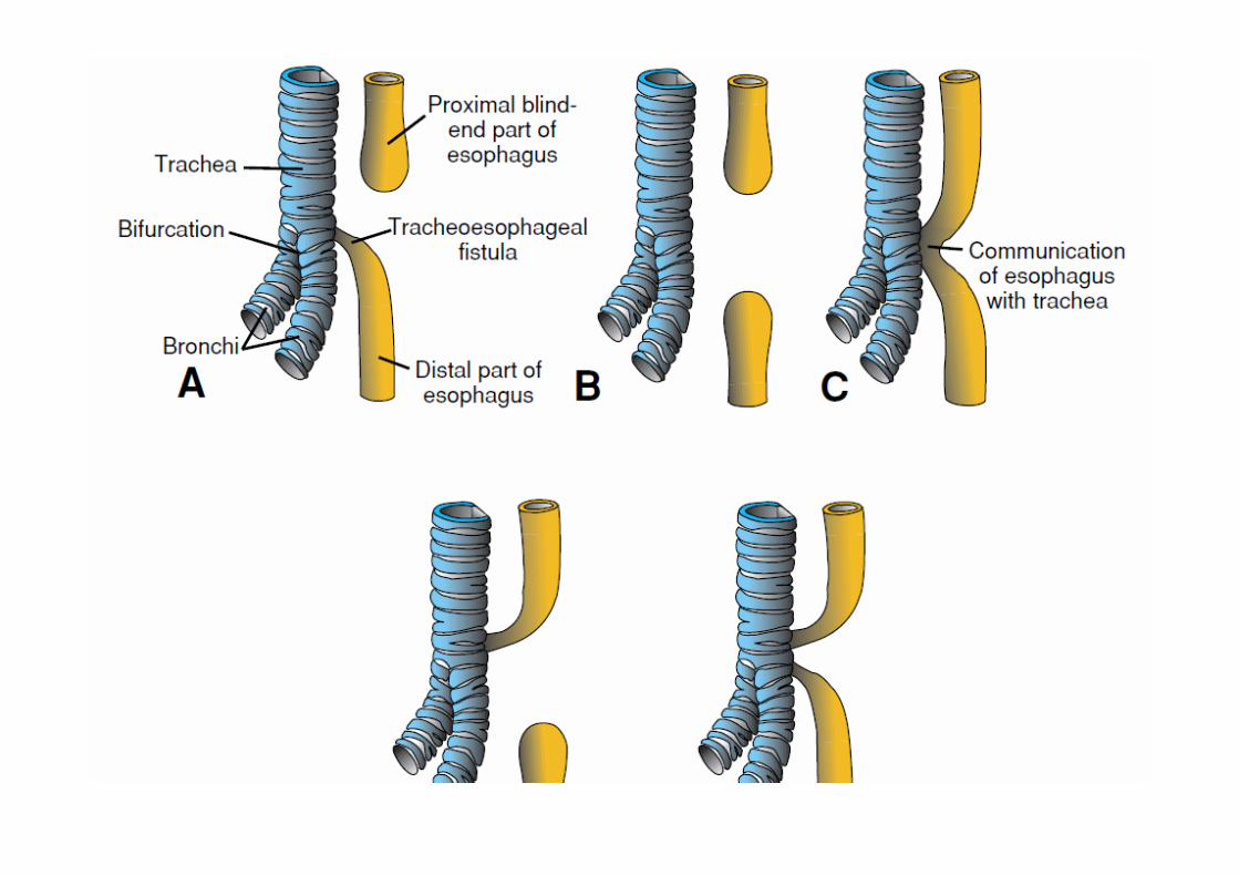
Congenital anomalies1. Foregut:
a) Tracheo-oesophageal fistulab) Stomach:
Congenital pyloric stenosis – Marked muscular thickening; projectile vomiting; surgery ( pyloromytomy)
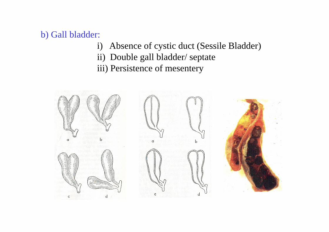
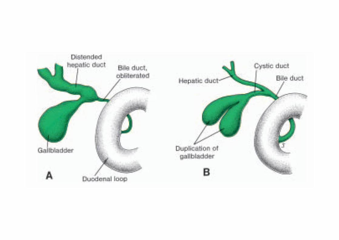
b) Gall bladder:i) Absence of cystic duct (Sessile Bladder)ii) Double gall bladder/ septateiii) Persistence of mesentery
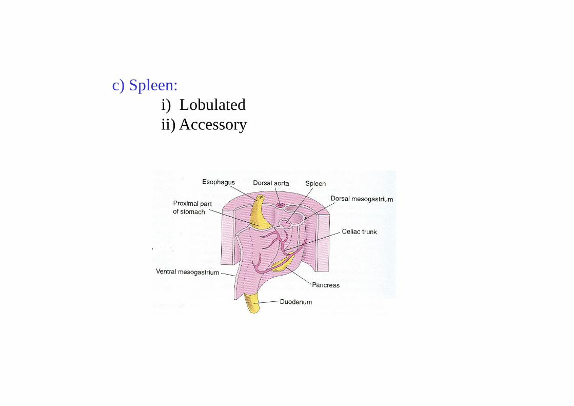
c) Spleen:i) Lobulatedii) Accessory
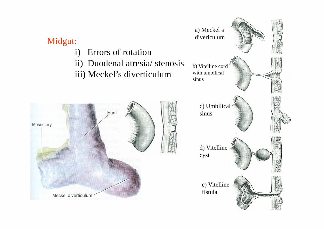
Midgut:i) Errors of rotationii) Duodenal atresia/ stenosisiii) Meckel’s diverticulum
a) Meckel’s divericulum
b) Vitelline cord with umbilical sinus
c) Umbilical sinus
d) Vitelline cyst
e) Vitelline fistula
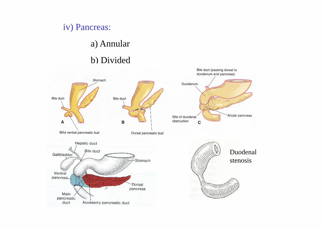
iv) Pancreas:
a) Annular
b) Divided
Duodenal stenosis
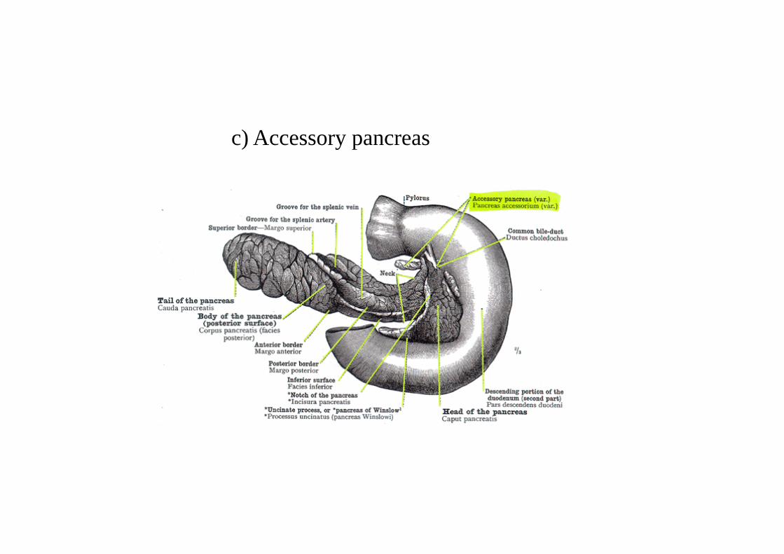
c) Accessory pancreas
v. Caecum
a) Anomalies of shape
i) Conical (Infantile: 2%)
ii) Quadrate (3%)
iii) Hyper position of appendix (4-5%)
b) Anomalies of position
(errors of rotation)
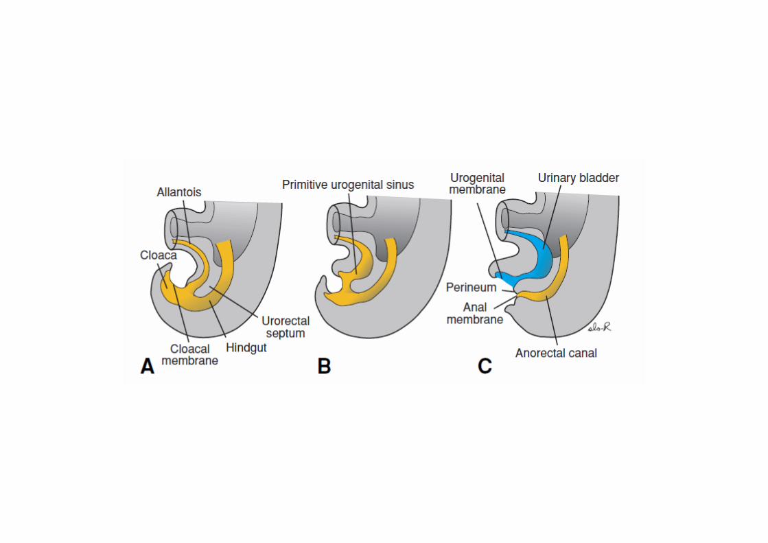
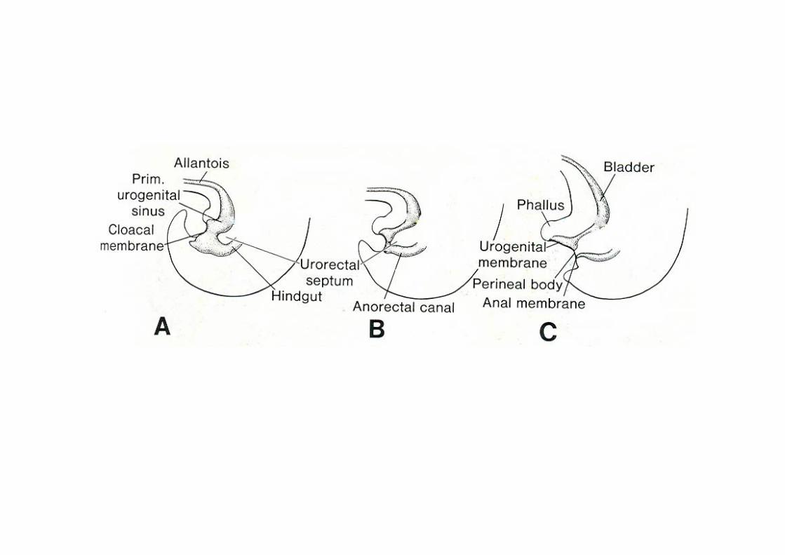
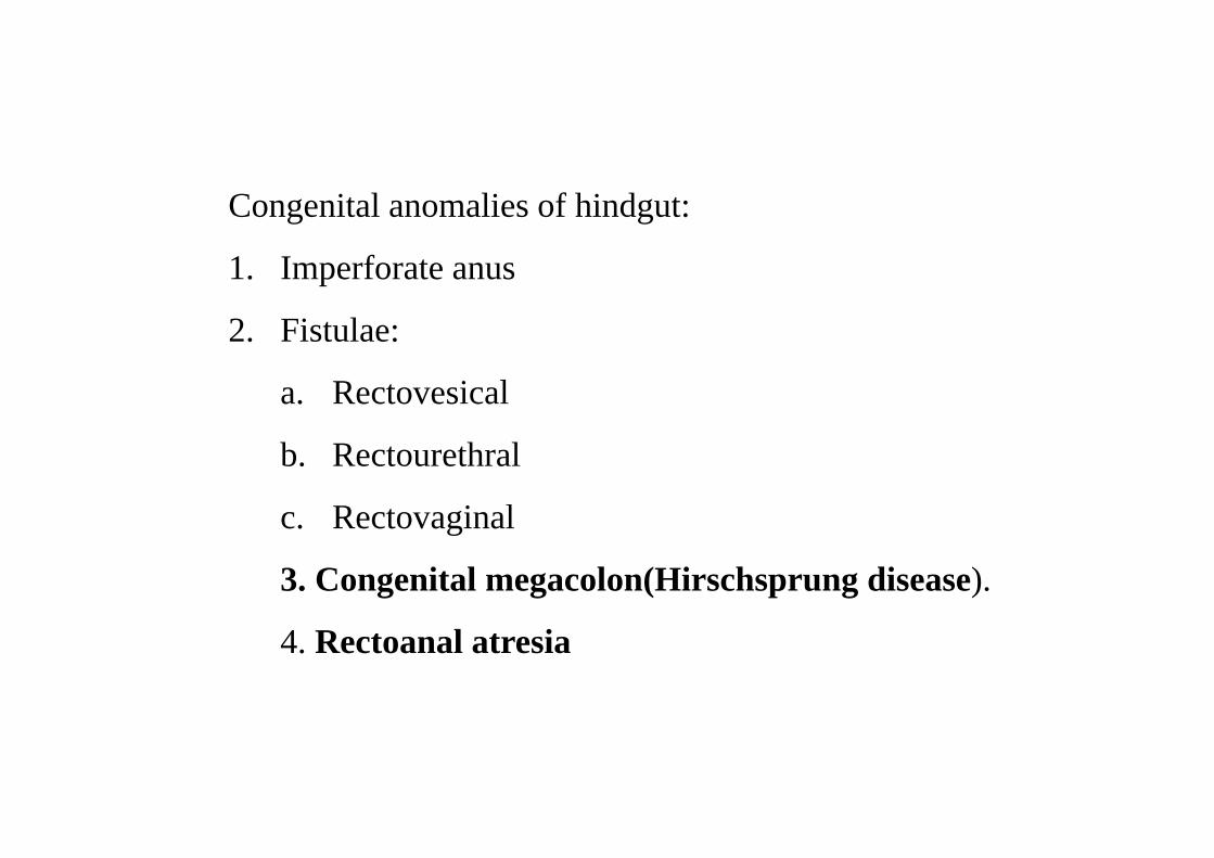
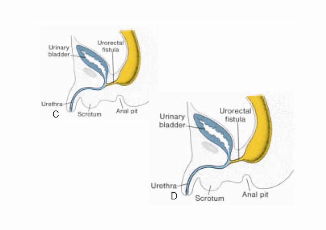
Congenital anomalies of hindgut:
1. Imperforate anus
2. Fistulae:
a. Rectovesical
b. Rectourethral
c. Rectovaginal
3. Congenital megacolon(Hirschsprung disease).
4. Rectoanal atresia
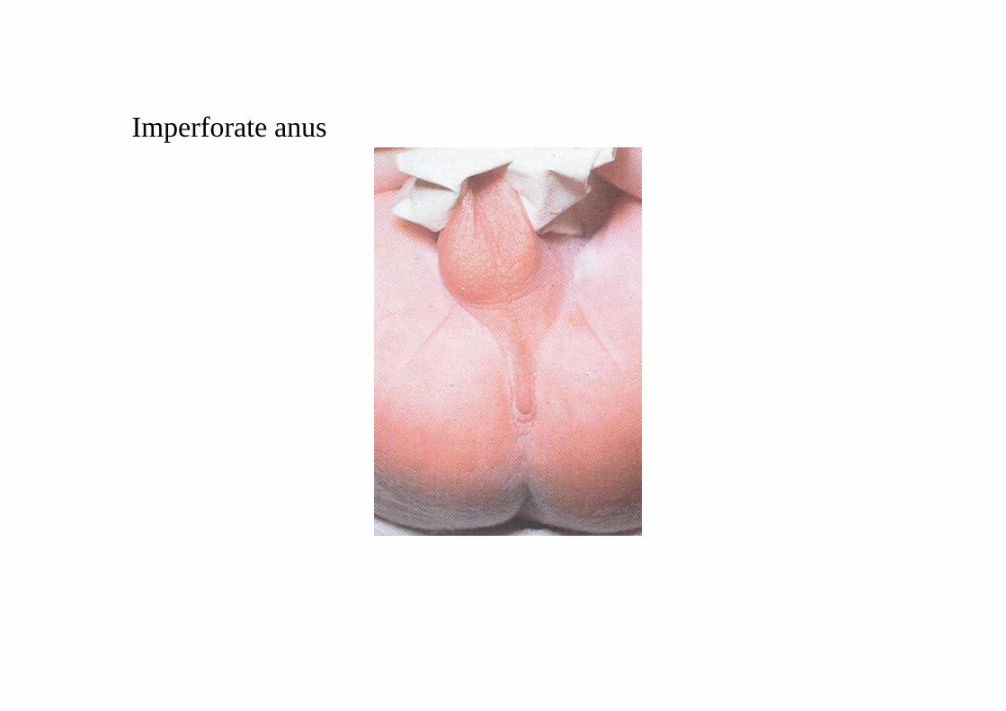
Imperforate anus


![Introduction à Git - [Groupe Calcul]calcul.math.cnrs.fr/IMG/pdf/git.pdf · Introduction Premièrescommandes Branches Synchronisation Contribueràunprojet Applications Conclusion](https://img.pdfslide.net/doc/110x75/5a78fbf97f8b9a68148d8a81/introduction-git-groupe-calcul-premirescommandes-branches-synchronisation-contribuerunprojet.jpg)