Embed Size (px)
Citation preview
Università degli Studi di Padova Dipartimento di Scienze Chimiche
Dottorato di Ricerca in Scienze Molecolari indirizzo Scienze Chimiche
Université Louis Pasteur de Strasbourg Faculté de Chimie
Doctorat en Sciences Chimiques
Ph.D. Thesis
Development of novel helical nucleopeptides
for applications as nucleic acid modulators Tutor: Prof. Fernando Formaggio (Università degli studi di Padova)
Cotutor: Dr. Alberto Bianco (ICT-IBMC. UPR 9021 CNRS, Strasbourg)
Ph.D. student: Piero Geotti-Bianchini
2th
February 2009
Referees:
Prof. Paolo Maria Scrimin, Dipartimento di Scienze Chimiche, Padova.
Prof. Nicolas Winssinger, Institut de Science et d'Ingénierie Supramoléculaires, Strasbourg.
Prof. Jean-Alain Fehrentz, Institut de Biomolécules Max Mousseron, Montpellier, France.
Prof. Roberto Corradini, Dipartimento di Chimica Organica e Industriale, Università di Parma, Italia.
Coordinator of the Doctoral School in Padova: Prof. Maurizio Casarin


Università degli Studi di Padova Dipartimento di Scienze Chimiche
Dottorato di Ricerca in Scienze Molecolari indirizzo Scienze Chimiche
Université Louis Pasteur de Strasbourg Faculté de Chimie
Doctorat en Sciences Chimiques
Ph.D. Thesis
Development of novel helical nucleopeptides
for applications as nucleic acid modulators
Tutor: Prof. Fernando Formaggio (Università degli studi di Padova)
Cotutor: Dr. Alberto Bianco (ICT-IBMC. UPR 9021 CNRS, Strasbourg)
Ph.D. student: Piero Geotti-Bianchini
2th
February 2009
Referees:
Prof. Paolo Maria Scrimin, Dipartimento di Scienze Chimiche, Padova.
Prof. Nicolas Winssinger, Institut de Science et d'Ingénierie Supramoléculaires, Strasbourg.
Prof. Jean-Alain Fehrentz, Institut de Biomolécules Max Mousseron, Montpellier, France.
Prof. Roberto Corradini, Dipartimento di Chimica Organica e Industriale, Università di Parma, Italia.
Coordinator of the Doctoral School in Padova: Prof. Maurizio Casarin


To Elisa,
who, by making me a better man,
has made me also a better scientist.

Foreword
Mr. Piero Geotti-Bianchini was a member of the European Doctoral College of the Universities
of Strasbourg during the preparatio of his Ph.D, from 2006 to 2008, class name Virginia Woolf.
He has benefited from specific financial supports offered by the College and, along with his
mainstream research, has followed a special course on topics of general European interest
presented by international experts.
This Ph.D research project has been led with the collaboration of two universities: the
Università di Padova, Italy and the Université Louis Pasteur in Strasbourg, France.


Index i
INDEX
Résumé ......................................................................................................................... iv
Riassunto...................................................................................................................... vi
Abstract ...................................................................................................................... viii
Abbreviations ................................................................................................................ x
PART A: INTRODUCTION AND AIM OF THE THESIS
1. Introduction and aim of the thesis .......................................................................... 1
1.1 Nucleotides and analogues ................................................................................... 1
1.1.1 Nucleic acids function and structure............................................................ 1
1.1.2 Nucleotide analogues for gene therapy ........................................................ 3
1.1.3 Peptide Nucleic Acids .................................................................................. 7
1.1.4 Nucleoamino acids and nucleopeptides ....................................................... 9
1.2 Peptide structure and C-tetrasubstituted -amino acids .................................... 15
1.2.1 Elements of folded peptide secondary structures....................................... 15
1.2.2 C-tetrasubstituted - amino acids ............................................................ 18
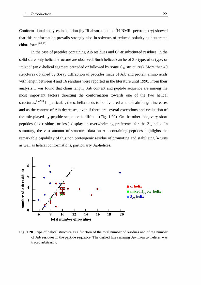
1.2.3 Conformational features of Aib-rich peptides ............................................ 20
1.3 Aim of the thesis ................................................................................................. 23
PART B: RESULTS AND DISCUSSION
2. Synthesis ................................................................................................................ 25
2.1 Synthesis design ................................................................................................. 25
2.1.1 Choice of the amino acids ......................................................................... 25
2.1.2 Choice of the synthetic strategy ................................................................. 26
2.1.3 Choice of suitable protecting groups ......................................................... 27
2.1.3.1 Nucleobase protecting groups .......................................................... 27
2.1.3.2 N- and carboxyl- protecting groups ................................................ 29
2.2 Nucleoamino acid synthesis ................................................................................ 31
2.2.1 Synthesis of N-protected serine -lactone .................................................. 32
2.2.2 Nucleobase alkylation ............................................................................... 33
2.3 Synthesis of rigid nucleopeptides ........................................................................ 36
2.3.1 Coupling methods ..................................................................................... 36
2.3.2 Synthesis of protected nucleotripeptides .................................................... 37
2.3.2 Synthesis of nucleopeptides with a C-terminal lysine ................................. 40
2.3.2 Synthesis of nucleopeptides bearing two nucleobases ................................ 41
Abstract ii
2.4 Synthesis of flexible nucleopeptides ..................................................................... 46
2.4.1 Model nucleopeptide libraries ................................................................... 46
2.4.1.1 Synthetic protocol optimisation ........................................................ 47
2.4.1.2 Incorporation of an Aib residue by SPPS ......................................... 48
2.4.2 Functionalised nucleopeptide libraries ..................................................... 50
2.4.2.1 Trials of cysteine-mediated nucleopeptide dimer formation .............. 55
3. Conformational characterisation ........................................................................... 58
3.1 X-ray diffraction ................................................................................................. 58
3.1.1 Crystal structure of Z-Aib-AlaT-Aib-OtBu ................................................ 58
3.2 IR absorption ...................................................................................................... 62
3.2.1 Nucleopeptide building blocks ................................................................... 63
3.2.1.1 Thymine-based nucleopeptides ......................................................... 63
3.2.1.2 Nucleopeptides containing other nucleobases .................................. 67
3.2.2 Lysine containing nucleopeptides .............................................................. 72
3.2.3 Nucleopeptides containing two nucleobases .............................................. 76
3.3 NMR analysis ...................................................................................................... 78
3.3.1 Protected nucleo-tripeptides ...................................................................... 79
3.3.2 Z-(Aib-AlaT-Aib)2-OtBu ............................................................................. 84
3.3.3 Thymine-based nucleo-heptapeptides ........................................................ 90
3.3.3.1 H-(Ala-AlaT-Ala)2-Lys(H)-NH2 ........................................................ 91
3.3.3.2 H-Ala-Alat-Ala-Aib-AlaT-Ala-Lys(H)-NH2 ...................................... 93
3.3.3.3 H-(Aib-AlaT-Aib)2-Lys(H)-NH2 ....................................................... 95
3.4 Circular Dichroism ............................................................................................. 98
3.4.1 Protected rigid nucleopeptides .................................................................. 99
3.4.2 Z-(Aib-AlaT-Aib)2-OtBu ........................................................................... 102
3.4.3 Lysine containing thymine-based nucleo-heptapeptides ........................... 104
3.4.4 Flexible nucleopeptides ........................................................................... 107
4. Pairing properties ................................................................................................. 112
4.1 Self-pairing properties ...................................................................................... 112
4.2 Selective Watson-Crick pairing ......................................................................... 114
4.2.1 Surface plasmon resonance measurements .............................................. 116
5. Biological tests...................................................................................................... 125
5.1 Apoptoticity tests ........................................................................................ 125
Index iii
5.2 Cytotoxicity evaluation ............................................................................... 126
5.3 Cell penetration experiments ...................................................................... 129
5.3.1 Mechanistic data on cell uptake ........................................................ 131
5.4 Stability tests .............................................................................................. 134
PART C: EXPERIMENTAL SECTION
6. Experimental section ........................................................................................... 136
6.1 Materials and methods ..................................................................................... 136
6.1.1 Reagents and solvents ............................................................................. 136
6.1.2 Instruments and methods ......................................................................... 139
6.2 Synthesis and characterization ......................................................................... 145
6.2.1 Solution phase synthesis .......................................................................... 145
6.2.1.1 Synthesis of protected nucleobases ................................................. 145
6.2.1.2 Synthesis of amino acid derivatives ................................................ 148
6.2.1.3 Synthesis of nucleoamino acids ...................................................... 152
6.2.1.4 Synthesis of model peptides and peptide building blocks ................ 159
6.2.1.5 Synthesis of nucleoamino acid derivatives and of nucleopeptides ... 162
6.2.2 Solid phase synthesis ............................................................................... 181
6.2.2.1 General methodology and remarks................................................. 181
6.2.2.2 Synthesis of model nucleopeptide libraries .................................... 182
6.2.2.3 Synthesis of functionalised nucleopeptide libraries......................... 184
6.2.2.4 Cysteine-mediated nucleopeptide dimerisation trials ...................... 188
6.3 Pairing and biological tests .............................................................................. 190
6.3.1 Surface plasmon resonance measurements .............................................. 190
6.3.2 DiOC6 test ............................................................................................... 191
6.3.3 MTS cytotoxicity tests .............................................................................. 191
6.3.4 Cell penetration experiments ................................................................... 192
6.3.4.1 Flow cytometric uptake analysis .................................................... 192
6.2.2.3 Epifluorescence and confocal microscopy uptake analysis ............. 192
6.3.5 Nucleopeptide stability tests .................................................................... 193
7. Conclusions ......................................................................................................... 195
8. References ........................................................................................................... 198
Publications and communications derived from the thesis ......................................... 208
Acknowledgements .................................................................................................... 209
Abstract iv
Abstract
Gene therapy aims at the treatment of genetic diseases at molecular level by interactions
with nucleic acids (DNA and RNA). Synthetic oligonucleotides can selectively recognize
complementary sequences and inhibit or modulate gene expression in vitro. However, it is
not possible to use synthetic oligonucleotides for in vivo applications, due to several
serious problems, particularly their rapid enzymatic hydrolysis and their extreme difficulty
to cross cell membranes.
To overcome such limitations, nucleotide analogues, which should have better biological
properties while retaining, or even improving, the affinity and selectivity towards
complementary strands characteristic of the natural nucleotide structures, have been
designed. Nucleopeptides are one of the recently developed classes of analogues; they are
peptides containing nucleoamino acid residues, non proteogenic amino acids carrying
nucleobases at their side chains.
The work presented in this thesis concerns the study of the properties of sequential helical
nucleopeptides containing alanyl-nucleoamino acid residues (AlaB) at i,i+3 positions in
view of applications as nucleic acid modulators. Indeed, if such nucleopeptides assume a
310-helical conformation, side chain nucleobases are aligned along the helical axis and this
might favour cooperative interactions with complementary functionalized strands.
To better evaluate the importance of structuration on the nucleopeptide pairing properties,
both rigid (based on the 310 helix promoting C-tetrasubstituted residue Aib) and flexible
(based in the less structuring proteogenic residue Ala) nucleopeptides have been
synthesized and conformational and biological properties of both kinds of nucleopeptides
have been investigated.
As regards the rigid nucleopeptides, this work reports on:
(I) The synthesis and characterization of protected nucleopeptides, each containing one
of the four DNA nucleobases;
(II) The synthesis of protected hexa-nucleopeptide and of a completely deprotected,
water-soluble hepta-nucleopeptide, both containing two thymines;
(III) The crystal structure of a protected thymine containing nucleo-tripeptide;
(IV) The conformational characterization in solution of the protected nucleo-tripeptides,
of the nucleo-hexapeptide and of the nucleo-heptapeptide.
As regards the flexible nucleopeptides, this work reports on:
Abstract v
(I) The design and solid-phase parallel synthesis of model nucleopeptide libraries, for
synthetic protocol optimization and conformational analysis;
(II) The design and solid-phase parallel synthesis of partially biotinylated or fluorescently
derivatized nucleopeptide libraries containing each of the four DNA nucleobases, for
pairing and biological tests;
(III) The conformational characterization by CD and 2D NMR of a thymine-based model
nucleo-heptapeptide containing two nucleobases, and of an analogue carrying an Aib
residue in the middle of its sequence.
(IV) The surface plasmon resonance analysis of nucleopeptide/nucleopeptide and of
nucleopeptide/nucleotide pairing properties, through immobilization of biotinylated
molecules on functionalized gold chips.
As regards the assessment of the nucleopeptide biological properties, this works reports on:
(I) Cytotoxicity tests carried on a rigid and on variously functionalized flexible thymine-
based nucleopeptides;
(II) Flow-cytometric and microscopy cell penetration experiments carried on fluorescent
and biotinylated thymine-based nucleopeptides, supported by colocalization tests and
by time, concentration, and energy dependence analysis of cell uptake.
(III) Stability tests in mouse serum carried on flexible, mixed, and rigid thymine
containing heptapeptides.
Finally, the whole of the data collected have been evaluated in order to give a global
judgement on the properties of the nucleopeptides studied as precursors for the
development of nucleic acid modulators.
Résumé vi
Résumé
La thérapie génique vise à traiter des maladies en agissant au niveau moléculaire sur les
acides nucléiques (ADN et ARN). Des oligonucléotides synthétiques peuvent reconnaître
des séquences complémentaires et ainsi inhiber ou moduler leur expression in vitro.
Pourtant, l‟application d‟oligonucléotides synthétiques in vivo n‟est pas possible, étant
donné leur dégradation rapide par les nucléases et leur faible capacité à pénétrer la
membrane cellulaire. Pour remédier à ces problèmes, des analogues des nucléotides doués
des meilleures propriétés biologiques, et gardant, ou même améliorant, l‟affinité et la
sélectivité caractéristiques des nucléotides naturels vis-à-vis des séquences
complémentaires ont été proposés.
Une classe d‟analogues récemment développée est constituée des nucléo-peptides, peptides
qui ont dans leur séquence des nucléo-acides aminés, contenant sur les chaines latérales les
bases azotées caractéristiques des acides nucléiques.
Le projet de recherche avait l‟objectif d‟étudier les propriétés des nucléo-peptides
séquentiels ayant des alanyl nucléo-acides aminés (AlaB), en position i,i+3, en vue
d‟applications comme modulateurs d‟acides nucléiques. En fait, si ces nucléo-peptides
adoptent une structure hélicoïdale de type 310, les nucléobases sur les chaines latérales sont
alignées et forment un côté fonctionnalisé de l‟hélice, de manière à interagir d‟une façon
coopérative avec des séquences complémentaires.
Pour mieux étudier les effets de la stabilité conformationelle sur les propriétés de
reconnaissance des nucléo-peptides, soit des nucléo-peptides rigides (basés sur l‟acide
aminé C-tetrasubstitué Aib, promoteur des structures hélicoïdales 310), soit des nucléo-
peptides flexibles (basés sur l‟acide aminé Ala, moins structurant), ont été synthétisés et
leur propriétés conformationnelles et biologiques ont été étudiées.
Par rapport aux nucléo-peptides rigides, cette thèse présente:
(I) La synthèse et caractérisation de nucléo-peptides protégés contenants chacun une des
quatre bases de l‟ADN ;
(II) La synthèse et caractérisation d‟un nucléo-hexapeptide protégé et d‟un nucléo-
heptapeptide complètement déprotégé renfermant deux thymines ;
(III) La structure cristalline d‟un nucléo-tripeptide protégé basé sur la thymine ;
(IV) La caractérisation conformationnelle en solution des nucléo-tripeptides protégés, du
nucléo-hexapeptide et du nucléo-heptapeptide.
Résumé vii
Par rapport aux nucléo-peptides flexibles, cette thèse présente :
(I) La conception et la synthèse parallèle sur phase solide de librairies de nucléo-
peptides modèles, pour l‟optimisation du protocole synthétique et pour les analyses
conformationnelles;
(II) La conception et la synthèse parallèle sur phase solide de librairies de nucléo-
peptides partiellement biotinilés ou dérivatisés avec un fluorophore, renfermant
chacune des quatre bases de l‟ADN, pour des études de reconnaissance et
biologiques;
(III) La caractérisation conformationnelle DC et RMN 2D d‟un nucléo-heptapeptide
modèle basé sur la thymine, et d‟un analogue avec un résidu d‟Aib au centre de sa
séquence;
(IV) L‟étude de la reconnaissance nucléo-peptide/nucléo-peptide et nucléo-peptide/
nucléotide mené par résonance plasmonique de surface, grâce à l‟immobilisation de
molécules biotinilées sur des puces d‟or fonctionnalisées.
Par rapport à l‟évaluation des propriétés biologiques des nucléo-peptides, cette thèse
présente:
(I) Des études de cytotoxicité sur un nucléo-peptide rigide et sur plusieurs nucléo-
peptides flexibles fonctionnalisés;
(II) Des essais de pénétration cellulaire suivis par cytométrie en flux et par microscopie,
menés sur les nucléo-peptides biotinilés et fluorescents basés sur la thymine,
supportés par des essais de co-localisation et par des mesures de pénétration en
fonction du temps, de la concentration et de l‟énergie cellulaire;
(III) Des mesures de stabilité en sérum murin menés sur les nucléo-heptapeptides flexible,
mixte et rigide, renfermant des thymines.
Enfin, l‟ensemble des données collectées a été évalué dans sa totalité afin de donner un
jugement global sur les propriétés des nucléo-peptides étudiés en tant que précurseurs pour
le développement de modulateurs d‟acides nucléiques.
Riassunto viii
Riassunto
La terapia genica mira a curare determinate malattie operando a livello molecolare sugli
acidi nucleici (DNA et RNA). Oligonucleotidi sintetici possono riconoscere selettivamente
sequenze complementari ed inibire o modulare l‟espressione genica in vitro. Non è tuttavia
possible utilizzare oligonucleotidi sintetici per applicazioni in vivo, a causa di svariati gravi
problemi, in particolare la loro rapida degradazione enzimatica e la grande difficoltà di
penetrazione attraverso le membrane cellulari. Per superare tali limiti sono stati proposti
analoghi di nucleotidi, che dovrebbero possedere migliori proprietà biologiche,
mantenendo, o perfino migliorando, l‟affinità e la selettività verso sequenze complementari
caratteristiche delle strutture nucleotidiche naturali.
Una tra le classi di analoghi sviluppate di recente sono i nucleopeptidi, peptidi contenenti
nucleoamminoacidi, ossia amminoacidi non proteogenici che portano una nucleobase in
catena laterale.
Il presente lavoro di tesi aveva l‟obiettivo di studiare le proprietà di nucleopeptidi
sequenziali in conformazione elicoidale contenenti alanil-nucleoamminoacidi (AlaB) in
posizione i,i+3 per applicazioni come modulatori di acidi nucleici. Infatti, se tali
nucleopeptidi assumono una conformazione elicoidale 310, le nucleobasi in catena laterale
si trovano allineate lungo l‟elica e ciò potrebbe favorire interazioni cooperative con
sequenze complementari.
Per meglio valutare l‟importanza della strutturazione sulle proprietà di riconoscimento dei
nucleopeptidi, si sono sintetizzati sia nucleopeptidi rigidi (basati sull‟amminoacido C-
tetrasostituito Aib, che favorisce l‟assunzione di strutture elicoidali 310), sia analoghi
flessibili (basati sull‟amminoacido proteogenico Ala, meno strutturante), e ne sono state
studiate sia le proprietà conformazionali che biologiche.
Riguardo ai nucleopeptidi rigidi sono descritte:
(I) La sintesi e la caratterizzazione di nucleopeptidi protetti contententi ciascuno una
delle quattro nucleobasi del DNA;
(II) La sintesi e la caratterizzazione di un nucleoesapeptide protetto e di un
nucleoeptapeptide idrosolubile completamente deprotetto, entrambi contenenti due
timine;
(III) La struttura cristallina di un nucleotripeptide protetto contenente timina;
(IV) La caratterizzazione conformazionale in soluzione dei nucleotripeptidi protetti, del
nucleoesapeptide e del nucleoeptapeptide.
Riassunto ix
Riguardo ai nucleopeptidi flessibili sono descritte:
(I) La progettazione e la sintesi in parallelo su fase solida di librerie di nucleopeptidi
modello per l‟ottimizzazione dei protocolli sintetici e per analisi conformazionali;
(II) La progettazione e la sintesi in parallelo su fase solida di librerie di nucleopeptidi
contenenti ciascuna delle quattro nucleobasi del DNA, parzialmente biotinilati o
derivatizzati con fluorofori, per studi di riconoscimento e biologici;
(III) La caratterizzazione conformazionale CD e 2D NMR di un nucleoeptapeptide
modello contenente due timine e di un analogo con un residuo di Aib al centro della
sequenza;
(IV) L‟analisi per risonanza plasmonica di superficie delle proprietà di riconoscimento tra
nucleopeptidi e tra nucleopeptidi e nucleotidi via immobilizzazione di molecole
biotinilate su lastrine di oro funzionalizzate.
Riguardo alla valutazione delle proprietà biologiche dei nucleopeptidi, sono descritti:
(I) Test di citotossicità svolti su un nucleopeptide rigido e su vari nucleopeptidi flessibili
basati sulla timina;
(II) Misure di citometria in flusso ed analisi di microscopia riguardo alle proprietà di
penetrazione cellulare di nucleopeptidi fluorescenti o biotinilati basati sulla timina,
supportate da prove di colocalizzazione e da un‟analisi della dipendenza da tempo,
concentrazione ed energia della penetrazione cellulare;
(III) Misure di stabilità in siero murino eseguite sugli eptapeptidi rigido, misto e flessibile
contenenti timina.
Infine l‟insieme dei dati raccolti è stato valutato nella sua totalità onde fornire un giudizio
globale sulle proprietà dei nucleopeptidi studiati per lo sviluppo di modulatori di acidi
nucleici.
Abbreviations
x
ABBREVIATIONS
1D = monodimensional
2Cl-Z = 2-chloro-benzyloxycarbonyl
2D = bidimensional
4-MePhOH = p-chresol
Abs = absorbance
Ac = acetyl
Ac2O = acetic anhydride
AcOEt = ethyl acetate
AcOH = acetic acid
AH
= 9-adeninyl
Aib = -aminoisobutyric acid
Ala = alanine
AlaAH
= 3-9-adenyl-alanine
AlaB = -alanyl nucleo amino acid
AlaCH
= 3-9-cytosyl-alanine
AlaGCl
= 3-(9-2-amino-6-chloro-purinyl)-alanine
AlaGOAt
= 3-(9-2-amino-6-oxy-O-(7-aza-benzotriazol-1-yl)-purinyl)-alanine
AlaGOH
= 3-9-guanyl-alanine
AlaT = 3-1-thymyl-alanine
All = allyl
AZ = 9-6-benzyloxycarbonyl-adenyl
B = generic nucleobase
BMB = 1,4-bis(maleimido)butane
Boc = tert-butyloxycarbonyl
BOP = (benzotriazol-1-yloxy) tris(dimethylamino) phosphonium
hexafluorophosphate
n-BuOH = 1- butanol
Bt = biotinyl
Bz = benzoyl
Bzl = benzyl
c = concentration
Abbreviations
xi
CBoc
= 1-N4-tert-butyloxycarbonyl-cytosyl
CD = circular dichroism
CH
= 1-cytosinyl
COSY = correlation spectroscopy
Cys = cysteine
CZ
= 1-N4-benzyloxycarbonyl-cytosyl
DAPI = 4',6-diamidino-2-phenylindole
DCM = dichloromethane
DEA = N,N-diethylamine
DIEA = N,N-diisopropylethylamine
DiOC6 = 3,3‟-dihexyloxacarbocyanine iodide
DKP = 2,5-dioxopiperazine
DMAD = N,N’-dimethylazodicarboxylate
DMAP = 4-(dimethylamino)-pyridine
DMF = N,N-dimethyformamide
DMHD = N,N’-dimethylhydrazodicarboxylate
DMSO = dimethylsulphoxyde
DNA = deoxyribonucleic acid
EDC = N-ethyl-N'-(3-dimethylamino)propyl-carbodiimide
EDTA = ethylenediaminetetraacetic acid
EP = petroleum ether
ESI = electron-spray ionization
Et2O = diethyl ether
EtOH = ethanol
FACS = fluorescence activated cell sorting
FBS = fetal bovine serum
FIT = fluorescein-5(6)amino-thiocarbonyl
FITC = fluorescein-5(6)isothiocyanate
FM4-64 = N-(3-triethylammoniumpropyl)-4-(6-(4-(diethylamino)
phenyl)
hexatrienyl) pyridinium dibromide
Fmoc = fluorenylmethyloxycarbonyl
FT = fourier transform
GCl
= 9-(2-amino-6-chloro)-purinyl
Abbreviations
xii
GOH
= 9-guaninyl
H-AH = adenine
HATU = O-(7-aza-benzotriazol-1-yl)-N,N,N′,N′-tetramethyluronium
hexafluorophosphate
HBTU = O-(benzotriazol-1-yl)-N,N,N′,N′-tetramethyluronium
hexafluorophosphate
H-CH
= cytosine
HEPES = N-2-hydroxyethylpiperazine-N'-2-ethanesulfonic acid
H-GCl
= 2-amino-6-chloro-purine
H-GOH
= guanine
HOAt = 7-aza-1-hydroxy-benzotriazol
HOBt = 1-hydroxy-benzotriazol
HOSu = N-hydroxysuccinimide
HPLC = high performance liquid chromatography
H-T = thymine
Hz = herz
iPrOH = 2-propanol
IR = infrared absorption
J = scalar coupling constant
L = optical path
Lys = lysine
MALDI = matrix assisted laser desorption ionization
MBHA = 4-methylbenzhydrylamine
MeCN = acetonitrile
MeOH = methanol
min = minute
Mp = melting point
Mr = molecular mass
MS = mass spectrometry
MTS = 3-(4,5-dimethylthiazol-2-yl)-5-(3-carboxymethoxyphenyl)-2-(4-
sulfophenyl)-2H-tetrazolium
NMM = N-methylmorpholine
NMR = nuclear magnetic resonance
Abbreviations
xiii
NOESY = nuclear Overhauser effect spectroscopy
OMe = methoxy
OEt = ethoxy
OSu = 1-oxysuccinimide
OtBu = tert-butoxy
Oxl = (4H)-oxazolidin-5-one or azlattone (oxazolone)
PBS = phosphate buffered saline
PhMe = toluene
PhOH = phenol
Pip = piperidine
Ppm = parts per million
py = pyridine
Rf = retention coefficient
RNA = ribonucleic acid
ROESY = rotational frame nuclear overhauser effect spectroscopy
RP = reverse phase
RPMI = Roswell Park Memorial Institute medium
rt = room temperature
RU = response unit
Ser = serine
Ser(Lactone) = serine--lactone
SPR = surface plasmon resonance
T = 1-thyminyl
TAll
= 1-(3-allyl)-thymyl
TEA = N,N,N-triethylamine
TFA = trifluoracetic acid
Tfa = trifluoroacetyl
(Tfa)2O = trifluoroacetic anhydride
THF = tetrahydrofurane
TLC = thin layer chromatography
TMP = 2,4,6-trimethylpyridine
TMS = tetramethylsilane
TMSOTf = trimethylsilyltrifluoromethanesulphonate
Abbreviations
xiv
TNBS = trinitrobenzensulfonic acid
TOCSY = total correlation spectroscopy
TOF = time of flight
tr = retention time
U = uracyl
UV-Vis = ultraviolet-visible
XRD = x-ray diffraction
Z = benzyloxycarbonyl
[] = optical rotation, specific optical rotation
= chemical shift
molecular absorbance
T = ellipticity, molar ellipticity
= wavelength
= frequency
P.S. The chiral amino acids are in the configuration L if not otherwise specified.
A: Introduction and aim of the thesis 1
1. INTRODUCTION
1.1 Nucleotides and analogues
1.1.1 Nucleic acid function
Nucleic acids (DNA and RNA) are nucleotide polymers, made of pentofuranose sugars (2-
deoxyribose and ribose respectively) linked by phosphate bridges, with a nitrogenous base
or nucleobase (adenine, cytosine, guanine, thymine or uracil, abbreviated A, C, G, T and
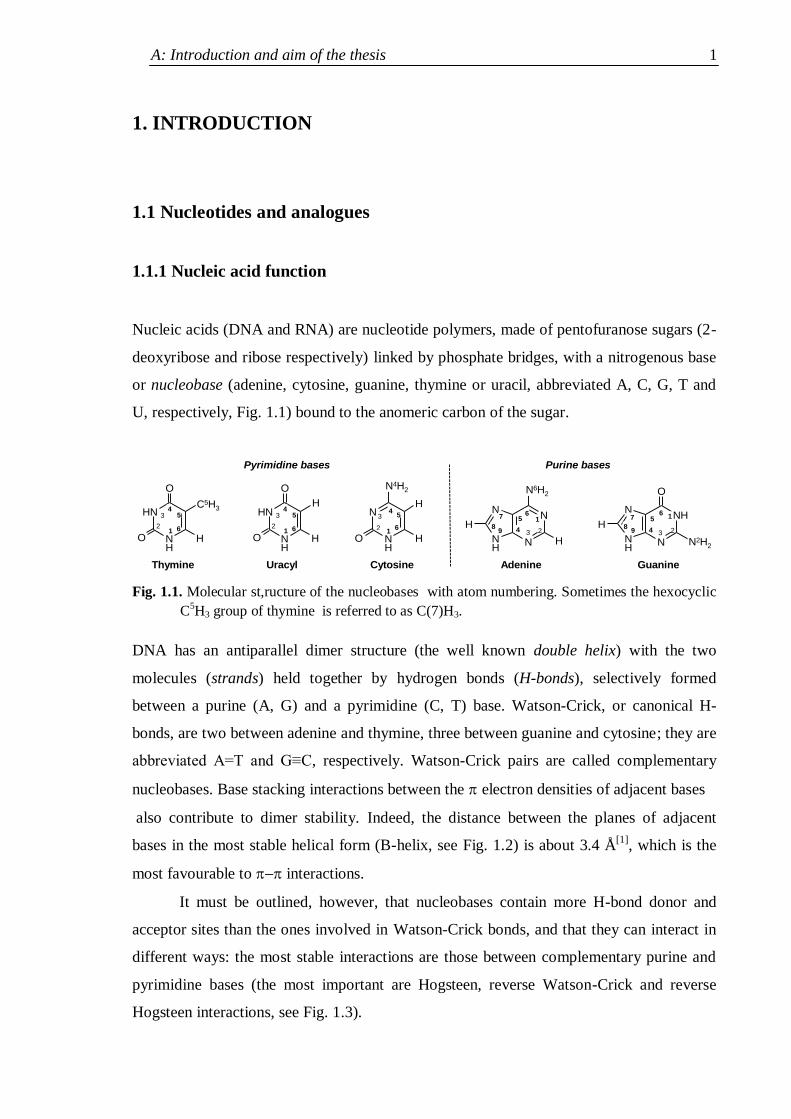
U, respectively, Fig. 1.1) bound to the anomeric carbon of the sugar.
NH
NH
O
O H
3
12
45
6
C5H3
N
NHN
NH
O
H3
1
24
56
7
89
N2H2
NH
NH
H
O
O H
3
12
45
6
N
NH
O H
H3
12
45
6
N4H2
N
NN
NH
H
H3
1
24
567
89
N6H2
Thymine Cytosine Adenine Guanine
Purine basesPyrimidine bases
Uracyl
Fig. 1.1. Molecular st,ructure of the nucleobases with atom numbering. Sometimes the hexocyclic
C5H3 group of thymine is referred to as C(7)H3.
DNA has an antiparallel dimer structure (the well known double helix) with the two
molecules (strands) held together by hydrogen bonds (H-bonds), selectively formed
between a purine (A, G) and a pyrimidine (C, T) base. Watson-Crick, or canonical H-
bonds, are two between adenine and thymine, three between guanine and cytosine; they are
abbreviated A=T and G≡C, respectively. Watson-Crick pairs are called complementary
nucleobases. Base stacking interactions between the electron densities of adjacent bases
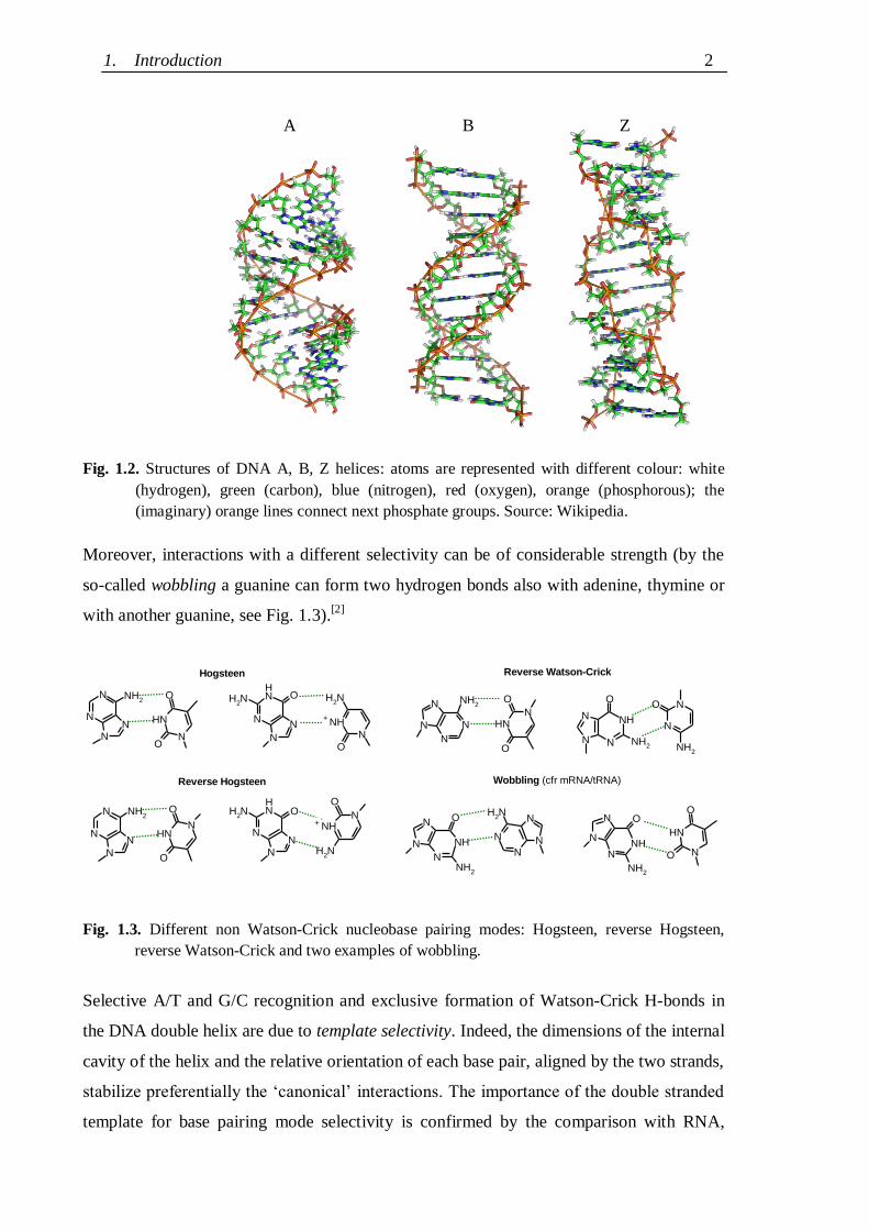
also contribute to dimer stability. Indeed, the distance between the planes of adjacent
bases in the most stable helical form (B-helix, see Fig. 1.2) is about 3.4 Å[1]
, which is the
most favourable to interactions.
It must be outlined, however, that nucleobases contain more H-bond donor and
acceptor sites than the ones involved in Watson-Crick bonds, and that they can interact in
different ways: the most stable interactions are those between complementary purine and
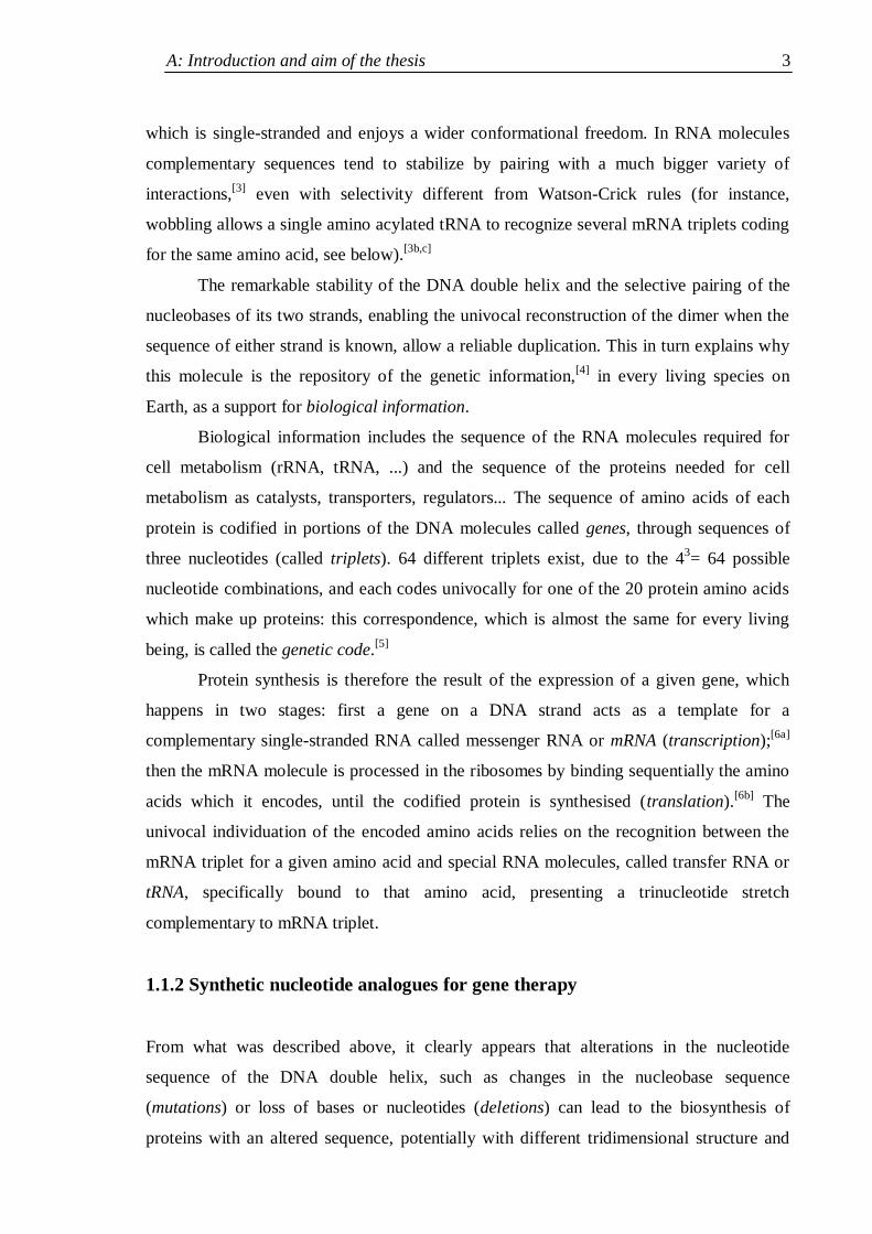
pyrimidine bases (the most important are Hogsteen, reverse Watson-Crick and reverse
Hogsteen interactions, see Fig. 1.3).
1. Introduction 2
Fig. 1.2. Structures of DNA A, B, Z helices: atoms are represented with different colour: white
(hydrogen), green (carbon), blue (nitrogen), red (oxygen), orange (phosphorous); the
(imaginary) orange lines connect next phosphate groups. Source: Wikipedia.
Moreover, interactions with a different selectivity can be of considerable strength (by the
so-called wobbling a guanine can form two hydrogen bonds also with adenine, thymine or
with another guanine, see Fig. 1.3).[2]
N
NN
N NH2
NNH
O
ON
NN
NH
ONH2 N
NH
O
NH2
N
N
N
N
NH2
NNH
O
ON N
N NH
O
NH2
N
N
O
NH2
N
N
N
NH
O
NH2
NN
N
N
NH2
N
N
N
NH
O
NH2
N
NH
O
O
N
NN
N NH2
NH
O
NO
N
NN
NH
ONH2
N
NH
O
NH2
+
Reverse Hogsteen
Reverse Watson-Crick
Wobbling (cfr mRNA/tRNA)
+
Hogsteen
Fig. 1.3. Different non Watson-Crick nucleobase pairing modes: Hogsteen, reverse Hogsteen,
reverse Watson-Crick and two examples of wobbling.
Selective A/T and G/C recognition and exclusive formation of Watson-Crick H-bonds in
the DNA double helix are due to template selectivity. Indeed, the dimensions of the internal
cavity of the helix and the relative orientation of each base pair, aligned by the two strands,
stabilize preferentially the „canonical‟ interactions. The importance of the double stranded
template for base pairing mode selectivity is confirmed by the comparison with RNA,
Z B A
A: Introduction and aim of the thesis 3
which is single-stranded and enjoys a wider conformational freedom. In RNA molecules
complementary sequences tend to stabilize by pairing with a much bigger variety of
interactions,[3]
even with selectivity different from Watson-Crick rules (for instance,
wobbling allows a single amino acylated tRNA to recognize several mRNA triplets coding
for the same amino acid, see below).[3b,c]
The remarkable stability of the DNA double helix and the selective pairing of the
nucleobases of its two strands, enabling the univocal reconstruction of the dimer when the
sequence of either strand is known, allow a reliable duplication. This in turn explains why
this molecule is the repository of the genetic information,[4]
in every living species on
Earth, as a support for biological information.
Biological information includes the sequence of the RNA molecules required for
cell metabolism (rRNA, tRNA, ...) and the sequence of the proteins needed for cell
metabolism as catalysts, transporters, regulators... The sequence of amino acids of each
protein is codified in portions of the DNA molecules called genes, through sequences of
three nucleotides (called triplets). 64 different triplets exist, due to the 43= 64 possible
nucleotide combinations, and each codes univocally for one of the 20 protein amino acids
which make up proteins: this correspondence, which is almost the same for every living
being, is called the genetic code.[5]
Protein synthesis is therefore the result of the expression of a given gene, which
happens in two stages: first a gene on a DNA strand acts as a template for a
complementary single-stranded RNA called messenger RNA or mRNA (transcription);[6a]
then the mRNA molecule is processed in the ribosomes by binding sequentially the amino
acids which it encodes, until the codified protein is synthesised (translation).[6b]
The
univocal individuation of the encoded amino acids relies on the recognition between the
mRNA triplet for a given amino acid and special RNA molecules, called transfer RNA or
tRNA, specifically bound to that amino acid, presenting a trinucleotide stretch
complementary to mRNA triplet.
1.1.2 Synthetic nucleotide analogues for gene therapy
From what was described above, it clearly appears that alterations in the nucleotide
sequence of the DNA double helix, such as changes in the nucleobase sequence
(mutations) or loss of bases or nucleotides (deletions) can lead to the biosynthesis of
proteins with an altered sequence, potentially with different tridimensional structure and
1. Introduction 4
polarity and therefore with impaired functionality. Indeed, research done in the last
decades has shown that the causes of a always growing number of pathologies reside in
alterations of the genetic information or in defects of its expression.[7]
This in turn raised a
growing interest towards gene therapy, a therapeutic strategy operating at molecular level
on genes with altered sequence or expression.
One of the first and more promising approaches of gene therapy is the control of
gene expression through RNA interference. RNA interference (RNAi) is a natural
mechanism of control of the gene expression which is found in all eukaryotes and even in
bacteria:[8]
if a mRNA molecule encounters another molecule (called antisense) able to
stably pair with it (even partially), translation is prevented and the mRNA will be
eventually degraded by special hydrolytic enzymes, so that the protein which it coded will
not be synthesised. This means that the introduction in a cell of a synthetic antisense RNA
strand complementary to the mRNA derived from a non functional or overexpressed gene
could avoid the synthesis of a non functional protein or prevent a harmful protein
overexpression.
Unfortunately the therapeutic use of synthetic oligonucleotides for RNA
intereference therapy has several problems preventing its practical implementation.[9]
The
two most severe problems are:
(i) Synthetic RNAi oligonucleotides are identical in structure to biological molecules
and are therefore rapidly degraded by nucleases (hydrolytic enzymes working on
phosphodiester bonds), present in every cell;
(ii) The nucleotide large negative charge (one for each phosphate group, i.e. one for
each residue) makes them so hydrophilic that they are not able to cross cell
membranes, formed by hydrophobic lipid bilayers, preventing them from entering
the cells.
To overcome such problems a wide range of studies has been carried on nucleotide
analogues[10,11]
with suitable properties for therapeutic applications, i.e.:
- Affinity to complementary strands comparable to the one of natural nucleotides;
- Pairing selectivity following Watson-Crick rules (A/T and G/C) and sensitivity to
single-base mismatched sequences;
- Good chemical stability and a better biological stability in comparison to nucleotides;
- Acceptable capability of biological membrane penetration in order to enter the cells;
- Reduced aspecific toxicity.
A: Introduction and aim of the thesis 5
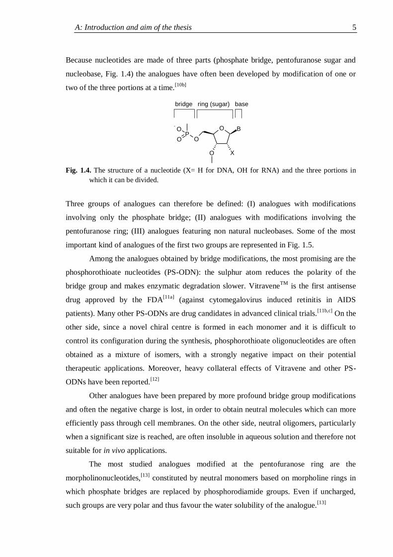
Because nucleotides are made of three parts (phosphate bridge, pentofuranose sugar and
nucleobase, Fig. 1.4) the analogues have often been developed by modification of one or
two of the three portions at a time.[10b]
Fig. 1.4. The structure of a nucleotide (X= H for DNA, OH for RNA) and the three portions in
which it can be divided.
Three groups of analogues can therefore be defined: (I) analogues with modifications
involving only the phosphate bridge; (II) analogues with modifications involving the
pentofuranose ring; (III) analogues featuring non natural nucleobases. Some of the most
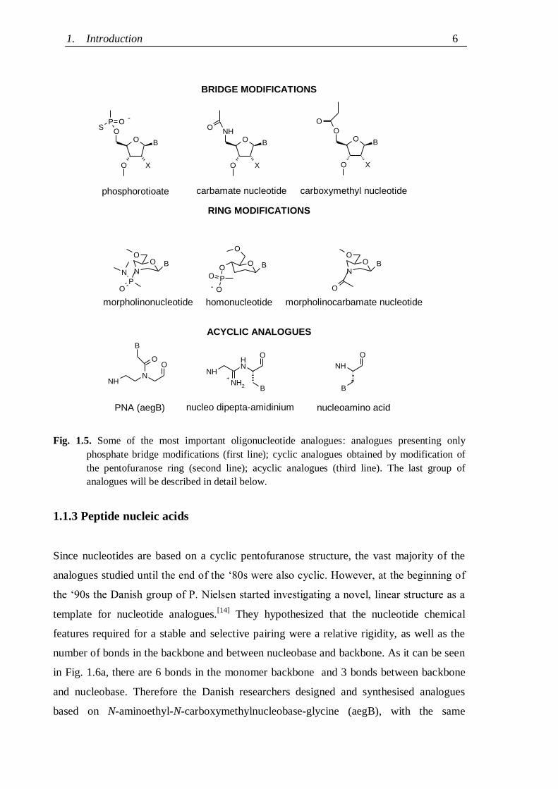
important kind of analogues of the first two groups are represented in Fig. 1.5.
Among the analogues obtained by bridge modifications, the most promising are the
phosphorothioate nucleotides (PS-ODN): the sulphur atom reduces the polarity of the
bridge group and makes enzymatic degradation slower. VitraveneTM
is the first antisense
drug approved by the FDA[11a]
(against cytomegalovirus induced retinitis in AIDS
patients). Many other PS-ODNs are drug candidates in advanced clinical trials.[11b,c]
On the
other side, since a novel chiral centre is formed in each monomer and it is difficult to
control its configuration during the synthesis, phosphorothioate oligonucleotides are often
obtained as a mixture of isomers, with a strongly negative impact on their potential
therapeutic applications. Moreover, heavy collateral effects of Vitravene and other PS-
ODNs have been reported.[12]
Other analogues have been prepared by more profound bridge group modifications
and often the negative charge is lost, in order to obtain neutral molecules which can more
efficiently pass through cell membranes. On the other side, neutral oligomers, particularly
when a significant size is reached, are often insoluble in aqueous solution and therefore not
suitable for in vivo applications.
The most studied analogues modified at the pentofuranose ring are the
morpholinonucleotides,[13]
constituted by neutral monomers based on morpholine rings in
which phosphate bridges are replaced by phosphorodiamide groups. Even if uncharged,
such groups are very polar and thus favour the water solubility of the analogue.[13]
PO
O O
O
O
B
X
-
bridge ring (sugar) base
1. Introduction 6
Fig. 1.5. Some of the most important oligonucleotide analogues: analogues presenting only
phosphate bridge modifications (first line); cyclic analogues obtained by modification of
the pentofuranose ring (second line); acyclic analogues (third line). The last group of
analogues will be described in detail below.
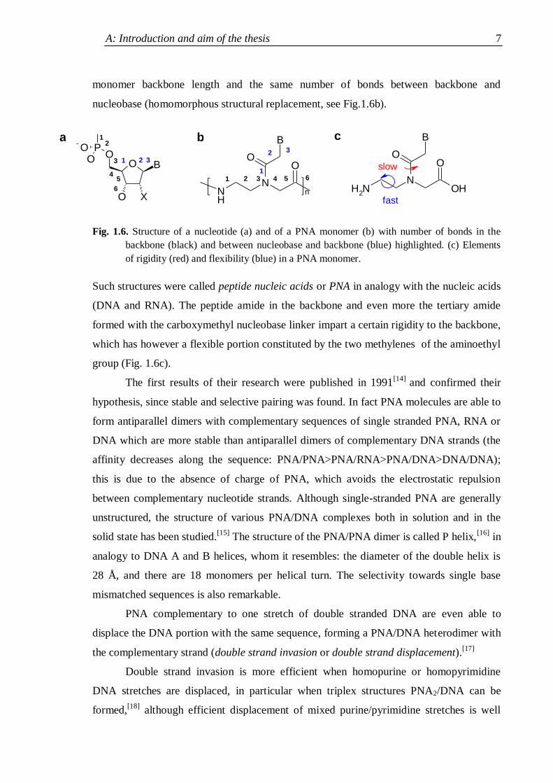
1.1.3 Peptide nucleic acids
Since nucleotides are based on a cyclic pentofuranose structure, the vast majority of the
analogues studied until the end of the „80s were also cyclic. However, at the beginning of
the „90s the Danish group of P. Nielsen started investigating a novel, linear structure as a
template for nucleotide analogues.[14]
They hypothesized that the nucleotide chemical
features required for a stable and selective pairing were a relative rigidity, as well as the
number of bonds in the backbone and between nucleobase and backbone. As it can be seen
in Fig. 1.6a, there are 6 bonds in the monomer backbone and 3 bonds between backbone
and nucleobase. Therefore the Danish researchers designed and synthesised analogues
based on N-aminoethyl-N-carboxymethylnucleobase-glycine (aegB), with the same
O
ONH
B
X
O
O
O
BO
PO
O
O
NB
O
N
PO
NH
B
O
NNH
O
B
ONH
B
NH
O
NH2
O
OO
B
X
OO
O
OO
B
PS
X
O
NB
O
O
carbamate nucleotide
homonucleotidemorpholinonucleotide
-
nucleoamino acidPNA (aegB)
+
nucleo dipepta-amidinium
carboxymethyl nucleotide
BRIDGE MODIFICATIONS
RING MODIFICATIONS
-
phosphorotioate
morpholinocarbamate nucleotide
ACYCLIC ANALOGUES
A: Introduction and aim of the thesis 7
monomer backbone length and the same number of bonds between backbone and
nucleobase (homomorphous structural replacement, see Fig.1.6b).
Fig. 1.6. Structure of a nucleotide (a) and of a PNA monomer (b) with number of bonds in the
backbone (black) and between nucleobase and backbone (blue) highlighted. (c) Elements
of rigidity (red) and flexibility (blue) in a PNA monomer.
Such structures were called peptide nucleic acids or PNA in analogy with the nucleic acids
(DNA and RNA). The peptide amide in the backbone and even more the tertiary amide
formed with the carboxymethyl nucleobase linker impart a certain rigidity to the backbone,
which has however a flexible portion constituted by the two methylenes of the aminoethyl
group (Fig. 1.6c).
The first results of their research were published in 1991[14]
and confirmed their
hypothesis, since stable and selective pairing was found. In fact PNA molecules are able to
form antiparallel dimers with complementary sequences of single stranded PNA, RNA or
DNA which are more stable than antiparallel dimers of complementary DNA strands (the
affinity decreases along the sequence: PNA/PNA>PNA/RNA>PNA/DNA>DNA/DNA);
this is due to the absence of charge of PNA, which avoids the electrostatic repulsion
between complementary nucleotide strands. Although single-stranded PNA are generally
unstructured, the structure of various PNA/DNA complexes both in solution and in the
solid state has been studied.[15]
The structure of the PNA/PNA dimer is called P helix,[16]
in
analogy to DNA A and B helices, whom it resembles: the diameter of the double helix is
28 Å, and there are 18 monomers per helical turn. The selectivity towards single base
mismatched sequences is also remarkable.
PNA complementary to one stretch of double stranded DNA are even able to
displace the DNA portion with the same sequence, forming a PNA/DNA heterodimer with
the complementary strand (double strand invasion or double strand displacement).[17]
Double strand invasion is more efficient when homopurine or homopyrimidine
DNA stretches are displaced, in particular when triplex structures PNA2/DNA can be
formed,[18]
although efficient displacement of mixed purine/pyrimidine stretches is well
B
NH
N
OO
n
O
XO
OB
PO
O
NH2
N
O
B
O
OH1 2 3 4 5 6
1
2 3-
12
3
45
6
1 2 3
fast
slow
a b c
1. Introduction 8
documented.[19]
Double strand invasion is more efficient when homopurine or
homopyrimidine DNA stretches are displaced, in particular when triplex structures
PNA2/DNA can be formed,[18]
although efficient displacement of mixed purine/pyrimidine
stretches is well documented.[19]
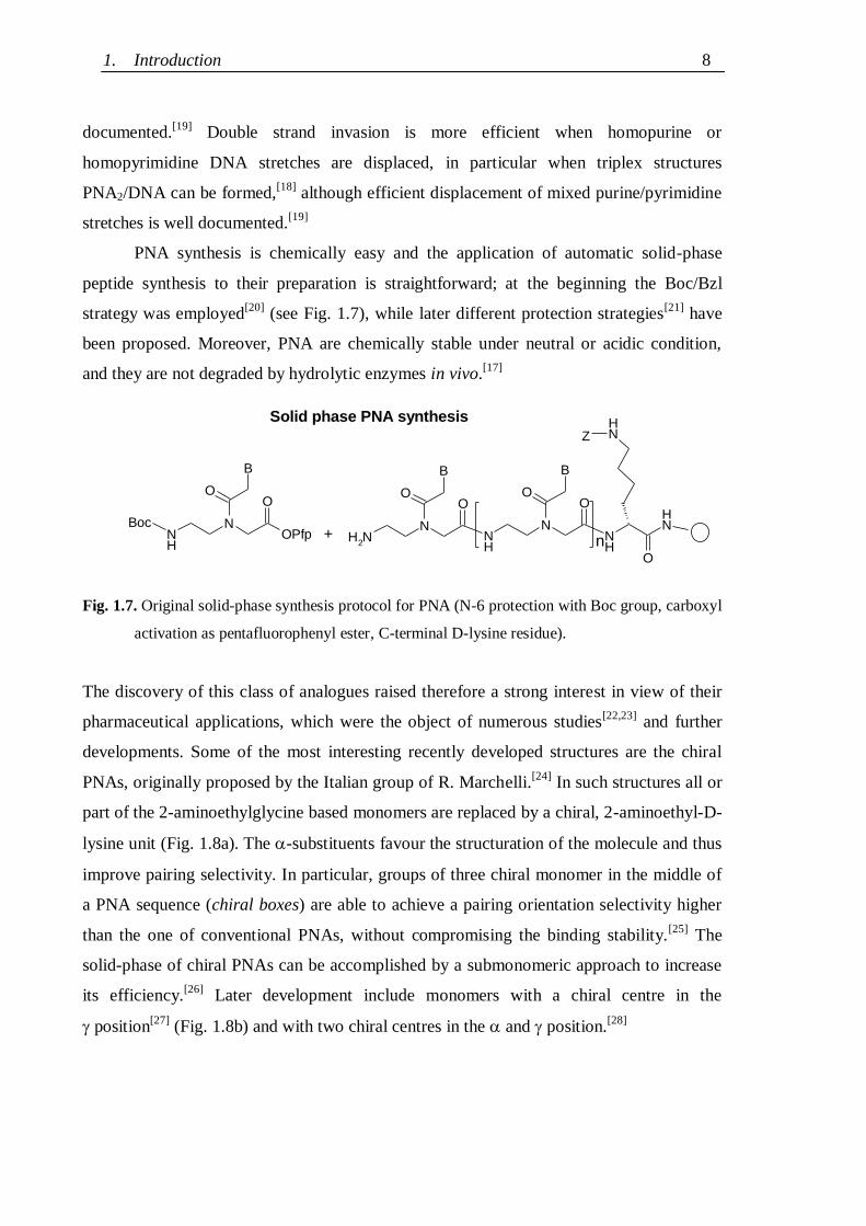
PNA synthesis is chemically easy and the application of automatic solid-phase
peptide synthesis to their preparation is straightforward; at the beginning the Boc/Bzl
strategy was employed[20]
(see Fig. 1.7), while later different protection strategies[21]
have
been proposed. Moreover, PNA are chemically stable under neutral or acidic condition,
and they are not degraded by hydrolytic enzymes in vivo.[17]
Fig. 1.7. Original solid-phase synthesis protocol for PNA (N-6 protection with Boc group, carboxyl
activation as pentafluorophenyl ester, C-terminal D-lysine residue).
The discovery of this class of analogues raised therefore a strong interest in view of their
pharmaceutical applications, which were the object of numerous studies[22,23]
and further
developments. Some of the most interesting recently developed structures are the chiral
PNAs, originally proposed by the Italian group of R. Marchelli.[24]
In such structures all or
part of the 2-aminoethylglycine based monomers are replaced by a chiral, 2-aminoethyl-D-
lysine unit (Fig. 1.8a). The -substituents favour the structuration of the molecule and thus
improve pairing selectivity. In particular, groups of three chiral monomer in the middle of
a PNA sequence (chiral boxes) are able to achieve a pairing orientation selectivity higher
than the one of conventional PNAs, without compromising the binding stability.[25]
The
solid-phase of chiral PNAs can be accomplished by a submonomeric approach to increase
its efficiency.[26]
Later development include monomers with a chiral centre in the
position[27]
(Fig. 1.8b) and with two chiral centres in the and position.[28]
B
NH2
N
O
NNH
O
NH
O
B
O
NH
O
n
NH
N
O
B
O
NH
OPfpBoc
Solid phase PNA synthesis
+
Z
A: Introduction and aim of the thesis 9
NH2
N
NH2
O
B
O
OH NH2
N
O
B
O
OH
NH2 N
HO
O
OH
B
O
OHNNH
2
B
-(R)-PNA
Chiral PNA Cyclic PNA
-(R)-PNA
(a) (b)(c)
(d)
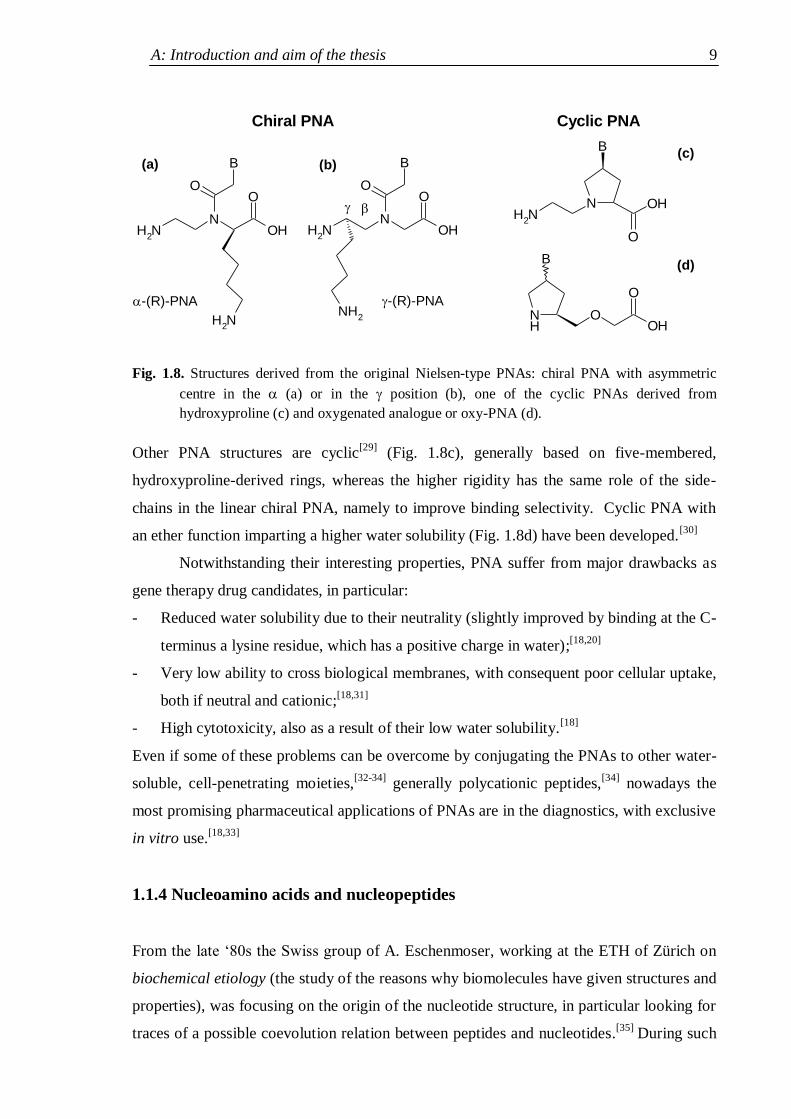
Fig. 1.8. Structures derived from the original Nielsen-type PNAs: chiral PNA with asymmetric
centre in the (a) or in the position (b), one of the cyclic PNAs derived from
hydroxyproline (c) and oxygenated analogue or oxy-PNA (d).
Other PNA structures are cyclic[29]
(Fig. 1.8c), generally based on five-membered,
hydroxyproline-derived rings, whereas the higher rigidity has the same role of the side-
chains in the linear chiral PNA, namely to improve binding selectivity. Cyclic PNA with
an ether function imparting a higher water solubility (Fig. 1.8d) have been developed.[30]
Notwithstanding their interesting properties, PNA suffer from major drawbacks as
gene therapy drug candidates, in particular:
- Reduced water solubility due to their neutrality (slightly improved by binding at the C-
terminus a lysine residue, which has a positive charge in water);[18,20]
- Very low ability to cross biological membranes, with consequent poor cellular uptake,
both if neutral and cationic;[18,31]
- High cytotoxicity, also as a result of their low water solubility.[18]
Even if some of these problems can be overcome by conjugating the PNAs to other water-
soluble, cell-penetrating moieties,[32-34]
generally polycationic peptides,[34]
nowadays the
most promising pharmaceutical applications of PNAs are in the diagnostics, with exclusive
in vitro use.[18,33]
1.1.4 Nucleoamino acids and nucleopeptides
From the late „80s the Swiss group of A. Eschenmoser, working at the ETH of Zürich on
biochemical etiology (the study of the reasons why biomolecules have given structures and
properties), was focusing on the origin of the nucleotide structure, in particular looking for
traces of a possible coevolution relation between peptides and nucleotides.[35]
During such
1. Introduction 10
researches, starting from L-serine, two non proteogenic alanyl amino acid derivatives were
synthesised, with a nucleobase (thymine or adenine) at the position, which were called
nucleoamino acids.[36]
At the time, nucleoamino acids were not completely unknown. Indeed, willardiine,
a nucleoamino acid carrying an uracil at the positon, is a naturally occurring compound
which was known since decades,[37]
as well as a 2-aminopyrimidine-4-yl analogue called
lathyrine.[38]
The occurrence in nature of nucleobase containing peptides,[39]
which had
been called nucleopeptides, had raised a great interest so that research on the biological
properties of the nucleoamino acids started,[40]
even if without conclusive results. A
thymine-based analogue of willardiine was thus synthesised as a racemate[41]
by Strecker
synthesis, partially resolved and polymerized,[42]
however the complete resolution of its L
enantiomer (with the same chirality as the natural compound) was not achieved. Similarly,
-nucleoamino acids and their polymers were synthesised as racemates.[43]
Even if
interaction experiments on the resulting raceme polymers with nucleotides were attempted,
no significant results were detected.[42,43]
On the other side, the synthetic strategy of
Eschenmoser relied on the nucleophilic ring opening of the protected L-serine--
lactone,[44]
a reaction which preserves the optical purity of the starting material.
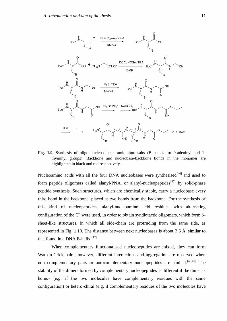
The nucleoamino acids thus obtained were coupled with 2-aminoethylnitrile to
form nucleo-dipeptanitriles, converted in nucleo-dipepta-amidines, which were
oligomerized under acidic conditions to form oligo nucleo-dipepta-amidinium salts (see
Fig. 1.9).[36,45]
As it can be noted in Fig. 1.9, this structure maintains the characteristic number of
bonds in the monomer backbone, whereas there are only two bonds between nucleobases
and backbone. The presence of a positive charge per residue in the salts of nucleo-
dipeptaamidinium was a key point in the design of such analogues, as it should have
imparted a higher affinity towards the polyanionic oligonucleotides. However, the
oligomer salts are chemically unstable under aqueous non acidic conditions and even under
acidic conditions (at pH≤ 4) they do not display significant structuration, nor any tendency
to dimer formation with complementary DNA or RNA molecules.[45]
Although these rather disappointing results, soon after the work of Eschenmoser,
the German group of U.Diederichsen carried on a wide research program on nucleoamino
acids.
A: Introduction and aim of the thesis 11
Fig. 1.9. Synthesis of oligo nucleo-dipepta-amidinium salts (B stands for 9-adeninyl and 1-
thyminyl groups). Backbone and nucleobase-backbone bonds in the monomer are
highlighted in black and red respectively.
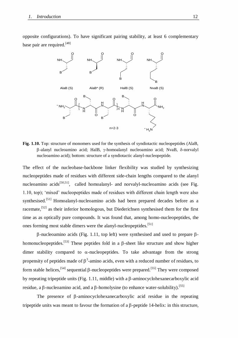
Nucleoamino acids with all the four DNA nucleobases were synthesised[46]
and used to
form peptide oligomers called alanyl-PNA, or alanyl-nucleopeptides[47]
by solid-phase
peptide synthesis. Such structures, which are chemically stable, carry a nucleobase every
third bond in the backbone, placed at two bonds from the backbone. For the synthesis of
this kind of nucleopeptides, alanyl-nucleoamino acid residues with alternating
configuration of the C were used, in order to obtain syndiotactic oligomers, which form -
sheet-like structures, in which all side-chain are protruding from the same side, as
represented in Fig. 1.10. The distance between next nucleobases is about 3.6 Å, similar to
that found in a DNA B-helix.[47]
When complementary functionalised nucleopeptides are mixed, they can form
Watson-Crick pairs; however, different interactions and aggregation are observed when
non complementary pairs or autocomplementary nucleopeptides are studied.[48,49]
The
stability of the dimers formed by complementary nucleopeptides is different if the dimer is
homo- (e.g. if the two molecules have complementary residues with the same
configuration) or hetero-chiral (e.g. if complementary residues of the two molecules have
O
OHN
Boc Boc
HN
OH
O
B
H-B, K2CO3/DBU
DMSO
Boc
HN
OH
O
B
+ H3N CN+ Cl- Boc
HN
NH
CN
O
B
DCC, HOSu, TEA
DMF
Boc
HN
NH
CN
O
B
Boc
HN
NH
O
B
NHH2S, TEA
MeOHS
Boc
HN
NH
O
B
NH
S
NaHCO3
Boc
HN
NH
O
B
S
NH
H3NNH
O
B
HN
NH2
TFA ...
NH
S
B
O
O+
+
n
n+1 TfaO-
Et3O+ PF6-
1 2 3 4 5 6
2
1
1. Introduction 12
opposite configurations). To have significant pairing stability, at least 6 complementary
base pair are required.[48]
Fig. 1.10. Top: structure of monomers used for the synthesis of syndiotactic nucleopeptides (AlaB,
-alanyl nucleoamino acid; HalB, -homoalanyl nucleoamino acid; NvaB, -norvalyl
nucleoamino acid); bottom: structure of a syndiotactic alanyl-nucleopeptide.
The effect of the nucleobase-backbone linker flexibility was studied by synthesizing
nucleopeptides made of residues with different side-chain lengths compared to the alanyl
nucleoamino acids[50,51]
, called homoalanyl- and norvalyl-nucleoamino acids (see Fig.
1.10, top); „mixed‟ nucleopeptides made of residues with different chain length were also
synthesised.[51]
Homoalanyl-nucleoamino acids had been prepared decades before as a
racemate,[52]
as their inferior homologous, but Diederichsen synthesised them for the first
time as as optically pure compounds. It was found that, among homo-nucleopeptides, the
ones forming most stable dimers were the alanyl-nucleopeptides.[51]
-nucleoamino acids (Fig. 1.11, top left) were synthesised and used to prepare -
homonucleopeptides.[53]
These peptides fold in a -sheet like structure and show higher
dimer stability compared to -nucleopeptides. To take advantage from the strong
propensity of peptides made of 3-amino acids, even with a reduced number of residues, to
form stable helices,[54]
sequential -nucleopeptides were prepared.[55]
They were composed
by repeating tripeptide units (Fig. 1.11, middle) with a -aminocyclohexanecarboxylic acid
residue, a -nucleoamino acid, and a -homolysine (to enhance water-solubility).[55]
The presence of -aminocyclohexanecarboxylic acid residue in the repeating
tripeptide units was meant to favour the formation of a -peptide 14-helix: in this structure,
B
O
NH
B
O
NH
B
O
NH
B
O
NH
O
n
H3N
NH
B
O
NH
B
O
NH
O
B
NH
O
B
NH3+
NH2
AlaB* (R)AlaB (S)
n=2-3
HalB (S) NvaB (S)
+
A: Introduction and aim of the thesis 13
residues every third position are aligned and therefore the side-chain nucleobases would be
able to interact cooperatively with complementary sequences of other molecules in the
same conformation. A detailed investigation[56]
confirmed the assumption of a 14-helical
comformation by the sequential oligomers. In general, multiple functionalization of a
nucleotide analogue with the same base allows various kind of pairing interactions.[35b,47]
It
was found however that Watson-Crick pairing, when possible, was largely prevalent over
non specific higher order interactions.
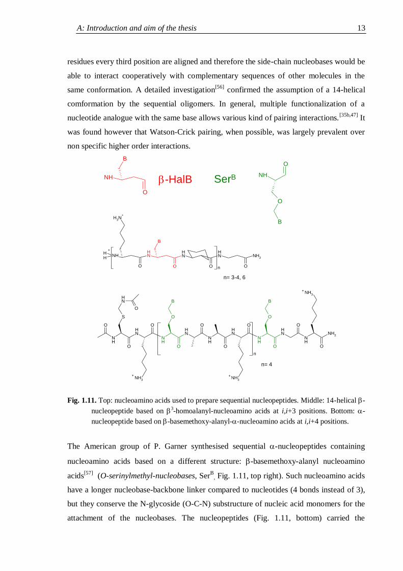
Fig. 1.11. Top: nucleoamino acids used to prepare sequential nucleopeptides. Middle: 14-helical -
nucleopeptide based on 3-homoalanyl-nucleoamino acids at i,i+3 positions. Bottom: -
nucleopeptide based on -basemethoxy-alanyl--nucleoamino acids at i,i+4 positions.
The American group of P. Garner synthesised sequential -nucleopeptides containing
nucleoamino acids based on a different structure: -basemethoxy-alanyl nucleoamino
acids[57]
(O-serinylmethyl-nucleobases, SerB
, Fig. 1.11, top right). Such nucleoamino acids
have a longer nucleobase-backbone linker compared to nucleotides (4 bonds instead of 3),
but they conserve the N-glycoside (O-C-N) substructure of nucleic acid monomers for the
attachment of the nucleobases. The nucleopeptides (Fig. 1.11, bottom) carried the
NH
O
B
NH
OO
NH
O
NH2
NH
O
NHH
H3N
n
H
B
NH
NH
NH
NH
NH
NH
NH
NH
O
O
O
O
O
O
O
O
O
NH
S
NH3
O
B
n
NH3
NH2
O
O
BNH
O
NH3
NH
O
O
B
-HalB
n= 3-4, 6
+ +
+
n= 4
SerB
+
+
1. Introduction 14
nucleoamino acids at i,i+4 positions and a lysine was present in each tetrapeptide unit,[58]
whose positive charge had the usual purposes both of increasing the water solubility of the
nucleopeptides and favouring their interactions with nucleotides by charge
complementarity.
The design of such sequential -nucleopeptide structures with the specific aim of
selective and stable nucleic acid recognition relied on two assumptions. Firstly, as
eicosapeptides rich in -helix promoting amino acids, it was hoped that the nucleopeptides
would have folded in -helices. The nucleoamino acids set every fourth position would
have therefore formed a right-handed superhelix, with the same handedness of the DNA B-
helix, thus favouring the the alignment of complementary nucleobases. Secondly, since the
distance of next nucleoamino acids along the described superhelix is significantly larger
than the distance of the planes of next nucleobases in double-stranded DNA, it was hoped
that the flexible O-methyl-serine-derived linker would have allowed a synchronized tilting
of the nucleobases in order to take the distance of their planes closer to the one of the
nucleobases in DNA. Furthermore, Garner and his group were confident that the
deformability of both -helices and DNA[59]
would have allowed an induced-fit driven by
pairing stabilization.
By studying the CD spectra of their -nucleopeptides, of complementary DNA
stretches, and of their mixtures, they found that while the analogue alone was not
structured, in the presence of one equivalent of complementary DNA, the CD spectrum
was close to the superimposition of the contribution of a peptide -helix[60]
and of DNA in
the B-form.[60a,61]
As nucleopeptide/nucleic acid complex formation was confirmed by
independent experiments performed with other techniques, the researchers interpreted this
result as a pairing-induced structuration of their analogue.
The last two examples illustrate clearly the importance of preparing
conformationally stable analogues, controlling and predicting their structures, in order to
favour cooperative nucleobase-nucleobase interactions as a prerequisite for selective
recognition, but also the effect that pairing forces can have on the conformation of an
analogue and its stability. Therefore in the following section elements of peptide structure
will be recalled and a class of amino acids known for their ability to adopt stable ordered
conformations will be presented, with particular attention for its simplest member and for
the helical structure its peptides generally adopt.
A: Introduction and aim of the thesis 15
1.2 Peptide structures and C-tetrasubstituted -amino acids
1.2.1. Elements of folded peptide secondary structures
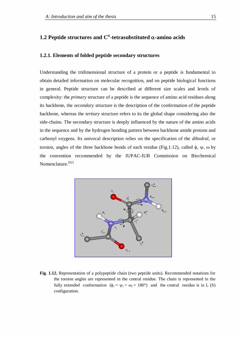
Understanding the tridimensional structure of a protein or a peptide is fundamental to
obtain detailed information on molecular recognition, and on peptide biological functions
in general. Peptide structure can be described at different size scales and levels of
complexity: the primary structure of a peptide is the sequence of amino acid residues along
its backbone, the secondary structure is the description of the conformation of the peptide
backbone, whereas the tertiary structure refers to its the global shape considering also the
side-chains. The secondary structure is deeply influenced by the nature of the amino acids
in the sequence and by the hydrogen bonding pattern between backbone amide protons and
carbonyl oxygens. Its univocal description relies on the specification of the dihedral, or
torsion, angles of the three backbone bonds of each residue (Fig.1.12), called , , by
the convention recommended by the IUPAC-IUB Commission on Biochemical
Nomenclature.[62]
Fig. 1.12. Representation of a polypeptide chain (two peptide units). Recommended notations for
the torsion angles are represented in the central residue. The chain is represented in the
fully extended conformation (i = i = i = 180°) and the central residue is in L (S)
configuration.

1. Introduction 16
The most important and widespread peptide secondary structures are[63]
the -helix, the -
structures, the -turns and the 310-helix; the most common organized secondary structures
are helical. Various helical structures differ in the dihedral angles and of each residue,
in the number of residues per turn, in the pitch and in the number of atoms involved in the
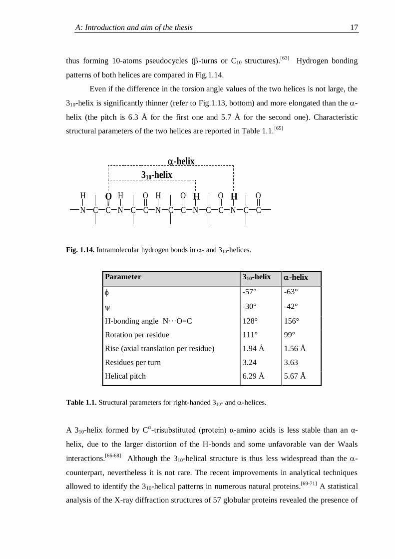
pseudocycles formed by intramolecular hydrogen bonds C=OH-N.[64]
Fig.1.13. Top: side view of right-handed -helix (a) and 310-helix (b); bottom: projections of the -
helix and of the 310-helix along the helical axis.
As mentioned above, the -helix and the 310-helix are the most common helical structures.
The -helix (Fig. 1.13a) is characterized by 3.63 residues per turn and it is stabilized by
intramolecular hydrogen bonds between the C=O group of residues in position i and the N-
H group of residues in position i+4 (i i+4 H-bonds), thus forming 13-atoms
pseudocycles (-turns or C13 structures). The 310-helix (Fig. 1.13b) has 3.24 residues per
turn and it is stabilized by intramolecular hydrogen bonds between the C=O group of
residues in position i and the N-H groups of residues in position i+3 (i i+3 H-bonds),
- Helix
x
310-Helix
1 2
3
4
5 1
2
3
4 5
(a) -helix
(b) 310-helix
A: Introduction and aim of the thesis 17
thus forming 10-atoms pseudocycles (-turns or C10 structures).[63]
Hydrogen bonding
patterns of both helices are compared in Fig.1.14.
Even if the difference in the torsion angle values of the two helices is not large, the
310-helix is significantly thinner (refer to Fig.1.13, bottom) and more elongated than the -
helix (the pitch is 6.3 Å for the first one and 5.7 Å for the second one). Characteristic
structural parameters of the two helices are reported in Table 1.1.[65]
N C C N C C N C C N C C N C C
H O H O H O H O H O
-helix
310-helix
Fig. 1.14. Intramolecular hydrogen bonds in - and 310-helices.
Parameter 310-helix -helix
-57° -63°
-30° -42°
H-bonding angle N···O=C 128° 156°
Rotation per residue 111° 99°
Rise (axial translation per residue) 1.94 Å 1.56 Å
Residues per turn 3.24 3.63
Helical pitch 6.29 Å 5.67 Å
Table 1.1. Structural parameters for right-handed 310- and -helices.
A 310-helix formed by C-trisubstituted (protein) α-amino acids is less stable than an α-
helix, due to the larger distortion of the H-bonds and some unfavorable van der Waals
interactions.[66-68]
Although the 310-helical structure is thus less widespread than the -
counterpart, nevertheless it is not rare. The recent improvements in analytical techniques
allowed to identify the 310-helical patterns in numerous natural proteins.[69-71]
A statistical
analysis of the X-ray diffraction structures of 57 globular proteins revealed the presence of
1. Introduction 18
71 310-helical motifs of different length. Interestingly, in most cases such structures were
found at the N- and C-termini of α-helices.
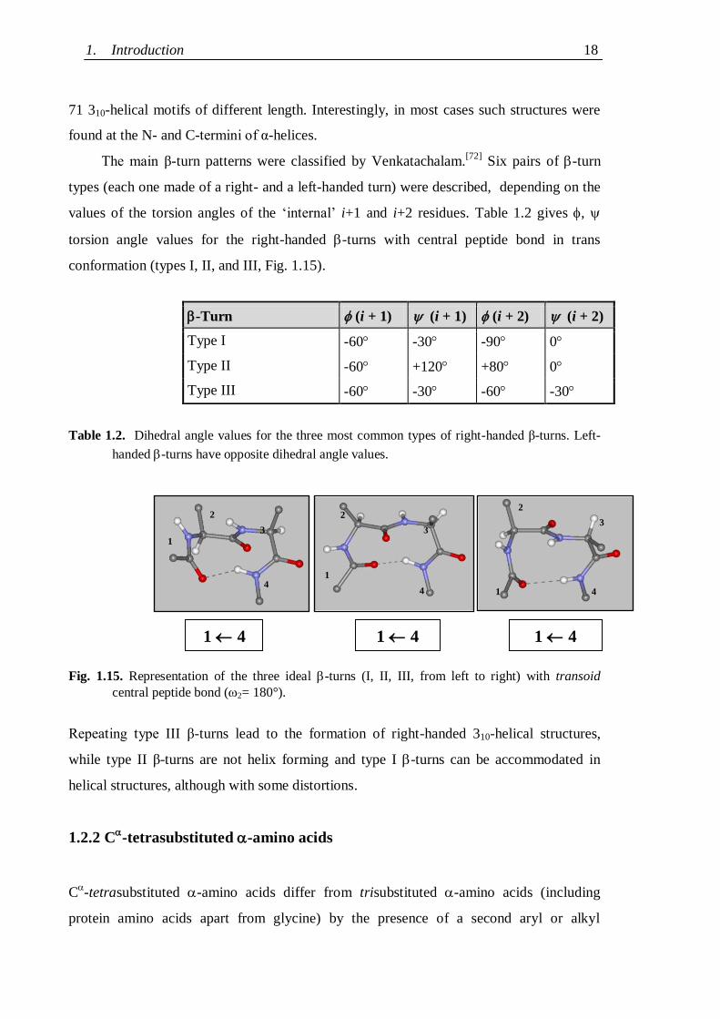
The main β-turn patterns were classified by Venkatachalam.[72]
Six pairs of -turn
types (each one made of a right- and a left-handed turn) were described, depending on the
values of the torsion angles of the „internal‟ i+1 and i+2 residues. Table 1.2 gives ,
torsion angle values for the right-handed -turns with central peptide bond in trans
conformation (types I, II, and III, Fig. 1.15).
Table 1.2. Dihedral angle values for the three most common types of right-handed β-turns. Left-
handed -turns have opposite dihedral angle values.
Fig. 1.15. Representation of the three ideal -turns (I, II, III, from left to right) with transoid
central peptide bond (2= 180°).
Repeating type III β-turns lead to the formation of right-handed 310-helical structures,
while type II β-turns are not helix forming and type I -turns can be accommodated in
helical structures, although with some distortions.
1.2.2 C-tetrasubstituted -amino acids
C-tetrasubstituted -amino acids differ from trisubstituted -amino acids (including
protein amino acids apart from glycine) by the presence of a second aryl or alkyl
-Turn (i + 1) (i + 1) (i + 2) (i + 2)
Type I -60 -30 -90 0
Type II -60 +120 +80 0
Type III -60 -30 -60 -30
1 4
(I)
1 4
(III)
1
2
3
4 1
2
3
4 1
2
3
4
1 4
(II)
A: Introduction and aim of the thesis 19
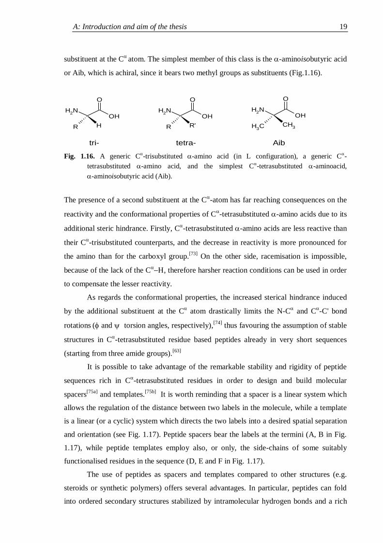
substituent at the C
atom. The simplest member of this class is the -aminoisobutyric acid
or Aib, which is achiral, since it bears two methyl groups as substituents (Fig.1.16).
NH2
R H
O
OHNH
2
R R'
O
OHNH
2
CH3
CH3
O
OH
tri- tetra- Aib
Fig. 1.16. A generic C-trisubstituted -amino acid (in L configuration), a generic C
-
tetrasubstituted -amino acid, and the simplest C-tetrasubstituted -aminoacid,
-aminoisobutyric acid (Aib).
The presence of a second substituent at the C-atomhas far reaching consequences on the
reactivity and the conformational properties of C-tetrasubstituted -amino acids due to its
additional steric hindrance. Firstly, C-tetrasubstituted -amino acids are less reactive than
their C-trisubstituted counterparts, and the decrease in reactivity is more pronounced for
the amino than for the carboxyl group.[73]
On the other side, racemisation is impossible,
because of the lack of the C, therefore harsher reaction conditions can be used in order
to compensate the lesser reactivity.
As regards the conformational properties, the increased sterical hindrance induced
by the additional substituent at the C atom drastically limits the N-C
and C
-C' bond
rotations ( and torsion angles, respectively),
[74] thus favouring the assumption of stable
structures in C-tetrasubstituted residue based peptides already in very short sequences
(starting from three amide groups).[63]
It is possible to take advantage of the remarkable stability and rigidity of peptide
sequences rich in C-tetrasubstituted residues in order to design and build molecular
spacers[75a]
and templates.[75b]
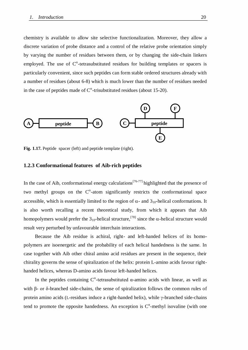
It is worth reminding that a spacer is a linear system which
allows the regulation of the distance between two labels in the molecule, while a template
is a linear (or a cyclic) system which directs the two labels into a desired spatial separation
and orientation (see Fig. 1.17). Peptide spacers bear the labels at the termini (A, B in Fig.
1.17), while peptide templates employ also, or only, the side-chains of some suitably
functionalised residues in the sequence (D, E and F in Fig. 1.17).
The use of peptides as spacers and templates compared to other structures (e.g.
steroids or synthetic polymers) offers several advantages. In particular, peptides can fold
into ordered secondary structures stabilized by intramolecular hydrogen bonds and a rich
1. Introduction 20
chemistry is available to allow site selective functionalization. Moreover, they allow a
discrete variation of probe distance and a control of the relative probe orientation simply
by varying the number of residues between them, or by changing the side-chain linkers
employed. The use of C-tetrasubstituted residues for building templates or spacers is
particularly convenient, since such peptides can form stable ordered structures already with
a number of residues (about 6-8) which is much lower than the number of residues needed
in the case of peptides made of C-trisubstituted residues (about 15-20).
Fig. 1.17. Peptide spacer (left) and peptide template (right).
1.2.3 Conformational features of Aib-rich peptides
In the case of Aib, conformational energy calculations[76-77]
highlighted that the presence of
two methyl groups on the C-atom significantly restricts the conformational space
accessible, which is essentially limited to the region of - and 310-helical conformations. It
is also worth recalling a recent theoretical study, from which it appears that Aib
homopolymers would prefer the 310-helical structure,[78]
since the -helical structure would
result very perturbed by unfavourable interchain interactions.
Because the Aib residue is achiral, right- and left-handed helices of its homo-
polymers are isoenergetic and the probability of each helical handedness is the same. In
case together with Aib other chiral amino acid residues are present in the sequence, their
chirality governs the sense of spiralization of the helix: protein L-amino acids favour right-
handed helices, whereas D-amino acids favour left-handed helices.
In the peptides containing C-tetrasubstituted α-amino acids with linear, as well as
with β- or δ-branched side-chains, the sense of spiralization follows the common rules of
protein amino acids (L-residues induce a right-handed helix), while -branched side-chains
tend to promote the opposite handedness. An exception is Cα-methyl isovaline (with one
peptide A B
F D
E
peptide C
A: Introduction and aim of the thesis 21
methyl and one ethyl side-chain), which does not show any noticeable screw sense
preference.
In the case of the study of the homo-octapeptide of L-C-methyl valine, or L-
(Me)Val, Ac-[L-(Me)Val]8-OtBu,
[79] it was obtained the first CD spectrum of a 310-
helix[80]
(Fig. 1.18), which was close to that theoretically predicted by Woody et al.[81]
-80180 260
Wavelength[nm]
[] T
x 1
0-3
x d
eg
x c
m2 x
dm
ol-1
200 220 240
-60
-40
-20
0
Fig. 1.18. CD spectrum of Ac-[L-(Me)Val]8-OtBu in 2,2,2-trifluoroethanol solution.
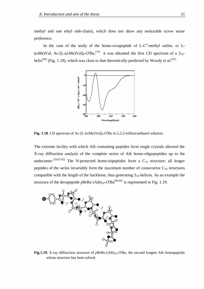
The extreme facility with which Aib containing peptides form single crystals allowed the
X-ray diffraction analysis of the complete series of Aib homo-oligopeptides up to the
undecamer.[29,82-92]
The N-protected homo-tripeptides form a C10 structure; all longer
peptides of the series invariably form the maximum number of consecutive C10 structures
compatible with the length of the backbone, thus generating 310-helices. As an example the
structure of the decapeptide pBrBz-(Aib)10-OtBu
[89,90] is represented in Fig. 1.19.
Fig.1.19. X-ray diffraction structure of pBrBz-(Aib)10-OtBu, the second longest Aib homopeptide
whose structure has been solved.
1. Introduction 22
Conformational analyses in solution (by IR absorption and 1H-NMR spectrometry) showed
that this conformation prevails strongly also in solvents of reduced polarity as deuterated
chloroform.[82,93]
In the case of peptides containing Aib residues and C-trisubstituted residues, in the
solid state only helical structure are observed. Such helices can be of 310 type, of type, or
„mixed‟ (an -helical segment preceded or followed by some C10 structures). More than 40
structures obtained by X-ray diffraction of peptides made of Aib and protein amino acids
with length between 4 and 16 residues were reported in the literature until 1990. From their
analysis it was found that chain length, Aib content and peptide sequence are among the
most important factors directing the conformation towards one of the two helical
structures.[94,95]
In particular, the -helix tends to be favoured as the chain length increases
and as the content of Aib decreases, even if there are several exceptions and evaluation of
the role played by peptide sequence is difficult (Fig. 1.20). On the other side, very short
peptides (six residues or less) display an overwhelming preference for the 310-helix. In
summary, the vast amount of structural data on Aib containing peptides highlights the
remarkable capability of this non proteogenic residue of promoting and stabilizing -turns
as well as helical conformations, particularly 310-helices.
Fig. 1.20. Type of helical structure as a function of the total number of residues and of the number
of Aib residues in the peptide sequence. The dashed line separing 310- from - helices was
traced arbitrarily.
A: Introduction and aim of the thesis 23
1.3 Aim of the thesis
Bearing in mind the above mentioned data, it is reasonable to think that sequential peptides
of limited length made of repeating tripeptide units with one C-trisubstituted residue
between two C-tetrasubstituted -amino acids, fold in a 310-helical conformation. In such
helical pepides the side-chains of each third residue would be almost aligned. Therefore, if
there is a functionalised residue every third position in the peptide sequence, a kind of
active side of the helix would be formed. Two such peptides, derivatised with
complementary chemical probes in the side-chains, could then recognize each other more
easily, since the dimer formation would be favoured by cooperative effects.
Moreover, in the long-term perspective of designing biologically active peptides, a
further advantage of employing C-tetrasubstituted -amino acids is that peptides rich in
C-tetrasubstituted residues are less easily degraded by proteolytic enzymes compared to
peptides formed by protein amino acids.[96]
A peptide composed completely of non natural
C-trisubstituted amino acids and C
-tetrasubstituted -amino acids should have therefore
a considerable stability in vivo.
The aim of this thesis has been to verify the hypothesis described above, in the
specific case of sequential nucleopeptides as nucleic acid analogues. Therefore, the
following targets were set:
- The synthesis of sequential rigid nucleopeptides made of repeating tripeptide
units composed of an -nucleoamino acid between two C-tetrasubstituted -amino
acids;
- Their conformational analysis, in order to verify the assumption of a folded
conformation, likely of 310-helical type, which could favour cooperative interactions
with complementary derivatised structures;
- The synthesis and conformational characterization of sequential flexible
nucleopeptides, in which the C-tetrasubstituted residues would have been replaced by
less structuring C-trisubstitued analogues, to evaluate the effect of backbone rigidity
on the peptide structure;
- The analysis of the pairing properties of both classes of nucleopeptides in order
to evaluate the effects of structuration, firstly towards complementary nucleopeptides,
then in case of encouraging results, towards complementary nucleotides;
1.3. Aim of the thesis 24
- The preliminary study of some relevant biological properties (cytotoxicity, cell
penetration, in vivo stability) of both classes of nucleopeptides, in order to evaluate the
possibility of in vivo applications for derivatives of nucleopeptides which should
display interesting pairing properties towards oligonucleotides in vitro.
The Ph.D. thesis project has been developed in co-direction between the Department of
Chemistry of the Università di Padova (Italy) and the “Laboratoire de Immunologie et
Chimie Thérapeutiques” (UPR 9021, CNRS) of Strasbourg (France). In particular, most of
the solution-phase synthesis of the nucleoamino acids and of the rigid nucleopeptides, as
well as their conformational characterization, has been performed in Padova, whereas the
solid-phase synthesis of the flexible nucleopeptides and the pairing and biological
experiments have been performed in Strasbourg.
B: Discussion and Results 25
2. SYNTHESIS
2.1 Synthesis design
A working and efficient synthesis design is crucial for the success of any chemical
synthesis. In this specific case, given the synthetic targets outlined in the preceding section,
it was required:
- To identify the most convenient -nucleoamino acid, C-tetrasubstitued -amino acid
and C-trisubstitued analogue for the preparation of rigid and flexible sequential
helical nucleopeptides;
- To design synthetic targets both for obtaining model compounds, for conformational
characterization and synthetic protocol evaluation, and for obtaining nucleotide
analogues soluble in physiological conditions;
- To identify useful functionalisations for pairing and biological tests;
- To devise efficient synthetic strategies for the synthesis of both classes of
nucleopeptides and for their functionalisation, in particular concerning to the choice of
the solution or solid phase, of suitable protecting groups, and of the activation
protocols.
Since the decisions taken about each point were conditioning the following steps, the
discussion will address one point a time.
2.1.1 Choice of the amino acids
On the basis of the literature[51]
, -alanyl -nucleoamino acids (AlaB) were identified as
the most convenient for interactions studies of complementary functionalized
nucleopeptides. DNA nucleobases rather than RNA nucleobases were chosen as pendants,
since uracil poses more synthetic problems than thymine.[97]
Aib, or -aminoisobutyric
acid, the simplest C-tetrasubstituted -amino acid, was chosen for the synthesis of the
rigid nucleopeptides, both because of its strong 310-helical promoting properties and
because the decrease in reactivity due to the sterical hindrance of the second methyl at the
C is not so dramatic as in C
-tetrasubstituted -amino acids carrying bulkier
substituents.[98]
Consequently, the protein amino acid alanine was chosen as the C-
trisubstituted analogue for the synthesis of the flexible nucleopeptides. The tripeptide units
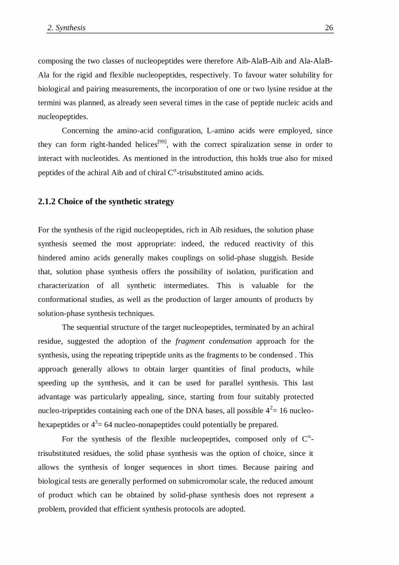
2. Synthesis 26
composing the two classes of nucleopeptides were therefore Aib-AlaB-Aib and Ala-AlaB-
Ala for the rigid and flexible nucleopeptides, respectively. To favour water solubility for
biological and pairing measurements, the incorporation of one or two lysine residue at the
termini was planned, as already seen several times in the case of peptide nucleic acids and
nucleopeptides.
Concerning the amino-acid configuration, L-amino acids were employed, since
they can form right-handed helices[99]
, with the correct spiralization sense in order to
interact with nucleotides. As mentioned in the introduction, this holds true also for mixed
peptides of the achiral Aib and of chiral C-trisubstituted amino acids.
2.1.2 Choice of the synthetic strategy
For the synthesis of the rigid nucleopeptides, rich in Aib residues, the solution phase
synthesis seemed the most appropriate: indeed, the reduced reactivity of this
hindered amino acids generally makes couplings on solid-phase sluggish. Beside
that, solution phase synthesis offers the possibility of isolation, purification and
characterization of all synthetic intermediates. This is valuable for the
conformational studies, as well as the production of larger amounts of products by
solution-phase synthesis techniques.
The sequential structure of the target nucleopeptides, terminated by an achiral
residue, suggested the adoption of the fragment condensation approach for the
synthesis, using the repeating tripeptide units as the fragments to be condensed . This
approach generally allows to obtain larger quantities of final products, while
speeding up the synthesis, and it can be used for parallel synthesis. This last
advantage was particularly appealing, since, starting from four suitably protected
nucleo-tripeptides containing each one of the DNA bases, all possible 42= 16 nucleo-
hexapeptides or 43= 64 nucleo-nonapeptides could potentially be prepared.
For the synthesis of the flexible nucleopeptides, composed only of C-
trisubstituted residues, the solid phase synthesis was the option of choice, since it
allows the synthesis of longer sequences in short times. Because pairing and
biological tests are generally performed on submicromolar scale, the reduced amount
of product which can be obtained by solid-phase synthesis does not represent a
problem, provided that efficient synthesis protocols are adopted.
B: Discussion and Results 27
2.1.3 Choice of suitable protecting groups
Generally speaking, the functions that require protection in peptide synthesis are the N-
terminal amino and C-terminal carboxyl group, as well as functional groups at the side
chains. The ideal conditions are satisfied when orthogonal protecting groups are
employed:[100]
two protecting groups are fully orthogonal if either group can be removed
under conditions which do not compromise the stability of the other; partial orthogonality
refers to the case that only one can be removed without causing removal of the other.
Concerning the terminal functionalities, in case of the solid-phase synthesis of the flexible
nucleopeptides, the C-terminal carboxyl group is bound to the resin until the cleavage at
the end of the synthesis; therefore only partial orthogonality with the -amino protecting
group is required. On the contrary, the solution-phase synthesis of the rigid nucleopeptides
by fragment condensation requires full orthogonality between N- and C-terminal protecting
groups, while the synthesis of the single fragments can be accomplished also in the case of
partial orthogonality.
Considering the side-chains of the target nucleopeptides, it appears that Aib and
Ala are not an issue, whereas the nucleobase pendants of the nucleoamino acid and the -
amino group of lysine (where present) must be taken into account. Indeed, three out of four
bases (adenine, cytosine, guanine) require protection: cytosine for chemoselectivity
reasons, the purine bases for solubility reasons. After their incorporation in the
nucleoamino acids, nucleobase deprotection is not required until the very end of the
synthesis, therefore partial orthogonality is sufficient for their protecting groups. The same
is true for the -amino group of lysine if it is introduced simply for providing a positively
charged ammonium moiety after complete deprotection; however, if this group is meant
for specific derivatization, full orthogonality must be ensured.
2.1.3.1 Nucleobase protecting groups
Nucleobases are difficult to manipulate, being highly hydrophilic, highly insoluble in the
most common organic solvents, and sparingly soluble and aggregation prone even in the
most polar solvents, such as MeCN, DMF or DMSO.[97]
The protecting groups chosen for
the nucleobases, in particular for the purines, should therefore make them less hydrophilic
and increase their solubility in polar organic solvents. This means that not all protecting
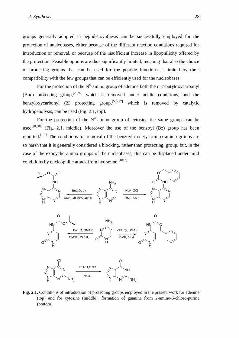
2. Synthesis 28
groups generally adopted in peptide synthesis can be successfully employed for the
protection of nucleobases, either because of the different reaction conditions required for
introduction or removal, or because of the insufficient increase in lipophilicity offered by
the protection. Feasible options are thus significantly limited, meaning that also the choice
of protecting groups that can be used for the peptide functions is limited by their
compatibility with the few groups that can be efficiently used for the nucleobases.
For the protection of the N6-amino group of adenine both the tert-butyloxycarbonyl
(Boc) protecting group,[20,97]
which is removed under acidic conditions, and the
benzyloxycarbonyl (Z) protecting group,[58b,97]
which is removed by catalytic
hydrogenolysis, can be used (Fig. 2.1, top).
For the protection of the N4-amino group of cytosine the same groups can be
used[20,58b]
(Fig. 2.1, middle). Moreover the use of the benzoyl (Bz) group has been
reported.[101]
The conditions for removal of the benzoyl moiety from -amino groups are
so harsh that it is generally considered a blocking, rather than protecting, group, but, in the
case of the exocyclic amino groups of the nucleobases, this can be displaced under mild
conditions by nucleophilic attack from hydrazine.[101b]
Fig. 2.1. Conditions of introduction of protecting groups employed in the present work for adenine
(top) and for cytosine (middle); formation of guanine from 2-amino-6-chloro-purine
(bottom).
NH N
NN
NHO
O
NH
N
O
NH2
NH
N
O
NH
O
O
NH N
NN
NH2
O
NH N
NN
NH
O
NH N
NNH
O
NH2
NH N
NN
Cl
NH2
NH
N
O
NH
O
O
NaH, ZCl
DMF, 5h rt
ZCl, py, DMAP
Boc2O, py
DMF, 1h 80°C,18h rt
TFA/H2O 3:1
Boc2O, DMAP
DMSO, 24h rt, DMF, 3d rt
3d rt
B: Discussion and Results 29
Concerning guanine, both the O6
protection as an ether,[102a]
or as a urethane,[102b]
and the
N2 protection as an amide
[103] have been reported, but neither solution ensures both
efficient and stable protection and removal under mild condition. In most cases dealing
with nucleotide analogues,[54,104]
guanine-based nucleotide analogues have been obtained
starting from 2-amino-6-chloro-purine (H-GCl
).[105]
The chlorine atom favours the
solubility in organic media compared to guanine and it can be replaced by an hydroxyl
under strongly acidic aqueous conditions (Fig. 2.1, bottom).
2.1.3.2 N-amino- and carboxyl- protecting groups
The protecting groups chosen for the preparation of the flexible nucleopeptides had to be
compatible with solid-phase synthesis conditions. The two most common strategies for the
synthesis of peptides on solid-phase are the Boc/Bzl or the Fmoc/tBu.
[106] In the first one,
-amino groups are protected with the acid-labile tert-butyloxycarbonyl (Boc)[107]
protecting group, in the second one the base labile fluorenylmethyloxycarbonyl (Fmoc)
protecting group is used.[108]
Both Boc-protected[47]
and Fmoc-protected[109]
-alanyl -
nucleoamino acids have been employed for the solid-phase synthesis of nucleopeptides.
However, two more steps are required in order to obtain the (non commercially available)
Fmoc-protected nucleoamino acids compared to the Boc-protected nucleoamino acids.
This is because their synthesis relies on the nucleobase alkylation under strongly basic
conditions (see section 2.2), which are not compatible with Fmoc-chemistry. Therefore
nucleoamino acids must be prepared at first as Boc- or Z- protected derivatives, then
deprotected and protected again with Fmoc. Consequently the Boc group has been chosen
as -amino protecting group for the flexible nucleopeptides, while protecting groups stable
to moderate acidic conditions were needed for the side-chains (e.g. for the adenine and
cytosine the Z protecting was employed).
Concerning the solution-phase synthesis of the rigid nucleopeptides, the situation
was more complex, because of the need of an additional orthogonal protection for the
carboxyl function. The most convenient urethane N-protecting group for solution-phase
synthesis is by far the Z group.[110]
The three main advantages of the use of this group are:
(i) the good physical and chemical stability of Z-protected peptides, (ii) the ease of
deprotection by catalytic hydrogenolysis, forming volatile co-products, which are smoothly
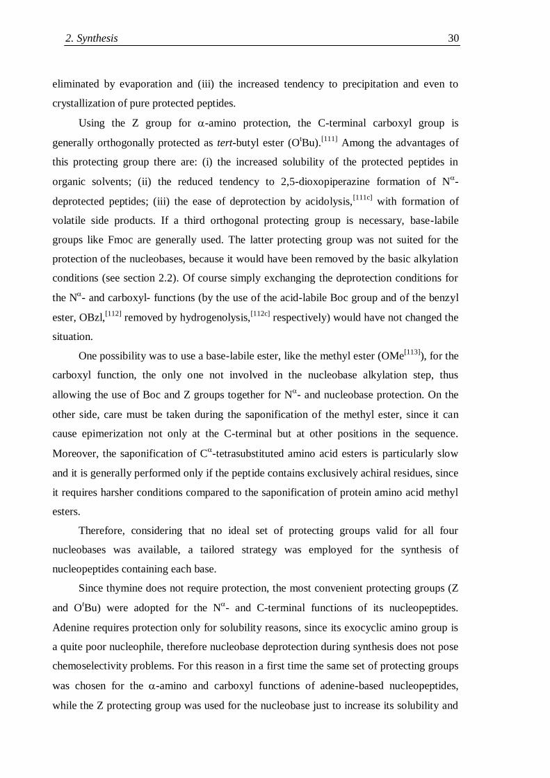
2. Synthesis 30
eliminated by evaporation and (iii) the increased tendency to precipitation and even to
crystallization of pure protected peptides.
Using the Z group for -amino protection, the C-terminal carboxyl group is
generally orthogonally protected as tert-butyl ester (OtBu).
[111] Among the advantages of
this protecting group there are: (i) the increased solubility of the protected peptides in
organic solvents; (ii) the reduced tendency to 2,5-dioxopiperazine formation of N-
deprotected peptides; (iii) the ease of deprotection by acidolysis,[111c]
with formation of
volatile side products. If a third orthogonal protecting group is necessary, base-labile
groups like Fmoc are generally used. The latter protecting group was not suited for the
protection of the nucleobases, because it would have been removed by the basic alkylation
conditions (see section 2.2). Of course simply exchanging the deprotection conditions for
the N- and carboxyl- functions (by the use of the acid-labile Boc group and of the benzyl
ester, OBzl,[112]
removed by hydrogenolysis,[112c]
respectively) would have not changed the
situation.
One possibility was to use a base-labile ester, like the methyl ester (OMe[113]
), for the
carboxyl function, the only one not involved in the nucleobase alkylation step, thus
allowing the use of Boc and Z groups together for N- and nucleobase protection. On the
other side, care must be taken during the saponification of the methyl ester, since it can
cause epimerization not only at the C-terminal but at other positions in the sequence.
Moreover, the saponification of C-tetrasubstituted amino acid esters is particularly slow
and it is generally performed only if the peptide contains exclusively achiral residues, since
it requires harsher conditions compared to the saponification of protein amino acid methyl
esters.
Therefore, considering that no ideal set of protecting groups valid for all four
nucleobases was available, a tailored strategy was employed for the synthesis of
nucleopeptides containing each base.
Since thymine does not require protection, the most convenient protecting groups (Z
and OtBu) were adopted for the N
- and C-terminal functions of its nucleopeptides.
Adenine requires protection only for solubility reasons, since its exocyclic amino group is
a quite poor nucleophile, therefore nucleobase deprotection during synthesis does not pose
chemoselectivity problems. For this reason in a first time the same set of protecting groups
was chosen for the -amino and carboxyl functions of adenine-based nucleopeptides,
while the Z protecting group was used for the nucleobase just to increase its solubility and
B: Discussion and Results 31
facilitate its manipulation up to the synthesis of the protected nucleo-dipeptide, then
simultaneous deprotection of both Z groups was planned. Unfortunately, the use of N-Z
protecting group for adenine-based nucleopeptides turned out to be not satisfactory (see
section 2.3.2). Therefore, as completely orthogonal protecting groups the Boc and OMe
moieties were employed for the backbone functions and the Z group was used for the
nucleobase. Cytosine required an orthogonal set of protecting groups, its amino group
being the most nucleophilic and basic (pKa 4.2)[97]
among nucleobases, therefore the same
set of orthogonal protecting groups employed in the case of adenine was used. Concerning
guanine-based nucleopeptides, it was chosen to use 2-amino-6-chloro-purine as the starting
material. As it was not clear whether the 6-chloro group would have resisted Boc or OtBu
deprotection conditions (strong acid in aprotic solvent), the Z group was chosen for N
protection and the carboxyl group was protected as OMe. In Fig. 2.2 nucleodipeptides
containing the four bases are represented with the planned protecting groups.
Fig. 2.2. The planned set of protecting groups for protected nucleodipeptides containing the four
DNA nucleobases. Different colours are used for different deprotection conditions:
hydrogenolysis (green), acidolysis (blue), saponification (red), acidic hydrolysis (purple).
2.2 Nucleoamino acid synthesis
The synthesis of the alanyl -nucleoamino acids relies on the nucleophilic attack of the
suitably protected nucleobases on the -carbon of the N-protected serine -lactone[44]
in
the presence of a strong, non-nucleophilic base. The lactone intermediate is not
O NH
NH
O
O
O
B
O
O NH
NH
O
O
O
O
B
O NH
NH
O
O
O
B
O
O NH
NH
O
O
O
B
O
Cl
AlaT nucleopeptides AlaAZ nucleopeptides
Z
AlaCZ, AlaAZ nucleopeptides
Z
AlaGCl nucleopeptides
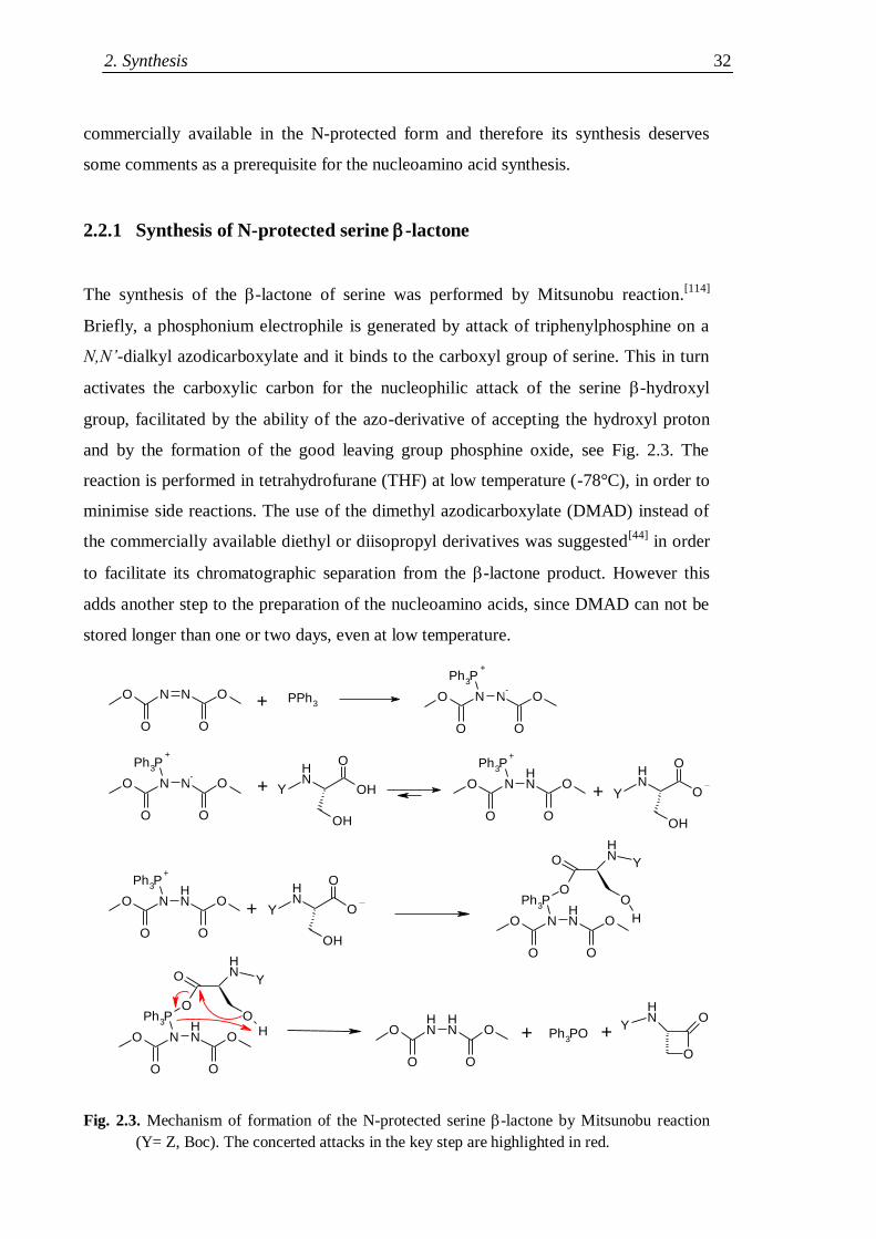
2. Synthesis 32
commercially available in the N-protected form and therefore its synthesis deserves
some comments as a prerequisite for the nucleoamino acid synthesis.
2.2.1 Synthesis of N-protected serine -lactone
The synthesis of the -lactone of serine was performed by Mitsunobu reaction.[114]
Briefly, a phosphonium electrophile is generated by attack of triphenylphosphine on a
N,N’-dialkyl azodicarboxylate and it binds to the carboxyl group of serine. This in turn
activates the carboxylic carbon for the nucleophilic attack of the serine -hydroxyl
group, facilitated by the ability of the azo-derivative of accepting the hydroxyl proton
and by the formation of the good leaving group phosphine oxide, see Fig. 2.3. The
reaction is performed in tetrahydrofurane (THF) at low temperature (-78°C), in order to
minimise side reactions. The use of the dimethyl azodicarboxylate (DMAD) instead of
the commercially available diethyl or diisopropyl derivatives was suggested[44]
in order
to facilitate its chromatographic separation from the -lactone product. However this
adds another step to the preparation of the nucleoamino acids, since DMAD can not be
stored longer than one or two days, even at low temperature.
Fig. 2.3. Mechanism of formation of the N-protected serine -lactone by Mitsunobu reaction
(Y= Z, Boc). The concerted attacks in the key step are highlighted in red.
O
O
N N
O
O PPh3
O
O
N N
O
O
Ph3P
NH
OH
O
OH
O
O
N NH
O
O
Ph3P
O
O
N N
O
O
Ph3P
NH
O
O
OH
O
O
N NH
O
O
Ph3P
NH
O
O
OH
O
O
N NH
O
O
Ph3P
O
O NH
O
H
O
O
N NH
O
O
Ph3P
O
O NH
OH O
O
NH
NH
O
O Ph3PO
O
NH
O
+
+
-
+ Y
++
-
+ Y
+
+ Y
Y
Y
+ + Y
B: Discussion and Results 33
The steric tension in the four member ring directs the selectivity of nucleophilic attacks
by soft nucleophiles towards the -carbon instead of towards the carbonyl group, as
expected for other esters,[44]
thus allowing the following synthetic steps. This makes
however the lactone extremely prone to decomposition. Indeed, the compound is not
stable in the reaction mixture, as it can be attacked by unreacted phosphine or by the
excess serine, and significant decomposition occurs even overnight at -20°C; therefore
it must be immediately purified by flash-chromatography. Moreover, the compound is
sensitive to nucleophilic impurities, particularly to moisture, so that the yield obtained
in a humid summer was systematically lower than in winter. Even worse, the compound
is extremely sensitive to radicals, even in traces, like the ones which are easily formed
by THF or by remnants of the NBS used for DMAD formation. In this cases no product
was obtained. Notwithstanding such serious problems, it was generally possible to
reach yields close to literature values (55%).[44]
2.2.2 Nucleobase alkylation
Performing the nucleobase alkylation with the serine -lactone in the presence of a strong,
non-nucleophilic base exposes to the risk of loss of the -proton of the lactone with
consequent racemisation. The reaction conditions must be carefully studied in order to
avoid or minimise this insidious side reaction.
The original paper of Eschenmoser[45]
was not rich in details on reaction
(nucleobase and non-nucleophilic base equivalents, addition order, etc.) and purification
conditions, and the same details were missing in the relevant papers of
Diederichsen.[47,48b,51]
Optimization of the synthesis protocol was therefore necessary and
thymine was chosen as the most convenient test nucleobase. However, after completing the
optimization process, a different, detailed alkylation protocol was found in the
literature,[109]
covering all nucleobases except guanine, even if using sometimes different
protecting groups. Such protocols used exactly one equivalent of non-nucleophilic base
(NaH for pyrimidine nucleobases, or DBU for the purines) and required the slow dropwise
addition of the serine -lactone to a DMF solution containing a slight excess of the
partially deprotonated nucleobase at -78°C. In order to compare the two methods, the
alkylation was performed on a preparative scale with both and two series of protected
nucleopeptides up to the hexamer were synthesised (see Section 2.3). By analyzing the
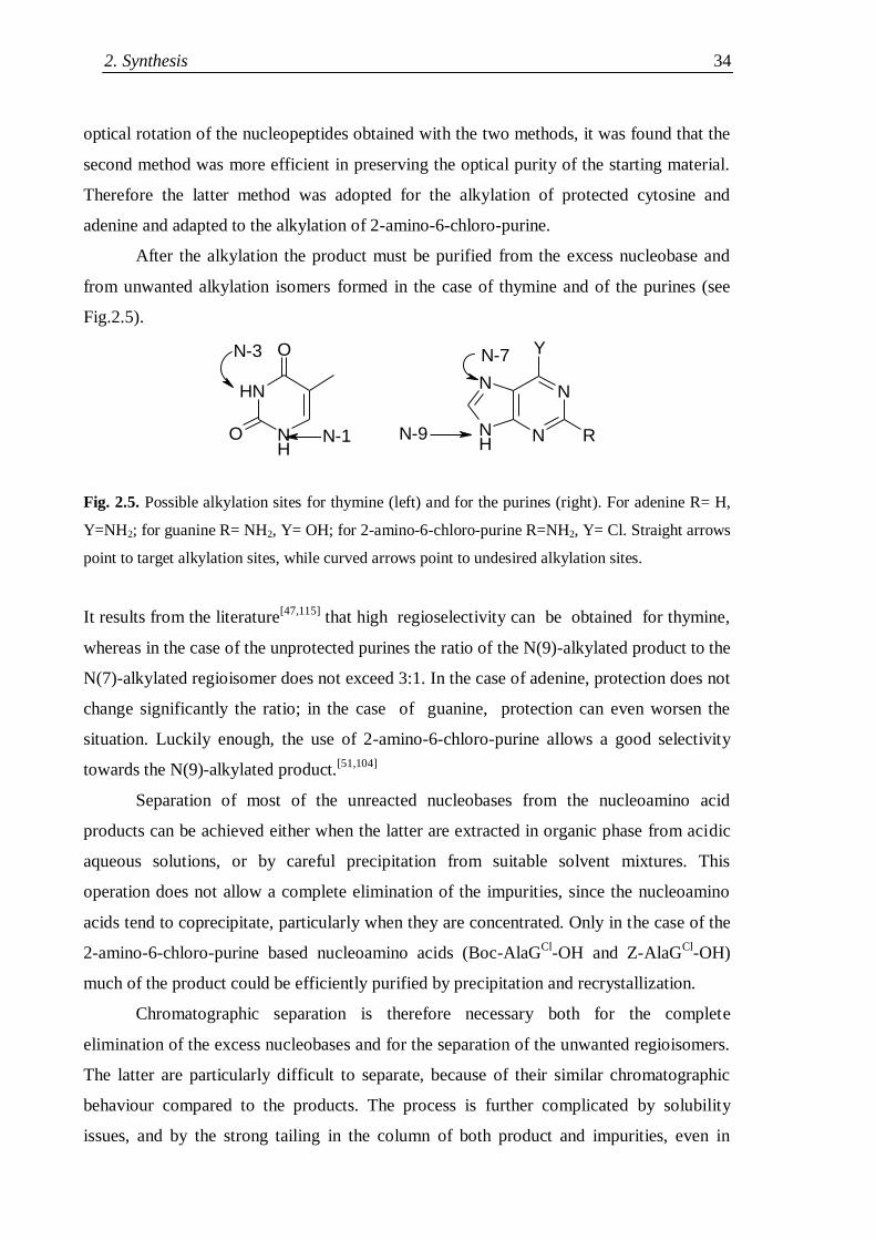
2. Synthesis 34
optical rotation of the nucleopeptides obtained with the two methods, it was found that the
second method was more efficient in preserving the optical purity of the starting material.
Therefore the latter method was adopted for the alkylation of protected cytosine and
adenine and adapted to the alkylation of 2-amino-6-chloro-purine.
After the alkylation the product must be purified from the excess nucleobase and
from unwanted alkylation isomers formed in the case of thymine and of the purines (see
Fig.2.5).
Fig. 2.5. Possible alkylation sites for thymine (left) and for the purines (right). For adenine R= H,
Y=NH2; for guanine R= NH2, Y= OH; for 2-amino-6-chloro-purine R=NH2, Y= Cl. Straight arrows
point to target alkylation sites, while curved arrows point to undesired alkylation sites.
It results from the literature[47,115]
that high regioselectivity can be obtained for thymine,
whereas in the case of the unprotected purines the ratio of the N(9)-alkylated product to the
N(7)-alkylated regioisomer does not exceed 3:1. In the case of adenine, protection does not
change significantly the ratio; in the case of guanine, protection can even worsen the
situation. Luckily enough, the use of 2-amino-6-chloro-purine allows a good selectivity
towards the N(9)-alkylated product.[51,104]
Separation of most of the unreacted nucleobases from the nucleoamino acid
products can be achieved either when the latter are extracted in organic phase from acidic
aqueous solutions, or by careful precipitation from suitable solvent mixtures. This
operation does not allow a complete elimination of the impurities, since the nucleoamino
acids tend to coprecipitate, particularly when they are concentrated. Only in the case of the
2-amino-6-chloro-purine based nucleoamino acids (Boc-AlaGCl
-OH and Z-AlaGCl
-OH)
much of the product could be efficiently purified by precipitation and recrystallization.
Chromatographic separation is therefore necessary both for the complete
elimination of the excess nucleobases and for the separation of the unwanted regioisomers.
The latter are particularly difficult to separate, because of their similar chromatographic
behaviour compared to the products. The process is further complicated by solubility
issues, and by the strong tailing in the column of both product and impurities, even in
NH
NH
O
O N
NN
NH
Y
RN-1
N-3 N-7
N-9
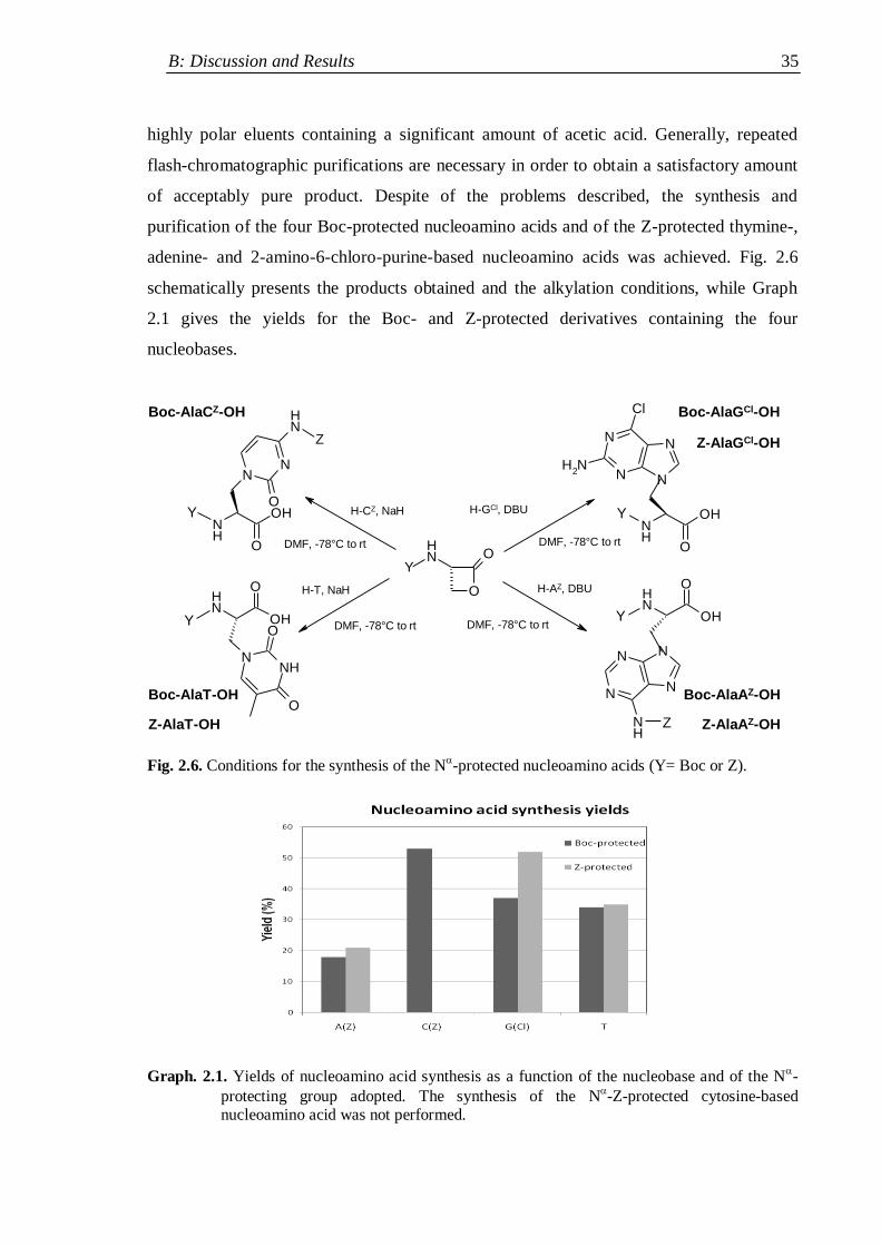
B: Discussion and Results 35
highly polar eluents containing a significant amount of acetic acid. Generally, repeated
flash-chromatographic purifications are necessary in order to obtain a satisfactory amount
of acceptably pure product. Despite of the problems described, the synthesis and
purification of the four Boc-protected nucleoamino acids and of the Z-protected thymine-,
adenine- and 2-amino-6-chloro-purine-based nucleoamino acids was achieved. Fig. 2.6
schematically presents the products obtained and the alkylation conditions, while Graph
2.1 gives the yields for the Boc- and Z-protected derivatives containing the four
nucleobases.
O
NH
O
NH
OH
O
NNH
O
O
N
NN
N
NH
NH
OH
O
NH
OH
O
NN
NH
O
N
NN
N
Cl
NH2
NH
OH
O
Y
Y
Y
H-T, NaH
DMF, -78°C to rt
Boc-AlaT-OH
H-AZ, DBU
DMF, -78°C to rt
Z
Boc-AlaAZ-OH
Boc-AlaCZ-OH
Z
H-CZ, NaH
DMF, -78°C to rt
Boc-AlaGCl-OH
H-GCl, DBU
DMF, -78°C to rt
Y
Y
Z-AlaT-OH
Z-AlaGCl-OH
Z-AlaAZ-OH
Fig. 2.6. Conditions for the synthesis of the N-protected nucleoamino acids (Y= Boc or Z).
Graph. 2.1. Yields of nucleoamino acid synthesis as a function of the nucleobase and of the N-
protecting group adopted. The synthesis of the N-Z-protected cytosine-based
nucleoamino acid was not performed.
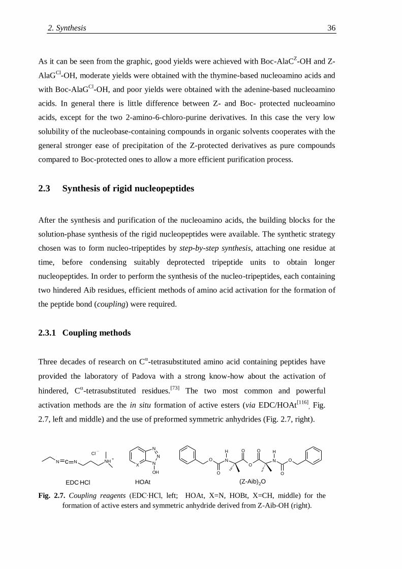
2. Synthesis 36
As it can be seen from the graphic, good yields were achieved with Boc-AlaCZ-OH and Z-
AlaGCl
-OH, moderate yields were obtained with the thymine-based nucleoamino acids and
with Boc-AlaGCl
-OH, and poor yields were obtained with the adenine-based nucleoamino
acids. In general there is little difference between Z- and Boc- protected nucleoamino
acids, except for the two 2-amino-6-chloro-purine derivatives. In this case the very low
solubility of the nucleobase-containing compounds in organic solvents cooperates with the
general stronger ease of precipitation of the Z-protected derivatives as pure compounds
compared to Boc-protected ones to allow a more efficient purification process.
2.3 Synthesis of rigid nucleopeptides
After the synthesis and purification of the nucleoamino acids, the building blocks for the
solution-phase synthesis of the rigid nucleopeptides were available. The synthetic strategy
chosen was to form nucleo-tripeptides by step-by-step synthesis, attaching one residue at
time, before condensing suitably deprotected tripeptide units to obtain longer
nucleopeptides. In order to perform the synthesis of the nucleo-tripeptides, each containing
two hindered Aib residues, efficient methods of amino acid activation for the formation of
the peptide bond (coupling) were required.
2.3.1 Coupling methods
Three decades of research on C-tetrasubstituted amino acid containing peptides have
provided the laboratory of Padova with a strong know-how about the activation of
hindered, C-tetrasubstituted residues.
[73] The two most common and powerful
activation methods are the in situ formation of active esters (via EDC/HOAt[116]
, Fig.
2.7, left and middle) and the use of preformed symmetric anhydrides (Fig. 2.7, right).
O
O
NO
N
H O O H
O
O
X N
N
OH
N
N N NH
Cl
(Z-Aib)2OHOAtEDC.HCl
+c
Fig. 2.7. Coupling reagents (EDC∙HCl, left; HOAt, X=N, HOBt, X=CH, middle) for the
formation of active esters and symmetric anhydride derived from Z-Aib-OH (right).
B: Discussion and Results 37
Active esters are easily formed, the use of HOAt efficiently suppresses racemisation due to
carbodiimide overactivation,[116]
and the side products deriving from the coupling reagents
can be eliminated by acidic or basic washing. However, the unreacted active ester is not
completely hydrolyzed or eliminated by washing and often chromatographic purification is
necessary.
On the contrary, couplings via symmetric anhydride form a smaller number of side
products, resulting in a simpler work-up, but they require more steps (anhydride synthesis
and purification, coupling, sometimes N-protected amino acid recovery). Indeed, 2
equivalent of N-protected amino acid are required in order to generate 1 equivalent of
acylating agent, and 1 equivalent of amino acid salt is formed after the coupling, thus
making the amino acid recovery necessary when working with large amounts or valuable
compounds.
The active ester coupling method was therefore always used for the activation of
the nucleoamino acids, while the activation as symmetric anhydride was often employed
for Aib activation. Large scale couplings involving Aib activation and couplings between
protein amino acids were performed via active esters, using the less expensive HOBt[117]
(Fig. 2.7, middle) instead of HOAt.
2.3.2 Synthesis of protected nucleo-tripeptides
The step-by-step synthesis of the thymine-based protected nucleo-tripeptide Z-Aib-
AlaT-Aib-OtBu was smoothly achieved by coupling the activated nucleoamino acid
with H-Aib-OtBu to form the protected nucleo-dipeptide Z-AlaT-Aib-O
tBu, which was
then hydrogenated and acylated with the symmetric anhydride (Z-Aib)2-O, as shown in
Scheme 2.1.
The same strategy was not efficient in the case of the adenine-based
nucleopeptides. Indeed, during the step of dipeptide deprotection, while the N6-Z
protecting group was rapidly removed by catalytic hydrogenation, the removal of the
N-Z group was extremely slow, so that the reaction was not complete even after 7
hours (Fig. 2.8), and the coupling yield was negatively affected. Indeed slow
hydrogenolysis of protecting groups next to the two most aromatic bases (adenine and
cytosine) have been reported,[109]
probably due to stacking interactions between the
nucleobase and the Z phenyl ring hindering the removal of the latter.
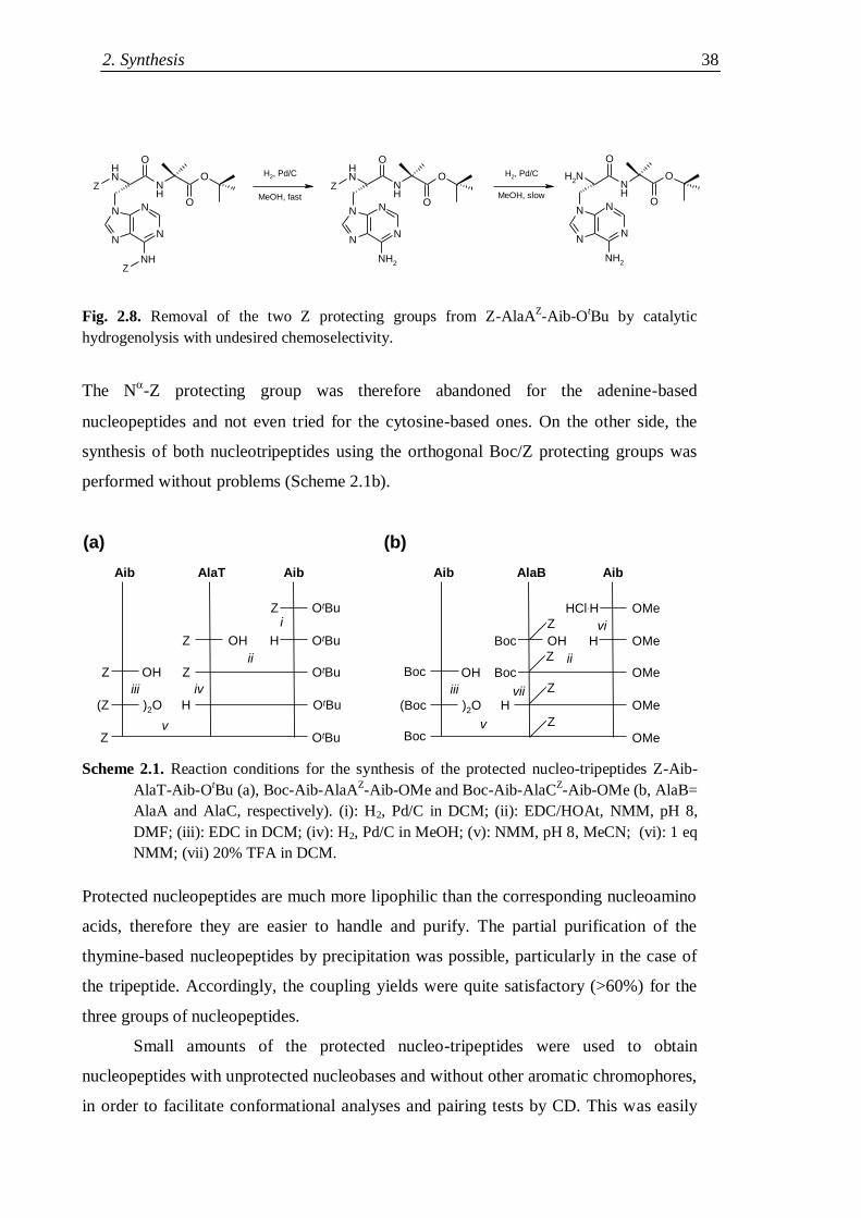
2. Synthesis 38
Fig. 2.8. Removal of the two Z protecting groups from Z-AlaAZ-Aib-O
tBu by catalytic
hydrogenolysis with undesired chemoselectivity.
The N-Z protecting group was therefore abandoned for the adenine-based
nucleopeptides and not even tried for the cytosine-based ones. On the other side, the
synthesis of both nucleotripeptides using the orthogonal Boc/Z protecting groups was
performed without problems (Scheme 2.1b).
Aib AlaT Aib
OtBu
OtBu
OtBu
OtBu
OtBu
Z
HOHZ
Z
H)2O(Z
Z
OHZii
iii iv
(a)
Aib AlaB Aib
OMe
OMe
OMe
OMe
OMe
HCl.H
HOHBoc
Boc
H)2O(Boc
OHBocii
iii
vBoc
(b)
Z
Z
Z
Zi vi
vii
v
Scheme 2.1. Reaction conditions for the synthesis of the protected nucleo-tripeptides Z-Aib-
AlaT-Aib-OtBu (a), Boc-Aib-AlaA
Z-Aib-OMe and Boc-Aib-AlaC
Z-Aib-OMe (b, AlaB=
AlaA and AlaC, respectively). (i): H2, Pd/C in DCM; (ii): EDC/HOAt, NMM, pH 8,
DMF; (iii): EDC in DCM; (iv): H2, Pd/C in MeOH; (v): NMM, pH 8, MeCN; (vi): 1 eq
NMM; (vii) 20% TFA in DCM.
Protected nucleopeptides are much more lipophilic than the corresponding nucleoamino
acids, therefore they are easier to handle and purify. The partial purification of the
thymine-based nucleopeptides by precipitation was possible, particularly in the case of
the tripeptide. Accordingly, the coupling yields were quite satisfactory (>60%) for the
three groups of nucleopeptides.
Small amounts of the protected nucleo-tripeptides were used to obtain
nucleopeptides with unprotected nucleobases and without other aromatic chromophores,
in order to facilitate conformational analyses and pairing tests by CD. This was easily
N N
NN
NH
NH
O
NH
O
ON N
NN
NH2
NH
O
NH
O
ON N
NN
NH2
NH2
O
NH
O
O
Z
Z
Z
H2, Pd/C
MeOH, fast
H2, Pd/C
MeOH, slow
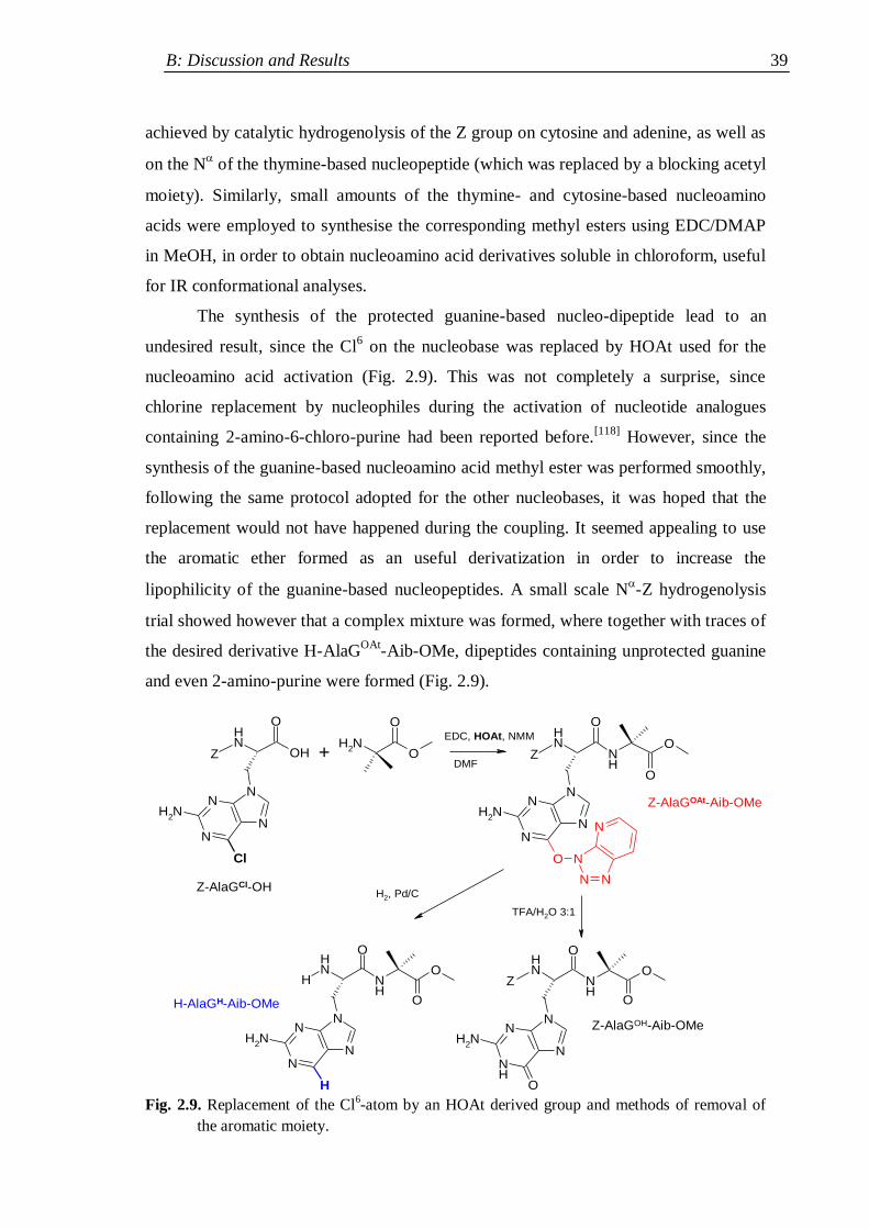
B: Discussion and Results 39
achieved by catalytic hydrogenolysis of the Z group on cytosine and adenine, as well as
on the N of the thymine-based nucleopeptide (which was replaced by a blocking acetyl
moiety). Similarly, small amounts of the thymine- and cytosine-based nucleoamino
acids were employed to synthesise the corresponding methyl esters using EDC/DMAP
in MeOH, in order to obtain nucleoamino acid derivatives soluble in chloroform, useful
for IR conformational analyses.
The synthesis of the protected guanine-based nucleo-dipeptide lead to an
undesired result, since the Cl6 on the nucleobase was replaced by HOAt used for the
nucleoamino acid activation (Fig. 2.9). This was not completely a surprise, since
chlorine replacement by nucleophiles during the activation of nucleotide analogues
containing 2-amino-6-chloro-purine had been reported before.[118]
However, since the
synthesis of the guanine-based nucleoamino acid methyl ester was performed smoothly,
following the same protocol adopted for the other nucleobases, it was hoped that the
replacement would not have happened during the coupling. It seemed appealing to use
the aromatic ether formed as an useful derivatization in order to increase the
lipophilicity of the guanine-based nucleopeptides. A small scale N-Z hydrogenolysis
trial showed however that a complex mixture was formed, where together with traces of
the desired derivative H-AlaGOAt
-Aib-OMe, dipeptides containing unprotected guanine
and even 2-amino-purine were formed (Fig. 2.9).
Fig. 2.9. Replacement of the Cl6-atom by an HOAt derived group and methods of removal of
the aromatic moiety.
NH
O
OH
N
N
N
N
Cl
NH2
NH2
O
O
N
N
N
N
O
NH2
N
N N
N
NH
O
ONH
O
N
N
N
N
NH2
H
NH
O
ONH
O
H
N
N
N
NH
NH2
O
NH
O
ONH
O
Z +EDC, HOAt, NMM
ZDMF
Z
TFA/H2O 3:1
H2, Pd/C
H-AlaGH-Aib-OMe
Z-AlaGOH-Aib-OMe
Z-AlaGOAt-Aib-OMe
Z-AlaGCl-OH
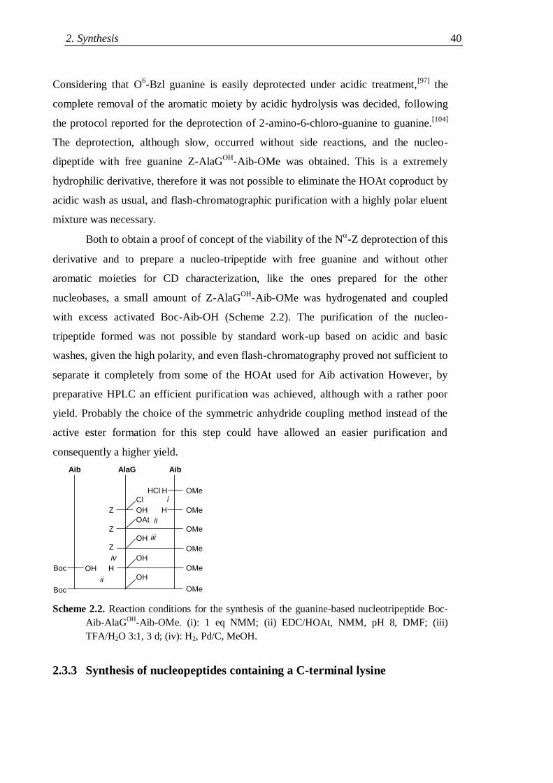
2. Synthesis 40
Considering that O6-Bzl guanine is easily deprotected under acidic treatment,
[97] the
complete removal of the aromatic moiety by acidic hydrolysis was decided, following
the protocol reported for the deprotection of 2-amino-6-chloro-guanine to guanine.[104]
The deprotection, although slow, occurred without side reactions, and the nucleo-
dipeptide with free guanine Z-AlaGOH
-Aib-OMe was obtained. This is a extremely
hydrophilic derivative, therefore it was not possible to eliminate the HOAt coproduct by
acidic wash as usual, and flash-chromatographic purification with a highly polar eluent
mixture was necessary.
Both to obtain a proof of concept of the viability of the N-Z deprotection of this
derivative and to prepare a nucleo-tripeptide with free guanine and without other
aromatic moieties for CD characterization, like the ones prepared for the other
nucleobases, a small amount of Z-AlaGOH
-Aib-OMe was hydrogenated and coupled
with excess activated Boc-Aib-OH (Scheme 2.2). The purification of the nucleo-
tripeptide formed was not possible by standard work-up based on acidic and basic
washes, given the high polarity, and even flash-chromatography proved not sufficient to
separate it completely from some of the HOAt used for Aib activation However, by
preparative HPLC an efficient purification was achieved, although with a rather poor
yield. Probably the choice of the symmetric anhydride coupling method instead of the
active ester formation for this step could have allowed an easier purification and
consequently a higher yield.
Aib AlaG Aib
OMe
OMe
OMe
OMe
OMe
HCl.H
HOHZ
Z
HOHBoc
ii
iv
i
ii
Z
OMeBoc
Cl
OAt
OH
OH
OH
iii
Scheme 2.2. Reaction conditions for the synthesis of the guanine-based nucleotripeptide Boc-
Aib-AlaGOH
-Aib-OMe. (i): 1 eq NMM; (ii) EDC/HOAt, NMM, pH 8, DMF; (iii)
TFA/H2O 3:1, 3 d; (iv): H2, Pd/C, MeOH.
2.3.3 Synthesis of nucleopeptides containing a C-terminal lysine
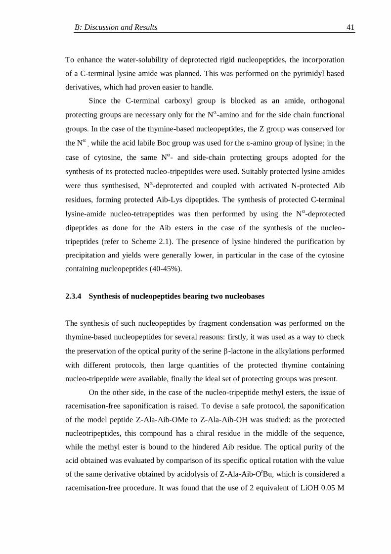
B: Discussion and Results 41
To enhance the water-solubility of deprotected rigid nucleopeptides, the incorporation
of a C-terminal lysine amide was planned. This was performed on the pyrimidyl based
derivatives, which had proven easier to handle.
Since the C-terminal carboxyl group is blocked as an amide, orthogonal
protecting groups are necessary only for the N-amino and for the side chain functional
groups. In the case of the thymine-based nucleopeptides, the Z group was conserved for
the N
, while the acid labile Boc group was used for the -amino group of lysine; in the
case of cytosine, the same N- and side-chain protecting groups adopted for the
synthesis of its protected nucleo-tripeptides were used. Suitably protected lysine amides
were thus synthesised, N-deprotected and coupled with activated N-protected Aib
residues, forming protected Aib-Lys dipeptides. The synthesis of protected C-terminal
lysine-amide nucleo-tetrapeptides was then performed by using the N-deprotected
dipeptides as done for the Aib esters in the case of the synthesis of the nucleo-
tripeptides (refer to Scheme 2.1). The presence of lysine hindered the purification by
precipitation and yields were generally lower, in particular in the case of the cytosine
containing nucleopeptides (40-45%).
2.3.4 Synthesis of nucleopeptides bearing two nucleobases
The synthesis of such nucleopeptides by fragment condensation was performed on the
thymine-based nucleopeptides for several reasons: firstly, it was used as a way to check
the preservation of the optical purity of the serine -lactone in the alkylations performed
with different protocols, then large quantities of the protected thymine containing
nucleo-tripeptide were available, finally the ideal set of protecting groups was present.
On the other side, in the case of the nucleo-tripeptide methyl esters, the issue of
racemisation-free saponification is raised. To devise a safe protocol, the saponification
of the model peptide Z-Ala-Aib-OMe to Z-Ala-Aib-OH was studied: as the protected
nucleotripeptides, this compound has a chiral residue in the middle of the sequence,
while the methyl ester is bound to the hindered Aib residue. The optical purity of the
acid obtained was evaluated by comparison of its specific optical rotation with the value
of the same derivative obtained by acidolysis of Z-Ala-Aib-OtBu, which is considered a
racemisation-free procedure. It was found that the use of 2 equivalent of LiOH 0.05 M
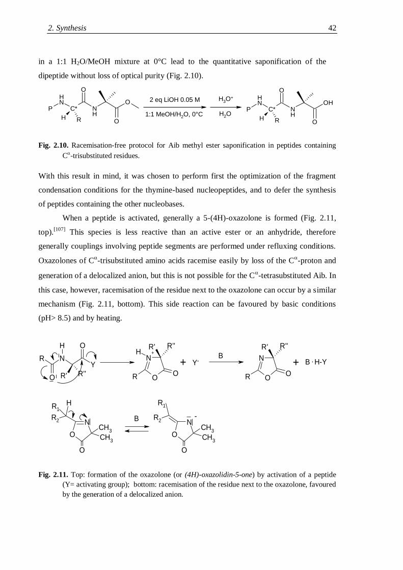
2. Synthesis 42
in a 1:1 H2O/MeOH mixture at 0°C lead to the quantitative saponification of the
dipeptide without loss of optical purity (Fig. 2.10).
Fig. 2.10. Racemisation-free protocol for Aib methyl ester saponification in peptides containing
C-trisubstituted residues.
With this result in mind, it was chosen to perform first the optimization of the fragment
condensation conditions for the thymine-based nucleopeptides, and to defer the synthesis
of peptides containing the other nucleobases.
When a peptide is activated, generally a 5-(4H)-oxazolone is formed (Fig. 2.11,
top).[107]
This species is less reactive than an active ester or an anhydride, therefore
generally couplings involving peptide segments are performed under refluxing conditions.
Oxazolones of C-trisubstituted amino acids racemise easily by loss of the C
-proton and
generation of a delocalized anion, but this is not possible for the C-tetrasubstituted Aib. In
this case, however, racemisation of the residue next to the oxazolone can occur by a similar
mechanism (Fig. 2.11, bottom). This side reaction can be favoured by basic conditions
(pH> 8.5) and by heating.
R NY
O
O
H
R' R''O
N
R
R'H
O
R''
O
N
R
R'
O
R''
CH3
O
N
O
CH3
CH3
O
N
O
CH3
H
++
Y- + B . H-YB
B R2
R1
-R2
R1
Fig. 2.11. Top: formation of the oxazolone (or (4H)-oxazolidin-5-one) by activation of a peptide
(Y= activating group); bottom: racemisation of the residue next to the oxazolone, favoured
by the generation of a delocalized anion.
PNH
C* NH
O
O
O
RH
PNH
C* NH
O
O
OH
RH
2 eq LiOH 0.05 M
1:1 MeOH/H2O, 0°C
H3O+
H2O
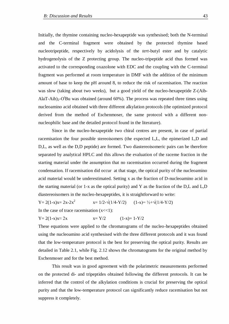
B: Discussion and Results 43
Initially, the thymine containing nucleo-hexapeptide was synthesised; both the N-terminal
and the C-terminal fragment were obtained by the protected thymine based
nucleotripeptide, respectively by acidolysis of the tert-butyl ester and by catalytic
hydrogenolysis of the Z protecting group. The nucleo-tripeptide acid thus formed was
activated to the corresponding oxazolone with EDC and the coupling with the C-terminal
fragment was performed at room temperature in DMF with the addition of the minimum
amount of base to keep the pH around 8, to reduce the risk of racemisation. The reaction
was slow (taking about two weeks), but a good yield of the nucleo-hexapeptide Z-(Aib-
AlaT-Aib)2-OtBu was obtained (around 60%). The process was repeated three times using
nucleoamino acid obtained with three different alkylation protocols (the optimized protocol
derived from the method of Eschenmoser, the same protocol with a different non-
nucleophilic base and the detailed protocol found in the literature).
Since in the nucleo-hexapeptide two chiral centres are present, in case of partial
racemisation the four possible stereoisomers (the expected L,L, the epimerized L,D and
D,L, as well as the D,D peptide) are formed. Two diastereoisomeric pairs can be therefore
separated by analytical HPLC and this allows the evaluation of the raceme fraction in the
starting material under the assumption that no racemisation occurred during the fragment
condensation. If racemisation did occur at that stage, the optical purity of the nucleoamino
acid material would be underestimated. Setting x as the fraction of D-nucleoamino acid in
the starting material (or 1-x as the optical purity) and Y as the fraction of the D,L and L,D
diastereoisomers in the nucleo-hexapeptides, it is straightforward to write:
Y= 2(1-x)x= 2x-2x2 x= 1/2-√(1/4-Y/2) (1-x)= ½+√(1/4-Y/2)
In the case of trace racemisation (x<<1):
Y= 2(1-x)x≈ 2x x= Y/2 (1-x)= 1-Y/2
These equations were applied to the chromatograms of the nucleo-hexapeptides obtained
using the nucleoamino acid synthesised with the three different protocols and it was found
that the low-temperature protocol is the best for preserving the optical purity. Results are
detailed in Table 2.1, while Fig. 2.12 shows the chromatograms for the original method by
Eschenmoser and for the best method.
This result was in good agreement with the polarimetric measurements performed
on the protected di- and tripeptides obtained following the different protocols. It can be
inferred that the control of the alkylation conditions is crucial for preserving the optical
purity and that the low-temperature protocol can significantly reduce racemisation but not
suppress it completely.
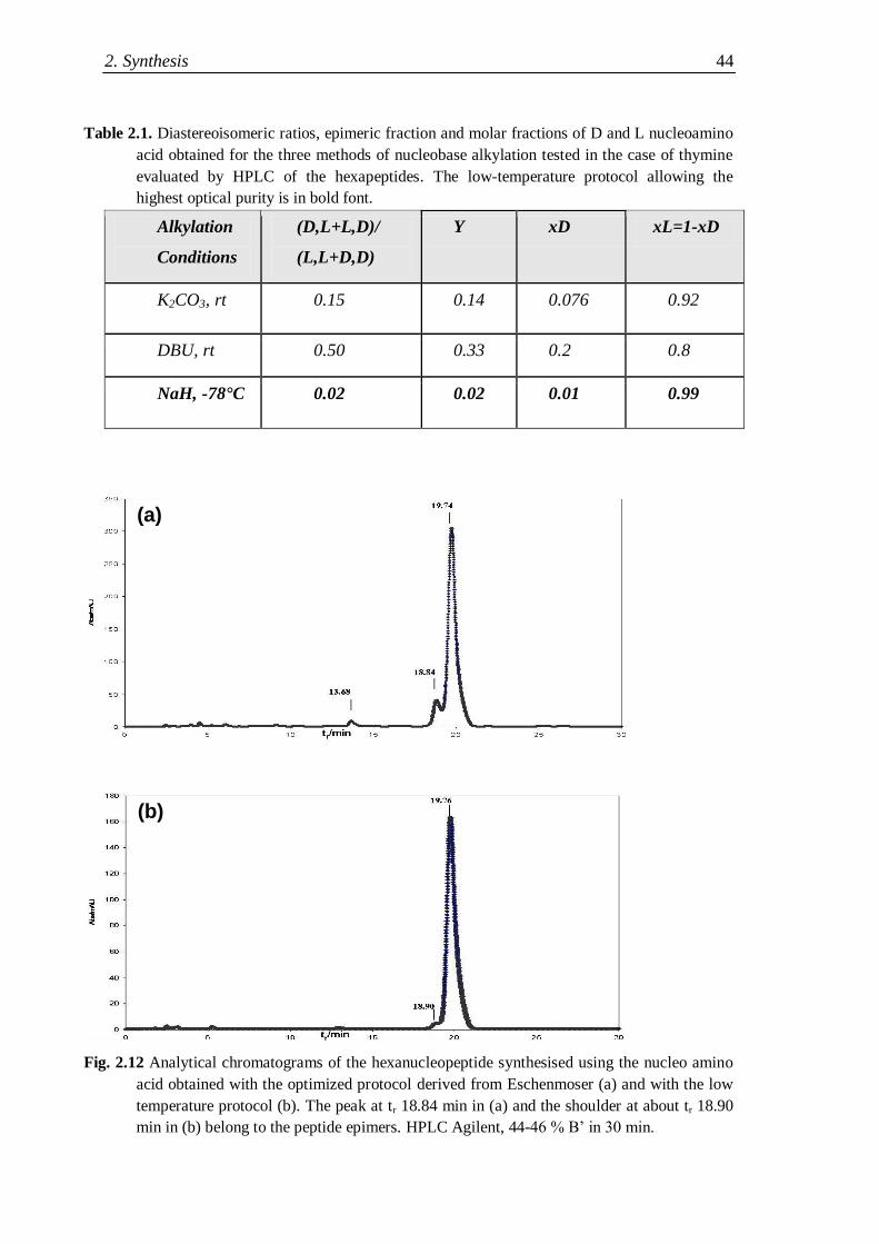
2. Synthesis 44
Table 2.1. Diastereoisomeric ratios, epimeric fraction and molar fractions of D and L nucleoamino
acid obtained for the three methods of nucleobase alkylation tested in the case of thymine
evaluated by HPLC of the hexapeptides. The low-temperature protocol allowing the
highest optical purity is in bold font.
Alkylation
Conditions
(D,L+L,D)/
(L,L+D,D)
Y xD xL=1-xD
K2CO3, rt 0.15 0.14 0.076 0.92
DBU, rt 0.50 0.33 0.2 0.8
NaH, -78°C 0.02 0.02 0.01 0.99
Fig. 2.12 Analytical chromatograms of the hexanucleopeptide synthesised using the nucleo amino
acid obtained with the optimized protocol derived from Eschenmoser (a) and with the low
temperature protocol (b). The peak at tr 18.84 min in (a) and the shoulder at about tr 18.90
min in (b) belong to the peptide epimers. HPLC Agilent, 44-46 % B‟ in 30 min.
(b)
(a)
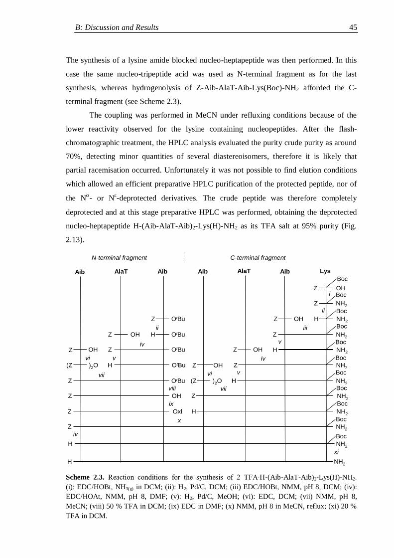
B: Discussion and Results 45
The synthesis of a lysine amide blocked nucleo-heptapeptide was then performed. In this
case the same nucleo-tripeptide acid was used as N-terminal fragment as for the last
synthesis, whereas hydrogenolysis of Z-Aib-AlaT-Aib-Lys(Boc)-NH2 afforded the C-
terminal fragment (see Scheme 2.3).
The coupling was performed in MeCN under refluxing conditions because of the
lower reactivity observed for the lysine containing nucleopeptides. After the flash-
chromatographic treatment, the HPLC analysis evaluated the purity crude purity as around
70%, detecting minor quantities of several diastereoisomers, therefore it is likely that
partial racemisation occurred. Unfortunately it was not possible to find elution conditions
which allowed an efficient preparative HPLC purification of the protected peptide, nor of
the N- or N
-deprotected derivatives. The crude peptide was therefore completely
deprotected and at this stage preparative HPLC was performed, obtaining the deprotected
nucleo-heptapeptide H-(Aib-AlaT-Aib)2-Lys(H)-NH2 as its TFA salt at 95% purity (Fig.
2.13).
Aib AlaT Aib Aib AlaT Aib Lys
Z OH
Z NH2
Z OH H NH2
Z NH2
H NH2Z OH
Z NH2
NH2
NH2
NH2
NH2
H)2O(Z
Z
H
OtBu
OtBu
OtBu
OtBu
OtBu
OH
Oxl
Z
HOHZ
Z
H)2O(Z
Z
Z
Z
Z
N-terminal fragment C-terminal fragment
Boc
Boc
Boc
Boc
Boc
Boc
Boc
Boc
Boc
Boc
NH2
Boc
NH2
OHZ
OHZ
H
H
i
ii
iiiii
iv v
vi v iv
vii vi v
viiviii
x
iv
ix
xi
Scheme 2.3. Reaction conditions for the synthesis of 2 TFA∙H-(Aib-AlaT-Aib)2-Lys(H)-NH2.
(i): EDC/HOBt, NH3(g) in DCM; (ii): H2, Pd/C, DCM; (iii) EDC/HOBt, NMM, pH 8, DCM; (iv):
EDC/HOAt, NMM, pH 8, DMF; (v): H2, Pd/C, MeOH; (vi): EDC, DCM; (vii) NMM, pH 8,
MeCN; (viii) 50 % TFA in DCM; (ix) EDC in DMF; (x) NMM, pH 8 in MeCN, reflux; (xi) 20 %
TFA in DCM.
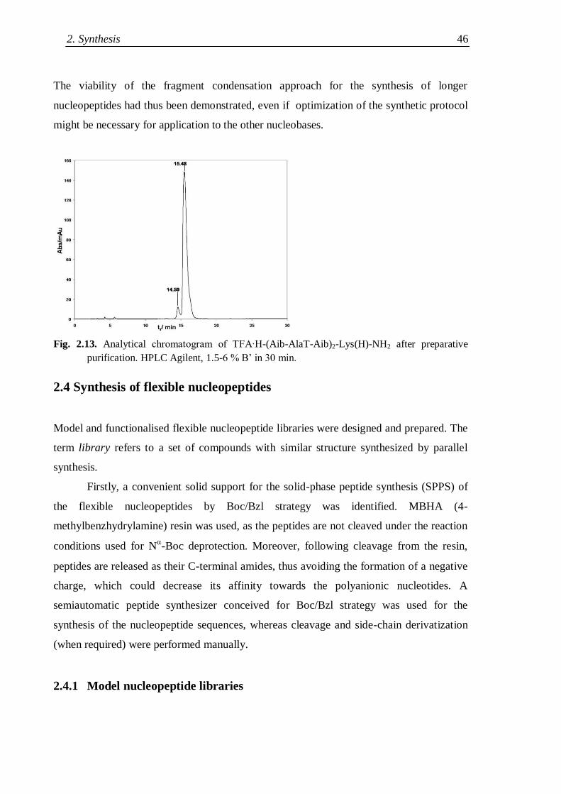
2. Synthesis 46
The viability of the fragment condensation approach for the synthesis of longer
nucleopeptides had thus been demonstrated, even if optimization of the synthetic protocol
might be necessary for application to the other nucleobases.
Fig. 2.13. Analytical chromatogram of TFA∙H-(Aib-AlaT-Aib)2-Lys(H)-NH2 after preparative
purification. HPLC Agilent, 1.5-6 % B‟ in 30 min.
2.4 Synthesis of flexible nucleopeptides
Model and functionalised flexible nucleopeptide libraries were designed and prepared. The
term library refers to a set of compounds with similar structure synthesized by parallel
synthesis.
Firstly, a convenient solid support for the solid-phase peptide synthesis (SPPS) of
the flexible nucleopeptides by Boc/Bzl strategy was identified. MBHA (4-
methylbenzhydrylamine) resin was used, as the peptides are not cleaved under the reaction
conditions used for N-Boc deprotection. Moreover, following cleavage from the resin,
peptides are released as their C-terminal amides, thus avoiding the formation of a negative
charge, which could decrease its affinity towards the polyanionic nucleotides. A
semiautomatic peptide synthesizer conceived for Boc/Bzl strategy was used for the
synthesis of the nucleopeptide sequences, whereas cleavage and side-chain derivatization
(when required) were performed manually.
2.4.1 Model nucleopeptide libraries
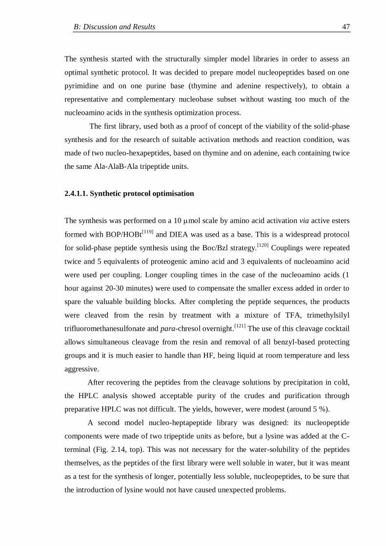
B: Discussion and Results 47
The synthesis started with the structurally simpler model libraries in order to assess an
optimal synthetic protocol. It was decided to prepare model nucleopeptides based on one
pyrimidine and on one purine base (thymine and adenine respectively), to obtain a
representative and complementary nucleobase subset without wasting too much of the
nucleoamino acids in the synthesis optimization process.
The first library, used both as a proof of concept of the viability of the solid-phase
synthesis and for the research of suitable activation methods and reaction condition, was
made of two nucleo-hexapeptides, based on thymine and on adenine, each containing twice
the same Ala-AlaB-Ala tripeptide units.
2.4.1.1. Synthetic protocol optimisation
The synthesis was performed on a 10 mol scale by amino acid activation via active esters
formed with BOP/HOBt[119]
and DIEA was used as a base. This is a widespread protocol
for solid-phase peptide synthesis using the Boc/Bzl strategy.[120]
Couplings were repeated
twice and 5 equivalents of proteogenic amino acid and 3 equivalents of nucleoamino acid
were used per coupling. Longer coupling times in the case of the nucleoamino acids (1
hour against 20-30 minutes) were used to compensate the smaller excess added in order to
spare the valuable building blocks. After completing the peptide sequences, the products
were cleaved from the resin by treatment with a mixture of TFA, trimethylsilyl
trifluoromethanesulfonate and para-chresol overnight.[121]
The use of this cleavage cocktail
allows simultaneous cleavage from the resin and removal of all benzyl-based protecting
groups and it is much easier to handle than HF, being liquid at room temperature and less
aggressive.
After recovering the peptides from the cleavage solutions by precipitation in cold,
the HPLC analysis showed acceptable purity of the crudes and purification through
preparative HPLC was not difficult. The yields, however, were modest (around 5 %).
A second model nucleo-heptapeptide library was designed: its nucleopeptide
components were made of two tripeptide units as before, but a lysine was added at the C-
terminal (Fig. 2.14, top). This was not necessary for the water-solubility of the peptides
themselves, as the peptides of the first library were well soluble in water, but it was meant
as a test for the synthesis of longer, potentially less soluble, nucleopeptides, to be sure that
the introduction of lysine would not have caused unexpected problems.
2. Synthesis 48
Assuming that the not satisfactory yields obtained with the first library were due to
insufficient nucleoamino acid activation, a more energetic activation protocol for the
formation of the nucleoamino acid active esters was adopted. Thus HATU/HOAt[122]
were
employed, while the activation of the proteogenic residues was performed with the usual
method. The synthesis was performed on 25 mol scale, in order to obtain sufficient
amounts of nucleopeptides for conformational analyses, not only by spectroscopic
techniques, but also by 2D NMR.
2.4.1.2 Incorporation of an Aib residue by SPPS
As mentioned in the Introduction, generally short nucleopeptides made of C-trisubstitued
residues are unfolded, while the presence of C-tetrasubstitued residues in a
C-trisubstitued residue-based sequence can substantially improve the conformational
stability of the peptides. To evaluate the ability of Aib to structure flexible nucleopeptides,
two more nucleopeptides were added to the library, each one carrying an Aib residue in the
middle of its sequence (Fig. 2.14, bottom). The central location was chosen in the hope that
it would have exerted deeper effects on the peptide conformation than peripheric positions.
Fig. 2.14. Flexible nucleo-heptapeptides based on thymine and adenine (A2A and T2A, top), and
analogues containing an Aib residue (circled) at position 4 (A2U and T2U, bottom).
Two positions were in principle available (3 and 4), both next to a nucleoamino acid
(AlaB2 or AlaB
5, respectively). Since the increased steric hindrance of Aib affects more the
reactivity of the amino- than that of the carboxyl-group, it was thought that coupling the
N
NH
O
O
H3N
H3N
NH
NH
O
O
N
NH
O
O
NH
O
NH
NH
O
O
O
NH
NH2
O
N
NH
O
O
H3N
H3N
NH
NH
O
O
N
NH
O
O
NH
O
NH
NH
O
O
O
NH
NH2
O
N
NN
N
NH2
H3N
H3N
NH
NH
O
O
N
NH
O
NH
NH
O
O
O
NH
NH2
O
N
N
N
NH2
N
NN
N
NH2
H3N
H3N
NH
NH
O
O
N
NH
O
NH
NH
O
O
O
NH
NH2
O
N
N
N
NH2
+
+
+
+
+
+
+
+
T2A
T2U
A2A
A2U
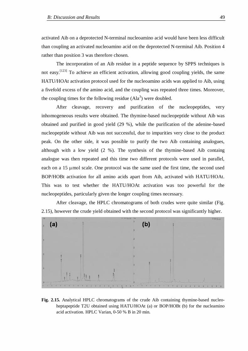
B: Discussion and Results 49
activated Aib on a deprotected N-terminal nucleoamino acid would have been less difficult
than coupling an activated nucleoamino acid on the deprotected N-terminal Aib. Position 4
rather than position 3 was therefore chosen.
The incorporation of an Aib residue in a peptide sequence by SPPS techniques is
not easy.[123]
To achieve an efficient activation, allowing good coupling yields, the same
HATU/HOAt activation protocol used for the nucleoamino acids was applied to Aib, using
a fivefold excess of the amino acid, and the coupling was repeated three times. Moreover,
the coupling times for the following residue (Ala3) were doubled.
After cleavage, recovery and purification of the nucleopeptides, very
inhomogeneous results were obtained. The thymine-based nucleopeptide without Aib was
obtained and purified in good yield (29 %), while the purification of the adenine-based
nucleopeptide without Aib was not successful, due to impurities very close to the product
peak. On the other side, it was possible to purify the two Aib containing analogues,
although with a low yield (2 %). The synthesis of the thymine-based Aib containg
analogue was then repeated and this time two different protocols were used in parallel,
each on a 15 mol scale. One protocol was the same used the first time, the second used
BOP/HOBt activation for all amino acids apart from Aib, activated with HATU/HOAt.
This was to test whether the HATU/HOAt activation was too powerful for the
nucleopeptides, particularly given the longer coupling times necessary.
After cleavage, the HPLC chromatograms of both crudes were quite similar (Fig.
2.15), however the crude yield obtained with the second protocol was significantly higher.
Fig. 2.15. Analytical HPLC chromatograms of the crude Aib containing thymine-based nucleo-
heptapeptide T2U obtained using HATU/HOAt (a) or BOP/HOBt (b) for the nucleamino
acid activation. HPLC Varian, 0-50 % B in 20 min.
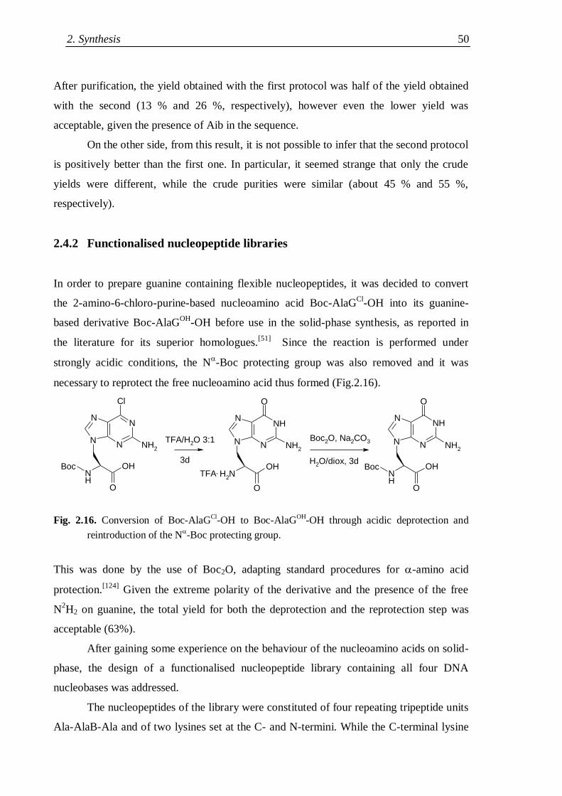
2. Synthesis 50
After purification, the yield obtained with the first protocol was half of the yield obtained
with the second (13 % and 26 %, respectively), however even the lower yield was
acceptable, given the presence of Aib in the sequence.
On the other side, from this result, it is not possible to infer that the second protocol
is positively better than the first one. In particular, it seemed strange that only the crude
yields were different, while the crude purities were similar (about 45 % and 55 %,
respectively).
2.4.2 Functionalised nucleopeptide libraries
In order to prepare guanine containing flexible nucleopeptides, it was decided to convert
the 2-amino-6-chloro-purine-based nucleoamino acid Boc-AlaGCl
-OH into its guanine-
based derivative Boc-AlaGOH
-OH before use in the solid-phase synthesis, as reported in
the literature for its superior homologues.[51]
Since the reaction is performed under
strongly acidic conditions, the N-Boc protecting group was also removed and it was
necessary to reprotect the free nucleoamino acid thus formed (Fig.2.16).
Fig. 2.16. Conversion of Boc-AlaGCl
-OH to Boc-AlaGOH
-OH through acidic deprotection and
reintroduction of the N-Boc protecting group.
This was done by the use of Boc2O, adapting standard procedures for -amino acid
protection.[124]
Given the extreme polarity of the derivative and the presence of the free
N2H2 on guanine, the total yield for both the deprotection and the reprotection step was
acceptable (63%).
After gaining some experience on the behaviour of the nucleoamino acids on solid-
phase, the design of a functionalised nucleopeptide library containing all four DNA
nucleobases was addressed.
The nucleopeptides of the library were constituted of four repeating tripeptide units
Ala-AlaB-Ala and of two lysines set at the C- and N-termini. While the C-terminal lysine
N
N
N
Cl
NH2
N
NH
OH
O
NH2
OH
O
N
N
N
NH
NH2
O
N
N NH2
O
NH
NH
OH
O
N
Boc Boc
TFA/H2O 3:1
3d
Boc2O, Na2CO3
H2O/diox, 3d
TFA.
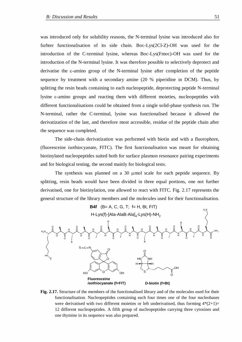
B: Discussion and Results 51
was introduced only for solubility reasons, the N-terminal lysine was introduced also for
furhter functionalisation of its side chain. Boc-Lys(2Cl-Z)-OH was used for the
introduction of the C-terminal lysine, whereas Boc-Lys(Fmoc)-OH was used for the
introduction of the N-terminal lysine. It was therefore possible to selectively deprotect and
derivatise the -amino group of the N-terminal lysine after completion of the peptide
sequence by treatment with a secondary amine (20 % piperidine in DCM). Thus, by
splitting the resin beads containing to each nucleopeptide, deprotecting peptide N-terminal
lysine -amino groups and reacting them with different moieties, nucleopeptides with
different functionalisations could be obtained from a single solid-phase synthesis run. The
N-terminal, rather the C-terminal, lysine was functionalised because it allowed the
derivatization of the last, and therefore most accessible, residue of the peptide chain after
the sequence was completed.
The side-chain derivatization was performed with biotin and with a fluorophore,
(fluoresceine isothiocyanate, FITC). The first functionalisation was meant for obtaining
biotinylated nucleopeptides suited both for surface plasmon resonance pairing experiments
and for biological testing, the second mainly for biological tests.
The synthesis was planned on a 30 mol scale for each peptide sequence. By
splitting, resin beads would have been divided in three equal portions, one not further
derivatised, one for biotinylation, one allowed to react with FITC. Fig. 2.17 represents the
general structure of the library members and the molecules used for their functionalisation.
B
NH
NH
NH
NH
NH
NH
NH
NH
NH
O
O
O
O
O
O
O
O
NH2
O
NH
B
NH
O
NH
NH
O
O
O
NH
NH2
O
B
B
NH2
O OHOH
O
ONS
NHNH
S
O
HH
O
OH
B4f (B= A, C, G, T; f= H, Bt, FIT)
f
D-biotin (f=Bt)
Fluoresceine
isothiocyanate (f=FIT)
H-Lys(f)-[Ala-AlaB-Ala]4-Lys(H)-NH2
C
Fig. 2.17. Structure of the members of the functionalised library and of the molecules used for their
functionalisation. Nucleopeptides containing each four times one of the four nucleobases
were derivatised with two different moieties or left underivatised, thus forming 4*(2+1)=
12 different nucleopeptides. A fifth group of nucleopeptides carrying three cytosines and
one thymine in its sequence was also prepared.
2. Synthesis 52
HBTU/HOBt[125]
were used for active ester formation for all amino acids, since side
reactions with guanine derivatives were reported by using phosphonium-based coupling
reagents, like BOP.[118]
This activation method has an intermediate reactivity between the
HATU/HOAt and BOP, therefore it was considered a suited general purpose protocol for
the synthesis. Biotin was coupled to the -amino group of lysine by activating its carboxyl
group with HBTU/HOBt, like the carboxyl groups of the protected amino acids. FITC,
being an electrophilic isocyanate, does not require activation to attack free amines,
therefore only pH adjustment to about 8 was necessary.
In order to prepare a single-base mismatch model, a nucleopeptide containing two
different nucleobases in its sequence (one thymine in position 5 and three cytosines in
position 2, 8 and 11) was designed and derivatised in the same way as the „homobase‟
nucleopeptides.
The crude mixtures were rather complex, making purification difficult. Therefore
the yields of the 15 functionalised nucleopeptides after purification were modest, ranging
between 3 % and 11 %. The (formal) average yields per step including the purification
were however comparable with the yield obtained for the best nucleoheptapeptide prepared
by SPPS (78-85 % against 84 %). Only the purification of the guanine-based biotinylated
peptide was not possible, because the lyophilized crude product turned insoluble in any
solvent mixture. A reason could be the well-known tendency to aggregation of guanine
derivatives,[97,116]
maybe increased by some serendipitously strong interaction with the
biotin ring. Apart from that case, the water solubility of the underivatised nucleopeptides
was high and even the more lipophilic FITC-derivatised peptides were well soluble up to 1
mM.
In some cases the derivatised nucleopeptides were obtained in higher yields
compared to the underivatised ones. This could be due to side reactions due to the free -
amino group of the N-terminal lysine in basic environment before the cleavage. Yields for
all the derivatives are presented in Table 2.2.
After this library, a novel library was designed, composed of thymine-based
nucleopeptides containing five tripeptide units between two lysines (Fig. 2.18, top).
Thymine was chosen for the preparation of this longer nucleopeptides because it had
proven the less problematic nucleobase (indeed it had the highest average yield among the
five nucleopeptide sequences tested, Table 2.2, last row) and because thymine-containing
peptides had shown interesting pairing properties in the surface plasmon resonance
experiments (see Section 4). If pairing is cooperative, a peptide with five nucleobases
B: Discussion and Results 53
should display a much stronger binding than a peptide with four. Partial biotinylation of
the peptide was therefore planned to carry on pairing tests. Moreover, a cysteine
containing, thymine-based (see Fig. 2.18, middle) nucleopeptide was designed to prepare a
nucleopeptide dimer linked by a disulphide bridge, as already reported in the literature with
other nucleotide analogues of peptide-like structure.[58]
Table 2.2. Yields of the various derivatised nucleopeptides obtained after purification.
Yield (%) A4 C4 G4 T4 CTCC
H 5 3 10 8 8
Bt 9 7 - 8 4
FIT 6 11 9 7 8
Average 6.7 7 6.3 7.7 6.7
Notes. Colums refer to the nucleobase sequence of the peptides (A4: adenine-based; C4: cytosine-
based; G4: guanine-based; T4: thymine-based; CTCC: mixed base peptide), rows to the
derivatization (H: underivatised; Bt: biotinylated; FIT: reacted with fluoresceine
isothiocyanate). The last row reports the average yield for the three derivatives.
This dimer molecule would have carried ten bases (on two strands) and it could in
principle have paired with complementary, adenine-based nucleopeptides or nucleotides
both by independent binding of the two strands to form Watson-Crick bonds with 1:1
stoichiometry and by cooperative pairing of both strands, one by Watson-Crick, the other
by Hogsteen interactions, with the same complementary sequence (triplex formation). The
latter possibility is represented in Fig. 2.18, bottom.
The formation of a homodimer through a disulphide bridge can be achieved both on
solid-phase and in solution. In the first case the protecting group for the thiol function is
removed under oxidizing conditions when the peptides are still bound to the resin, so that a
disulphide bridges are formed between next molecules. In the second case the peptide is
synthesized, cleaved and purified as a monomer, then allowed to dimerize in solution. In
this case, either the thiol protecting group is stable to the cleavage and removed after the
purification stage, or the cleavage and the purification are performed under non-oxidizing
conditions. The on-resin dimerization requires one purification step less than the
dimerization in solution, on the other side the dimer is generally less pure and solubility
problems can appear during the purification. Therefore, it was chosen to perform the
dimerization in solution, using a protecting group that was removed during the cleavage,
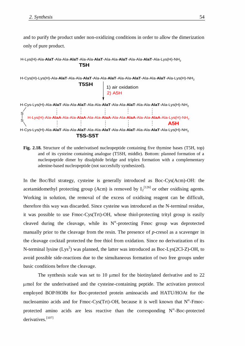
2. Synthesis 54
and to purify the product under non-oxidizing conditions in order to allow the dimerization
only of pure product.
Caratterizzazione e prove biologiche sui
Fig. 2.18. Structure of the underivatised nucleopeptide containing five thymine bases (T5H, top)
and of its cysteine containing analogue (T5SH, middle). Bottom: planned formation of a
nucleopeptide dimer by disulphide bridge and triplex formation with a complementary
adenine-based nucleopeptide (not succesfully synthesized).
In the Boc/Bzl strategy, cysteine is generally introduced as Boc-Cys(Acm)-OH: the
acetamidomethyl protecting group (Acm) is removed by I2[126]
or other oxidising agents.
Working in solution, the removal of the excess of oxidising reagent can be difficult,
therefore this way was discarded. Since cysteine was introduced as the N-terminal residue,
it was possible to use Fmoc-Cys(Trt)-OH, whose thiol-protecting trityl group is easily
cleaved during the cleavage, while its N-protecting Fmoc group was deprotected
manually prior to the cleavage from the resin. The presence of p-cresol as a scavenger in
the cleavage cocktail protected the free thiol from oxidation. Since no derivatization of its
N-terminal lysine (Lys2) was planned, the latter was introduced as Boc-Lys(2Cl-Z)-OH, to
avoid possible side-reactions due to the simultaneous formation of two free groups under
basic conditions before the cleavage.
The synthesis scale was set to 10 mol for the biotinylated derivative and to 22
mol for the underivatised and the cysteine-containing peptide. The activation protocol
employed BOP/HOBt for Boc-protected protein aminoacids and HATU/HOAt for the
nucleoamino acids and for Fmoc-Cys(Trt)-OH, because it is well known that N-Fmoc-
protected amino acids are less reactive than the corresponding N-Boc-protected
derivatives.[107]
S
S
H-Cys(H)-Lys(H)-Ala-AlaT-Ala-Ala-AlaT-Ala-Ala-AlaT-Ala-Ala-AlaT-Ala-Ala-AlaT-Ala-Lys(H)-NH2
H-Cys-Lys(H)-Ala-AlaT-Ala-Ala-AlaT-Ala-Ala-AlaT-Ala-Ala-AlaT-Ala-Ala-AlaT-Ala-Lys(H)-NH2
H-Cys-Lys(H)-Ala-AlaT-Ala-Ala-AlaT-Ala-Ala-AlaT-Ala-Ala-AlaT-Ala-Ala-AlaT-Ala-Lys(H)-NH2
1) air oxidationT5SH
T5S-S5T
H-Lys(H)-Ala-AlaT-Ala-Ala-AlaT-Ala-Ala-AlaT-Ala-Ala-AlaT-Ala-Ala-AlaT-Ala-Lys(H)-NH2
T5H
H-Lys(H)-Ala-AlaA-Ala-Ala-AlaA-Ala-Ala-AlaA-Ala-Ala-AlaA-Ala-Ala-AlaA-Ala-Lys(H)-NH2
A5H
2) A5H
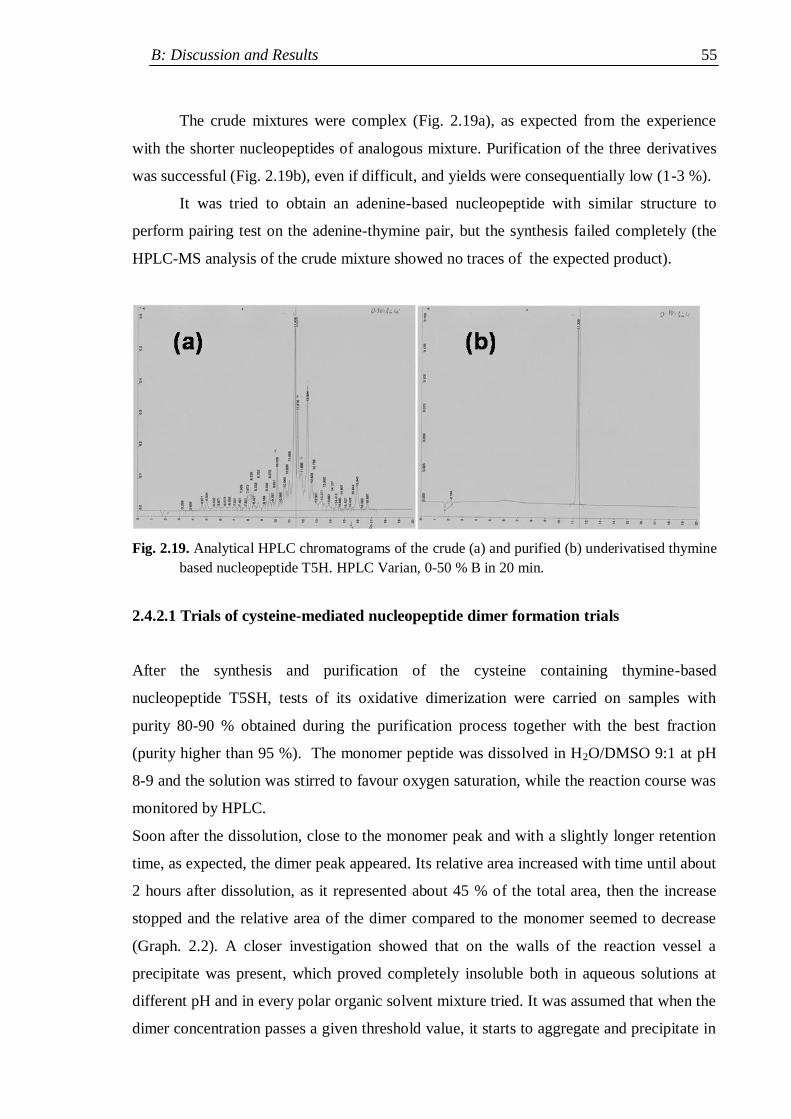
B: Discussion and Results 55
The crude mixtures were complex (Fig. 2.19a), as expected from the experience
with the shorter nucleopeptides of analogous mixture. Purification of the three derivatives
was successful (Fig. 2.19b), even if difficult, and yields were consequentially low (1-3 %).
It was tried to obtain an adenine-based nucleopeptide with similar structure to
perform pairing test on the adenine-thymine pair, but the synthesis failed completely (the
HPLC-MS analysis of the crude mixture showed no traces of the expected product).
Fig. 2.19. Analytical HPLC chromatograms of the crude (a) and purified (b) underivatised thymine
based nucleopeptide T5H. HPLC Varian, 0-50 % B in 20 min.
2.4.2.1 Trials of cysteine-mediated nucleopeptide dimer formation trials
After the synthesis and purification of the cysteine containing thymine-based
nucleopeptide T5SH, tests of its oxidative dimerization were carried on samples with
purity 80-90 % obtained during the purification process together with the best fraction
(purity higher than 95 %). The monomer peptide was dissolved in H2O/DMSO 9:1 at pH
8-9 and the solution was stirred to favour oxygen saturation, while the reaction course was
monitored by HPLC.
Soon after the dissolution, close to the monomer peak and with a slightly longer retention
time, as expected, the dimer peak appeared. Its relative area increased with time until about
2 hours after dissolution, as it represented about 45 % of the total area, then the increase
stopped and the relative area of the dimer compared to the monomer seemed to decrease
(Graph. 2.2). A closer investigation showed that on the walls of the reaction vessel a
precipitate was present, which proved completely insoluble both in aqueous solutions at
different pH and in every polar organic solvent mixture tried. It was assumed that when the
dimer concentration passes a given threshold value, it starts to aggregate and precipitate in
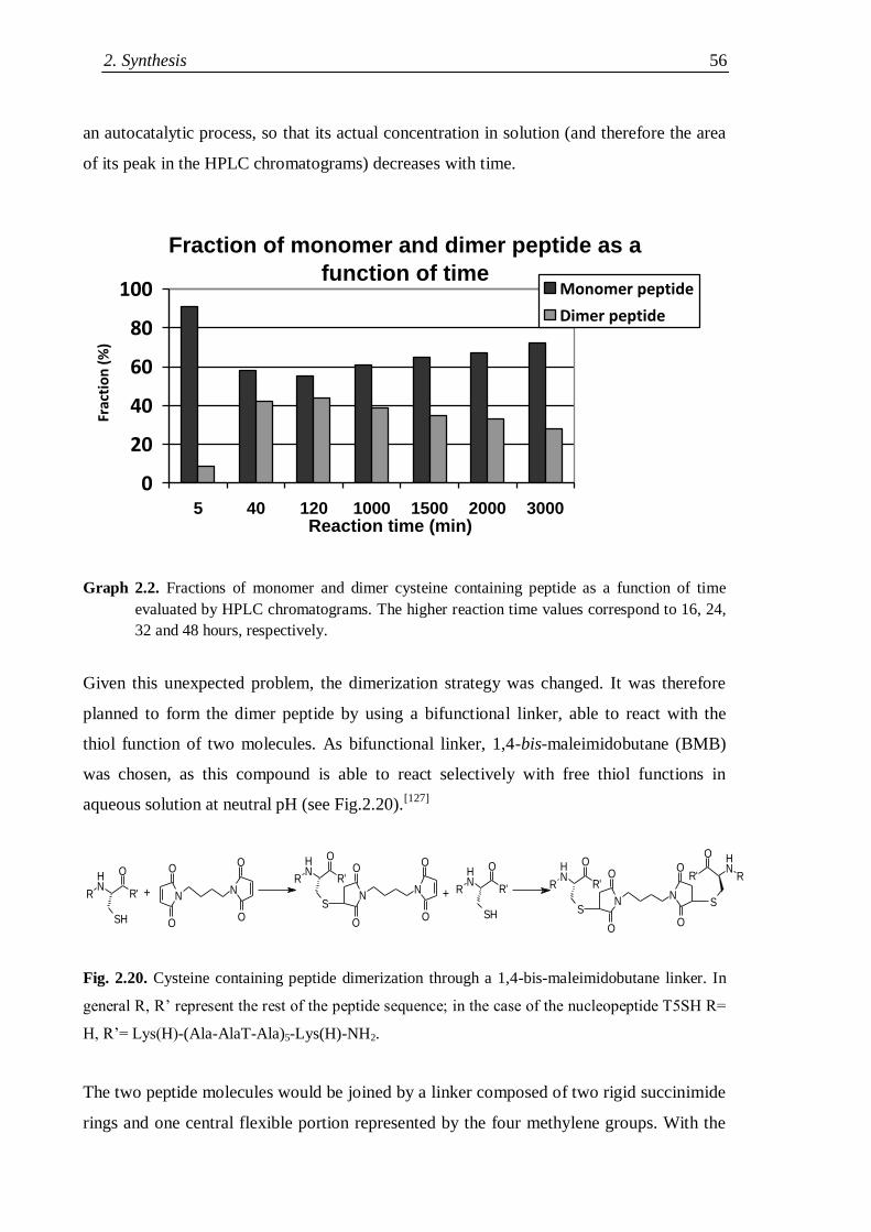
2. Synthesis 56
an autocatalytic process, so that its actual concentration in solution (and therefore the area
of its peak in the HPLC chromatograms) decreases with time.
Fraction of monomer and dimer peptide as a
function of time
0
20
40
60
80
100
5 40 120 1000 1500 2000 3000Reaction time (min)
Frac
tio
n (
%)
Monomer peptide
Dimer peptide
Graph 2.2. Fractions of monomer and dimer cysteine containing peptide as a function of time
evaluated by HPLC chromatograms. The higher reaction time values correspond to 16, 24,
32 and 48 hours, respectively.
Given this unexpected problem, the dimerization strategy was changed. It was therefore
planned to form the dimer peptide by using a bifunctional linker, able to react with the
thiol function of two molecules. As bifunctional linker, 1,4-bis-maleimidobutane (BMB)
was chosen, as this compound is able to react selectively with free thiol functions in
aqueous solution at neutral pH (see Fig.2.20).[127]
NH
R'
O
SH
R N
O
O
N
O
O
NH
R'
O
S
R
N
O
O
N
O
O
NH
R'
O
SH
RNH
R'
O
S
R
N
O
O
N
O
O
NH
R'
O
S
R
+ +
Fig. 2.20. Cysteine containing peptide dimerization through a 1,4-bis-maleimidobutane linker. In
general R, R‟ represent the rest of the peptide sequence; in the case of the nucleopeptide T5SH R=
H, R‟= Lys(H)-(Ala-AlaT-Ala)5-Lys(H)-NH2.
The two peptide molecules would be joined by a linker composed of two rigid succinimide
rings and one central flexible portion represented by the four methylene groups. With the
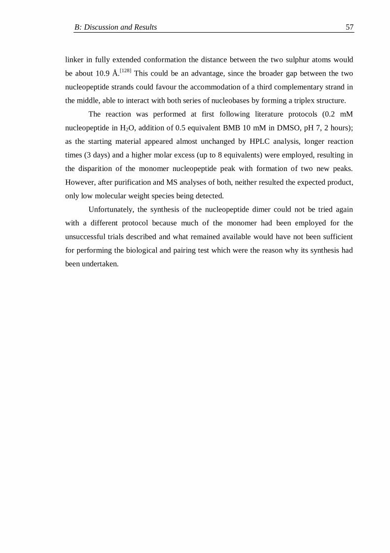
B: Discussion and Results 57
linker in fully extended conformation the distance between the two sulphur atoms would
be about 10.9 Å.[128]
This could be an advantage, since the broader gap between the two
nucleopeptide strands could favour the accommodation of a third complementary strand in
the middle, able to interact with both series of nucleobases by forming a triplex structure.
The reaction was performed at first following literature protocols (0.2 mM
nucleopeptide in H2O, addition of 0.5 equivalent BMB 10 mM in DMSO, pH 7, 2 hours);
as the starting material appeared almost unchanged by HPLC analysis, longer reaction
times (3 days) and a higher molar excess (up to 8 equivalents) were employed, resulting in
the disparition of the monomer nucleopeptide peak with formation of two new peaks.
However, after purification and MS analyses of both, neither resulted the expected product,
only low molecular weight species being detected.
Unfortunately, the synthesis of the nucleopeptide dimer could not be tried again
with a different protocol because much of the monomer had been employed for the
unsuccessful trials described and what remained available would have not been sufficient
for performing the biological and pairing test which were the reason why its synthesis had
been undertaken.
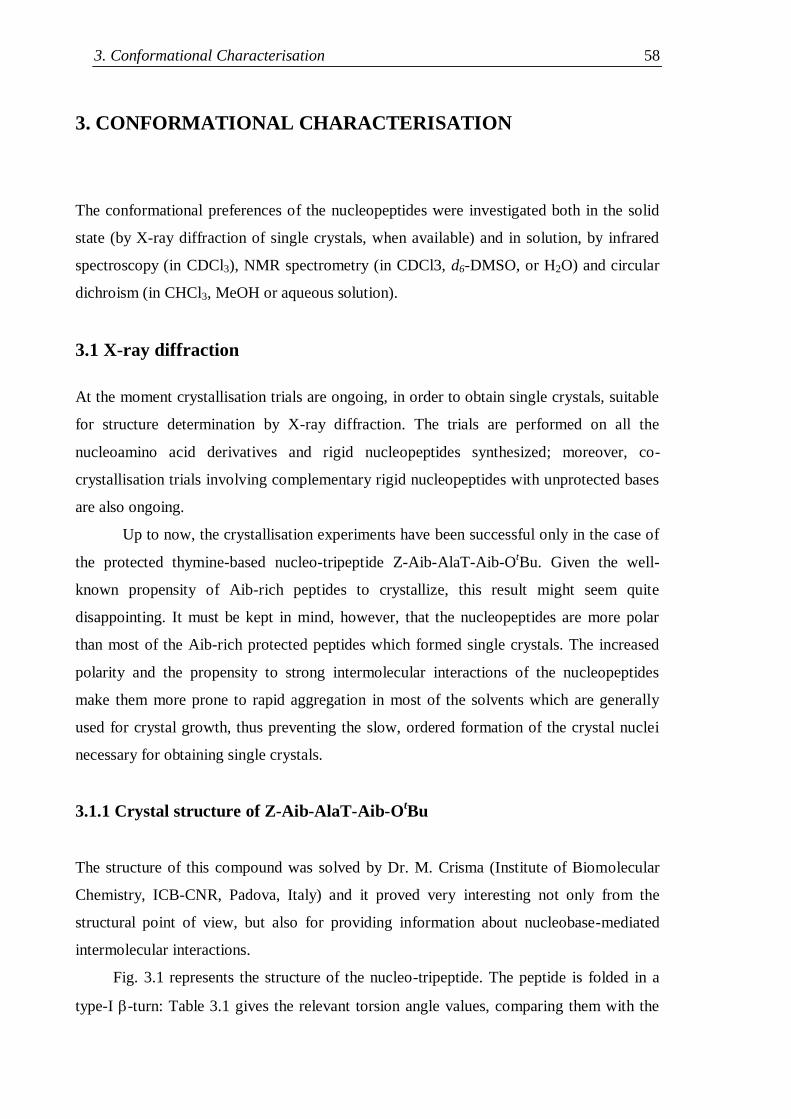
3. Conformational Characterisation 58
3. CONFORMATIONAL CHARACTERISATION
The conformational preferences of the nucleopeptides were investigated both in the solid
state (by X-ray diffraction of single crystals, when available) and in solution, by infrared
spectroscopy (in CDCl3), NMR spectrometry (in CDCl3, d6-DMSO, or H2O) and circular
dichroism (in CHCl3, MeOH or aqueous solution).
3.1 X-ray diffraction
At the moment crystallisation trials are ongoing, in order to obtain single crystals, suitable
for structure determination by X-ray diffraction. The trials are performed on all the
nucleoamino acid derivatives and rigid nucleopeptides synthesized; moreover, co-
crystallisation trials involving complementary rigid nucleopeptides with unprotected bases
are also ongoing.
Up to now, the crystallisation experiments have been successful only in the case of
the protected thymine-based nucleo-tripeptide Z-Aib-AlaT-Aib-OtBu. Given the well-
known propensity of Aib-rich peptides to crystallize, this result might seem quite
disappointing. It must be kept in mind, however, that the nucleopeptides are more polar
than most of the Aib-rich protected peptides which formed single crystals. The increased
polarity and the propensity to strong intermolecular interactions of the nucleopeptides
make them more prone to rapid aggregation in most of the solvents which are generally
used for crystal growth, thus preventing the slow, ordered formation of the crystal nuclei
necessary for obtaining single crystals.
3.1.1 Crystal structure of Z-Aib-AlaT-Aib-OtBu
The structure of this compound was solved by Dr. M. Crisma (Institute of Biomolecular
Chemistry, ICB-CNR, Padova, Italy) and it proved very interesting not only from the
structural point of view, but also for providing information about nucleobase-mediated
intermolecular interactions.
Fig. 3.1 represents the structure of the nucleo-tripeptide. The peptide is folded in a
type-I -turn: Table 3.1 gives the relevant torsion angle values, comparing them with the
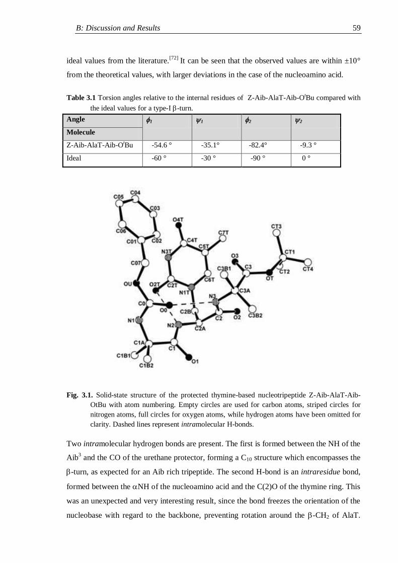
B: Discussion and Results 59
ideal values from the literature.[72]
It can be seen that the observed values are within ±10°
from the theoretical values, with larger deviations in the case of the nucleoamino acid.
Table 3.1 Torsion angles relative to the internal residues of Z-Aib-AlaT-Aib-OtBu compared with
the ideal values for a type-I -turn.
Angle 1 1 2 2
Molecule
Z-Aib-AlaT-Aib-OtBu -54.6 ° -35.1° -82.4° -9.3 °
Ideal -60 ° -30 ° -90 ° 0 °
Fig. 3.1. Solid-state structure of the protected thymine-based nucleotripeptide Z-Aib-AlaT-Aib-
OtBu with atom numbering. Empty circles are used for carbon atoms, striped circles for
nitrogen atoms, full circles for oxygen atoms, while hydrogen atoms have been omitted for
clarity. Dashed lines represent intramolecular H-bonds.
Two intramolecular hydrogen bonds are present. The first is formed between the NH of the
Aib3 and the CO of the urethane protector, forming a C10 structure which encompasses the
-turn, as expected for an Aib rich tripeptide. The second H-bond is an intraresidue bond,
formed between the NH of the nucleoamino acid and the C(2)O of the thymine ring. This
was an unexpected and very interesting result, since the bond freezes the orientation of the
nucleobase with regard to the backbone, preventing rotation around the -CH2 of AlaT.
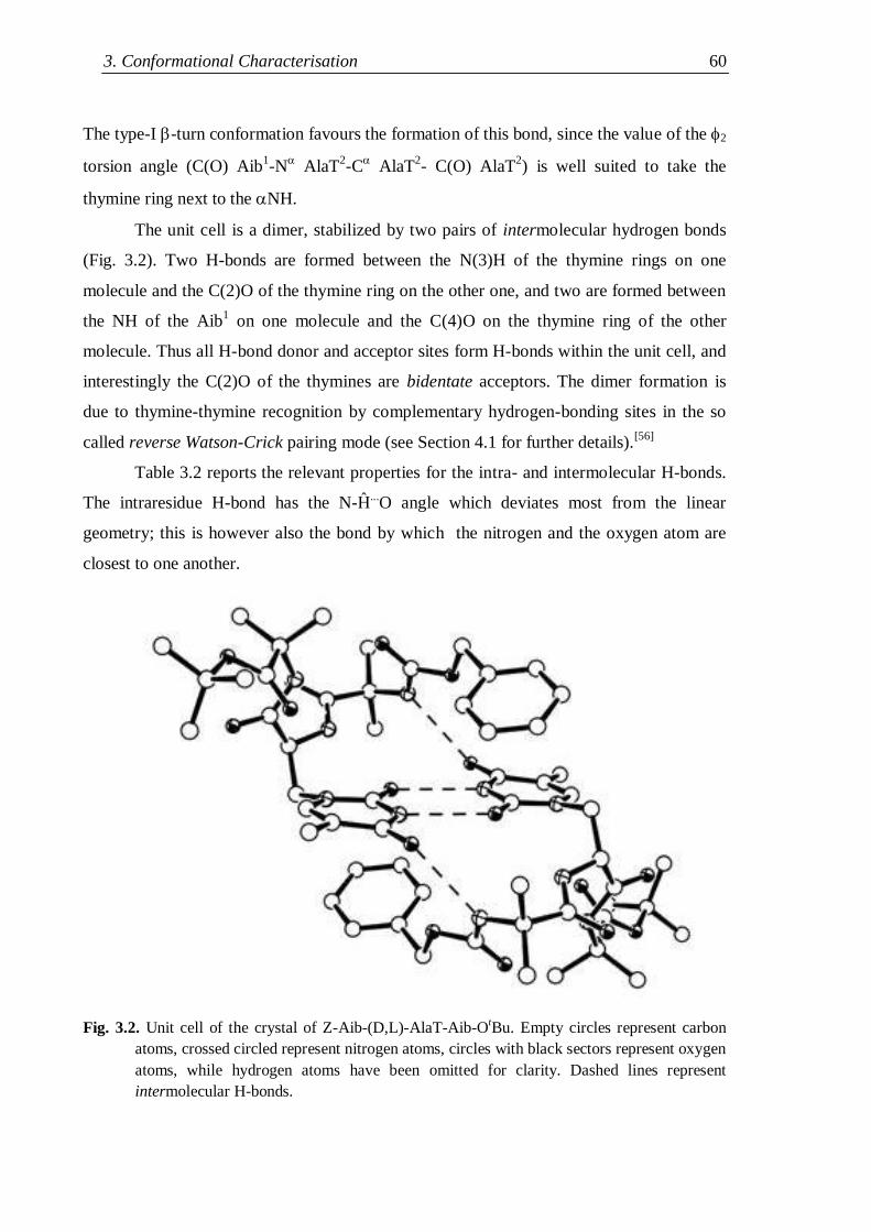
3. Conformational Characterisation 60
The type-I -turn conformation favours the formation of this bond, since the value of the 2
torsion angle (C(O) Aib1-N
AlaT
2-C
AlaT
2- C(O) AlaT
2) is well suited to take the
thymine ring next to the NH.
The unit cell is a dimer, stabilized by two pairs of intermolecular hydrogen bonds
(Fig. 3.2). Two H-bonds are formed between the N(3)H of the thymine rings on one
molecule and the C(2)O of the thymine ring on the other one, and two are formed between
the NH of the Aib1 on one molecule and the C(4)O on the thymine ring of the other
molecule. Thus all H-bond donor and acceptor sites form H-bonds within the unit cell, and
interestingly the C(2)O of the thymines are bidentate acceptors. The dimer formation is
due to thymine-thymine recognition by complementary hydrogen-bonding sites in the so
called reverse Watson-Crick pairing mode (see Section 4.1 for further details).[56]
Table 3.2 reports the relevant properties for the intra- and intermolecular H-bonds.
The intraresidue H-bond has the N-Ĥ...O angle which deviates most from the linear
geometry; this is however also the bond by which the nitrogen and the oxygen atom are
closest to one another.
Fig. 3.2. Unit cell of the crystal of Z-Aib-(D,L)-AlaT-Aib-O
tBu. Empty circles represent carbon
atoms, crossed circled represent nitrogen atoms, circles with black sectors represent oxygen
atoms, while hydrogen atoms have been omitted for clarity. Dashed lines represent
intermolecular H-bonds.
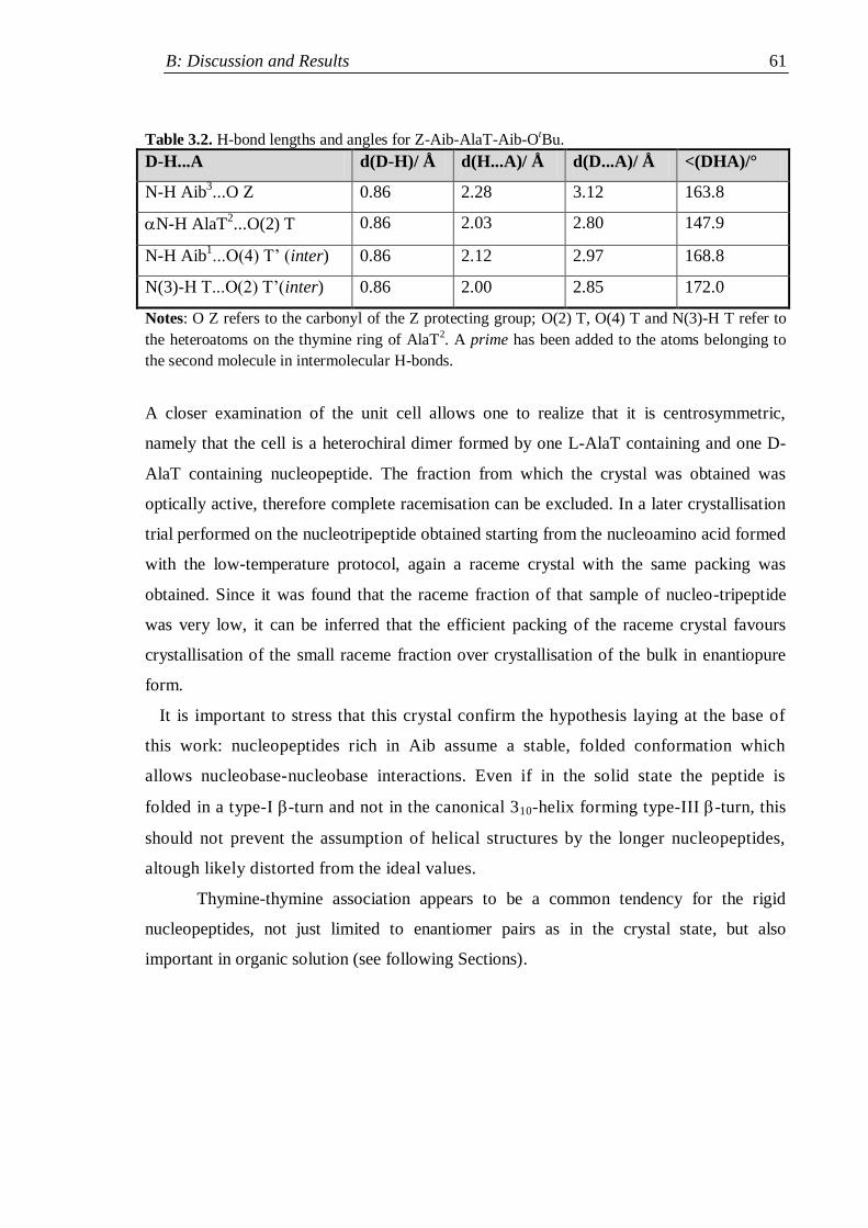
B: Discussion and Results 61
Table 3.2. H-bond lengths and angles for Z-Aib-AlaT-Aib-OtBu.
D-H...A d(D-H)/ Å d(H...A)/ Å d(D...A)/ Å <(DHA)/°
N-H Aib3...O Z 0.86 2.28 3.12 163.8
N-H AlaT2...O(2) T 0.86 2.03 2.80 147.9
N-H Aib1...O(4) T‟ (inter) 0.86 2.12 2.97 168.8
N(3)-H T...O(2) T‟(inter) 0.86 2.00 2.85 172.0
Notes: O Z refers to the carbonyl of the Z protecting group; O(2) T, O(4) T and N(3)-H T refer to
the heteroatoms on the thymine ring of AlaT2. A prime has been added to the atoms belonging to
the second molecule in intermolecular H-bonds.
A closer examination of the unit cell allows one to realize that it is centrosymmetric,
namely that the cell is a heterochiral dimer formed by one L-AlaT containing and one D-
AlaT containing nucleopeptide. The fraction from which the crystal was obtained was
optically active, therefore complete racemisation can be excluded. In a later crystallisation
trial performed on the nucleotripeptide obtained starting from the nucleoamino acid formed
with the low-temperature protocol, again a raceme crystal with the same packing was
obtained. Since it was found that the raceme fraction of that sample of nucleo-tripeptide
was very low, it can be inferred that the efficient packing of the raceme crystal favours
crystallisation of the small raceme fraction over crystallisation of the bulk in enantiopure
form.
It is important to stress that this crystal confirm the hypothesis laying at the base of
this work: nucleopeptides rich in Aib assume a stable, folded conformation which
allows nucleobase-nucleobase interactions. Even if in the solid state the peptide is
folded in a type-I -turn and not in the canonical 310-helix forming type-III -turn, this
should not prevent the assumption of helical structures by the longer nucleopeptides,
altough likely distorted from the ideal values.
Thymine-thymine association appears to be a common tendency for the rigid
nucleopeptides, not just limited to enantiomer pairs as in the crystal state, but also
important in organic solution (see following Sections).
3. Conformational Characterisation 62
3.2 Infrared absorption spectroscopy
The IR absorption study was performed in deuterochloroform (a low polarity solvent, often
employed for conformational studies on peptides). The two frequency intervals richest in
conformational information are:[129]
(i) 3550-3200 cm-1
(Amide A band), related to the stretching vibrations of the N-H
bonds belonging to the urethane and amide groups;
(ii) 1800-1600 cm-1
(Amide I band), related to the stretching vibrations of the C=O
bonds of the ester, urethane and amide groups.
The presence of an hydrogen-bond between a donor (e.g. the amide N-H) and one acceptor
(e.g. the amide C=O group) modifies the bond strength constant of both groups involved,
causing an alteration of their bending and stretching vibration frequencies. Under the
harmonic approximation for the oscillators, vibration frequencies are given by the Hooke‟s
law:
where : νvibr = vibration frequency
c = speed of light
k = bond strength constant
μ = reduced mass [(m1∙m2)/(m1+m2)]
More precisely, a general displacement to lower vibration frequencies (red shift) is
observed, with formation of more intense and broader bands. From the experimental
observation of this phenomenon it is possible to obtain information on the presence of NH
groups engaged in hydrogen bonds or exposed to the solvent (the so-called „free NHs‟).
Indeed, it is generally assumed that in deuterochloroform solvated NH groups
resonate at wavenumbers higher than 3400 cm-1
, whereas H-bonded NHs resonate at lower
wavelengths.[130,131]
Moreover, it is possible to discriminate between intra- and inter-molecular
hydrogen bonds through the analysis of the variation of the relative intensity of the bands
of the bonded and „free‟ NHs as the concentration is changed, since intramolecular H-
bonds are insensitive to dilution, whereas intermolecular H-bonds are favoured at higher
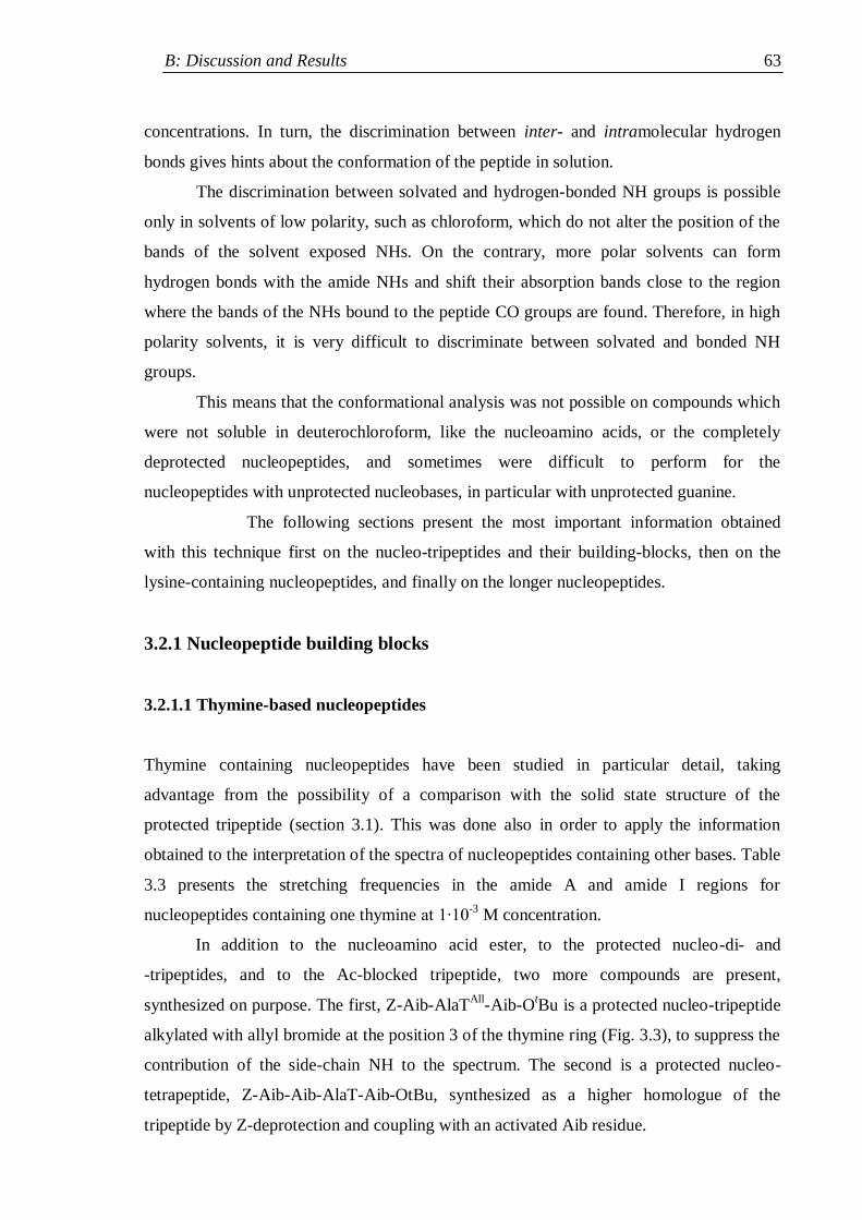
B: Discussion and Results 63
concentrations. In turn, the discrimination between inter- and intramolecular hydrogen
bonds gives hints about the conformation of the peptide in solution.
The discrimination between solvated and hydrogen-bonded NH groups is possible
only in solvents of low polarity, such as chloroform, which do not alter the position of the
bands of the solvent exposed NHs. On the contrary, more polar solvents can form
hydrogen bonds with the amide NHs and shift their absorption bands close to the region
where the bands of the NHs bound to the peptide CO groups are found. Therefore, in high
polarity solvents, it is very difficult to discriminate between solvated and bonded NH
groups.
This means that the conformational analysis was not possible on compounds which
were not soluble in deuterochloroform, like the nucleoamino acids, or the completely
deprotected nucleopeptides, and sometimes were difficult to perform for the
nucleopeptides with unprotected nucleobases, in particular with unprotected guanine.
The following sections present the most important information obtained
with this technique first on the nucleo-tripeptides and their building-blocks, then on the
lysine-containing nucleopeptides, and finally on the longer nucleopeptides.
3.2.1 Nucleopeptide building blocks
3.2.1.1 Thymine-based nucleopeptides
Thymine containing nucleopeptides have been studied in particular detail, taking
advantage from the possibility of a comparison with the solid state structure of the
protected tripeptide (section 3.1). This was done also in order to apply the information
obtained to the interpretation of the spectra of nucleopeptides containing other bases. Table
3.3 presents the stretching frequencies in the amide A and amide I regions for
nucleopeptides containing one thymine at 1∙10-3
M concentration.
In addition to the nucleoamino acid ester, to the protected nucleo-di- and
-tripeptides, and to the Ac-blocked tripeptide, two more compounds are present,
synthesized on purpose. The first, Z-Aib-AlaTAll
-Aib-OtBu is a protected nucleo-tripeptide
alkylated with allyl bromide at the position 3 of the thymine ring (Fig. 3.3), to suppress the
contribution of the side-chain NH to the spectrum. The second is a protected nucleo-
tetrapeptide, Z-Aib-Aib-AlaT-Aib-OtBu, synthesized as a higher homologue of the
tripeptide by Z-deprotection and coupling with an activated Aib residue.
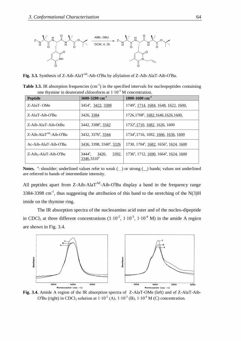
3. Conformational Characterisation 64
Fig. 3.3. Synthesis of Z-Aib-AlaTAll
-Aib-OtBu by allylation of Z-Aib-AlaT-Aib-O
tBu.
Table 3.3. IR absorption frequencies (cm
-1) in the specified intervals for nucleopeptides containing
one thymine in deuterated chloroform at 1∙10-3 M concentration.
Peptide 3600-3200 cm-1
1800-1600 cm-1
Z-AlaT- OMe 3454a, 3422, 3388 1749a, 1714, 1684, 1648, 1622, 1600,
Z-AlaT-Aib-OtBu 3426, 3384 1726,1708a, 1682,1646,1626,1600,
Z-Aib-AlaT-Aib-OtBu 3442, 3388a, 3342 1732a,1710, 1682, 1626, 1600
Z-Aib-AlaTAll-Aib-OtBu 3432, 3376a, 3344 1734a,1716, 1692, 1666, 1636, 1600
Ac-Aib-AlaT-Aib-OtBu 3436, 3398, 3340a, 3326 1730, 1704a, 1682, 1656a, 1624, 1600
Z-Aib2-AlaT-Aib-OtBu 3444a, 3420, 3392, 3346,3310a
1736a, 1712, 1690, 1664a, 1624, 1600
Notes. a: shoulder; underlined values refer to weak ( ) or strong ( ) bands; values not underlined
are referred to bands of intermediate intensity.
All peptides apart from Z-Aib-AlaTAll
-Aib-OtBu display a band in the frequency range
3384-3398 cm-1
, thus suggesting the attribution of this band to the stretching of the N(3)H
imide on the thymine ring.
The IR absorption spectra of the nucleoamino acid ester and of the nucleo-dipeptide
in CDCl3 at three different concentrations (1∙10-2
, 1∙10-3
, 1∙10-4
M) in the amide A region
are shown in Fig. 3.4.
Fig. 3.4. Amide A region of the IR absorption spectra of Z-AlaT-OMe (left) and of Z-AlaT-Aib-
OtBu (right) in CDCl3 solution at 1∙10
-2 (A), 1∙10
-3 (B), 1∙10
-4 M (C) concentration.
NH
NH
NH
O
O
O
O
NH
O
O
NH
NH
NH
O
O
O
O
N
O
O
Z ZAllBr, DBU
DCM, rt, 2h
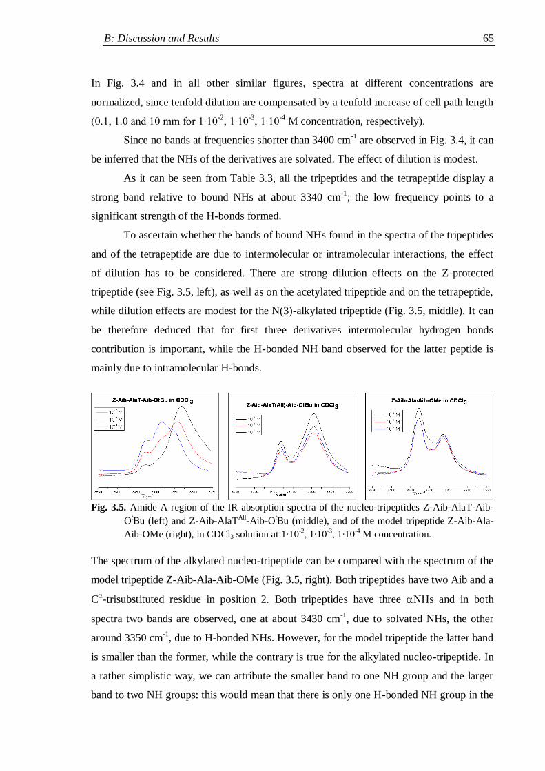
B: Discussion and Results 65
In Fig. 3.4 and in all other similar figures, spectra at different concentrations are
normalized, since tenfold dilution are compensated by a tenfold increase of cell path length
(0.1, 1.0 and 10 mm for 1∙10-2
, 1∙10-3
, 1∙10-4
M concentration, respectively).
Since no bands at frequencies shorter than 3400 cm-1
are observed in Fig. 3.4, it can
be inferred that the NHs of the derivatives are solvated. The effect of dilution is modest.
As it can be seen from Table 3.3, all the tripeptides and the tetrapeptide display a
strong band relative to bound NHs at about 3340 cm-1
; the low frequency points to a
significant strength of the H-bonds formed.
To ascertain whether the bands of bound NHs found in the spectra of the tripeptides
and of the tetrapeptide are due to intermolecular or intramolecular interactions, the effect
of dilution has to be considered. There are strong dilution effects on the Z-protected
tripeptide (see Fig. 3.5, left), as well as on the acetylated tripeptide and on the tetrapeptide,
while dilution effects are modest for the N(3)-alkylated tripeptide (Fig. 3.5, middle). It can
be therefore deduced that for first three derivatives intermolecular hydrogen bonds
contribution is important, while the H-bonded NH band observed for the latter peptide is
mainly due to intramolecular H-bonds.
Fig. 3.5. Amide A region of the IR absorption spectra of the nucleo-tripeptides Z-Aib-AlaT-Aib-
OtBu (left) and Z-Aib-AlaT
All-Aib-O
tBu (middle), and of the model tripeptide Z-Aib-Ala-
Aib-OMe (right), in CDCl3 solution at 1∙10-2, 1∙10
-3, 1∙10
-4 M concentration.
The spectrum of the alkylated nucleo-tripeptide can be compared with the spectrum of the
model tripeptide Z-Aib-Ala-Aib-OMe (Fig. 3.5, right). Both tripeptides have two Aib and a
C-trisubstituted residue in position 2. Both tripeptides have three NHs and in both
spectra two bands are observed, one at about 3430 cm-1
, due to solvated NHs, the other
around 3350 cm-1
, due to H-bonded NHs. However, for the model tripeptide the latter band
is smaller than the former, while the contrary is true for the alkylated nucleo-tripeptide. In
a rather simplistic way, we can attribute the smaller band to one NH group and the larger
band to two NH groups: this would mean that there is only one H-bonded NH group in the
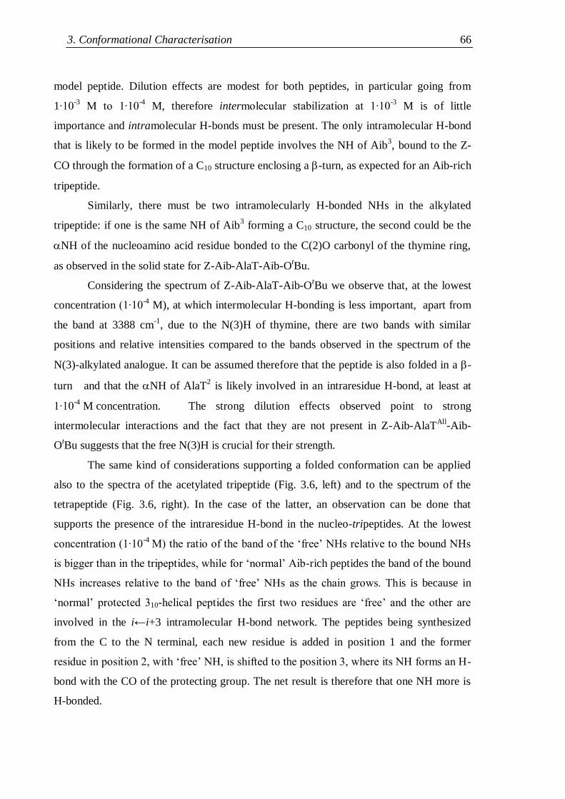
3. Conformational Characterisation 66
model peptide. Dilution effects are modest for both peptides, in particular going from
1∙10-3
M to 1∙10-4
M, therefore intermolecular stabilization at 1∙10-3
M is of little
importance and intramolecular H-bonds must be present. The only intramolecular H-bond
that is likely to be formed in the model peptide involves the NH of Aib3, bound to the Z-
CO through the formation of a C10 structure enclosing a -turn, as expected for an Aib-rich
tripeptide.
Similarly, there must be two intramolecularly H-bonded NHs in the alkylated
tripeptide: if one is the same NH of Aib3 forming a C10 structure, the second could be the
NH of the nucleoamino acid residue bonded to the C(2)O carbonyl of the thymine ring,
as observed in the solid state for Z-Aib-AlaT-Aib-OtBu.
Considering the spectrum of Z-Aib-AlaT-Aib-OtBu we observe that, at the lowest
concentration (1∙10-4
M), at which intermolecular H-bonding is less important, apart from
the band at 3388 cm-1
, due to the N(3)H of thymine, there are two bands with similar
positions and relative intensities compared to the bands observed in the spectrum of the
N(3)-alkylated analogue. It can be assumed therefore that the peptide is also folded in a -
turn and that the NH of AlaT2 is likely involved in an intraresidue H-bond, at least at
1∙10-4
M concentration. The strong dilution effects observed point to strong
intermolecular interactions and the fact that they are not present in Z-Aib-AlaTAll
-Aib-
OtBu suggests that the free N(3)H is crucial for their strength.
The same kind of considerations supporting a folded conformation can be applied
also to the spectra of the acetylated tripeptide (Fig. 3.6, left) and to the spectrum of the
tetrapeptide (Fig. 3.6, right). In the case of the latter, an observation can be done that
supports the presence of the intraresidue H-bond in the nucleo-tripeptides. At the lowest
concentration (1∙10-4
M) the ratio of the band of the „free‟ NHs relative to the bound NHs
is bigger than in the tripeptides, while for „normal‟ Aib-rich peptides the band of the bound
NHs increases relative to the band of „free‟ NHs as the chain grows. This is because in
„normal‟ protected 310-helical peptides the first two residues are „free‟ and the other are
involved in the i←i+3 intramolecular H-bond network. The peptides being synthesized
from the C to the N terminal, each new residue is added in position 1 and the former
residue in position 2, with „free‟ NH, is shifted to the position 3, where its NH forms an H-
bond with the CO of the protecting group. The net result is therefore that one NH more is
H-bonded.
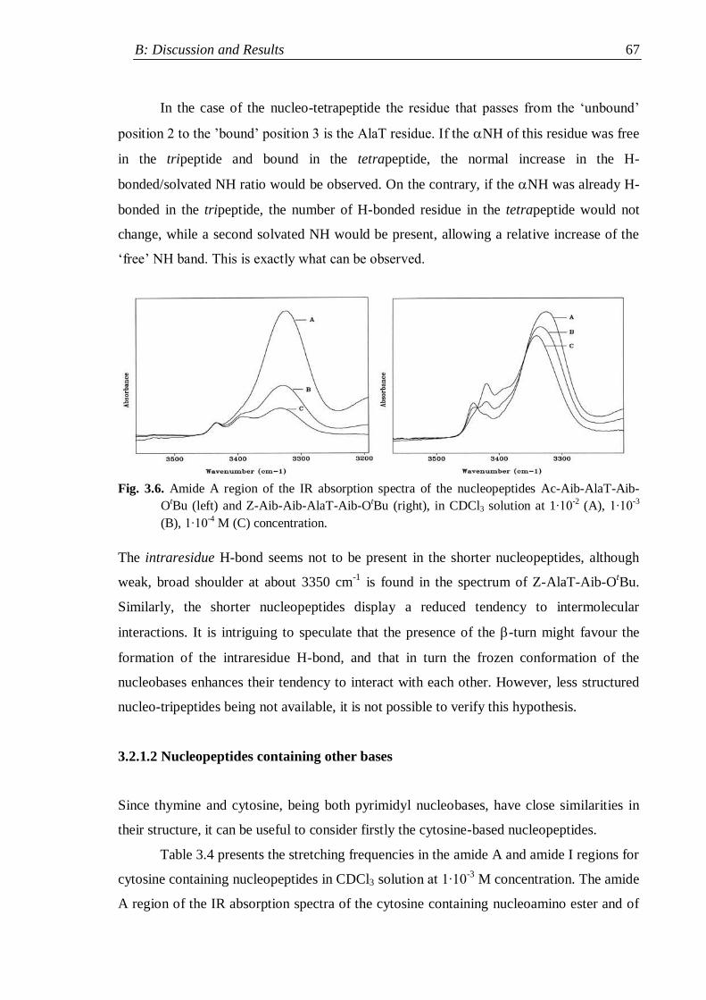
B: Discussion and Results 67
In the case of the nucleo-tetrapeptide the residue that passes from the „unbound‟
position 2 to the ‟bound‟ position 3 is the AlaT residue. If the NH of this residue was free
in the tripeptide and bound in the tetrapeptide, the normal increase in the H-
bonded/solvated NH ratio would be observed. On the contrary, if the NH was already H-
bonded in the tripeptide, the number of H-bonded residue in the tetrapeptide would not
change, while a second solvated NH would be present, allowing a relative increase of the
„free‟ NH band. This is exactly what can be observed.
Fig. 3.6. Amide A region of the IR absorption spectra of the nucleopeptides Ac-Aib-AlaT-Aib-
OtBu (left) and Z-Aib-Aib-AlaT-Aib-O
tBu (right), in CDCl3 solution at 1∙10
-2 (A), 1∙10
-3
(B), 1∙10-4
M (C) concentration.
The intraresidue H-bond seems not to be present in the shorter nucleopeptides, although
weak, broad shoulder at about 3350 cm-1
is found in the spectrum of Z-AlaT-Aib-OtBu.
Similarly, the shorter nucleopeptides display a reduced tendency to intermolecular
interactions. It is intriguing to speculate that the presence of the -turn might favour the
formation of the intraresidue H-bond, and that in turn the frozen conformation of the
nucleobases enhances their tendency to interact with each other. However, less structured
nucleo-tripeptides being not available, it is not possible to verify this hypothesis.
3.2.1.2 Nucleopeptides containing other bases
Since thymine and cytosine, being both pyrimidyl nucleobases, have close similarities in
their structure, it can be useful to consider firstly the cytosine-based nucleopeptides.
Table 3.4 presents the stretching frequencies in the amide A and amide I regions for
cytosine containing nucleopeptides in CDCl3 solution at 1∙10-3
M concentration. The amide
A region of the IR absorption spectra of the cytosine containing nucleoamino ester and of
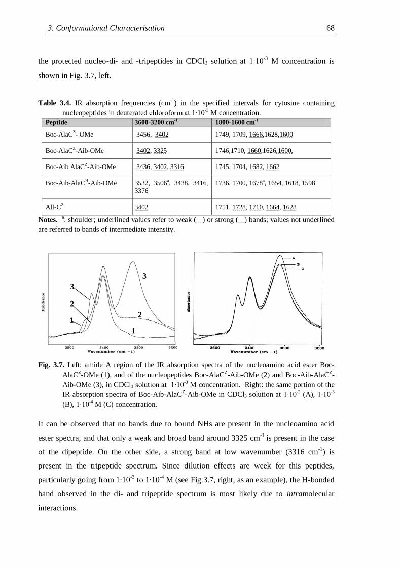
3. Conformational Characterisation 68
the protected nucleo-di- and -tripeptides in CDCl3 solution at 1∙10-3
M concentration is
shown in Fig. 3.7, left.
Table 3.4. IR absorption frequencies (cm-1
) in the specified intervals for cytosine containing
nucleopeptides in deuterated chloroform at 1∙10-3 M concentration.
Peptide 3600-3200 cm-1
1800-1600 cm-1
Boc-AlaCZ- OMe 3456, 3402 1749, 1709, 1666,1628,1600
Boc-AlaCZ-Aib-OMe 3402, 3325 1746,1710, 1660,1626,1600,
Boc-Aib AlaCZ-Aib-OMe 3436, 3402, 3316 1745, 1704, 1682, 1662
Boc-Aib-AlaCH-Aib-OMe 3532, 3506a, 3438, 3416,
3376
1736, 1700, 1678a, 1654, 1618, 1598
All-CZ 3402 1751, 1728, 1710, 1664, 1628
Notes. a: shoulder; underlined values refer to weak ( ) or strong ( ) bands; values not underlined
are referred to bands of intermediate intensity.
Fig. 3.7. Left: amide A region of the IR absorption spectra of the nucleoamino acid ester Boc-
AlaCZ-OMe (1), and of the nucleopeptides Boc-AlaC
Z-Aib-OMe (2) and Boc-Aib-AlaC
Z-
Aib-OMe (3), in CDCl3 solution at 1∙10-3 M concentration. Right: the same portion of the
IR absorption spectra of Boc-Aib-AlaCZ-Aib-OMe in CDCl3 solution at 1∙10
-2 (A), 1∙10
-3
(B), 1∙10-4
M (C) concentration.
It can be observed that no bands due to bound NHs are present in the nucleoamino acid
ester spectra, and that only a weak and broad band around 3325 cm-1
is present in the case
of the dipeptide. On the other side, a strong band at low wavenumber (3316 cm-1
) is
present in the tripeptide spectrum. Since dilution effects are week for this peptides,
particularly going from 1∙10-3
to 1∙10-4
M (see Fig.3.7, right, as an example), the H-bonded
band observed in the di- and tripeptide spectrum is most likely due to intramolecular
interactions.
1
2
3
1
2
3
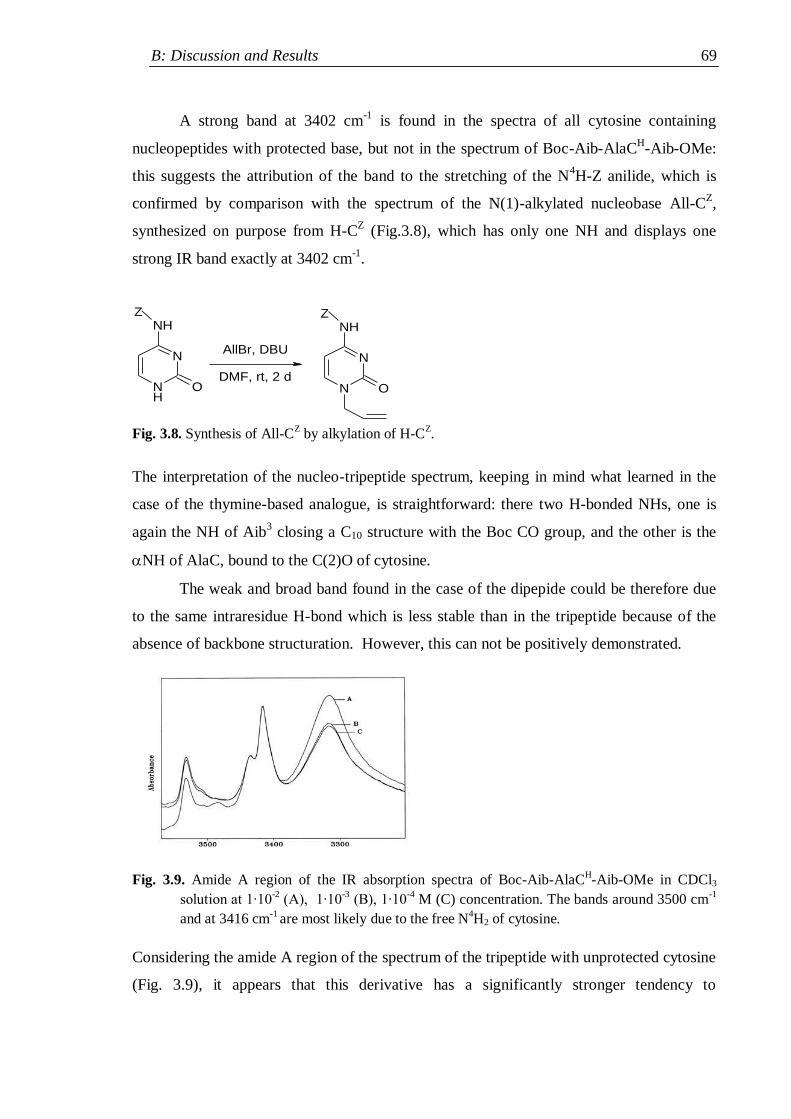
B: Discussion and Results 69
A strong band at 3402 cm-1
is found in the spectra of all cytosine containing
nucleopeptides with protected base, but not in the spectrum of Boc-Aib-AlaCH-Aib-OMe:
this suggests the attribution of the band to the stretching of the N4H-Z anilide, which is
confirmed by comparison with the spectrum of the N(1)-alkylated nucleobase All-CZ,
synthesized on purpose from H-CZ (Fig.3.8), which has only one NH and displays one
strong IR band exactly at 3402 cm-1
.
NH
N
NH
O N
N
NH
O
Z Z
AllBr, DBU
DMF, rt, 2 d
Fig. 3.8. Synthesis of All-C
Z by alkylation of H-C
Z.
The interpretation of the nucleo-tripeptide spectrum, keeping in mind what learned in the
case of the thymine-based analogue, is straightforward: there two H-bonded NHs, one is
again the NH of Aib3 closing a C10 structure with the Boc CO group, and the other is the
NH of AlaC, bound to the C(2)O of cytosine.
The weak and broad band found in the case of the dipepide could be therefore due
to the same intraresidue H-bond which is less stable than in the tripeptide because of the
absence of backbone structuration. However, this can not be positively demonstrated.
Fig. 3.9. Amide A region of the IR absorption spectra of Boc-Aib-AlaCH-Aib-OMe in CDCl3
solution at 1∙10-2
(A), 1∙10-3 (B), 1∙10
-4 M (C) concentration. The bands around 3500 cm
-1
and at 3416 cm-1
are most likely due to the free N4H2 of cytosine.
Considering the amide A region of the spectrum of the tripeptide with unprotected cytosine
(Fig. 3.9), it appears that this derivative has a significantly stronger tendency to
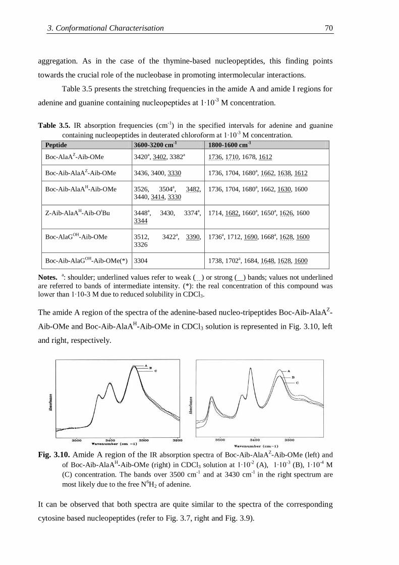
3. Conformational Characterisation 70
aggregation. As in the case of the thymine-based nucleopeptides, this finding points
towards the crucial role of the nucleobase in promoting intermolecular interactions.
Table 3.5 presents the stretching frequencies in the amide A and amide I regions for
adenine and guanine containing nucleopeptides at 1∙10-3
M concentration.
Table 3.5. IR absorption frequencies (cm
-1) in the specified intervals for adenine and guanine
containing nucleopeptides in deuterated chloroform at 1∙10-3 M concentration.
Peptide 3600-3200 cm-1
1800-1600 cm-1
Boc-AlaAZ-Aib-OMe 3420a, 3402, 3382a 1736, 1710, 1678, 1612
Boc-Aib-AlaAZ-Aib-OMe 3436, 3400, 3330 1736, 1704, 1680a, 1662, 1638, 1612
Boc-Aib-AlaAH-Aib-OMe 3526, 3504a, 3482,
3440, 3414, 3330
1736, 1704, 1680a, 1662, 1630, 1600
Z-Aib-AlaAH-Aib-OtBu 3448a, 3430, 3374a,
3344
1714, 1682, 1660a, 1650a, 1626, 1600
Boc-AlaGOH-Aib-OMe 3512, 3422a, 3390,
3326
1736a, 1712, 1690, 1668a, 1628, 1600
Boc-Aib-AlaGOH-Aib-OMe(*) 3304 1738, 1702a, 1684, 1648, 1628, 1600
Notes. a: shoulder; underlined values refer to weak ( ) or strong ( ) bands; values not underlined
are referred to bands of intermediate intensity. (*): the real concentration of this compound was lower than 1∙10-3 M due to reduced solubility in CDCl3.
The amide A region of the spectra of the adenine-based nucleo-tripeptides Boc-Aib-AlaAZ-
Aib-OMe and Boc-Aib-AlaAH-Aib-OMe in CDCl3 solution is represented in Fig. 3.10, left
and right, respectively.
Fig. 3.10. Amide A region of the IR absorption spectra of Boc-Aib-AlaAZ-Aib-OMe (left) and
of Boc-Aib-AlaAH-Aib-OMe (right) in CDCl3 solution at 1∙10
-2 (A), 1∙10
-3 (B), 1∙10
-4 M
(C) concentration. The bands over 3500 cm-1
and at 3430 cm-1
in the right spectrum are
most likely due to the free N6H2 of adenine.
It can be observed that both spectra are quite similar to the spectra of the corresponding
cytosine based nucleopeptides (refer to Fig. 3.7, right and Fig. 3.9).
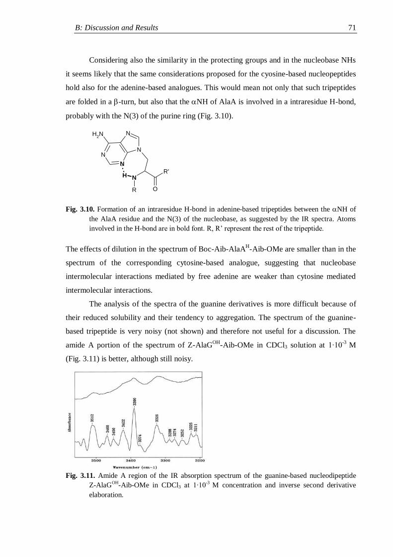
B: Discussion and Results 71
Considering also the similarity in the protecting groups and in the nucleobase NHs
it seems likely that the same considerations proposed for the cyosine-based nucleopeptides
hold also for the adenine-based analogues. This would mean not only that such tripeptides
are folded in a -turn, but also that the NH of AlaA is involved in a intraresidue H-bond,
probably with the N(3) of the purine ring (Fig. 3.10).
Fig. 3.10. Formation of an intraresidue H-bond in adenine-based tripeptides between the NH of
the AlaA residue and the N(3) of the nucleobase, as suggested by the IR spectra. Atoms
involved in the H-bond are in bold font. R, R‟ represent the rest of the tripeptide.
The effects of dilution in the spectrum of Boc-Aib-AlaAH-Aib-OMe are smaller than in the
spectrum of the corresponding cytosine-based analogue, suggesting that nucleobase
intermolecular interactions mediated by free adenine are weaker than cytosine mediated
intermolecular interactions.
The analysis of the spectra of the guanine derivatives is more difficult because of
their reduced solubility and their tendency to aggregation. The spectrum of the guanine-
based tripeptide is very noisy (not shown) and therefore not useful for a discussion. The
amide A portion of the spectrum of Z-AlaGOH
-Aib-OMe in CDCl3 solution at 1∙10-3
M
(Fig. 3.11) is better, although still noisy.
Fig. 3.11. Amide A region of the IR absorption spectrum of the guanine-based nucleodipeptide
Z-AlaGOH
-Aib-OMe in CDCl3 at 1∙10-3
M concentration and inverse second derivative
elaboration.
N
O
R'
R
N
N
N
N
NH2
H
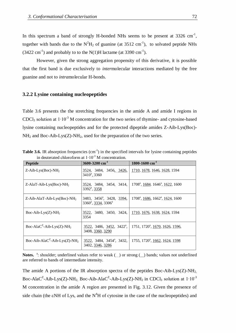
3. Conformational Characterisation 72
In this spectrum a band of strongly H-bonded NHs seems to be present at 3326 cm-1
,
together with bands due to the N2H2 of guanine (at 3512 cm
-1), to solvated peptide NHs
(3422 cm-1
) and probably to to the N(1)H lactame (at 3390 cm-1
).
However, given the strong aggregation propensity of this derivative, it is possible
that the first band is due exclusively to intermolecular interactions mediated by the free
guanine and not to intramolecular H-bonds.
3.2.2 Lysine containing nucleopeptides
Table 3.6 presents the the stretching frequencies in the amide A and amide I regions in
CDCl3 solution at 1∙10-3
M concentration for the two series of thymine- and cytosine-based
lysine containing nucleopeptides and for the protected dipeptide amides Z-Aib-Lys(Boc)-
NH2 and Boc-Aib-Lys(Z)-NH2, used for the preparation of the two series.
Table 3.6. IR absorption frequencies (cm-1
) in the specified intervals for lysine containing peptides
in deuterated chloroform at 1∙10-3
M concentration.
Peptide 3600-3200 cm-1
1800-1600 cm-1
Z-Aib-Lys(Boc)-NH2 3524, 3484, 3456, 3426,
3410a, 3360
1710, 1678, 1646, 1628, 1594
Z-AlaT-Aib-Lys(Boc)-NH2 3524, 3484, 3454, 3414,
3392a, 3358
1708a, 1684. 1646a, 1622, 1600
Z-Aib-AlaT-Aib-Lys(Boc)-NH2 3483, 3456a, 3428, 3394,
3360a, 3334, 3306a
1708a, 1686, 1662a, 1624, 1600
Boc-Aib-Lys(Z)-NH2 3522, 3480, 3450, 3424,
3354
1710, 1676, 1638, 1624, 1594
Boc-AlaCZ-Aib-Lys(Z)-NH2 3522, 3486, 3452, 3422a,
3408, 3360, 3290
1751, 1720a, 1670, 1626, 1596,
Boc-Aib-AlaCZ-Aib-Lys(Z)-NH2 3522, 3484, 3454a, 3432,
3402, 3346, 3286
1755, 1720a, 1662, 1624, 1598
Notes. a: shoulder; underlined values refer to weak ( ) or strong ( ) bands; values not underlined
are referred to bands of intermediate intensity.
The amide A portions of the IR absorption spectra of the peptides Boc-Aib-Lys(Z)-NH2,
Boc-AlaCZ-Aib-Lys(Z)-NH2, Boc-Aib-AlaC
Z-Aib-Lys(Z)-NH2 in CDCl3 solution at 1∙10
-3
M concentration in the amide A region are presented in Fig. 3.12. Given the presence of
side chain (the NH of Lys, and the N4H of cytosine in the case of the nucleopeptides) and
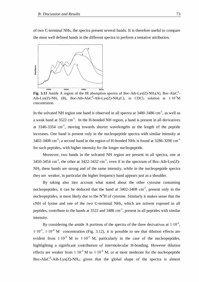
B: Discussion and Results 73
of two C-terminal NHs, the spectra present several bands. It is therefore useful to compare
the most well defined bands in the different spectra to perform a tentative attribution.
Fig. 3.11 Amide A region of the IR absorption spectra of Boc-Aib-Lys(Z)-NH2(A), Boc-AlaC
Z-
Aib-Lys(Z)-NH2 (B), Boc-Aib-AlaCZ-Aib-Lys(Z)-NH2(C), in CDCl3 solution at 1∙10
-3M
concentration.
In the solvated NH region one band is observed in all spectra at 3480-3486 cm-1
, as well as
a weak band at 3522 cm-1
. In the H-bonded NH region, a band is present in all derivatives
at 3346-3354 cm-1
, moving towards shorter wavelengths as the length of the peptide
increases. One band is present only in the nucleopeptide spectra with similar intensity at
3402-3408 cm-1
; a second band in the region of H-bonded NHs is found at 3286-3290 cm-1
for such peptides, with higher intensity for the longer nucleopeptide.
Moreover, two bands in the solvated NH region are present in all spectra, one at
3450-3454 cm-1
, the other at 3422-3432 cm-1
, even if in the spectrum of Boc-Aib-Lys(Z)-
NH2 these bands are strong and of the same intensity, while in the nucleopeptide spectra
they are weaker, in particular the higher frequency band appears just as a shoulder.
By taking also into account what stated about the other cytosine containing
nucleopeptides, it can be deduced that the band at 3402-3408 cm-1
, present only in the
nucleopeptides, is most likely due to the N4H of cytosine. Similarly it makes sense that the
NH of lysine and one of the two C-terminal NHs, which are solvent exposed in all
peptides, contribute to the bands at 3522 and 3486 cm-1
, present in all peptides with similar
intensity.
By considering the amide A portions of the spectra of the three derivatives at 1∙10-2
,
1∙10-3
, 1∙10-4
M concentration (Fig. 3.12), it is possible to see that dilution effects are
evident from 1∙10-2
M to 1∙10-3
M, particularly in the case of the nucleopeptides,
highlighting a significant contribution of intermolecular H-bonding. However dilution
effects are weaker from 1∙10-3
M to 1∙10-4
M, or at most moderate for the nucleopeptide
Boc-AlaCZ-Aib-Lys(Z)-NH2; given that the global shape of the spectra is almost
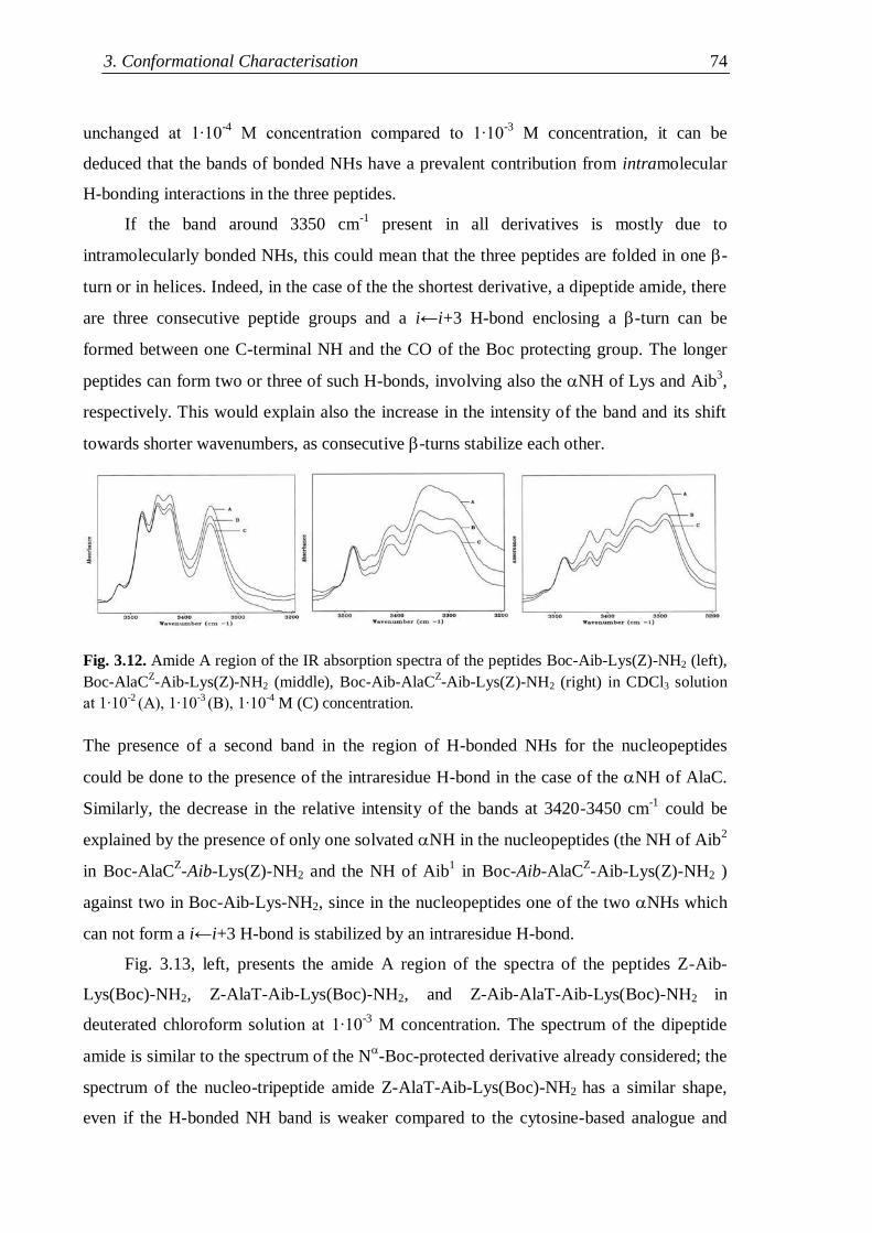
3. Conformational Characterisation 74
unchanged at 1∙10-4
M concentration compared to 1∙10-3
M concentration, it can be
deduced that the bands of bonded NHs have a prevalent contribution from intramolecular
H-bonding interactions in the three peptides.
If the band around 3350 cm-1
present in all derivatives is mostly due to
intramolecularly bonded NHs, this could mean that the three peptides are folded in one -
turn or in helices. Indeed, in the case of the the shortest derivative, a dipeptide amide, there
are three consecutive peptide groups and a i←i+3 H-bond enclosing a -turn can be
formed between one C-terminal NH and the CO of the Boc protecting group. The longer
peptides can form two or three of such H-bonds, involving also the NH of Lys and Aib3,
respectively. This would explain also the increase in the intensity of the band and its shift
towards shorter wavenumbers, as consecutive -turns stabilize each other.
Fig. 3.12. Amide A region of the IR absorption spectra of the peptides Boc-Aib-Lys(Z)-NH2 (left),
Boc-AlaCZ-Aib-Lys(Z)-NH2 (middle), Boc-Aib-AlaC
Z-Aib-Lys(Z)-NH2 (right) in CDCl3 solution
at 1∙10-2
(A), 1∙10-3
(B), 1∙10-4
M (C) concentration.
The presence of a second band in the region of H-bonded NHs for the nucleopeptides
could be done to the presence of the intraresidue H-bond in the case of the NH of AlaC.
Similarly, the decrease in the relative intensity of the bands at 3420-3450 cm-1
could be
explained by the presence of only one solvated NH in the nucleopeptides (the NH of Aib2
in Boc-AlaCZ-Aib-Lys(Z)-NH2 and the NH of Aib
1 in Boc-Aib-AlaC
Z-Aib-Lys(Z)-NH2 )
against two in Boc-Aib-Lys-NH2, since in the nucleopeptides one of the two NHs which
can not form a i←i+3 H-bond is stabilized by an intraresidue H-bond.
Fig. 3.13, left, presents the amide A region of the spectra of the peptides Z-Aib-
Lys(Boc)-NH2, Z-AlaT-Aib-Lys(Boc)-NH2, and Z-Aib-AlaT-Aib-Lys(Boc)-NH2 in
deuterated chloroform solution at 1∙10-3
M concentration. The spectrum of the dipeptide
amide is similar to the spectrum of the N-Boc-protected derivative already considered; the
spectrum of the nucleo-tripeptide amide Z-AlaT-Aib-Lys(Boc)-NH2 has a similar shape,
even if the H-bonded NH band is weaker compared to the cytosine-based analogue and
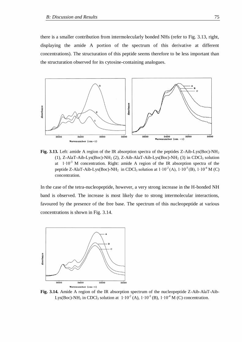
B: Discussion and Results 75
there is a smaller contribution from intermolecularly bonded NHs (refer to Fig. 3.13, right,
displaying the amide A portion of the spectrum of this derivative at different
concentrations). The structuration of this peptide seems therefore to be less important than
the structuration observed for its cytosine-containing analogues.
Fig. 3.13. Left: amide A region of the IR absorption spectra of the peptides Z-Aib-Lys(Boc)-NH2
(1), Z-AlaT-Aib-Lys(Boc)-NH2 (2), Z-Aib-AlaT-Aib-Lys(Boc)-NH2 (3) in CDCl3 solution
at 1∙10-3
M concentration. Right: amide A region of the IR absorption spectra of the
peptide Z-AlaT-Aib-Lys(Boc)-NH2 in CDCl3 solution at 1∙10-2
(A), 1∙10-3
(B), 1∙10-4 M (C)
concentration.
In the case of the tetra-nucleopeptide, however, a very strong increase in the H-bonded NH
band is observed. The increase is most likely due to strong intermolecular interactions,
favoured by the presence of the free base. The spectrum of this nucleopeptide at various
concentrations is shown in Fig. 3.14.
Fig. 3.14. Amide A region of the IR absorption spectrum of the nucleopeptide Z-Aib-AlaT-Aib-
Lys(Boc)-NH2 in CDCl3 solution at 1∙10-2
(A), 1∙10-3
(B), 1∙10-4
M (C) concentration.
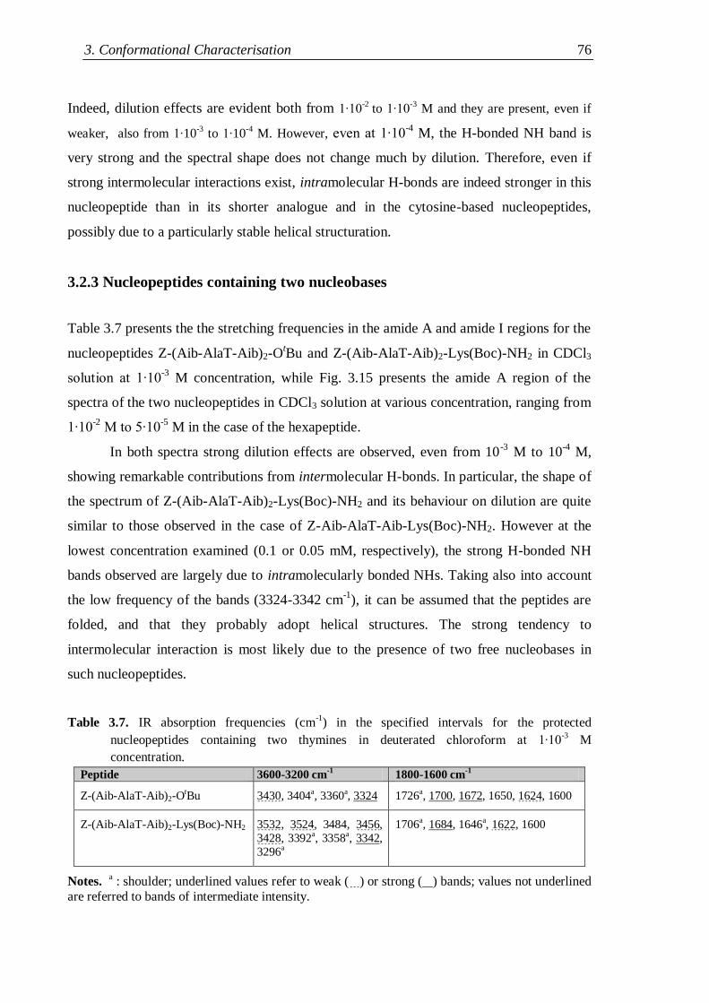
3. Conformational Characterisation 76
Indeed, dilution effects are evident both from 1∙10-2
to 1∙10-3
M and they are present, even if
weaker, also from 1∙10-3
to 1∙10-4
M. However, even at 1∙10-4
M, the H-bonded NH band is
very strong and the spectral shape does not change much by dilution. Therefore, even if
strong intermolecular interactions exist, intramolecular H-bonds are indeed stronger in this
nucleopeptide than in its shorter analogue and in the cytosine-based nucleopeptides,
possibly due to a particularly stable helical structuration.
3.2.3 Nucleopeptides containing two nucleobases
Table 3.7 presents the the stretching frequencies in the amide A and amide I regions for the
nucleopeptides Z-(Aib-AlaT-Aib)2-OtBu and Z-(Aib-AlaT-Aib)2-Lys(Boc)-NH2 in CDCl3
solution at 1∙10-3
M concentration, while Fig. 3.15 presents the amide A region of the
spectra of the two nucleopeptides in CDCl3 solution at various concentration, ranging from
1∙10-2
M to 5∙10-5
M in the case of the hexapeptide.
In both spectra strong dilution effects are observed, even from 10-3
M to 10-4
M,
showing remarkable contributions from intermolecular H-bonds. In particular, the shape of
the spectrum of Z-(Aib-AlaT-Aib)2-Lys(Boc)-NH2 and its behaviour on dilution are quite
similar to those observed in the case of Z-Aib-AlaT-Aib-Lys(Boc)-NH2. However at the
lowest concentration examined (0.1 or 0.05 mM, respectively), the strong H-bonded NH
bands observed are largely due to intramolecularly bonded NHs. Taking also into account
the low frequency of the bands (3324-3342 cm-1
), it can be assumed that the peptides are
folded, and that they probably adopt helical structures. The strong tendency to
intermolecular interaction is most likely due to the presence of two free nucleobases in
such nucleopeptides.
Table 3.7. IR absorption frequencies (cm-1
) in the specified intervals for the protected
nucleopeptides containing two thymines in deuterated chloroform at 1∙10-3 M
concentration.
Peptide 3600-3200 cm-1
1800-1600 cm-1
Z-(Aib-AlaT-Aib)2-OtBu 3430, 3404a, 3360a, 3324 1726a, 1700, 1672, 1650, 1624, 1600
Z-(Aib-AlaT-Aib)2-Lys(Boc)-NH2 3532, 3524, 3484, 3456,
3428, 3392a, 3358a, 3342,
3296a
1706a, 1684, 1646a, 1622, 1600
Notes. a : shoulder; underlined values refer to weak ( ) or strong ( ) bands; values not underlined
are referred to bands of intermediate intensity.
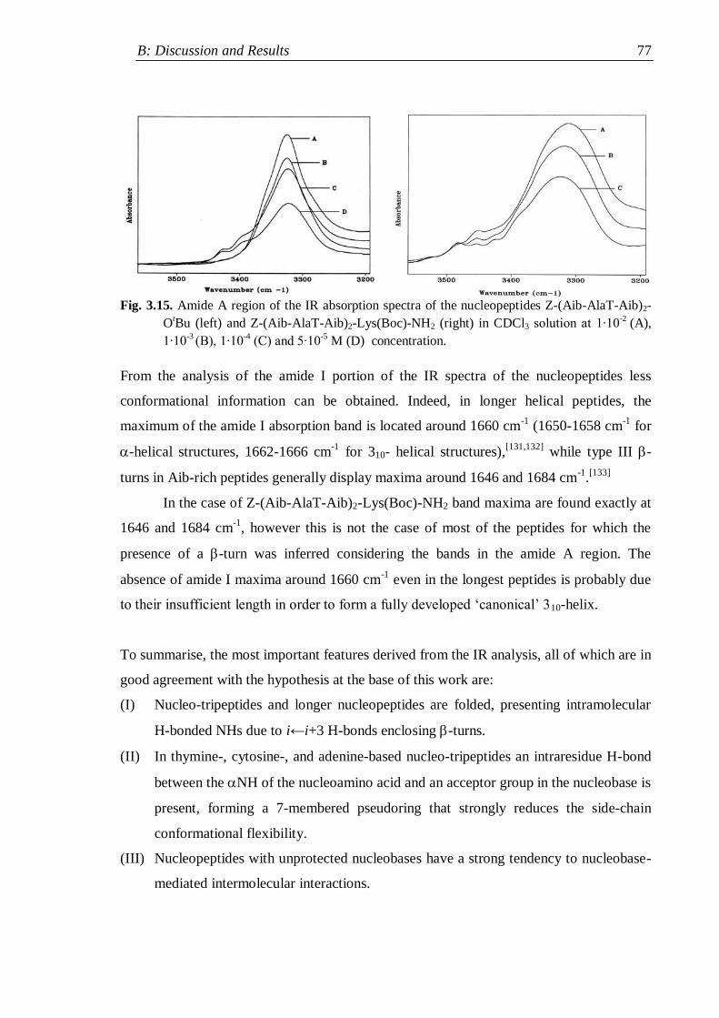
B: Discussion and Results 77
Fig. 3.15. Amide A region of the IR absorption spectra of the nucleopeptides Z-(Aib-AlaT-Aib)2-
OtBu (left) and Z-(Aib-AlaT-Aib)2-Lys(Boc)-NH2 (right) in CDCl3 solution at 1∙10
-2 (A),
1∙10-3
(B), 1∙10-4
(C) and 5∙10-5
M (D) concentration.
From the analysis of the amide I portion of the IR spectra of the nucleopeptides less
conformational information can be obtained. Indeed, in longer helical peptides, the
maximum of the amide I absorption band is located around 1660 cm-1
(1650-1658 cm-1
for
-helical structures, 1662-1666 cm-1
for 310- helical structures),[131,132]
while type III -
turns in Aib-rich peptides generally display maxima around 1646 and 1684 cm-1
.[133]
In the case of Z-(Aib-AlaT-Aib)2-Lys(Boc)-NH2 band maxima are found exactly at
1646 and 1684 cm-1
, however this is not the case of most of the peptides for which the
presence of a -turn was inferred considering the bands in the amide A region. The
absence of amide I maxima around 1660 cm-1
even in the longest peptides is probably due
to their insufficient length in order to form a fully developed „canonical‟ 310-helix.
To summarise, the most important features derived from the IR analysis, all of which are in
good agreement with the hypothesis at the base of this work are:
(I) Nucleo-tripeptides and longer nucleopeptides are folded, presenting intramolecular
H-bonded NHs due to i←i+3 H-bonds enclosing -turns.
(II) In thymine-, cytosine-, and adenine-based nucleo-tripeptides an intraresidue H-bond
between the NH of the nucleoamino acid and an acceptor group in the nucleobase is
present, forming a 7-membered pseudoring that strongly reduces the side-chain
conformational flexibility.
(III) Nucleopeptides with unprotected nucleobases have a strong tendency to nucleobase-
mediated intermolecular interactions.
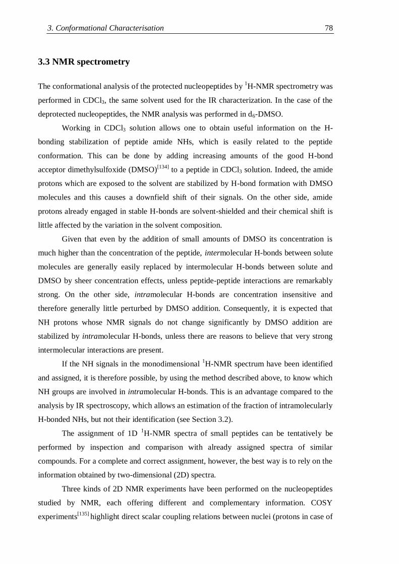
3. Conformational Characterisation 78
3.3 NMR spectrometry
The conformational analysis of the protected nucleopeptides by 1H-NMR spectrometry was
performed in CDCl3, the same solvent used for the IR characterization. In the case of the
deprotected nucleopeptides, the NMR analysis was performed in d6-DMSO.
Working in CDCl3 solution allows one to obtain useful information on the H-
bonding stabilization of peptide amide NHs, which is easily related to the peptide
conformation. This can be done by adding increasing amounts of the good H-bond
acceptor dimethylsulfoxide (DMSO)[134]
to a peptide in CDCl3 solution. Indeed, the amide
protons which are exposed to the solvent are stabilized by H-bond formation with DMSO
molecules and this causes a downfield shift of their signals. On the other side, amide
protons already engaged in stable H-bonds are solvent-shielded and their chemical shift is
little affected by the variation in the solvent composition.
Given that even by the addition of small amounts of DMSO its concentration is
much higher than the concentration of the peptide, intermolecular H-bonds between solute
molecules are generally easily replaced by intermolecular H-bonds between solute and
DMSO by sheer concentration effects, unless peptide-peptide interactions are remarkably
strong. On the other side, intramolecular H-bonds are concentration insensitive and
therefore generally little perturbed by DMSO addition. Consequently, it is expected that
NH protons whose NMR signals do not change significantly by DMSO addition are
stabilized by intramolecular H-bonds, unless there are reasons to believe that very strong
intermolecular interactions are present.
If the NH signals in the monodimensional 1H-NMR spectrum have been identified
and assigned, it is therefore possible, by using the method described above, to know which
NH groups are involved in intramolecular H-bonds. This is an advantage compared to the
analysis by IR spectroscopy, which allows an estimation of the fraction of intramolecularly
H-bonded NHs, but not their identification (see Section 3.2).
The assignment of 1D 1H-NMR spectra of small peptides can be tentatively be
performed by inspection and comparison with already assigned spectra of similar
compounds. For a complete and correct assignment, however, the best way is to rely on the
information obtained by two-dimensional (2D) spectra.
Three kinds of 2D NMR experiments have been performed on the nucleopeptides
studied by NMR, each offering different and complementary information. COSY
experiments[135]
highlight direct scalar coupling relations between nuclei (protons in case of
B: Discussion and Results 79
2D-1H NMR) generally less than three chemical bonds apart. TOCSY experiments
[136]
allow the identification of spin systems (i.e. groups of nuclei connected by scalar coupling
relations). ROESY experiments[137]
are instead sensitive to the spatial proximity of the
nuclei.
In general, the backbone and the alkyl chain portion of the side-chain of each amino
acid residue form a spin system, whose signals can be related by TOCSY experiments.
ROESY experiments can then connect residues which are next to each other along the
backbone, allowing the complete assignment. In folded peptides made of protein residues a
specific process, called sequential assignment,[138]
is well established. It employs the
through-space connectivity along the backbone of the kind CH(i)NH(i+1) and
NH(i)NH(i+1), derived from ROESY experiments, in order to obtain the sequence of the
residues forming a peptide, whose signals have been connected by TOCSY experiments.
COSY experiments are useful particularly for the assignment of the resonances within
single spin systems in case this is not evident from the single chemical shifts.
Once the attribution has been completed, the information about spatial proximity
obtained by ROESY experiments can be usefully employed. In particular, when nuclei
which are many bonds apart appear to be next to each other, additional data on the peptide
conformation are obtained. This can be used also for gaining insight in the side-chain
conformation, on which it is more difficult to gather information by other techniques.
Since 2D NMR experiments can be performed in every solvent, the possibility of
obtaining conformational information is not restricted by solubility in organic solvents of
low polarity, as in the case of the 1D NMR or of the IR analysis. Therefore this method has
been employed for studying also the conformation of the deprotected nucleopeptides.
3.3.1 Protected nucleo-tripeptides
N- and C-terminally protected thymine-, cytosine- and adenine-based tripeptides have been
studied. The nucleopeptide with protected cytosine Boc-Aib-AlaCZ-Aib-OMe and the
nucleopeptide with unprotected adenine Boc-Aib-AlaAH-Aib-OMe have been considered.
The latter two nucleopeptides were selected to check whether the nucleobase protection
affects the conformation of the backbone. Indeed, it was shown by the analysis of their IR
spectra (Section 3.2.1.2) that adenine- and cytosine-based tripeptides, both with protected
and unprotected base, had a similar behaviour. It was therefore assumed that the analysis of
3. Conformational Characterisation 80
the two derivatives would have given information which is also valid for the two other
nucleopeptides (Boc-Aib-AlaCH-Aib-OMe and Boc-Aib-AlaA
Z-Aib-OMe).
The data gathered by IR spectroscopy on the three nucleopeptides which needed to
be supported by the NMR analysis were the assumption of a folded conformation and the
presence of an intraresidue H-bond involving the NH of the nucleoamino acid residues.
Firstly, the 1D NMR spectra of the nucleo-tripeptides were completely assigned by
performing 2D TOCSY and ROESY experiments. Interestingly, in all spectra the NH
protons of the nucleoamino acid residues have the highest chemical shift among the
backbone NHs (≈ 8.5 ppm for the pyrimidyl derivatives, = 9 ppm for the adenine
derivative), suggesting the involvement in strong hydrogen bonds.
Fig. 3.16 presents the variation of the chemical shifts of the NH protons of the three
peptides by addition of increasing amounts of d6- DMSO.
Fig. 3.16. Variation of the chemical shift of the NH protons of Z-Aib-AlaT-Aib-OtBu (left), Boc-
Aib-AlaCZ-Aib-OMe (middle), Boc-Aib-AlaA
H-Aib-OMe (right) in CDCl3 solution at
1∙10-3 M concentration by addition of increasing amounts of d6-DMSO.
All peptides have three backbone NHs (black, red and green) and one more signal (blue)
due to side chain NHs (the imide N(3)H of thymine, the anilide N4H-Z of the protected
cytosine, the aromatic amino group N6H2 of the unprotected adenine) and the three graphs
display parallel trends for the three nucleopeptides. Indeed, in all graphs the signals of the
side chain NHs and of the NH of Aib1 are shifted downfields as the DMSO concentration
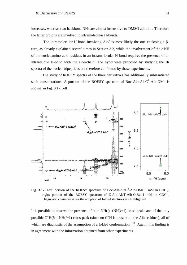
B: Discussion and Results 81
increases, whereas two backbone NHs are almost insensitive to DMSO addition. Therefore
the latter protons are involved in intramolecular H-bonds.
The intramolecular H-bond involving Aib3 is most likely the one enclosing a -
turn, as already explained several times in Section 3.2, while the involvement of the NH
of the nucleoamino acid residues in an intramolecular H-bond requires the presence of an
intraresidue H-bond with the side-chain. The hypotheses proposed by studying the IR
spectra of the nucleo-tripeptides are therefore confirmed by these experiments.
The study of ROESY spectra of the three derivatives has additionally substantiated
such considerations. A portion of the ROESY spectrum of Boc-Aib-AlaCZ-Aib-OMe is
shown in Fig. 3.17, left.
Fig. 3.17. Left: portion of the ROESY spectrum of Boc-Aib-AlaCZ-Aib-OMe 1 mM in CDCl3;
right: portion of the ROESY spectrum of Z-Aib-AlaT-Aib-OtBu 1 mM in CDCl3.
Diagnostic cross-peaks for the adoption of folded stuctures are highlighted.
It is possible to observe the presence of both NH(i)NH(i+1) cross-peaks and of the only
possible CH(i)→NH(i+1) cross-peak (since no C
H is present on the Aib residues), all of
which are diagnostic of the assumption of a folded conformation.[138]
Again, this finding is
in agreement with the information obtained from other experiments.
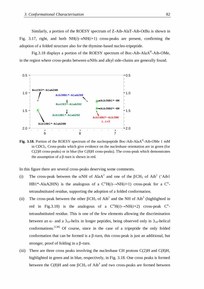
3. Conformational Characterisation 82
Similarly, a portion of the ROESY spectrum of Z-Aib-AlaT-Aib-OtBu is shown in
Fig. 3.17, right, and both NH(i)NH(i+1) cross-peaks are present, confirming the
adoption of a folded structure also for the thymine-based nucleo-tripeptide.
Fig.3.18 displays a portion of the ROESY spectrum of Boc-Aib-AlaAH-Aib-OMe,
in the region where cross-peaks between NHs and alkyl side-chains are generally found.
2.0
1.5
1.0
0.5
1
-
1
H
(
p
pm
)
Aib3HB1*-HN
Aib3HB2*-HN
Aib1HB2*-Aib3HN
i,i+2
Aib1HB1*-AlaA2H2
Aib1HB1*-AlaA2HN
Aib3HB1*-AlaA2H8
BocCH3*-AlaA2H2
BocCH3*-AlaA2HN
2.0
1.5
1.0
0.5
9 8 7
Fig. 3.18. Portion of the ROESY spectrum of the nucleopeptide Boc-Aib-AlaAH-Aib-OMe 1 mM
in CDCl3. Cross-peaks which give evidence on the nucleobase orientation are in green (for
C(2)H cross-peaks) or in blue (for C(8)H cross-peaks). The cross-peak which demonstrates
the assumption of a -turn is shown in red.
In this figure there are several cross-peaks deserving some comments.
(i) The cross-peak between the NH of AlaA2 and one of the CH3 of Aib
1 („Aib1
HB1*-AlaA2HN) is the analogous of a CH(i)→NH(i+1) cross-peak for a C
-
tetrasubstituted residue, supporting the adoption of a folded conformation.
(ii) The cross-peak between the other CH3 of Aib1 and the NH of Aib
3 (highlighted in
red in Fig.3.18) is the analogous of a CH(i)→NH(i+2) cross-peak C
-
tetrasubstituted residue. This is one of the few elements allowing the discrimination
between an - and a 310-helix in longer peptides, being observed only in 310-helical
conformations.[138]
Of course, since in the case of a tripeptide the only folded
conformation that can be formed is a -turn, this cross-peak is just an additional, but
stronger, proof of folding in a -turn.
(iii) There are three cross peaks involving the nucleobase CH protons C(2)H and C(8)H,
highlighted in green and in blue, respectively, in Fig. 3.18. One cross peaks is formed
between the C(8)H and one CH3 of Aib3 and two cross-peaks are formed between
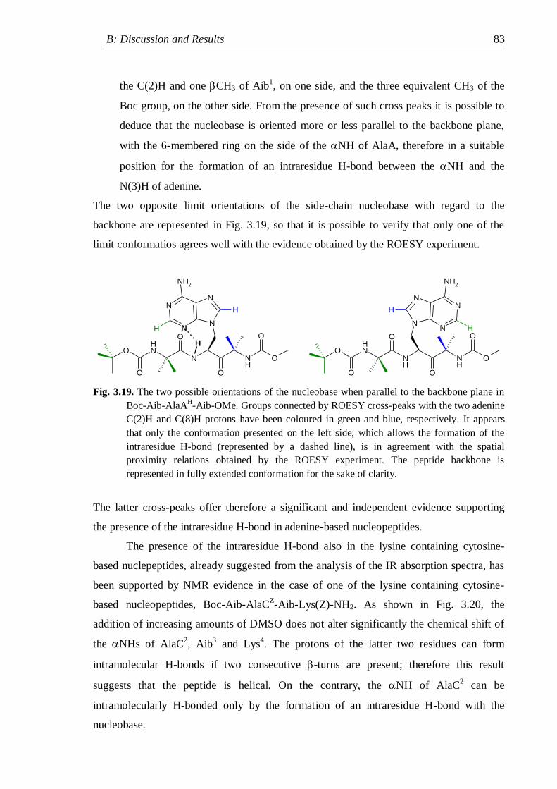
B: Discussion and Results 83
the C(2)H and one CH3 of Aib1, on one side, and the three equivalent CH3 of the
Boc group, on the other side. From the presence of such cross peaks it is possible to
deduce that the nucleobase is oriented more or less parallel to the backbone plane,
with the 6-membered ring on the side of the NH of AlaA, therefore in a suitable
position for the formation of an intraresidue H-bond between the NH and the
N(3)H of adenine.
The two opposite limit orientations of the side-chain nucleobase with regard to the
backbone are represented in Fig. 3.19, so that it is possible to verify that only one of the
limit conformatios agrees well with the evidence obtained by the ROESY experiment.
N
O
NH
N
NN
N
NH2
O
O
ONH
O
O
H
H
H
NH
O
NH
O
O
ONH
O
O
N
NN
N
NH2
H
H
Fig. 3.19. The two possible orientations of the nucleobase when parallel to the backbone plane in
Boc-Aib-AlaAH-Aib-OMe. Groups connected by ROESY cross-peaks with the two adenine
C(2)H and C(8)H protons have been coloured in green and blue, respectively. It appears
that only the conformation presented on the left side, which allows the formation of the
intraresidue H-bond (represented by a dashed line), is in agreement with the spatial
proximity relations obtained by the ROESY experiment. The peptide backbone is
represented in fully extended conformation for the sake of clarity.
The latter cross-peaks offer therefore a significant and independent evidence supporting
the presence of the intraresidue H-bond in adenine-based nucleopeptides.
The presence of the intraresidue H-bond also in the lysine containing cytosine-
based nuclepeptides, already suggested from the analysis of the IR absorption spectra, has
been supported by NMR evidence in the case of one of the lysine containing cytosine-
based nucleopeptides, Boc-Aib-AlaCZ-Aib-Lys(Z)-NH2. As shown in Fig. 3.20, the
addition of increasing amounts of DMSO does not alter significantly the chemical shift of
the NHs of AlaC2, Aib
3 and Lys
4. The protons of the latter two residues can form
intramolecular H-bonds if two consecutive -turns are present; therefore this result
suggests that the peptide is helical. On the contrary, the NH of AlaC2 can be
intramolecularly H-bonded only by the formation of an intraresidue H-bond with the
nucleobase.
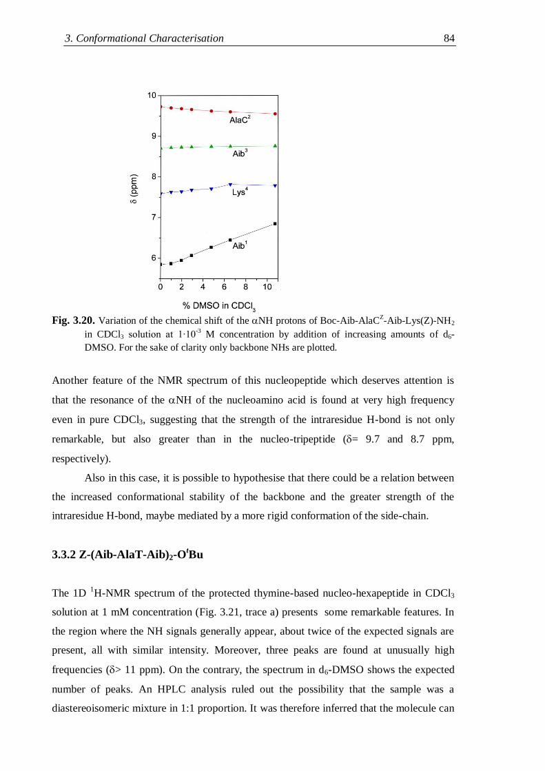
3. Conformational Characterisation 84
Fig. 3.20. Variation of the chemical shift of the NH protons of Boc-Aib-AlaCZ-Aib-Lys(Z)-NH2
in CDCl3 solution at 1∙10-3
M concentration by addition of increasing amounts of d6-
DMSO. For the sake of clarity only backbone NHs are plotted.
Another feature of the NMR spectrum of this nucleopeptide which deserves attention is
that the resonance of the NH of the nucleoamino acid is found at very high frequency
even in pure CDCl3, suggesting that the strength of the intraresidue H-bond is not only
remarkable, but also greater than in the nucleo-tripeptide (= 9.7 and 8.7 ppm,
respectively).
Also in this case, it is possible to hypothesise that there could be a relation between
the increased conformational stability of the backbone and the greater strength of the
intraresidue H-bond, maybe mediated by a more rigid conformation of the side-chain.
3.3.2 Z-(Aib-AlaT-Aib)2-OtBu
The 1D 1H-NMR spectrum of the protected thymine-based nucleo-hexapeptide in CDCl3
solution at 1 mM concentration (Fig. 3.21, trace a) presents some remarkable features. In
the region where the NH signals generally appear, about twice of the expected signals are
present, all with similar intensity. Moreover, three peaks are found at unusually high
frequencies (> 11 ppm). On the contrary, the spectrum in d6-DMSO shows the expected
number of peaks. An HPLC analysis ruled out the possibility that the sample was a
diastereoisomeric mixture in 1:1 proportion. It was therefore inferred that the molecule can
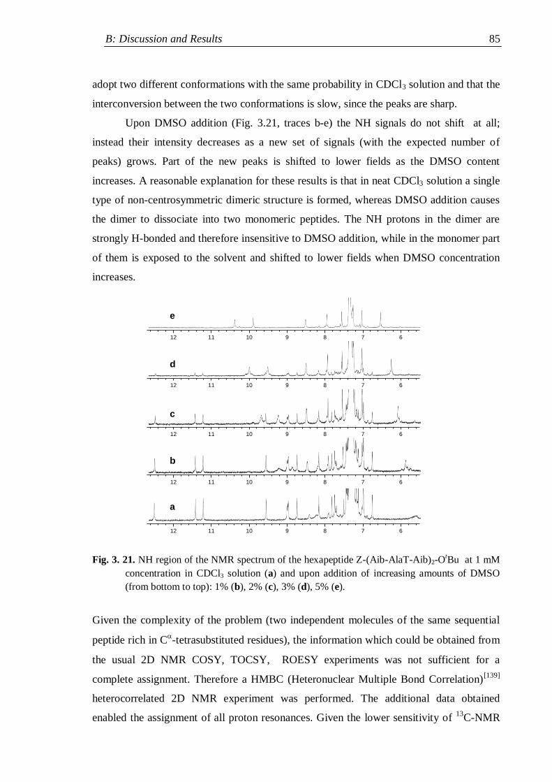
B: Discussion and Results 85
adopt two different conformations with the same probability in CDCl3 solution and that the
interconversion between the two conformations is slow, since the peaks are sharp.
Upon DMSO addition (Fig. 3.21, traces b-e) the NH signals do not shift at all;
instead their intensity decreases as a new set of signals (with the expected number of
peaks) grows. Part of the new peaks is shifted to lower fields as the DMSO content
increases. A reasonable explanation for these results is that in neat CDCl3 solution a single
type of non-centrosymmetric dimeric structure is formed, whereas DMSO addition causes
the dimer to dissociate into two monomeric peptides. The NH protons in the dimer are
strongly H-bonded and therefore insensitive to DMSO addition, while in the monomer part
of them is exposed to the solvent and shifted to lower fields when DMSO concentration
increases.
Fig. 3. 21. NH region of the NMR spectrum of the hexapeptide Z-(Aib-AlaT-Aib)2-OtBu at 1 mM
concentration in CDCl3 solution (a) and upon addition of increasing amounts of DMSO
(from bottom to top): 1% (b), 2% (c), 3% (d), 5% (e).
Given the complexity of the problem (two independent molecules of the same sequential
peptide rich in C-tetrasubstituted residues), the information which could be obtained from
the usual 2D NMR COSY, TOCSY, ROESY experiments was not sufficient for a
complete assignment. Therefore a HMBC (Heteronuclear Multiple Bond Correlation)[139]
heterocorrelated 2D NMR experiment was performed. The additional data obtained
enabled the assignment of all proton resonances. Given the lower sensitivity of 13
C-NMR
12 11 10 9 8 7 6
12 11 10 9 8 7 6
12 11 10 9 8 7 6
12 11 10 9 8 7 6
12 11 10 9 8 7 6
a
b
c
d
e
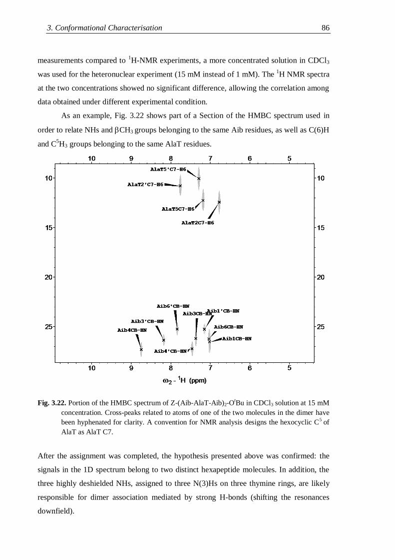
3. Conformational Characterisation 86
measurements compared to 1H-NMR experiments, a more concentrated solution in CDCl3
was used for the heteronuclear experiment (15 mM instead of 1 mM). The 1H NMR spectra
at the two concentrations showed no significant difference, allowing the correlation among
data obtained under different experimental condition.
As an example, Fig. 3.22 shows part of a Section of the HMBC spectrum used in
order to relate NHs and CH3 groups belonging to the same Aib residues, as well as C(6)H
and C5H3 groups belonging to the same AlaT residues.
Fig. 3.22. Portion of the HMBC spectrum of Z-(Aib-AlaT-Aib)2-OtBu in CDCl3 solution at 15 mM
concentration. Cross-peaks related to atoms of one of the two molecules in the dimer have
been hyphenated for clarity. A convention for NMR analysis designs the hexocyclic C5
of
AlaT as AlaT C7.
After the assignment was completed, the hypothesis presented above was confirmed: the
signals in the 1D spectrum belong to two distinct hexapeptide molecules. In addition, the
three highly deshielded NHs, assigned to three N(3)Hs on three thymine rings, are likely
responsible for dimer association mediated by strong H-bonds (shifting the resonances
downfield).
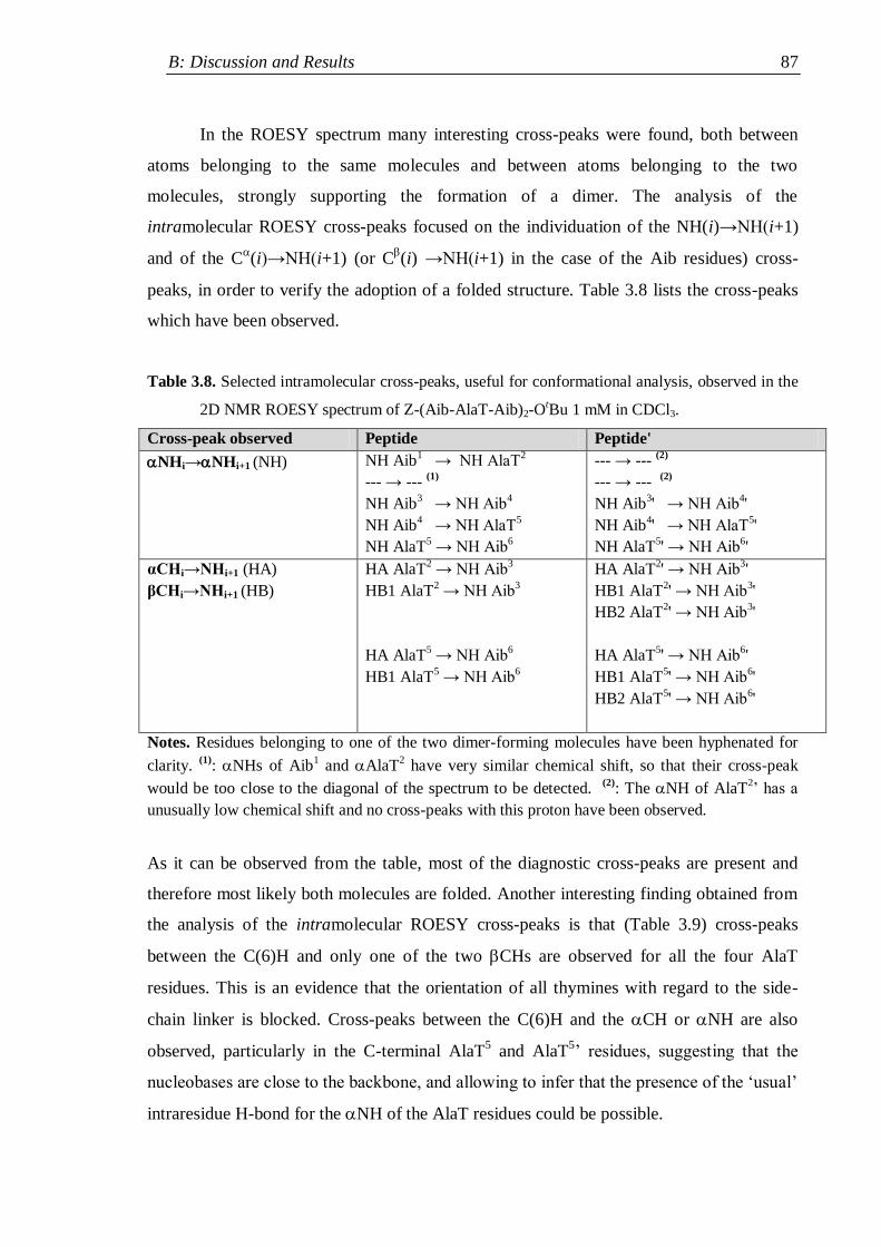
B: Discussion and Results 87
In the ROESY spectrum many interesting cross-peaks were found, both between
atoms belonging to the same molecules and between atoms belonging to the two
molecules, strongly supporting the formation of a dimer. The analysis of the
intramolecular ROESY cross-peaks focused on the individuation of the NH(i)→NH(i+1)
and of the C(i)→NH(i+1) (or C
(i) →NH(i+1) in the case of the Aib residues) cross-
peaks, in order to verify the adoption of a folded structure. Table 3.8 lists the cross-peaks
which have been observed.
Table 3.8. Selected intramolecular cross-peaks, useful for conformational analysis, observed in the
2D NMR ROESY spectrum of Z-(Aib-AlaT-Aib)2-OtBu 1 mM in CDCl3.
Cross-peak observed Peptide Peptide'
NHi→NHi+1 (NH) NH Aib1 → NH AlaT
2
--- → --- (1)
NH Aib3 → NH Aib
4
NH Aib4 → NH AlaT
5
NH AlaT5 → NH Aib
6
--- → --- (2)
--- → --- (2)
NH Aib3' → NH Aib
4'
NH Aib4' → NH AlaT
5'
NH AlaT5' → NH Aib
6'
αCHi→NHi+1 (HA)
βCHi→NHi+1 (HB)
HA AlaT2 → NH Aib
3
HB1 AlaT2 → NH Aib
3
HA AlaT5 → NH Aib
6
HB1 AlaT5 → NH Aib
6
HA AlaT2' → NH Aib
3'
HB1 AlaT2' → NH Aib
3'
HB2 AlaT2' → NH Aib
3'
HA AlaT5' → NH Aib
6'
HB1 AlaT5' → NH Aib
6'
HB2 AlaT5' → NH Aib
6'
Notes. Residues belonging to one of the two dimer-forming molecules have been hyphenated for
clarity. (1)
: NHs of Aib1 and AlaT
2 have very similar chemical shift, so that their cross-peak
would be too close to the diagonal of the spectrum to be detected. (2)
: The NH of AlaT2‟ has a
unusually low chemical shift and no cross-peaks with this proton have been observed.
As it can be observed from the table, most of the diagnostic cross-peaks are present and
therefore most likely both molecules are folded. Another interesting finding obtained from
the analysis of the intramolecular ROESY cross-peaks is that (Table 3.9) cross-peaks
between the C(6)H and only one of the two CHs are observed for all the four AlaT
residues. This is an evidence that the orientation of all thymines with regard to the side-
chain linker is blocked. Cross-peaks between the C(6)H and the CH or NH are also
observed, particularly in the C-terminal AlaT5 and AlaT
5‟ residues, suggesting that the
nucleobases are close to the backbone, and allowing to infer that the presence of the „usual‟
intraresidue H-bond for the NH of the AlaT residues could be possible.
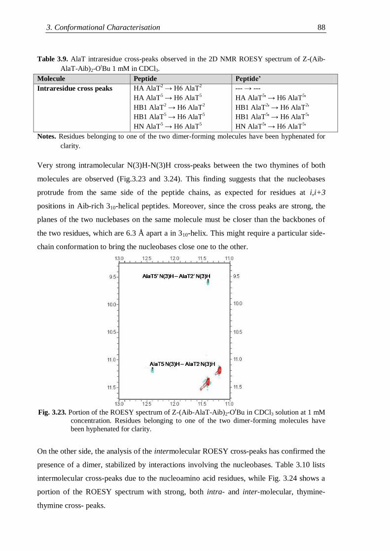
3. Conformational Characterisation 88
Table 3.9. AlaT intraresidue cross-peaks observed in the 2D NMR ROESY spectrum of Z-(Aib-
AlaT-Aib)2-OtBu 1 mM in CDCl3.
Molecule Peptide Peptide’
Intraresidue cross peaks
HA AlaT2 → H6 AlaT
2
HA AlaT5 → H6 AlaT
5
HB1 AlaT2 → H6 AlaT
2
HB1 AlaT5 → H6 AlaT
5
HN AlaT5 → H6 AlaT
5
--- → ---
HA AlaT5' → H6 AlaT
5'
HB1 AlaT2' → H6 AlaT
2'
HB1 AlaT5' → H6 AlaT
5'
HN AlaT5' → H6 AlaT
5'
Notes. Residues belonging to one of the two dimer-forming molecules have been hyphenated for
clarity.
Very strong intramolecular N(3)H-N(3)H cross-peaks between the two thymines of both
molecules are observed (Fig.3.23 and 3.24). This finding suggests that the nucleobases
protrude from the same side of the peptide chains, as expected for residues at i,i+3
positions in Aib-rich 310-helical peptides. Moreover, since the cross peaks are strong, the
planes of the two nuclebases on the same molecule must be closer than the backbones of
the two residues, which are 6.3 Å apart a in 310-helix. This might require a particular side-
chain conformation to bring the nucleobases close one to the other.
Fig. 3.23. Portion of the ROESY spectrum of Z-(Aib-AlaT-Aib)2-OtBu in CDCl3 solution at 1 mM
concentration. Residues belonging to one of the two dimer-forming molecules have
been hyphenated for clarity.
On the other side, the analysis of the intermolecular ROESY cross-peaks has confirmed the
presence of a dimer, stabilized by interactions involving the nucleobases. Table 3.10 lists
intermolecular cross-peaks due to the nucleoamino acid residues, while Fig. 3.24 shows a
portion of the ROESY spectrum with strong, both intra- and inter-molecular, thymine-
thymine cross- peaks.
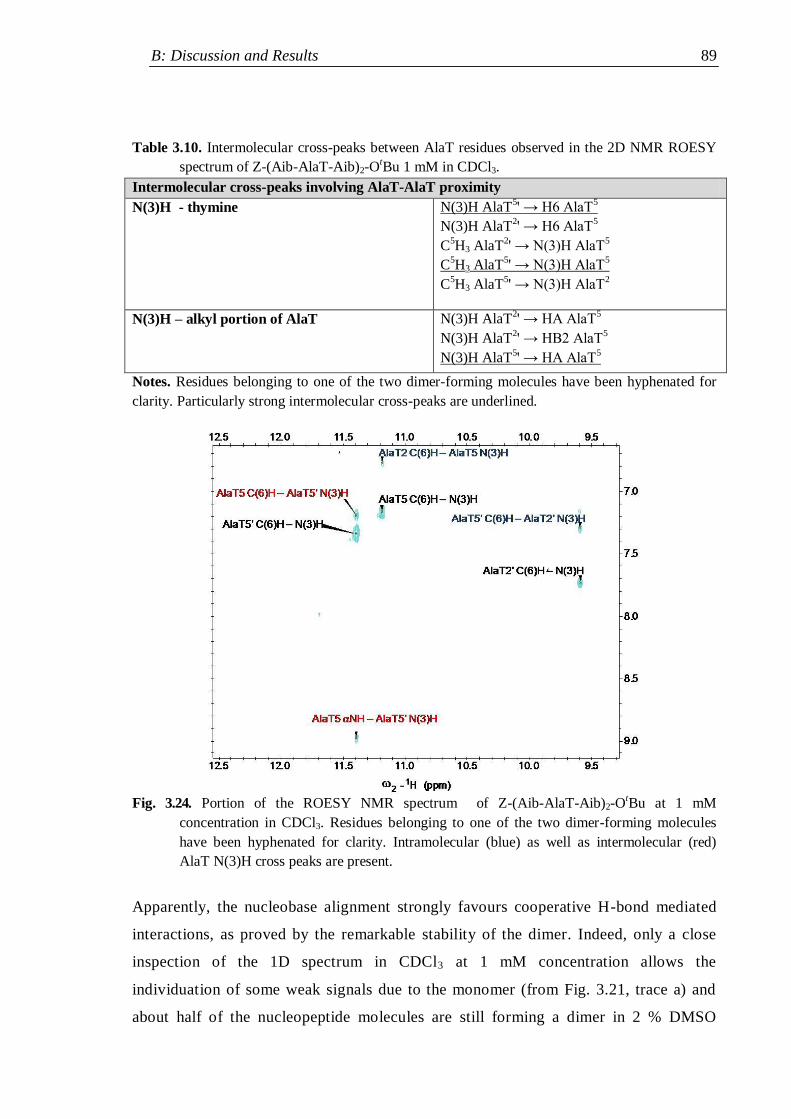
B: Discussion and Results 89
Table 3.10. Intermolecular cross-peaks between AlaT residues observed in the 2D NMR ROESY
spectrum of Z-(Aib-AlaT-Aib)2-OtBu 1 mM in CDCl3.
Intermolecular cross-peaks involving AlaT-AlaT proximity
N(3)H - thymine
N(3)H AlaT5' → H6 AlaT
5
N(3)H AlaT2' → H6 AlaT
5
C5H3 AlaT
2' → N(3)H AlaT
5
C5H3 AlaT
5' → N(3)H AlaT
5
C5H3 AlaT
5' → N(3)H AlaT
2
N(3)H – alkyl portion of AlaT N(3)H AlaT2' → HA AlaT
5
N(3)H AlaT2' → HB2 AlaT
5
N(3)H AlaT5' → HA AlaT
5
Notes. Residues belonging to one of the two dimer-forming molecules have been hyphenated for
clarity. Particularly strong intermolecular cross-peaks are underlined.
Fig. 3.24. Portion of the ROESY NMR spectrum of Z-(Aib-AlaT-Aib)2-OtBu at 1 mM
concentration in CDCl3. Residues belonging to one of the two dimer-forming molecules
have been hyphenated for clarity. Intramolecular (blue) as well as intermolecular (red)
AlaT N(3)H cross peaks are present.
Apparently, the nucleobase alignment strongly favours cooperative H-bond mediated
interactions, as proved by the remarkable stability of the dimer. Indeed, only a close
inspection of the 1D spectrum in CDCl3 at 1 mM concentration allows the
individuation of some weak signals due to the monomer (from Fig. 3.21, trace a) and
about half of the nucleopeptide molecules are still forming a dimer in 2 % DMSO
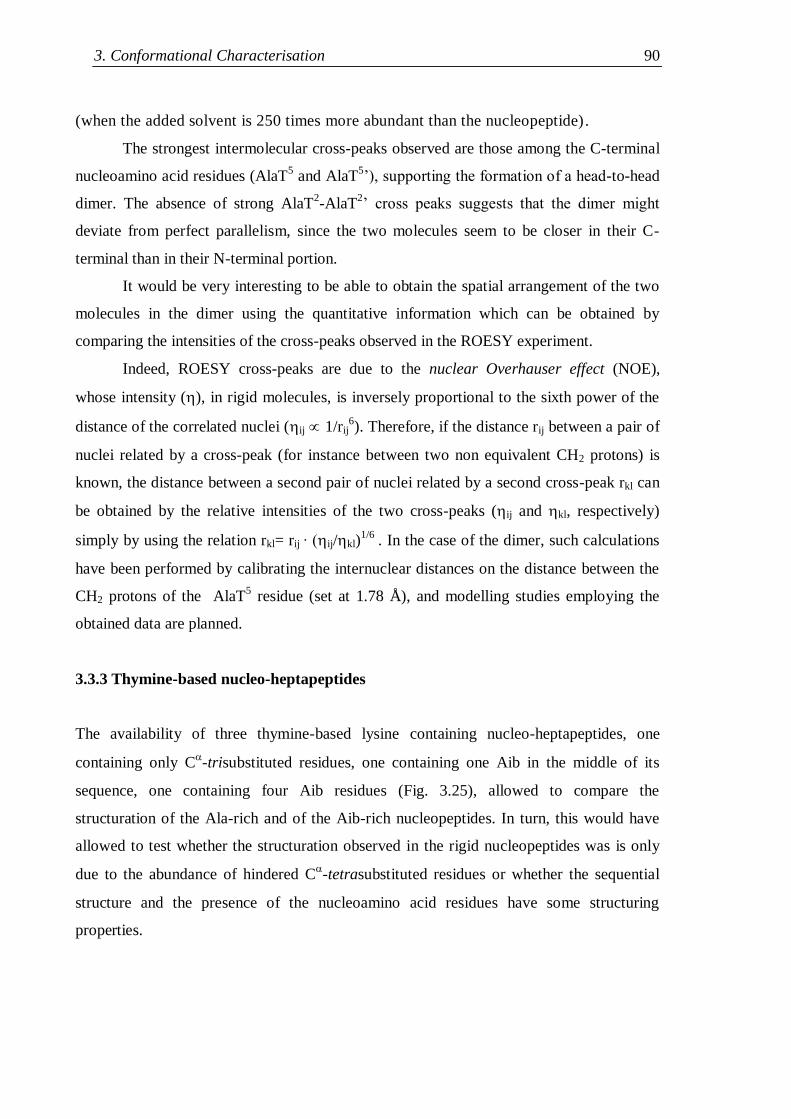
3. Conformational Characterisation 90
(when the added solvent is 250 times more abundant than the nucleopeptide).
The strongest intermolecular cross-peaks observed are those among the C-terminal
nucleoamino acid residues (AlaT5 and AlaT
5‟), supporting the formation of a head-to-head
dimer. The absence of strong AlaT2-AlaT
2‟ cross peaks suggests that the dimer might
deviate from perfect parallelism, since the two molecules seem to be closer in their C-
terminal than in their N-terminal portion.
It would be very interesting to be able to obtain the spatial arrangement of the two
molecules in the dimer using the quantitative information which can be obtained by
comparing the intensities of the cross-peaks observed in the ROESY experiment.
Indeed, ROESY cross-peaks are due to the nuclear Overhauser effect (NOE),
whose intensity (), in rigid molecules, is inversely proportional to the sixth power of the
distance of the correlated nuclei (ij 1/rij6). Therefore, if the distance rij between a pair of
nuclei related by a cross-peak (for instance between two non equivalent CH2 protons) is
known, the distance between a second pair of nuclei related by a second cross-peak rkl can
be obtained by the relative intensities of the two cross-peaks (ij and kl, respectively)
simply by using the relation rkl= rij ∙ (ij/kl)1/6
. In the case of the dimer, such calculations
have been performed by calibrating the internuclear distances on the distance between the
CH2 protons of the AlaT5 residue (set at 1.78 Å), and modelling studies employing the
obtained data are planned.
3.3.3 Thymine-based nucleo-heptapeptides
The availability of three thymine-based lysine containing nucleo-heptapeptides, one
containing only C-trisubstituted residues, one containing one Aib in the middle of its
sequence, one containing four Aib residues (Fig. 3.25), allowed to compare the
structuration of the Ala-rich and of the Aib-rich nucleopeptides. In turn, this would have
allowed to test whether the structuration observed in the rigid nucleopeptides was is only
due to the abundance of hindered C-tetrasubstituted residues or whether the sequential
structure and the presence of the nucleoamino acid residues have some structuring
properties.
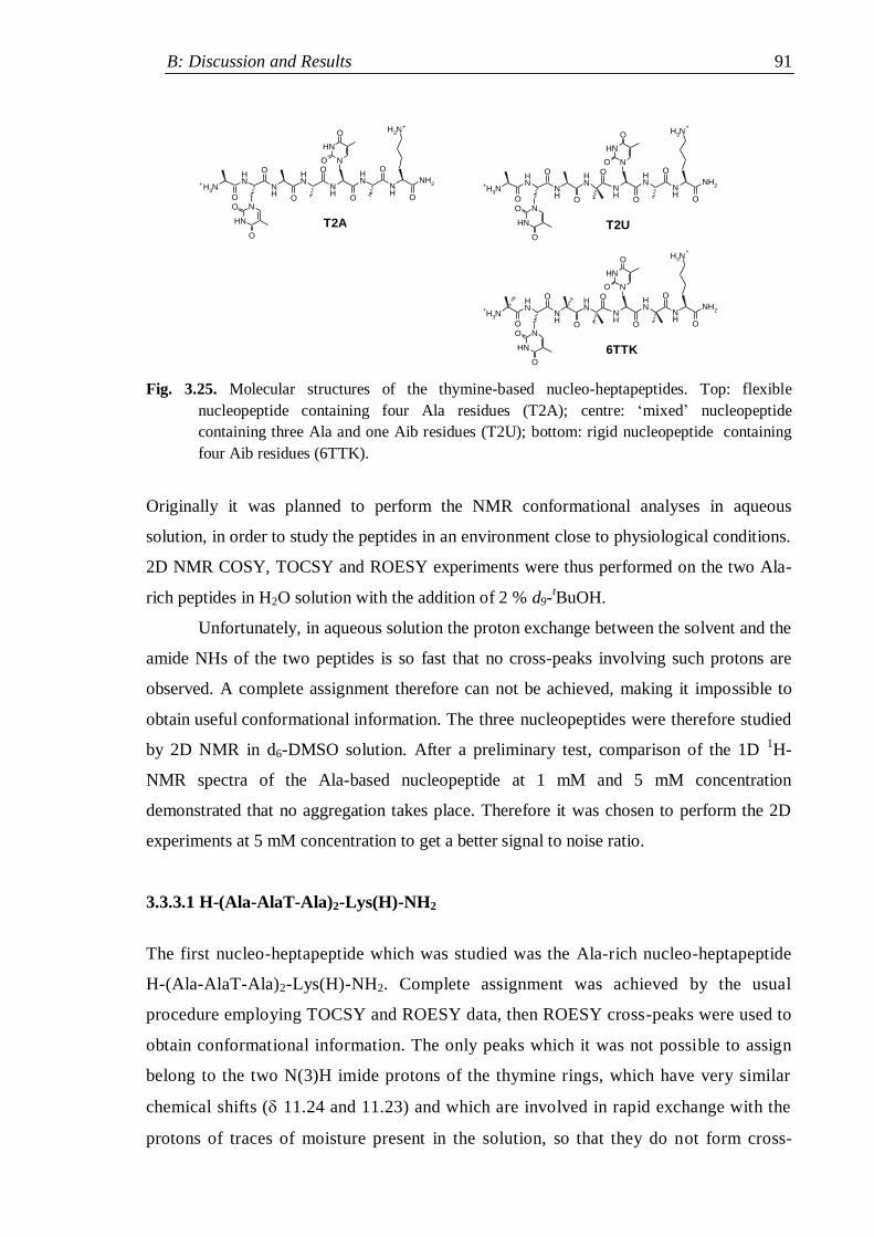
B: Discussion and Results 91
N
NH
O
O
H3N
H3N
NH
NHO
ON
NH
O
O
NH
O
NH
NHO
O
O
NH
NH2
O
N
NH
O
O
H3N
H3N
NH
NHO
ON
NH
O
O
NH
O
NH
NHO
O
O
NH
NH2
ON
NH
O
O
H3N
H3N
NH
NHO
ON
NH
O
O
NH
O
NH
NH
O
O
O
NH
NH2
O
+
+
6TTK
+
+
T2U
+
+
T2A
Fig. 3.25. Molecular structures of the thymine-based nucleo-heptapeptides. Top: flexible
nucleopeptide containing four Ala residues (T2A); centre: „mixed‟ nucleopeptide
containing three Ala and one Aib residues (T2U); bottom: rigid nucleopeptide containing
four Aib residues (6TTK).
Originally it was planned to perform the NMR conformational analyses in aqueous
solution, in order to study the peptides in an environment close to physiological conditions.
2D NMR COSY, TOCSY and ROESY experiments were thus performed on the two Ala-
rich peptides in H2O solution with the addition of 2 % d9-tBuOH.
Unfortunately, in aqueous solution the proton exchange between the solvent and the
amide NHs of the two peptides is so fast that no cross-peaks involving such protons are
observed. A complete assignment therefore can not be achieved, making it impossible to
obtain useful conformational information. The three nucleopeptides were therefore studied
by 2D NMR in d6-DMSO solution. After a preliminary test, comparison of the 1D 1H-
NMR spectra of the Ala-based nucleopeptide at 1 mM and 5 mM concentration
demonstrated that no aggregation takes place. Therefore it was chosen to perform the 2D
experiments at 5 mM concentration to get a better signal to noise ratio.
3.3.3.1 H-(Ala-AlaT-Ala)2-Lys(H)-NH2
The first nucleo-heptapeptide which was studied was the Ala-rich nucleo-heptapeptide
H-(Ala-AlaT-Ala)2-Lys(H)-NH2. Complete assignment was achieved by the usual
procedure employing TOCSY and ROESY data, then ROESY cross-peaks were used to
obtain conformational information. The only peaks which it was not possible to assign
belong to the two N(3)H imide protons of the thymine rings, which have very similar
chemical shifts ( 11.24 and 11.23) and which are involved in rapid exchange with the
protons of traces of moisture present in the solution, so that they do not form cross-
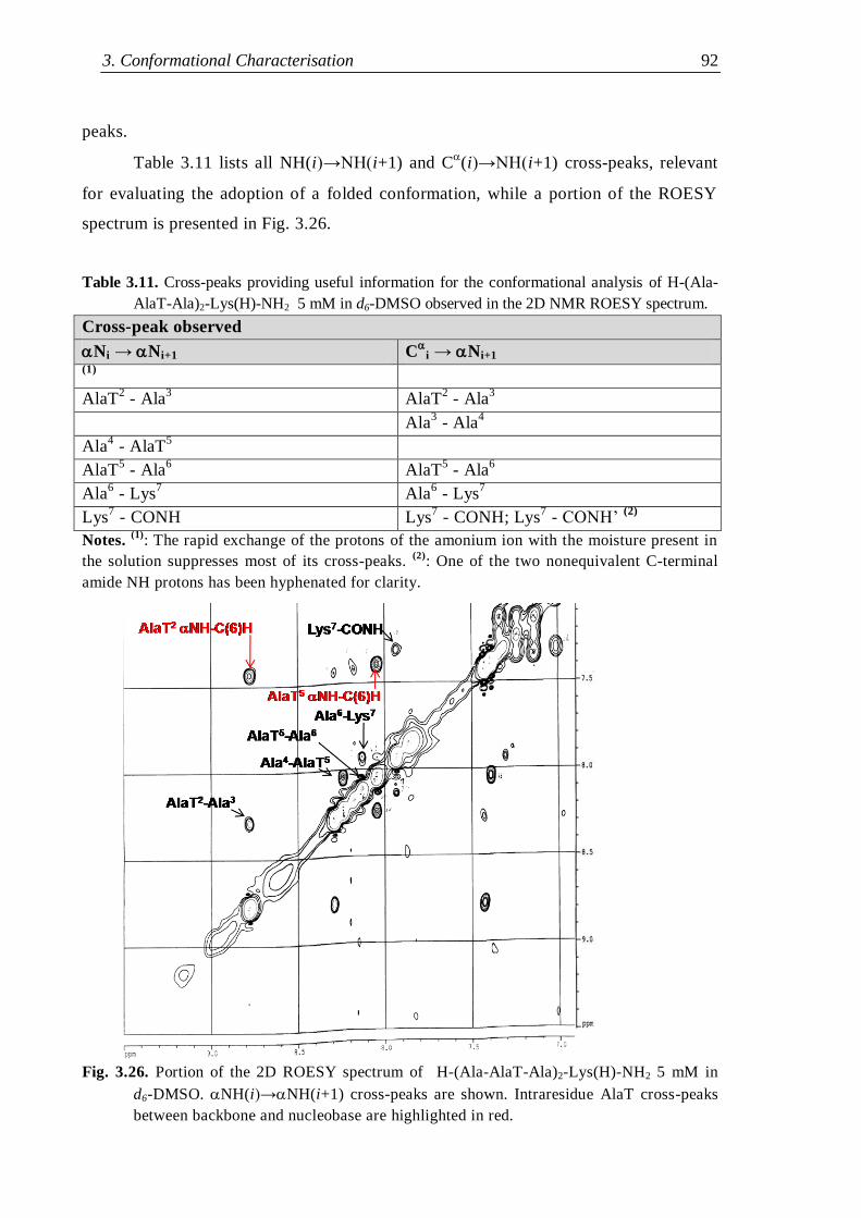
3. Conformational Characterisation 92
peaks.
Table 3.11 lists all NH(i)→NH(i+1) and C(i)→NH(i+1) cross-peaks, relevant
for evaluating the adoption of a folded conformation, while a portion of the ROESY
spectrum is presented in Fig. 3.26.
Table 3.11. Cross-peaks providing useful information for the conformational analysis of H-(Ala-
AlaT-Ala)2-Lys(H)-NH2 5 mM in d6-DMSO observed in the 2D NMR ROESY spectrum.
Cross-peak observed
Ni → Ni+1 C
i → Ni+1 (1)
AlaT2 - Ala
3 AlaT
2 - Ala
3
Ala3 - Ala
4
Ala4 - AlaT
5
AlaT5 - Ala
6 AlaT
5 - Ala
6
Ala6 - Lys
7 Ala
6 - Lys
7
Lys7 - CONH Lys
7 - CONH; Lys
7 - CONH‟
(2)
Notes. (1)
: The rapid exchange of the protons of the amonium ion with the moisture present in
the solution suppresses most of its cross-peaks. (2)
: One of the two nonequivalent C-terminal
amide NH protons has been hyphenated for clarity.
Fig. 3.26. Portion of the 2D ROESY spectrum of H-(Ala-AlaT-Ala)2-Lys(H)-NH2 5 mM in
d6-DMSO. NH(i)→NH(i+1) cross-peaks are shown. Intraresidue AlaT cross-peaks
between backbone and nucleobase are highlighted in red.
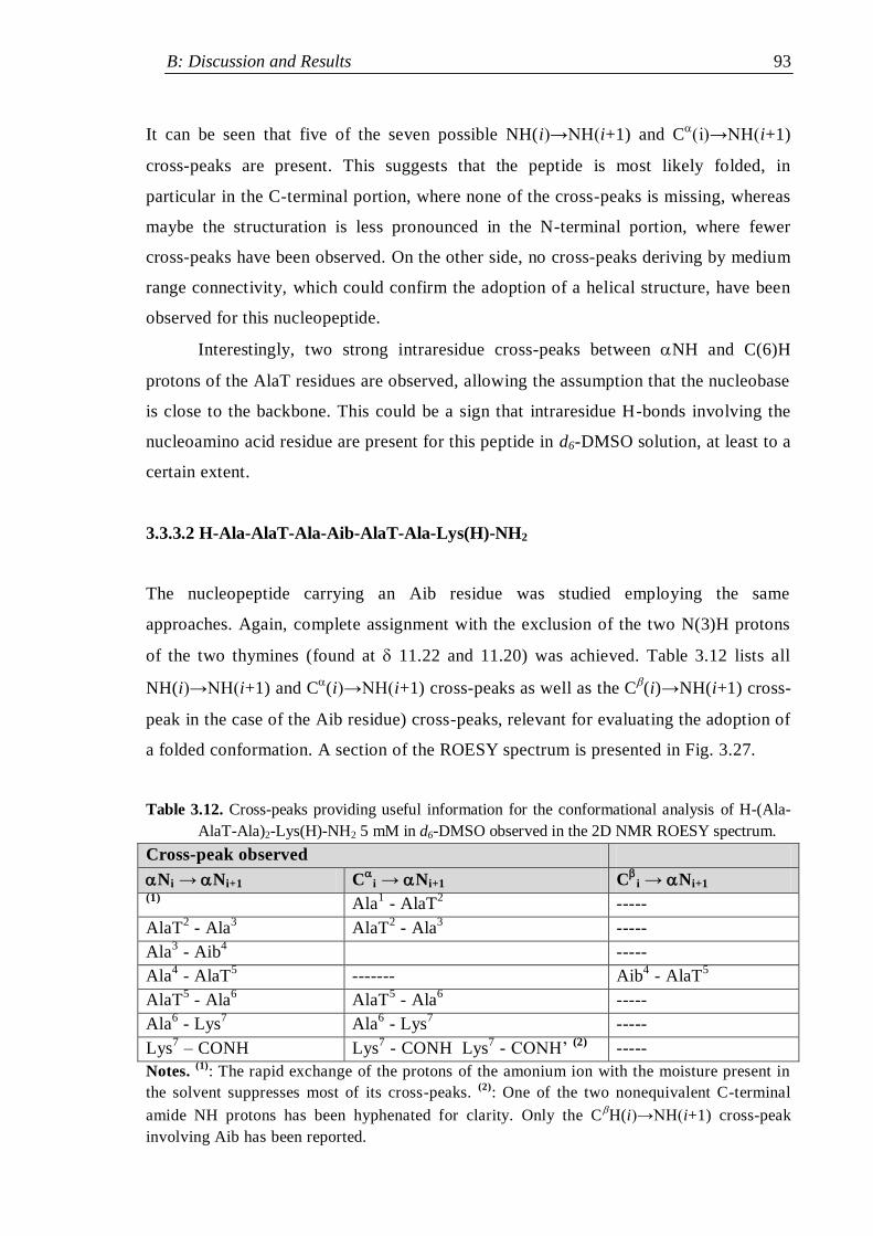
B: Discussion and Results 93
It can be seen that five of the seven possible NH(i)→NH(i+1) and C(i)→NH(i+1)
cross-peaks are present. This suggests that the peptide is most likely folded, in
particular in the C-terminal portion, where none of the cross-peaks is missing, whereas
maybe the structuration is less pronounced in the N-terminal portion, where fewer
cross-peaks have been observed. On the other side, no cross-peaks deriving by medium
range connectivity, which could confirm the adoption of a helical structure, have been
observed for this nucleopeptide.
Interestingly, two strong intraresidue cross-peaks between NH and C(6)H
protons of the AlaT residues are observed, allowing the assumption that the nucleobase
is close to the backbone. This could be a sign that intraresidue H-bonds involving the
nucleoamino acid residue are present for this peptide in d6-DMSO solution, at least to a
certain extent.
3.3.3.2 H-Ala-AlaT-Ala-Aib-AlaT-Ala-Lys(H)-NH2
The nucleopeptide carrying an Aib residue was studied employing the same
approaches. Again, complete assignment with the exclusion of the two N(3)H protons
of the two thymines (found at 11.22 and 11.20) was achieved. Table 3.12 lists all
NH(i)→NH(i+1) and C(i)→NH(i+1) cross-peaks as well as the C
(i)→NH(i+1) cross-
peak in the case of the Aib residue) cross-peaks, relevant for evaluating the adoption of
a folded conformation. A section of the ROESY spectrum is presented in Fig. 3.27.
Table 3.12. Cross-peaks providing useful information for the conformational analysis of H-(Ala-
AlaT-Ala)2-Lys(H)-NH2 5 mM in d6-DMSO observed in the 2D NMR ROESY spectrum.
Cross-peak observed
Ni → Ni+1 C
i → Ni+1 C
i → Ni+1 (1) Ala
1 - AlaT
2 -----
AlaT2 - Ala
3 AlaT
2 - Ala
3 -----
Ala3 - Aib
4 -----
Ala4 - AlaT
5 ------- Aib
4 - AlaT
5
AlaT5 - Ala
6 AlaT
5 - Ala
6 -----
Ala6 - Lys
7 Ala
6 - Lys
7 -----
Lys7 – CONH Lys
7 - CONH Lys
7 - CONH‟
(2) -----
Notes. (1)
: The rapid exchange of the protons of the amonium ion with the moisture present in
the solvent suppresses most of its cross-peaks. (2)
: One of the two nonequivalent C-terminal
amide NH protons has been hyphenated for clarity. Only the C(i)→NH(i+1) cross-peak
involving Aib has been reported.
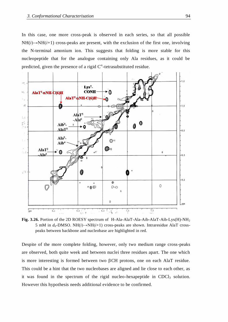
3. Conformational Characterisation 94
In this case, one more cross-peak is observed in each series, so that all possible
NH(i)→NH(i+1) cross-peaks are present, with the exclusion of the first one, involving
the N-terminal amonium ion. This suggests that folding is more stable for this
nucleopeptide that for the analogue containing only Ala residues, as it could be
predicted, given the presence of a rigid C-tetrasubstituted residue.
Fig. 3.26. Portion of the 2D ROESY spectrum of H-Ala-AlaT-Ala-Aib-AlaT-Aib-Lys(H)-NH2
5 mM in d6-DMSO. NH(i)→NH(i+1) cross-peaks are shown. Intraresidue AlaT cross-
peaks between backbone and nucleobase are highlighted in red.
Despite of the more complete folding, however, only two medium range cross-peaks
are observed, both quite week and between nuclei three residues apart. The one which
is more interesting is formed between two CH protons, one on each AlaT residue.
This could be a hint that the two nucleobases are aligned and lie close to each other, as
it was found in the spectrum of the rigid nucleo-hexapeptide in CDCl3 solution.
However this hypothesis needs additional evidence to be confirmed.
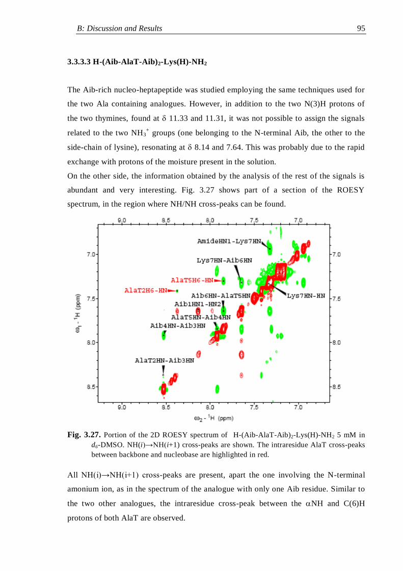
B: Discussion and Results 95
3.3.3.3 H-(Aib-AlaT-Aib)2-Lys(H)-NH2
The Aib-rich nucleo-heptapeptide was studied employing the same techniques used for
the two Ala containing analogues. However, in addition to the two N(3)H protons of
the two thymines, found at 11.33 and 11.31, it was not possible to assign the signals
related to the two NH3+ groups (one belonging to the N-terminal Aib, the other to the
side-chain of lysine), resonating at 8.14 and 7.64. This was probably due to the rapid
exchange with protons of the moisture present in the solution.
On the other side, the information obtained by the analysis of the rest of the signals is
abundant and very interesting. Fig. 3.27 shows part of a section of the ROESY
spectrum, in the region where NH/NH cross-peaks can be found.
Fig. 3.27. Portion of the 2D ROESY spectrum of H-(Aib-AlaT-Aib)2-Lys(H)-NH2 5 mM in
d6-DMSO. NH(i)→NH(i+1) cross-peaks are shown. The intraresidue AlaT cross-peaks
between backbone and nucleobase are highlighted in red.
All NH(i)→NH(i+1) cross-peaks are present, apart the one involving the N-terminal
amonium ion, as in the spectrum of the analogue with only one Aib residue. Similar to
the two other analogues, the intraresidue cross-peak between the NH and C(6)H
protons of both AlaT are observed.
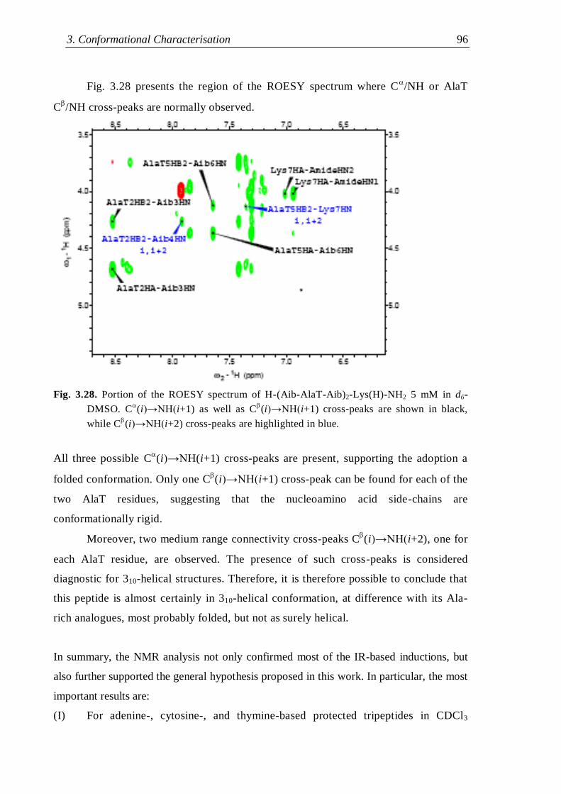
3. Conformational Characterisation 96
Fig. 3.28 presents the region of the ROESY spectrum where C/NH or AlaT
C/NH cross-peaks are normally observed.
Fig. 3.28. Portion of the ROESY spectrum of H-(Aib-AlaT-Aib)2-Lys(H)-NH2 5 mM in d6-
DMSO. C(i)→NH(i+1) as well as C
(i)→NH(i+1) cross-peaks are shown in black,
while C(i)→NH(i+2) cross-peaks are highlighted in blue.
All three possible C(i)→NH(i+1) cross-peaks are present, supporting the adoption a
folded conformation. Only one C(i)→NH(i+1) cross-peak can be found for each of the
two AlaT residues, suggesting that the nucleoamino acid side-chains are
conformationally rigid.
Moreover, two medium range connectivity cross-peaks C(i)→NH(i+2), one for
each AlaT residue, are observed. The presence of such cross-peaks is considered
diagnostic for 310-helical structures. Therefore, it is therefore possible to conclude that
this peptide is almost certainly in 310-helical conformation, at difference with its Ala-
rich analogues, most probably folded, but not as surely helical.
In summary, the NMR analysis not only confirmed most of the IR-based inductions, but
also further supported the general hypothesis proposed in this work. In particular, the most
important results are:
(I) For adenine-, cytosine-, and thymine-based protected tripeptides in CDCl3
B: Discussion and Results 97
solution, strong evidence supporting the presence of intraresidue H-bonds
involving the NH protons of the nucleoamino acid residues has been obtained.
(II) In CDCl3 solution all protected nucleopeptides adopt a folded conformation,
stabilized by intramolecular H-bonds, and data suggesting the formation of
helical structures have been obtained in several cases.
(III) A stable parallel dimer is formed by the protected thymine-based nucleo-
hexapeptide in CDCl3 solution and dimerization through intermolecular H-
bonds is mediated by the side-chain nucleobases, which are aligned along the
backbone and can therefore bind cooperatively to the other molecule.
(IV) The thymine-based nucleo-heptapeptides are folded, and for the Aib-rich
peptide the predicted adoption of a 310-helical conformation has been confirmed.
3. Conformational Characterisation 98
3.4 Circular Dichroism
Circular dichroism is an analytical technique based on the differential absorption of
circularly polarized light by chromophores set in an asymmetric environment. The spectral
region most commonly investigated for the conformational analysis of peptides lies in the
far UV region ( 190-260 nm). The amide chromophore of peptides displays two bands
in this interval, corresponding to the n→* and →* transitions. The amide group is
planar and therefore its transitions are optically inactive, but the presence of chiral
elements (such as the C atoms of chiral amino acids) next to the cromophore, as well as
its incorporation in helical secondary structures, which are intrinsically dissymmetric,
make it optically active, therefore liable to CD analysis.
The interactions among consecutive amide chromophores along the backbone
modulate their optical transitions in a way that depends on their relative orientation,
induced by the peptide secondary structure. The global outcome is that the spectral features
depend on the peptide secondary structure, so that useful information can be obtained both
from the shape and from the intensity of the dichroic bands of peptides.[60, 140]
The molar
ellipticity []T (deg∙cm2∙dmol
-1) is the parameter used for comparing the intensity of the
CD signal () by normalization with regard to concentration (c, mol/L) and optical path (l,
cm), using the relation [T= /(c∙l).
When applying circular dichroic techniques in the far UV to study the
nucleopeptide conformation, however, it must be considered that chromophores other than
the peptide amide groups are present. Indeed the side-chain nucleobases are powerful
chromofores, absorbing already at < 300 nm. Like the amide groups, they are planar and
optically inactive, but the proximity of chiral elements makes their transitions optically
active. The superimposition of contributions from the nucleobases and from the amide
groups of the backbone significantly complicates the CD spectra of the nucleopeptides,
making their interpretation in terms of peptide secondary structures difficult, when not
impossible.
On the other side, the nucleobase contribution to the CD spectra is largely prevalent
in the region at > 250 nm, therefore the analysis of the bands observed in this region can
be used to infer data on the side-chain nucleobase orientation. Indeed, the interaction of the
nucleobase chromophore with the asymmetric centre in the backbone is modulated by the
nucleobase orientation, so that different orientations can result in a different band shape.
B: Discussion and Results 99
Therefore, if all side-chains of a given compound in a sample are rigid and oriented
in the same way relative to the backbone, contributions of the same kind from all
molecules sum up in the sample spectrum and a strong band results. On the contrary, if the
side-chains are flexible and no prevalent orientation exists, each molecule contributes in a
different way to the spectrum, so that part the signals cancel each other, at least partially,
and a weaker and less featured band results. Consequently, by comparing the dichroic
bands observed in the region of the nucleobase chromophore in the spectra of analogous
nucleopeptides acquired under similar experimental conditions, it should be possible to
have an idea of the relative conformational order of their side-chains. Moreover,
particularly strong dichroic bands in the nucleobase chromophore region can suggest the
presence of strong intermolecular nucleobase-mediated interactions forcing the assumption
of a rigid conformation of the side-chains of the groups involved.
3.4.1 Protected rigid nucleopeptides
Conformational investigations by CD on the protected nucleopeptides have been
performed both in CHCl3 and in MeOH. The first solvent, employed for the IR and NMR
studies, is generally not suited for CD studies on peptides, since it absorbs strongly the UV
radiation from 240 nm, where most of the bands due to the peptide chromophore are
observed. This is not a problem, however, if the nucleobase bands are of interest. MeOH,
on the other side, allows the observation also of the lower wavelength portion of the
spectrum. The low polarity of CHCl3 favours intermolecular interactions mediated by
dipolar effects (including hydrogen bonding interactons) among solute molecules. On the
contrary MeOH, being a polar solvent, able to accept and to donate H-bonds, tends to
disrupt such interactions.
All CD experiments on the protected nucleopeptides were performed at room
temperature. Given the strong absorbance of the nucleobases in the far UV region, and
since the IR experiments showed the propensity of the nucleopeptides to aggregation in
CDCl3, the spectra were acquired at concentrations not higher than 1 mM in both solvents.
The nucleo-tripeptides with free nucleobases and without the Z protecting group
were considered more suited for the CD analysis than their corresponding Z-protected
analogues, because the aromatic chromophore of the Z protector is active in the spectral
region where the bands due to the nucleobases are found, and in principle this could
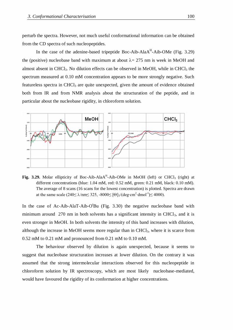
3. Conformational Characterisation 100
perturb the spectra. However, not much useful conformational information can be obtained
from the CD spectra of such nucleopeptides.
In the case of the adenine-based tripeptide Boc-Aib-AlaAH-Aib-OMe (Fig. 3.29)
the (positive) nucleobase band with maximum at about = 275 nm is week in MeOH and
almost absent in CHCl3. No dilution effects can be observed in MeOH, while in CHCl3 the
spectrum measured at 0.10 mM concentration appears to be more strongly negative. Such
featureless spectra in CHCl3 are quite unexpected, given the amount of evidence obtained
both from IR and from NMR analysis about the structuration of the peptide, and in
particular about the nucleobase rigidity, in chloroform solution.
Fig. 3.29. Molar ellipticity of Boc-Aib-AlaAH-Aib-OMe in MeOH (left) or CHCl3 (right) at
different concentrations (blue: 1.04 mM, red: 0.52 mM, green: 0.21 mM, black: 0.10 mM).
The average of 8 scans (16 scans for the lowest concentration) is plotted. Spectra are drawn
at the same scale (240≤ nm≤ 325, -8000≤ [T/(deg∙cm2∙dmol
-1)≤ 4000).
In the case of Ac-Aib-AlaT-Aib-OtBu (Fig. 3.30) the negative nucleobase band with
minimum around 270 nm in both solvents has a significant intensity in CHCl3, and it is
even stronger in MeOH. In both solvents the intensity of this band increases with dilution,
although the increase in MeOH seems more regular than in CHCl3, where it is scarce from
0.52 mM to 0.21 mM and pronounced from 0.21 mM to 0.10 mM.
The behaviour observed by dilution is again unexpected, because it seems to
suggest that nucleobase structuration increases at lower dilution. On the contrary it was
assumed that the strong intermolecular interactions observed for this nucleopeptide in
chloroform solution by IR spectroscopy, which are most likely nucleobase-mediated,
would have favoured the rigidity of its conformation at higher concentrations.
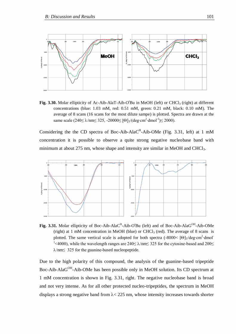
B: Discussion and Results 101
Fig. 3.30. Molar ellipticity of Ac-Aib-AlaT-Aib-OtBu in MeOH (left) or CHCl3 (right) at different
concentrations (blue: 1.03 mM, red: 0.51 mM, green: 0.21 mM, black: 0.10 mM). The
average of 8 scans (16 scans for the most dilute sampe) is plotted. Spectra are drawn at the
same scale (240≤ nm≤ 325, -20000≤ [T/(deg∙cm2∙dmol
-1)≤ 2000).
Considering the the CD spectra of Boc-Aib-AlaCH-Aib-OMe (Fig. 3.31, left) at 1 mM
concentration it is possible to observe a quite strong negative nucleobase band with
minimum at about 275 nm, whose shape and intensity are similar in MeOH and CHCl3.
Fig. 3.31. Molar ellipticity of Boc-Aib-AlaCH-Aib-O
tBu (left) and of Boc-Aib-AlaG
OH-Aib-OMe
(right) at 1 mM concentration in MeOH (blue) or CHCl3 (red). The average of 8 scans is
plotted. The same vertical scale is adopted for both spectra (-8000< [T/deg∙cm2∙dmol
-
1<4000), while the wavelength ranges are 240≤ nm≤ 325 for the cytosine-based and 200≤
nm≤ 325 for the guanine-based nucleopeptide.
Due to the high polarity of this compound, the analysis of the guanine-based tripeptide
Boc-Aib-AlaGOH
-Aib-OMe has been possible only in MeOH solution. Its CD spectrum at
1 mM concentration is shown in Fig. 3.31, right. The negative nucleobase band is broad
and not very intense. As for all other protected nucleo-tripeptides, the spectrum in MeOH
displays a strong negative band from < 225 nm, whose intensity increases towards shorter
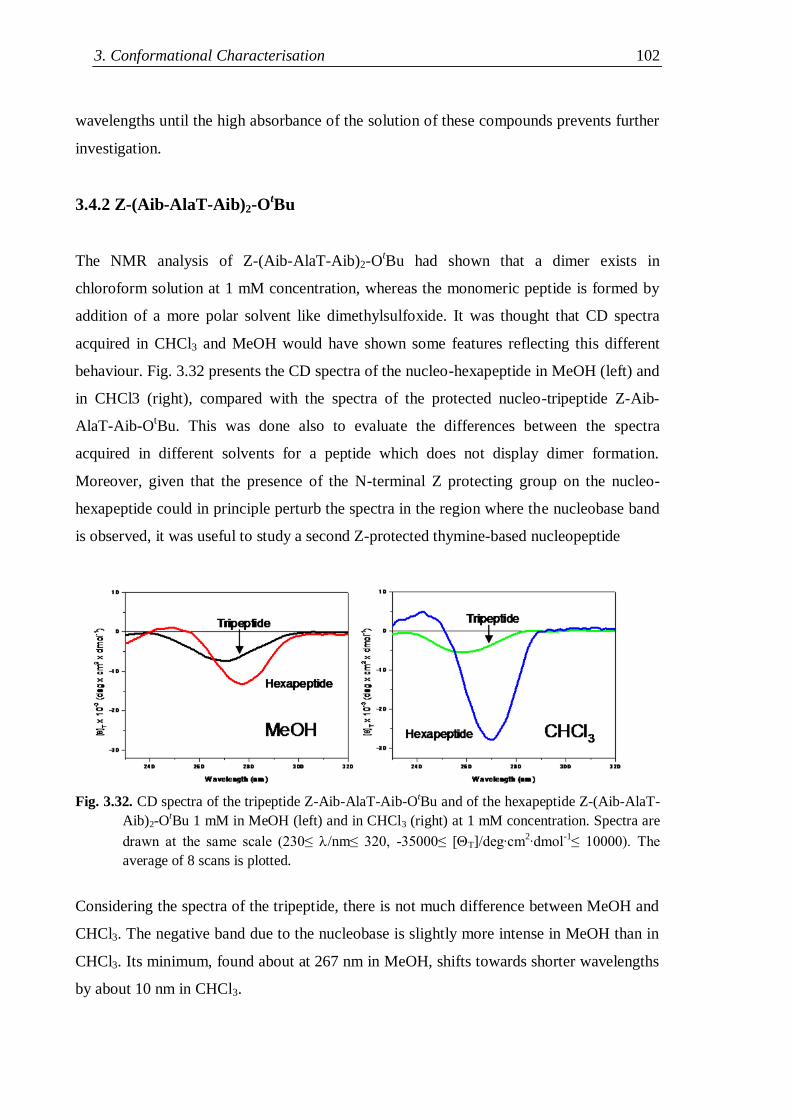
3. Conformational Characterisation 102
wavelengths until the high absorbance of the solution of these compounds prevents further
investigation.
3.4.2 Z-(Aib-AlaT-Aib)2-OtBu
The NMR analysis of Z-(Aib-AlaT-Aib)2-OtBu had shown that a dimer exists in
chloroform solution at 1 mM concentration, whereas the monomeric peptide is formed by
addition of a more polar solvent like dimethylsulfoxide. It was thought that CD spectra
acquired in CHCl3 and MeOH would have shown some features reflecting this different
behaviour. Fig. 3.32 presents the CD spectra of the nucleo-hexapeptide in MeOH (left) and
in CHCl3 (right), compared with the spectra of the protected nucleo-tripeptide Z-Aib-
AlaT-Aib-OtBu. This was done also to evaluate the differences between the spectra
acquired in different solvents for a peptide which does not display dimer formation.
Moreover, given that the presence of the N-terminal Z protecting group on the nucleo-
hexapeptide could in principle perturb the spectra in the region where the nucleobase band
is observed, it was useful to study a second Z-protected thymine-based nucleopeptide
Fig. 3.32. CD spectra of the tripeptide Z-Aib-AlaT-Aib-O
tBu and of the hexapeptide Z-(Aib-AlaT-
Aib)2-OtBu 1 mM in MeOH (left) and in CHCl3 (right) at 1 mM concentration. Spectra are
drawn at the same scale (230≤ nm≤ 320, -35000≤ [T]/deg∙cm2∙dmol
-1≤ 10000). The
average of 8 scans is plotted.
Considering the spectra of the tripeptide, there is not much difference between MeOH and
CHCl3. The negative band due to the nucleobase is slightly more intense in MeOH than in
CHCl3. Its minimum, found about at 267 nm in MeOH, shifts towards shorter wavelengths
by about 10 nm in CHCl3.
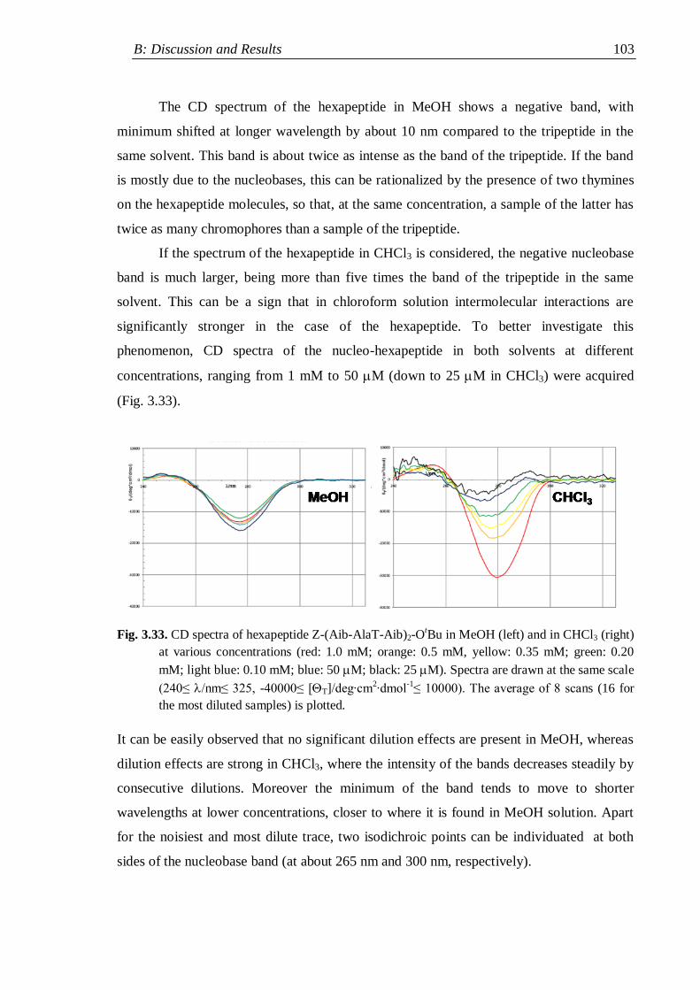
B: Discussion and Results 103
The CD spectrum of the hexapeptide in MeOH shows a negative band, with
minimum shifted at longer wavelength by about 10 nm compared to the tripeptide in the
same solvent. This band is about twice as intense as the band of the tripeptide. If the band
is mostly due to the nucleobases, this can be rationalized by the presence of two thymines
on the hexapeptide molecules, so that, at the same concentration, a sample of the latter has
twice as many chromophores than a sample of the tripeptide.
If the spectrum of the hexapeptide in CHCl3 is considered, the negative nucleobase
band is much larger, being more than five times the band of the tripeptide in the same
solvent. This can be a sign that in chloroform solution intermolecular interactions are
significantly stronger in the case of the hexapeptide. To better investigate this
phenomenon, CD spectra of the nucleo-hexapeptide in both solvents at different
concentrations, ranging from 1 mM to 50 M (down to 25 M in CHCl3) were acquired
(Fig. 3.33).
Fig. 3.33. CD spectra of hexapeptide Z-(Aib-AlaT-Aib)2-O
tBu in MeOH (left) and in CHCl3 (right)
at various concentrations (red: 1.0 mM; orange: 0.5 mM, yellow: 0.35 mM; green: 0.20
mM; light blue: 0.10 mM; blue: 50 M; black: 25 M). Spectra are drawn at the same scale
(240≤ nm≤ 325, -40000≤ [T]/deg∙cm2∙dmol
-1≤ 10000). The average of 8 scans (16 for
the most diluted samples) is plotted.
It can be easily observed that no significant dilution effects are present in MeOH, whereas
dilution effects are strong in CHCl3, where the intensity of the bands decreases steadily by
consecutive dilutions. Moreover the minimum of the band tends to move to shorter
wavelengths at lower concentrations, closer to where it is found in MeOH solution. Apart
for the noisiest and most dilute trace, two isodichroic points can be individuated at both
sides of the nucleobase band (at about 265 nm and 300 nm, respectively).
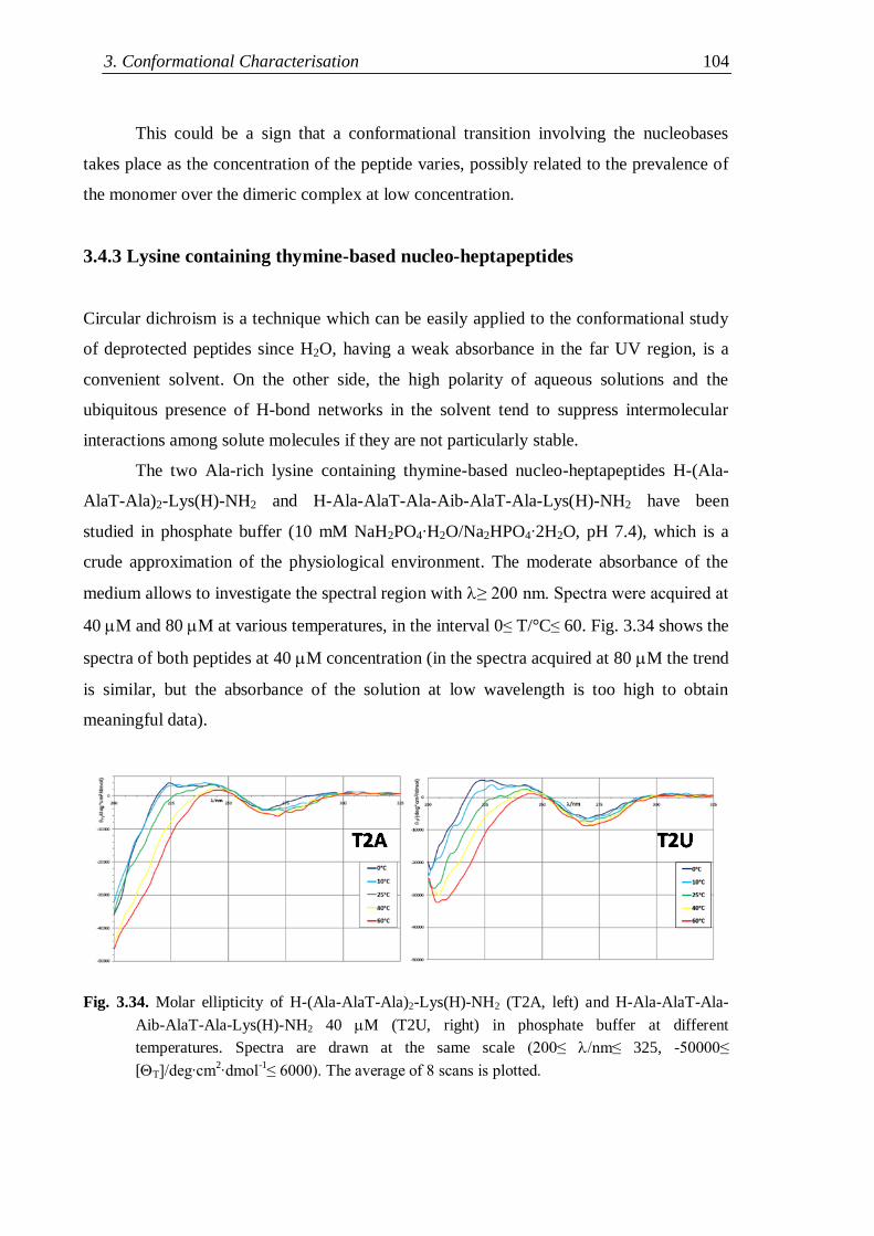
3. Conformational Characterisation 104
This could be a sign that a conformational transition involving the nucleobases
takes place as the concentration of the peptide varies, possibly related to the prevalence of
the monomer over the dimeric complex at low concentration.
3.4.3 Lysine containing thymine-based nucleo-heptapeptides
Circular dichroism is a technique which can be easily applied to the conformational study
of deprotected peptides since H2O, having a weak absorbance in the far UV region, is a
convenient solvent. On the other side, the high polarity of aqueous solutions and the
ubiquitous presence of H-bond networks in the solvent tend to suppress intermolecular
interactions among solute molecules if they are not particularly stable.
The two Ala-rich lysine containing thymine-based nucleo-heptapeptides H-(Ala-
AlaT-Ala)2-Lys(H)-NH2 and H-Ala-AlaT-Ala-Aib-AlaT-Ala-Lys(H)-NH2 have been
studied in phosphate buffer (10 mM NaH2PO4∙H2O/Na2HPO4∙2H2O, pH 7.4), which is a
crude approximation of the physiological environment. The moderate absorbance of the
medium allows to investigate the spectral region with ≥ 200 nm. Spectra were acquired at
40 M and 80 M at various temperatures, in the interval 0≤ T/°C≤ 60. Fig. 3.34 shows the
spectra of both peptides at 40 M concentration (in the spectra acquired at 80 M the trend
is similar, but the absorbance of the solution at low wavelength is too high to obtain
meaningful data).
Fig. 3.34. Molar ellipticity of H-(Ala-AlaT-Ala)2-Lys(H)-NH2 (T2A, left) and H-Ala-AlaT-Ala-
Aib-AlaT-Ala-Lys(H)-NH2 40 M (T2U, right) in phosphate buffer at different
temperatures. Spectra are drawn at the same scale (200≤ nm≤ 325, -50000≤
[T]/deg∙cm2∙dmol
-1≤ 6000). The average of 8 scans is plotted.
B: Discussion and Results 105
The main features of both spectra are similar: the negative band due to the nucleobase can
be observed at about 270 nm, at low temperature a weak positive band is present for 225<
/nm< 250 while from < 225 nm a strong negative band is found. The variation of the
spectra with the temperature is also similar, since the weak positive band tends to
disappear as the temperature rises, while both negative bands increase their intensity. The
increase in intensity of the nucleobase band with the temperature, which should favour the
flexibility of the side-chain, is quite unexpected, like the increase of the same band with
decreasing concentration in the case of the acetylated thymine-based nucleo-tripeptide. A
closer closer comparison of the spectra reveals however some interesting differences: (i) at
every given temperature the nucleobase band is stronger for the Aib containing
nucleopeptide; (ii) while its maximum tends to move towards longer wavelengths for the
Ala-based peptide, it does not change for the Aib containing peptide.
The different behaviour could be related to a different conformation of the side-chains,
even if, given the variation of the nucleobase band with the temperature, it is questionable
whether a stronger band can be related to a more structured side-chain.
The second difference lies in the low wavelength portion of the spectra: for the Ala-
based nucleopeptide the dichroic signal decreases steadily with decreasing wavelength and
no minimum can be observed until 200 nm. On the other side, for the nucleopeptide with
one Aib residue, a minimum is found for all spectra in the range 201-203 nm. For peptides
made only of protein amino acids it is generally accepted[140a]
that unstructured molecules
give negative bands with minima at <200 nm, whereas helical structures like the - and
the 310-helix have a minimum at about 208 nm. If the low wavelength region of the CD
spectra of the nucleopeptides can be interpreted using the same criteria (as done in the
literature[56,58]
for other kinds of nucleopeptides) this finding could suggest that the Aib-
containing nucleopeptide is more structured than the Ala-based analogue.
The Aib-rich analogue H-(Aib-AlaT-Aib)2-Lys(H)-NH2 has been studied in pure
H2O solution at different concentrations at room temperature. A reason for this choice is
that the effect of temperature proved difficult to rationalize in case of the two Ala-rich
analogues. On the other side, it was considered interesting to test whether dilution effects
could be detected in aqueous solution for the deprotected nucleopeptides, and the need to
explore a wider concentration range forced to abandon the phosphate buffer to use the less
UV-absorbing pure solvent.
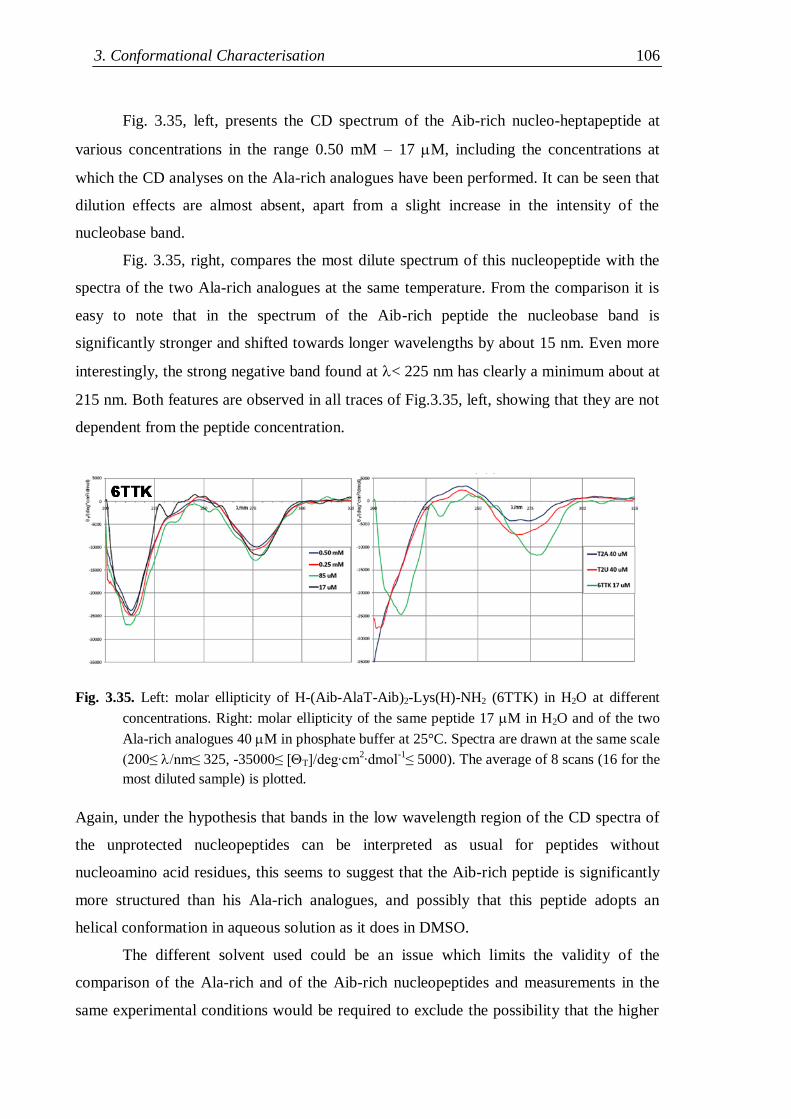
3. Conformational Characterisation 106
Fig. 3.35, left, presents the CD spectrum of the Aib-rich nucleo-heptapeptide at
various concentrations in the range 0.50 mM – 17 M, including the concentrations at
which the CD analyses on the Ala-rich analogues have been performed. It can be seen that
dilution effects are almost absent, apart from a slight increase in the intensity of the
nucleobase band.
Fig. 3.35, right, compares the most dilute spectrum of this nucleopeptide with the
spectra of the two Ala-rich analogues at the same temperature. From the comparison it is
easy to note that in the spectrum of the Aib-rich peptide the nucleobase band is
significantly stronger and shifted towards longer wavelengths by about 15 nm. Even more
interestingly, the strong negative band found at < 225 nm has clearly a minimum about at
215 nm. Both features are observed in all traces of Fig.3.35, left, showing that they are not
dependent from the peptide concentration.
Fig. 3.35. Left: molar ellipticity of H-(Aib-AlaT-Aib)2-Lys(H)-NH2 (6TTK) in H2O at different
concentrations. Right: molar ellipticity of the same peptide 17 M in H2O and of the two
Ala-rich analogues 40 M in phosphate buffer at 25°C. Spectra are drawn at the same scale
(200≤ nm≤ 325, -35000≤ [T]/deg∙cm2∙dmol
-1≤ 5000). The average of 8 scans (16 for the
most diluted sample) is plotted.
Again, under the hypothesis that bands in the low wavelength region of the CD spectra of
the unprotected nucleopeptides can be interpreted as usual for peptides without
nucleoamino acid residues, this seems to suggest that the Aib-rich peptide is significantly
more structured than his Ala-rich analogues, and possibly that this peptide adopts an
helical conformation in aqueous solution as it does in DMSO.
The different solvent used could be an issue which limits the validity of the
comparison of the Ala-rich and of the Aib-rich nucleopeptides and measurements in the
same experimental conditions would be required to exclude the possibility that the higher
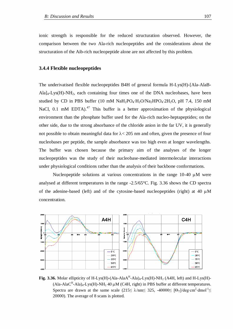
B: Discussion and Results 107
ionic strength is responsible for the reduced structuration observed. However, the
comparison between the two Ala-rich nucleopeptides and the considerations about the
structuration of the Aib-rich nucleopeptide alone are not affected by this problem.
3.4.4 Flexible nucleopeptides
The underivatised flexible nucleopeptides B4H of general formula H-Lys(H)-[Ala-AlaB-
Ala]4-Lys(H)-NH2, each containing four times one of the DNA nucleobases, have been
studied by CD in PBS buffer (10 mM NaH2PO4∙H2O/Na2HPO4∙2H2O, pH 7.4, 150 mM
NaCl, 0.1 mM EDTA).47
This buffer is a better approximation of the physiological
environment than the phosphate buffer used for the Ala-rich nucleo-heptapeptides; on the
other side, due to the strong absorbance of the chloride anion in the far UV, it is generally
not possible to obtain meaningful data for < 205 nm and often, given the presence of four
nucleobases per peptide, the sample absorbance was too high even at longer wavelengths.
The buffer was chosen because the primary aim of the analyses of the longer
nucleopeptides was the study of their nucleobase-mediated intermolecular interactions
under physiological conditions rather than the analysis of their backbone conformations.
Nucleopeptide solutions at various concentrations in the range 10-40 M were
analysed at different temperatures in the range -2.5/65°C. Fig. 3.36 shows the CD spectra
of the adenine-based (left) and of the cytosine-based nucleopeptides (right) at 40 M
concentration.
Fig. 3.36. Molar ellipticity of H-Lys(H)-(Ala-AlaA
H-Ala)4-Lys(H)-NH2 (A4H, left) and H-Lys(H)-
(Ala-AlaCH-Ala)4-Lys(H)-NH2 40 M (C4H, right) in PBS buffer at different temperatures.
Spectra are drawn at the same scale (215≤ nm≤ 325, -40000≤ [T]/deg∙cm2∙dmol
-1≤
20000). The average of 8 scans is plotted.
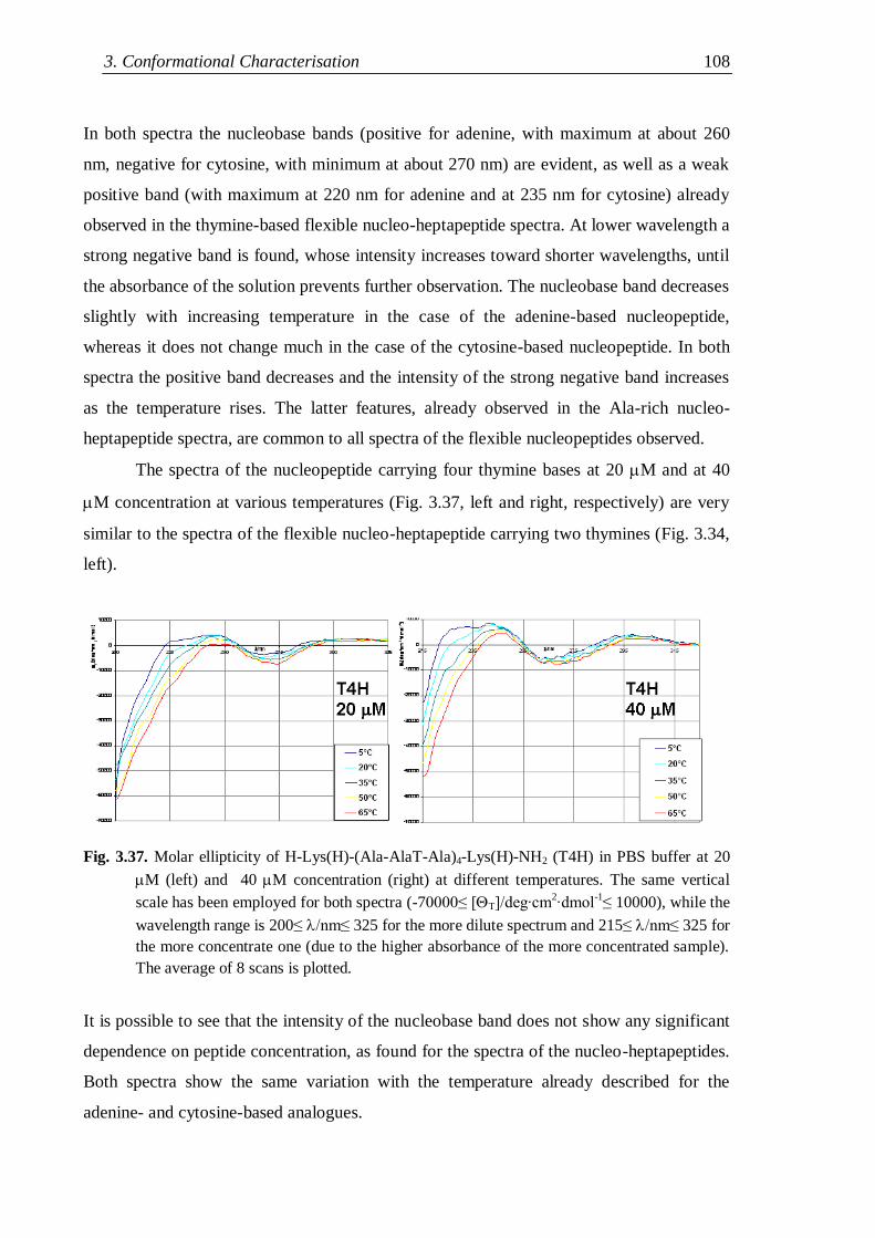
3. Conformational Characterisation 108
In both spectra the nucleobase bands (positive for adenine, with maximum at about 260
nm, negative for cytosine, with minimum at about 270 nm) are evident, as well as a weak
positive band (with maximum at 220 nm for adenine and at 235 nm for cytosine) already
observed in the thymine-based flexible nucleo-heptapeptide spectra. At lower wavelength a
strong negative band is found, whose intensity increases toward shorter wavelengths, until
the absorbance of the solution prevents further observation. The nucleobase band decreases
slightly with increasing temperature in the case of the adenine-based nucleopeptide,
whereas it does not change much in the case of the cytosine-based nucleopeptide. In both
spectra the positive band decreases and the intensity of the strong negative band increases
as the temperature rises. The latter features, already observed in the Ala-rich nucleo-
heptapeptide spectra, are common to all spectra of the flexible nucleopeptides observed.
The spectra of the nucleopeptide carrying four thymine bases at 20 M and at 40
M concentration at various temperatures (Fig. 3.37, left and right, respectively) are very
similar to the spectra of the flexible nucleo-heptapeptide carrying two thymines (Fig. 3.34,
left).
Fig. 3.37. Molar ellipticity of H-Lys(H)-(Ala-AlaT-Ala)4-Lys(H)-NH2 (T4H) in PBS buffer at 20
M (left) and 40 M concentration (right) at different temperatures. The same vertical
scale has been employed for both spectra (-70000≤ [T]/deg∙cm2∙dmol
-1≤ 10000), while the
wavelength range is 200≤ nm≤ 325 for the more dilute spectrum and 215≤ nm≤ 325 for
the more concentrate one (due to the higher absorbance of the more concentrated sample).
The average of 8 scans is plotted.
It is possible to see that the intensity of the nucleobase band does not show any significant
dependence on peptide concentration, as found for the spectra of the nucleo-heptapeptides.
Both spectra show the same variation with the temperature already described for the
adenine- and cytosine-based analogues.
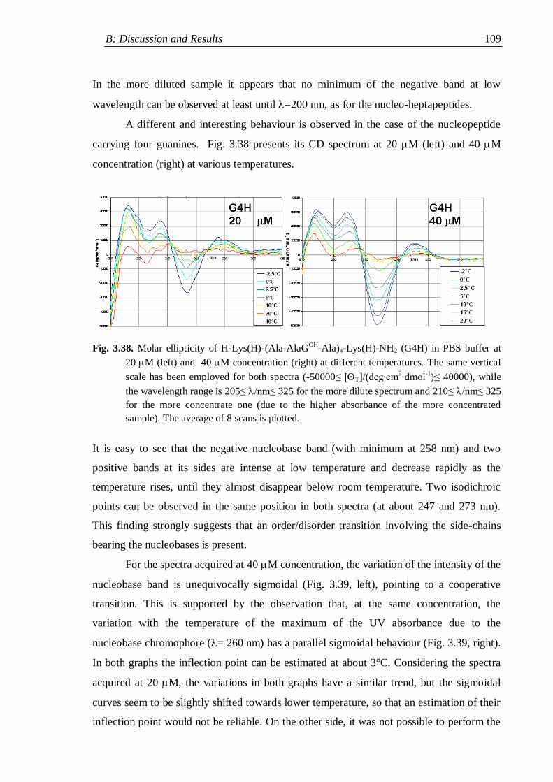
B: Discussion and Results 109
In the more diluted sample it appears that no minimum of the negative band at low
wavelength can be observed at least until =200 nm, as for the nucleo-heptapeptides.
A different and interesting behaviour is observed in the case of the nucleopeptide
carrying four guanines. Fig. 3.38 presents its CD spectrum at 20 M (left) and 40 M
concentration (right) at various temperatures.
Fig. 3.38. Molar ellipticity of H-Lys(H)-(Ala-AlaGOH
-Ala)4-Lys(H)-NH2 (G4H) in PBS buffer at
20 M (left) and 40 M concentration (right) at different temperatures. The same vertical
scale has been employed for both spectra (-50000≤ [T]/(deg∙cm2∙dmol
-1)≤ 40000), while
the wavelength range is 205≤ nm≤ 325 for the more dilute spectrum and 210≤ nm≤ 325
for the more concentrate one (due to the higher absorbance of the more concentrated
sample). The average of 8 scans is plotted.
It is easy to see that the negative nucleobase band (with minimum at 258 nm) and two
positive bands at its sides are intense at low temperature and decrease rapidly as the
temperature rises, until they almost disappear below room temperature. Two isodichroic
points can be observed in the same position in both spectra (at about 247 and 273 nm).
This finding strongly suggests that an order/disorder transition involving the side-chains
bearing the nucleobases is present.
For the spectra acquired at 40 M concentration, the variation of the intensity of the
nucleobase band is unequivocally sigmoidal (Fig. 3.39, left), pointing to a cooperative
transition. This is supported by the observation that, at the same concentration, the
variation with the temperature of the maximum of the UV absorbance due to the
nucleobase chromophore (= 260 nm) has a parallel sigmoidal behaviour (Fig. 3.39, right).
In both graphs the inflection point can be estimated at about 3°C. Considering the spectra
acquired at 20 M, the variations in both graphs have a similar trend, but the sigmoidal
curves seem to be slightly shifted towards lower temperature, so that an estimation of their
inflection point would not be reliable. On the other side, it was not possible to perform the

3. Conformational Characterisation 110
CD measurements at temperatures lower than -2.5°C, due to the risk of freezing the
solution. Consequently, given the uncertainty in the evaluation of the transition point for
the nucleopeptide at lower concentration, it is not possible to state whether concentration
effects are significant (pointing to the involvement of intermolecular interactions for the
stabilization of the „ordered‟ state at low temperature, as suggested by the cooperativity of
the transition) or not.
Fig. 3.39. Left: molar ellipticity at 258 nm of H-Lys(H)-(Ala-AlaGOH
-Ala)4-Lys(H)-NH2 in PBS at
20 M and 40 M concentration as a function of the temperature. Right: molar absorbance
of the same nucleopeptide at 260 nm in PBS at 20 M and 40 M concentration as a
function of the temperature.
The CD spectra of the underivatised nucleopeptide carrying five thymines T5H in
phosphate buffer at 20 M concentration at different temperatures in the range 0-65°C are
shown in Fig. 3.40.
Fig. 3.40. Molar ellipticity of H-Lys(H)-(Ala-AlaT-Ala)5-Lys(H)-NH2 (T5H) in phosphate buffer
at 20 M (left) at different temperatures. Moalr ellipticity range is -110000≤
[T]/(deg∙cm2∙dmol
-1)≤ 10000), while the wavelength range is 205≤ nm≤ 325. The
average of 8 scans is plotted.
B: Discussion and Results 111
It is possible to observe that both the spectral shape and its variation with the temperature
are very similar to what found for the spectra of the shorter analogue carrying four
thymines, even if both the nucleobase and the strong negative band at low wavelength are
significantly stronger. This could suggest that the conformational structuration of the Ala-
based nucleopeptides does not change susbstantially with the backbone length.
The most important results deriving from the CD analyses can be summarised as follows:
(I) In general, it has not been observed a simple correlation between the intensity of
the nucleobase band and the rigidity of the side-chains bearing the nucleobases.
(II) In the case of the nucleo-hexapeptide, the strong propensity to aggregation with
formation of dimers in CHCl3 solution is reflected in the nucleobase band both by
intense concentration effects and by its much stronger intensity of relative to the
nucleo-tripeptide bearing the same base. On the contrary, no effect has been
observed in a polar solvent like methanol, where the dimer is likely not formed.
(III) The comparison of the low wavelength portion of the spectra of the thymine-based
nucleo-heptapeptides in aqueous solution could suggest that the Aib-rich peptide is
helical, as observed in DMSO solution by NMR, whereas the Ala-rich analogues
seem to be less structured.
(IV) A cooperative order/disorder transition involving the side-chains of the
nucleopeptide carrying four guanines has been observed at low temperature in
aqueous solution.
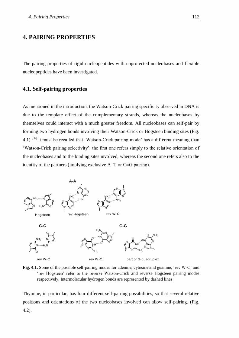
4. Pairing Properties 112
4. PAIRING PROPERTIES
The pairing properties of rigid nucleopeptides with unprotected nucleobases and flexible
nucleopeptides have been investigated.
4.1. Self-pairing properties
As mentioned in the introduction, the Watson-Crick pairing specificity observed in DNA is
due to the template effect of the complementary strands, whereas the nucleobases by
themselves could interact with a much greater freedom. All nucleobases can self-pair by
forming two hydrogen bonds involving their Watson-Crick or Hogsteen binding sites (Fig.
4.1).[56]
It must be recalled that „Watson-Crick pairing mode‟ has a different meaning than
„Watson-Crick pairing selectivity‟: the first one refers simply to the relative orientation of
the nucleobases and to the binding sites involved, whereas the second one refers also to the
identity of the partners (implying exclusive A=T or C≡G pairing).
N
NN
N NH2
N
NN
NNH2 N N
N N
NH2
N N
NN
NH2
N N
N N
NH2
NN
NN
NH2
N
N
O
NH2
N
N
O
NH2
N
N
N
NH
O
NH2
N
N
N
NH
O
NH2
N
N
N
NH
O
NH2
NN
NNH
O
NH2
A-A
rev W-Crev HogsteenHogsteen
rev W-C
C-C G-G
rev W-C part of G-quadruplex
Fig. 4.1. Some of the possible self-pairing modes for adenine, cytosine and guanine; „rev W-C‟ and
„rev Hogsteen‟ refer to the reverse Watson-Crick and reverse Hogsteen pairing modes
respectively. Intermolecular hydrogen bonds are represented by dashed lines
Thymine, in particular, has four different self-pairing possibilities, so that several relative
positions and orientations of the two nucleobases involved can allow self-pairing. (Fig.
4.2).
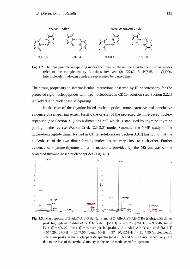
B: Discussion and Results 113
NNH
O
ON
NH
O
O
NNH
O
O
NNH
O
O
NNH
O
ON
NH
O
ON
NH
O
O
NNH
O
O
Watson - Crick Reverse Watson-Crick
3,4-2,3 3,4-3,4 2,3-2,32,3-3,4
Fig. 4.2. The four possible self-pairing modes for thymine; the numbers under the different modes
refer to the complementary functions involved (2: C(2)O, 3: N(3)H, 4: C(4)O).
Intermolecular hydrogen bonds are represented by dashed lines
The strong propensity to intermolecular interactions observed by IR spectroscopy for the
protected rigid nucleopeptides with free nucleobases in CDCl3 solution (see Section 3.2.1)
is likely due to nucleobase self-pairing.
In the case of the thymine-based nucleopeptides, more extensive and conclusive
evidence of self-pairing exists. Firstly, the crystal of the protected thymine-based nucleo-
tripeptide (see Section 3.1) has a dimer unit cell which is stabilised by thymine-thymine
pairing in the reverse Watson-Crick „2,3-2,3‟ mode. Secondly, the NMR study of the
nucleo-hexapeptide dimer formed in CDCl3 solution (see Section 3.3.2) has found that the
nucleobases of the two dimer-forming molecules are very close to each-other. Further
evidence of thymine-thymine dimer formation is provided by the MS analysis of the
protected thymine-based nucleopeptides (Fig. 4.3).
Fig. 4.3. Mass spectra of Z-AlaT-Aib-OtBu (left) and of Z-Aib-AlaT-Aib-O
tBu (right), with dimer
peak highlighted. Z-AlaT-Aib-OtBu: calcd [M+H]
+ = 489.23, [2M+H]
+ = 977.46, found
[M+H]+
= 489.22, [2M+H]+
= 977.49 (circled peak). Z-Aib-AlaT-Aib-OtBu: calcd [M+H]
+
= 574.29, [2M+H]+
= 1147.56, found [M+H]+
= 574.30, [2M+H]+
= 1147.55 (circled peak).
The main peaks in the nucleopeptide spectra (at 433.16 and 518.23 m/z respectively) are
due to the loss of the tertbutyl moiety in the acidic media used for injection.
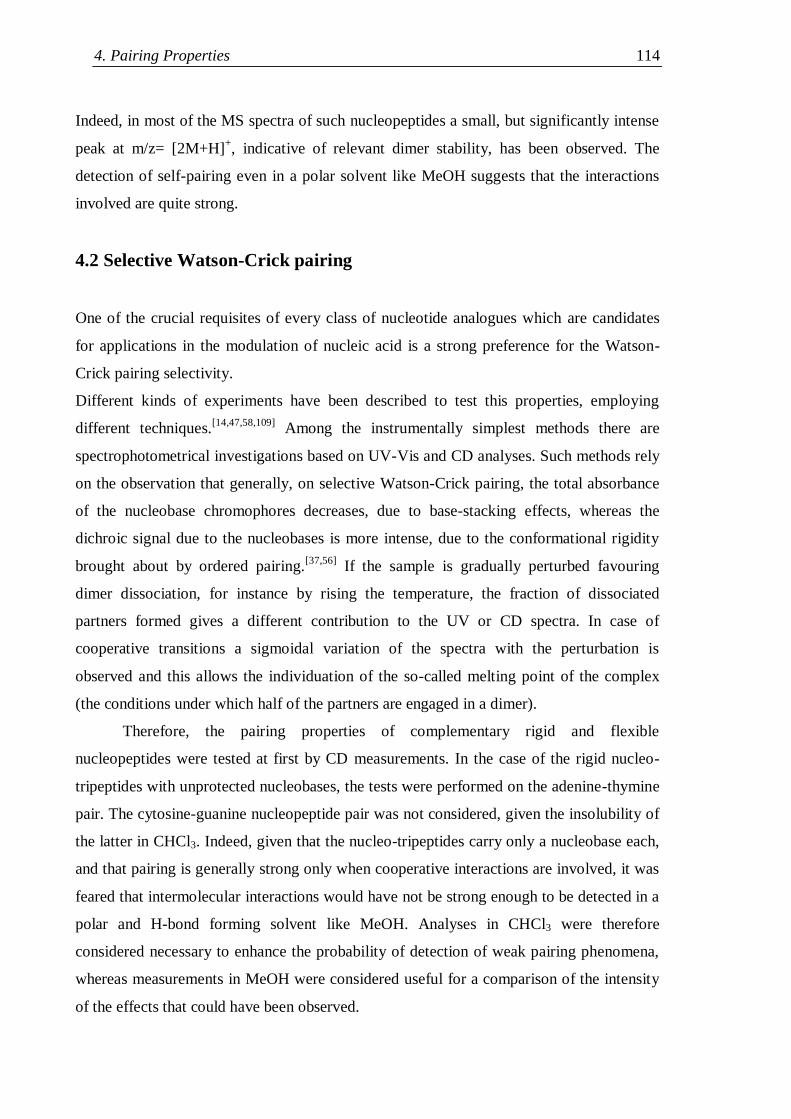
4. Pairing Properties 114
Indeed, in most of the MS spectra of such nucleopeptides a small, but significantly intense
peak at m/z= [2M+H]+, indicative of relevant dimer stability, has been observed. The
detection of self-pairing even in a polar solvent like MeOH suggests that the interactions
involved are quite strong.
4.2 Selective Watson-Crick pairing
One of the crucial requisites of every class of nucleotide analogues which are candidates
for applications in the modulation of nucleic acid is a strong preference for the Watson-
Crick pairing selectivity.
Different kinds of experiments have been described to test this properties, employing
different techniques.[14,47,58,109]
Among the instrumentally simplest methods there are
spectrophotometrical investigations based on UV-Vis and CD analyses. Such methods rely
on the observation that generally, on selective Watson-Crick pairing, the total absorbance
of the nucleobase chromophores decreases, due to base-stacking effects, whereas the
dichroic signal due to the nucleobases is more intense, due to the conformational rigidity
brought about by ordered pairing.[37,56]
If the sample is gradually perturbed favouring
dimer dissociation, for instance by rising the temperature, the fraction of dissociated
partners formed gives a different contribution to the UV or CD spectra. In case of
cooperative transitions a sigmoidal variation of the spectra with the perturbation is
observed and this allows the individuation of the so-called melting point of the complex
(the conditions under which half of the partners are engaged in a dimer).
Therefore, the pairing properties of complementary rigid and flexible
nucleopeptides were tested at first by CD measurements. In the case of the rigid nucleo-
tripeptides with unprotected nucleobases, the tests were performed on the adenine-thymine
pair. The cytosine-guanine nucleopeptide pair was not considered, given the insolubility of
the latter in CHCl3. Indeed, given that the nucleo-tripeptides carry only a nucleobase each,
and that pairing is generally strong only when cooperative interactions are involved, it was
feared that intermolecular interactions would have not be strong enough to be detected in a
polar and H-bond forming solvent like MeOH. Analyses in CHCl3 were therefore
considered necessary to enhance the probability of detection of weak pairing phenomena,
whereas measurements in MeOH were considered useful for a comparison of the intensity
of the effects that could have been observed.
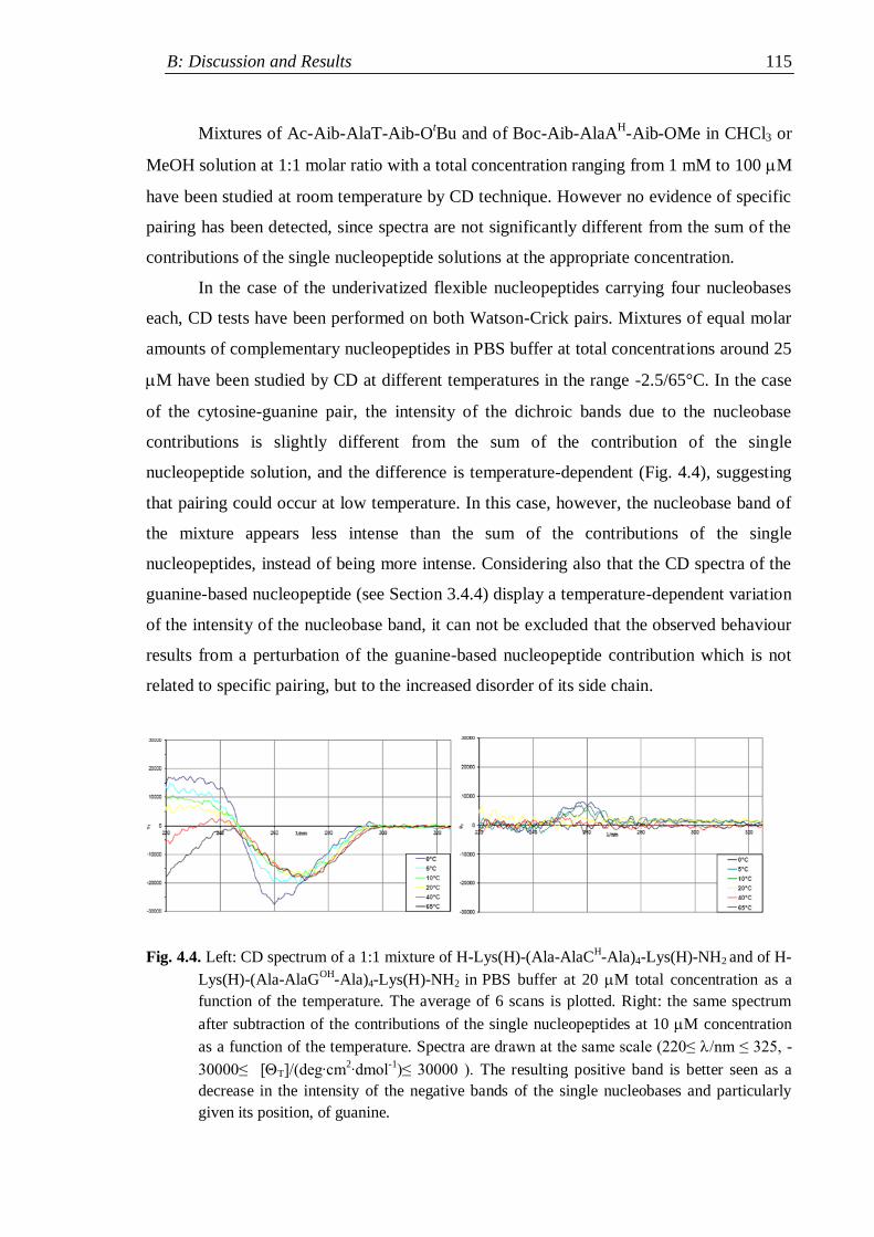
B: Discussion and Results 115
Mixtures of Ac-Aib-AlaT-Aib-OtBu and of Boc-Aib-AlaA
H-Aib-OMe in CHCl3 or
MeOH solution at 1:1 molar ratio with a total concentration ranging from 1 mM to 100 M
have been studied at room temperature by CD technique. However no evidence of specific
pairing has been detected, since spectra are not significantly different from the sum of the
contributions of the single nucleopeptide solutions at the appropriate concentration.
In the case of the underivatized flexible nucleopeptides carrying four nucleobases
each, CD tests have been performed on both Watson-Crick pairs. Mixtures of equal molar
amounts of complementary nucleopeptides in PBS buffer at total concentrations around 25
M have been studied by CD at different temperatures in the range -2.5/65°C. In the case
of the cytosine-guanine pair, the intensity of the dichroic bands due to the nucleobase
contributions is slightly different from the sum of the contribution of the single
nucleopeptide solution, and the difference is temperature-dependent (Fig. 4.4), suggesting
that pairing could occur at low temperature. In this case, however, the nucleobase band of
the mixture appears less intense than the sum of the contributions of the single
nucleopeptides, instead of being more intense. Considering also that the CD spectra of the
guanine-based nucleopeptide (see Section 3.4.4) display a temperature-dependent variation
of the intensity of the nucleobase band, it can not be excluded that the observed behaviour
results from a perturbation of the guanine-based nucleopeptide contribution which is not
related to specific pairing, but to the increased disorder of its side chain.
Fig. 4.4. Left: CD spectrum of a 1:1 mixture of H-Lys(H)-(Ala-AlaCH-Ala)4-Lys(H)-NH2 and of H-
Lys(H)-(Ala-AlaGOH
-Ala)4-Lys(H)-NH2 in PBS buffer at 20 M total concentration as a
function of the temperature. The average of 6 scans is plotted. Right: the same spectrum
after subtraction of the contributions of the single nucleopeptides at 10 M concentration
as a function of the temperature. Spectra are drawn at the same scale (220≤ /nm ≤ 325, -
30000≤ [T]/(deg∙cm2∙dmol
-1)≤ 30000 ). The resulting positive band is better seen as a
decrease in the intensity of the negative bands of the single nucleobases and particularly
given its position, of guanine.
4. Pairing Properties 116
Considering that it was possible that pairing between nucleopeptides and complementary
nucleotides was stronger and therefore easier to detect than pairing among complementary
nucleopeptides, CD tests of nucleopeptide/nucleotide pairing have been performed. The
longest flexible nucleopeptide synthesized, carrying five thymine nucleobases, was used
and a synthetic DNA oligonucleotide carrying ten adenine nucleobases (dA10) was chosen
as a complementary partner. The choice of a longer nucleotide was done in order to take
advantage of the flanking base effect: in a nucleotide strand the nucleobases next to the 5‟-
and 3‟-termini do not contribute to pairing, but stabilise the conformation of the internal
bases, which are therefore capable of a more efficient binding.
Mixtures of the two compounds at various nucleopeptide/nucleotide molar ratios
(1:2, 1:1, 2:1, 4:1) in phosphate buffer at a total concentration of 10 M have been
prepared and their CD spectra have been measured at different temperatures in the range 0-
65°C. The use of different molar ratios could give useful information for the understanding
of the pairing mode in case interactions were detected. For instance, if A:T pairing
following Watson-Crick rules was prevalent, the strongest effects would be detected for a
1:1 nucleopeptide/nucleotide ratio, whereas in case of (T:A)T triplex formation, the
strongest effects would be observed for a 2:1 nucleopeptide/nucleotide ratio. The spectra
obtained from the mixtures, however, do not significantly differ from the weighted sums of
the contributions of the nucleopeptide and of the nucleotide.
4.2.1 Surface plasmon resonance measurements
An alternative technique for detecting and evaluating pairing is surface plasmon resonance
(SPR).[141]
This techniques relies on the variations of the optical properties of a gold chip in
response to the variation of mass brought about by binding of ligands to the suitably
functionalised surface of the chip. The instrumental response can be followed against time
to obtain the binding kinetics in the association phase, during which a solution of the
ligand is allowed to flow on the chip and bind to its surface, and in the dissociation phase,
during which a solution without ligand flows on the chip, allowing elution of the ligand.
By fitting the instrumental data collected during the association and dissociation phase of
the experiment it is possible to calculate the kinetic association and dissociation constant
(ka and kd, respectively) and their ratio allows to obtain the thermodynamic dissociation
constant (Kd= kd/ka). After the experiments, the chip surface can be regenerated by
B: Discussion and Results 117
washing with an energic eluent, in general aqueous HCl, in order to eliminate all
substances not covalently bound, so that the chip is ready for new experiments.
Since a BiacoreTM
SPR system was available in Strasbourg, it has been employed
for pairing measurements on the flexible nucleopeptides. As mentioned above, in order to
perform such SPR measurements, it is necessary to immobilise one of the two partners on
solid-phase, whereas the other is allowed to flow in solution. For the immobilisation of the
nucleopeptides, the biotinylated flexible nucleopeptides have been used, after preliminary
activation of the dextran-functionalized gold surface to obtain covalent binding of
streptavidin,[142]
which in turn complexes almost irreversibly (Kd≈ 10-15
M) the biotinylated
nucleopeptides.
Two advantages of this method are that it is well-established and easy to apply.
Moreover, since the biotinylated compounds are stable to the regeneration phase, several
test can be performed without the need to repeat the immobilisation procedure.
On the other side, this method brings about limitations to the instrumental
sensitivity. Indeed, the bulkier is the ligand and the shorter is the distance between the
ligand and the gold surface, the stronger is the instrumental signal obtained Given that
streptavidin is a protein with Mr≈ 50000 and the nucleopeptide ligands have Mr≈ 2000, this
means that the effects of the binding of small ligands are communicated to gold surface
through a bulky linker, negatively affecting the instrumental sensitivity.
Adenine-, cytosine- and thymine-based biotinylated nucleopeptides with four
nucleobases were bound to the functionalised gold sensor chip. For the first series of
experiments, solutions of guanine-based and thymine-based underivatised nucleopeptides
in the concentration range 12.5-100 M have been allowed to flow on the chip. Binding
has been detected in the case of the adenine-thymine Watson-Crick pair (Fig. 4.5) with a
dissociation constant Kd≈ 30 M.
Unfortunately, pairing formation has not been detected between the
cytosine- and the guanine-based nucleopeptide, whose nucleotide analogues display a
stronger binding compared to adenine-thymine pairing in DNA.
On the other side, pairing between non complementary nucleopeptides has not been
observed (see Fig. 4.6 as an example). Given that four non complementary pairs have been
screened (T4H against T4Bt and C4Bt, G4H against A4H and A4Bt), respect of Watson-
Crick selectivity, in this preliminary test, is considered encouraging.
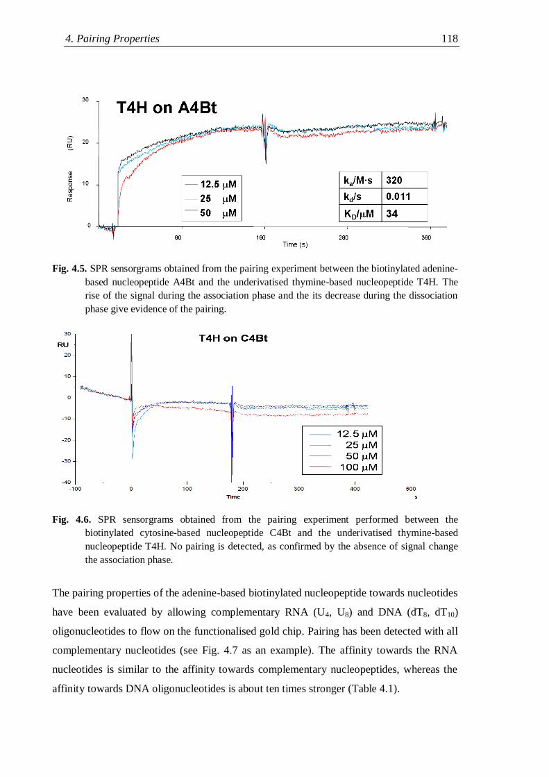
4. Pairing Properties 118
Fig. 4.5. SPR sensorgrams obtained from the pairing experiment between the biotinylated adenine-
based nucleopeptide A4Bt and the underivatised thymine-based nucleopeptide T4H. The
rise of the signal during the association phase and the its decrease during the dissociation
phase give evidence of the pairing.
Fig. 4.6. SPR sensorgrams obtained from the pairing experiment performed between the
biotinylated cytosine-based nucleopeptide C4Bt and the underivatised thymine-based
nucleopeptide T4H. No pairing is detected, as confirmed by the absence of signal change
the association phase.
The pairing properties of the adenine-based biotinylated nucleopeptide towards nucleotides
have been evaluated by allowing complementary RNA (U4, U8) and DNA (dT8, dT10)
oligonucleotides to flow on the functionalised gold chip. Pairing has been detected with all
complementary nucleotides (see Fig. 4.7 as an example). The affinity towards the RNA
nucleotides is similar to the affinity towards complementary nucleopeptides, whereas the
affinity towards DNA oligonucleotides is about ten times stronger (Table 4.1).
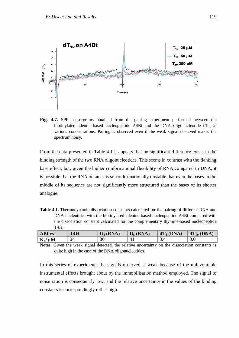
B: Discussion and Results 119
Fig. 4.7. SPR sensorgrams obtained from the pairing experiment performed between the
biotinylated adenine-based nucleopeptide A4Bt and the DNA oligonucleotide dT10 at
various concentrations. Pairing is observed even if the weak signal observed makes the
spectrum noisy.
From the data presented in Table 4.1 it appears that no significant difference exists in the
binding strength of the two RNA oligonucleotides. This seems in contrast with the flanking
base effect, but, given the higher conformational flexibility of RNA compared to DNA, it
is possible that the RNA octamer is so conformationally unstable that even the bases in the
middle of its sequence are not significantly more structured than the bases of its shorter
analogue.
Table 4.1. Thermodynamic dissociation constants calculated for the pairing of different RNA and
DNA nucleotides with the biotinylated adenine-based nucleopeptide A4Bt compared with
the dissociation constant calculated for the complementary thymine-based nucleopeptide
T4H.
ABt vs T4H U4 (RNA) U8 (RNA) dT8 (DNA) dT10 (DNA)
Kd/ M 34 36 41 3.4 3.0
Notes. Given the weak signal detected, the relative uncertainty on the dissociation constants is
quite high in the case of the DNA oligonucleotides.
In this series of experiments the signals observed is weak because of the unfavourable
instrumental effects brought about by the immobilisation method employed. The signal to
noise ration is consequently low, and the relative uncertainty in the values of the binding
constants is correspondingly rather high.
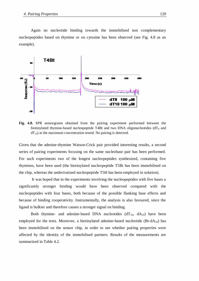
4. Pairing Properties 120
Again no nucleotide binding towards the immobilised non complementary
nucleopeptides based on thymine or on cytosine has been observed (see Fig. 4.8 as an
example).
Fig. 4.8. SPR sensorgrams obtained from the pairing experiment performed between the
biotinylated thymine-based nucleopeptide T4Bt and two DNA oligonucleotides (dT8 and
dT10) at the maximum concentration tested. No pairing is detected.
Given that the adenine-thymine Watson-Crick pair provided interesting results, a second
series of pairing experiments focusing on the same nucleobase pair has been performed.
For such experiments two of the longest nucleopeptides synthesized, containing five
thymines, have been used (the biotinylated nucleopeptide T5Bt has been immobilised on
the chip, whereas the underivatised nucleopeptide T5H has been employed in solution).
It was hoped that in the experiments involving the nucleopeptides with five bases a
significantly stronger binding would have been observed compared with the
nucleopeptides with four bases, both because of the possible flanking base effects and
because of binding cooperativity. Instrumentally, the analysis is also favoured, since the
ligand is bulkier and therefore causes a stronger signal on binding.
Both thymine- and adenine-based DNA nucleotides (dT10, dA10) have been
employed for the tests. Moreover, a biotinylated adenine-based nucleotide (Bt-dA10) has
been immobilised on the sensor chip, in order to see whether pairing properties were
affected by the identity of the immobilised partners. Results of the measurements are
summarized in Table 4.2.
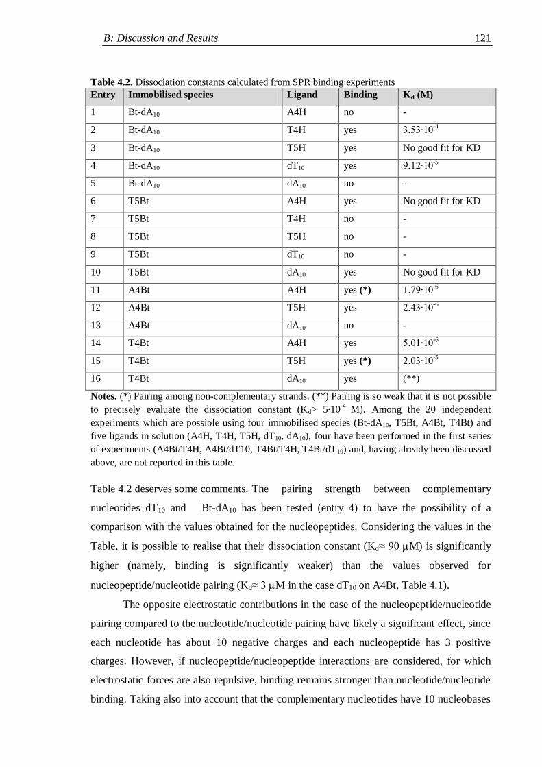
B: Discussion and Results 121
Table 4.2. Dissociation constants calculated from SPR binding experiments
Entry Immobilised species Ligand Binding Kd (M)
1 Bt-dA10 A4H no -
2 Bt-dA10 T4H yes 3.53∙10-4
3 Bt-dA10 T5H yes No good fit for KD
4 Bt-dA10 dT10 yes 9.12∙10-5
5 Bt-dA10 dA10 no -
6 T5Bt A4H yes No good fit for KD
7 T5Bt T4H no -
8 T5Bt T5H no -
9 T5Bt dT10 no -
10 T5Bt dA10 yes No good fit for KD
11 A4Bt A4H yes (*) 1.79∙10-6
12 A4Bt T5H yes 2.43∙10-6
13 A4Bt dA10 no -
14 T4Bt A4H yes 5.01∙10-6
15 T4Bt T5H yes (*) 2.03∙10-5
16 T4Bt dA10 yes (**)
Notes. (*) Pairing among non-complementary strands. (**) Pairing is so weak that it is not possible
to precisely evaluate the dissociation constant (Kd> 5∙10-4
M). Among the 20 independent
experiments which are possible using four immobilised species (Bt-dA10, T5Bt, A4Bt, T4Bt) and
five ligands in solution (A4H, T4H, T5H, dT10, dA10), four have been performed in the first series
of experiments (A4Bt/T4H, A4Bt/dT10, T4Bt/T4H, T4Bt/dT10) and, having already been discussed
above, are not reported in this table.
Table 4.2 deserves some comments. The pairing strength between complementary
nucleotides dT10 and Bt-dA10 has been tested (entry 4) to have the possibility of a
comparison with the values obtained for the nucleopeptides. Considering the values in the
Table, it is possible to realise that their dissociation constant (Kd≈ 90 M) is significantly
higher (namely, binding is significantly weaker) than the values observed for
nucleopeptide/nucleotide pairing (Kd≈ 3 M in the case dT10 on A4Bt, Table 4.1).
The opposite electrostatic contributions in the case of the nucleopeptide/nucleotide
pairing compared to the nucleotide/nucleotide pairing have likely a significant effect, since
each nucleotide has about 10 negative charges and each nucleopeptide has 3 positive
charges. However, if nucleopeptide/nucleopeptide interactions are considered, for which
electrostatic forces are also repulsive, binding remains stronger than nucleotide/nucleotide
binding. Taking also into account that the complementary nucleotides have 10 nucleobases
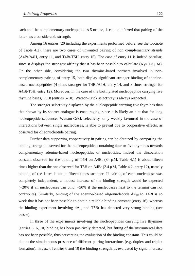
4. Pairing Properties 122
each and the complementary nucleopeptides 5 or less, it can be inferred that pairing of the
latter has a considerable strength.
Among 16 entries (20 including the experiments performed before, see the footnote
of Table 4.2), there are two cases of unwanted pairing of non complementary strands
(A4Bt/A4H, entry 11, and T4Bt/T5H, entry 15). The case of entry 11 is indeed peculiar,
since it displays the strongest affinity that it has been possible to calculate (Kd≈ 1.8 M).
On the other side, considering the two thymine-based partners involved in non-
complementary pairing of entry 15, both display significant stronger binding of adenine-
based nucleopeptides (4 times stronger for T4Bt/A4H, entry 14, and 8 times stronger for
A4Bt/T5H, entry 12). Moreover, in the case of the biotinylated nucleopeptide carrying five
thymine bases, T5Bt (entries 6-10), Watson-Crick selectivity is always respected.
The stronger selectivity displayed by the nucleopeptide carrying five thymines than
that shown by its shorter analogue is encouraging, since it is likely an hint that for long
nucleopeptide sequences Watson-Crick selectivity, only weakly favoured in the case of
interactions between single nucleobases, is able to prevail due to cooperative effects, as
observed for oligonucleotide pairing.
Further data supporting cooperativity in pairing can be obtained by comparing the
binding strength observed for the nucleopeptides containing four or five thymines towards
complementary adenine-based nucleopeptides or nucleotides. Indeed the dissociation
constant observed for the binding of T4H on A4Bt (34 M, Table 4.1) is about fifteen
times higher than the one observed for T5H on A4Bt (2.4 M, Table 4.2, entry 12), namely
binding of the latter is about fifteen times stronger. If pairing of each nucleobase was
completely independent, a modest increase of the binding strength would be expected
(+20% if all nucleobases can bind, +50% if the nucleobases next to the termini can not
contribute). Similarly, binding of the adenine-based oligonucleotide dA10 to T4Bt is so
week that it has not been possible to obtain a reliable binding constant (entry 16), whereas
the binding experiment involving dA10 and T5Bt has detected very strong binding (see
below).
In three of the experiments involving the nucleopeptides carrying five thymines
(entries 3, 6, 10) binding has been positively detected, but fitting of the instrumental data
has not been possible, thus preventing the evaluation of the binding constant. This could be
due to the simultaneous presence of different pairing interactions (e.g. duplex and triplex
formation). In case of entries 6 and 10 the binding strength, as evaluated by signal increase
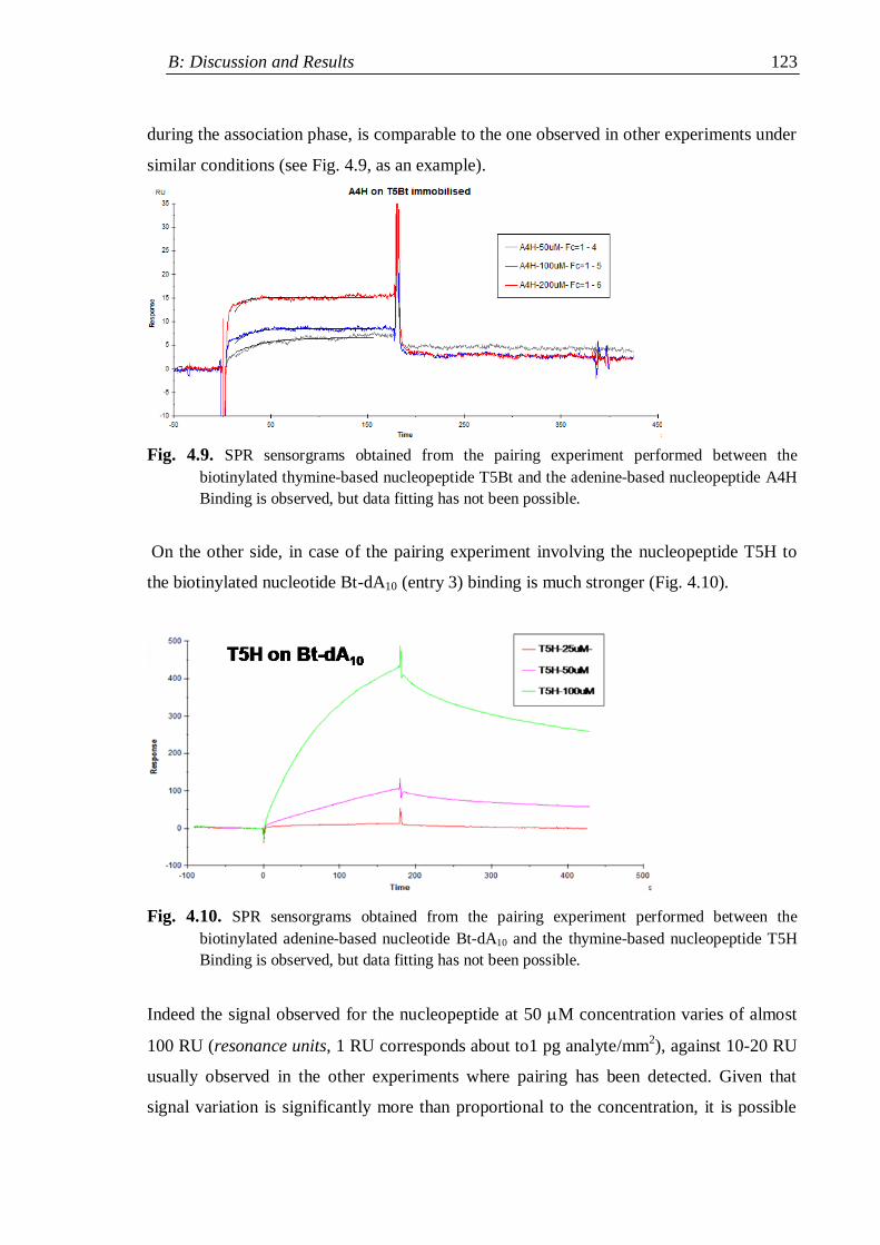
B: Discussion and Results 123
during the association phase, is comparable to the one observed in other experiments under
similar conditions (see Fig. 4.9, as an example).
Fig. 4.9. SPR sensorgrams obtained from the pairing experiment performed between the
biotinylated thymine-based nucleopeptide T5Bt and the adenine-based nucleopeptide A4H
Binding is observed, but data fitting has not been possible.
On the other side, in case of the pairing experiment involving the nucleopeptide T5H to
the biotinylated nucleotide Bt-dA10 (entry 3) binding is much stronger (Fig. 4.10).
Fig. 4.10. SPR sensorgrams obtained from the pairing experiment performed between the
biotinylated adenine-based nucleotide Bt-dA10 and the thymine-based nucleopeptide T5H
Binding is observed, but data fitting has not been possible.
Indeed the signal observed for the nucleopeptide at 50 M concentration varies of almost
100 RU (resonance units, 1 RU corresponds about to1 pg analyte/mm2), against 10-20 RU
usually observed in the other experiments where pairing has been detected. Given that
signal variation is significantly more than proportional to the concentration, it is possible
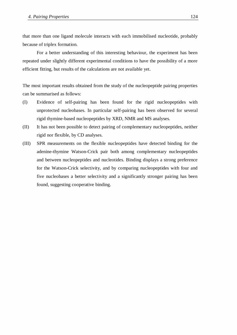
4. Pairing Properties 124
that more than one ligand molecule interacts with each immobilised nucleotide, probably
because of triplex formation.
For a better understanding of this interesting behaviour, the experiment has been
repeated under slightly different experimental conditions to have the possibility of a more
efficient fitting, but results of the calculations are not available yet.
The most important results obtained from the study of the nucleopeptide pairing properties
can be summarised as follows:
(I) Evidence of self-pairing has been found for the rigid nucleopeptides with
unprotected nucleobases. In particular self-pairing has been observed for several
rigid thymine-based nucleopeptides by XRD, NMR and MS analyses.
(II) It has not been possible to detect pairing of complementary nucleopeptides, neither
rigid nor flexible, by CD analyses.
(III) SPR measurements on the flexible nucleopeptides have detected binding for the
adenine-thymine Watson-Crick pair both among complementary nucleopeptides
and between nucleopeptides and nucleotides. Binding displays a strong preference
for the Watson-Crick selectivity, and by comparing nucleopeptides with four and
five nucleobases a better selectivity and a significantly stronger pairing has been
found, suggesting cooperative binding.
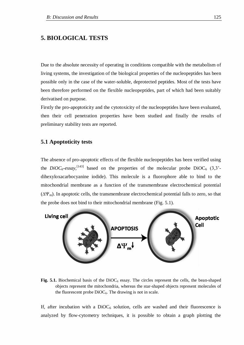
B: Discussion and Results 125
5. BIOLOGICAL TESTS
Due to the absolute necessity of operating in conditions compatible with the metabolism of
living systems, the investigation of the biological properties of the nucleopeptides has been
possible only in the case of the water-soluble, deprotected peptides. Most of the tests have
been therefore performed on the flexible nucleopeptides, part of which had been suitably
derivatised on purpose.
Firstly the pro-apoptoticity and the cytotoxicity of the nucleopeptides have been evaluated,
then their cell penetration properties have been studied and finally the results of
preliminary stability tests are reported.
5.1 Apoptoticity tests
The absence of pro-apoptotic effects of the flexible nucleopeptides has been verified using
the DiOC6-essay,[143]
based on the properties of the molecular probe DiOC6 (3,3‟-
dihexyloxacarbocyanine iodide). This molecule is a fluorophore able to bind to the
mitochondrial membrane as a function of the transmembrane electrochemical potential
(m). In apoptotic cells, the transmembrane electrochemical potential falls to zero, so that
the probe does not bind to their mitochondrial membrane (Fig. 5.1).
Fig. 5.1. Biochemical basis of the DiOC6 essay. The circles represent the cells, the bean-shaped
objects represent the mitochondria, whereas the star-shaped objects represent molecules of
the fluorescent probe DiOC6. The drawing is not in scale.
If, after incubation with a DiOC6 solution, cells are washed and their fluorescence is
analyzed by flow-cytometry techniques, it is possible to obtain a graph plotting the
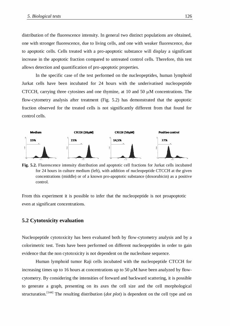
5. Biological tests 126
distribution of the fluorescence intensity. In general two distinct populations are obtained,
one with stronger fluorescence, due to living cells, and one with weaker fluorescence, due
to apoptotic cells. Cells treated with a pro-apoptotic substance will display a significant
increase in the apoptotic fraction compared to untreated control cells. Therefore, this test
allows detection and quantification of pro-apoptotic properties.
In the specific case of the test performed on the nucleopeptides, human lymphoid
Jurkat cells have been incubated for 24 hours with the underivatised nucleopeptide
CTCCH, carrying three cytosines and one thymine, at 10 and 50 M concentrations. The
flow-cytometry analysis after treatment (Fig. 5.2) has demonstrated that the apoptotic
fraction observed for the treated cells is not significantly different from that found for
control cells.
Fig. 5.2. Fluorescence intensity distribution and apoptotic cell fractions for Jurkat cells incubated
for 24 hours in culture medium (left), with addition of nucleopeptide CTCCH at the given
concentrations (middle) or of a known pro-apoptotic substance (doxorubicin) as a positive
control.
From this experiment it is possible to infer that the nucleopeptide is not proapoptotic
even at significant concentrations.
5.2 Cytotoxicity evaluation
Nucleopeptide cytotoxicity has been evaluated both by flow-cytometry analysis and by a
colorimetric test. Tests have been performed on different nucleopeptides in order to gain
evidence that the non cytotoxicity is not dependent on the nucleobase sequence.
Human lymphoid tumor Raji cells incubated with the nucleopeptide CTCCH for
increasing times up to 16 hours at concentrations up to 50 M have been analyzed by flow-
cytometry. By considering the intensities of forward and backward scattering, it is possible
to generate a graph, presenting on its axes the cell size and the cell morphological
structuration.[144]
The resulting distribution (dot plot) is dependent on the cell type and on
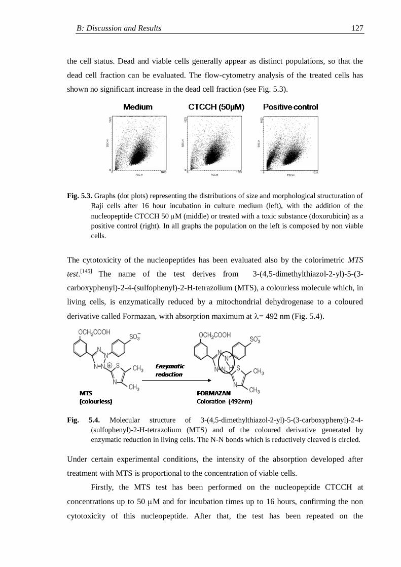
B: Discussion and Results 127
the cell status. Dead and viable cells generally appear as distinct populations, so that the
dead cell fraction can be evaluated. The flow-cytometry analysis of the treated cells has
shown no significant increase in the dead cell fraction (see Fig. 5.3).
Fig. 5.3. Graphs (dot plots) representing the distributions of size and morphological structuration of
Raji cells after 16 hour incubation in culture medium (left), with the addition of the
nucleopeptide CTCCH 50 M (middle) or treated with a toxic substance (doxorubicin) as a
positive control (right). In all graphs the population on the left is composed by non viable
cells.
The cytotoxicity of the nucleopeptides has been evaluated also by the colorimetric MTS
test.[145]
The name of the test derives from 3-(4,5-dimethylthiazol-2-yl)-5-(3-
carboxyphenyl)-2-4-(sulfophenyl)-2-H-tetrazolium (MTS), a colourless molecule which, in
living cells, is enzymatically reduced by a mitochondrial dehydrogenase to a coloured
derivative called Formazan, with absorption maximum at = 492 nm (Fig. 5.4).
Fig. 5.4. Molecular structure of 3-(4,5-dimethylthiazol-2-yl)-5-(3-carboxyphenyl)-2-4-
(sulfophenyl)-2-H-tetrazolium (MTS) and of the coloured derivative generated by
enzymatic reduction in living cells. The N-N bonds which is reductively cleaved is circled.
Under certain experimental conditions, the intensity of the absorption developed after
treatment with MTS is proportional to the concentration of viable cells.
Firstly, the MTS test has been performed on the nucleopeptide CTCCH at
concentrations up to 50 M and for incubation times up to 16 hours, confirming the non
cytotoxicity of this nucleopeptide. After that, the test has been repeated on the
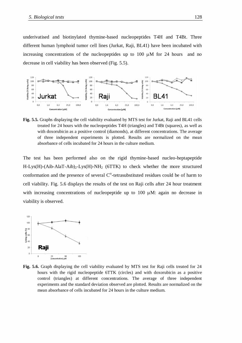
5. Biological tests 128
underivatised and biotinylated thymine-based nucleopeptides T4H and T4Bt. Three
different human lymphoid tumor cell lines (Jurkat, Raji, BL41) have been incubated with
increasing concentrations of the nucleopeptides up to 100 M for 24 hours and no
decrease in cell viability has been observed (Fig. 5.5).
Fig. 5.5. Graphs displaying the cell viability evaluated by MTS test for Jurkat, Raji and BL41 cells
treated for 24 hours with the nucleopeptides T4H (triangles) and T4Bt (squares), as well as
with doxorubicin as a positive control (diamonds), at different concentrations. The average
of three independent experiments is plotted. Results are normalized on the mean
absorbance of cells incubated for 24 hours in the culture medium.
The test has been performed also on the rigid thymine-based nucleo-heptapeptide
H-Lys(H)-(Aib-AlaT-Aib)2-Lys(H)-NH2 (6TTK) to check whether the more structured
conformation and the presence of several C-tetrasubstituted residues could be of harm to
cell viability. Fig. 5.6 displays the results of the test on Raji cells after 24 hour treatment
with increasing concentrations of nucleopeptide up to 100 M: again no decrease in
viability is observed.
Fig. 5.6. Graph displaying the cell viability evaluated by MTS test for Raji cells treated for 24
hours with the rigid nucleopeptide 6TTK (circles) and with doxorubicin as a positive
control (triangles) at different concentrations. The average of three independent
experiments and the standard deviation observed are plotted. Results are normalized on the
mean absorbance of cells incubated for 24 hours in the culture medium.
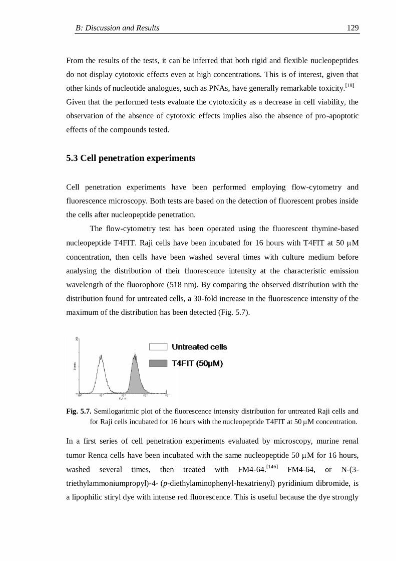
B: Discussion and Results 129
From the results of the tests, it can be inferred that both rigid and flexible nucleopeptides
do not display cytotoxic effects even at high concentrations. This is of interest, given that
other kinds of nucleotide analogues, such as PNAs, have generally remarkable toxicity.[18]
Given that the performed tests evaluate the cytotoxicity as a decrease in cell viability, the
observation of the absence of cytotoxic effects implies also the absence of pro-apoptotic
effects of the compounds tested.
5.3 Cell penetration experiments
Cell penetration experiments have been performed employing flow-cytometry and
fluorescence microscopy. Both tests are based on the detection of fluorescent probes inside
the cells after nucleopeptide penetration.
The flow-cytometry test has been operated using the fluorescent thymine-based
nucleopeptide T4FIT. Raji cells have been incubated for 16 hours with T4FIT at 50 M
concentration, then cells have been washed several times with culture medium before
analysing the distribution of their fluorescence intensity at the characteristic emission
wavelength of the fluorophore (518 nm). By comparing the observed distribution with the
distribution found for untreated cells, a 30-fold increase in the fluorescence intensity of the
maximum of the distribution has been detected (Fig. 5.7).
Fig. 5.7. Semilogaritmic plot of the fluorescence intensity distribution for untreated Raji cells and
for Raji cells incubated for 16 hours with the nucleopeptide T4FIT at 50 M concentration.
In a first series of cell penetration experiments evaluated by microscopy, murine renal
tumor Renca cells have been incubated with the same nucleopeptide 50 M for 16 hours,
washed several times, then treated with FM4-64.[146]
FM4-64, or N-(3-
triethylammoniumpropyl)-4- (p-diethylaminophenyl-hexatrienyl) pyridinium dibromide, is
a lipophilic stiryl dye with intense red fluorescence. This is useful because the dye strongly
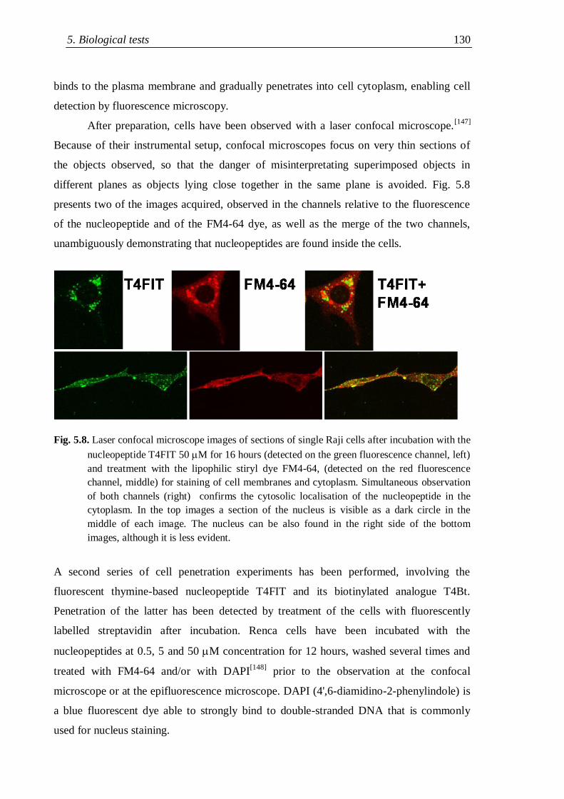
5. Biological tests 130
binds to the plasma membrane and gradually penetrates into cell cytoplasm, enabling cell
detection by fluorescence microscopy.
After preparation, cells have been observed with a laser confocal microscope.[147]
Because of their instrumental setup, confocal microscopes focus on very thin sections of
the objects observed, so that the danger of misinterpretating superimposed objects in
different planes as objects lying close together in the same plane is avoided. Fig. 5.8
presents two of the images acquired, observed in the channels relative to the fluorescence
of the nucleopeptide and of the FM4-64 dye, as well as the merge of the two channels,
unambiguously demonstrating that nucleopeptides are found inside the cells.
Fig. 5.8. Laser confocal microscope images of sections of single Raji cells after incubation with the
nucleopeptide T4FIT 50 M for 16 hours (detected on the green fluorescence channel, left)
and treatment with the lipophilic stiryl dye FM4-64, (detected on the red fluorescence
channel, middle) for staining of cell membranes and cytoplasm. Simultaneous observation
of both channels (right) confirms the cytosolic localisation of the nucleopeptide in the
cytoplasm. In the top images a section of the nucleus is visible as a dark circle in the
middle of each image. The nucleus can be also found in the right side of the bottom
images, although it is less evident.
A second series of cell penetration experiments has been performed, involving the
fluorescent thymine-based nucleopeptide T4FIT and its biotinylated analogue T4Bt.
Penetration of the latter has been detected by treatment of the cells with fluorescently
labelled streptavidin after incubation. Renca cells have been incubated with the
nucleopeptides at 0.5, 5 and 50 M concentration for 12 hours, washed several times and
treated with FM4-64 and/or with DAPI[148]
prior to the observation at the confocal
microscope or at the epifluorescence microscope. DAPI (4',6-diamidino-2-phenylindole) is
a blue fluorescent dye able to strongly bind to double-stranded DNA that is commonly
used for nucleus staining.
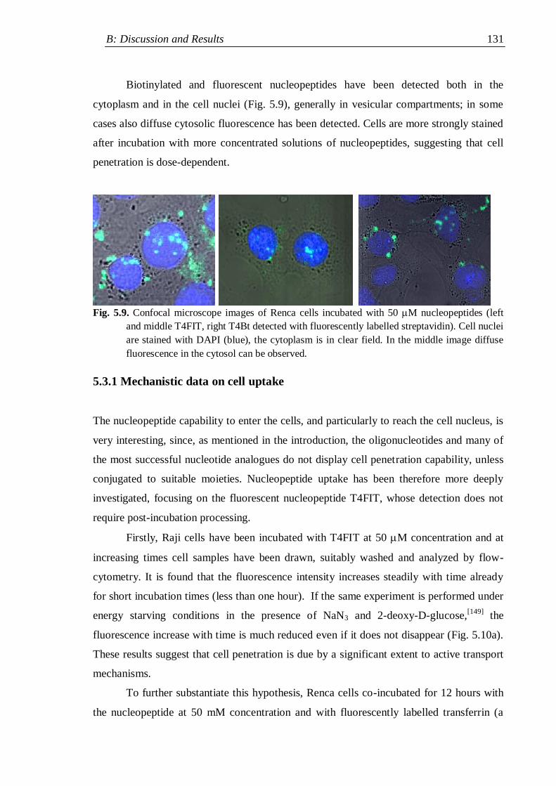
B: Discussion and Results 131
Biotinylated and fluorescent nucleopeptides have been detected both in the
cytoplasm and in the cell nuclei (Fig. 5.9), generally in vesicular compartments; in some
cases also diffuse cytosolic fluorescence has been detected. Cells are more strongly stained
after incubation with more concentrated solutions of nucleopeptides, suggesting that cell
penetration is dose-dependent.
Fig. 5.9. Confocal microscope images of Renca cells incubated with 50 M nucleopeptides (left
and middle T4FIT, right T4Bt detected with fluorescently labelled streptavidin). Cell nuclei
are stained with DAPI (blue), the cytoplasm is in clear field. In the middle image diffuse
fluorescence in the cytosol can be observed.
5.3.1 Mechanistic data on cell uptake
The nucleopeptide capability to enter the cells, and particularly to reach the cell nucleus, is
very interesting, since, as mentioned in the introduction, the oligonucleotides and many of
the most successful nucleotide analogues do not display cell penetration capability, unless
conjugated to suitable moieties. Nucleopeptide uptake has been therefore more deeply
investigated, focusing on the fluorescent nucleopeptide T4FIT, whose detection does not
require post-incubation processing.
Firstly, Raji cells have been incubated with T4FIT at 50 M concentration and at
increasing times cell samples have been drawn, suitably washed and analyzed by flow-
cytometry. It is found that the fluorescence intensity increases steadily with time already
for short incubation times (less than one hour). If the same experiment is performed under
energy starving conditions in the presence of NaN3 and 2-deoxy-D-glucose,[149]
the
fluorescence increase with time is much reduced even if it does not disappear (Fig. 5.10a).
These results suggest that cell penetration is due by a significant extent to active transport
mechanisms.
To further substantiate this hypothesis, Renca cells co-incubated for 12 hours with
the nucleopeptide at 50 mM concentration and with fluorescently labelled transferrin (a
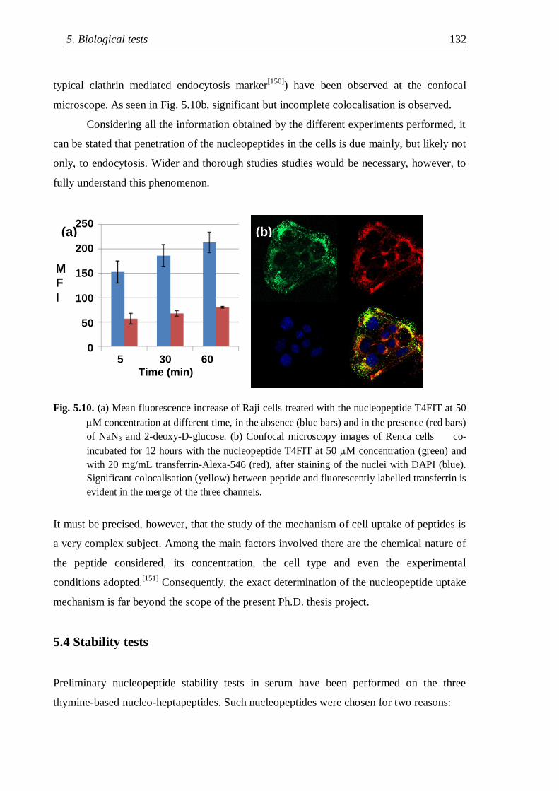
5. Biological tests 132
typical clathrin mediated endocytosis marker[150]
) have been observed at the confocal
microscope. As seen in Fig. 5.10b, significant but incomplete colocalisation is observed.
Considering all the information obtained by the different experiments performed, it
can be stated that penetration of the nucleopeptides in the cells is due mainly, but likely not
only, to endocytosis. Wider and thorough studies studies would be necessary, however, to
fully understand this phenomenon.
Fig. 5.10. (a) Mean fluorescence increase of Raji cells treated with the nucleopeptide T4FIT at 50
M concentration at different time, in the absence (blue bars) and in the presence (red bars)
of NaN3 and 2-deoxy-D-glucose. (b) Confocal microscopy images of Renca cells co-
incubated for 12 hours with the nucleopeptide T4FIT at 50 M concentration (green) and
with 20 mg/mL transferrin-Alexa-546 (red), after staining of the nuclei with DAPI (blue).
Significant colocalisation (yellow) between peptide and fluorescently labelled transferrin is
evident in the merge of the three channels.
It must be precised, however, that the study of the mechanism of cell uptake of peptides is
a very complex subject. Among the main factors involved there are the chemical nature of
the peptide considered, its concentration, the cell type and even the experimental
conditions adopted.[151]
Consequently, the exact determination of the nucleopeptide uptake
mechanism is far beyond the scope of the present Ph.D. thesis project.
5.4 Stability tests
Preliminary nucleopeptide stability tests in serum have been performed on the three
thymine-based nucleo-heptapeptides. Such nucleopeptides were chosen for two reasons:
5 30 60
Time (min)
250
200
150
100
50
0
M F
I
(a) (b)))
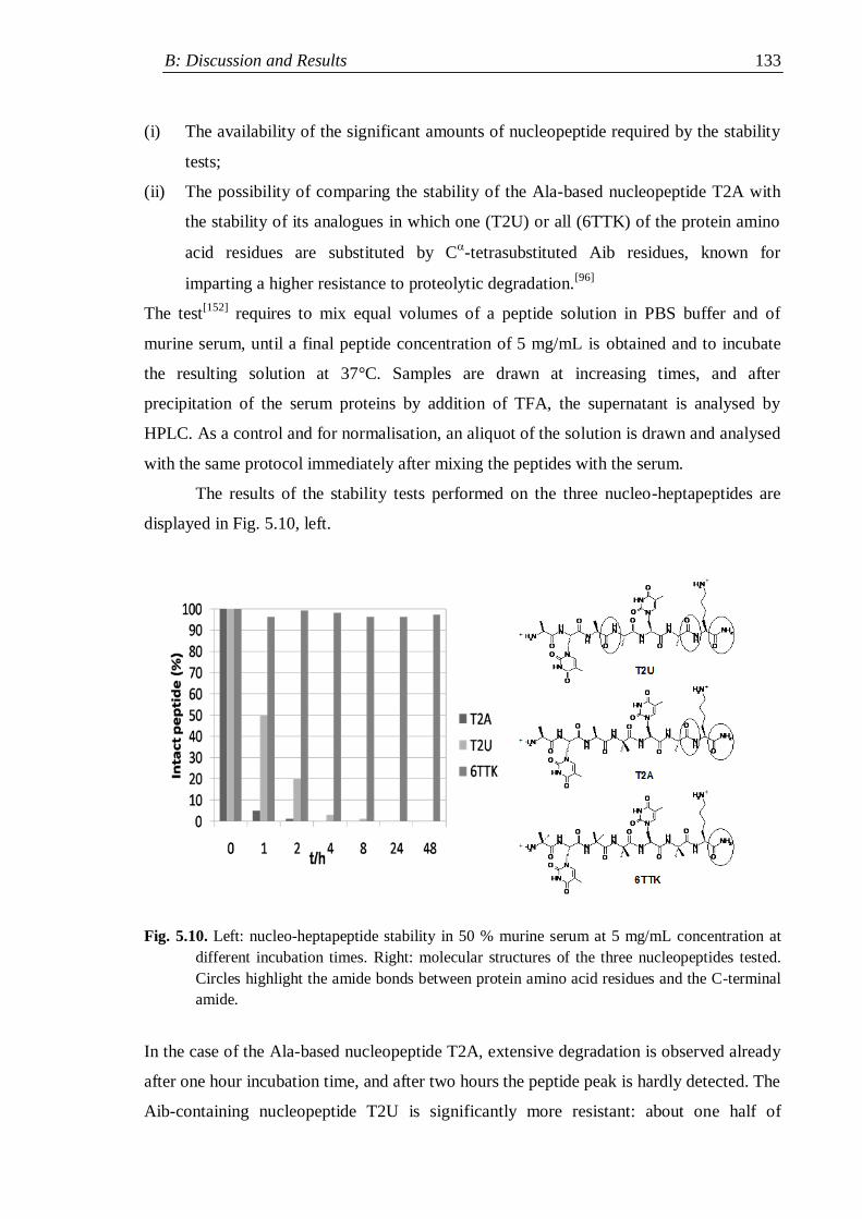
B: Discussion and Results 133
(i) The availability of the significant amounts of nucleopeptide required by the stability
tests;
(ii) The possibility of comparing the stability of the Ala-based nucleopeptide T2A with
the stability of its analogues in which one (T2U) or all (6TTK) of the protein amino
acid residues are substituted by C-tetrasubstituted Aib residues, known for
imparting a higher resistance to proteolytic degradation.[96]
The test[152]
requires to mix equal volumes of a peptide solution in PBS buffer and of
murine serum, until a final peptide concentration of 5 mg/mL is obtained and to incubate
the resulting solution at 37°C. Samples are drawn at increasing times, and after
precipitation of the serum proteins by addition of TFA, the supernatant is analysed by
HPLC. As a control and for normalisation, an aliquot of the solution is drawn and analysed
with the same protocol immediately after mixing the peptides with the serum.
The results of the stability tests performed on the three nucleo-heptapeptides are
displayed in Fig. 5.10, left.
Fig. 5.10. Left: nucleo-heptapeptide stability in 50 % murine serum at 5 mg/mL concentration at
different incubation times. Right: molecular structures of the three nucleopeptides tested.
Circles highlight the amide bonds between protein amino acid residues and the C-terminal
amide.
In the case of the Ala-based nucleopeptide T2A, extensive degradation is observed already
after one hour incubation time, and after two hours the peptide peak is hardly detected. The
Aib-containing nucleopeptide T2U is significantly more resistant: about one half of
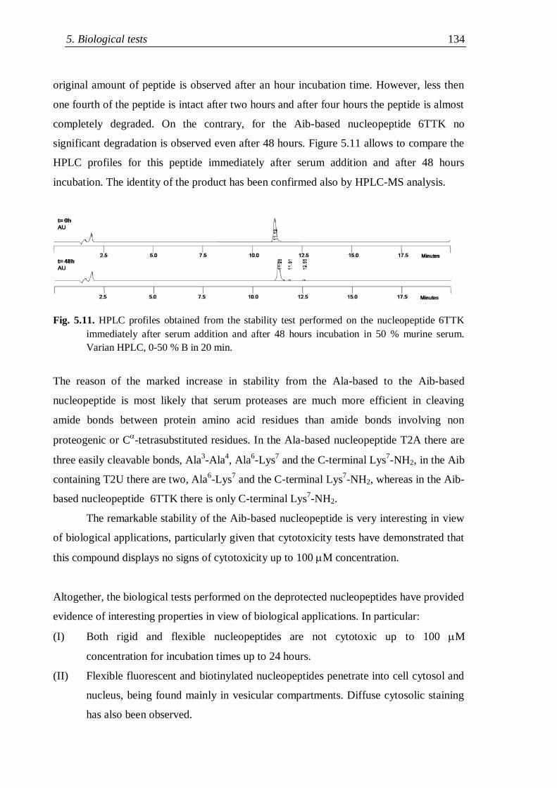
5. Biological tests 134
original amount of peptide is observed after an hour incubation time. However, less then
one fourth of the peptide is intact after two hours and after four hours the peptide is almost
completely degraded. On the contrary, for the Aib-based nucleopeptide 6TTK no
significant degradation is observed even after 48 hours. Figure 5.11 allows to compare the
HPLC profiles for this peptide immediately after serum addition and after 48 hours
incubation. The identity of the product has been confirmed also by HPLC-MS analysis.
Fig. 5.11. HPLC profiles obtained from the stability test performed on the nucleopeptide 6TTK
immediately after serum addition and after 48 hours incubation in 50 % murine serum.
Varian HPLC, 0-50 % B in 20 min.
The reason of the marked increase in stability from the Ala-based to the Aib-based
nucleopeptide is most likely that serum proteases are much more efficient in cleaving
amide bonds between protein amino acid residues than amide bonds involving non
proteogenic or C-tetrasubstituted residues. In the Ala-based nucleopeptide T2A there are
three easily cleavable bonds, Ala3-Ala
4, Ala
6-Lys
7 and the C-terminal Lys
7-NH2, in the Aib
containing T2U there are two, Ala6-Lys
7 and the C-terminal Lys
7-NH2, whereas in the Aib-
based nucleopeptide 6TTK there is only C-terminal Lys7-NH2.
The remarkable stability of the Aib-based nucleopeptide is very interesting in view
of biological applications, particularly given that cytotoxicity tests have demonstrated that
this compound displays no signs of cytotoxicity up to 100 M concentration.
Altogether, the biological tests performed on the deprotected nucleopeptides have provided
evidence of interesting properties in view of biological applications. In particular:
(I) Both rigid and flexible nucleopeptides are not cytotoxic up to 100 M
concentration for incubation times up to 24 hours.
(II) Flexible fluorescent and biotinylated nucleopeptides penetrate into cell cytosol and
nucleus, being found mainly in vesicular compartments. Diffuse cytosolic staining
has also been observed.

B: Discussion and Results 135
(III) Cell uptake is concentration, time and energy dependent. Significant colocalisation
with transferrin has been observed, suggesting that cell penetration is due mainly to
endocytosis, but likely this is not the only mechanism involved.
(IV) Whereas the Ala-based nucleo-heptapeptide is rapidly degraded in serum, the
introduction of a single Aib residue significantly improves resistance to proteolytic
degradation, and the Aib-based analogue is stable even after 48 hour incubation.
6. Experimental Section 136
6. EXPERIMENTAL SECTION
6.1 Materials and methods
6.1.1 Reagents and solvents
Acros-Janssen (Geel, Belgium):
Acetic anhydride, allyl bromide, benzyl chloroformate, biotine, bromocresol green,
para-cresol, ethanolamine hydrochloride, ethylenediaminetetraacetic acid, 1-
hydroxy-1H-benzotriazole, N-2-hydroxyethylpiperazine-N'-2-ethanesulfonic acid,
methyl chloroformate, phosphoric anhydride, piperidine, potassium bromide,
trifluoroacetic acid,.
Alfa Aesar (Karlsruhe, Germany):
2-amino-6-chloro-purine, adenine, cytosine, thymine.
Applied Biosystems - Invitrogen (Carlsbad, USA):
4-methylbenzhydrylamine hydrochloride salt resin (loading 0.62 mmol/g), transferrin-Alexa-
546, N- (3-triethylammoniumpropyl) -4- (6-(4-(diethylamino)-phenyl) -
hexatrienyl)-pyridinium dibromide.
Bachem (Bubendorf, Switzerland):
N-tertbutyloxycarbonyl-serine.
BD Pharmingen (Franklin Lakes, New Jersey, USA):
Fluorescein-5(6)-aminothiocarbonyl-streptavidin (streptavidin-FITC).
Biacore (Uppsala, Sweden):
N-hydroxysuccinimide, surfactant P-20.
Cambridge Isotope Laboratories (Cambridge, England):
Deuterochloroform, hexadeuterated dimethylsulphoxide, trideuterated acetonitrile.
C: Experimental Section 137
Carlo Erba (Milan, Italy):
Acetic acid, ammonia (28% aqueous solution), 1-butanol, calcium chloride
anhydrous, chloroform, dichloromethane, diethyl ether, ethanol, ethyl acetate,
hydrochloric acid (37% aqueous solution), methanol, petroleum ether (boiling
range 40-60°C), potassium hydrogenocarbonate, potassium hydrogenosulphate, 2-
propanol, sodium carbonate dodecahydrate, sodium hydroxyde, sodium
hypochlorite, sodium sulphate anhydrous, sulphuric acid (98% aqueous solution),
toluene, triethylamine.
EMS-Chemie (Domat-Ems, Switzerland):
Paraformaldehyde.
Eurogentec (Seraing, Belgium):
Oligoribonucleotides (U4, U8), oligodeoxyribonucleotides (dA10, 5‟-biotinyl-
phosphoramidite-dA10 or Bt-dA10, dT8, dT10).
Fluka (Buchs, Switzerland):
Acetonitrile, -amino isobutyric acid, chloroform (spectrophotometric grade),
N,N-diisopropylethylamine, hydrazine hydrate, methanol, spectrophotometric
grade, N-methylmorpholine, ninhydrin, palladium catalyst (10% on activated
charcoal), serine, tetrahydrofurane.
GL Biochem Lt (Shangai, China):
7-aza-1-hydroxy-1H-benzotriazole, N-benzyloxycarbonyl- N
-
tertbutyloxycarbonyl-lysine, N-tertbutyloxycarbonyl- N
-benzyloxycarbonyl-
lysine, O-(7-aza-benzotriazol-1-yl)- N,N,N′,N′-tetramethyluronium
hexafluorophosphate.
Interchim (Montluçon, France):
3,3‟-dihexyloxacarbocyanine iodide.
6. Experimental Section 138
Iris Biotech (Marktredwitz, Germany):
N-benzyloxycarbonyloxysuccinimide, N-ethyl-N'-(3-dimethylamino)propyl-
carbodiimide chlorohydrate.
J. T. Baker (Phillipsburg, New Jersey, USA):
N,N-dimethylformamide, pyridine.
Merck (Darmstadt, Germany):
N-bromosuccinimide, magnesium acetate, sodium azide, disodium
hydrogenphosphate dihydrate, potassium carbonate, sodium azide, sodium
dihydrogenphosphate monohydrate, sodium formate.
NeoMPS (Strasbourg, France):
N-tertbutyloxycarbonyl-alanine, N
-tertbutyloxycarbonyl- N
-(2-
chlorobenzyloxycarbonyl)- lysine, N-tertbutyloxycarbonyl- N
-
fluorenylmethyoxycarbonyl- lysine, O-(benzotriazol-1-yl)- N,N,N’,N’-
tetramethyluronium hexafluoro-phosphate.
Prolabo (Paris, France):
Sodium chloride.
Promega (Madison, Wisconsin, USA):
3-(4,5-dimethylthiazol-2-yl)-5-(3-carboxymethoxyphenyl)-2-(4-sulfophenyl)-2H-
tetrazolium, reagent solution.
Siad (Bergamo, Italy):
Isobutene.
Sigma-Aldrich (Milwaukee, USA):
2-deoxy-D-glucose, 4',6-diamidino-2-phenylindole 1,8-diazabicyclo[5.4.0]undec-
7-ene, 4-(dimethylamino)-pyridine, formic acid, 2-hydroxy-cinnamic acid,
ninhydrin, iodine, sodium hydride (60% dispersion in mineral oil), streptavidin,
N,N,N',N'-tetramethyl-4,4'-diamino-diphenylmethane, trinitrobenzensulfonic acid,
trimethylsilyl trifluoromethansulphonate, triphenylphosphine, Triton X-100.
C: Experimental Section 139
6.1.2 Instruments and methods
Solid-phase synthesis
Solid-phase synthesis of the nucleopeptide sequences was accomplished using a
PSP 4000 semiautomatic peptide synthesizer for Boc/Bzl strategy.
MBHA resin (loading 0.62 mmol/g) was used as solid support.
Completion of coupling or deprotection steps was checked by qualitative Kaiser
test[153]
or by TNBS test[154]
.
Melting point determination
Melting points were measured on a Leitz apparatus model Laborlux 12 and are
uncorrected.
Thin layer chromatography
Silica gel 60 F254 (Merck) or Alugram® Sil G UV 254 (Macherey-Nagel) on
aluminium foil was used to follow the reactions.
Silica gel 60 F254 (Merck) on glass was used for TLC characterization. Retention
factors (Rf) have been measured using three different solvent mixtures as eluents.
Rf1: CHCl3/EtOH 9:1; Rf2: BuOH/AcOH/H2O 3:1:1; Rf3: PhMe/EtOH 7:1.
Products were detected either by UV lamp irradiation or with exposition to I2
vapours or by warming with a heat gun and spraying firstly with a 1.5% NaClO
solution and then with a ninhydrin-TDM solution.[155]
N-protected serine--lactone was detected by spraying a 0.06% (w/v) solution of
bromocresol green in EtOH.
Flash chromatography
For FC purifications silica gel 60 Merck (40-63 m diameter, mesh 230-400) was
used. For the various purifications, one of the following loading methods was used:
1) direct loading on top of the column of the crude mixture dissolved in a small
amount of the eluent used for the purification;
2) direct loading on top of the column of the crude mixture dissolved in the
minimum amount of a different solvent mixture;
3) loading on top of the column of the crude mixture adsorbed on silica gel („dry-
loading‟).
6. Experimental Section 140
HPLC
Analytical chromatograms were recorded either on a Pharmacia LKB-LCC 2252
instrument, provided with a Kromasil Phenomenex C18 column (4.6250 mm, 100
Å, flow 1.0 mL/min) and a UV Uvicord SD detector operating at 214 nm, or on a
Agilent Technologies 1200 Series instrument, provided with a Kromasil
Phenomenex C18 column (4.6250 mm, 100 Å, flow 1.0 mL/min) or on a Varian
ProStar 410 instrument, provided with a Nucleosil 100-5 C18 column (4.6150
mm, 100 Å, flow 1.2 mL/min) and a Varian ProStar 330.71 PDA detector.
Semi-preparative scale chromatographic purifications were performed either on a
Agilent Technologies 1200 Series instrument, provided with a Vydac C18 Protein
and Peptides column (10250 mm, flow 2.5 mL/min) or on a Beckmann System
Gold HPLC 166 instrument, provided with a SDS C18 Macherey-Nagel column
(10250 mm, 100 Å, flow 6.0 mL/min) and a Beckmann System Gold UV detector
operating at 230 nm.
Preparative scale HPLC purifications were performed on a Shimadzu SCL-6B
instrument, provided with Shimadzu LC-8A pumps, a Delta Pack C18 (19300
mm, 100 Å, flow 17 mL/min) column and a Shimadzu SPD-64 detector operating
at 265 nm. As eluents following solvent mixtures were used:
- HPLC Varian and Beckmann: A: H2O + 0.1% TFA; B: MeCN + 0.08% TFA.
- HPLC Pharmacia, Agilent and Shimadzu: A‟: H2O/MeCN 9:1 + 0.05% TFA;
B‟: MeCN/H2O 9:1 + 0.05% TFA.
MilliQ H2O was used for eluent preparation.
Polarimetric measurements
Optical rotations were measured on a a Perkin-Elmer model 241 polarimeter with
an Haake model D8 thermostat at the sodium D line wavelength, using a cell with
an optical pathlength of 10 cm. Concentrations are expressed in g/100 mL. []25
D
are calculated using the formula []= /(c∙l), where c is the concentration (in g/mL)
and l is the optical path (in dm). Spectrophotometric grade MeOH was used as a
solvent.
C: Experimental Section 141
Circular dichroism
CD spectra were recorded either on a Jasco dichrograph model J-715 provided
with a Haake model F3 thermostat or on Jasco dichrograph model J-810, provided
with a JASCO ETC-505S/PTC-423S temperature controller using Hellma quartz
cells (pathlenghts 0.1, 0.2, 0.5 or 1.0 cm). Instrumental parameters were as follow:
response 2 sec, standard sensitivity, scan speed 100 nm/min, data pitch 0.5 nm.
Experimental values are expressed in molar ellipticity [Θ]T (degrees∙cm2∙dmol
-1)
and were calculated, using the relation [Θ]T = Θ/(l∙c) , where Θ is the observed
ellipticity (in degrees), l is the optical path (in dm) and c is the peptide
concentration (in mol/L).
UV spectroscopy
UV-Vis spectra were recorded on a Shimadzu UV 2501PC spectrophotometer or on
a Cary 5000 spectrophotometer using Hellma 1.0 cm optical path quartz cell.
Mass spectrometry (MS)
ESI-TOF mass spectra were collected on a Mariner ESI-TOF mass spectrometer
(Perseptive Biosystems). Prior to injection, samples were dissolved in a 1:1 mixture
of water-MeOH containing 0.1 % formic acid. The positive ions were accelerated at
10, 15, 20 or 30 keV.
MALDI mass spectra of nucleopeptides were collected on a Bruker Protein TOF
Biflex II MALDI-TOF spectrometer. 2-hydroxy-cinnamic acid (saturated solution
in acetone) was used as matrix. Equal amounts (0.6 L each) of matrix solution and
of dilute aqueous nucleopeptide solution were mixed and the same volume of the
resulting mixture was loaded on target disk.
HPLC-MS
HPLC-MS ESI-TOF analyses were performed on a Finnigan LCQ Advantage
Thermo Scientific LC-MS, provided with a Macherey-Nagel EC 100/2 Nucleodur
C18 column (2100 mm, 100 Å, flow 300 L/min).
As eluents, H2O + 0.1 % formic acid (A‟‟) and MeCN (B‟‟) were used.
MilliQ H2O was used for eluent preparation.
6. Experimental Section 142
IR absorption spectroscopy
IR absorption measurements on KBr pellets were performed on a Perkin-Elmer
model 580 B spectrophotometer, equipped with a Perkin-Elmer 3600 IR data
station. IR absorption measurements in deuterated chloroform solution were
performed on a Perkin-Elmer model 1720 X FT-IR spectrophotometer, equipped
with a sample-shuttle device, at 2 cm-1
nominal resolution, averaging 100 scans,
nitrogen flushed, connected with a IBM PS/2 model 50 Z personal computer.
Solvent (baseline) spectra were obtained under the same conditions. Cells with
pathlengths of 0.1, 1 and 10 mm (with CaF2 windows) were used. Absorption
maxima, shoulder and partially overlapping bands were detected with the inverse
second derivative method. Spectral elaborations (solvent subtraction and
derivatization) were performed using the program SpectraCalc by Galactic (Salem,
MA, USA).
NMR spectrometry
1H-NMR and 2D-NMR spectra were recorded on Bruker AC 200, Bruker AC 250,
Bruker AC 300 or DRX 400 spectrometers. 13
C-NMR spectra were recorded on a
Bruker AC 300 spectrometer. 2D-NMR spectra were recorded on DRX 400 Bruker
or DMX 600 spectrometers. Chemical shifts () are expressed in parts per million
with regard to tetramethylsilane signal. Solvent residual peaks (CHCl3, 7.26 ppm,
or d6-DMSO, 2.50 ppm, for 1H-NMR, H3
13CS(O)CH3, 39.52 ppm for
13C-
NMR) were used to calibrate the spectra. Peak multiplicity is described as follows:
s (singlet), br s (broad singlet), d (doublet), dd (doublet of doublets), m (multiplet);
coupling constants (J) are expressed in Hz.
For the elaboration of the spectra the programs XwinNMR e MestRe-C2.3 were
applied.
X-ray diffraction (XRD)
Diffraction data were collected on a Philips PW1100 four-circle diffractometer,
using graphite-monochromated CuK radiation. Unit cell parameters were obtained
by least-squares refinement of the angular settings of 48 carefully centred
reflections in the 12 – 18° range. Intensities were corrected for Lorentz and
polarisation effects, not for absorption.
C: Experimental Section 143
The structure was solved by direct methods of the SIR 2002 program.[156]
Refinement was carried out by full-matrix least-squares procedures on F2, using all
data, by application of the SHELXL-97 program,[157]
with all non-H atoms
anisotropic. The phenyl ring of the N-terminal Z-protecting group was constrained
to the idealized geometry. H-atoms atoms were calculated at idealized positions and
refined using a riding model.
Centrifuges
Hettich Zentrifugen Mikroliter Typ 2021 or Sigma 2MK centrifuges were used for
Eppendorf tubes (stability tests) or 1.5 mL Starsted cryotubes (lyophilized purified
products); an Eppendorf 5804 R centrifuge was used for Falcon tubes (crude
peptide recovery after cleavage) .
Surface Plasmon Resonance
Surface Plasmon Resonance measurements were performed on a BIACORE 3000
system using sensor chips model CM5 (Biacore, Uppsala, Sweden).
Kinetic parameters were calculated using the BIAeval 4.1 software on a personal
computer. Global analysis was performed using the simple Langmuir binding
model. The specific binding profiles were obtained after subtracting the response
signal from the channel control (streptavidin free). The fitting to each model was
judged by the reduced chi square and randomness of residue distribution.
Microscopy imaging
Fluorescently labelled cells were observed on a Zeiss LSM 510 Meta confocal
microscopy or on a Zeiss Axiovert 200M epifluorescence microscopy equipped
with a Zeiss Apotome module.
Fluorescence Activated Cell Sorting
Cells were analyzed with a FACSCalibur® flow cytometer. At least 10.000 events
were acquired for each experiment using the CellQuest 3.3 software developed by
Becton Dickinson, Pont de Claix, France and data were processed with the
WinMDI 2.8 freeware developed by Joseph Trotter, Scripps Research Institute
(http://facs.scripps.edu/software.html).

6. Experimental Section 144
Microplate biological testing
For colorimetric cell viability tests a Wallac Victor2 microplate reader was used.
C: Experimental Section 145
6.2 Synthesis and characterization
6.2.1 Solution phase synthesis
6.2.1.1 Synthesis of protected nucleobases and lactone precursors
H-ABoc [109]
H-AH (5.4 g, 40 mmol) is suspended in DMF/py 2:1 (total volume 150 mL) and Boc2O
(17.5 g, 80 mmol) is added. The mixture is heated at 70-80°C for 1 hour then stirred at
room temperature overnight. The solvent is evaporated under vacuum, the remaining solid
is dissolved in H2O/MeOH 4:1 (total volume 65 mL) by heating under reflux for 15 min.
The mixture is evaporated and the resulting oil is diluted in MeOH/DCM 6:1 (total volume
90 mL) then, after extensive stirring, it is filtered. The mother liquors are evaporated and
the crude product is purified by flash-chromatography (eluent: DCM/MeOH 98:2).
Yield: 28 %. Mp> 250°C (dec.). Rf1: 0.10; Rf2: 0.80; Rf3: 0.10.
IR (KBr): 3418, 1748, 1624, 1595, 1557 cm-1
.
1H-NMR (d6-DMSO, 200 MHz), /ppm: 12.27 (br s, 1H, N
6H-Boc), 10.55 (br s, 1H, N(9)-
H), 8.57 (s, 1H, C(2)H), 8.43 (s, 1H, C(8)H), 1.51 (s, 9H, CH3 Boc).
H-AZ [20]
NaH (6.4 g, 60 % in hexanes, 160 mmol) is suspended in 160 mL of anhydrous DMF and
stirred under N2 for 10 min, then H-AH (5.4 g, 40 mmol) is added. Stirring continues for 2
hours until a yellow suspension is formed, then the mixture is cooled to 0°C in a water/ice
bath and ZCl (13.5 mL, 100 mmol) are added dropwise in 20 min. After 4 hours the
mixture is poured in 320 mL of chilled H2O and stirred for 10 min, then it is neutralized
with HCl 1N with precipitation of a solid. The precipitate is filtrated, washed with H2O and
Et2O, then purified through double recrystallization from MeOH/CHCl3.
Yield: 51 %. Mp: 222°C. Rf1: 0.40; Rf2: 0.80; Rf3: 0.30.
IR (KBr):3360, 1780, 1618, 1575 cm-1
.
UV: 15100 M-1
cm-1
at = 275.4 nm (7.1 M in MeOH).
1H-NMR (d6-DMSO, 200 MHz), /ppm: 12.31 (br s, 1H, N
6H-Z), 11.01 (br s, 1H, N(9)H),
8.63 (s, 1H, C(2)H), 8.48(s, 1H, C(8)H), 7.48 (s, 5H, C6H5-Z), 5.32 (s, 2H, CH2-Z).
6. Experimental Section 146
H-CBoc [58b]
H-CH
(4.5 g, 40 mmol) and DMAP (0.25 g, 20 mmol) are suspended in 76 mL anhydrous
DMSO under N2 and the resulting yellow slurry is stirred for 10 min. Boc2O (10.7 g, 48
mmol) is added under N2 to the mixture, the flask is closed with a rubber septum pierced
by a syringe needle and rapid dissolution with CO2 development occurs. After stirring 1
day at room temperature, the yellow mixture is poured in 300 mL H2O and vigorously
stirred with decoloration and precipitation of the crude product after a while. The
precipitate is filtered, washed with H2O, AcOEt and finally EP, than it is dried overnight
under vacuum. More product is recovered by concentration of the mother liquors.
Yield: 43 %. Mp: 170°C. Rf1: 0.45; Rf2: 0.85; Rf3: 0.25.
IR (KBr): 3427, 1738, 1626, 1580, 1512 cm-1
.
UV : 36 M-1
cm-1
at =265.0 nm (17 M in MeOH).
NMR (d6-DMSO, 200 MHz), δ/ppm: 7.79-7.76 (d, J= 7.8 Hz, 1H, C(6)H), 7.60 (br s, 1H,
N4H), 5.79-5.76 (d, J= 7.8 Hz, 1H, C(5)H), 1.49-1.44 (2 s, CH3 Boc cis-trans).
H-CBz [101]
H-CH (1.1 g, 10 mmol) is suspended in DMF, py is added (0.9 mL, 11 mmol) under
stirring, then BzCl is added dropwise (2 mL, 20 mmol). A solution is suddenly formed,
then the product precipitates. After stirring for 1 hour, the product is filtered and washed
with plenty of H2O, AcOEt and Et2O, then dried under vacuum overnight.
Yield: 90 %. Mp> 250°C. Rf1: 0.15; Rf2: 0.80; Rf3: 0.05.
IR (KBr): 3423, 3227, 1720, 1695, 1641, 1621, 1589, 1582, 1513 cm-1
.
UV: 22400 M-1
cm-1
at =250.4 nm, shoulder at = 294 nm (26 M in MeOH).
1H-NMR (d6-DMSO, 200 MHz), δ/ppm: 8.01-7.96 (m, 2H, H-orto Bz), 7.93-7.89 (d, J=
7.0 Hz, 1H, C(6)H), 7.69-7.62 (m, 1H, H-para Bz), 7.58-7.50 (m, 2H, H-meta Bz), 7.26-
7.22 (d, J= 7.0 Hz, 1H, C(5)H).
H-CZ [20]
H-CH (5.0 g, 45 mmol) is dissolved in 90 mL py (2 ml/mmol H-C
H) and DMAP (1.0 g,
8.0 mmol) are added. The solution is chilled in a water/ice bath, then Z-Cl (15 ml, 102
mmol) is added dropwise. The mixture is stirred three days at room temperature, then 180
mL chilled H2O are added and the precipitate is filtered and washed with H2O, then
desiccated.
C: Experimental Section 147
Yield: 40 %. Mp: 273°C. Rf1: 0.30; Rf2: 0.80; Rf3: 0.30.
IR (KBr): 3475, 3416, 1744, 1688, 1627, 1587 cm-1
.
UV: 18800 M-1
cm-1
at =236.1 nm, 7490 M-1
cm-1
at = 289.3 nm (7.1 M in
MeOH).
1H-NMR (d6-DMSO, 200 MHz), δ/ppm: 8.31 (s, 1H, N
4H), 7.83 (d, 1H, C(6)H), 7.43 (m,
5H, C6H5-Z), 6.96 (d, 1H, C(5)H) , 5.21 (s, 2H, CH2-Z).
All-CZ
H-CZ (0.10 g, 4.2 mmol) is suspended in 5 mL anhydrous DMF, DBU (65 L, 4.2 mmol)
is added and sudden dissolution occurs. AllBr (36 L, 4.1 mmol) is added and the mixture
is stirred at room temperature with exclusion of moisture. After 2 days the reaction is
quenched by adding 1 drop H2O, the solvent is evaporated at reduced pressure and the
resulting oil is taken up in AcOEt, then neutralized by washing with KHSO4 5 %, with
precipitation of some unreacted material, which is filtered off. The organic phase is
desiccated on Na2SO4, then evaporated at reduced pressure and an oil results. The product
is purified by flash-chromatography (eluent: DCM/MeOH, gradient from 40:1 to 25:1).
Yield: 31 %. Mp: 119-121°C. Rf1: 0.85. Rf2: 0.90. Rf3: 0.45.
IR (KBr): 3515, 3427, 1756, 1646, 1606, 1587, 1500 cm-1
.
UV: 16000 M-1
cm-1
at = 240.3 nm, 8630 M-1
cm-1
at = 293.3 nm (11.0 M in
MeOH).
1H-NMR (CDCl3, 200 MHz), /ppm: 7.58-7.51 (s and d, J= 7.1 Hz, 2H, C(6)H and N
4H),
7.38 (m, 5H, C6H5-Z), 7.23-7.20 (d, J= 7.1 Hz, C(5)H), 6.05-5.85 (m, 1H, C(2‟)H All),
5.36-5.35, 5.31-5.30 (2 d, J= 1.1 Hz, 2H, 2 C(3‟)H All), 5.22 (s, 2H, CH2-Z), 4.52-4.49 (d,
J= 5.8 Hz, 2H, C(1‟)H2 All).
DMHD [158]
A solution of NH2NH2∙H2O (25 mL, 380 mmol) in 100 mL EtOH is cooled in a water/ice
bath and MeOC(O)Cl (29.5 mL, 380 mmol) is added dropwise. After that an aqueous
solution of Na2CO3∙10 H2O (109 g, 380 mmol) in 150 mL H2O is added dropwise
simultaneously to the dropwise addition of a second aliquot of MeOC(O)Cl (29.5 mL, 380
mmol). The mixture is stirred for 1 hour, then stirring is turned off and the product
precipitates on standing overnight. The precipitate is filtered with cold H2O and desiccated
6. Experimental Section 148
under vacuum. More product is obtained by precipitation after evaporating the EtOH at
reduced pressure from the solution.
Yield: 85 %. Mp: 133-135°C. Rf1: 0.55; Rf2: 0.90; Rf3: 0.35.
IR (KBr): 3288, 1746, 1719 1539 cm-1
.
1H-NMR (d6-DMSO, 200 MHz), δ/ppm: 9.07, 8.72 (2 br s, 2H, 2 NH transoid/ cisoid
5.5:1), 3.58 (s, 6H, 2 OMe).
DMAD[158, 159]
NBS (3.0 g, 16.9 mmol) is suspended in 50 mL DCM, py (1.5 mL, 18.1 mmol) is added
and after stirring a solution is formed. DMHD (2,66 g, 17.9 mmol) is suspended in 50 mL
DCM and the NBS solution is slowly added to the suspension. The resulting orange
solution is stirred for 3 hours closed and protected from the light. The solution is then
washed with KHSO4 0.3 % (pH=3) and several times with H2O, desiccated on Na2SO4,
and the solvent is removed at low temperature. The resulting crude product is a red oil,
unstable to chromatographic treatment and which must be used quickly without further
purification. Purity: 90% (extimated from TLC).
Yield: 90 % (taking into account crude purity).
IR (KBr): 2964, 1781, 1437 cm-1
.
6.2.1.2 Synthesis of amino acid derivatives
Boc-Aib-OH [160]
H-Aib-OH (15.3 g, 146 mmol) is dissolved in 40 mL 4 N NaOH and cooled to 0°C in a
water/ice bath. Boc2O (12.5 g, 57 mmol) is dissolved in 20 mL dioxane and slowly added
to the solution, which is allowed to return to room temperature and stirred overnight. The
day after, the same amount of Boc2O is dissolved in dioxane and added to the mixture,
NaOH (2.0 g, 50 mmol) dissolved in 50 mL H2O is added and the mixture is heated at
45°C for 6 hours, then it is allowed to return to room temperature and stirred overnight.
The day after, the same amount of Boc2O is dissolved in dioxane and added to the mixture,
which is heated at 45°C for 6 hours, then allowed to return to room temperature and stirred
overnight. The day after the dioxane is evaporated and the remaining solution is diluted to
300 mL with H2O, then washed with Et2O and acidified to pH 3 with solid KHSO4 until
C: Experimental Section 149
latescence occurs. The product is extracted with AcOEt, organic extracts are washed with
KHSO4 5 % and H2O, desiccated on Na2SO4, and evaporated to dryness.
Yield: 63 %. Mp: 116-118°C. Rf1: 0.65; Rf2: 0.80; Rf3: 0.40.
IR (KBr): 3313, 1739, 1661, 1546 cm-1
.
NMR (CDCl3, 200 MHz), /ppm: 5.11 (br s, 1H, NH), 1.53 (s, 6H, 2 CH3), 1.45 (s, 9H,
CH3 Boc).
Z-Aib-OH [73,161]
To a solution of H-Aib-OH (26.4 g, 253 mmol) and TEA (36 mL, 253 mmol) in 160 mL
H2O, Z-OSu (44.5 g, 177 mmol) dissolved in 50 mL MeCN is added under stirring. The
mixture is stirred at room temperature, the pH is kept around 8.5 by addition of TEA. More
Z-OSu dissolved in the minimum amount of MeCN is added after 1, 2 and 3 days (19.2 g,
76 mmol, 6.6 g, 26 mmol e 3.3 g, 14 mmol respectively), and each time the pH is adjusted
by addition of TEA. After 5 days the MeCN is removed at reduced pressure, the pH is
adjusted to 9 with NaOH (3 g, 75 mmol) dissolved in brine (200 mL). Unreacted Z-OSu is
extracted with Et2O and the aqueous phase is acidified to pH 2 with solid KHSO4 until
latescence occurs. The product is extracted with AcOEt and the organic extracts are
washed with KHSO4 5 %, then several times with H2O. The mixture is desiccated on
Na2SO4 and concentrated under vacuum. The product precipitates from AcOEt/EP.
Yield: 78 %. Mp: 84-85 C (from AcOEt/EP).
Rf1: 0.65; Rf2: 0.90. Rf3: 0.40.
IR (KBr): 3317, 1719, 1674, 1550 cm-1
.
1H-NMR (CDCl3, 200 MHz), /ppm: 11.38 (s, 1H, COOH), 7.34 (m, 5H, C6H5-Z), 5.47 (s,
1H, NH), 5.10 (m, 2H, Z CH2), 1.57 (s, 6H, 2 CH3).
HCl∙H-Aib-OMe[162]
H-Aib-OH (20.7 g, 199 mmol) is suspended in 270 mL MeOH then cooled to -15°C in a
MeOH/ice/NaCl bath. SOCl2 (29.5 mL, 402 mmol) is added dropwise to the mixture under
stirring, with gradual dissolution. After adding the reagent, the mixture is allowed to return
to room temperature, then it is heated at gentle reflux for 5 hours. The solvent is
evaporated, the residue is taken up with Et2O and evaporated again several times. Finally
the residue is dissolved in the minimum amount of MeOH and the product precipitates by
Et2O addition.
6. Experimental Section 150
Yield: 93 %. Mp: 163-165°C. Rf1: 0.30; Rf2: 0.60; Rf3: 0.20.
IR (KBr): 3386, 1738, 1586 cm-1
.
1H-NMR (d6-DMSO, 250 MHz), /ppm: 8.788 (br s, 3H, NH3
+), 3.74 (s, 3H, OMe), 1.48
(s, 6H, 2 CH3).
Z-Aib-OtBu
[161]
Z-Aib-OH (28.96 g, 122 mmol) is dissolved in 330 mL DCM in a pressure-proof reaction
vessel, the mixture is cooled to –60 oC in an acetone/dry ice bath, isobutene (140 mL, 1.48
mol) is bubbled, then H2SO4 98 % is added (1.30 mL, 24 mmol) and the vessel is closed
and allowed to return to room temperature. After 7 days, the vessel is cooled to -15°C in a
NaCl/ice bath, it is opened and the solution is poured into 100 mL cold NaHCO3 5 %.
Phases are separated, the organic phase is concentrated under vacuum, the aqueous phase
is extracted with AcOEt. Organic phases are pooled, washed with NaHCO3 5 %, KHSO4
5 % and H2O, then desiccated on Na2SO4 and evaporated to dryness.
Yield: 85 %. Mp: 63-64oC (from Et2O/EP).
Rf1: 0.95; Rf2: 0.95. Rf3: 0.40.
IR (KBr): 3370, 1712, 1519 cm-1
.
1H-NMR (CDCl3, 200 MHz), /ppm: 7.34 (m, 5H, C6H5-Z ), 5.45 (s, 1H, NH), 5.08 (s, 2H,
Z CH2), 1.51 (s, 6H, 2 CH3), 1.43 (s, 9H, OtBu).
(Z-Aib)2O[73,161]
To a solution of Z-Aib-OH (2.01 g, 8.5 mmol) in 5 mL of anhydrous MeCN EDC.HCl
(0.83 g, 4.2 mmol) is added. After stirring for 6 hours at room temperature with the
exclusion of moisture, the solvent is evaporated under vacuum. The residue is taken up
with AcOEt, rapidly washed with KHSO4 5 %, H2O, NaHCO3 5 % and H2O, desiccated on
Na2SO4 and evaporated to dryness.
Yield: 77 %. Mp: 101-102oC (from AcOEt/EP).
Rf1: 0.95; Rf2: 0.95; Rf3: 0.60.
IR (KBr): 3288, 1815, 1740, 1697, 1681, 1538 cm-1
.
1H-NMR (CDCl3, 200 MHz), /ppm: 7.35 (m, 5H, C6H5-Z), 5.07 (s, 2H, Z CH2), 4.95 (s,
1H, NH), 1.45 (s, 6H, 2 CH3).
Boc-Lys(Z)-NH2
C: Experimental Section 151
To a solution of Boc-Lys(Z)-OH (6.5 g, 17 mmol) in 30 mL distilled DCM (30 ml) cooled
to 0°C in a water/ice bath, HOBt (2.4 g, 17.4 mmol) and EDC∙HCl (3.50 g, 17.9 mmol) are
added, then NH3 (generated by adding excess solid NaOH to 80 mL NH3 30 % aqueous
solution, 1.25 mol) is bubbled in the solution. After stirring for 1 hour, the solvent is
evaporated, the residue is taken up with AcOEt and washed with KHSO4 5 %, H2O,
NaHCO3 5 % and H2O, desiccated on Na2SO4, and evaporated to dryness. The resulting
product is a waxy solid.
Yield: 85 %. []25
D = +0.92°, []25
436 = +1.5° (c= 0.75 in MeOH).
Rf1: 0.75; Rf2: 0.95; Rf3: 0.60.
IR (KBr): 3377, 3335, 1693, 1657 cm-1
.
1H-NMR (CDCl3, 250 MHz), δ/ppm: 7.35 (m, 5H, C6H5-Z), 6.20 (s, 1H, 1 CONH2), 5.53
(s, 1H, 1 CONH2), 5.22 (d, 1H, αNH), 5.09 (s, 2H, CH2-Z), 4.93 (t, 1H, NH), 4.1 (m, 1H,
αCH), 3.20 (q, 2H, εCH2), 1.89 (m, 1H, 1 βCH2), 1.62 (m, 1H, 1 βCH2), 1.57 (m, 2H,
CH2), 1.43 (s, 9H, Boc CH3), 1.37 (m, 2H, CH2).
Z-Lys(Boc)-NH2
Z-Lys(Boc)-OH (6.2 g, 16.1 mmol) is dissolved in distilled DCM, then cooled in a
water/ice bath. HOBt (2.40 g, 17.6 mmol) and EDC∙HCl (3.6 g, 18.3 mmol) are added,
then NH3 (formed by addition of excess solid NaOH to 80 mL NH3 30 % aqueous solution,
1.25 mol) is bubbled in the solution. After stirring for 1 hour, the solvent is evaporated,
the residue is taken up with AcOEt, washed with KHSO4 5 %, H2O, NaHCO3 5 %, H2O,
desiccated on Na2SO4 and evaporated to dryness.
Yield: 98 %. Mp: 141-143°C. []25
D = -4.6° (c= 0.4 in MeOH).
Rf1: 0.60; Rf2: 0.90; Rf3: 0.40.
IR (KBr): 3379, 3320, 3203, 1684, 1656, 1538 cm-1
.
NMR (CDCl3, 250 MHz), /ppm: 7.35 (m, 5H, C6H5-Z), 6.28 (br s, 1H, CONH), 5.63-5.62
(br s, 2H, 1 CONH and NH), 5.10 (s, 2H, CH2-Z), 4.66 (t, 1H, NH), 4.19-4.17 (m, 1H,
CH), 3.12-3.09 (m, 2H, CH2), 1.83-1.65 (m, 6H, CH2, CH2 and CH2), 1.42 (s, 9H,
CH3 Boc).
Z-Ser-OH
H-Ser-OH (5.3 g, 50 mmol) is dissolved in H2O after addition of TEA (7.0 mL, 50 mmol).
Z-OSu (11.3 g, 45 mmol) is dissolved in the minimum amount of MeCN and slowly added
6. Experimental Section 152
to the mixture. The pH is adjusted to about 8 with TEA and the reaction is stirred at room
temperature overnight. The MeCN is evaporated, the pH is adjusted again with Na2CO3
10 % and the solution is washed with Et2O. The mixture is acidified to pH 3 with solid
KHSO4, until latescence occurs. The product is extracted with AcOEt , washed with KHSO4
and several times with H2O, desiccated on Na2SO4 and the concentrated. The product
precipitates from AcOEt/EP.
Yield: 62 %. Mp:116-118 °C []25
D = 5.9 ° (c= 0.5 in MeOH).
Rf1: 0.35; Rf2: 0.65; Rf3: 0.25.
IR (KBr): 3400, 1768, 1693, 1520 cm-1
.
NMR (d6-DMSO, 200 MHz), /ppm: 7.40 (m, 5H, C6H5-Z), 5.08 (s, 2H, CH2-Z), 4.96-4.93
(d, J= 7.2 Hz, 1H NH), 4.14-4.05 (m, 1H, CH), 3.70-3.68 (m, 2H, CH2).
6.2.1.3 Synthesis of nucleoamino acids
Boc-Ser(Lactone) [44a]
PPh3 (3.80 g, 14.5 mmol) is dissolved in 58 mL of distilled THF under N2 and the solution
is cooled to -78°C in an acetone/dry ice bath. DMAD (2.43 g, 15.0 mmol) diluted in 15 mL
of anhydrous THF is added dropwise in 10 min and after 10 min a solution of Boc-Ser-OH
(3.12 g, 15.2 mmol) in 26 mL of distilled THF is added dropwise in 20 min. The
temperature is kept at -78° for additional 20 min, and then the mixture is allowed to return
to room temperature in 2.5 hours. The solution is concentrated to a pale yellow oil and the
crude product is immediately purified by flash-chromatography (eluent: EP/AcOEt,
gradient from 3:1 to 3:2).
Yield: 55 %. Mp: 117-119°C. []25
D = -26.6° (c= 0.5 in MeOH).
Rf1: 0.70; Rf2: 0.80; Rf3: 0.40.
IR (KBr): 3360, 1844, 1752, 1678, 1643, 1532 cm-1
.
1H-NMR (CDCl3, 200 MHz), /ppm: 5.19 (br s, 1H, NH), 5.13-5.05 (m, 1H, CH), 4.49-
4.40 (m, 2H, CH2), 1.46 (s, 9H 3 CH3 Boc).
Z-Ser(Lactone) [44a]
PPh3 (3.90 g, 14.8 mmol) is dissolved in 60 mL anhydrous THF and the solution is cooled
to -78°C in an acetone/dry ice bath. A solution of DMAD (2.20 g, 15.2 mmol) in THF (4
ml) is added dropwise in 10 min. After 10 min a solution of Z-Ser-OH (3.80 g, 15.8 mmol)
C: Experimental Section 153
in THF (15 ml) is added dropwise in 20 min. The temperature is kept at -78°C for
additional 20 min, then the mixture is allowed to return to room temperature in 2,5 hours.
The THF is evaporated at reduced temperature and the mixture is immediately purified by
flash-chromatography (eluent: EP:AcOEt, gradient from 7:3 to 11:9).
Yield: 54 %. Mp: 129°C. []D 25
= -26.8° (c = 0.5, MeOH).
Rf1: 0.90; Rf2: 0.95; Rf3: 0.50.
IR (KBr): 3366, 1845, 1830, 1685, 1530 cm-1
.
1H-NMR (CDCl3, 250 MHz), δ/ppm: 7.36 (s, 5H, C6H5-Z), 5.45-5.42 (d, J= 5.9 Hz, 1H,
NH), 5.15 (s, 2H, CH2-Z), 5.11 (s, 1H, α-CH), 4.58-4.38 (2m, 2H, β-CH2).
Boc-AlaAZ-OH
[163]
H-AZ (1.63 g, 6.1 mmol) is suspended in 61 mL anhydrous DMF, DBU (0.84 mL, 5.5
mmol) are added and the yellow solution is stirred for 2 hours. After cooling to -78°C in an
acetone/dry ice bath, a solution of Boc-Ser(Lactone) (1.04 g, 5.5 mmol) in 11 mL of
anhydrous DMF is added dropwise in 1 hour. The mixture is allowed to return to room
temperature and stirred overnight. The solvent is evaporated and the resulting oil is taken
up with 90 mL H2O, with precipitation of some excess H-AZ, which is filtered off. The
solution is cooled in a water/ice bath and acidified carefully with HCl 2N until latescence
is observed. The resulting suspension is extracted with AcOEt, NaCl is added to the acidic
phase until latescence and the suspension is extracted four times with AcOEt. The
combined organic extracts are washed with KHSO4 5 % saturated with NaCl and brine,
desiccated on Na2SO4 and the solvent is evaporated to obtain a wet solid which is purified
by flash-chromatography (eluent: CHCl3/MeOH/AcOH).
Yield: 18 %. Mp> 250°C (dec.). []25
D = -17.6° (c= 0.3 in MeOH).
Rf1: 0.05; Rf2: 0.50; Rf3: 0.05. MS calcd [M+H]+: 457.18, found: 457.12.
IR (KBr): 3391, 3330, 1758, 1752, 1716, 1678, 1514 cm-1
.
UV: 17200 M-1
cm-1
at = 268.8 nm (17 M in MeOH).
HPLC (Pharmacia, 10 - 70% B‟ in 30 min) tr: 16.1 min .
1H-NMR (d6-DMSO, 200 MHz), /ppm: 13.07 (br s, 1H, COOH), 10.65 (br s, 1H, N
6H
adenine), 8.63 (s, 1H, C(2)H adenine), 8.31 (s, 1H, C(8)H adenine), 7.48-7.32 (m, 6H,
C6H5-Z and NH AlaA), 5.22 (s, 2H, CH2-Z), 4.67-4.64 (m, 1H, CH AlaA), 4.47-4.44
(m, 2H CH2 AlaA), 1.27-1.13 (2 s, 9H, 3CH3 Boc cis-trans).
6. Experimental Section 154
13C-NMR (d6-DMSO), /ppm: 172.01 (COOH), 155.97, 152.92, 152.24, 150.11 (C(2)
adenine), 145.37 (C(8) adenine), 137.20 (C-ipso C6H5), 129.19, 128.76, 128.61, 124.02
(C(5) adenine), 79.29 (C(CH3)3 Boc), 67.00 (CH2-Z), 53.62 (CH AlaA), 44.50 (CH2
AlaA), 28.80-28.37 (CH3 Boc cis-trans).
Z-AlaAZ-OH
H-AZ (2.88 g 10.7 mmol) is suspended in 160 mL anhydrous DMF. Z-Ser(Lactone) (1.17 g
5.3 mmol) is added, then K2CO3 (1.48 g, 10.7 mmol) is added and the resulting suspension
is stirred at room temperature with the exclusion of moisture. After 3 days it is filtered and
evaporated to an oil, from which some excess base precipitates, which is filtered off. The
oil is taken up in MeOH/AcOEt and some more H-AZ precipitates on standing overnight.
The crude mixture is purified by repeated flash-chromatography (eluent:
DCM/MeOH/AcOH).
Yield: 21 %. Mp> 250°C (dec.). [α]D25
= -1.2°, []43625
= -1.3° (c= 0.2, MeOH).
Rf1: 0.05; Rf2: 0.55; Rf3: 0.05.
IR (KBr): 3419, 3262, 1721, 1685, 1607, 1585, 1522 cm-1
.
1H-NMR (d6-DMSO, 200 MHz), /ppm: 12.93 (s br, 1H, N
6H adenine), 8.76 (s, 1H, C(2)H
adenine), 8.61 (s, 1H, C(8)H adenine), 7.60-7.56 (d, J= 6.0 Hz, 1H, NH AlaA), 7.32 (m,
10H, 2 C6H5-Z), 5.26, 4.91 (2 s, 4H, 2 CH2-Z), 4.57-4.35 (m, 3H, CH and CH2).
Boc-AlaCZ-OH
H-CZ (2 g, 8.4 mmol) is suspended in 100 mL DMF under N2 and NaH (0.29 g, 7.15
mmol) is added. After stirring 2 hours at room temperature, the suspension is cooled to
-78°C in an acetone/dry ice bath and a solution of Boc-Ser(Lactone) (1.5 g, 8 mmol) in
DMF is added dropwise in 1 hour. The mixture is allowed to return to room temperature
and stirred overnight with exclusion of moisture. The solvent is removed at reduced
pressure and 120 mL H2O are added. The solution is cooled in a water/ice bath and
acidified with careful addition of HCl 2 N to pH 3, when latescence occurs. The mixture is
extracted with AcOEt, saturated with NaCl and extracted several times with AcOEt. The
organic extracts are pooled, washed with KHSO4 5 % and with brine, desiccated on
Na2SO4 and evaporated to dryness. The crude product is purified by flash-chromatography
(eluent: DCM/MeOH/AcOH 15:1:0.2).
Yield: 53 %. Mp:167-180°C. []D 25
= -85° (c = 0.5, MeOH).
C: Experimental Section 155
Rf1: 0.60; Rf2: 0.70; Rf3: 0.55.
IR (KBr): 3416, 1746, 1700, 1627, 1564 cm-1
.
UV: 18100 M-1
cm-1
at = 297.6 nm (18.5 M in MeOH).
1H-NMR (CDCl3, 200 MHz), /ppm: 10.81 (br,1H, OH) 7.83 (d, 1H, C(6)H cytosine), 7.38
(m, 5H, C6H5-Z), 7.20 (d, 1H, αNH), 6.97 (d, 1H, C(5)H cytosine), 5.17 (s, 2H, AlaC(Z)
CH2-Z), 4.30 (m, 2H, 1H AlaC(Z) αCH and 1 βCH2), 3.67 (m, 1H, 1 βCH2), 1.19 (s, 9H,
Boc CH3).
Boc-AlaGCl
-OH
H-GCl
(1.42 g, 8.3 mmol) is suspended in 91 mL anhydrous DMF, DBU (1.15 mL, 7.5
mmol) is added and a pale yellow solution is formed. After stirring for 2 hours, the solution
is cooled to -78°C in an acetone/dry ice bath and a solution of Boc-Ser(Lactone) (1.44 g,
7.5 mmol) in DMF is added dropwise in 1 hour. The mixture is allowed to return to room
temperature and stirred overnight with exclusion of moisture. The solvent is removed at
reduced pressure and 100 mL H2O are added until a clear solution is formed, which is
cooled in a water/ice bath and carefully acidified with HCl 2 N to pH 2, when latescence
occurs. The mixture is extracted with AcOEt, saturated with NaCl and extracted several
times. The organic extracts are washed with KHSO4 5 % added with plenty of NaCl and
with brine, desiccated on Na2SO4 and evaporated to dryness. Part of the product
precipitates from CHCl3/MeOH/AcOEt, the rest is purified by flash-chromatography
(eluent: CHCl3/MeOH 12:1 + 0.3% AcOH).
Yield: 37%. Mp> 250°C. []25
D = -70° (c= 0.17 in MeOH).
Rf1: 0.00; Rf2: 0.70; Rf3: 0.00.
IR (KBr): 3441, 3402, 3325, 3203, 1692, 1637, 1609, 1572, 1506 cm-1
.
HPLC (Pharmacia, 15-55 % B‟ in 30 min) tr: 14.7 min.
UV: 22900 M-1
cm-1
at = 223.8, 6800 M-1
cm-1
at = 310.2 nm (15 M in MeOH).
1H-NMR (d6-DMSO, 200 MHz), δ/ppm: 13.10 (s, 1H, COOH) , 7.93 (s, 1H, C(8)H
guanine), 7.34-7.30 (d, J= 7.6 Hz, 1H, NH), 6.97 (br s, 2H, N2H2 guanine), 4.48-4.35 (m,
2H, 1H αCH and 1H βCH2), 4.25-4.15 (m, 1H, 1 βCH2), 1.28-1.11 (2 s, 9H, CH3 Boc cis-
trans).
Z-AlaGCl
-OH
6. Experimental Section 156
H-GCl
(1.2 g, 7 mmol) is suspended in 80 mL anhydrous DMF, DBU (1 mL, 6.3 mmol) is
added and the resulting solution is stirred for 2 hours at room temperature with the
exclusion of moisture, then it is cooled to -78°C in an acetone/dry ice bath. A solution of
Z-Ser(Lactone) (1.4 g, 6.3 mmol) dissolved in 30 mL DMF is added dropwise in 1 hour.
The mixture is allowed to return to room temperature and stirred overnight. The solvent is
evaporated under vacuum and the residue is taken up with 200 mL H2O, cooled to 0°C,
then acidified with HCl 2 N until pH 3, when latescence occurs. The resulting suspension
is extracted several times with AcOEt. Organic extracts are pooled, washed with KHSO4
5 % and with brine, desiccated on Na2SO4, then evaporated to dryness. The crude product
is purified partially by recrystallization from MeOH (hot/cold), partially by flash-
chromatography (eluent: DCM/MeOH/AcOH 15:1:0.3).
Yield: 52 %. Mp: 138-140°C. []25
D = -32.6° (c= 0.23 in MeOH).
Rf1: 0.00; Rf2: 0.65; Rf3: 0.00.
IR (KBr): 3472, 3320, 3202, 1717, 1687, 1630, 1572, 1518 cm-1
.
UV: 7550 M-1
cm-1
at = 243.4, 4900 M-1
cm-1
at = 305.3 nm (47 M in MeOH).
1H-NMR (d6-DMSO, 200 MHz), δ/ppm: 7.96 (s, 1H, C(8)H guanine) , 7.82 (d, 1H, NH),
7.35 (m, 5H, C6H5-Z), 6.98 (s, 2H, N2H2 guanine), 4.97 (s, 2H, Z-CH2), 4.46 (m, 1H, 1
βCH), 4.27 (m, 2H, αCH and 1 βCH).
Boc-AlaGOH
-OH
Boc-AlaGCl
-OH (0.82 g, 2.3 mmol) is dissolved in 8.0 mL TFA/H2O 3:1 and the mixture is
stirred at room temperature for 60 hours. The solution is evaporated, taken up with PhMe
and evaporated again until a solid results, then taken up again with AcOEt and evaporated
to dryness. It is then redissolved in 20 mL NaOH 2 % (pH 9-10), cooled to 0°C in a
water/ice bath and Boc2O (0.50 g, 2.3 mmol) dissolved in 12 mL MeCN is slowly added.
The mixture is allowed to return to room temperature and stirred for 3 days. The MeCN is
evaporated at reduced pressure, the mixture is diluted with 20 mL H2O and washed with
Et2O, then cooled to 0°C in a water/ice bath and acidified carefully with HCl 1 N to pH 3,
until latescence and precipitation of a solid is observed. 180 mL AcOEt are added and the
mixture is stirred for 5 min, then it is allowed to stand, the part of the product which is not
dissolved is filtered and washed with HCl 1 mM, acetone, CHCl3, MeOH, and Et2O then
desiccated under vacuum overnight. The organic layer is concentrated at reduced pressure.
C: Experimental Section 157
From the concentrated organic layer and from the acidic aqueous solution more product
precipitates on standing in the cold overnight.
Yield: 63 %. Mp> 250 °C (dec.). []25
D= -73°
Rf1: 0.00. Rf2: 0.30. Rf3: 0.00.
IR (KBr): 3422, 3220, 1706, 1634, 1595, 1539 cm-1
.
UV: 12900 M-1
cm-1
at = 255.5 nm (10 M in MeOH).
HPLC (Varian, 1-31 % B in 20 min): tr 15.81 min.
1H-NMR (d6-DMSO, 300 MHz), /ppm: 13.03 (br s, 1H, COOH), 10.56 (s, 1H, N(1)H
guanine), 7.51 (s, 1H, C(8)H guanine), 7.31-7.28 (d, J= 8.4 Hz, NH), 6.49 (s, 2H, N2H2
guanine), 4.36-4.28 (m, 2H, CH and 1 CH), 4.11-4.08 (m, 1H, 1 CH), 1.32-1.19 (2 s,
9H, CH3 Boc cis-trans).
13C-NMR (d6-DMSO, 76 MHz), /ppm: 171.90 (COOH), 157.21, 155.77, 154.06, 151.62,
138.23 (C(8) guanine), 116.91 (C(5) guanine), 78.94 (OC(CH3)3 Boc), 53.42 (CH), 43.52
(CH2), 28.53-28.07 (CH3 Boc cis-trans).
Boc-AlaT-OH [109]
H-T (0.99 g, 7.6 mmol) is suspended in 68 mL anhydrous DMF, NaH (0.27 g 60 % in
mineral oil, 6.7 mmol) is added, and the resulting suspension is stirred for 2 hours. The
mixture is then cooled to -78°C in an acetone/dry ice bath, and a solution of Boc-
Ser(Lactone) (1.30 g, 6.8 mmol) in 20 mL DMF is added dropwise in 1 hour. The mixture
is allowed to return to room temperature and stirred overnight. After evaporation of the
solvent, the resulting yellowish oil is dissolved in the minimum amount of H2O, cooled in
a water/ice bath and acidified carefully with HCl 2 N until latescence is observed. The
emulsion is then extracted four times with AcOEt. The combined organic phases are then
washed with KHSO4 5 % and brine, desiccated on Na2SO4 and concentrated to an oil. The
product is purified by flash-chromatography (eluent: DCM/MeOH/AcOH).
Yield: 34%. Mp> 250°C (dec.). []25
D = -55.8° (c= 0.3 in MeOH).
Rf1: 0.0; Rf2: 0.55; Rf3: 0.0. MS calcd [M+Na]+: 336.12, found: 335.98.
IR (KBr): 3426, 1681, 1515 cm-1
.
UV: 8960 M-1
cm-1
at = 271.4 nm (14 M in MeOH).
HPLC (Varian, 0-50 % B in 20 min): tr 12.1 min.
1H-NMR (d6-DMSO, 200 MHz), /ppm: 11.16 (br s, 1H, N(3)H thymine), 7.33 (s, 1H,
C(6)H thymine), 6.76 (d, J= 8.0 Hz, 1H, NH), 4.27-4.18 (m, 2H, CH and 1 CH), 3.48-
6. Experimental Section 158
3.39 (dd, J= 14.2, 11.0 Hz, 1H, 1 CH), 1.71 (s, 3H, C5H3 thymine), 1.38-1.30 (2 s, 9H, 3
CH3 Boc, cis-trans).
13C-NMR (d6-DMSO): 171.79 (COOH), 164.82 (C(4)O thymine), 155.60 (CO Boc),
151.36 (C(2)O thymine), 142.56 (C(6)H thymine) 107.95 (C(5) thymine), 78.59 (C(CH3)3
Boc), 52.48 (CH), 49.96 (CH), 28.48-28.19 (CH3 Boc, cis-trans), 12.48 (C5H3
thymine).
Z-AlaT-OH
Method A:[45]
H-T (1.51 g, 11.6 mmol) is dissolved in 230 mL anhydrous DMF. Z-
Ser(Lactone) (1.22 g, 5.5 mmol) is added, then K2CO3 (1.60 g, 11.6 mmol) is added and
the resulting suspension is stirred with the exclusion of moisture for 2 days at room
temperature, then it is filtered and evaporated to an oil, from which some excess H-T
precipitates, which is filtered off. The oil is taken up in MeOH/AcOEt and some more H-T
precipitates on standing overnight. The crude mixture is purified through repeated flash-
chromatography (eluent:DCM/MeOH/AcOH).
Yield: 27 %. Mp> 250°C (dec.). [α]D25
= -33° (c= 0.5, MeOH).
Rf1: 0.0; Rf2: 0.55; Rf3: 0.0. MS calcd [M+H]+: 348.12; found: 348.14.
IR (KBr): 3415,1684, 1610, 1521 cm-1
.
UV: 8960 M-1
cm-1
at = 271.4 nm (14 M in MeOH).
1H-NMR (d6-DMSO, 200 MHz), /ppm: 11.12 (s br, 1H, N(3)H thymine), 7.42 (s, 1H,
C(6)H thymine), 7.34-7.32 (m, 5H, C6H5-Z), 6.70-6.66 (d, J= 6.6 Hz, 1H, NH), 5.05-4.88
(2 d, J= 12.0 Hz, 2H, CH2-Z), 4.36-4.29 (d, J= 14 Hz, 1H, CH), 4.08 (m, 1H, 1 CH),
3.85 (m, 1H, 1 CH), 1.79 (s, 3 H, C5H3 thymine).
Method B:[109]
H-T (1.07 g, 8.2 mmol) is dissolved in 75 mL anhydrous DMF under N2.
NaH (0.30 g, 60 % dispersion in mineral oil, 7.6 mmol) is added and the heterogeneous
reaction mixture is stirred for 2 hours, then cooled to -78°C in an acetone/dry ice
bath. Z-Ser(Lactone) (1.66 g, 7.5 mmol) is dissolved in 20 mL anhydrous DMF and added
dropwise in 1 hour, while the temperature is kept at -78°C, then the reaction is allowed to
come back to room temperature and it is stirred overnight. The solvent is evaporated at
reduced pressure to a yellow oil, it is taken up in 100 mL H2O, then acidified with 2N HCl
up to pH=3, when strong latescence occurs. The emulsion is extracted with AcOEt four
times until the acidic phase is clear. Organic extracts are pooled and washed with KHSO4 5
C: Experimental Section 159
% added with plenty of NaCl and then with brine. They are concentrated to a deliquescent
yellowish solid, which is purified by repeated flash-chromatography (eluent:
CHCl3/MeOH/AcOH).
Yield: 35%. Mp> 250°C (dec.). [α]D25
= -91° (c= 0.2, MeOH).
Rf1: 0.0; Rf2: 0.55; Rf3: 0.0.
IR (KBr): 3416, 1681, 1626 cm-1
.
UV: 8960 M-1
cm-1
at = 271.4 nm (14 M in MeOH).
1H-NMR (d6-DMSO, 200 MHz), /ppm: 11.30 (br s, 1H, N(3)H thymine), 7.57-7.53 (d, J=
8.2 Hz, NH), 7.39 (s, 1H, C(6)H thymine), 7.35 (m, 5H, C6H5-Z), 5.04 (s, 2H, CH2-Z),
4.37-4.22 (m, 2H, CH and 1 CH), 3.67-3.55 (dd, J= 13.2, 9.7 Hz, 1H, 1 CH), 1.71 (s,
3H, C5H3 thymine) .
6.2.1.4 Synthesis of model peptides and peptide building blocks
Boc-Aib-Lys(Z)-NH2
The active ester formed by dissolving in the minimum amount of distilled DCM Boc-Aib-
OH (3.0 g, 14.5 mmol) and adding HOAt (2.0 g, 15 mmol), HCl∙EDC (3.1 g, 15.4 mmol)
and NMM (4.6 mL, 41 mmol) is added to a solution of TFA∙H-Aib-Lys(Z)-NH2 (obtained
by acidolysis with TFA in DCM of 5 g, 13.0 mmol of the corresponding Boc-protected
derivative) in distilled DCM. After stirring 1 day at room temperature with the exclusion of
moisture, the solvent is evaporated under vacuum and the residue is taken up with AcOEt.
The mixture is washed with KHSO4 5 %, H2O, NaHCO3 5 % and H2O, desiccated on
Na2SO4 and evaporated to dryness. The crude product is purified by flash-chromatography
(eluent: DCM/MeOH, gradient from 30:1 to 18:1) to give a waxy solid.
Yield: 71%. [α]D25
= -15° (c= 1.2, MeOH).
Rf1: 0.70; Rf2: 0.90; Rf3: 0.55.
IR (KBr): 3404, 3334, 1694, 1672.
1H-NMR (CDCl3, 200 MHz), δ/ppm: 7.35 (m, 5H, C6H5-Z), 7.08 (s, 1H, NH Aib), 6.66 (d,
1H, αNH Lys), 5.35 (br s,1H, NH Lys), 5.10 (d, 2H, CH2-Z), 4.98 (br s, 2H, CONH2),
4.40 (m,1H, αCH Lys), 3.20 (m, 2H, εCH2 Lys), 2.00 (m, 1H, 1 βCH2 Lys), 1.69 (m, 3H,
1 βCH2 and δCH2 Lys), 1.49 (s, 4H, 1 CH3 Aib and 1 CH2 Lys), 1.43 (s, 10H, 3 CH3 Boc
and 1 CH2 Lys), 1.40 (s, 3H, 1 CH3 Aib).
6. Experimental Section 160
Z-Aib-Lys(Boc)-NH2
The active ester formed by dissolving Z-Aib-OH (1.89 g, 8.0 mmol) in 15 mL distilled
DCM cooled to 0°C in a water/ice bath and adding HOBt (1.11 g, 8.1 mmol) and EDC∙HCl
(1.61 g, 8.2 mmol) is added to a H-Lys(Boc)-NH2 solution (obtained by catalytic
hydrogenolysis of 2.68 g, 7.1 mmol, of the corresponding Z-protected peptide). NMM (1.0
mL, 9.2 mmol) is added to adjust the pH to about 8.5 and the mixture is stirred at room
temperature for 3 days. The solvent is evaporated, the residue is taken up in AcOEt, the
mixture is washed with KHSO4 5 %, H2O, NaHCO3 5 % and H2O, then desiccated on
Na2SO4 and evaporated to dryness. The product is an oil.
Yield: 85 %. [α]D25
= -12.5° (c= 1.0, MeOH).
Rf1: 0.55; Rf2: 0.85; Rf3: 0.35.
IR (KBr): 3340, 1685, 1525 cm-1
.
1H-NMR (CDCl3, 250 MHz), δ/ppm: 7.34 (m, 5H, C6H5-Z), 6.86 (br s, 1H, NH Aib), 6.64-
6.61 (d, J= 7.7 Hz, 1H, αNH Lys), 5.44, 5.38 (2s, 2H, CONH2), 5.13-5.03 (2 d, 2H, CH2-
Z), 4.74-4.70 (t, 1H, NH Lys), 4.42-4.31 (m, 1H, CH Lys), 3.11-3.03 (q, J= 6.2 Hz, 2H,
CH2 Lys), 1.97-1.95 (m, 1H, 1 CH Lys) 1.67-1.58 (m, 1H, 1 CH Lys), 1.52, 1.49 (2 s,
7H, 2 CH3 Aib and 1 CH Lys), 1.42 (s, 10H, 3 CH3 Boc and 1 CH Lys), 1.34-1.31 (m,
2H, CH2 Lys).
Z-Ala-Aib-OMe
Z-Ala-OH (0.39 g, 1.74 mmol) is dissolved in the minimum amount of distilled DCM, it is
cooled to 0°C in a water/ice bath and HOBt (0.24 g, 1.8 mmol) and EDC∙HCl (0.36 g, 1.8
mmol) are added. After 10 min HCl∙H-Aib-OMe (0.57 g, 3.7 mmol) is added to the
mixture and the pH is adjusted to 8 with NMM (0.73 mL, 6.5 mmol). After 6 hours the
solvent is evaporated, the residue is taken up with AcOEt, the mixture is washed with
KHSO4 5 %, H2O, NaHCO3 5 % and H2O, then desiccated on Na2SO4 and evaporated to
dryness. The product is an oil.
Yield: 74 %. [α]D25
= -30° (c= 0.45, MeOH).
Rf1: 0.85; Rf2: 0.90; Rf3: 0.60.
IR (KBr): 3321, 1741, 1726, 1666, 1587, 1532 cm-1
.
1H-NMR (CDCl3, 200 MHz), δ/ppm: 7.35 (m, 5H, C6H5-Z), 6.87 (br s, 1H, NH Aib),
5.41-5.38 (d, J= 6.6 Hz, 1H, NH Ala), 5.12 (s, 2H, CH2-Z), 4.30-4.19 (m, 1H, CH Ala),
3.72 (s, 3H, OMe), 1.52 (s, 6H, 2 CH3 Aib), 1.38-1.35 (d, J= 7.0 Hz, CH3 Ala).
C: Experimental Section 161
Z-Ala-Aib-OtBu
Z-Ala-OH (0.25 g, 1.12 mmol) is dissolved in the minimum amount of distilled DCM, it is
cooled to 0°C in a water/ice bath and HOBt (0.16 g, 1.2 mmol) and EDC∙HCl (0.23 g, 1.3
mmol) are added. After 10 min H-Aib-OtBu (obtained by catalytic hydrogenolysis of 0.91
g, 3.1 mmol, of the corresponding Z-protected derivative) is added to the mixture and the
pH is adjusted to 8 with NMM (0.25 mL, 2.2 mmol). After stirring overnight at room
temperature with the exclusion of moisture, the solvent is evaporated, the residue is taken
up with AcOEt, the mixture is washed with KHSO4 5 %, H2O, NaHCO3 5 % and H2O, then
desiccated on Na2SO4 and evaporated to dryness. The product precipitates from AcOEt/EP.
Yield: 74 %. Mp: 97-98°C. [α]D25
= -30° (c= 0.45, MeOH).
Rf1: 0.90; Rf2: 0.90; Rf3: 0.55.
IR (KBr): 3343, 3306, 1738, 1715, 1691, 1678, 1653, 1551, 1536 cm-1
.
1H-NMR (CDCl3, 200 MHz), δ/ppm: 7.35 (m, 5H, C6H5-Z), 6.59 (br s, 1H, NH Aib),
5.34-5.31 (d, J= 5.5 Hz, 1H, NH Ala), 5.12 (s, 2H, CH2-Z), 4.22-4.15 (m, 1H, CH Ala), ,
1.52 (s, 6H, 2 CH3 Aib), 1,45 (s, 9H, CH3 OtBu), 1.39-1.35 (d, J= 7.0 Hz, CH3 Ala).
Z-Ala-Aib-OH
Method A: Z-Ala-Aib-OMe (0.057 g, 0.18 mmol) is dissolved in 3.6 mL MeOH, cooled to
0°C in a water/ice bath and the same volume of 0.1 M solution of LiOH in H2O (prepared
by dissolving LiOH∙H2O, 0.21 g, 0.50 mmol, in 5.0 mL H2O) is slowly added under
stirring. The reaction monitored by TLC (DCM/MeOH 20:1) and after 2 hours the reaction
is complete. The solution is neutralized by addition of KHSO4 5 %, then the MeOH is
evaporated at reduced pressure, the solution is acidified to pH 3 by addition of solid
KHSO4 with turbidity and the acidic phase is extracted with AcOEt. The organic extracts
are pooled and washed with KHSO4 5 % and H2O, then desiccated on Na2SO4 and
evaporated to dryness. The product precipitates from DCM/Et2O.
Yield: 92 %. Mp: 126°C. [α]D25
= -18.8° (c= 0.80, MeOH).
Rf1: 0.30; Rf2: 0.90; Rf3: 0.20.
IR (KBr): 3352, 3317, 1719, 1695, 1663, 1622, 1528 cm-1
.
1H-NMR (CDCl3, 200 MHz), δ/ppm: 7.34 (m, 5H, C6H5-Z), 6.95 (br s, 1H, NH Aib), 6.74
(br s, 1H, OH), 5.69-5.65 (d, J= 7.4 Hz, 1H, NH Ala), 5.11 (s, 2H, CH2-Z), 4.35-4.23 (m,
1H, CH Ala), 1.54, 1.51 (2s, 6H, 2 CH3 Aib), 1.38-1.34 (d, J= 7.0 Hz, CH3 Ala).
6. Experimental Section 162
Method B: Z-Ala-Aib-OtBu (0.102 g, 0.28 mmol) is dissolved in the minimum amount of
DCM, the solution is cooled, and TFA (0.42 mL, 5.6 mmol) is added, then the mixture is
allowed to return to room temperature and stirred for 2 hours. N2 is bubbled in the mixture
in order to strip the excess TFA. The residue is taken up in DCM twice and again N2 is
bubbled in the mixture. The residue is taken up in DCM and evaporated at reduced
pressure, then taken up with Et2O and evaporated several times, until a colourless solid is
obtained.
Yield: 98 %. [α]D25
= -18.6° (c= 0.60, MeOH).
All other characterization data are the same as those found for Method A.
Z-Aib-Ala-Aib-OMe [164]
Z-Aib-OH (0.33 g, 1.4 mmol) is dissolved in the minimum amount of distilled DCM, they
are cooled to 0°C, then HOBt (0.19 g, 1.4 mmol) and EDC∙HCl (0.31 g, 1.6 mmol) are
added. After 10 min HCl∙H-Ala-Aib-OMe (1.1 mmol, obtained by idrogenolysis of 0.39 g
Z-Ala-Aib-OMe in the presence of 1 equivalent of HCl to avoid diketopiperazine
formation) is dissolved in 1 mL anhydrous MeCN and transferred into the reaction vessel.
NMM (0.40 mL, 3.6 mmol) is added in order to adjust the pH to 8-9, the heterogeneous
reaction mixture starts to dissolve and it is allowed to return to room temperature. After 6
days the solvent is evaporated, the mixture is taken up in AcOEt and washed with KHSO4
5 %, H2O, NaHCO3 5 % and H2O, then desiccated on Na2SO4 and evaporated to dryness.
The crude product is purified by flash-chromatography (eluent: DCM/MeOH 30:1).
Yield: 44 %. Mp: 47-49°C. [α]D25
= -16° (c= 0.7, MeOH).
Rf1: 0.65. Rf2: 0.80. Rf3: 0.40.
IR (KBr): 3517, 3468, 3321, 3257, 1719, 1694, 1653, 1540, 1503 cm-1
.
1H-NMR (CDCl3, 200 MHz), /ppm: 7.34 (m, 5H, C6H5-Z), 7.07 (br s, 1H, NH Aib
3),
6.53-6.49 (d, J= 8.0 Hz, 1H, NH Ala), 5.23 (br s, 1H, NH Aib1), 5.08 (s, 2H, CH2-Z),
4.49-4.38 (q, J= 7.2, CH Ala), 3.70 (s, 3H, OMe), 1.52 (2 s, 6H, 2 CH3 Aib), 1.49 (2 s,
6H, 2 CH3 Aib), 1.34-1.31 (d, J= 7.2 Hz, 3H, CH3 Ala).
6.2.1.5 Synthesis of nucleoamino acid derivatives and of nucleopeptides
Boc-AlaAZ-Aib-OMe
C: Experimental Section 163
Boc-AlaAZ-OH (0.91 g, 2.0 mmol) is dissolved in anhydrous DMF under N2, cooled to 0°
in a water/ice bath and HOAt (0.32 g, 2.3 mmol) and EDC∙HCl (0.45 g, 2.3 mmol) are
added. After 10 min, HCl∙H-Aib-OMe (0.61 gm 4.0 mmol) is added and NMM (0.9 mL,
8.1 mmol) is added until a homogeneous mixture is formed, at pH 8, then it is allowed to
return to room temperature. After 7 days the solvent is evaporated at reduced pressure, the
remaining yellow oil is taken up with AcOEt, washed with KHSO4 5 %, H2O, NaHCO3
5 % and H2O, then desiccated on Na2SO4 and evaporated to pale yellow oil, which is
diluted with a little AcOEt. Pure colourless product precipitates on standing, more product
can be obtained from the mother liquors by flash-chromatography (eluent: DCM/MeOH
35:1).
Yield: 67 %. Mp: 184-187°C (from AcOEt).
[α]D25
= -4.1°, [α]43625
= -5.0° (c= 0.7, MeOH). Rf1: 0.75. Rf2: 0.90. Rf3: 0.45.
MS calcd [M+H]+: 556.25; found: 556.24.
IR (KBr): 3416, 3266, 3231, 1747, 1716, 1689, 1672, 1610, 1577, 1538 cm-1
.
UV: 25000 M-1
cm-1
at = 268.4 nm, shoulder at = 278 nm (12.6 M in MeOH).
1H-NMR (CDCl3, 250 MHz), /ppm: 8.75 (s, 1H, C(2)H adenine), 8.36 (s, 1H, N
6H
adenine), 7.97 (s, 1H, C(8)H adenine), 7.43-7.39 (m, 5H, C6H5-Z), 7.12 (br s, 1H, NH
Aib), 6.10 (br s, 1H, NH AlaA), 5.30 (s, 2H, CH2-Z), 4.61-4.59 (m, 3H, CH and CH2
AlaA), 3.68 (s, 3H, OMe), 1.45 (s, 6H, 2 CH3 Aib), 1.40 ( s, 9H, CH3 Boc).
Z-AlaAZ-Aib-O
tBu
Z-AlaAZ-OH (0.21 g, 0.43 mmol) is dissolved in the minimum amount of anhydrous DMF,
cooled to 0°C in a water/ice bath and HOAt (0.07 g, 0.50 mmol) and EDC∙HCl (0.11 g,
0.55 mmol) are added. After 10 min H-Aib-OtBu (obtained by catalytic hydrogenolysis of
0.32 g, 1.1 mmol of the corresponding Z-protected derivative in DCM) is added and the
mixture is allowed to return to room temperature. After 1 day the pH is adjusted at 8 by
addition of NMM (90 L, 0.8 mmol). After 5 days the solvent is evaporated at reduced
pressure, the residue is taken up with AcOEt, washed with KHSO4 5 %, H2O, NaHCO3
5 % and H2O, then desiccated on Na2SO4 and evaporated to an oil. The crude product is
purified by flash-chromatography (eluent: DCM/MeOH 30:1).
Yield: 45 %. Mp: 191-193°C. [α]D25
= -12.9° (c= 0.5, MeOH).
Rf1: 0.70. Rf2: 0.85. Rf3: 0.40.
IR (KBr): 3425, 3267, 1731, 1709, 1693, 1648, 1601, 1577, 1538 cm-1
.
6. Experimental Section 164
1H-NMR (CDCl3, 200 MHz), /ppm: 8.74 (s, 1H, C(2)H adenine), 8.24 (s, 1H, N
6H
adenine), 7.95 (s, 1H, C(8)H adenine), 7.43-7.33 (m, 10H, 2 C6H5-Z), 7.16 (br s, 1H, NH
Aib), 6.56-6.53 (d, J= 6.0 Hz, 1H, NH AlaA), 5.30 (s, 2H, CH2-Z AlaA), 5.10 (s, 2H,
CH2-Z N6H adenine), 4.69-4.60 (m, 3H, CH and CH2 AlaA), 1.40 ( s, 9H, CH3 Boc),
1.36 (s, 6H, 2 CH3 Aib).
Boc-Aib-AlaAZ-Aib-OMe
Boc-Aib-OH (0.38 g , 1.9 mmol) is dissolved in the minimum amont of distilled DCM,
cooled to 0°C in a water/ice bath and HOAt (0.28 g, 2.1 mmol) and EDC∙HCl (0.43 g, 2.2
mmol) are added. The active ester formed is added to TFA∙H-AlaAZ-Aib-OMe (obtained
by acidolysis with TFA in DCM of 0.68 g, 1.16 mmol, of the corresponding Boc-protected
peptide) and NMM (0.33 mL, 2.9 mmol) is added to adjust the pH to 9. After 9 days the
solvent is evaporated, the residue is taken up with AcOEt, washed with KHSO4 5 %, H2O,
NaHCO3 5 % and H2O, then desiccated on Na2SO4 and evaporated to an oil, from which
the product is obtained by flash-chromatography (eluent: DCM/MeOH, gradient from 45:1
to 22:1).
Yield: 63 %. Mp: 109-111°C. [α]D25
= -69° (c= 0.4, MeOH).
Rf1: 0.75. Rf2: 0.90. Rf3: 0.35.
IR (KBr): 3445, 3327, 1742, 1691, 1689, 1612, 1589, 1527 cm-1
.
UV: 15300 M-1
cm-1
at = 268.2 nm, shoulder at = 279 nm (10.1 M in MeOH).
1H-NMR (CDCl3, 200 MHz), /ppm: 8.81-8.78 (s and d, J= 6.4 Hz, 2H, C(2)H adenine and
NH AlaA), 8.16 (br s, 1H, N6H adenine), 7.95 (s, 1H, C(8)H adenine), 7.54 (br s, 1H, NH
Aib3), 7.43-7.36 (m, 5H, C6H5-Z), 5.31 (s, 2H, CH2-Z), 4.95-4.90 (m, 3H, CH and 1 CH
AlaA, NH Aib1), 4.46-4.40 (d, J= 12.6 Hz, 1H, 1 CH AlaA), 3.54 (s, 3H, OMe), 1.51,
1.47, 1.44, 1.24 (4 s, 12H, 4 CH3 2 Aib), 1.22 (s, 9H, CH3 Boc).
Boc-Aib-AlaAH
-Aib-OMe
Boc-Aib-AlaAZ-Aib-OMe (0.056 g, 0.087 mmol) is dissolved in abundant MeOH and N2
is fluxed in the mixture, Pd catalyst (0.030 g, 10 % on activated charcoal) is added under
N2, then after 10 min H2 is bubbled. After 2,5 hours H2 bubbling is stopped, N2 is bubbled
for 10 min and the catalyst is filtered on a celite pad. The mother liquors are evaporated at
reduced temperature, then taken up with AcOEt and evaporated again until a solid is
obtained, which is desiccated under vacuum.
C: Experimental Section 165
Yield: 95 %. Mp: 203-205°C. [α]D25
= -72° (c= 0.33, MeOH).
Rf1: 0.30. Rf2: 0.80. Rf3: 0.10.
IR (KBr): 3402, 1739, 1694, 1645, 1603, 1578, 1533 cm-1
.
UV: 13300 M-1
cm-1
at = 259.3 nm (26 M in MeOH).
1H-NMR (CDCl3/DMSO 3:1, 250 MHz), /ppm: 9.38-9.35 (d, J= 6.4 Hz, 1H, NH AlaA),
8.32 (s, 1H, C(2)H adenine), 7.65 (s, 1H, C(8)H adenine), 7.63 (br s, 1H, NH Aib3), 6.63
(br s, 1H, NH Aib1), 6.49 (br s, 2H, N
6H2 adenine), 4.80-4.72 (dd, J= 14.0, 5.0 Hz, 1H, 1
CH AlaA), 4.63 (m, 1H, CH AlaA), 4.31-4.25 (d, J= 14.0, 1H, 1 CH AlaA), 3.41 (s,
3H, OMe), 1.36 (s, 6H, 2 CH3 Aib), 1.30 (s, 3H, 1 CH3 Aib), 1.23 (s, 12H, 1 CH3 Aib and
CH3 Boc).
Z-Aib-AlaAH
-Aib-OtBu
(Z-Aib)2O (0.215 g, 0.47 mmol) is dissolved in the minimum amount of distilled DCM and
added to H-AlaAH-Aib-O
tBu (obtained by catalytic hydrogenolysis of 0.118 g, 0.19 mmol
of the corresponding N,N
6-bis-Z-protected peptide) dissolved in anhydrous DMF and
cooled to 0°C in a water/ice bath, then NMM (60 L, 0.53 mmol) is added and the reaction
is allowed to return to room temperature and stirred with exclusion of moisture. After 6
days the solvent is evaporated at reduced pressure, the residue is taken up with AcOEt,
washed with KHSO4 5 % and H2O. The organic phase is stirred for 30 min in NaHCO3 5 %
in order to hydrolyze the excess anhydride, then phases are separated, the organic phase is
washed again with NaHCO3 5 % and with H2O, desiccated on Na2SO4 and concentrated
under vacuum. The product is purified by flash-chromatography (eluent: CHCl3/MeOH).
Yield: 31 %. Mp: 183-185°C. [α]D25
= -19° (c= 0.3, MeOH).
Rf1: 0.30. Rf2: 0.50. Rf3: 0.20.
IR (KBr): 3390, 3270, 1733, 1709, 1650, 1602, 1576, 1539 cm-1
.
UV: 10600 M-1
cm-1
at = 259.6 nm (10.1 M in MeOH).
1H-NMR (CDCl3, 200 MHz), /ppm: 9.10-9.06 (d, J= 6.4 Hz, 1H, NH AlaA), 8.32 (s, 1H,
C(2)H adenine), 7.83 (s, 1H, C(8)H adenine), 7.37 (br s, 1H, NH Aib3), 7.30 (m, 5H, C6H5-
Z), 5.67 (s, 2H, N6H2 adenine), 5.27 (br s, 1H, NH Aib
1) 5.04-4.82 (2 d and m, 4H, CH2-Z,
CH and 1 CH AlaA), 4.39-4.33 (d, J= 12.0 Hz, 1H, 1 CH AlaA), 1.54 (s, 3H, 1 CH3 1
Aib), 1.50 (s, 3H, 1 CH3 1 Aib), 1.38 (s, 9H, CH3 OtBu), 1.30 (s, 3H, 1 CH3 Aib), 1.21 (s,
3H, 1 CH3 1 Aib).
6. Experimental Section 166
Boc-AlaCZ-OMe
After dissolving Boc-AlaCZ-OH (80 mg, 0.17 mmol) in the minimum amount of
anhydrous DMF, DMAP (15 mg, 0.12 mmol) is added. The mixture is cooled to 0°C and
EDC∙HCl (46 mg, 0.24 mmol) then MeOH (0.22 ml, 5.1 mmol) are added. After stirring at
room temperature for 3 hours, the solvent is evaporated. The residue is taken up in AcOEt
and washed with KHSO4 5 %, desiccated on Na2SO4, and evaporated to dryness.
Yield: 65 %. Mp: 95-98°C. [α]D25
= -74° (c= 0.6, MeOH).
Rf1: 0.80; Rf2: 0.90; Rf3: 0.40.
IR (KBr): 3385, 3274, 1748, 1700, 1665, 1627, 1556 cm-1
.
UV: 13400 M-1
cm-1
at = 242.4 nm, 6520 M-1
cm-1
at = 291.9 nm (44 M in
MeOH).
1H-NMR (CDCl3, 250 MHz), δ/ppm: 7.80 (br, 1H, NH-Z), 7.53 (d, 1H, C(6)H cytosine),
7.40 (m, 5H, C6H5-Z), 7.20 (d, 1H, C(5)H cytosine), 5.76 (d, 1H αNH), 5.23 (s, 2H, CH2-
Z), 4.57 (m, 1H, 1 βCH), 4.32 (m, 2H, αCH and 1 βCH), 3.79 (s, 3H, OMe), 1.44 (s, 9H,
CH3 Boc).
Boc-AlaCZ-Aib-OMe
The active ester formed by dissolving in the minimum amount of anhydrous DMF Boc-
AlaCZ-OH (1.4 g, 3.0 mmol) and adding HOBt (0.45 g, 3.35 mmol) and EDC∙HCl (0.70 g,
3.6 mmol) is added to a solution of HCl∙H-Aib-OMe in DMF (0.7 g, 4.5 mmol). The pH
is adjusted to about 8 by adding NMM (0.9 mL, 8.9 mmol) and the mixture is stirred
overnight at room temperature with the exclusion of moisture. The solvent is evaporated,
the residue is taken up with AcOEt, washed with KHSO4 5 %, H2O, NaHCO3 5 % and
H2O, desiccated on Na2SO4 and evaporated to dryness. The crude product is purified by
flash-chromatography (eluent: DCM/MeOH 15:1).
Yield: 60 %. Mp: 173-176C. []25
D = -35.1° (c = 0.13, MeOH).
Rf1: 0.80; Rf2: 0.90; Rf3: 0.50.
IR (KBr): 3425, 3259, 1744, 1704, 1676, 1627, 1554 cm-1
.
UV: 13400 M-1
cm-1
at = 242.5 nm, 6180 M-1
cm-1
at = 292.4 nm (24 M in
MeOH).
1H-NMR (CDCl3, 200 MHz), δ/ppm: 7.99 (br, 1H, NH AlaC), 7.91 (s, 1H, N
4H
cytosine), 7.69 (d, 1H, C(6)H cytosine), 7.37 (m, 5H, C6H5-Z), 7.21 (d, 1H, C(5)H
C: Experimental Section 167
cytosine), 6.36 (br, 1H, NH Aib), 5.20 (s, 2H, CH2-Z), 4.57 (m, 1H, 1 βCH AlaC), 4.44
(m, 2H, αCH and 1 βCH AlaC), 1.50 (s, 6H, 2 CH3 Aib), 1.38 (s, 9H, CH3 Boc).
Boc-Aib-AlaCZ-Aib-OMe
To a chilled solution of TFA∙ H-AlaCZ-Aib-OMe (obtained by treating with TFA in DCM
1.0 g, 1.7 mmol the corresponding Boc-protected peptide) in 5 mL distilled DCM, (Boc-
Aib)2O (1.0 g, 2.5 mmol) dissolved in 5 mL distilled DCM is added and the pH is adjusted
to about 8 with NMM. After 2 days, more (Boc-Aib)2O (0.4 g, 1 mmol) is added and the
solution is stirred overnight. The solvent is evaporated under vacuum, the residue is taken
up with AcOEt, washed with KHSO4 5 %, H2O, NaHCO3 5 % and H2O, desiccated on
Na2SO4 and evaporated to dryness. The crude product is purified by flash-chromatography
(eluent: DCM/MeOH 30:1).
Yield: 85 %. Mp: 173-176 C. []25
D= -53.9° (c = 0.5, MeOH).
Rf1 = 0.80; Rf2 = 0.95; Rf3 = 0.70.
IR (KBr): 3313, 1744, 1664, 1626, 1552 cm-1
.
UV: 13400 M-1
cm-1
at = 240.4 nm, 6000 M-1
cm-1
at = 294.3 nm (18 M in
MeOH).
1H-NMR (CDCl3, 200 MHz), δ/ppm: 8.66 (d, 1H,
NH AlaC), 7.97 (s, 1H, NH Aib
3),
7.75 (d, 1H, C(6)H cytosine), 7.53 (s, 1H, N4H cytosine), 7.38 (m, 5H, C6H5-Z), 7.21 (d,
1H, C(5)H cytosine), 5.25 (s, 2H, CH2-Z), 5.10 (s, 1H, NH Aib1), 4.68 (m, 1H, 1 βCH
AlaC), 4.23 (m, 2H, αCH and 1 βCH AlaC), 3.62 (s, 3H, OMe), 1.48 (s, 6H, 2 CH3 Aib),
1.40 (2 s, 15H, 3 CH3 Boc and 2 CH3 Aib).
Boc-Aib-AlaCH
-Aib-OMe
Boc-Aib-AlaCZ-Aib-OMe (0.047 g, 0.076 mmol) is dissolved in abundant MeOH, N2 is
fluxed in the mixture, Pd catalyst (0.018 g, 10 % on activated charcoal) is added under N2,
then after 10 min H2 is bubbled. After 2,5 hours H2 bubbling is stopped, N2 is bubbled for
10 min and the catalyst is filtered on a celite pad. The mother liquors are evaporated at
reduced temperature, taken up with AcOEt and evaporated again, obtaining a solid, then
taken up with Et2O/EP and evaporated to dryness, finally desiccated under vacuum
overnight.
Yield: 95 %. Mp: 205-207°C. [α]D25
= -70° (c= 0.6, MeOH).
Rf1: 0.10. Rf2: 0.65. Rf3: 0.05.
6. Experimental Section 168
IR (KBr): 3423, 1744, 1685, 1647, 1525 cm-1
.
UV: 7200 M-1
cm-1
at = 273.2 nm (24.5 M in MeOH).
1H-NMR (CDCl3, 250 MHz), /ppm: 8.83-8.81 (d, J= 5.5 Hz, 1H, NH AlaC), 7.98 (br s,
1H, NH Aib3), 7.42-7.39 (d, J= 7.0 Hz, 1H, C(6)H cytosine), 5.72-5.69 (d, J= 7.0 Hz, 1H,
C(5)H cytosine), 5.30 (br s, 1H, NH Aib1), 4.59 (m, 1H, CH AlaC), 4.31-4.25 (d, J= 13.8
Hz, 1 C AlaC), 4.17-4.09 (dd, J= 13.8, 6.0 Hz, 1 C AlaC), 3.65 (s, 3H, OMe), 2.20-
1.90 (br s, 2H, N4H2 cytosine), 1.49 (s, 6H, 2 CH3 Aib), 1.41 (s, 15H, 2 CH3 Aib and CH3
Boc).
Boc-AlaCZ-Aib-Lys(Z)-NH2
A solution in 5 mL distilled DCM of TFA∙ H-Aib-Lys(Z)NH2 (obtained by treatment with
TFA in DCM of 1.0 g, 1.85 mmol of the corresponding Boc-protected peptide) is cooled in
a water/ice bath and basified to pH 8 with NMM (0.61 mL, 5.6 mmol). The active ester
formed by dissolving in the minimum amount of anhydrous DMF Boc-AlaCZ-OH (0.42 g,
0.98 mmol) and adding HOAt (0.15 g, 1.13 mmol) and EDC∙HCl (0.23 g, 1.19 mmol) is
added to the solution and the pH is adjusted again to 8 with NMM (0.2 mL, 1.19 mmol).
The mixture is stirred overnight with the exclusion of moisture. The solvent is evaporated
under vacuum, the residue is taken up with AcOEt, washed with KHSO4 5 %, H2O,
NaHCO3 5 % and H2O, desiccated on Na2SO4, and evaporated to dryness. The crude
product is purified by flash-chromatography (eluent: DCM/EtOH, gradient from 20:1 to
10:1).
Yield: 46 %. Mp: 118-121C. []25
D= -37.6° (c = 0.35, MeOH).
Rf1: 0.75; Rf2: 0.95; Rf3: 0.70.
IR (KBr): 3322, 1751, 1658, 1626, 1554 cm-1
.
UV: 17400 M-1
cm-1
at = 241.5 nm, 8230 M-1
cm-1
at = 290.2 nm (17 M in
MeOH).
1H-NMR (d6-DMSO, 200 MHz), δ/ppm: 10.75 (br s, 1H, NH AlaC), 8.21 (s, 1H, N
4H
cytosine), 7.83 (d, 1H, C(6)H cytosine), 7.53 (d, 1H, NH Lys), 7.38 (m, 5H, C6H5-Z N4H
cytosine), 7.33 (m, 5H, C6H5-Z NH Lys), 7.22 (d, 1H, C(5)H cytosine), 7.20-6.90 (3 br s,
4H, CONH2, NH Aib, NH Lys), 5.18 (s, 2H, CH2-Z N
4H cytosine), 4.97 (s, 2H, CH2-Z
NH Lys), 4.35 (m, 1H, αCH AlaC), 4.28 (m, 1H, αCH Lys), 4.03, 3.68 (2 m, 2H, βCH2
AlaC), 2.96 (m, 2H, CH2 Lys), 1.76, 1.54 (2m, 2H, CH2 Lys), 1.30 (m, 19H, 3 CH3 Boc,
2 CH3 Aib, CH2 and CH2 Lys).
C: Experimental Section 169
Boc-Aib-AlaCZ-Aib-Lys(Z)-NH2
A solution of TFA∙H-AlaCZ-Aib-Lys(Z)NH2 (obtained by treatment with TFA in DCM of
0.3 g, 0.39 mmol of the corresponding Boc-protected peptide) in 5 mL distilled DCM is
cooled in a water/ice bath and NMM (0.86 mL, 0.78 mmol) is added to adjust the pH to
about 8. The active ester formed by dissolving Boc-Aib-OH (0.16 g, 0.78 mmol) in the
minimum amount of anhydrous DCM and adding HOAt (55 mg, 0.41 mmol) and
EDC∙HCl (89 mg, 0.46 mmol) is added to the solution and more NMM (50L, 0.46
mmol) is added to keep the pH around 8. After stirring at room temperature overnight with
the exclusion of moisture, the solvent is evaporated under vacuum, the residue is taken up
with AcOEt, washed with KHSO4 5 %, H2O, NaHCO3 5 % and H2O, desiccated on
Na2SO4 and evaporated to dryness. The product is purified by flash-chromatography
(eluent: DCM/MeOH 15:1).
Yield: 40 %. Mp: 127-134 C. []25
D= -34.5° (c= 0.45, MeOH).
Rf1: 0.50; Rf2: 0.90; Rf3: 0.25.
IR (KBr): 3315, 1748, 1663, 1626, 1525 cm-1
.
UV: 17000 M-1
cm-1
at = 241.3 nm, 8010 M-1
cm-1
at = 289.9 nm (17 M in
MeOH).
1H-NMR (CDCl3, 200 MHz), δ/ppm: 9.23 (d, 1H, NH AlaC), 8.21 (s, 1H, NH Aib
3), 7.55
(d, 1H, C(6)H cytosine), 7.65 (d, 1H, NH Lys), 7.55-7.20 (m, 11H, 2 C6H5-Z and N4H
cytosine), 7.09 (d, 1H, C(5)H cytosine), 5.35 (s, 1H, NH Aib1), 5.22 (s, 2H, CH2-Z N
4H
cytosine), 5.17 (t, 1H, NH Lys), 5.05 (s, 2H, CH2-Z NH Lys), 4.40-4.00 (m, 4H, CH
Lys, CH and CH2 AlaC), 3.19 (m, 2H, εCH2 Lys), 1.60-1.25 (m, 27H, 3 CH3 Boc, 4
CH3 2 Aib, CH2, CH2 and CH2 Lys).
Z-AlaGCl
-OMe
After dissolving Z-AlaGCl
-OH (0.60 g, 0.17 mmol) in the minimum amount of anhydrous
DMF, DMAP (15 mg, 0.12 mmol) is added. The solution is cooled to 0 °C and EDC∙HCl
(45 mg, 0.24 mmol), then MeOH (0.25 mL, 5.1 mmol) are added. The mixture is allowed
to retun to room temperature and stirred overnight, then evaporated to dryness and the
crude product is purified by flash-chromatography (eluent: DCM/MeOH/AcOH 20:1:1).
Yield: 30 %. Mp: 125-130 °C. [α]D25
= -42.7° (c= 0.2, MeOH).
Rf1: 0.65; Rf2: 0.90; Rf3: 0.35.
6. Experimental Section 170
IR (KBr): 3388, 3324, 3210, 1713, 1699, 1611, 1566, 1524 cm-1
.
UV: 9800 M-1
cm-1
at = 246.7 nm, 8350 M-1
cm-1
at = 305.1 nm (44 M in MeOH).
1H-NMR (CDCl3, 200 MHz), δ/ppm: 7.36 (s, 1H, C(8)H guanine), 7.36 (m, 5H, C6H5-Z),
5.88 (d, 1H, αNH), 5.10 (m, 2H, Z-CH2), 5.02 (s, 2H, N2H2 guanine), 4.75 (m, 1H, 1 βCH),
4.54 (m, 2H, αCH and 1 βCH), 3.80 (s, 3H, OMe).
Z-AlaGOAt
-Aib-OMe
The active ester obtained by dissolving Z-AlaGCl
-OH (0.49 g, 1.25 mmol) in the minimum
amount of anhydrous DMF and adding HOAt (0.19 g, 1.4 mol) and EDC∙HCl (0.29 g, 1.48
mmol) is added to a solution of HCl∙H-Aib-OMe (0.28 g, 1.8 mmol) in distilled DCM.
NMM (0.3 mL, 3.28 mmol) is added to adjust the pH to about 8 and the mixture is stirred
at room temperature with the exclusion of moisture for 7 days. The solvent is evaporated
under vacuum, the residue is taken up with DCM, washed with KHSO4 5 %, H2O,
NaHCO3 5 % and H2O, desiccated on Na2SO4 and evaporated to dryness. The product is
purified by flash-chromatography (eluent: DCM/MeOH, gradient from 30:1 to 25:1).
Yield: 53 %. Mp: 113-118C. []D25
= - 48.8° (c = 0.35, MeOH).
Rf1: 0.60; Rf2: 0.90; Rf3 = 0.35.
IR (KBr): 3387, 1730, 1681, 1639, 1570, 1520 cm-1
.
UV: 23600 M-1
cm-1
at = 290.7 nm, 22500 M-1
cm-1
at = 245.8 nm (48 M in
MeOH).
1H-NMR (CDCl3, 200 MHz), δ/ppm: 8.68 (dd, 1H, H-6‟ OAt), 8.45 (dd, 1H, H-4‟ OAt),
7.68 (s, 1H, C(8)H guanine), 7.43 (q, 1H, H-5‟ OAt), 7.30 (m, 5H, C6H5-Z), 6.99 (br s, 1H,
Aib NH), 6.49 (d, 1H, αNH AlaG), 5.09 (s, 2H, CH2-Z), 4.80 (s, 2H, N2H2 guanine), 4.61
(m, 1H, αCH AlaG), 4.40 (m, 2H, βCH2 AlaG), 3.66 (s, 3H, OMe), 1.42 (2 s, 6H, CH3
Aib).
Z-AlaGOH
-Aib-OMe
Z-AlaGOAt
-Aib-OMe (0.17 g, 2.9 mmol) is dissolved in 4 mL TFA/H2O 3:1 with
formation of a strongly red solution and stirred at room temperature for 3 days, with partial
decoloration of the mixture. The excess TFA is stripped with N2, the solution is diluted
with H2O and neutralized with NaHCO3 5%, then extracted with AcOEt several times after
addition of NaCl. Organic extracts are pooled, washed with NaHCO3 5%, brine, KHSO4
5% and brine, then desiccated on Na2SO4 and evaporated to dryness to yield reddish crude
C: Experimental Section 171
product and HOAt (about 1:2). The acidic aqueous phase is neutralized, added with plenty
of NaCl and extracted several times with AcOEt, the organic extracts are washed with
brine, then desiccated on Na2SO4 and evaporated to dryness to yield colourless pure
product. The crude product is dissolved in CHCl3 and purified by flash-chromatography
(eluent: DCM/MeOH 10:1).
Yield: 67%. Mp: 135-137°C. [α]D25
= -20.5° (c= 0.4, MeOH).
Rf1: 0.10. Rf2: 0.75. Rf3: 0.05.
IR (KBr): 3424, 1683, 1627, 1594, 1574, 1533 cm-1
.
UV: 15500 M-1
cm-1
at = 255.7 nm, shoulder at = 278 nm (47 M in MeOH).
HPLC (Agilent, 10-70 % B‟ in 20 min): tr 16.0 min.
1H-NMR (d6-DMSO, 250 MHz), /ppm: 10.55 (br s, 1H, N(1)H guanine), 8.39 (br s, 1H,
NH Aib), 7.64 (d, J= 9.0 Hz, 1H, NH AlaG), 7.51 (s, 1H, C(8)H guanine), 7.35-7.24 (m,
5H, C6H5-Z), 6.47 (s, 2H, N2H2 guanine), 4.98 (s, 2H, CH2-Z), 4.46-4.41 (m, 1H, CH
AlaG), 4.29-4.23 (dd, J= 15.3, 13.6 Hz, 1H, 1 CH AlaG), 4.07-3.98 (dd, J= 13.6, 8.8 Hz,
1H, 1 CH AlaG), 3.55 (s, 3H, OMe), 1.33 (s, 3H, 1 CH3 Aib), 1.30 (s, 3H, 1 CH3 Aib).
Boc-Aib-AlaGOH
-Aib-OMe
Boc-Aib-OH (0.05 g, 0.25 mmol) is dissolved in the minimum amount of distilled DCM,
cooled to 0°C in a water/ice bath and HOAt (0.042 g, 0.30 mmol) and EDC∙HCl (0.061 g,
0.31 mmol) are added. The active ester formed is added to H-AlaGOH
-Aib-OMe (obtained
by catalytic hydrogenolysis of 0.030 g, 0.064 mmol, of the corresponding Z-protected
peptide) suspended in 1 mL of anhydrous DMF and NMM (25 L, 0.22 mmol) is added to
take the pH to about 9. Further 3 mL anhydrous DMF are added and the slowly dissolving
the mixture is stirred at room temperature with exclusion of moisture. After 7 days the
solvent is evaporated at reduced pressure. The resulting oil is directly purified by flash-
chromatography (eluent: CHCl3/MeOH, gradient from 10:1 to 7:1, dry loading). Tubes
containing the product are pooled and evaporated to yield pinkish crude product, which is
purified by preparative HPLC (Shimadzu, isocratic 25 % B‟) and lyophilized to yield a
colourless solid.
Yield: 15 %. Mp: 168-170°C. [α]D25
= -28.5° (c= 0.165, H2O).
Rf1: 0.05. Rf2: 0.70. Rf3: 0.00. MS calcd[M+H]+: 523.26; found: 523.26.
IR (KBr): 3426, 1685, 1635, 1538, 1534 cm-1
.
UV: = 12900 M-1
cm-1
at = 255.3 nm, shoulder at = 274.4 nm (32.7 M in MeOH).
6. Experimental Section 172
HPLC (Agilent, 10-70 % B‟ in 20 min): tr 10.7 min.
1H-NMR (d6-DMSO, 250 MHz), /ppm: 10.69 (br s, 1H, N(1)H guanine), 8.29-8.26 (d, J=
6.6 Hz, 1H, NH AlaG), 7.77 (br s, 1H, NH Aib), 7.54 (s, 1H, C(8)H AlaG), 7.36 (s, 1H,
NH Aib), 6.54 (s, 2H, N2H2 guanine), 4.48-4.43 (m, 1H, CH AlaG), 4.31-4.16 (m, 2H,
CH2 AlaG), 3.54 (s, 3H, OMe), 1.34 (2 s, 12H, 1 CH3 1 Aib and CH3 Boc), 1.28 (s, 3H, 1
CH3 1 Aib), 1.20 (s, 6H, 2 CH3 Aib).
Z-AlaT-OMe
Z-AlaT-OH (0.05 g, 0.14 mmol) is dissolved in the minimum amount of MeOH, DMAP
(0.01 g, 0.08 mmol) is added, and the solution is cooled to 0°C in a water/ice bath.
EDC∙HCl (0.10 g, 0.50 mmol) is dissolved in the minimum amount of distilled DCM and
added dropwise to the mixture, which is allowed to return to room temperature and stirred
with exclusion of moisture for 2 hours. AcOEt is added to the mixture, which is thereafter
concentrated to an oil and purified by flash-chromatography (eluent: DCM/MeOH 20:1).
Yield: 48 %. Mp: 159-161°C. [α]D25
= -57.6° (c= 0.5, MeOH).
Rf1: 0.70. Rf2: 0.85. Rf3: 0.45. MS calcd[M+H]+: 362.14; found: 362.15.
IR (KBr): 3354, 1744, 1718, 1698, 1682, 1534 cm-1
.
UV: 4490 M-1
cm-1
at = 267.4 nm (13.3 M in MeOH).
1H-NMR (CDCl3, 250 MHz), /ppm: 8.64 (br s, 1H, N(3)H thymine), 7.35-7.32 (m, 5H,
C6H5-Z), 6.90 (s, 1H, C(6)H thymine), 5.80-5.77 (d, J= 6.8 Hz, 1H, NH), 5.17-5.05 (2 d,
J= 12.2 Hz, 2H, CH2-Z), 4.59-4.52 (m, 1H, CH), 4.13-4.08 (m, 2H, CH2), 3.78 (s, 3H,
OMe), 1.82 (s, 3H, C5H3 thymine).
Z-AlaT-Aib-OtBu
Z-AlaT-OH (0.50 g, 1.4 mmol) is dissolved in 6 mL anhydrous DMF, the solution is
cooled to 0°C in a water/ice bath then HOAt (0.224 g, 1.6 mmol) and EDC∙HCl (0.327 g,
1.7 mmol) are added. H-Aib-OtBu (obtained by catalytic hydrogenolysis of 0.73 g, 2.4
mmol, of the corresponding Z-protected derivative) is added and the pH is adjusted to 8.5
with NMM (0.15 mL, 1.3 mmol). After 5 days the solvent is evaporated, the mixture is
taken up in AcOEt, then washed with KHSO4 5 %, H2O, NaHCO3 5 %/brine 1:2 and H2O.
The solvent is evaporated and some product precipitates from MeOH. The rest of the
crude is purified by flash-chromatography (eluent: 13:9 AcOEt/EP + 1% MeOH).
Yield: 64 %. Mp: 148-150°C. [α]D25
= -38° (c= 0.2, MeOH).
C: Experimental Section 173
Rf1: 0.60; Rf2: 1.00; Rf3: 0.45. MS calcd [M+H]+: 489.18; found: 489.22.
IR (KBr): 3400, 3354, 1730, 1684, 1653, 1530 cm-1
.
UV: 650 M-1
cm-1
at = 268.0 nm (13 M in MeOH).
1H-NMR (CDCl3, 200 MHz), /ppm: 8.22 (br s, 1H, N(3)H thymine), 7.34 (m, 5H, C6H5-
Z), 7.16 (br s, 1H, NH Aib), 7.04 (s, 1H, C(6)H thymine), 6.20-6.16 (d, J= 7.2 Hz, 1H,
NH AlaT), 5.11 (s, 2H, CH2-Z), 4.54-4.45 (m, 1H, CH AlaT), 4.22-4.13 (dd, J= 14.2,
3.7 Hz, 1H, 1 CH AlaT), 3.96-3.85 (dd, J= 14.2, 3.7 Hz, 1H, 1 CH AlaT), 1.85 (s, 3H,
C5H3 thymine), 1.49-1.47 (2 s, 6H, 2 CH3 Aib), 1.45 (s, 9H, O
tBu).
Z-Aib-AlaT-Aib-OtBu
(Z-Aib)2O (0.52 g, 1.2 mmol) is dissolved in 2 mL anhydrous MeCN, then added to
H-AlaT-Aib-OtBu (obtained by hydrogenolysis of 0.330 g, 0.68 mmol, of the
corresponding Z-protected peptide). NMM (0.125 mL, 1.1 mol) is added, then the mixture
is stirred at room temperature with the exclusion of moisture for 3 days. The solvent is
evaporated, the mixture is taken up with AcOEt and washed with KHSO4 5 %, H2O. The
organic phase is stirred for 30 min in NaHCO3 5 %/H2O 1:2 in order to hydrolyze the
excess anhydride, then phases are separated, the organic phase is washed again with
NaHCO3 5 %/H2O 1:2 and with H2O, desiccated on Na2SO4 and concentrated under
vacuum. Pure product precipitates from AcOEt/EP, more product is obtained from the
mother liquors by flash-chromatography (eluent: DCM/EtOH 20:1) .
Yield: 73 %. Mp 114-116°C (from AcOEt/EP). [α]D25
= -43° (c= 0.4, MeOH).
Rf1: 0.55 Rf2: 0.95 Rf3: 0.40. MS calcd[M+H]+: 574.29; found: 574.30.
IR (KBr): 3423, 3336, 1734, 1682, 1673, 1529 cm-1
.
UV: M-1
cm-1
at = 268.2 nm (13 M in MeOH).
1H-NMR (CDCl3, 200 MHz), /ppm: 9.71 (br s, 1H, N(3)H thymine), 8.23-8.20 (d, J= 7.2
Hz, 1H, NH AlaT), 7.49 (s, 1H, C(6)H thymine), 7.30 (m, 5H, C6H5-Z), 7.04 (br s, 1H,
NH Aib3), 6.23 (br s, 1H, NH Aib
1), 5.02 (s, 2H, CH2-Z), 4.64 (m, 1H, CH AlaT), 4.03-
4.01 (m, 2H, CH2 AlaT), 1.88 (s, 3H, C5H3 thymine) , 1.54 (s, 3H, 1 CH3 Aib
3), 1.45-1.44
(2s, 15H, 2 CH3 Aib1 and 3 CH3 O
tBu), 1.38 (s, 3H, 1 CH3 Aib
3).
Z-Aib-AlaT-Aib-OH
Z-Aib-AlaT-Aib-OtBu (0.125 g, 0.22 mmol) is dissolved in the minimum amount of DCM,
an equal volume of deprotecting mixture (TFA/H2O 25:1) is added, then the solution is
6. Experimental Section 174
stirred at room temperature for 1,5 hours. The solvent mixture is stripped by bubbling N2
in the solution, then the residue is taken up with DCM and stripped again with N2 several
times. The residue is taken up with Et2O and evaporated at reduced pressure several times,
until a dry odourless solid is obtained, which is desiccated under vacuum overnight.
Yield: 98%. Mp> 250°C. [α]D25
= -38° (c= 0.6, MeOH).
Rf1: 0.05. Rf2: 0.80. Rf3: 0.05.
IR (KBr): 3403, 1782, 1697,1676, 1528 cm-1
.
1H-NMR (CDCl3, 200 MHz), /ppm: 9.98 (s, 1H, N(3)H thymine), 8.30-8.27 (d, J= 6.8 Hz,
1H, NH AlaT), 7.68 (br s, 1H, NH Aib3), 7.29 (s, 1H, C(6)H thymine), 6.04 (br s, 1H,
NH Aib1), 5.11-4.95 (2 d, J= 12.4 Hz, 2H, CH2-Z), 4.72 (m, 1H, CH AlaT), 4.07 (m, 2H,
CH2 AlaT), 1.85 (s, 3H, C5H3 AlaT), 1.54 (s, 3H, 1 CH3 1 Aib), 1.50 (s, 3H, 1 CH3 1
Aib), 1.47 (s, 3H, 1 CH3 1 Aib), 1.45 (s, 3H, 1 CH3 1 Aib).
Z-Aib-AlaTAll
-Aib-OtBu
Z-Aib-AlaT-Aib-OtBu (0.059 g, 0.10 mmol) is dissolved in 2.0 mL DCM, DBU (16 L,
0.10 mmol) and AllBr (16 L, 0.18 mmol) are added. The mixture is stirred 3 days at room
temperature with the exclusion of moisture. After quenching the reaction with the addition
of 0.05 mL MeOH, the mixture is directly purified by flash-chromatography (eluent:
DCM/MeOH 60:1 to 40:1) to yield an oil.
Yield: 27%. [α]D25
= -23° (c 0.3, MeOH).
Rf1: 0.70. Rf2: 0.90. Rf3: 0.45. MS calcd [M-tBu]+: 556.24, found: 556.23.
IR (KBr): 3397, 1732, 1700, 1666, 1639, 1529 cm-1
.
HPLC (Agilent, 10-70 % B‟ in 30 min): tr 16.37 min.
1H-NMR (CDCl3, 200 MHz), /ppm: 8.06-8.03 (d, J= 7.0 Hz, 1H, NH AlaT), 7.45 (s, 1H,
C(6)H thymine), 7.34 (m, 5H, C6H5-Z), 7.27 (s, 1H, NH Aib3), 5.90-5.75 (m, 1H, C(2‟)H
All), 5.25 (s, 1H, NH Aib1), 5.23 (m, 1H, 1 C(3‟)H All), 5.16 (m, 1H, 1 C(3‟)H All), 5.11-
5.00 (m, 2H, CH2-Z), 4.67-4.61 (m, 1H, CH AlaT), 4.51-4.49 (2 s, 2H, CH2 AlaT),
4.20-4.09 (m, 2H, C(1‟)H2 All), 1.90 (s, 3H, C5H3 AlaT), 1.51 (s, 3H, 1 CH3 1 Aib), 1.47
(s, 3H, 1 CH3 1 Aib), 1.45 (s, 3H, 1 CH3 1 Aib), 1.43 (s, 9H, CH3 OtBu), 1.39 (s, 3H, 1
CH3 1 Aib).
Ac-Aib-AlaT-Aib-OtBu
C: Experimental Section 175
To H-Aib-AlaT-Aib-OtBu (obtained by catalytic hydrogenolysis of 0.055 g, 0.096 mmol,
of the corresponding Z-protected tripeptide) dissolved in anhydrous MeCN, Ac2O (0.20
mL, 2.0 mmol) is added and the reaction is stirred at room temperature with exclusion of
moisture for 3 hours, then it is quenched by addition of MeOH. The mixture is evaporated
at reduced pressure and the resulting oil is taken up with PhMe and evaporated again, until
a solid is obtained, from which the product is obtained by flash-chromatography (eluent:
CHCl3/MeOH, gradient from 22:1 to 15:1, dry loading).
Yield: 63 %. Mp> 250°C. [α]D25
= -26.3° (c= 0.38, MeOH).
Rf1: 0.40. Rf2: 0.80. Rf3: 0.20.
IR (KBr): 3421, 3315, 1671, 1533, 1515 cm-1
.
UV: = 10000 M-1
cm-1
at = 267.5 (31.6 M in MeOH).
HPLC (Agilent, 10-70 % B‟ in 20 min) tr 11.1 min.
1H-NMR (CDCl3, 250 MHz), /ppm: 10.73 (br s, 1H, N(3)H thymine), 8.31-8.28 (d, J= 7.2
Hz, 1H, NH AlaT), 7.68 (br s, 1H, NH Aib), 7.60 (s, 1H, C(6)H thymine), 7.20 (br s, 1H,
NH Aib), 4.76-4.71 (m, 1H, CH AlaT), 4.24-4.07 (m, 2H, CH2 AlaT), 1.91 (s, 6H, CH3
Ac and C5H3 thymine), 1.55 (s, 3H, 1 CH3 1 Aib), 1.47 (s, 3H, 1 CH3 1 Aib), 1.44 (s, 3H, 1
CH3 1 Aib), 1.41 (s, 9H, CH3 OtBu) 1.36 (s, 3H, 1 CH3 1 Aib).
Z-AlaT-Aib-Lys(Boc)-NH2
Z-AlaT-OH (0.41 g, 1.2 mmol) is dissolved in anhydrous DMF, cooled to 0°C in a
water/ice bath and HOAt (0.19 g, 1.4 mmol) and EDC∙HCl (0.28 g, 1.5 mmol) are added.
H-Aib-Lys(Boc)-NH2 (obtained by catalytic hydrogenolysis of 0.89 g, 1.7 mmol, of the
corresponding Z-protected derivative) is added and the pH is adjusted to about 8.5 with
NMM (0.09 mL, 0.8 mmol). The mixture allowed to return to room temperature and stirred
with exclusion of moisture. After 6 days, the solvent is removed at reduced pressure, the
resulting oil is taken up with AcOEt, washed with KHSO4 5 %, H2O, NaHCO3 5 %/H2O
1:2 and H2O, then desiccated on Na2SO4 and evaporated to an oil. The crude product is
purified by flash-chromatography (eluent: DCM/MeOH, gradient from 18:1 to 9:1).
Yield: 58 %. Mp: 142-145°C (from EP). [α]D25
= -35.9° (c= 0.34, MeOH).
Rf1: 0.30. Rf2: 0.85. Rf3: 0.20.
IR (KBr): 3410, 1675, 1521 cm-1
.
UV: 15000 M-1
cm-1
at = 267.3 nm (16.5 M in MeOH).
HPLC (Agilent, 10-50 % B‟ in 25 min): tr 20.0 min.
6. Experimental Section 176
1H-NMR (d6-DMSO, 200 MHz), /ppm: 8.32 (br s, 1H, N(3)H thymine), 7.64-7.60, 7.47-
7.43 (2 d, J= 6.2 Hz, 2H NH AlaT and NH Lys), 7.35-7.30 (m, 7H, C6H5-Z, C(6)H
thymine and NH Aib), 7.11 (br s, 1H, 1 CONH), 7.00 (br s, 1H, 1 CONH), 6.76-6.72 (t, J=
4.3 Hz, 1H, NH Lys), 5.06-4.89 (m, 2H, CH2-Z), 4.41-4.33 (m, 1H, 1 CH), 4.23-4.19
(m, 2H, 1 CH and 1 CH AlaT), 3.64-3.55 (m, 1H, 1 CH AlaT), 2.88-2.80 (m, 2H,
CH2 Lys), 1.67 (s, 4H, C5H3 thymine and 1 CH Lys), 1.59-1.49 (m, 1H, 1 CH Lys),
1.34 (s and m, 16H, 1 CH3 Aib, 3 CH3 OtBu, CH2 and CH2 Lys), 1.30 (s, 3H, 1 CH3
Aib).
Z-Aib-Aib-AlaT-Aib-OtBu
(Z-Aib)2O (0.31 g, 0.61 mmol) is dissolved in anhydrous MeCN and added to H-Aib-
AlaT-Aib-OtBu (obtained by catalytic hydrogenolysis of 0.145 g, 0.26 mmol, of the
corresponding Z-protected derivative) dissolved in anhydrous MeCN. NMM (0.135 mL,
0.12 mmol) is added to adjust the pH at about 8.5 and the mixture is stirred at room
temperature with the exclusion of moisture. After 5 days the solvent is evaporated at
reduced pressure, the residue is taken up with AcOEt, washed with KHSO4 5 % and H2O,
stirred with NaHCO3 5 % for 1 hour, then H2O is added and the organic layer is washed
with NaHCO3 5 %/H2O 1:2 and H2O, desiccated on Na2SO4 and evaporated to an oil. The
product is purified by flash-chromatography (eluent: DCM/MeOH, gradient from 35:1 to
25:1).
Yield: 25 %. Mp: 131-133°C. Rf1: 0.65. Rf2: 0.90. Rf3: 0.30.
IR (KBr): 3421, 1732, 1689, 1674, 1532 cm-1
.
UV: 10500 M-1
cm-1
at = 267.9 nm (47 M in MeOH).
HPLC (Agilent, 10-70 % B‟ in 20 min): tr 19.4 min.
1H-NMR (CDCl3, 250 MHz), /ppm: 10.38 (br s, 1H, N(3)H thymine), 8.10 (br s, 1H, NH
Aib), 7.77-7.74 (d, J= 8.0 Hz, 1H, NH AlaT), 7.51 (s, 1H, C(6)H thymine), 7.19 (m, 5H,
C6H5-Z), 7.16 (br s, 1H, NH Aib), 5.36 (br s, 1H, NH Aib1), 5.17-5.04 (2 d, J= 12.5 Hz,
2H, CH2-Z), 4.63-4.56 (m, 1H, NH AlaT), 4.50-4.46 (m, 1H, 1 CH AlaT), 4.05-4.00 (m,
1H, 1 CH AlaT), 1.87 (s, 3H, C5H3 thymine), 1.73 (s, 3H, 1 CH3 1 Aib), 1.53 (s, 3H, 1
CH3 1 Aib), 1.52 (s, 3H, 1 CH3 1 Aib), 1.51 (s, 3H, 1 CH3 1 Aib), 1.49 (s, 3H, 1 CH3 1
Aib), 1.46 (s, 9H, CH3 OtBu), 1.26 (s, 3H, 1 CH3 1 Aib).
Z-Aib-AlaT-Aib-Lys(Boc)-NH2
C: Experimental Section 177
(Z-Aib)2O (0.58 g, 1.26 mmol) dissolved in anhydrous MeCN is added to H-AlaT-Aib-
Lys(Boc)-NH2 (obtained by catalytic hydrogenolysis of 0.46 g, 0.65 mmol, of the
corresponding Z-protected peptide), cooled to 0°C in a water/ice bath, then 1.5 mL
anhydrous DMF are added and a solution is obtained. The pH is adjusted to 8 with NMM
(0.08 mL, 0.71 mmol), the mixture is allowed to return to room temperature and stirred
with the exclusion of moisture for 7 days. The solvent is evaporated at reduced pressure,
the residue is taken up with AcOEt, washed with KHSO4 5 % and H2O, stirred with
NaHCO3 5 %/H2O 1:2 for 1 hour, then washed with NaHCO3 5 %/H2O 1:2 and H2O,
desiccated on Na2SO4 and evaporated to a solid foam, which is purified by flash-
chromatography (eluent: DCM/MeOH, gradient from 15:1 to 13:1).
Yield: 75 %. Mp: 124-126°C. [α]D25
= -55.6° (c= 0.25, MeOH).
Rf1: 0.25. Rf2: 0.80. Rf3: 0.15. MS calcd[M+H]+: 745.39; found: 745.38.
IR (KBr): 3329, 1675, 1525 cm-1
.
UV: 13400 M-1
cm-1
at = 267.3 nm (17.8 M in MeOH).
HPLC (Agilent, 20-60 % B‟ in 20 min): tr 19.4 min.
1H-NMR (CDCl3, 250 MHz), /ppm: 9.22 (br s, 1H, N(3)H thymine), 8.63-8.62 (d, J= 3.5
Hz, 1H, NH AlaT), 7.92 (s, 1H, C(6)H thymine), 7.34-7.33 (m, 5H, C6H5-Z), 7.17-7.13
(d, J= 8.0 Hz, 1H, NH Lys), 7.03 (br s, 2H, CONH2), 6.59 (br s, 1H, NH Aib3), 5.78 (br s,
1H, NH Aib1), 5.05 (s, 2H, CH2-Z), 4.62 (br s, 1H, NH Lys), 4.28-4.15 (m, 3H, CH Lys,
CH and 1 CH AlaT), 4.06-3.98 (dd, J= 15.0, 6.3 Hz, 1H, CH AlaT), 3.14-3.09 (m, 1H,
1 CH Lys), 3.03-2.95 (m, 1H, 1 CH Lys), 2.05-1.95 (m, 1H, 1 CH Lys), 1.88 (s, 3H,
C5H3 thymine), 1.87-1.80 (m, 1H, 1 CH Lys), 1.52-1.43 (2 s and m, 13H, 3 CH3 Aib,
CH2 and CH2 Lys), 1.42-1.41 (2 s, 12H, 1 CH3 1 Aib and 3 CH3 Boc).
Z-(Aib-AlaT-Aib)2-OtBu
H-Aib-AlaT-Aib-OtBu (obtained by catalytic hydrogenolysis of 0.105 g, 0.18 mmol, of the
corresponding Z-protected peptide) is dissolved in 1 mL anhydrous DMF and added to a
solution of Z-Aib-AlaT-Aib∙Oxl (0.088g, 0.18 mmol, obtained by the activation of Z-Aib-
AlaT-Aib-OH, 0.093 g, 0.18 mmol, with EDC∙HCl, 0.045 g, 0.23 mmol) in 1.5 mL DMF.
NMM (0.11 mL, 0.10 mmol) is added and the mixture is stirred at room temperature with
the exclusion of moisture for 10 days. The solvent is evaporated and the resulting oil is
taken up in AcOEt, washed with KHSO4 5 %, H2O, NaHCO3 5 %/H2O 1:2 and H2O, then
6. Experimental Section 178
desiccated on Na2SO4 and concentrated under vacuum. The product precipitates from
AcOEt/EP.
Yield: 61 %. Mp: 156-157°C. [α]D25
= -22° (c= 0.4, MeOH).
Rf1: 0.40; Rf2: 0.80; Rf3: 0.25. MS calcd[M+H]+: 939.46; found: 939.42.
IR (KBr): 3417, 1674, 1528 cm-1
.
UV: M-1
cm-1
at = 268.8 nm (11 M in MeOH).
HPLC (Agilent, 44-46 % B‟ in 30 min): method A, tr 18.84 min (D,L + L,D) 9.5 %, tr 18.75
min (L,L + D,D) 88.7 %; method B: tr 18.90 min (D,L + L,D) 1.0 %, tr 18.76 min (L,L +
D,D) 99.0 %.
1H-NMR (d6-DMSO, 200 MHz), /ppm: 11.32-11.30 (2 br s, 2H, 2 N(3)H of 2 thymines),
8.60-8.57 (d, J= 6.8 Hz, 1H, NH 1 AlaT), 7.99 (s, 1H, NH 1 Aib), 7.86 (s, 1H, NH Aib),
7.72-7.60 (2 s and d, J= 8 Hz, 3H, 2 C(6)H of 2 thymines and NH 1 AlaT), 7.37 (m, 5H,
C6H5-Z), 7.26 (s, 1H, NH 1 Aib), 7.19 (s, 1H, NH 1 Aib), 5.15-4.99 (2 d, J= 12.4 Hz, 2H,
CH2-Z), 4.51-4.25 (m, 4H, 2 CH and 2 CH of 2 AlaT), 3.89-3.77 (dd, J= 13.0, 10.4 Hz,
1H, 1 CH 1 AlaT), 3.67-3.55 (m, 1H, 1 CH 1 AlaT), 1.72 (s, 3H, 1 C5H3 1 AlaT), 1.70
(s, 3H, 1 C5H3 1 AlaT), 1.37-1.23 (m, 33H, 8 CH3 of 4 Aib and 3 CH3 O
tBu).
Z-(Aib-AlaT-Aib)2-Lys(Boc)-NH2
Z-Aib-AlaT-Aib∙Oxl (0.17 g, 0.34 mmol, obtained by activation of Z-Aib-AlaT-Aib-OH,
0.30 g, 0.58 mmol, with EDC∙HCl, 0.18 g, 0.94 mmol) is suspended in anhydrous MeCN
and added to H-Aib-AlaT-Aib-Lys(Boc)-NH2 (obtained by catalytic hydrogenolysis of
0.33 g, 0.40 mmol, of the corresponding Z-protected peptide) and the resulting suspension
is heated at gentle reflux, with gradual dissolution. After 2 days more Z-Aib-AlaT-Aib∙Oxl
(0.086 g, 0.17 mmol, obtained by activation of Z-Aib-AlaT-Aib-OH, 0.10 g, 0.19 mmol,
with EDC∙HCl, 0.065 g, 0.32 mmol) is added and the pH is adjusted to about 8 with TEA
(0.07 mL, 0.50 mmol). The reaction is carried on 2 days at room temperature and 2 days at
gentle reflux. After 6 days the solvent is evaporated at reduced pressure to a reddish oil,
which is purified by flash-chromatography (eluent: CHCl3/MeOH, gradient 15:1 to 7:1, dry
loading).
The product contains minor quantities of several diastereoisomers (HPLC analysis) which
can not be easily separated by preparative techniques. Purity (estimated by HPLC): 70%.
Crude yield: 34 %. Yield: 24 % (taking into account product purity).
Rf1: 0.10. Rf2: 0.85. Rf3: 0.05. MS calcd[M+H]+: 1110.56; found: 1110.55.
C: Experimental Section 179
IR (KBr): 3422, 1674, 1526 cm-1
.
UV: 20100 M-1
cm-1
at = 270.5 nm (13.7 M in MeOH).
HPLC (Agilent, 20-50 % B‟ in 30 min): tr 26.45 min, 12 % , tr 27.07 min, 69 % (product),
tr 27.84 min, 10 % .
1H-NMR (d3-MeCN/CDCl3 3:1, 250 MHz), /ppm: 9.94 (br s, 1H, N(3)H 1 AlaT), 9.61 (br
s, 1H, N(3)H 1 AlaT), 7.98-7.59 (m, 3H, 2 NH 2 AlaT and 1 NH), 7.34 (m, 6H, C6H5-Z
and 1 C(6)H 1 thymine), 7.21-6.82 (m, 4H, CONH2, 1 C(6)H 1 thymine and 1 NH), 6.39
(br s, 1H, NH 1 Aib), 5.75 (br s, 1H, NH Aib1), 5.32 (t, 1H, NH Lys), 5.15-5.01 (m, 2H,
Z-CH2), 4.42-4.37 (m, 1H, 1 CH), 4.22-4.04 (m, 4H, 2 CH and 2 CH AlaT), 3.98-3.93
(m, 1H, 1 CH 1 AlaT), 3.81-3.75 (m, 1H, 1 CH 1 AlaT), 2.99 (m, 2H, CH2 Lys), 1.78
(2 s, 6H, 2 C5H3 2 thymines), 1.52-1.46 (m, 8H, 2 CH3 Aib and CH2 Lys), 1.41 (s, 8H, 2
CH3 Aib and CH2 Lys), 1.36 (s, 18H, 3 CH3 Aib and 3 CH3 Boc), 1.32 (s, 3H, 1 CH3 1
Aib).
H-(Aib-AlaT-Aib)2-Lys(Boc)-NH2
Z-(Aib-AlaT-Aib)2-Lys(Boc)-NH2 (0.10 g, 0.09 mmol crude) is dissolved in abundant
MeOH, N2 is fluxed in the mixture, Pd catalyst (0.038 g, 10% on activated charcoal) is
added under N2, then after 10 min H2 is bubbled. After 5,5 hours H2 bubbling is stopped,
N2 is bubbled for 10 min and the catalyst is filtered on a celite pad. The mother liquors are
evaporated at reduced temperature, taken up with MeCN, then with AcOEt until a solid is
obtained, which is desiccated under vacuum overnight. The minor diastereoisomers present
can not be easily separated by preparative techniques. Purity (extimated by HPLC): 70%.
Yield: 98 % (crude).
MS calcd[M+H]+: 976.52; found: 976.51.
IR (KBr): 3421, 1663, 1523 cm-1
.
HPLC (Agilent, 10-25 % B‟ in 30 min): tr 22.65 min, 72 % (product), tr 23.78 min, 8 %, tr
25.20 min, 17 %, tr 26.34 min, 2 % .
TFA∙Z-(Aib-AlaT-Aib)2-Lys(H)-NH2
Z-(Aib-AlaT-Aib)2-Lys(Boc)-NH2 (2.5 mg, 2.3 mol) is dissolved in the minimum
amount of DCM, then an equal volume of TFA is added and the mixture is stirred for 10
min. The solvents are stripped with N2, the residue is taken up with DCM and stripped
again with N2, finally taken up with MeOH and evaporated to a solid, which is desiccated
6. Experimental Section 180
under vacuum overnight. The minor diastereoisomers present can not be easily separated
by preparative techniques. Purity (estimated by HPLC): 70%.
Yield: 97 % (crude).
MS calcd[M+H]+: 1010.50; found: 1010.46.
HPLC (Agilent, 10-30% B‟ in 30 min): tr 27.23 min, 6 %, tr 28.00 min, 15 %, tr 29.18 min,
72 % (product), tr 30.1 min, 5 % .
2 TFA∙H-(Aib-AlaT-Aib)2-Lys(H)-NH2
To H-(Aib-AlaT-Aib)2-Lys(Boc)-NH2 (0.08 g, 0.08 mmol) 0.52 mL deprotecting mixture
TFA/H2O 25:1 is added with sudden dissolution and pale pink coloration. After 1 hour the
mixture is diluted with DCM, solvents are stripped with N2, the residue is taken up with
DCM and stripped again with N2 2 times, then taken up with MeOH. The crude product is
purified by preparative HPLC (Shimadzu, 1.5-4.8 % B‟ in 22 min), then lyophilized.
Yield: 95 % (crude). Yield: 18 % (purified). Mp: 176-178°C
[α]D25
= -27° (c= 0.18, MeOH). Rf1: 0.00. Rf2: 0.05. Rf3: 0.00.
MS calcd[M+H]+: 876.47; found: 876.52.
IR (KBr): 3418, 1674, 1532 cm-1
.
UV: 15800 M-1
cm-1
at = 269.3 nm (17.1 M in H2O).
HPLC (Agilent, 1.5-6 % B‟ in 30 min), crude: tr 17.44 min, 7 %, tr 18.52 min (product),
71 %, tr 19.49 min, 11 %; purified: tr 17.44 min, 4.6 %, tr 18.52 min (product), 95.4 %.
1H-NMR (d6-DMSO, 400 MHz), /ppm: 11.33 (br s, 1H, N(3)H 1 AlaT), 11.31 (br s, 1H,
N(3)H 1 AlaT), 8.56, (br s, 1H, NH Aib3), 8.38 (d, 1H, NH AlaT
2), 8.14 (br s, 3 NH, 1
NH3+), 7.94 (br s, 1H, NH Aib
4), 7.85 (d, 1H, NH AlaT
5), 7.64 (br s, 4H, NH Aib
6 and 1
NH3+), 7.42 (s, 1H, C(6)H AlaT
2), 7.33 (d, 1H, NH Lys7), 7.31 (s, 1H, C(6)H AlaT
5),
7.04 (br s, 1H, 1 CONH), 6.94 (br s, 1H, 1 CONH), 4.68 (m, 1H, CH AlaT2), 4.36 (m,
1H, CH AlaT5), 4.28-4.26 (m, 1H, 1 CH AlaT
2), 4.11 (m, 1H, CH Lys
7), 4.01-3.96 (m,
2H, CH2 AlaT5), 3.73 (m, 1H, 1 CH AlaT
2), 2.73 (m, 2H, CH2 Lys
7), 1.80 (m, 1H, 1
CH Lys7), 1.72 (s, 3H, C
5H3 AlaT
2), 1.72-1.70 (s, 3H, C
5H3 AlaT
5), 1.59 (m, 1H, 1 CH
Lys7), 1.48 (m, 2H, CH2 Lys
7), 1.42 (s, 3H, 1 CH3 Aib
1), 1.40 (s, 6H, 1 CH3 Aib
1 and 1
CH3 Aib6), 1.36 (s, 3H, 1 CH3 Aib
3), 1.34 (s, 7H, 1 CH Lys
7, 1 CH3 Aib
3 and 1 CH3
Aib6), 1.28 (s, 7H, 1 CH Lys
7 and 2 CH3 Aib
4).
C: Experimental Section 181
6.2.2 Solid phase synthesis
6.2.2.1 General methodology and remarks
General synthesis protocol
Nucleopeptide sequences are synthesized using the semiautomatic peptide synthesizer.[165]
for the Boc/Bzl strategy; splitting, Fmoc group removal, -NH2-functionalisation (where
necessary) and cleavage are performed manually.
As a solid support MBHA∙HCl resin (loading 0.62 mmol/g) is used.
Swelling: Before starting the synthesis, the resin is swollen by washing with DMF and
DCM several times.
Deprotection: N-Boc protecting groups are removed by treating the resin twice with 2 mL
100% TFA (firstly for 1 min, then for 3 min). After Boc-deprotection the resins are washed
with iPrOH, DMF and DCM.
Coupling: Activated N-Boc-protected amino acids in DMF solution are transferred into
the reactor, then DIEA is added. Preactivation is avoided, since it is known to favor
racemisation.[166]
Couplings are allowed to proceed for 20 min for proteogenic activated amino acids on N-
terminal proteogenic amino acids, 30 min for proteogenic activated amino acid on N-
terminal nucleoamino acids or 60 min for activated nucleoamino acids. Each coupling is
repeated twice and subsequently qualitative Kaiser test[153]
or TNBS test[154]
are used to
verify the completion of the reaction. When a negative response to the test is obtained, the
coupling is repeated once more, generally allowing a longer coupling time.
After completing the peptide sequences, the resin beads are transferred to 20 mL syringes
provided with sintered glass filters in order to operate the remaining steps manually.
Cleavage: A mixture of TFA/TMSOTf/4-MePhOH (80:25:10, v/v/w)[167]
is prepared and
0.8 mL of this mixture are transferred to each syringe. Cleavage is allowed to proceed
overnight at room temperature.
Recovery: The crude nucleopeptides are precipitated from cleavage solutions using cold
Et2O and after centrifugation (Eppendorf 5804 R centrifuge, 5 min at 4000 rpm) the
supernatant is eliminated. The precipitate is resuspended with cold Et2O, allowed to stand
1 hour in the cold and after centrifugation (5 min at 4000 rpm) the supernatant is
eliminated again. After waiting 1 hour to allow the remaining Et2O to evaporate, the crude
6. Experimental Section 182
nucleopeptides are dissolved in 1 mL of H2O milliQ, analyzed with HPLC (Varian, 1-100
% B in 20 min) and lyophilized.
Purification: The lyophilized crude nucleopeptides are dissolved in H2O milliQ or in H2O
milliQ/MeCN (total volume 2.5-3.0 mL) and AcOH (0.1-0.2 mL) is added. 0.5-1.0 mL of
the resulting solution are injected in the semipreparative HPLC (Beckmann) per run and
0.5-2.5 mL fractions are collected. Gradient: 1-41 % B in 30 min for FIT-nucleopeptides,
1-16 %, 1-21 % or 1-31 % B in 30 min for all other nucleopeptides. The purity of the
collected fractions is checked by analytical HPLC (Varian) after 1:1 dilution with H2O
milliQ. Gradients: 0-40 %, 0-50% or 5-65 % B in 20 min. The pure fractions are screened
by MALDI-MS spectrometry in order to confirm the presence of the desired product. Pure
fractions are finally pooled, lyophilized, redissolved in 1.0 mL of a 1 mM solution of HCl
in H2O milliQ and relyophilized.
6.2.2.2 Synthesis of model nucleopeptides libraries
6BB library (6AA and 6TT peptides)
General sequence: H-(Ala-AlaB-Ala)2-NH2, where AlaB= AlaAH, AlaT.
Synthesis scale: 10 mol resin.
Activation protocol: 5 eq each of Boc-protected proteogenic amino acid (Boc-Ala-OH),
BOP, HOBt and 15 eq DIEA; 3 eq each of N-Boc-protected nucleoamino acid (Boc-
AlaAZ-OH, Boc-AlaT-OH), BOP, HOBt, 10 eq DIEA.
6AA [H-(Ala-AlaAH-Ala)2-NH2] Yield: 5 % (0.30 mg).
MS calcd [M+H]+: 692.31, found: 692.2 [MALDI-MS].
HPLC (Varian, 0-40 % B in 20 min): tr 8.10
6TT [H-(Ala-AlaT-Ala)2-NH2] Yield: 5 % (0.35 mg).
MS calcd [M+H]+: 710.34 found: 710.5 [MALDI-MS].
HPLC (Varian, 0-40 % B in 20 min): tr 8.41 min.
B2f library (T2A, T2U, A2A and A2U peptides)
General sequence: H-Ala-AlaB-Ala-Xxx-AlaB-Ala-Lys(H)-NH2, where AlaB= AlaAH,
AlaT, Xxx= Ala, Aib.
C: Experimental Section 183
Synthesis scale: 25 mol resin (for T2A, A2A, A2U); 15 mol resin in each of two
reactors used for a parallel resynthesis of T2U following two different activation protocols
(protocol x and y, respectively).
Activation protocol: 5 eq each of Boc-protected proteogenic amino acid (Boc-Ala-OH,
Boc-Lys(2Cl-Z)-OH), BOP, HOBt and 15 eq DIEA; 5 eq each of Boc-protected C-
tetrasubstituted amino acid (Boc-Aib-OH), HATU, HOAt and 15 eq DIEA; 3 eq each of
N-Boc-protected nucleoamino acid (Boc-AlaA
Z-OH, Boc-AlaT-OH), HATU, HOAt, 10
eq DIEA (T2A, A2U, A2A, T2U reactor x) or 3 eq each of N-Boc-protected nucleoamino
acid (Boc-AlaT-OH), BOP, HOBt (T2U reactor y) and 10 eq DIEA.
Special coupling procedures: Triple coupling are used for Aib introduction. Coupling
times of 40 min are used for activated proteogenic amino acids on N-terminal nucleoamino
acids, coupling times of 60 min are used for Aib introduction and coupling times of 60 min
are used for activated proteogenic amino acids on N-terminal Aib.
A2A [H-(Ala-AlaAH-Ala)2-Lys(H)-NH2]
MS calcd [M+H]+: 838.43, found: 838.5 [Finnigan HPLC-MS, 1-31 % B in 10 min].
HPLC (Varian, 0-100 % B in 20 min): tr 6.27 min.
It is not possible to obtain sufficiently pure fractions. Crude yield: 88 % (7.4 mg)
A2U [H-Ala-AlaAH-Ala-Aib-AlaA
H-Ala-Lys(H)-NH2] Yield: 2 % (0.4 mg).
MS calcd [M+H]+: 852.45, found: 852.5 [Finnigan HPLC-MS, 1-31 % B in 10 min].
HPLC (Varian, 0-40 % B in 20 min): tr 8.80 min.
T2A [H-(Ala-AlaT-Ala)2-Lys(H)-NH2] Yield: 29 % (5.9 mg).
MS calcd [M+H]+: 820.41, found: 820.5 [Finnigan HPLC-MS, 1-31 % B in 10 min].
HPLC (Varian, 0-50 % B in 20 min): tr 8.41 min.
NMR (3 mM, d6-DMSO, 500 MHz), /ppm: 11.22, 11.20 (2 br s, 2H, N(3)H AlaT2 and
AlaT5), 8.80-8.79 (d, 1H, NH AlaT
2), 8.30-8.28 (d, 1H, NH Ala
3), 8.25-8.24 (d, 1H, NH
Ala3), 8.19 (br s, 3H, NH3
+ Ala
1), 8.14-8.13 (d, 1H, NH Ala
6), 8.06-8.04 (d, 1H ,NH
AlaT5), 7.95-7.93 (d, 1H, NH Lys
7), 7.87 (br s, 3H, NH3
+ Lys
7), 7.44 (s, 1H, C(6)H
thymine AlaT2), 7.40 (s, 1H, C(6)H thymine AlaT
5), 7.31 (br s, 1H, 1 CONH), 7.01 (br s,
1H, 1 CONH), 4.66-4.63 (m, 1H, CH AlaT2), 4.61-4.57 (m, 1H, CH AlaT
5), 4.27-4.24
(m, 1H, CH Ala3), 4.24-4.21 (m, 1H, CH Ala
6), 4.17-4.14 (m, 1H, CH Ala
4), 4.13-
6. Experimental Section 184
4.11 (m, 1H, CH Lys7), 4.09-4.06 (m, 1H, 1 CH AlaT
5), 4.04-4.03 (m, 1H, 1 CH
AlaT2), 3.84-3.79 (m, 2H, 1 CH Ala
1 and 1 CH AlaT
2), 3.74-3.69 (m, 1H, 1 CH
AlaT5), 2.77-2.74 (m, 2H, CH2 Lys
7), 1.72 (m, 3H, C
5H3 thymine AlaT
2), 1.71 (m, 3H,
C5H3 thymine AlaT
5), 1.68-1.64 (m, 2H, CH2 Lys
7), 1.55-1.52 (m, 2H, CH2 Lys
7), 1.34-
1.33 (d, 3H, CH3 Ala1), 1.31-1.25 (m, 2H, CH2 Lys
7),1.23-1.21 (2 d, 6H, CH3 Ala
3 and
Ala6), 1.19-1.18 (d, 3H, CH3 Ala
4).
T2U [H-Ala-AlaT-Ala-Aib-AlaT-Ala-Lys(H)-NH2]
Yield: 13 % (1.7 mg), method x; yield: 26 % (3.4 mg), method y.
MS calcd [M+H]+: 834.42, found: 834.5 [Finnigan HPLC-MS, 1-31 % B in 10 min].
HPLC (Varian, 0-50 % B in 20 min): tr 9.10 min.
NMR (5 mM, d6-DMSO, 500 MHz), /ppm: 11.24, 11.23 (2 br s, 2H, N(3)H AlaT2 and
AlaT5), 8.86-8.84 (d, 1H, NH AlaT
2), 8.43-8.42 (d, 1H, NH Ala
3), 8.36 (br s, 1H, NH
Aib3), 8.21 (br s, 3H, NH3
+ Ala
1), 7.92-7.91 (d, 1H, NH AlaT
5), 7.86-7.84 (d, 1H, NH
Lys7), 7.84-7.82 (br s, 3H, NH3
+ Lys
7), 7.74-7.72 (d, 1H, NH Ala
6), 7.50 (d, 1H, C(6)H
thymine AlaT2), 7.36 (s, 1H, C(6)H thymine AlaT
5), 7.22 (br s, 1H, 1 CONH), 7.02 (br s,
1H, 1 CONH), 4.66-4.62 (m, 1H, CH AlaT2), 4.49-4.44 (m, 1H, CH AlaT
5), 4.28-4.24
(m, 1H, 1 CH AlaT5), 4.22-4.21 (m, 1H, CH Ala
3), 4.21-4.19 (m, 1H, CH Ala
6), 4.15-
4.11 (m, 1H, CH Lys7), 4.02-3.98 (m, 1H, 1 CH AlaT
2), 3.92-3.87 (m, 1H, 1 CH
AlaT2), 3.86-3.80 (m, 2H, CH Ala
1 and 1 CH AlaT
5), 2.78-2.72 (m, 2H, CH2 Lys
7),
1.73 (d, 3H, C5H3 thymine AlaT
2), 1.71 (d, 3H, C
5H3 thymine AlaT
5), 1.68-1.65 (m, 2H,
CH2 Lys7), 1.58-1.52 (m, 2H, CH2 Lys
7), 1.36-1.34 (m, 2H, CH2 Lys
7), 1.33-1.32 (d,
3H, CH3 Ala1), 1.29 (s, 6H, 2 CH3 Aib), 1.27-1.25 (d, 3H, CH3 Ala
6), 1.25-1.24 (d, 3H,
CH3 Ala3).
6.2.2.3 Synthesis of functionalised nucleopeptide libraries
In order to allow selective derivatisation of N-terminal Lys--NH2 (B4f and CTCCF
libraries, T5Bt peptide), C-terminal Lys are introduced as Boc-Lys(2Cl-Z)-OH, while N-
terminal Lys
are introduced as Boc-Lys(Fmoc)-OH. To allow the side-chain
functionalisation of the nucleopeptides with different moieties (B4f and CTCCf libraries),
after completion of the peptide sequence resin beads are split in order to perform the
different treatments required.
C: Experimental Section 185
In case dimerisation through disulfide bridge formation is planned (T5SH peptide), a Cys
residue is introduced as Fmoc-Cys(Trt)-OH at the N-terminal position of the sequence. For
this peptide the N-terminal lysine is introduced as Boc-Lys(2Cl-Z)-OH, as -derivatisation
is not required. The Fmoc group is removed manually prior to cleavage procedure, which
causes the contextual Trt group removal. Unwanted oxidation of the thiol function during
purification is avoided by operating always under acidic conditions.
Additional steps
Resin splitting: The resin beads are split by suspending them in a DMF/DCM mixture and
transferring parts of the suspension into 2 mL syringes provided with sintered glass filters.
The volumes transferred into different syringes are proportional to the fraction of the
peptide to be derivatised with the specific treatment planned for each syringe.
Fmoc deprotection: Fmoc group on Lys--NH2 or on Cys--NH2 is removed by treating
the resin with 2 mL of a 20% (v/v) solution of piperidine in DCM for 30 min, then washed
four times with DCM. The procedure is repeated twice.
Derivatisation with FITC: 5 eq FITC are dissolved in 1 mL DMF and transferred into the
syringe, then 15 eq DIEA are added. The coupling is allowed to proceed for 60 min.
Subsequently, qualitative Kaiser test is used to verify the completion of the reaction. When
a negative response to the test is obtained, the coupling is repeated a second time.
Biotinylation: 5 eq each of biotin, HBTU, HOBt are dissolved in 1 mL of a 1:1
DMF/DMSO mixture and transferred into the syringe, then 15 eq DIEA are added. The
coupling is allowed to proceed for 30 min and it is repeated twice. Subsequently,
qualitative Kaiser test is used to verify the completion of the reaction. When a negative
response to the test is obtained, the coupling is repeated a third time.
B4f library (A4H, A4Bt, A4FIT; C4H, C4Bt, C4FIT; G4H, G4Bt, G4FIT; T4H, T4Bt,
T4FIT peptides)
General sequence: H-Lys(f)-(Ala-AlaB-Ala)4-Lys(H)-NH2, where f= H, FIT, Bt and
AlaB= AlaAH, AlaC
H, AlaG
OH, AlaT.
Synthesis scale: 30 mol resin per peptide sequence, 10 mol resin for each derivative
after splitting.
Activation protocol: 5 eq each of Boc-protected proteogenic amino acid (Boc-Ala-OH,
Boc-Lys(2Cl-Z)-OH, Boc-Lys(Fmoc)-OH), HBTU, HOBt and 15 eq DIEA; 3 eq each of
6. Experimental Section 186
N-Boc-protected nucleoamino acid (Boc-AlaA
Z-OH, Boc-AlaC
Z-OH, Boc-AlaG
OH-OH,
Boc-AlaT-OH), HBTU, HOBt and 10 eq DIEA.
A4H [H-Lys(H)-(Ala-AlaAH-Ala)4-Lys(H)-NH2] Yield: 5 % (0.9 mg).
MS calcd [M+H]+: 1658.82, found: 1658.8 [MALDI MS].
HPLC (Varian, 0-40 % B in 20 min): tr 9.10 min.
A4Bt [H-Lys(Bt)-(Ala-AlaAH-Ala)4-Lys(H)-NH2] Yield: 9 % (1.7 mg).
MS calcd [M]+: 1883.88, found: 1884.1 [MALDI MS].
HPLC (Varian, 5-65 % B in 20 min): tr 13.48 min.
A4FIT [H-Lys(FIT)-(Ala-AlaAH-Ala)4-Lys(H)-NH2] Yield: 6 % (1.2 mg).
MS calcd [M+H]+: 2047.86, found: 2048.3 [MALDI MS].
HPLC (Varian, 5-65 % B in 20 min): tr 10.19 min.
C4H [H-Lys(H)-(Ala-AlaCH-Ala)4-Lys(H)-NH2] Yield: 3 % (0.4 mg).
MS calcd [M]+: 1561.77, found: 1561.4 [MALDI MS].
HPLC (Varian, 5-65 % B in 20 min): tr 5.63 min.
C4Bt [H-Lys(Bt)-(Ala-AlaCH-Ala)4-Lys(H)-NH2]: Yield: 7 % (1.3 mg).
MS calcd [M+H]+: 1788.86, found: 1789.5 [MALDI MS].
HPLC (Varian, 5-65 % B in 20 min): tr 6.83 min.
C4FIT [H-Lys(FIT)-(Ala-AlaCH-Ala)4-Lys(H)-NH2] Yield: 11 % (2.1 mg).
MS calcd [M+H]+: 1951.82, found: 1951.9 [MALDI MS].
HPLC (Varian, 5-65 % B in 20 min): tr 9.90 min.
G4H [H-Lys(H)-(Ala-AlaGOH
-Ala)4-Lys(H)-NH2] Yield: 10 % (1.8 mg).
MS calcd [M+H]+: 1722.80, found: 1723.2 [MALDI MS].
HPLC (Varian, 5-65 % B in 20 min): tr 6.51 min.
G4Bt [H-Lys(Bt)-(Ala-AlaGOH
-Ala)4-Lys(H)-NH2]
After the first lyophilization the crude product can not be redissolved in any solvent
mixture. Crude yield: 94 % (18 mg).
C: Experimental Section 187
HPLC (Varian, 0-50 % B in 20 min): crude, tr 10.63 min, 53 %.
G4FIT [H-Lys(FIT)-(Ala-AlaGOH
-Ala)4-Lys(H)-NH2] Yield: 9 % (2.0 mg).
MS calcd [M+H]+: 2111.83, found: 2111.6 [MALDI MS].
HPLC (Varian, 5-65 % B in 20 min): tr 10.24 min.
T4H [H-Lys(H)-(Ala-AlaT-Ala)4-Lys(H)-NH2]: Yield: 8 % (1.3 mg).
MS calcd [M+H]+: 1622.78, found: 1622.8 [MALDI MS].
HPLC (Varian, 5-65 % B in 20 min): tr 7.36 min.
T4Bt [H-Lys(Bt)-(Ala-AlaT-Ala)4-Lys(H)-NH2]: Yield: 8 % (1.5 mg).
MS calcd [M+H]+: 1848.86, found: 1848.8 [MALDI MS].
HPLC (Varian, 5-65 % B in 20 min): tr 8.78 min.
T4FIT [H-Lys(FIT)-(Ala-AlaT-Ala)4-Lys(H)-NH2]: Yield: 7 % (1.4 mg).
MS calcd [M+H]+: 2011.81, found: 2011.7 [MALDI MS].
HPLC (Varian, 5-65 % B in 20 min): tr 11.20 min.
CTCCf library (CTCCH, CTCCBt, CTCCFIT peptides)
General sequence: H-Lys(f)-Ala-AlaCH-Ala-Ala-AlaT-Ala-(Ala-AlaC
H-Ala)2-Lys(H)-
NH2, where f= H, FIT, Bt.
Synthesis scale: 30 mol resin per peptide sequence, 10 mol resin for each derivative
after splitting.
Activation protocol: 5 eq each of Boc-protected proteogenic amino acid (Boc-Ala-OH,
Boc-Lys(2Cl-Z)-OH, Boc-Lys(Fmoc)-OH), HBTU, HOBt and 15 eq DIEA; 3 eq each of
N-Boc-protected nucleoamino acid (Boc-AlaC
Z-OH, Boc-AlaT-OH), HBTU, HOBt and
10 eq DIEA.
CTCCH [H-Lys(H)-Ala-AlaC-Ala-Ala-AlaT-Ala-(Ala-AlaC-Ala)2-Lys(H)-NH2]
Yield: 8 % (1.2 mg). MS calcd [M+H]+: 1577.77, found: 1577.9 [MALDI MS].
HPLC (Varian, 5-65 % B in 20 min): tr 6.09 min.
CTCCBt [H-Lys(Bt)-Ala-AlaC-Ala-Ala-AlaT-Ala-(Ala-AlaC-Ala)2-Lys(H)-NH2]
Yield: 4 % (0.8 mg). MS calcd [M+H]+: 1803.82, found: 1803.7 [MALDI MS].
6. Experimental Section 188
HPLC (Varian, 5-65 % B in 20 min): tr 7.63 min.
CTCCFIT [H-Lys(FIT)-Ala-AlaC-Ala-Ala-AlaT-Ala-(Ala-AlaC-Ala)2-Lys(H)-NH2]
Yield: 8 % (1.6 mg). MS calcd [M+H]+: 1966.82, found: 1966.7 [MALDI MS].
HPLC (Varian, 5-65 % B in 20 min): tr 10.43 min.
T5f library (T5H, T5Bt, T5SH peptides)
General sequence: X-Lys(f)-[Ala-AlaT-Ala]5-Lys(H)-NH2 where X=H, H-Cys(H), f= H,
Bt.
Synthesis scale: 22 mol resin for T5H and T5SH, 10 mol resin for T5Bt.
Activation protocol: 5 eq each of Boc-protected proteogenic amino acid (Boc-Ala-OH,
Boc-Lys(2Cl-Z)-OH, Boc-Lys(Fmoc)-OH, Fmoc-Cys(Trt)-OH), BOP, HOBt and 15 eq
DIEA; 3 eq each of N-Boc-protected nucleoamino acid (Boc-AlaT-OH), HATU, HOAt
and 10 eq DIEA.
T5H [H-Lys(H)-(Ala-AlaT-Ala)4-Lys(H)-NH2] Yield: 5 % (2.3 mg).
MS calcd [M+2H]2+
: 980.46, found: 980.8 [Finnigan HPLC-MS, 0-100 % B in 10 min].
HPLC (Varian, 0-50 % B in 20 min): tr 11.30 min.
T5Bt [H-Lys(Bt)-(Ala-AlaT-Ala)4-Lys(H)-NH2] Yield: 3 % (0.7 mg).
MS calcd [M+H]+: 2185.99, found: 2187.1 [MALDI MS].
HPLC (Varian, 0-50 % B in 20 min): tr 10.21 min.
T5SH [H-Cys(H)-Lys(H)-(Ala-AlaT-Ala)4-Lys(H)-NH2] Yield: 3 % (1.5 mg).
MS calcd [M+2H]2+
: 1031.96, found: 1032.3 [Finnigan HPLC-MS, 0-100 % B in 10 min].
HPLC (Varian, 0-50 % B in 20 min): tr 12.07 min.
6.2.2.4 Cysteine-mediated nucleopeptide dimerisation trials (T5S)2 [H-Cys(┐)-Lys(H)-(Ala-AlaT-Ala)4-Lys(H)-NH2 | H-Cys(┘)-Lys(H)-(Ala-AlaT-Ala)4-Lys(H)-NH2]
T5SH (0.5 mg, 0.25 mol) is dissolved in 1.5 mL H2O/DMSO 9:1 added with TEA to set
the pH at 9 and stirred in an open reaction vessel in order to allow atmospheric oxidation.
The reaction is monitored by HPLC (Varian, 0-50 % B in 20 min). After reaching a critical
C: Experimental Section 189
concentration, the compound precipitates, while the unreacted starting material stays in
solution, therefore HPLC conversion measurements give decreasing values with time. It is
not possible to redissolve the dimer in any solvent mixture in order to purify and fully
characterize it
HPLC (Varian, 0-50% B in 20 min): reaction mixture after 2 hours (maximum apparent
conversion), tr 12.07 min, 54.8 % [T5SH], tr 12.59 min, 43.8% [(T5S)2].
T5S-BMB-S5T [H-Cys(┐)-Lys(H)-(Ala-AlaT-Ala)4-Lys(H)-NH2 BMB H-Cys(┘)-Lys(H)-(Ala-AlaT-Ala)4-Lys(H)-NH2]
T5SH (0.4 mg, 0.21 mol) is dissolved in 1.0 mL milliQ H2O and the pH is set to 7 by
addition of a 0.01 M solution of TEA in H2O (10 L, 1 mol). A solution of BMB (2.5 mg,
0.01 mmol) in 1.0 mL DMSO is prepared (sonication and heating are necessary) and 10 L
of this solution (0.5 equivalents) are added to the mixture, which is stirred in a closed
vessel in order to avoid atmospheric oxidation of the thiol functions. After 2 hours the
HPLC analysis (Agilent, 0-50 % B‟ in 20 min) doesn‟t show significant changes in the
product peak. After 1 hour 10 L of the BMB solution (0.5 equivalents) are added. After 8
hours the product peak starts to decrease and a new peak („B‟) is observed. 40 L of BMB
solution are added (2.0 equivalents) and the mixture is stirred overnight. The day after the
new peak has grown, but the increase doesn‟t continue during the day, and a new peak is
observed. 100 L BMB (5 equivalents) are added and the mixture is stirred overnight. The
day after the new peak is prevalent and the second has grown. The two peaks are separed
semipreparative HPLC (Agilent, 1-10 % B‟ in 40 min, peak „A‟ tr 15.8-17.6 min, peak „B‟
tr 25.3-26.5 min) and analyzed by HPLC and MS (Mariner). The HPLC analysis of the
content of the second peak shows two well separed peaks with a different retention time
from the original, pointing to substantial degradation. Both seem to be low-molecular
weight impurities.
Peak ‘A’
HPLC (Agilent, 0-40 % B in 20 min): tr 10.5 min.
MS: observed m/z: 278.58, 518.23, 540.15.
Peak ‘B’
HPLC (Agilent, 0-40 % B in 20 min), before purification: tr 13.1 min.
HPLC (Agilent, 0-40 % B in 20 min), after purification: tr 5.02 min, tr 19.7 min.
MS: observed m/z: 185.1, 201.12.
6. Experimental Section 190
6.3 Pairing and biological tests
6.3.1 Surface plasmon resonance measurements
Experimental conditions
Biosensor assays are performed with HEPES-buffered saline (HBS-EP) as running buffer
(10 mM HEPES, 150 mM NaCl, 3 mM Mg(AcO)2, 3 mM EDTA, 0.005% surfactant P20,
pH 7.4).
Two series of experiments are performed, requiring three sensor chips:
- For the first series of experiments, A4Bt, T4Bt and C4Bt are immobilised on the sensor
chip surface and solutions of G4H, T4H, U4, U8, dT8 and dT10 are injected;
- For the second series of experiments, T5Bt, Bt-dA10 and biotin (as a
control) are immobilised on a first sensor chip surface and solutions of A4H, T4H,
T5H, dT10 or dA10 are injected; similarly, A4Bt, T4Bt and biotin (as a control) are
immobilised on a second sensor chip and solutions of A4H, T5H, dT10 or dA10 are
injected.
All nucleopeptide and oligonucleotide solutions are prepared in running buffer.
Immobilisation on streptavidin coated chips
For each experiment a single four channel sensor chip CM5 is used.
Immobilisation of streptavidin is performed by injecting onto the surface of the sensor
chip, activated with EDC/HOSu, 35 L of streptavidin (100 µg/mL in acetate buffer, pH
4.9), which gives a signal of approximately 4000-5000 RU, followed by 20 µL of
ethanolamine hydrochloride, pH 8.5, to saturate the free activated sites of the matrix.
Biotinyled ligands (1 M in HEPES buffer) are allowed to interact with streptavidin until a
response of approximately 600-700 RU is obtained (60 RU for biotin).
Kinetic experiments
Binding experiments are carried out at 25°C with a constant flow rate of 20 L/min.
Nucleopeptides and DNA oligonucleotides are dissolved in running buffer to form stock
solutions (about 1 mM), from which samples at different concentrations (from 12.5 to 200
M for T4H, T5H, dA10, dT10, from 25 to 200 M for A4H, dT8 ) are prepared by
C: Experimental Section 191
appropriate dilutions with running buffer and injected. RNA oligonucleotides are dissolved
in running buffer to form 100 M solutions which are injected.
The association phase is allowed to run for 3 min, followed by a dissociation phase of 3
min. After each run, the channels are completely regenerated with 10 M HCl.
Data analysis
The kinetic parameters are calculated using the BIAeval 3.1 software on a personal
computer. The analysis is performed using the simple Langmuir binding model
with/without separate ka/kd. The specific binding profiles are obtained after subtracting the
response signal from the control channel and from blank-buffer injection. The fitting to
each model is judged by the chi square value and randomness of residue distribution
compared to the theoretical model.
6.3.2 DiOC6 test
Apoptotic effects of the flexible nucleopeptides are evaluated by the 3,3‟-
dihexyloxacarbocyanine iodide (DiOC6) uptake test.[143]
Jurkat lymphoid cells are
incubated in 10% FBS/RPMI for 24 hours at 37°C in 5 % CO2 in the presence of the
nucleopeptide CTCCH (10 M or 50 M). The same cells incubated in 10% FBS/RPMI
for 24 hours at 37°C in 5 % CO2 with or without doxorubicin are used as a positive and
negative control, respectively.
To evaluate apoptosis induction approximately 1∙106 cells washed in PBS are resuspended
in 300 µL of PBS containing 40 nM DiOC6 and incubated at 37°C in 5 % CO2 for 30 min.
Cells are then washed 3 times with PBS and analyzed with a FACSCalibur® flow-
cytometer (at least 10‟000 events for each experiment are acquired) by flow cytometry and
the apoptotic fraction is compared with the apoptotic fraction found in negative control
cells.
6.3.3 MTS cytotoxicity tests
Lymphoid Raji and Jurkat and tumor BL41 cells are used for nucleopeptide cytotoxicity
evaluation by the 3-(4,5-dimethylthiazol-2-yl)-5-(3-carboxymethoxyphen-yl)-2-(4-
sulfophenyl)-2H-tetrazolium (MTS) test.[145]
6. Experimental Section 192
Cells are seeded at a density of 50.000 cells per well in 96-well plates, incubated in 10%
FBS/RPMI and treated for 24 hours at 37°C in 5% CO2 with flexible (CTCCH, T4H,
T4Bt) or rigid (2 HCl∙H-(Aib-AlaT-Aib)2-Lys(H)-NH2) nucleopeptides at concentrations
up to 100 M. Doxorubicin at the same concentrations is used as a positive control. After
the treatment, the cells are incubated for 2 hours at 37°C with 20 L of MTS reagent
solution. Absorbance is measured at 490 nm wavelength using a microplate reader.
Results are the average of three independent experiments.
6.3.4 Cell penetration experiments
Raji cells are used for flow-cyotometric nucleopeptide uptake tests, while murine renal
cancer Renca cells are used for the microscopy uptake analysis.
6.3.4.1 Flow cytometry uptake analysis
For preliminary uptake assessment, cells are seeded at a density of 1∙105 cells per well in
96-well plates, incubated in 10% FBS/RPMI at 37°C in 5 % CO2 and treated with T4FIT
(50 M) for 16 hours. Following incubation, cells are washed 3 times with PBS and
analyzed with a FACSCalibur®
flow-cytometer (at least 10‟000 events for each experiment
are acquired). Raji cells incubated in the same conditions in the absence of nucleopeptide
are used as a control.
To evaluate uptake dependence on time, concentration and energy, cells are seeded at a
density of 1∙105 cells per well in 96-well plates, incubated in 10% FBS/RPMI at 37°C in 5
% CO2 and treated with T4FIT (5 or 50 M) for 5, 30 and 60 min in the presence or in the
absence of 2-deoxy-D-glucose (50 mM) and NaN3 (10 mM). Following incubation, cells
are washed 3 times with PBS and analyzed with a FACSCalibur®
flow-cytometer (at least
10‟000 events for each experiment are acquired).
6.3.4.2 Epifluorescence and confocal microscopy uptake analysis
Cells are seeded onto glass coverslips at 20.000 cells per well in 24-well plates, grown
overnight in 10% FBS/RPMI medium at 37°C in 5 % CO2, then treated for 12 hours with
increasing concentration (0.5, 5.0 and 50 M) of T4Bt or T4FIT at 37°C in 5% CO2. The
C: Experimental Section 193
coverslips are then washed 3 times with cold PBS and fixed with 4% paraformaldehyde
for 10 min at 4°C.
In the case of T4Bt, after fixation, the cells are washed 3 times with cold PBS, and then
permeabilized at room temperature with PBS containing 0.1% Triton X-100 for 10 min.
Following another ish, the cells are incubated with streptavidin-FITC for 1 hour at room
temperature.
The cells are stained with 0.5 g/mL DAPI solution for 10 min at room temperature to
identify nuclei. In some experiments the cell are also stained with the lipophilic dye
FM4-64 (25 M) for 10 min in order to label membranes. The coverslips are then
transferred to slides and cells are visualized by confocal microscopy or epifluorescence
microscopy.
In order to perform co-localization experiments, cells are seeded onto glass coverslips at
20.000 cells per well in 24-well plates, grown overnight in 10% FBS/RPMI medium at
37°C in 5 % CO2, then co-treated with transferrin-Alexa-546 at 20 g/mL and T4FIT (50
M) for 30 min at 37°C in 5% CO2. The coverslips are then washed 3 times with cold PBS
and fixed with 4% paraformaldehyde for 10 min at 4°C. The cells are also stained with 0.5
g/mL DAPI solution for 10 min at room temperature to identify nuclei and their cell
membranes labeled with the lipophilic dye FM4-64 before visualization by confocal
microscopy.
6.3.5 Stability tests
Flexible nucleo-heptapeptides T2A and T2U (about 2.5 mg) and the rigid nucleo-
heptapeptide 6TTK (1.0 mg) are dissolved in PBS buffer at 10 mg/mL concentration, an
equal volume of freshly prepared mouse serum is added and time count starts. The
solutions are kept in closed vials at 37°C in a thermostatic bath and sampled immediately
after serum addition (as a control) and 1, 2, 4, 8, 24 and 48 hours thereafter.
For flexible nucleopeptide solutions, 50 L solution are transferred into an Eppendorf vial,
5 L TFA are added in order to quench and precipitate serum enzymes, the suspension is
diluted with 200 L PBS, then centrifugated (Sigma centrifuge, 10 min at 5000 rpm) and
the supernatant is diluted with 300 L H2O milliQ. In the case of the rigid nucleopeptide
solution, 20 L solution are used, 2 L TFA and 80 L PBS are added, then after
centrifugation the supernatant is diluted with 120 L H2O milliQ.

6. Experimental Section 194
The prepared samples are analyzed by HPLC (Varian, 0-50 % B in 20 min).
7. Conclusions 195
7. CONCLUSIONS
This project aimed at studying the properties of sequential helical nucleopeptides
containing alanyl nucleoamino acid residues (AlaB) at i,i+3 positions in view of
applications as nucleic acid modulators. The working hypothesis to be tested was that, if
such nucleopeptides assume a 310-helical conformation, side chain nucleobases are aligned
along the helical axis and this might favour cooperative interactions with complementary
functionalised strands.
To better evaluate the importance of structuration on the nucleopeptide pairing
properties, both rigid (based on the 310-helix promoting C-tetrasubstituted residue Aib)
and flexible (based on the less structuring proteogenic residue Ala) nucleopeptides have
been synthesised.
Protected rigid tripeptides containing each of the four DNA nucleobases have been
synthesised and characterized. A protected hexapeptide and a completely deprotected,
water-soluble heptapeptide, both containing two thymines, have been synthesised and
studied. The solid state structure of a protected thymine-based tripeptide has been studied
by X-ray diffraction. The tripeptide is folded in a -turn and shows a backbone-to-side-
chain intraresidue H-bond freezing the nucleobase orientation. The unit cell is a dimer
stabilized by intermolecular thymine involving H-bonds.
The conformational analysis by IR spectroscopy and NMR spectrometry in CDCl3
solution has confirmed that protected nucleo-tripeptides are folded and an intraresidue
backbone-to-side-chain H-bond, stabilizing the NHs of adenine-, cytosine- and thymine-
based nucleoamino acid residues, has been detected in such peptides. It has been found that
nucleopeptides with free nucleobases have a strong tendency to intermolecular
interactions; in case of the thymine-based nucleopeptides, this favours the formation of
dimers, as observed in the crystal state. In particular the thymine containing nucleo-
hexapeptide forms a well defined and very stable dimeric structure in CDCl3, which has
been studied by 2D-NMR.
The completely deprotected lysine containing thymine-based nucleo-heptapeptide
has been studied by 2D NMR in DMSO solution, providing very strong evidence in
support of the adoption of a 310-helical structure. CD data suggesting the adoption of an
helical structure in aqueous solution H2O have also been obtained.
7. Conclusions 196
Flexible model nucleopeptide libraries have designed and synthesised by solid-
phase parallel synthesis, for synthetic protocol optimization and conformational analysis.
Library members are made based on repeating Ala-AlaB-Ala tripeptide units containing
the same base (adenine or thymine), in some cases with one lysine at the C-terminus to
enhance their water solubility. A thymine-based nucleo-heptapeptide containing two
tripeptide units and an analogue, carrying an Aib residue in the middle of its sequence,
have been conformationally characterized by CD in aqueous solution and by 2D NMR in
DMSO solution. In aqueous solution data suggesting some degree of folding have been
obtained for the Aib containing nucleopeptide, while in DMSO both adopt a folded
conformation.
Functionalised nucleopeptide libraries, for pairing and biological tests, have been
designed and synthesised by solid-phase parallel synthesis. Library members are made of
repeating Ala-AlaB-Ala tripeptide units containing the same base, with two lysines at the
termini in order to enhance their water solubility and to allow for selective partial
derivatisation of the N-terminal lysine -amino group with biotin or with a fluorophore.
Functionalised nucleopeptides containing the four DNA nucleobases with four tripeptide
units and thymine-based nucleopeptides with five tripeptide units have been prepared.
Flexible functionalised nucleopeptide pairing properties have been studied by
surface plasmon resonance techniques through immobilization of biotinylated
nucleopeptides or nucleotides on a functionalised gold chip. Selective adenine/thymine
recognition has been detected both in the case of nucleopeptide/nucleopeptide interactions
and in the case of nucleopeptide/nucleotide interactions. Pairing is significantly stronger
for the thymine-based nucleopeptide with five tripeptide units than for the nucleopeptide
with four tripeptide units, thus suggesting cooperative binding.
Aiming at future applications as novel biomedical tools, cytotoxicity tests have
been performed on a rigid and on variously functionalised flexible thymine-based
nucleopeptides. No decrease in the viability of different tumor and lymphoid cell lines has
been detected after cells were treated with the nucleopeptides at concentrations up to 100
M for 24 hours.
Cell penetration experiments, carried on fluorescent and biotinylated thymine-based
nucleopeptides at concentrations 0.5-50 M, with 12 hours incubation time, have shown
that the nucleopeptides enter the cells and are found both in the cytosol and in the nucleus,
generally in vesicular compartments. A deeper investigation has demonstrated that cell
7. Conclusions 197
uptake is time, concentration and energy dependent, since it is significantly reduced when
cells are energy-starved by treatment with NaN3 and 2-deoxy-D-glucose. Significant
colocalization with transferrin, a typical clathrin mediated endocytosis marker, has been
observed, thus suggesting that nucleopeptide uptake is due mainly, but likely not only, to
endocytosis.
Stability tests in mouse serum on the thymine-based nucleo-heptapeptides have
shown that the Ala-based peptide is rapidly degraded, the peptide containing one Aib
residue in its sequence is much more resistant and the Aib-based analogue is stable even
after 48 hours.
Altogether, this work shows that sequential nucleopeptides adopt a stable
conformation that favours intermolecular interactions among the nucleobases. In particular
the rigid nucleopeptides, rich in Aib residues, tend to fold into 310-helical structures and
display interesting biological properties, making them promising for the development of
analogues with applications as nucleic acid modulators.
8. References 198
8. REFERENCES
1. (a) Mandelkern, M., Elias, J., Eden, D., and Crothers, D., J. Mol. Biol., 152, 153
(1981); (b) Ghosh, A. and Bansal, M., Acta Crystallogr. D Biol. Crystallogr., 59,
620 (2003).
2. Sponer, J., Leszczinski, J., and Hobza, P., Biopolymers, 61, 3 (2001).
3. (a) Lee, J.C. and Gutell, R.R., J. Mol. Biol., 344, 1225 (2004); (b) Crick, F., J. Mol.
Biol., 19, 548, (1966); (c) Varani, G. and McClain, W., EMBO Rep., 1, 18 (2000).
4. Fox, S.W., Mol. Cell. Biochem., 15, 129 (1974).
5. (a) Zubay, G., Nature, 182, 112 (1958); (b) Knight, R.D., Freeland, S.J., and
Landweber, L.F., Trends Biochem. Sci., 24, 241 (1999).
6. (a) Jukes, T.H., Adv. Biol. Med. Phys., 9, 1 (1963); (b) Schreiber, G., Angew. Chem.
Int. Ed. Engl., 10, 638 (1971).
7. Pagon, R.A., Bird, T.C., Dolan, C.R., Smith, R.H. and Stephens, K. (Eds.),
GeneReviews, University of Washington, Seattle (WA, USA), 1993-2008.
8. (a) Mizuno, T., Chou, M.-Y., and Inouye, M., Proc. Natl Acad. Sci. USA, 81, 1966
(1984); (b) Fire, A., Xu, S., Montgomery, M.K., Kostas, S.A., Driver, S.E., and
Mello, C.C., Nature, 391, 806 (1998).
9. Uhlmann, E. and Peyman, A., Chem. Rev., 90, 543 (1991).
10. (a) Halford, M.H., and Jones, A.S., Nature, 217, 638 (1968); (b) Miller, P.S.,
Cushman, C.D., Levis, J.T., and Eckstein, F. (Eds.), Oligonucleotides and
analogues. A practical approach. JRC Press (European Union), 1991.
11. (a) Detrick, B., Nagineni, C.N., Grillone, L.R., Anderson, K.P., Henry S.P., and
Hooks, J.J., Invest. Ophthalmol. Vis. Sci., 42, 163 (2001); (b) Takemoto, H. and
Inahi, Y., Adv. in Polymer Science, 41, 123 (1981); (c) Ravikumar, V.T., Lima,
W.F., Van Sooy, K., and Turney, B., Nucleosides Nucleotides Nucl. Ac., 23, 149
(2004); (d) Ho, P.T. and, Parkinson, D.R., Semin. Oncol., 24, 187 (1997).
12. (a) Waters, J.S., Webb, A., Cunningham, D, Clarke, P.A., Raynaud, F., di Stefano,
F., and Cotter, F.E., J. Clin. Oncol., 18, 1812 (2000); (b) Chi, K.N., Gleave, M.E.,
Klasa, R., Murray, J., Bryce, C., Lopes de Menezes, D.E., D‟Aloisio, S., and
Tolcher, A.W., Clin. Cancer Res., 7, 3920 (2001).
13. Summerton, J. And Weller, D., Antisense Nucleic Acid Drug Dev., 7, 187 (1995);
(b) Amantano, A. and Iversen, P.L. Curr. Opin. Pharm., 5, 550 (2005); (c)
Moulton, J.D. and Yan, Y.-L., Curr. Protoc. Mol. Biol., 83, 26.8.1 (2008).
14. Nielsen, P.E., Egholm, M., Berg, R.H., and Buchardt, O., Science, 254, 1497
(1991).
8. References 199
15. (a) Betts, L., Josey, S.A., Veal, J.M., and Jordan, S.R., Science, 270, 1838 (1995);
(b) Eriksson, M. and Nielsen, P.E., Nature Struct. Biol., 3, 5, 410 (1996); (c)
Brown,S.C., Thomnson S.A., Veal, J.M., and Davis, D.G., Science, 265, 777
(1994).
16. Rasmussen, H., Sandholm, O., Kastrup, J., and Nielsen, P.E., Nature Struct. Biol.,
4, 2, 98 (1997).
17. Peffer, N.J., Hanvey, J.C., Biss, J.E., Thomson, S.A., Hassman, C.F., Noble, S.A.,
and Babiss, L.E., Proc. Natl Acad. Sci. USA, 90, 10649 (1993).
18. Uhlman, E., Peyman, A., Breipohl, G., and Will, D.W., Angew. Chem. Int. Ed., 37,
2796 (1998).
19. (a) Boffa, L.C., Carpaneto, E.M., and Allfrey, V.G., Proc. Natl. Acad. Sci. USA, 92,
1901 (1995); (b) Boffa, L.C., Morris, P.L., Carpaneto, E.M., Louissaunt, M., and
Allfrey, V.G., J. Biol. Chem., 271, 13228 (1996); (c) Ishihara, T. and Corey, D.R.,
J. Am. Chem. Soc., 121, 2012 (1999); (d) Zhang, X., Ishihara, T., and Corey, D.R.,
Nucleic Acids Res., 28, 3332 (2000); (e) Nielsen, P.E. and Egholm, M., Bioorg.
Med. Chem., 9, 2429 (2001).
20. Egholm, M., Buchardt, O., Nielsen, P.E., and Berg, R.H., J. Am. Chem. Soc., 114,
1895 (1992).
21. (a) Will, D.W., Breipohl, G., Langner, D., Knolle, J., and Uhlmann, E.,
Tetrahedron, 51, 12069 (1995); (b) Planas, M., Bardaji, E., Jensen, K.J., and
Barany, G., J. Org. Chem., 64, 7281 (1999); (c) Debraen, F.W. and Winssinger, N.,
Org. Lett., 5, 4445 (2003).
22. (a) Noble, S.A., Bonham, M.A., Bisi, J.E., Bruckenstein, D.A., Brown, P.H.,
Brown, S.C., Cadilla, R., Gaul, M.D., Hanvey, J.C., Hassman, C.F., Josey, J.A.,
Luzzio, M.J., Myers, P.M., Pipe, A.J., Ricca, D.J., Su, C.W., Stevenson, C.L.,
Thomson, S.A., Wiethe, R.W., and Babiss, L.E., Drug Dev. Res., 34, 184 (1995);
(b) Nielsen, P.E., Mol. Biotechol., 26, 233 (2004).
23. (a) Wang, L., Zhang, Q.-F., Wang, X.-S., Xue, Y.-W., Pang, D., and Fu, S.-B.,
Chin. Med. J., 117, 566 (2004); (b) Kaihatsu, K., Shah, R.H., Zhao, X., and Corey,
D.R., Biochemistry 42, 13396 (2003).
24. Sforza, S., Haaima, G., Marchelli, R., and Nielsen, P. E., Eur. J. Org. Chem., 1999,
197.
25. (a) Sforza, S., Corradini, R., Ghirardi, S., Dossena, A., and Marchelli, R., Eur. J.
Org. Chem., 2000, 2905; (b) Sforza, S., Tedeschi, T., Corradini, R., Dossena, A.,
and Marchelli, R., Chem. Commun., 2003, 1102.
26. Sforza, S., Tedeschi, T., Corradini, R., Ciavardelli D., Dossena, A., and Marchelli,
R., Eur. J. Org. Chem., 1999, 197.
27. Dragulescu-Andrasi, A., Rapireddy, S., Frezza, B.M., Gayathri, C., Gil, R.R., and
Ly, D.H., J. Am. Chem. Soc., 128, 10258 (2006).
8. References 200
28. Tedeschi, T., Sforza, S., Corradini, R., and Marchelli, R., Tetrahedron Lett., 46,
8935 (2005).
29. (a) Gangamani, B.P., Kumar, V.A., and Ganesh, K.N., Tetrahedron, 52, 15017
(1996); (b) D‟Costa, M., Kumar, V.A., and Ganesh, K.N., Org. Lett., 3, 1281
(2001); (c) Vilaivan T. and Srisuwannaket, C., Org. Lett., 8, 1897 (2006).
30. (a) Kitamatsu, M., Shigeyasu, M., Saitoh, M., and Sisido, M., Biopolymers (Pept.
Sci.), 84, 267 (2006); (b) Kitamatsu, M., Kashiwagi, T., Matsuzaki, R. and Sisido,
M., Chem. Lett., 35, 300 (2006).
31. (a) Koppelhus, U., Awasthi, S., Zachar, V., Holst, H.U., Ebbesen, P., and Nielsen,
P.E., Antisense Nucleic Acid Drug Dev., 12, 1236 (2002); (b) Eriksson, M.,
Nielsen, P. E., and Gond, L. B., Biol. Chem., 277, 7144 (2002).
32. (a) Boffa, L.C., Scarfì, S., Mariani, M.R., Damonte, G., Allfrey, V.G., Benatti, U.,
and Morris, P.L., Cancer Res., 60, 2258 (2000); (b) Cutrona, G., Carpaneto, E.M.,
Ulivi, M. Roncella, S., Landt, O., Ferrarini, M., and Boffa, L.C., Nat. Biotechnol.,
18, 300 (2000).
33. Eriksson, M., Nielsen, P.E., and Good, L., J. Biol. Chem., 277, 7144 (2002).
34. (a) Oehlke, J., Wallukat, T.G., Wolf, Y., Ehrlich, A., Wiesner, B. , Berger, K., and
Bienert, M., Eur. J. Biochem., 271, 3043 (2004); (b) Braun, K., Peschke, P.,
Pipkorn, R., Lampel, S., Wachsmuth, M., Waldeck, W., Friedrich, E., and Debus,
J., J. Mol. Biol., 318, 237 (2002); (c) Pelleston, F., Paulasova, P., Chromosome,
112, 375 (2004).
35. (a) Eschenmoser, A., Nachr. Chem.. Tec. Lab., 39, 795 (1991); (b) Eschenmoser,
A. and Dobler, M., Helv. Chim. Acta, 25, 258 (1992); (c) Eschenmoser, A., Pure
Appl. Chem., 65, 1199 (1993); (d) Böhringer, M, Roth, H.-J., Hunziker, J., and
Eschenmoser, A., Helv. Chim. Acta, 75, 1416 (1992); (e) Eschenmoser, A. and
Krishnamurthy, R., Pure Appl. Chem., 72, 343 (2000).
36. Lohse P., Ph.D. thesis, ETH-Zürich (Switzerland), 1992.
37. Gmelin, R., Z. Physiol. Chem., 316, 164 (1959).
38. (a) Bell, E.A. and Foster, R.G., Nature, 194, 91 (1962); (b) Giller, S.A., Yu Lidak,
M., Shluke, Y.Y., and Shvachkin, T., Chem. Heterocyclic Compd., 2, 124 (1968);
(c) Yu Lidak, M., Paegle, R.A., Petz, K.Y., and Shvachkin, T., Ibid., 2, 193 (1968);
(d) Yu Lidak, M., Shluke, Y.Y., and Shvachkin, T., Ibid., 2, 955 (1968).
39. Bonnen, J., Dahmus, M.E., Fainbrough, D., Huang, R.C., Marushige, K., and Tuan
D.H.T., Science, 159, 47 (1968).
40. Beiser, S.M and Erlanger, B.F., Cancer Res., 26, 2012 (1966).
41. (a) Doel, M.T., Jones, A.S., and Taylor, N., Tetrahedron Lett., 10, 2285 (1969); (b)
Doel, M.T., Jones, A.S., and Walker, R.T., Tetrahedron, 30, 2755 (1974).
42. Battrey, J.O, Jones, A.S., and Walker, R.T., Tetrahedron, 31, 73 (1975).
8. References 201
43. (a) De Koning, H. and Pandit, U.K., Recueil, 91, 875 (1971); (b) De Koning, H. and
Pandit, U.K., Ibid., 1069.
44. (a) Arnold, L.D., Kalantar, T.H., and Vederas, J.C., J. Am. Chem. Soc., 107, 7105
(1985); (b) Arnold, L.D., May, R.G., and Vederas, J.C., J. Am. Chem. Soc., 110,
2237 (1988); (c) Pansare, S., Huyer, G., Arnold, L.D., and Vederas, J.C., Organic
Syntheses, Coll. Vol. 9 p. 58 (1998), Vol. 7 p. 70 (1992).
45. Oberhauser, B., Oberhauser, H., Hofbauer, B., Baschang, G., and Eschenmoser, A.,
Croat. Chem. Acta, 69, 2, 535 (1996).
46. Diederichsen, U., Angew. Chem. Int. Ed., 36, 1886 (1997).
47. Diederichsen, U., Angew. Chem. Int. Ed., 35, 445 (1996).
48. (a) Diederichsen, U., Bioorg. Med. Chem. Lett., 7, 1743 (1997); (b) Diederichsen,
U., Angew. Chem. Int. Ed., 37, 2273 (1998).
49. Hoffmann, M.F.H., Brückner, A.M., Hupp, T., Engels, B., and Diederichsen, U.,
Helv. Chim. Acta, 83, 2580 (2000).
50. Diederichsen, U. and Schmitt, H.W., Tetrahedron Lett., 37, 475 (1996).
51. Diederichsen, U., Weicherding, D., and Diezemann, N., Org. Biomol. Chem., 3,
1058 (2005).
52. (a) Nollet, A.J.H., Huting, C.M., and Pandit, U.K., Tetrahedron, 25, 5971 (1969);
(b-c) Nollet, A.J.H. and Pandit, U.K., Ibid., 5983 and 5989.
53. (a) Diederichsen, U. and Schmitt, H.W., Angew. Chem. Int. Ed., 37, 302 (1998); (b)
Diederichsen, U. and Schmitt, H.W., Eur. J. Org. Chem. 1998, 827.
54. (a) Seebach, D. and Matthews, J.L, Chem. Commun., 1997, 2015; (b) Gellman,
S.H., Acc. Chem. Res., 31, 173 (1998); (c) Chen, R.P., Gellman, S.H., and
DeGrado, W.F., Chem. Rev., 101, 3219 (2001).
55. (a) Brückner, A.M., Schmitt, H.W., and Diederichsen, U., Helv. Chim. Acta, 85,
3855 (2002); (b) Brückner, A.M., Garcia, M. , Marsh, A., Gellman, S.H., and
Diederichsen, U., Eur. J. Org. Chem., 2003, 3555.
56. Weiß, A. and Diederichsen, U., Eur. J. Org. Chem., 2007, 5531.
57. Garner, P., Dey, S., Huang, Y., and Zhang, X., Org. Lett., 1, 403 (1999).
58. (a) Garner, P., Dey, S., and Huang Y., J. Am. Chem. Soc., 122, 2405 (2000); (b)
Huang, Y., Dey, S., Zhang, X., Sönnichsen, F., and Garner, P., J. Am. Chem. Soc.,
126, 4626 (2004).
59. Curran, T. and Kerppola, T.K., Science, 254, 1210 (1991).
60. (a) Fasman, G.D., Circular Dichroism and the Conformational Analysis of
Biomolecules, Plenum Press, New York (NY, USA), 1996; (b) Woody, A.W. and
Tinoco, I.jr., J. Chem. Phys., 46, 4927 (1967).
61. Usatyi, A.F. and Shlyakhtenko, L.S., Biopolymers, 12,45 (1973).
8. References 202
62. IUPAC-IUB Commission on Biochemical Nomenclature, Biochemistry, 9, 3471
(1970).
63. Toniolo, C. and Benedetti, E., Trends Biochem. Sci., 16, 350 (1991).
64. Ramachandran, G.N.and Sasisekharan, V., Adv. Protein Chem., 23, 283 (1968).
65. Pavone, V., Di Blasio, B., Pedone, C., Santini, A., Benedetti, E., Formaggio, F.,
Crisma, M., and Toniolo, C., Gazz. Chim. Ital., 121, 21 (1991).
66. Donohue, J., Proc. Natl. Acad. Sci. USA, 39, 470 (1953).
67. Ramachandran, G.N., Venkatachalam, C.M., and Krimm, S., Biophys. J., 6, 849
(1966).
68. Baker, E.N. and Hubbard, E., Prog. Biophys. Mol. Biol., 44, 97 (1984).
69. Robbins, A.H. and Stout, C.D., Protein Struct. Funct. Genet., 5, 289 (1989).
70. Pathak, D. and Ollis, D., J. Mol. Biol., 214, 497 (1990).
71. Kostrikis, L.G., Liu, D.J., and Day, L.A., Biochemistry, 33 (1994).
72. Venkatachalam, C.M., Biopolymers, 6, 1425 (1968).
73. Valle, G., Formaggio, F., Crisma, M., Bonora, G.M., Toniolo, C., Bavoso, A.,
Benedetti, E., Di Blasio, B., Pavone, V., and Pedone, C., J. Chem. Soc., Perkin
Trans. 2, 1371 (1986).
74. (a) Ramachandran, G.N. and Chandrasekharan, R., Indian J. Biochem. Biophys., 9,
1 (1972); (b) Burgess, A. W. and Leach, S.J., Biopolymers., 12, 2599 (1973).
75. (a) Toniolo, C., Crisma, M., Formaggio, F., and Peggion, C., Biopolymers (Pept.
Sci.)., 60, 396 (2001); (b) Toniolo, C., Crisma, M., Formaggio, F., Peggion, C.,
Broxterman, Q.B., and Kaptein, B., Biopolymers (Pept. Sci.), 76, 162 (2004).
76. (a) Marshall, G.R., in Intra-Science Chemistry Reports, Kharasch, N. (Ed.), Gordon
and Breach, New York, (NY, USA), Vol. 5, pp. 305-316 (1971); (b) Venkataram
Prasad, B.V.and Sasisekharan, V., Macromolecules, 12, 1107 (1979).
77. (a) Paterson, Y., Rumsey, S.M., Benedetti, E., Némethy, G., and Scheraga, H.A., J.
Am. Chem.Soc., 103, 2947 (1981); (b) Improta, R., Rega, N., Aleman, C., and
Barone, V., Macromolecules, 34, 7550 (2001).
78. Improta, R., Barone, V., Kudin, K.N., and Scuseria, G.E., J. Am. Chem. Soc., 123,
3311 (2002).
79. Polese, A., Formaggio, F., Crisma, M., Valle, G., Toniolo, C., Bonora, G.M.,
Broxtermann, Q.B., and Kamphuis, J., Chem. Eur. J., 2, 1104 (1996).
80. Toniolo, C., Polese, A., Formaggio, F., Crisma, M., and Kamphuis, J., J. Am.
Chem. Soc., 118, 2744 (1996).
81. Manning, M. and Woody, R., Biopolymers, 31, 569 (1991).
8. References 203
82. Benedetti, E., Bavoso, A., Di Blasio, B., Pavone, V., Pedone, C., Crisma, M.,
Bonora, G.M., and Toniolo, C., J. Am. Chem. Soc., 104, 2437 (1982).
83. Toniolo, C., Valle, G., Bonora, G.M., Crisma, M., Formaggio, F., Bavoso, A.,
Benedetti, E., Di Blasio, B., Pavone, V., and Pedone, C., Biopolymers, 25, 2237
(1986).
84. Valle, G., Crisma, M., Toniolo, C., Beißwenger, R., Rieker, A., and Jung, G.,
Liebigs Ann. Chem., 1989, 337.
85. Valle, G., Crisma, M., and Toniolo, C., Zeit. Kristallogr., 188, 261 (1989).
86. Souhassou, M., Smith, G.D., Leplawy, M.T., and Marshall, G.R., Acta Crystallogr.,
A46, suppl. C-140, PS-04.01.16 (1990).
87. Di Blasio, B., Santini, A., Pavone, V., Pedone, C., Benedetti, E., Moretto, V.,
Crisma, M., and Toniolo, C., Struct. Chem., 2, 523 (1991).
88. Bavoso, A., Benedetti, E., Di Blasio, B., Pavone, V., Pedone, C., Toniolo, C., and
Bonora G.M., Proc. Natl. Acad. Sci. USA, 83, 1988 (1986).
89. Pavone, V., Di Blasio, B., Santini, A., Benedetti, E., Pedone, C., Toniolo, C., and
Crisma, M., J. Mol. Biol., 214, 633 (1990).
90. Toniolo, C., Crisma, M., Bonora, G.M., Benedetti, E., Di Blasio, B., Pavone, V.,
Pedone, C., and Santini, A., Biopolymers, 31, 129 (1991).
91. Geßmann, R., Brückner, H., and Kokkinidis, M., Acta Crystallogr., B54, 300
(1998).
92. Geßmann, R., Brückner, H. and Petratos, K., J. Pept. Sci., 9, 753 (2003).
93. (a) Paterson, Y., Stimson, E.R., Evans, D.J., Leach, S.J. and Scheraga, H.A., Int. J.
Pept. Protein Res., 20, 468 (1982); (b) Toniolo, C., Bonora, G.M., Barone, V.,
Bavoso, A., Benedetti, E., Di Blasio, B., Grimaldi, P., Lelj, F., Pavone, V., and
Pedone, C., Macromolecules, 18, 895 (1985).
94. Karle, I.L. and Balaram, P., Biochemistry, 29, 6747 (1990).
95. Benedetti, E., Di Blasio, B., Pavone, V., Pedone, C., Santini, A., Crisma, M., and
Toniolo, C., in Molecular Conformation and Biological Interactions, Balaram, P.
and Ramaseshan, S. (Eds.), Indian Academy of Science, Bangalore (India), 1998,
pp. 497-502.
96. Zikou, S., Koukkou, A.I., Mastora, P., Sakarellos-Daitsiotis, M., Sakarellos, C.,
Drainas, C., and Panou-Pomonis, E., J. Pept. Sci., 13, 481 (2007).
97. Agrawal, S. (Ed.), Methods in Molecular Biology., Vol. 26, Humana Press Inc.,
Totowa (NJ, USA), 1994.
98. (a) Benedetti, E., Barone, V., Bavoso, A., Di Blasio, B., Lelj, F., Pavone, V.,
Pedone, C., Bonora, G.M., Toniolo, C., Leplawy, M.T., Kaczmarek, K., and
Redlinski, A., Biopolymers, 27, 357 (1988); (b) Toniolo, C., Bonora, G.M., Bavoso,
8. References 204
A., Benedetti, E., Di Blasio, B., Pavone, V., Pedone, C., Barone, V., Lelj, F.,
Leplawy, M.T., Kaczmarek, K.E., and Redlinski, A., Biopolymers, 27, 373 (1988).
99. Ramachandran, G.N., Ramakrishnan, C., and Sasisekharan, V., J. Mol. Biol., 7, 95
(1963).
100. (a) Merrifield, R.B., Barany, G., Cosand, W.L., Engelhard, M., and Mojsov, S.,
Pept.: Proc. Am. Pept. Symp., 5th, 1977; (b) Green, T.W. and Wuts, P.G., Protective
Groups in Organic Synthesis, 3rd
edn. Wiley, Hoboken (NJ, USA), 1999.
101. (a) Schaller, H., Weimann, G., Lerch, W.B., and Khorana, H.G., J. Am. Chem. Soc.,
85, 3821 (1963); (b) Urdea, M.S. and Won, T., Tetrahedron Lett., 27, 2933 (1986).
102. (a) Zhou, X.-X., Sandström, A., and Chattopadyhaya, J., Chem. Scr., 26, 241
(1986); (b) Kamimura, T., Tsuchiya, M., Koura, K., Sekine, M., and Hata, T.,
Tetrahedron Lett., 24, 2775 (1983).
103. (a) Xu, Y.-Z. and Swann, P.F., Nucleic Acids Res., 18, 4061; (b) Büchi, H. and
Khorana, H.G., J. Mol. Biol., 72, 251 (1972).
104. Dueholm, K.L., Egholm, M., Behrens, C., Christensen, L., and Nielsen, P.E., J.
Org. Chem., 59, 5767 (1994).
105. Von Saltza, M.H., Ph.D. Thesis, University of Madison (WI, USA), 1959.
106. Benoiton, N.L., Chemistry of peptide synthesis, CRC Press, Boca Raton (FL, USA),
2006, chap. 5, pp.125-156.
107. Bodanszky, M., Principles of Peptide Chemistry, Springer-Verlag, New York (NY,
USA), 1984, p.99.
108. Carpino, L.A. and Han, G.Y., J. Am. Chem. Soc., 93, 5748 (1970).
109. Chaltin, P., Lescrinier, E., Lescrinier, T., Rozanski, J., Hendrix, C., Rosemeyer, H.,
Busson, R., Van Aershot, A., and Herdewijn, T., Helv. Chim. Acta, 85, 2258
(2002).
110. Bergmann, M. and Zervas, L., Ber. Dtsch. Chem. Ges., 65, 1192 (1932).
111. (a) Anderson, G.W. and Callahan, F.M., J. Am. Chem. Soc., 82, 3359 (1960); (b)
McGahren, W.J. and Goodman, M., Tetrahedron, 23, 2017 (1967); (c) Bryan, D.B.,
Hall, R.F., Holden, K.G., Huffman, W.F., and Gleason, J.G., J. Am. Chem. Soc., 99,
2353 (1977).
112. (a) Nemes, B., Andries, T., and Steglich, W., J. Chem. Soc., Chem. Commun.,
1982, 1132; (b) Vedejs, E., Bennatt, N.S., Conn, L.M., Owen, S.T., Gingras, M.,
Lèis, S., Oliver, P.A., and Paterson, M.J., J. Org. Chem., 58, 7286 (1993); (c)
Hartung, W.H. and Simoneff, R., Org. React., VII, 263 (1953).
113. (a) Leplawy, M.T., Jones, D.S., Kenner, G.W., and Sheppard, R.C., Tetrahedron,
11, 39 (1960); (b) Boissonnas, R.A., Guttmann, S., Jaquenoud, P.-A., and Waller,
J.-P., Helv. Chim. Acta, 39, 1421 (1956); Corey, E.J., Székely, I., and Shiner, C.S.,
Tetrahedron Lett., 16, 3529 (1977).
8. References 205
114. (a) Mitsunobu, O. Synthesis, 1981, 1; (b) Grochowski, E., Hilton, B.D., De Kupper,
R.J., and Michejda, C.J., J. Am. Chem. Soc., 104, 6876 (1982).
115. (a) Vorbrüggen, H. and Bennua, B., Chem. Ber., 114, 1279 (1981); (b) Zou, R. and
Robins, M.J., Can. J. Chem., 65, 1436 (1987).
116. Carpino, L.A., J. Am. Chem. Soc, 115, 4397 (1993).
117. (a-b) König, W. and Geiger, R., Ber. Dtsch. Chem. Ges., 103, 788; 2024 (1970).
118. Pritz, S., Wolf, Y., Klemm, C., and Bienert, M., Tetrahedron Lett., 47, 5893 (2006).
119. Castro, B., Dormay, J.R., Evin, R., and Selve, C., Tetrahedron Lett., 14, 1219
(1975).
120. Hudson, D., J. Org. Chem., 53, 617 (1988).
121. Fujii, N., Otaka, A., Kamura, O., Akaji, K., Funakoshi, S., Hayashi, Y., Kuroda, Y.,
and Yajima, H., J. Chem. Soc., Chem. Commun., 1987, 274.
122. (a) Carpino, L.A., J. Am. Chem. Soc., 115, 4397 (1993); (b) Miranda, L.P. and
Alewood, P.F., Procl. Nat. Acad. Sci. USA, 96, 1181 (1999).
123. (a) Bellon, P., Cotton, R., Ciler, M.B., Atherdon, E., Horton, J., and Richards, J.D.,
Peptides 1988, Jung., G., Bayer, E. (Eds.), Walter de Gruyer, Berlin-New York,
1989, p. 699; (b) Spencer, J.R., Antonenko, V.V., Delael, N.G.J., and Goodman,
M., Int. J. Pept. Prot. Res., 40, 282 (1992); (c) Wenschuh, H., Beyermann, M.,
Krause, E., Brudel, M., Winter, R., Schümann, M., Carpino, L.A., and Bienert, M.,
J. Org. Chem., 59, 3275 (1994); (d) Kamiński, Z.J., Kolesińska, B., Kolesińska, J.,
Sabatino, G., Chelli, M., Rovero, P., Błaszczyk, M., Główka, M.L., and Papini,
A.M., J. Am. Chem. Soc., 127, 16912 (2005).
124. Tarbell, D.S., Yamamoto, Y., and Pope, B.M., Procl. Nat. Acad. Sci. USA, 69, 730
(1972).
125. (a) Knorr, R., Trzeciak, A., Bannawarth, W., and Gillessen, D. Tetrahedron Lett.,
30, 1927 (1989); (b) Fields, G.C., Lloyd, D.H., Macdonald, R.C., Otteson, K.M.,
and Noble, R.L., Pept. Res., 4, 95 (1991).
126. (a) Kamber, B., Helv. Chim. Acta, 54, 927 (1971); (b) Kamber, B., Hartmann, A.,
Eisler, K., Reiniker, B., Rink, H., Sieber, P., and Rittle, W., Helv. Chim. Acta, 63,
899 (1980).
127. (a) Schmid, S.L. and Carter, L.L, J. Cell. Biol., 111, 2307 (1990); (b) Smith, B.A.,
Daniels, D.S., Coplin, A.E., Jordan, G.E., McGregor, L.M., and Schepartz, A., J.
Am. Chem. Soc., 130, 2948 (2008).
128. Green, N.S., Reisler, E., and Houk, K.N., Protein Sci., 10, 1293 (2001).
129. Bellamy, M., The Infra-Red Spectra of Complex Molecules, Methuen, London
(U.K.), 1956.
130. Palumbo, M., Da Rin, S., Bonora, G.M., and Toniolo, C., Makromol. Chem., 177,
1477 (1976).
8. References 206
131. Bonora, G.M., Mapelli, C., Toniolo, C., Wilkening, R.R. and Stevens, E.S., Int. J.
Biol. Macromol., 6, 179 (1984).
132. Kennedy, D.F, Crisma, M., Toniolo, C., and Chapman, D., Biochemistry, 30, 654
(1991).
133. Dwivedi, A.M., Krimm, S., and Malcom, B.R., Biopolymers, 23, 2305 (1984).
134. (a) Martin, R. and Hauthal, G., Dimethyl Sulphoxide, Van Nostrand-Reinhold,
Wokingham (U.K.), 1975; (b) Pitner, T.P. and Urry, D.W., J. Am. Chem. Soc., 94,
1399 (1972).
135. (a) Griesinger, C., Otting, G., Ernst, R.R., and Wütrich, K., J. Am. Chem. Soc., 110,
7870 (1988); (b) Williams, K.R. and King, R.W., J. Chem. Ed., 67, A125 (1990).
136. (a) Braunschweiler, L. and Ernst, R.R., J. Magn. Reson., 53, 521 (1985); (b) Bazzo,
R., Boyd, J., Campbell, I.D., and Soffe, N., J. Magn. Res., 73, 369 (1987).
137. (a) Bothner-By, A.A., Stephens, R.L., Lee, J., Warren, C.D., and Jeanloz, R.W., J.
Am. Chem. Soc., 106, 811 (1984); (b) Bax, D. and Davis, D.J., J. Magn. Reson., 63,
207 (1985).
138. Wüthrich, K., NMR of Proteins and Nucleic Acids, Wiley, New York (NY, USA),
1986.
139. Bernassau, N.M. and Nuzillard, J.M., J. Magn. Reson. Ser. B, 103, 77 (1994).
140. Sudha, T.S., Vijayakumar, E.K.S., and Balaram, P., Int. J. Pept. Prot. Res., 22, 464
(1983).
141. Surface plasmons on smooth and rough surfaces and on gratings, Springer Verlag,
Berlin (Germany), 1988.
142. Wilchek, M. and Bayer, E.A., Protein Recognition of Immobilized Ligands,
Hutchins, T.W. (Ed.), Alan R. Liss, Inc., New York (NY, USA), 1989, pp. 83-90.
143. Koning, A.J., Lum, P.Y., Willliams, J.M., and Wright, R., Cell. Motil.
Cytoskeleton, 25, 111 (1993).
144. Fathman, C.G., Cone, J.L., Sharrow, S.O., Tyrer, H., and Sachs, D.H., J. Immunol.,
115, 584 (1975).
145. Cory, A.H., Owen, T.C., Barltrop, J.A., and Cory, J.G., Cancer Commun., 3, 207
(1991).
146. Vida, T. A. and Emr, S.D., J. Cell Biol., 128, 779 (1995).
147. (a) Sheppard, C.J. and Wilson, T., J. Microsc., 124, 107 (1981); (b) Pawley, J.B.
(Ed.), Handbook of Biological Confocal Microscopy, 3th
ed., Springer, Berlin
(Germany), 2006.
148. Kapuscinski, J., Biotech. Histochem., 70, 220 (1995).
8. References 207
149. (a) Schmid, S.L. and Carter, L.L., J. Cell. Biol., 111, 2307 (1990); (b) Smith, B.A.,
Daniels, D.S., Coplin, A.E., Jordan, G.E., McGregor, L.M., and Schepartz, A., J.
Am. Chem. Soc., 130, 2948 (2008).
150. Rolfs, A. and Hediger, M.A., J. Physiol., 518,1 (1999).
151. (a) Duchardt, F., Fotin-Mleczek, M., Schwarz, H., Fischer, R., and Brock, R.,
Traffic, 8, 848 (2007); (b) Futaki, S., Nakase, I., Tadokoro, A., Takeuchi, T., and
Jones, A.T., Biochem. Soc. Trans., 35, 784 (2007); (c) Bareford, L.M., Swaan,
P.W., Adv. Drug Del. Rev., 59, 748 (2007).
152. Guichard, G., Benkirane, N., Zeder-Lutz, G., van Regenmortel, M.H., Briand, J.-P.,
and Muller, S., Proc. Nat. Ac. Sci.(USA), 91, 9765 (1994).
153. Kaiser, E. Colescott, R.L., Bossinger, C.D., and Cook, P.I., Anal. Biochem. 34, 595
(1970).
154. Hancock, W.S. and Battersby, J.E., Anal. Biochem., 71, 260 (1976).
155. Von Arx, M. Faupel, M., and Brugger, M., J. Chromatogr., 120, 224 (1976).
156. Burla, M.C., Camalli, M., Carrozzini, B., Cascarano, G.L., Giacovazzo, C. Polidori,
G., and Spagna, R., J. Appl. Crystallogr., 36, 1103 (2003).
157. Sheldrick, G.M., Acta Crystallogr. 2008, A64, 112.
158. Tognon, S., Master thesis, Departement of Organic Chemistry, Padova (Italy),
2001-2002.
159. MacKay, D. and McIntyre, D.D., Can. J. Chem., 62, 355 (1984).
160. Valle, G., Bonora, G.M., and Toniolo, C., Can. J. Chem., 62, 2661 (1984).
161. McGahren, W.J. and Goodman, M., Tetrahedron, 23, 2017 (1967).
162. Leplawy, M.T., Jones, D.S., Kenner, G.W., and Sheppard, R.C., Tetrahedron, 11,
39 (1960).
163. Geotti-Bianchini, P., Beyrath, J., Chaloin, O., Formaggio, F., and Bianco, A., Org.
Biomol. Chem., 6, 3661 (2008).
164. Benedetti, E., Bavoso, A., Di Blasio, B., Pavone, V., Pedone, C., Toniolo, C.,
Bonora, G.M., and Crisma, M., Int. J. Pept. Protein Res., 22, 385 (1983).
165. Neimark, J. and Briand, J.-P., Pept. Res., 6, 219 (1993).
166. Corradini, R., Sforza, S., Dossena, A., Palla, G., Rocchi, R., Filira, F., Nastri, F.,
and Marchelli, R., J. Chem. Soc., Perkin Trans. 1, 2690 (2001).
167. Fujii, N., Otaka, A., Kamura, O., Akaji, K., Funakoshi, S., Hayashi, Y., Kuroda, Y.,
and Yajima, H., J. Chem. Soc., Chem. Commun., 274 (1987).
Publications and communications 208
Publications et communications derived from the thesis
Publications:
Geotti-Bianchini, P., Beyrath, J., Chaloin, O., Formaggio, F., Bianco, A., Design and
Synthesis of Intrinsically Cell-Penetrating Nucleopeptides, Org. Biomol. Chem., 6,
3661 (2008) [DOI: 10.1039/b811639c].
Geotti-Bianchini, P., Crisma, M., Peggion, C., Bianco, A., Formaggio, F.,
Conformationally controlled, thymine-based -nucleopeptides, Chem. Commun.,
submitted.
Geotti-Bianchini, P., Crisma, M., Peggion, C., Bianco, A., Formaggio, F., Synthesis and
3D-structure of conformationally controlled nucleopeptides, proceedings of the 20th
American Peptide Symposium, E. Escher and W. Lubell (Eds.) 2008, in press.
Geotti-Bianchini, P., Beyrath, J., Chaloin, O., Bianco, A., Formaggio, F., Design and
synthesis of cell-penetrating nucleopeptides, proceeding of the 30th
European Peptide
Symposium in Peptides 2008, in press.
Posters :
Geotti-Bianchini, P., Crisma, M., Peggion, C., Bianco, A., Formaggio, F., Synthesis of
helical, conformationally controlled nucleopeptides, Poster P 58 at the 15th
Réunion du
Groupement Français de Peptides et Protéines, Dinard (France), 20-25 May 2007.
Geotti-Bianchini, P., Crisma, M., Bianco, A., Formaggio, F., Synthesis and 3D-structure
of conformationally controlled nucleopeptides, Poster 138 at the 20th American Peptide
Society Symposium, Montréal (Canada), 26-30 June 2007.
Geotti-Bianchini, P., Beyrath, J., Chaloin, O., Decossas, M., Bianco, A., Formaggio, F.,
Design and synthesis of cell-penetrating nucleopeptides, poster 14 at the National
Meeting of Chemistry of Biological Systems - Italian Chemical Society (SCI- Section CSB), Riccione (Italy) 23-24 September 2008.
Oral communications:
Geotti-Bianchini, P., Beyrath, J., Chaloin, O., Decossas, M., Bianco, A., Formaggio, F.,
Conformationally controlled nucleopeptides: synthesis, 3D-structure and biological
studies, National Meeting of Chemistry of Biological Systems - Italian Chemical Society
(SCI- Section CSB), Montagnana (Italy), 8-9 September 2007.
Geotti-Bianchini, P., Beyrath, J., Chaloin, O., Bianco, A., Formaggio, F., Design and
synthesis of cell-penetrating nucleopeptides, abstract Y12 at the Young Investigator
Mini Symposium at the 30th
European Peptide Symposium, Helsinki (Finland), 30 August
- 5 September 2008.
Acknowledgements 209
Acknowledgements
This Ph.D. thesis project was funded by the French-Italian University (UIF-UFI) through a
Vinci Ph.D. scholarship and a „bourse d‟accompagnement‟.
I wish to thank my tutor, professor Fernando Formaggio and my cotutor, doctor
Alberto Bianco, for having supervised my thesis project, for all what they have taught me,
professionally and, maybe more importantly, as persons, and for their unlimited readiness
to give me help, advice, and confort every time I was in need (i.e. just too often).
I would like to thank professor Claudio Toniolo for having disclosed to me the
charme of peptide research and for allowing me to work in his group for my master and
Ph.D. thesis. I would like to thank doctor Sylviane Muller for allowing me to work in her
laboratory during my stay in Strasbourg, and for the high opinion of me she has displayed.
I thank doctor Jean-Paul Briand for having taught me very much about semi-automated
peptide synthesis and peptide purification through preparative HPLC.
I thank doctor Marco Crisma for the resolution of the structure of the only
nucleopeptide crystal I was able to obtain, and for the patience in considering many failed
crystallization trials, as well as for what I learned from him about peptide structure. I thank
doctor Cristina Peggion for the 2D NMR spectra, in particular for the huge amount of
experiments and inductive work she has done in order to understand the arrangement of the
hexapeptide dimer. I owe enormous gratitude to doctor Alessandro Moretto, who has given
me precious help and advice, but also moral assistance, from the beginning to the end of
my synthetic work in Padova, representing for me, with his words and even more with his
example, the model of the successful junior researcher.
I thank doctor Barbara Biondi for performing MS analyses and trying to decipher
my orrible handwriting. I thank Salvatore Spatola for the IR spectra. I thank doctor Andrea
Caporale for introducing me to the use of the preparative HPLC and for his valuable
synthetic expertise, as well as for the friendly talks in Padova and Helsinki.
I thank doctor Olivier Chaloin for the SPR measurements on my nucleopeptides,
for his patience in understanding which kind of experiments I wanted to perform and in
explaining their results, as well as for his sympathy. I would like to thank doctor Marion
Decossas for her expertise about the microscopy experiments. I thank Julien Beyrath for
the biological tests he performed and for the thousand questions he answered me about
them, but also for having shared my emotions and disappointments in the six refusals of
our first manuscript. I thank doctor engineer Jean Neimark for explaining me about his
peptide synthesizer and for his kindness and nice attitude. I would like to thank also the
specialists of the NMR facility in Strasbourg, and specially doctor Lionel Allouche, for
performing the 2D NMR experiments and teaching me about spectral processing, starting
from the most elementary operations.
I would like to thank two graduate students who shared a part of this project: Enrica
Venturelli fought with me against the serine -lactone sensitivity during her BA thesis,
while Elena Piva had to deal with the lability of the 6-chloro group of the guanine-based
nucleopeptides and with the quest for a working orthogonal set of protectors for cytosine-
based nucleopeptides during her MA thesis. I think I was really lucky to work with such
motivated, skilled and hard-working students, which are also kind and nice persons, friends
as well as coworkers.
I thank the members of my doctoral commission in Padova, in particular its
president, professor Stefano Mammi, for the attention given to my research and for their
stimulating criticism and suggestions.
Acknowledgements 210
I wish to thank the European Doctoral College of Strasbourg for accepting my application,
providing support for the accommodation during my stay in Strasbourg and for offering me
the possibility of attending conferences about various aspects of European integration,
history and culture. I would like to thank particularly Ms. Céline Montibeller for her
incredible ability to know everything, remember everything and provide everything I could
need and doctor Michael Vorbeck for his generous engagement in finding interesting
speakers about interesting subjects, for his humour and sympathy.
Terminati i ringraziamenti istituzionali, mi ritengo libero di proseguire nella mia lingua: le
persone che vorrei ringraziare sono davvero tantissime, e purtroppo lo spazio di queste
pagine non basta. Per primi non posso non citare i dottorandi che mi hanno preceduto nel
nostro laboratorio: Ivan, sempre simpatico, e capace di sorridere, e ridere, pur nelle
situazioni più stressanti, Chiara, che nella sua saggezza mi ha insegnato che occorre
sempre avere pronto un „piano B‟, Marta, di cui in questi anni ho imparato a conoscere la
generosità, la disponibilità, e l‟umiltà, ma anche la competenza professionale, e che
ringrazio anche per avermi sempre ricordato di „togliere la trave‟ al momento giusto…
Vorrei poi salutare con un grande „in bocca al lupo‟ i dottorandi che rimangono: Morelle,
di cui ammiro sempre la saggezza e la pazienza (che nello stress degli ultimi mesi ho a
volte messo a dura prova), e Matteo, sempre efficiente e simpatico.
Grazie alle due „laureande giovani‟, Claudia e Vanessa (o „Vane&Clau‟), che
hanno portato in laboratorio una ventata di freschezza e allegria, ed hanno sofferto tante
volte la fame per attendere „ancora cinque minuti‟ che fossi pronto per andare a mensa e lì
atteggiarmi a „vecchio saggio del paese‟. In bocca al lupo a Edoardo, il nuovo arrivato.
I thank the lab mates, but also the friends, I had the opportunity to know in
Strasbourg: Shou-Ping, always nice and smiling, Raquel, never losing her sympathy, even
when her dwarfs were growing, Prabhpreet, so kind and ready to help, and Cristian
„l‟Argentin‟, for his fascinating climbing stories and photos.
Merci beaucoup à Claire, qui à toujours été prête à m‟aider et qui est une des personnes
plus sympathiques que je connais, à Lucile, qui m‟a appris beaucoup, à Marie-Charlotte,
qui à lutté avec moi contre l‟HPLC, à Paul, qui a partagé l‟expérience Finlandaise, et à
Cristophe, pour son dessin. Ringrazio Joël per le lezioni di calcio (teoria e pratica) e le
risate al congresso di Dinard. Grazie molte a Cristian, per la sua generosità e simpatia.
Vorrei ringraziare mia madre per il continuo supporto morale (divenuto in realtà „sopporto‟
nelle ultime settimane di redazione della tesi), nonché per avermi sempre invitato a dare il
giusto valore agli impegni professionali rispetto a tutto il resto. In questi tre anni mio padre
è stato forse chi ha ascoltato di più l‟evoluzione delle mie ricerche, delle mie speranze,
delusioni e soddisfazioni; anche senza poterlo esprimere a parole, è riuscito ad
incoraggiarmi ed a farmi comprendere il suo interesse per il mio lavoro. Costanza ha
sempre cercato di aiutarmi a sdrammatizzare e vivere con più ironia ed autoironia, e mi ha
aiutato anche concretamente nella revisione delle bozze. Ringrazio tutti i parenti, e
soprattutto i nonni, per la fiducia e gli incoraggiamenti, nonché per il loro affetto.
Per ultimo il ringraziamento più importante, a Elisa. Non so se senza di lei avrei
avuto la forza di imboccare il cammino di dottorato e di percorrerlo fino alla fine, mentre
sono certo di avere vissuto diversamente il mio percorso di crescita scientifica grazie ai
suoi suggerimenti ed alle sue critiche sempre costruttive, anche se spesso volutamente
provocatorie. Il mio livello di consapevolezza nello studio delle proprietà biologiche dei
nucleopeptidi è migliorato molto grazie ai suoi stimoli ed alla sua disponibilità a chiarire
molti dei miei dubbi da rozzo chimico. Questa tesi, che le deve così tanto in qualità e
quantità, è dunque dedicata a lei.