Embed Size (px)
Citation preview
Mahatma Jyotiba Fule College of Veterinary Science and
Animal Husbandry, Chomu (RAJ.)
PRESENTED BY:
Dr. Lakshmi kant
2019-2020
Assistant professor
DEPARTMENT OF VETERINARY ANATOMY AND HISTOLOGYCOLLEGE OF VETERINARY SCIENCE & ANIMAL HUSBANDRY, CHOMU
(RAJ.)
Development of Reproductive
System
ORGANOGENESIS
Development of Reproductive System
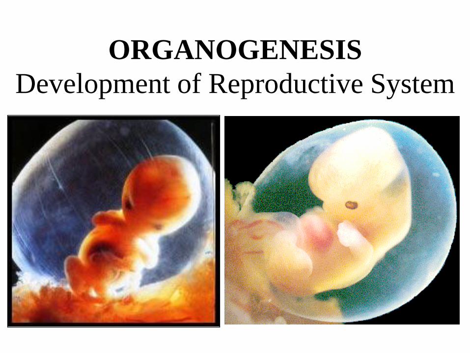
• Urinary and genital systems are closely associated.
• Both develop from inter-mediate mesoderm.
• At first a bulging appears on the abdominal wall atthe lateral aspect of dorsal mesentery of the gutwhich extends from cervical region to sacral regionof the embryo.
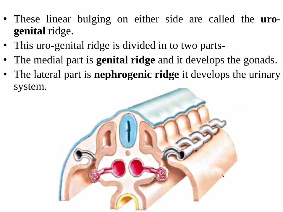
• These linear bulging on either side are called the uro-genital ridge.
• This uro-genital ridge is divided in to two parts-
• The medial part is genital ridge and it develops the gonads.
• The lateral part is nephrogenic ridge it develops the urinarysystem.
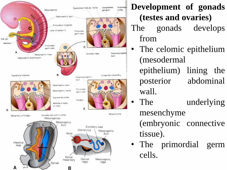
Development of gonads
(testes and ovaries)
The gonads develops
from
• The celomic epithelium
(mesodermal
epithelium) lining the
posterior abdominal
wall.
• The underlying
mesenchyme
(embryonic connective
tissue).
• The primordial germ
cells.
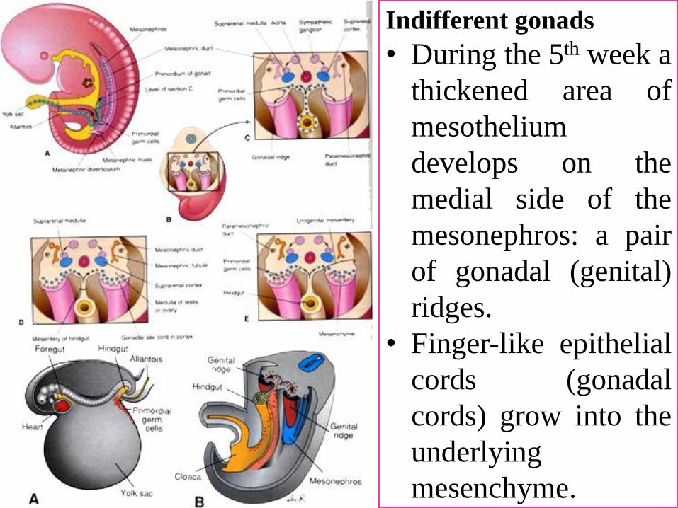
Indifferent gonads
• During the 5th week a
thickened area of
mesothelium
develops on the
medial side of the
mesonephros: a pair
of gonadal (genital)
ridges.
• Finger-like epithelial
cords (gonadal
cords) grow into the
underlying
mesenchyme.
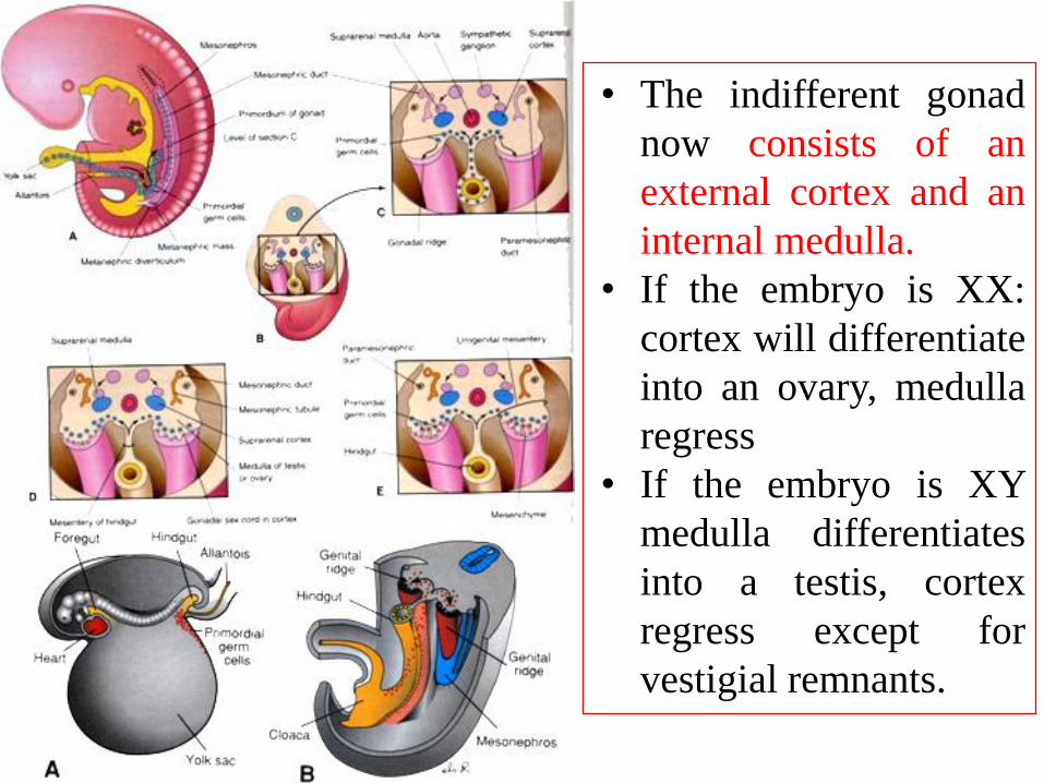
• The indifferent gonad
now consists of an
external cortex and an
internal medulla.
• If the embryo is XX:
cortex will differentiate
into an ovary, medulla
regress
• If the embryo is XY
medulla differentiates
into a testis, cortex
regress except for
vestigial remnants.
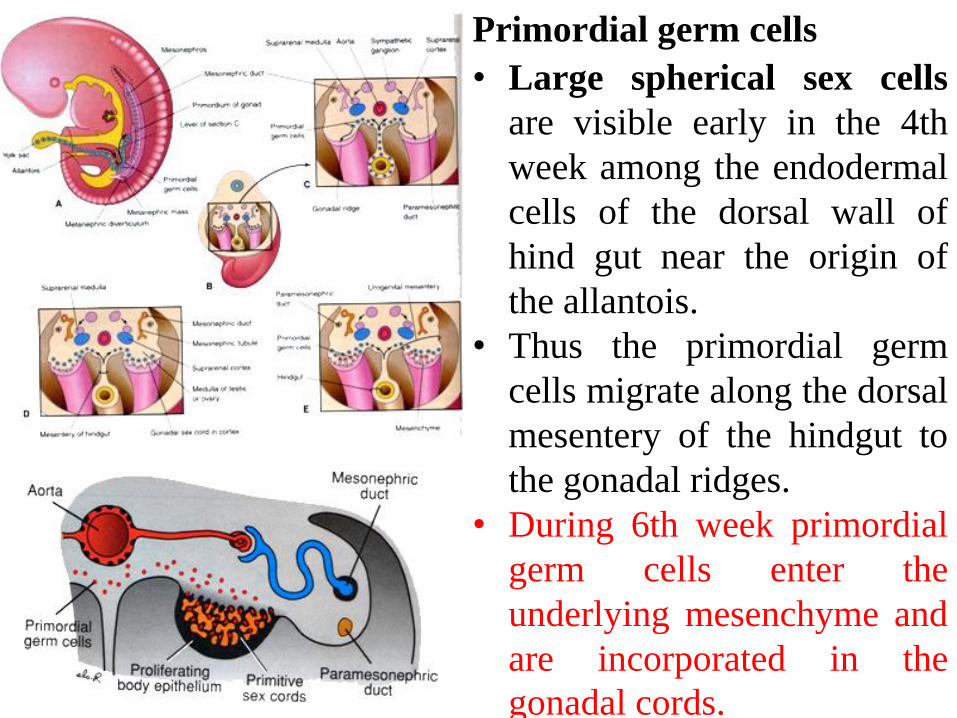
Primordial germ cells
• Large spherical sex cells
are visible early in the 4th
week among the endodermal
cells of the dorsal wall of
hind gut near the origin of
the allantois.
• Thus the primordial germ
cells migrate along the dorsal
mesentery of the hindgut to
the gonadal ridges.
• During 6th week primordial
germ cells enter the
underlying mesenchyme and
are incorporated in the
gonadal cords.
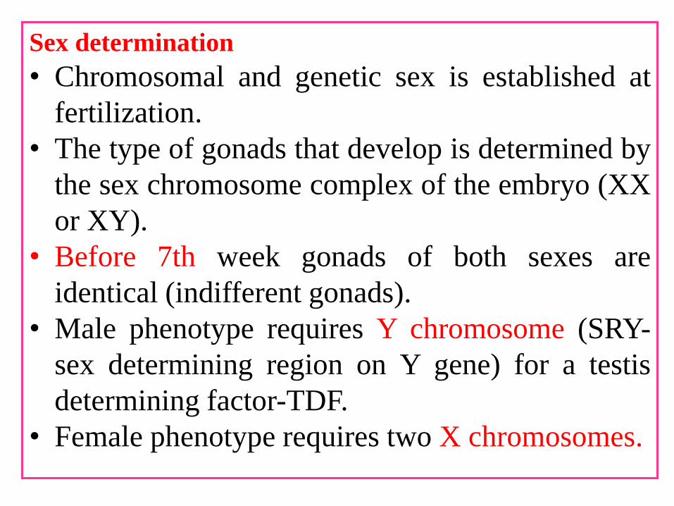
Sex determination
• Chromosomal and genetic sex is established at
fertilization.
• The type of gonads that develop is determined by
the sex chromosome complex of the embryo (XX
or XY).
• Before 7th week gonads of both sexes are
identical (indifferent gonads).
• Male phenotype requires Y chromosome (SRY-
sex determining region on Y gene) for a testis
determining factor-TDF.
• Female phenotype requires two X chromosomes.
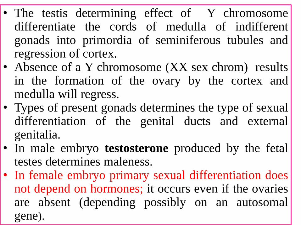
• The testis determining effect of Y chromosomedifferentiate the cords of medulla of indifferentgonads into primordia of seminiferous tubules andregression of cortex.
• Absence of a Y chromosome (XX sex chrom) resultsin the formation of the ovary by the cortex andmedulla will regress.
• Types of present gonads determines the type of sexualdifferentiation of the genital ducts and externalgenitalia.
• In male embryo testosterone produced by the fetaltestes determines maleness.
• In female embryo primary sexual differentiation doesnot depend on hormones; it occurs even if the ovariesare absent (depending possibly on an autosomalgene).
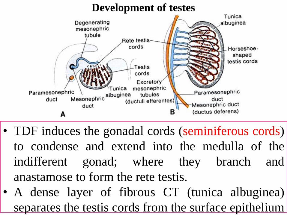
Development of testes
• TDF induces the gonadal cords (seminiferous cords)
to condense and extend into the medulla of the
indifferent gonad; where they branch and
anastamose to form the rete testis.
• A dense layer of fibrous CT (tunica albuginea)
separates the testis cords from the surface epithelium
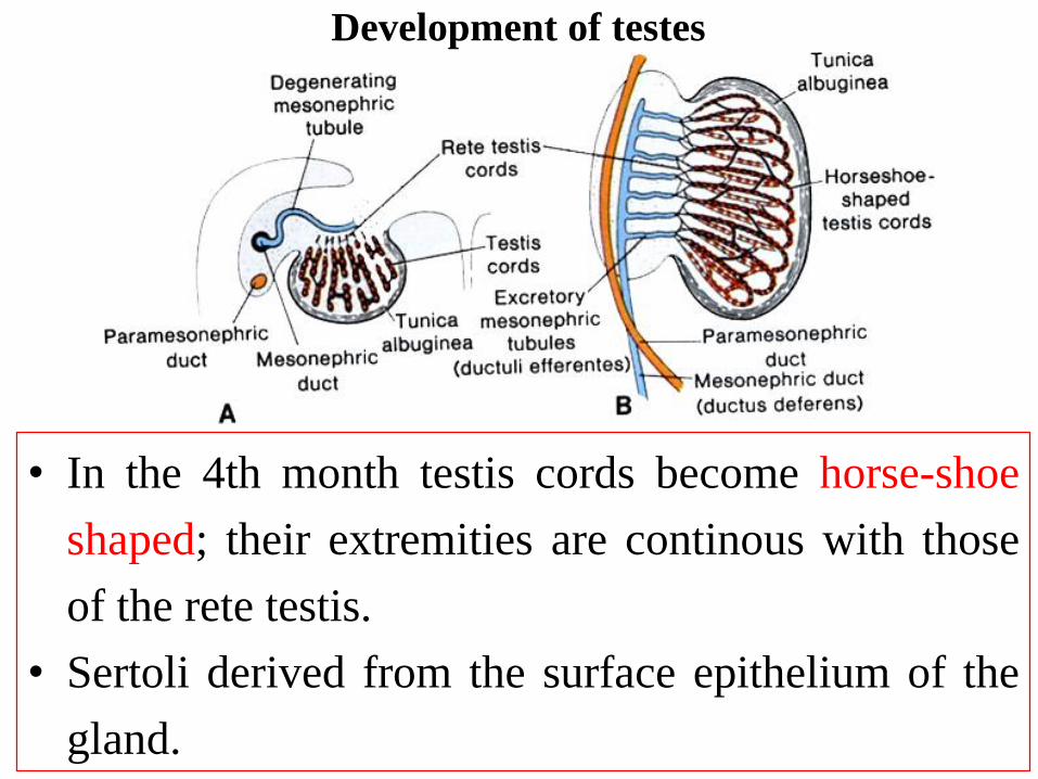
• In the 4th month testis cords become horse-shoe
shaped; their extremities are continous with those
of the rete testis.
• Sertoli derived from the surface epithelium of the
gland.
Development of testes
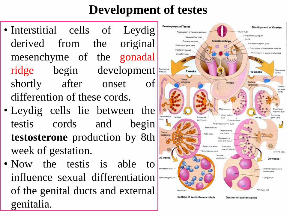
• Interstitial cells of Leydig
derived from the original
mesenchyme of the gonadal
ridge begin development
shortly after onset of
differention of these cords.
• Leydig cells lie between the
testis cords and begin
testosterone production by 8th
week of gestation.
• Now the testis is able to
influence sexual differentiation
of the genital ducts and external
genitalia.
Development of testes
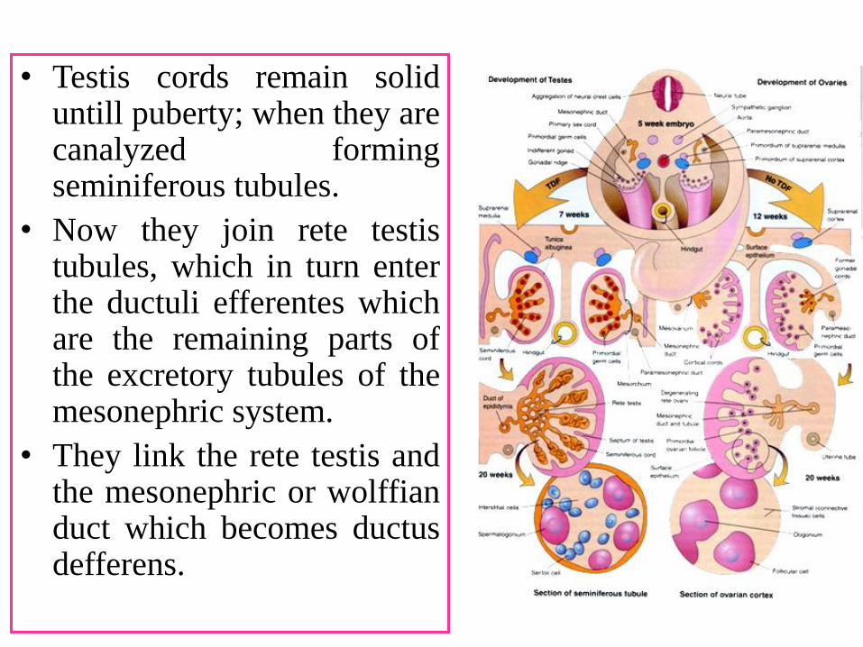
• Testis cords remain soliduntill puberty; when they arecanalyzed formingseminiferous tubules.
• Now they join rete testistubules, which in turn enterthe ductuli efferentes whichare the remaining parts ofthe excretory tubules of themesonephric system.
• They link the rete testis andthe mesonephric or wolffianduct which becomes ductusdefferens.
• In female gonadal development occurs
slowly.
• In XX embryo primitive sex cords
dissociate into irregular cell clusters
containing groups of primitive germ cells
in the medullary part of ovary.
• Later they disappear and are replaced by a
vascular stroma thet forms the ovarian
medulla.
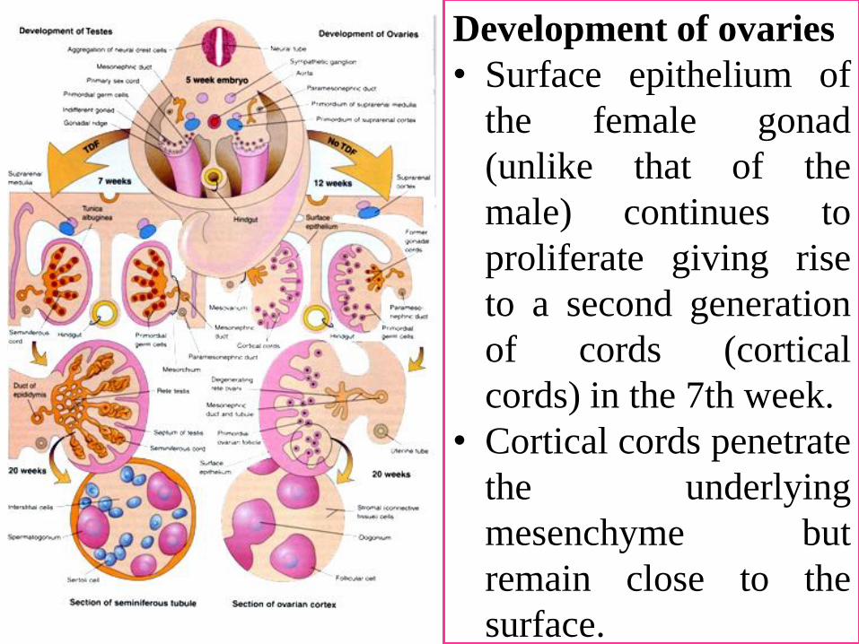
Development of ovaries
• Surface epithelium of
the female gonad
(unlike that of the
male) continues to
proliferate giving rise
to a second generation
of cords (cortical
cords) in the 7th week.
• Cortical cords penetrate
the underlying
mesenchyme but
remain close to the
surface.
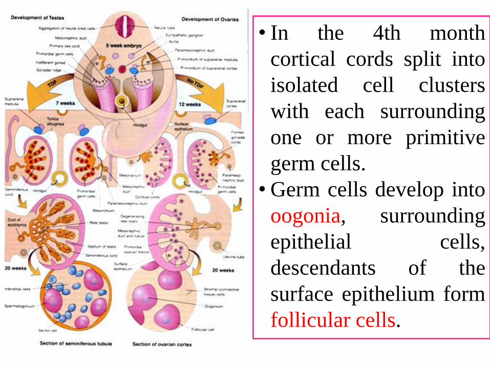
• In the 4th month
cortical cords split into
isolated cell clusters
with each surrounding
one or more primitive
germ cells.
• Germ cells develop into
oogonia, surrounding
epithelial cells,
descendants of the
surface epithelium form
follicular cells.
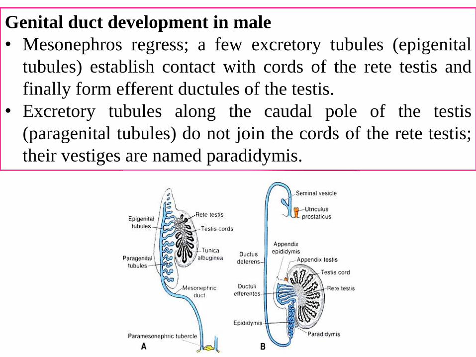
Genital duct development in male
• Mesonephros regress; a few excretory tubules (epigenital
tubules) establish contact with cords of the rete testis and
finally form efferent ductules of the testis.
• Excretory tubules along the caudal pole of the testis
(paragenital tubules) do not join the cords of the rete testis;
their vestiges are named paradidymis.
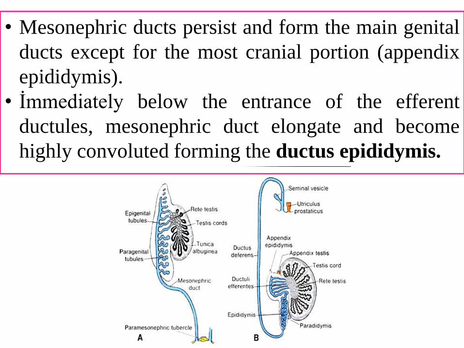
• Mesonephric ducts persist and form the main genital
ducts except for the most cranial portion (appendix
epididymis).
• İmmediately below the entrance of the efferent
ductules, mesonephric duct elongate and become
highly convoluted forming the ductus epididymis.
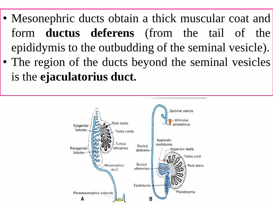
• Mesonephric ducts obtain a thick muscular coat and
form ductus deferens (from the tail of the
epididymis to the outbudding of the seminal vesicle).
• The region of the ducts beyond the seminal vesicles
is the ejaculatorius duct.
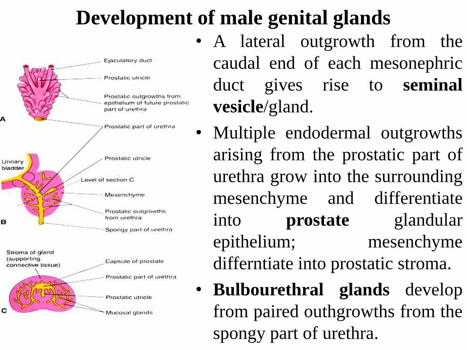
Development of male genital glands• A lateral outgrowth from the
caudal end of each mesonephric
duct gives rise to seminal
vesicle/gland.
• Multiple endodermal outgrowths
arising from the prostatic part of
urethra grow into the surrounding
mesenchyme and differentiate
into prostate glandular
epithelium; mesenchyme
differntiate into prostatic stroma.
• Bulbourethral glands develop
from paired outhgrowths from the
spongy part of urethra.
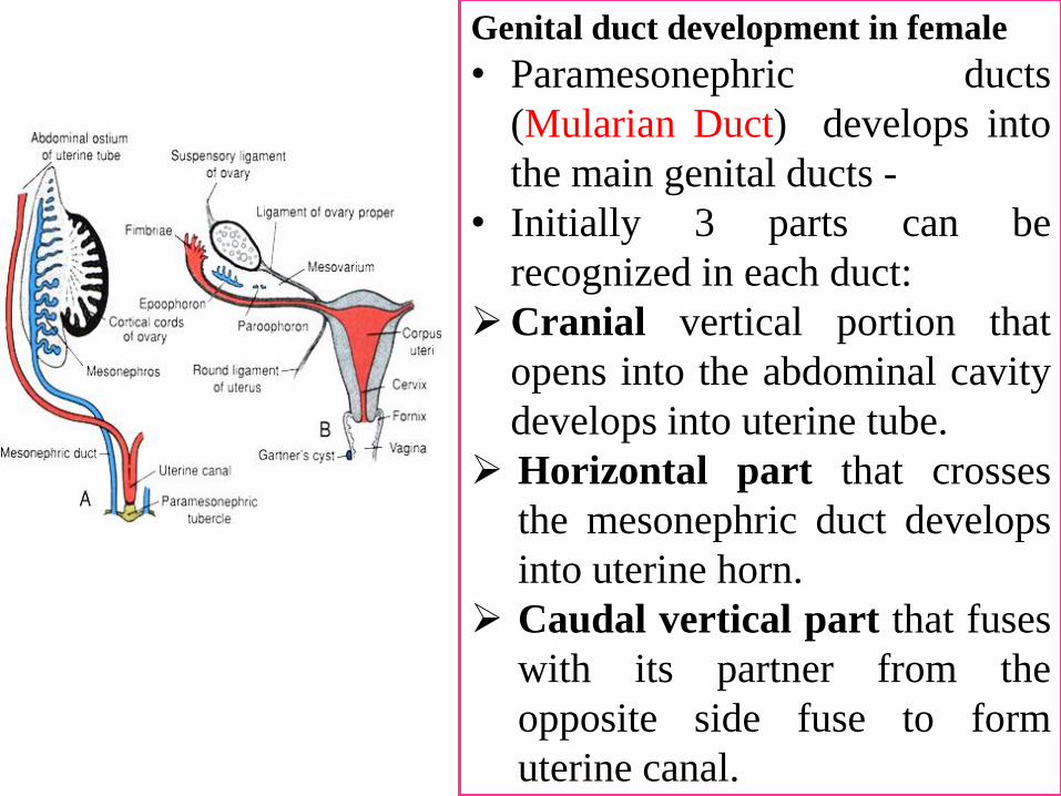
Genital duct development in female
• Paramesonephric ducts
(Mularian Duct) develops into
the main genital ducts -
• Initially 3 parts can be
recognized in each duct:
Cranial vertical portion that
opens into the abdominal cavity
develops into uterine tube.
Horizontal part that crosses
the mesonephric duct develops
into uterine horn.
Caudal vertical part that fuses
with its partner from the
opposite side fuse to form
uterine canal.
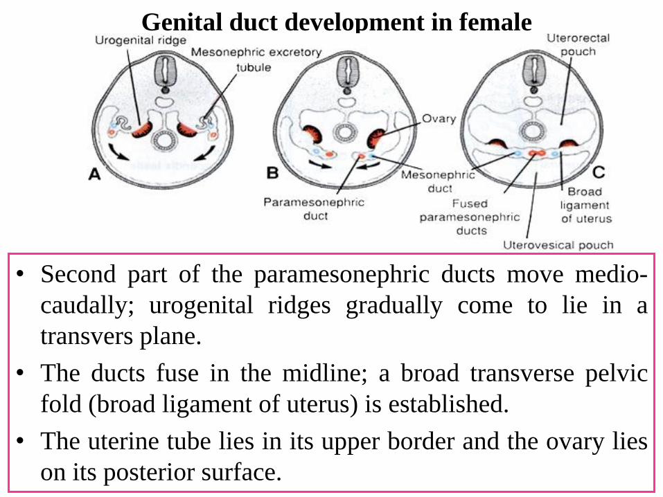
Genital duct development in female
• Second part of the paramesonephric ducts move medio-
caudally; urogenital ridges gradually come to lie in a
transvers plane.
• The ducts fuse in the midline; a broad transverse pelvic
fold (broad ligament of uterus) is established.
• The uterine tube lies in its upper border and the ovary lies
on its posterior surface.
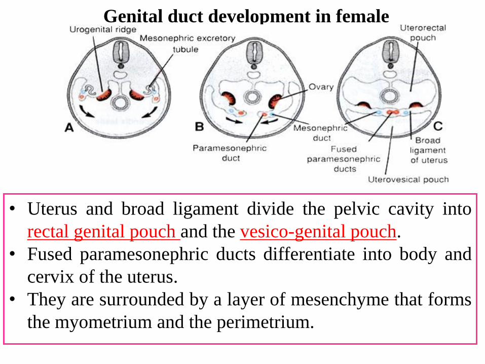
• Uterus and broad ligament divide the pelvic cavity into
rectal genital pouch and the vesico-genital pouch.
• Fused paramesonephric ducts differentiate into body and
cervix of the uterus.
• They are surrounded by a layer of mesenchyme that forms
the myometrium and the perimetrium.
Genital duct development in female
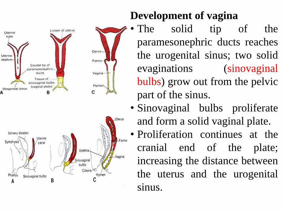
Development of vagina
• The solid tip of the
paramesonephric ducts reaches
the urogenital sinus; two solid
evaginations (sinovaginal
bulbs) grow out from the pelvic
part of the sinus.
• Sinovaginal bulbs proliferate
and form a solid vaginal plate.
• Proliferation continues at the
cranial end of the plate;
increasing the distance between
the uterus and the urogenital
sinus.
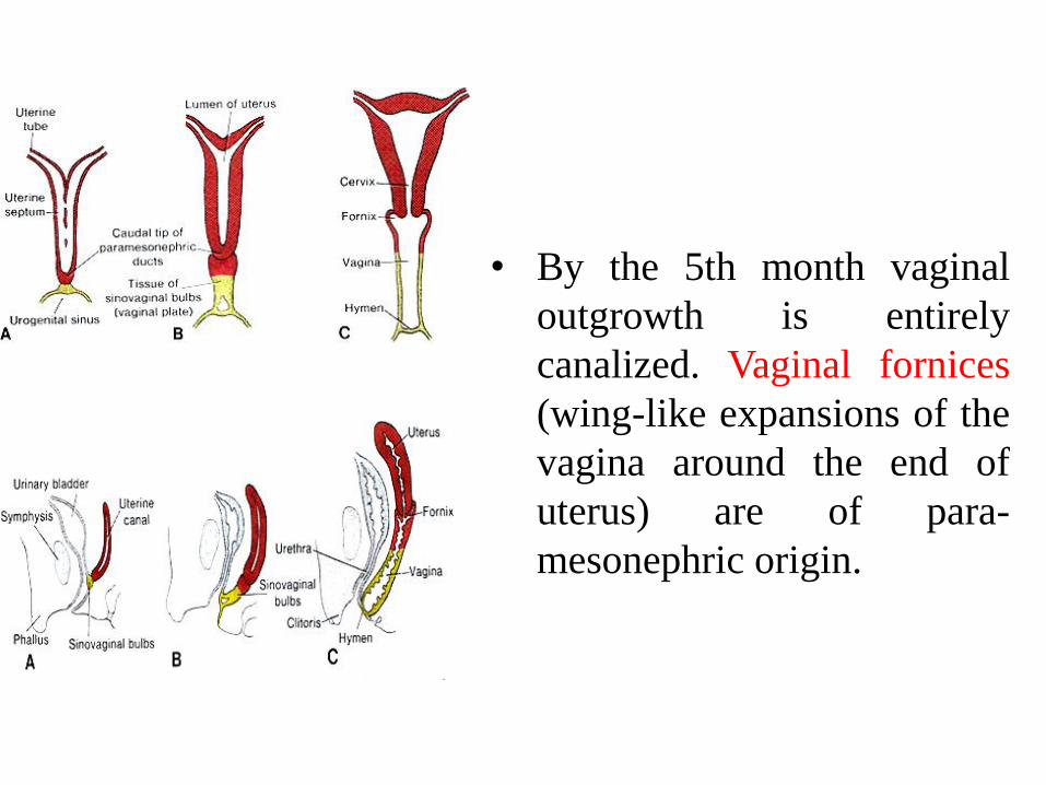
• By the 5th month vaginal
outgrowth is entirely
canalized. Vaginal fornices
(wing-like expansions of the
vagina around the end of
uterus) are of para-
mesonephric origin.
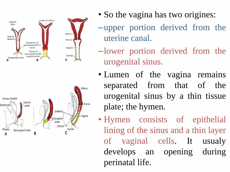
• So the vagina has two origines:
–upper portion derived from the
uterine canal.
–lower portion derived from the
urogenital sinus.
• Lumen of the vagina remains
separated from that of the
urogenital sinus by a thin tissue
plate; the hymen.
• Hymen consists of epithelial
lining of the sinus and a thin layer
of vaginal cells. It usualy
develops an opening during
perinatal life.
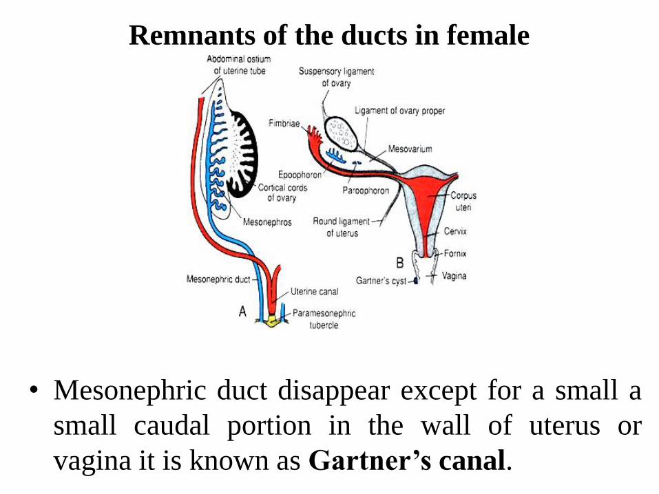
Remnants of the ducts in female
• Mesonephric duct disappear except for a small a
small caudal portion in the wall of uterus or
vagina it is known as Gartner’s canal.