Embed Size (px)
Citation preview
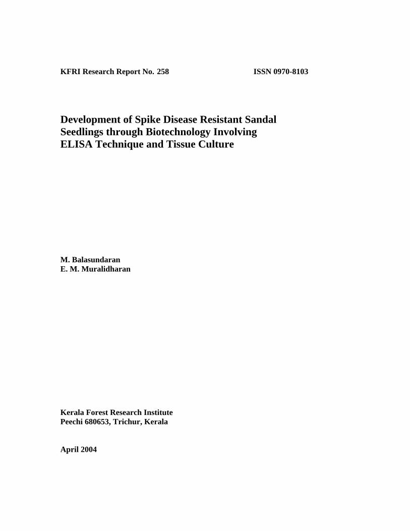
KFRI Research Report No. 258 ISSN 0970-8103 Development of Spike Disease Resistant Sandal Seedlings through Biotechnology Involving ELISA Technique and Tissue Culture
M. Balasundaran E. M. Muralidharan
Kerala Forest Research Institute
Peechi 680653, Trichur, Kerala April 2004
KFRI
KFRI Research Report No. 258 ISSN 0970-8103 Development of Spike Disease Resistant Sandal Seedlings through Biotechnology Involving ELISA Technique and Tissue Culture M. Balasundaran E. M. Muralidharan
Kerala Forest Research Institute Peechi 680653, Trichur, Kerala April 2004

KFRI Research Report No. 258 (Final Report of the Project KFRI 247/96)
Development of Spike Disease Resistant Sandal Seedlings through Biotechnology Involving ELISA Technique and Tissue Culture M. Balasundaran E. M. Muralidharan Biotechnology Division of Sustainable Natural and Plantation Forest Management Kerala Forest Research Institute Peechi 680653, Trichur, Kerala April 2004

CONTENTS
Abstract of the project proposal i Acknowledgements ii Abstract iii
1. Development of ELISA Technique for Detection of Sandal Spike Phytoplasma 1 2. Commercial Exploitation of ELISA Technique for Detection of Sandal Spike Phytoplasma 36
3. Identification of ‘Disease Resistant’ Trees using ELISA Technique 39
4. Micropropagation of Disease-Evaded Genotypes 43
5. Conclusion 52
6. Research Results of Practical/Field Application 54
7. References 55

i
Abstract of the Project Proposal
1. Project No. : KFRI 247/96
2. Title : Development of Spike Disease Resistant
Sandal Seedlings through Biotechnology Involving ELISA Technique and Tissue Culture
3. Principal investigator : Dr. M. Balasundaran 4. Associate : Dr. E.M. Muralidharan 5. Research Fellows : T. B. Suma
Sunil Thomas 6. Objectives :
i) Development of ELISA technique for detection of Mycoplasma-like-organisms in tissues/sap of spike diseased sandal, host plants showing phyllody symptoms and in insect vectors.
ii) Identification of disease resistant trees using ELISA technique.
iii) Micro-propagation of disease resistant superior genotypes
iv) Development and standardization of technology for production of
ELISA kits for detection of spike disease. 7. Date of commencement : December 1995 8. Duration : 3 Years 9. Funding Agency : Department of Biotechnology

ii
ACKNOWLEDGEMENTS We are grateful to Dr. K. S. S. Nair, former Director and Dr. J. K. Sharma, Director for
their keen interest and support during the project work. We are thankful to Department of
Biotechnology, Government of India for funding the research project. We acknowledge
the permission given by the Kerala Forest Department for doing the fieldwork in Marayur
Forest Range. The kind help provided by the Divisional Forest Officer, Munnar and
Forest Range Officer, Marayur are duly acknowledged.
Dr. T.B. Suma and Dr. Sunil Thomas were associated with this project as Research
Fellows throughout the project period. We are thankful to them for carrying out
experiments sincerely and meticulously in forest area and laboratories.
We record our gratitude to Dr. P. Nandakumar, Associate Professor, Veterinary College,
Mannuthy for helping us in antibody production in rabbits. The services of
Dr. Sreekumar, Veterinary College, Mannuthy and staff of the Department of Electron
Microscopy, All India Institute of Medical Sciences are duly acknowledged.
We are also thankful to Dr. R. Gnanaharan, Dr. Jose Kallarackal, Dr. C. Mohanan and
Dr. E. P. Indira for suggestions to improve the manuscript.
M. Balasundaran
E. M. Muralidharan

iii
Abstract
Sandal (Santalum album L.), the root hemi-parasitic tree is famous for its highly
valuable heartwood and oil. Spike disease is the most serious disease of the species;
infected trees die within one to two years after the appearance of disease symptoms.
The disease is caused by a non-culturable phytoplasma, seen exclusively in the
phloem tissues. The present study was undertaken to develop immunological
techniques for the detection of sandal spike phytoplasma, identify disease resistant
trees among disease-evaded trees and multiply the disease evaded trees through tissue
culture via somatic embryogenesis.
Stem and leaf tissues from diseased sandal and its host plants were screened for the
presence of phytoplasma in situ using fluorescence microscopy employing the
fluorochrome, 4’-6- diamidino-2-phenyl indole (DAPI). The fluorescent spots were
detected exclusively in the phloem of diseased sandal tissues and not in healthy
tissues. No fluorescent spots were seen in the phloem of the host plants of spike
disease affected sandal. Ultrastructural studies using scanning electron microscopy
confirmed the pleomorphic nature of sandal spike phytoplasma.
Phytoplasma from diseased sandal tissues was purified using a differential filtration
technique. The technique takes advantage of the property of phytoplasma to pass
through 0.45 μm pore size membrane filters. The purity of phytoplasma pellet
obtained after ultracentrifugation was confirmed using transmission and scanning
electron microscopy.
The purified phytoplasma was injected to rabbits to raise polyclonal antibody.
Precipitin bands were seen against the extract of diseased sandal only and not against
healthy sandal when the polyclonal antibody was subjected to Ouchterlony double
diffusion test.
Direct and indirect enzyme linked immunosorbent assay (ELISA) was standardised to
detect phytoplasma in diseased sandal. Indirect ELISA utilised both HRP conjugated

iv
anti-rabbit antibodies as well as avidin and streptavidin amplification systems.
Indirect ELISA was found to be very sensitive for detecting the pathogen compared to
direct ELISA. For indirect ELISA, one hour was found to be optimum for both
antigen coating and incubation of antibody. Antibodies at a dilution of 1:2000 could
be used as a probe in indirect ELISA and could detect a minimum of 25 ng of
phytoplasma protein. The techniques could not detect phytoplasma in the host plants
of spike disease affected sandal and in witches’ broom affected Zizyphus oenoplia.
The techniques confirmed that sandal spike phytoplasma is specific to sandal and is
not transmitted through the host plants. Sandal spike phytoplasma was not detected
using ELISA tests in salivary gland and intestine of the insect vector, Redarator
bimaculatus, collected from diseased trees.
The reagents developed for detecting sandal spike phytoplasma have the potential to
be commercially marketed as kits which may be beneficial to the sandalwood
industry. The kits, viz., Sandal Spike Phytoplasma Purification Kit and Immuno
Detection Kit could be used for purifying sandal spike phytoplasma and detection of
the pathogen in sandal.
In sandal forest Reserve 51 of Marayoor Range, 15 mature trees which evaded the
disease for more than 20 years were identified in highly diseased area. Six-monthly
monitoring of infection in these trees for four years using indirect ELISA tests
showed infection in three trees, six months prior to external expression of disease
symptoms, confirming the potential of early detection of disease through ELISA tests.
Tissue culture experiments using internodal explants from disease-evaded trees
through somatic embryogenesis produced plantlets. But the conversion rate of somatic
embryos to plantlets was very low and many of such plantlets were abnormal. When
the hardened plantlets were out-planted in sterile potting medium, the percentage of
survival was extremely poor. Co-culturing in vitro of sandal with Cajanus cajan as
host plant and subsequent out-planting did not improve the survival of tissue culture-
raised plantlets.

1
1. Development of ELISA Technique for Detection of Sandal Spike Phytoplasma
1.1. Introduction
Sandal (Santalum album L.) (Family: Santalaceae), the xylem tapping root hemi-parasitic
tree is the source of the aromatic East Indian sandalwood and oil. Sandalwood oil,
formed in the heartwood of the tree has a characteristic pleasant, woody odour. The oil is
widely employed in the perfume industry, particularly in high-priced perfumes. Both the
wood and oil are used in incense and medicine; besides, the wood is used in carving
(Srinivasan et al., 1992; Coppen, 1995). Sandal is considered as a royal tree and has been
rated as the most precious and valuable tree (Fig.1).
1.1.1. Distribution of Santalum sp.
The genus Santalum consists of 25 species, distributed between India in the West to Juan
Fernandez Islands in the East and from Hawaiian Archipelago in the North to New
Zealand in the South (George, 1984; Srinivasan et al., 1992). They vary greatly in habit,
from small shrubs to large trees (Radomiljac, 1994). The commercially valuable
sandalwood, Santalum album L. occurs naturally in Southern India and in the islands of
eastern Indonesia, notably Timor and both the countries are the major producers and
exporters of East Indian sandalwood and oil (Fox et al., 1994; Coppen, 1995).
In India, sandal is found mainly in the Deccan Plateau and its extension, and in small
numbers in almost all regions, except the Himalayas. Large natural stands of sandal
occurs in Karnataka (5,245 km2) and Tamil Nadu (3,040 km2) accounting for nearly 90%
of sandal in India (Venkatesan, 1981). Sandal forests in Kerala are chiefly distributed in
the Anjanad Valley in the eastern side of Western Ghats falling in Marayoor forest range
of Munnar forest division with an extent of 15.42 km2 in reserved forests and 47.26 km2
in revenue lands (Mathew, 1995). In India, production of sandalwood has plummeted

2
from around 3000 tones per annum during 1985 to around 1000 tones in 1997; similarly
oil production also declined from 140 tones in 1985 to 40 tones in 1997 (Jain et al.,
1999). The sandal tree grows at altitudes from sea level to about 1200 m above mean sea
level. It grows to a height of 10-15 m and a girth of 100 cm and attains full maturity at an
age of 60-80 years (Ghosh et al., 1985; Jain et al. 1999). India has a monopoly in the
world sandalwood market. Most of the existing sandal populations are not dense and are
devoid of larger-girth-class trees, due to illegal felling, encroachment of sandal forests
and due to spike disease.
1.1. 2. Diseases of sandal
Diseases of sandal include seedling diseases and leaf spot diseases caused by different
fungal species, leaf curl disease caused by virus, and the spike disease (Srinivasan et al.,
1992). Collar rot caused by Fusarium spp. is a serious disease affecting seedlings.
1.1.2.1. Spike disease
Spike disease, the most serious disease of S. album is characterised by extreme reduction
in the size of leaves and internodes accompanied by stiffening of the leaves. In advanced
stage, owing to the progressive reduction in leaf size and internodes, the whole shoot
looks like a `spike inflorescence’ (Fig.2).
Spiked plants do not bear flowers or fruits; occasionally phyllody or abortive flowers are
developed. Spiked trees usually die within 1 – 2 years after the appearance of the
symptoms. Although, in Kerala, the production of sandalwood has not declined markedly
because of the extraction of dead trees (dead trees increased in number as a result of
spike disease), the stock in the forest is depleted considerably (Ghosh et al., 1992). In
Karnataka, the growing stock has been reduced to 25 per cent of its initial level in the last
two decades (Swaminathan et al., 1998). The disease is not known in Timor and does
not affect the other species of Santalum (Fox et al., 1994).

3
Although, spike disease was first observed in Coorg by McCarthy in 1899 (McCarthy,
1899; Barber, 1903) subsequent investigations showed that the disease had made its
appearance in Coorg several years before McCarthy noticed it. The disease was observed
in North Coimbatore in 1903, in Salem in 1913, and in Tirupathur Javadis in 1917
(Srinivasan et al., 1992). In Kerala, the disease was first noticed at Marayoor in 1980
(Ghosh et al., 1985).
1.1.2.2. The pathogen
Sandal spike disease was thought to be caused by a virus until 1969, when three
independent groups confirmed through electron microscopic studies that the disease was
caused by a phytoplasma (Dijkstra and Ie, 1969; Hull et al., 1969; Verma et al., 1969).
Phytoplasmas were first reported by Japanese workers in 1967 (Doi et al., 1967). These
pathogens are seen exclusively in the sieve tubes of phloem tissues of leaves, petioles,
stem (Fig.3) and root, causing symptoms such as yellowing of leaf, little leaf, phyllody,
witches’ broom, etc. Phytoplasmas have been implicated as pathogens in more than
300 plant diseases worldwide (McCoy et al., 1989).
Morphologically, phytoplasmas resemble animal or human mycoplasmas (Class:
Mollicutes) and share several characteristics with mycoplasmas. These include
unicellular and pleomorphic nature and absence of cell wall, the cells being delimited
only by a membrane, passage through bacteriological filters and resistance to antibiotics
that interfere with cell wall formation (Neimark and Kirckpatrick, 1993). Phytoplasmas
have remained uncultured despite extensive efforts over many years. The inability to
grow these agents in vitro has severely hindered their study. As a result, phytoplasmas
are among the most poorly characterised groups of plant pathogens. The pathogen could
be visualized by electron microscopy, and their presence in phloem tissues demonstrated
by fluorochromic DNA stains, but these methods cannot discriminate among different
groups of phytoplasmas (Clark et al., 1989; Neimark and Kirckpatrick, 1993).

4
1.1.3. Diagnosis of phytoplasma diseases
Accurate diagnosis is a necessary prelude to any successful disease control. However
diagnosis of plant mollicute diseases has often been one of the difficult aspects in the
study of these diseases. It has long been known that most plant pathogens possess, as
part of their structure, specific antigenic determinant in the form of proteins or other
antigenic moieties. Recognition of the diagnostic potential of such determinants for both
experimental and applied investigations in plant pathology has resulted in an array of
techniques, collectively referred to as immunoassays. However, for this technique,
extremely pure preparations of the organisms are required.
Initial efforts of a few laboratories to purify phytoplasma for the production of polyclonal
antisera (Sinha, 1979; Caudwell et al., 1982; Sinha and Chiykowski, 1984) were
discouraging due to high contamination with plant proteins which resulted in inferior
quality and non-specific antisera. Hobbs et al. (1987) used antibody raised against
healthy plant to remove plant specific proteins from the semi-pure phytoplasma pellet by
cross absorption. Since then, several modifications of the protocol have been used in the
purification of different phytoplasmas (Jiang and Chen, 1987; Jiang et al., 1988; Clark et
al., 1989). All these techniques use several centrifugation steps (differential
centrifugation) for the purification of phytoplasma which invariably leads to the loss of
phytoplasma cells during successive centrifugation.
1.1.4. Diagnosis of spike disease of sandal
Spike disease of sandal is generally diagnosed by the manifestation of external
symptoms. Attempts have been made to detect the diseased plants by determining the
length/breadth ratio of leaves (Iyengar, 1961), histochemical tests using Mann’s stain
(Parthasarathi et al., 1966), Dienes’ stain (Ananthapadmanabha et al., 1973) aniline blue
and Hoechst 33258 (Ghosh et al., 1985, Rangaswami, 1995). But most of these

5
techniques are insensitive indirect detection methods leading to misinterpretation of
results. Highly sensitive techniques are needed to detect the presence of the pathogen.
This chapter reports detection of sandal spike phytoplasma through immunological
methods such as double immunodiffusion test and direct and indirect enzyme linked
immunosorbent assay (ELISA).
1.2. Review of literature
1.2.1. Sandal spike disease
Sandal spike is one of the most serious yellows-type diseases of forest trees known in the
world. The disease has spread progressively over the years, devastating large forest tracts
and threatening the entire sandal industry in India (Raychaudhuri and Varma, 1980).
According to Subba Rao (1980) the percentage incidence of spike disease ranged from 1
to 55 in Karnataka.
1.2.2. Etiology Initially the spike disease was suspected to be a root disease and a physiological disorder
caused by unbalanced sap circulation brought about by adverse factors such as forest fires
(Hole, 1917). Latham (1918) was of the opinion that the disease was due to a fungus,
whereas Fischer (1918) thought that the disease was caused by some ultramicroscopic
bacteria. Coleman (1917, 1923) attributed the disease to a virus.
The association of phytoplasmas with yellows diseases by Japanese scientists (Doi et al.,
1967) gave impetus to a reconsideration of the causative organisms of plant diseases of

6
unknown and unconfirmed etiology. The viral theory of sandal spike disease was
disproved when three groups of workers showed phytoplasma in the phloem tissues of
spike diseased plants through transmission electron microscopy studies (Dijkstra and Ie,
1969; Hull et al., 1969; Verma et al., 1969). The pleomorphic bodies with 40 to 750 nm
size were devoid of cell walls; the cytoplasm was bound by a unit membrane of 10 to 12
nm thick. The organism contained a fibrillar network of DNA and ribosomal bodies. The
remission of spike disease symptoms after the infusion of tetracycline antibiotics further
confirmed the phytoplasmal etiology of the disease (Raychaudhuri et al., 1972).
1.2.3. In vitro culture of the pathogen
Nayar and Ananthapadmanabha (1970) reported successful culturing of sandal spike
phytoplasma in vitro using PPLO broth. They claimed to have reproduced spike
symptoms on sandal and Stachytarpheta inoculated with the culture. However
Muniyappa et al. (1980), Subba Rao (1980) and Ghosh et al. (1985) could not reproduce
the results and the pathogen still is considered as a non-culturable organism.
1.2.4. Disease detection
Spike disease is generally diagnosed by the manifestation of external symptoms.
Diseased plants can be detected by light and fluorescent microscopic techniques. The
detection of abnormal levels of wound callose produced in response to injury to phloem
cells had been suggested as an indirect method of diagnosis of phytoplasma.
Ananthapadmanabha et al. (1973) employed Giemsa and Dienes’ stain for the detection
of the pathogen. In spike diseased sandal, aniline blue stained sections showed large
number of fluorescent spots throughout the phloem tissue; a DNA binding fluorochrome,
Hoechst 33258, has been used to detect the pathogen (Ghosh et al., 1985; Rangaswamy,
1995). Ghosh et al. (1985) used shigometer to detect spike diseased sandal. The
electrical resistance of the inner bark of diseased trees was correlated with the intensity of
visual symptoms.

7
1.2.4.1. Immunological techniques
According to Brock and Madigan (1988) antigens are substances that can be bound
specifically by antibodies of the immune system of vertebrates. Injected into animals in
the appropriate manner, antigens elicit an immune response, thus initiating the synthesis
of specific antibodies. Antibodies are directed towards restricted parts of a macro-
molecule, namely the antigenic determinants or epitopes (Roitt et al., 1993). Antibodies
are found in blood serum. They are referred to as immunoglobulin molecules (Ig) and can
be divided into five classes. Every warm blooded animal is capable of producing
antibodies. Rabbits (Saeed et al., 1992a, 1993) and mice (Jiang and Chen, 1987; Jiang et
al., 1988) are generally used to raise antiserum against phytoplasma. Two types of
antibodies can be distinguished - polyclonal and monoclonal antibodies. Polyclonal
antibodies are purified from the raw serum fraction of the blood of an immunised animal.
Heterogeneity, is the outstanding feature of polyclonal antisera. Whereas homogeneity is
the important feature of monoclonal antibodies since they are produced from a single
antibody producing B-lymphocyte and multiplied as clones.
Immunological methods are among the simplest to use and interpret and are most
valuable for diagnosis for diseases with inconsistent and undeveloped symptoms (Fox,
1998). Particularly, since the report of Engvall and Perlmann (1971) on the use of
antibody-enzyme conjugate, the technique has become foundational to some of the most
sensitive immunoassays in use today, including enzyme linked immunosorbent assay
(ELISA), dot immunobinding assay (DIBA), immuno microscopy and immunoblotting
(Western blotting). Immunoassays are used in plant pathology for diagnosis of disease,
and identification and quantitation of microorganisms (Barbara and Clark, 1986).
Immunological methods of detection of plant mollicutes have been used depending on the
availability of specific antiserum. Various laboratories have reported the production of
polyclonal antibodies to selected phytoplasma derived from plant tissue extracts (Sinha,
1979; Hobbs et al., 1987; Clark et al., 1989; Saeed et al., 1993). Such polyclonal
antibodies (Pab) are capable of distinguishing the phytoplasma affected diseased plants

8
from healthy ones. But some of the antisera obtained were highly contaminated with anti-
plant antibodies and were of poor quality. The use of monoclonal antibodies (Mab) (Lin
and Chen, 1986) circumvents many of the problems encountered with polyclonal
antibodies, but the production of suitable Mabs require specialised laboratory,
considerable time, effort and a degree of luck (Clark et al., 1989). Nayar and
Ananthapadmanabha (1975) purified sandal spike phytoplasma by ammonium sulphate
precipitation method and used the same to raise polyclonal antibodies in rabbit. The
polyclonal antibody was used in gel diffusion and agglutination tests.
1.2.4.2. Disease transmission
The transmission of sandal spike disease in the field was suspected to be caused by
several insect vectors such as Moonia albimaculata (Dover and Appanna, 1933), Jassus
indicus (Rangaswami and Griffith, 1941) and Nephotettix virescens (Shivaramakrishnan
and Sen-Sarma, 1978). But subsequent studies could not confirm the findings (Lasrado,
1955; Subba Rao, 1980; Muniyappa et al., 1980). Ghosh et al., (1985) reported
Redarator bimaculatus as the insect vector. Most of the earlier workers suspected host
plants as the agents transmitting the spike disease. Nayar and Srimathi (1968) were of the
opinion that Lantana acted as a symptomless carrier. It was also felt that sandal, in
association with certain hosts was more susceptible to disease than others. Studies
conducted by Subba Rao (1980) proved that phytoplasma could not be transmitted from
diseased sandal to healthy sandal through haustorial connection. Laboratory transmission
of the disease is achieved mainly through grafting and dodder. Coleman (1923) was the
first to demonstrate the graft transmissibility of the disease to healthy trees. The
establishment of the scion was found to be a prerequisite for disease transmission (Ghosh
et al., 1992).
1.2.5. Disease control
With the discovery of phytoplasma in sandal, attempts were made to control the disease
using antibiotics. Raychaudhuri et al. (1972) used dimethyl chlorotetracycline

9
hydrochloride, tetracycline hydrochloride and benlate (methyl 1-(butylcarbaucyl)-2-
bensimidazole carbamate, a systemic fungicide to treat the disease by girdling and
spraying, but appreciable recovery of diseased plants were not observed. The antibiotics
achromycin, aureomycin, and ledermycin also did not show positive response (Nayar et
al., 1973; Nayar and Ananthapadmanabha, 1974). But Ali et al. (1987) reported
temporary remission of spike disease in various degrees by injection method using five
tetracycline antibiotics - tetracycline hydrochloride, oxytetracycline HCl, ledermycin,
aureomycin and doxycycline. However, the disease reappeared within 3-7 months.
Infusion of digitonin also gave the same results.
1.3. Materials and Methods 1.3.1. Plant material
Spike disease-affected sandal, and the host plants, Lantana camara and witches’ broom-
affected Zizyphus oenoplia were collected from Marayoor, Munnar Forest Division,
Kerala. The tissues were transported either in ice or the excised branches dipped
vertically in water and covered using polythene bags. The plant materials were stored at
40C.
1.3.2. Disease transmission
To make diseased plant material available in glass house for experiments, the spike
disease-affected sandal twigs (scion), were wedge grafted to one-to two-year-old healthy
sandal seedlings grown in glass house. Pongamia glabra and Pterocarpus marsupium
were provided as the host species.
1.3.3. Disease detection
10
DAPI staining: 4’,6-diamidino-2-phenyl indole, a DNA binding fluorochrome specific
for staining phytoplasma was used to detect sandal spike phytoplasma in apparently
diseased sandal plants. Healthy and spike disease affected sandal and host plants in the
field and glass house were screened for the presence of phytoplasma using DAPI stain
(Seemuller, 1976). Tissues were fixed in 5% formaldehyde in 0.1M phosphate buffer, pH
7.0 for 30 minutes, then washed in phosphate buffer, pH 7.0, for 3 minutes. Free-hand
sections of 20 μm thickness (approx.) were stained with 0.001% DAPI (Sigma, USA) in
0.01M phosphate buffered saline, pH 7.4, for one hour, mounted in water or glycerine
and viewed under Leitz Dialux fluorescence microscope using HBO 50 W bulb.
Scanning electron microscopy: Healthy and diseased sandal stem tissues were cut into
pieces of 1 mm3 and fixed in 2.5% glutaraldehyde for 1 hour, washed thrice in the same
buffer and post fixed in 2% potassium permanganate in 0.2 M phosphate buffer, pH 7.0,
for 90 minutes at 40C. The blocks were then washed in the same buffer, critical point
dried, followed by gold coating and viewed in Leo 435 VP scanning electron microscope
(LEO, UK).
1.3.4. Purification of phytoplasma
Inner bark and stem tissues from diseased branches, collected from Marayoor were
utilized for the isolation and purification of phytoplasma. Tissues were washed in running
tap water for 10 minutes, treated with Extran (Merck, India) for 3 minutes and again
washed thoroughly with tap water. Subsequent steps were carried out at 40 C. Diced
tissues were homogenised in ice-cold 0.3 M glycine-sodium hydroxide buffer, pH 8.0
containing 0.02M magnesium chloride (Clark et al., 1989) (1g fresh weight tissue/4 ml
buffer). The extract was passed through two layers of cheese cloth, followed by filtration
through Whatman 1 and 5 filter papers (Whatman, UK). The clear extract was then
passed through 0.45 μm pore size Millipore filter (Millipore, USA) and centrifuged
(Sorvall OTD 65 B, USA) at 45,000g (r.av) for 45 minutes. The re-suspended pellet was
incubated with undiluted antiserum prepared against extract from healthy sandal for 2
hours at room temperature for cross absorption of any plant debris present in the

11
phytoplasma pellet. After low speed centrifugation at 4700g for 20 minutes the pellet was
discarded and the supernatant centrifuged at 65,000g for 45 minutes. The pale yellow
pellet was re-suspended in 1.0 ml of the same buffer. The sedimentation constant
(Payment et al., 1991) was calculated using the equation: t (hours) = K/S20w, where K is a
factor relative to the specific rotor provided by the manufacturer and S 20w, the
sedimentation constant.
Electron microscopy: For scanning electron microscopy (SEM) of the purified
phytoplasma, the centrifuged pellet was embedded in 3% agarose (Sigma, USA) in 0.2 M
phosphate buffer, pH 7.0. The agarose block was cut into pieces of 1mm3 and fixed in
2.5% glutaraldehyde for 1 hour, washed thrice in the same buffer and post fixed in 2%
potassium permanganate in 0.2 M phosphate buffer, pH 7.0 for 90 minutes at 40C. The
blocks were then washed in the same buffer and subsequently in distilled water, dried in a
desiccator followed by gold coating. Specimens were then viewed in Philips 501 B
scanning electron microscope.
For transmission electron microscopy (TEM), the technique of Jiang and Chen (1987)
was followed with slight modification. The pellet obtained after centrifugation was fixed
in 2.0% glutaraldehyde in 0.3 M mannitol-20mM MOPS buffer, pH 7.0 for 1 hour at
40C. The suspension was centrifuged at 45,000g for 30 minutes. The supernatant was
aspirated and the pellet rinsed twice with the same buffer and post fixed with 1% osmium
tetroxide at 40C for 6 hours, followed by washing in buffer. The pellet was mixed with
3% agarose and the solidified block cut into pieces of 1 mm3, and suspended in 0.5%
uranyl acetate for 12 hours at 40C. Dehydration and embedding were done as described
by Cole (1983). Ultra thin sections were stained with 2% lead citrate for 10 minutes,
washed and examined using Philips CM10 transmission electron microscope.
1.3.5. Total protein estimation
For total protein estimation, the phytoplasma was pelleted as stated above, but during the
second centrifugation step, instead of glycine buffer, 0.1M phosphate buffer, pH 8.0 was

12
added followed by centrifugation at 4700g for 20 minutes. The pellet was discarded and
the supernatant centrifuged at 65,000g for 45 minutes. The pale yellow pellet was re-
suspended in 1.0 ml of 0.1M phosphate buffer, pH 8.0. Total protein of phytoplasma cells
obtained from 6.25 g tissue in 25 ml buffer was estimated at A205 and A280 (Simonian,
1996) in 0.025 M phosphate buffer, pH 7.0 using a spectrophotometer (Unicam, U.K), by
Lowry method (Lowry et al., 1951), and silver binding method (Krystal et al., 1985).
1.3.6. Production of polyclonal antibody
For polyclonal antibody production the method of Saeed et al. (1993) was followed with
modifications. Sandal spike phytoplasma purified by the differential filtration method
was suspended in 1.0 ml of glycine buffer without magnesium chloride and sonicated
thrice (Vibracell, USA), 30 seconds each with an interval of two minutes at ice-cold
temperature. New Zealand White rabbits were injected intramuscularly in the hind legs at
two sites with an emulsion of equal volume of sonicated phytoplasma preparation and
Freund’s complete adjuvant (Sigma, USA) in the first week and Freund’s incomplete
adjuvant in the subsequent injections given at two-week interval. Blood was collected
from the ear vein after 12 weeks of the first injection.
Pellet from healthy plant extract, purified as antigen to raise antibody for cross absorption
of partially purified phytoplasma was also injected into another rabbit two months prior
to the actual purification of phytoplasma following the same method. Booster injections
were given once in a month after bleeding the rabbit to maintain high antibody titre.
1.3.7. Purification of polyclonal antibody
Serum was processed from the blood according to the method of Ball et al. (1990). IgG
was purified using protein-A affinity chromatography using an IgG purification kit
(Bangalore Genei, India) following manufacturers’ instructions.
1.3.8. Double immunodiffusion

13
The Ouchterlony method of double immunodiffusion as described by Ball (1990) was
employed for preliminary detection of the presence of the antigen in the test plants -
healthy and diseased sandal and the host plants viz., Lantana and witches’ broom-
affected Zizyphus.
1.3.9. Enzyme linked immunosorbent assay (ELISA)
Both the direct and indirect methods of ELISA techniques were used to detect the
pathogen.
1.3.10. Direct ELISA
IgG was conjugated to horseradish peroxidase (HRP) (Sigma, USA) following the
method of Mackenzie (1990). For direct ELISA, the method of Saeed et al. (1993) was
followed with modifications. Polystyrene ELISA strips (Polysorp-Nunc, Denmark) were
used to coat antigen. The ELISA strip wells were coated with purified antigen or crude
extract (100 μl) at different dilutions in PBS, pH 7.4, at 370C for one hour followed by
washing thrice with PBS buffer containing 0.025% Tween 20 (wash buffer). Each strip
had 8 wells, of which 3 were coated with purified healthy sandal pellet or extract of
healthy sandal (crude extract – without purification) and 5 with purified phytoplasma or
the extract of diseased sandal (crude extract – without purification). Coating was
followed by blocking the strips in phosphate buffered saline-tween (PBS-T) containing
0.2% BSA for 30 minutes and again washing thrice. The strips were then incubated with
diluted HRP-IgG conjugate in conjugate buffer (PBS, pH 7.4 containing 0.025% tween
and 0.20% BSA) at 370C for one hour. After washing, the substrate, O-phenylene
diamine (OPDA) (Sigma, USA) and hydrogen peroxide in citrate buffer, pH 5.0, was
added and incubated in dark at room temperature for one hour. The reaction was stopped
by the addition of 2M sulphuric acid (25 μl) and the absorbance read at 490 nm (Span

14
Autoreader, India). Values greater than the threshold value (mean of healthy plant antigen
± twice the standard deviation) were considered positive (Sutula et al., 1986).
Initial experiments were conducted to assess the optimum time for coating antigen and
incubation with polyclonal antibody. In these experiments, the ELISA strips were coated
with purified antigen (1:100 dilution) followed by incubation with conjugated antibody
(1:250 dilution). In another experiment, different dilutions of conjugated antibody (1:250
and 1:500 dilutions) was tested to detect the presence of phytoplasma in purified antigen.
In the final test, the crude extract (obtained by pooling healthy or diseased sandal from
three different trees and homogenised in glycine buffer at the ratio of 1g sample : 4 ml
buffer- without further purification) was used to detect the presence of phytoplasma
using conjugated antibody of dilutions- 1:100, 1:250 and 1:500.
1.3.11. Indirect ELISA
Indirect ELISA techniques consisted of using HRP-conjugated anti-rabbit antibody,
biotin-avidin and biotin-streptavidin systems. As in direct ELISA, polystyrene strips were
used to coat antigen in PBS, pH 7.4 for one hour at 370C. After washing thrice, the strips
were blocked using PBS-tween-BSA (0.2%) for 30 minutes, washed thrice and the strips
incubated with phytoplasma specific antibody in PBS for one hour at 370C. Subsequently,
they were incubated with either goat anti-rabbit IgG-HRP conjugate (Sigma, USA) or
biotinylated goat anti-rabbit IgG (B. Genei, India) for one hour. This was followed by
incubation in either avidin or streptavidin conjugated HRP (B. Genei, India). Finally the
HRP coated strips were treated with the substrate OPDA and hydrogen peroxide in citrate
buffer, pH 5.0, and kept in dark at room temperature for 1 hour. The reaction was stopped
by the addition of 2M sulphuric acid (25μl) and the absorbance read at 490 nm. Each
strip had 8 wells, of which 3 were coated with purified healthy sandal pellet or extract of
healthy sandal (crude extract – without purification) or its hosts and 5 with purified
phytoplasma or extract of diseased sandal (crude extract – without purification) or the
host plants. Values greater than the threshold value (mean of healthy plant antigen ±
twice the standard deviation) were considered positive (Sutula et al., 1986).

15
Initial tests were conducted to assess the optimum concentration of the specific
polyclonal antibody (1:500 to 1:10,000 dilution) required for indirect ELISA using a
constant amount of purified phytoplasma (1:1000 dilution). An experiment was
conducted to probe for different dilutions of purified antigen (1:50-1:10000 dilution)
using polyclonal antibody at dilutions 1:500, 1:1000 and 1:2000. In the test, HRP
conjugated to streptavidin (1:2000 dilution) was used as the secondary probe. The
efficiency of goat anti-rabbit antibody HRP conjugate and avidin -HRP was also tested
using the purified phytoplasma. For the final test involving detection of phytoplasma in
crude extract of sandal (obtained by pooling samples from three different trees and
homogenised in glycine buffer at the ratio of 1g sample : 4 ml buffer) polyclonal
antibody at a dilution of 1:2000 was used as the primary probe and goat anti-rabbit
antibody conjugated to HRP (1:2000 dilution) or streptavidin-HRP (1:2000 dilution) was
used as the secondary probe. For confirmation of the efficiency of the polyclonal
antibody, indirect ELISA was used to test 24 individual plant samples (crude extract –
1:1000 dilution) selected at random from Marayoor using streptavidin amplification
system (1:2000 dilution).
1.3.12. Testing host plants of spike diseased sandal and insect vector
Since indirect ELISA was found to be superior to direct ELISA, the host plants of spike
disease-affected sandal viz., Lantana and witches’ broom affected Zizyphus were used to
detect the presence of phytoplasma using the technique. For the preliminary tests three
host plants were pooled and the extract was used for the test. The final test involved
detecting the presence of pathogen in 10 individual host samples.
Redarator bimaculatus, supposed to be the insect vector of sandal spike disease (Ghosh
et al., 1985) were collected from spike diseased sandal. Twelve adult insects were
collected from diseased sandal of Reserve 54, where large number of trees were infected.
The insects were brought to the laboratory, keeping them in cages provided with
moistened diseased twigs covered with fine plastic nets. The insects were homogenized

16
in the extraction buffer, centrifuged at 5000 rpm and the supernatant diluted to 1:100 to
1:1000. The solutions were used for indirect ELISA test.
1.4. Results 1.4.1. Infected plant material
Spike disease-affected tissues transported in ice had a shelf life of only three weeks when
stored at 40C; the tissues got damaged after three weeks. Whereas, the samples which
were transported vertically by placing the base of the stem in water and thereafter stored
at 40C, remained fresh even after four weeks.
1.4.2. DAPI staining
The xylem and sclerenchymatous stone cells of both the healthy and diseased sections
showed green autofluorescence under UV light. While all the tissues of spike disease-
affected sandal showed the characteristic yellow-green fluorescent spots (Fig.4.) in the

17
(a) (b)
Fig. 4. Fluorescent photomicrograph of (a) diseased and (b) healthy sandal stem (cross
section) stained with DAPI (X70). Note the fluorescent spots in the phloem of
diseased tissue.
phloem region after being stained with DAPI, the intensity of fluorescence was high in
the stem and inner bark compared to petiole, leaf and root.
1.4.3. Scanning electron microscopy
(a) (b) Fig. 5. Scanning electron micrograph of (a) heathy and (b) diseased sandal stem. The
phytoplasma cells are seen exclusively in the phloem tissues of diseased sandal.
With this technique, it could be confirmed that phytoplasmas in the infected samples
were similar in size, shape and colonization pattern to those observed by transmission
electron microscopy (Ghosh et al., 1985). The microorganisms were present only in the

18
sieve tube elements of diseased plants but not in healthy plants (Fig. 5). The average size
of the organism was found to be around 1 μm; the pathogens were generally
pleomorphic, with some cells taking the shape of the cell wall of the phloem cells.
1.4.4. Disease transmission
Grafts established on 75% of healthy plants and the spike disease symptoms appeared
within 60 days after grafting (Fig.6). Generally, grafts were found to establish readily
during the monsoon months rather than the summer months.
1.4.5. Purification of phytoplasma
Phytoplasma pellet was not obtained after passing the extract from diseased plant through
0.2 μm membrane or centrifugation below 45,000g. Optimum amount of pellet was
collected from the 0.45 μm membrane filtered diseased-sap after 45 minutes of
centrifugation. Increasing the quantity of plant tissues (>1g/4
ml buffer) blocked the 0.45 μm membrane.
1.4.6. Electron microscopy
Scanning electron micrographs (Fig. 7) of partially purified pellets of phytoplasma prior
(a) (b)
to cross absorption with antiserum against healthy plant extract showed plant debris
among phytoplasma cells whereas impurities were sparse when the pellets were
subjected to cross absorption with antiserum raised against healthy plant extract. The
mollicutes showed an elliptical structure as seen in the electron micrograph. Passing the
diseased plant extract through 0.45 μm membrane filter followed by centrifugation had
19
slightly altered the phytoplasma morphology due to the pleomorphic property of the
pathogen (Fig.7 (b). No phytoplasma cell was observed in pellets obtained from extract
of healthy sandal.
Transmission electron micrographs confirmed the presence of purified phytoplasma cells
(Fig. 8), whereas no such structure was present in healthy control. The sedimentation
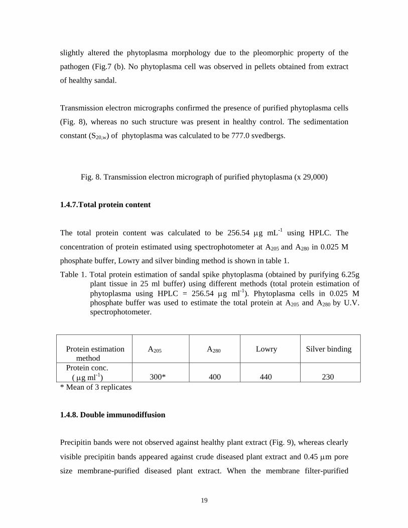
constant (S20,w) of phytoplasma was calculated to be 777.0 svedbergs.
Fig. 8. Transmission electron micrograph of purified phytoplasma (x 29,000)
1.4.7.Total protein content
The total protein content was calculated to be 256.54 μg mL-1 using HPLC. The
concentration of protein estimated using spectrophotometer at A205 and A280 in 0.025 M
phosphate buffer, Lowry and silver binding method is shown in table 1.
Table 1. Total protein estimation of sandal spike phytoplasma (obtained by purifying 6.25g plant tissue in 25 ml buffer) using different methods (total protein estimation of phytoplasma using HPLC = 256.54 μg ml-1). Phytoplasma cells in 0.025 M phosphate buffer was used to estimate the total protein at A205 and A280 by U.V. spectrophotometer.
Protein estimation method
A205
A280
Lowry
Silver binding
Protein conc. ( μg ml-1)
300*
400
440
230
* Mean of 3 replicates 1.4.8. Double immunodiffusion
Precipitin bands were not observed against healthy plant extract (Fig. 9), whereas clearly
visible precipitin bands appeared against crude diseased plant extract and 0.45 μm pore
size membrane-purified diseased plant extract. When the membrane filter-purified

20
diseased plant extract was diluted, precipitin bands appeared only against concentrated
and 1:2 diluted plant extracts whereas no precipitin band was observed at higher
dilutions. Clear bands were also visible against phytoplasma of diseased sandal collected
from Mysore. No band was visible against the extract of the host plants of spike disease
affected sandal - Lantana and witches’ broom-affected Zizyphus.
1.4.9. Enzyme linked immunosorbent assay (ELISA)
1.4.10. Direct ELISA
Initial experiments on the effect of time on coating the antigen in wells of polystyrene
strips showed that one hour of coating was optimum (Fig. 10). Also, one hour duration
was found to be ideal for incubating the conjugated antibody (Fig. 11). The results
Fig. 10. Effect of time on efficiency of antigen coating in direct ELISA. Plates were
coated with antigen (1:100 dilution-1 hour incubation) followed by incubation with conjugated antibody (1:250 dilution). Each value represents the mean of 5 replicates. : Healthy plant antigen, : diseased plant antigen, : ratio of absorbency values of diseased and healthy plants.

21
Fig. 11. Effect of time on efficiency of incubation of the conjugated antibody in direct
ELISA. Plates were coated with antigen (1:100 dilution) followed by incubation with conjugated antibody (1:250 dilution-1hour incubation). Each value represents the mean of 5 replicates. : Healthy plant antigen, : diseased plant antigen, : ratio of absorbance values of diseased and healthy plants.
obtained when the purified samples were probed for the presence of phytoplasma using
the conjugated antibody (1:250 and 1:500 dilutions) are shown in table 2.
Table 2. Direct ELISA values of different dilutions of purified diseased antigen. The antigens were probed with HRP conjugated phytoplasma specific antibodies (C.Ab). (Value for buffer=0.010).
Dilution 1:10 1:50 1:100 1:250 1:500 1:750 C.Ab-1:250 0.281*
(0.243)** 0.276
(0.097) 0.172
(0.075) 0.088
(0.042) 0.062
(0.038) 0.055
(0.034) C.Ab- 1:500 0.188
(0.212) 0.173 (0.075)
0.122 (0.059)
0.056 (0.035)
0.041 (0.038)
0.034 (0.027)
• Mean of 5 replicates. ** Threshold value which represents the mean of 3 healthy sandal (purified antigen) ±
twice the standard deviation.
22
The sensitivity of 1:250 dilution of conjugated antibody was higher compared to the
dilutions at 1:500. When the crude sample was used to detect the presence of
phytoplasma at different dilutions of conjugated antibody (1:100, 1:250; and 1:500), the
dilution at 1:250 was found to be better compared to the other dilutions (Table 3).
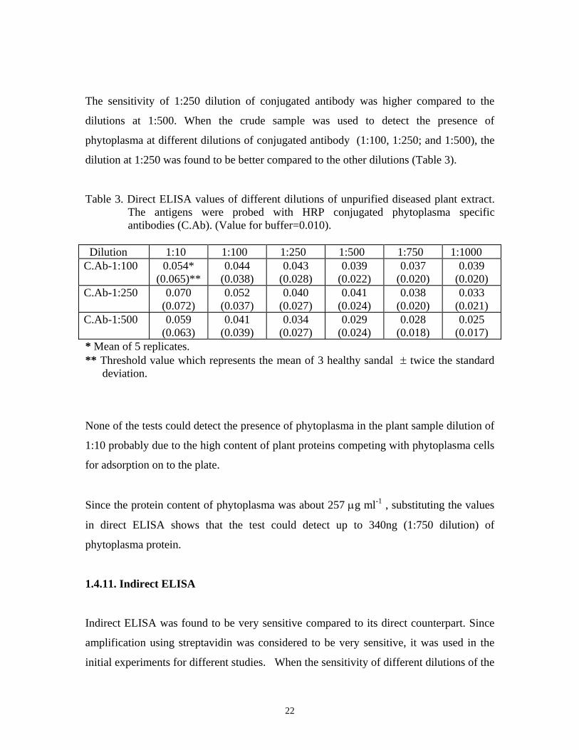
Table 3. Direct ELISA values of different dilutions of unpurified diseased plant extract. The antigens were probed with HRP conjugated phytoplasma specific antibodies (C.Ab). (Value for buffer=0.010).
Dilution 1:10 1:100 1:250 1:500 1:750 1:1000 C.Ab-1:100 0.054*
(0.065)** 0.044
(0.038) 0.043
(0.028) 0.039
(0.022) 0.037
(0.020) 0.039
(0.020) C.Ab-1:250 0.070
(0.072) 0.052
(0.037) 0.040
(0.027) 0.041
(0.024) 0.038
(0.020) 0.033
(0.021) C.Ab-1:500 0.059
(0.063) 0.041
(0.039) 0.034
(0.027) 0.029
(0.024) 0.028
(0.018) 0.025
(0.017) * Mean of 5 replicates. ** Threshold value which represents the mean of 3 healthy sandal ± twice the standard
deviation.
None of the tests could detect the presence of phytoplasma in the plant sample dilution of
1:10 probably due to the high content of plant proteins competing with phytoplasma cells
for adsorption on to the plate.
Since the protein content of phytoplasma was about 257 μg ml-1 , substituting the values
in direct ELISA shows that the test could detect up to 340ng (1:750 dilution) of
phytoplasma protein.
1.4.11. Indirect ELISA
Indirect ELISA was found to be very sensitive compared to its direct counterpart. Since
amplification using streptavidin was considered to be very sensitive, it was used in the
initial experiments for different studies. When the sensitivity of different dilutions of the

23
polyclonal antibody was tested using a constant dilution (1:1000) of purified antigen,
antibody dilution at 1: 2000 was found to be the most sensitive for the test (Fig. 12).
Fig. 12. Comparison of different antibody titre (dilution) in indirect ELISA. Plates were
coated with antigen (1:1000 dilution) followed by incubation with antibody at different dilutions(1. 1:500, 2. 1:1000, 3.1:2000, 4.1:5000, 5.1:10000 dilution-1hour). This was followed by incubation in biotinylated goat anti-rabbit antibody (1:2000 dilution-1 hour incubation) and finally in HRP-streptavidin conjugate (1:2000 dilution-1 hour). The results were read at 490nm. Each value represents the mean of 5 replicates. : Healthy plant antigen, : diseased plant antigen, : ratio of absorbance values of diseased and healthy plants.
The purified antigen at different dilutions (1:50-1:10,000) were probed with different
dilutions of polyclonal antibody (1:500, 1:1000 and 1:2000) using the streptavidin
amplification system (Table 4). The results confirm that polyclonal antibody at a dilution
of 1:2000 was the most sensitive. From the result it is inferred that a minimum of 25ng
phytoplasma protein could be detected by indirect ELISA, since purified phytoplasma of
1:10,000 dilution could be detected.
The results obtained using the avidin system and goat anti-rabbit HRP conjugate for
detection of the presence of phytoplasma in purified sample is shown in table 5. The
indirect test employing goat anti-rabbit HRP was found to be more sensitive compared to
the avidin-biotin system. Hence, to detect the presence of phytoplasma in crude extract,
only goat anti-rabbit HRP and the biotin-streptavidin systems were considered. The

24
results are shown in table 6. From the results it is confirmed that the test employing
biotin-streptavidin system is the most sensitive of the tests. This system was utilised to
detect the pathogen in 24 individual sandal plants. The polyclonal antibody could detect
the pathogen in diseased plants and the result is shown in table 7.
Table 4. Indirect ELISA values of different dilutions of purified diseased antigen using streptavidin amplification system. The antigens were probed with different dilutions of sandal spike phytoplasma specific antibody, biotinylated antirabbit antibody (1:2000 dilution) followed by HRP conjugated streptavidin (1:2000 dilution). (Buffer=0.010).
Dilution 1:50 1:100 1:250 1:500 1:1000 1:2000 1:5000 1:10000 Ab- 1:500 2.671*
(0.180)** 2.468
(0.111) 1.472
(0.072) 1.009
(0.056) 0.649
(0.044) 0.317
(0.024) 0.147
(0.037) 0.080
(0.031) Ab-1:1000 2.780
(0.148) 2.609
(0.102) 1.370
(0.063) 0.997
(0.053) 0.602
(0.038) 0.298
(0.027) 0.137
(0.038) 0.072
(0.036) Ab- 1:2000 2.554
(0.153) 2.447
(0.087) 1.861
(0.066) 1.321
(0.055) 0.746
(0.053) 0.404
(0.044) 0.198
(0.040) 0.118
(0.044) * Mean of 5 replicates. ** Threshold value which represents the mean of 3 healthy sandal (purified antigen) ±
twice the standard deviation.
Table 5. Indirect ELISA values of different dilutions of purified diseased antigen. The antigens were probed with sandal spike phytoplasma specific antibody (Ab) (1:2000 dilution) followed by either (A) goat anti-rabbit antibody conjugated to HRP (1:2000 dilution) or (B) biotinylated anti-rabbit antibody (1:2000 dilution). Avidin conjugated to HRP (1:2000 dilution) was used to probe the biotinylated anti-rabbit antibody. (Value for buffer=0.010).
Dilution of purified diseased antigen
Antibody systems
1:50 1:100 1:250 1:500 1:1000 1:2000 1:5000 1:10000 A 1.738*
(0.058)**
1.763 (0.041)
1.359 (0.028)
0.977 (0.022)
0.586 (0.022)
0.274 (0.023)
0.121 (0.021)
0.061 (0.020)
B 2.213 (1.326)
2.044 (0.924)
1.922 (0.792)
1.716 (0.663)
1.415 (0.564)
1.219 (0.523)
0.807 (0.499)
0.635 (0.437)
* Mean of 5 replicates.

25
** Threshold value which represents the mean of 3 healthy sandal (purified antigen) ± twice the standard deviation.
Table 6. Indirect ELISA values of different dilutions of unpurified diseased plant extract. The antigens were probed with sandal spike phytoplasma specific antibody (Ab) (1:2000 dilution) followed by either (A) goat anti-rabbit antibody conjugated to HRP (1:2000 dilution) and (B) biotinylated anti-rabbit antibody (1:2000 dilution). Streptavidin conjugated to HRP (1:2000 dilution) was used to probe the biotinylated anti-rabbit antibody. (Value for buffer=0.010).
Dilution of unpurified diseased antigen
Antibody systems
1:10 1:100 1:250 1:500 1:1000 1:2000 1:5000 1:10000 A 0.564*
(0.268)** 0.481
(0.241) 0.408
(0.188) 0.280
(0.162) 0.209
(0.131) 0.140
(0.114) 0.114
(0.097) 0.116
(0.088) B 0.571
(0.177) 0.371
(0.139) 0.304
(0.071) 0.253
(0.055) 0.190
(0.057) 0.142
(0.042) 0.084
(0.032) 0.054
(0.025) * Mean of 5 replicates. ** Threshold value which represents the mean of 3 healthy sandal ± twice the standard
deviation. Table 7. Detection of sandal spike phytoplasma in individual plant samples using indirect
ELISA.
Sample No. ELISA
Value
Sample No. ELISA
Value
1 2 3 4 5 6 7 8 9 10 11
12
1.235* 1.205 1.595 1.483 1.724 1.962 1.589 1.828 1.479 1.637 1.357
0.324**
13 14 15 16 17 18 19 20 21 22 23 24
1.286 1.350 1.582 1.669 1.498 1.749 2.143 1.581 1.934 1.523 1.265
0.415**
* Mean of 3 replicates
** Healthy trees

26
Since the indirect ELISA employing biotin-streptavidin system was found to be very
sensitive, the same was utilised in the detection of phytoplasma in the host plants of spike
disease affected sandal. The results are shown in table 8. The host plants of diseased
Table 8. Indirect ELISA values of different dilutions of extract of host plants of spike disease affected sandal using streptavidin system. (Value for buffer=0.010).
Dilution of host plant extract
Host plant
1:10 1:100 1:500 1:1000 Lantana 0.157*
(0.140)** 0.060 (0.058)
0.033 (0.029)
0.027 (0.033)
Zizyphus 0.134 (0.203)
0.136 (0.149)
0.086 (0.080)
0.052 (0.054)
* Mean of 5 replicates. ** Threshold values which represents the mean of 3 host plants of healthy sandal ± twice the standard deviation. sandal did not show variation in results compared to the host plants of healthy sandal.
When individual host plants were screened for the presence of phytoplasma, the test
could not detect the presence of the pathogen in any of the hosts (Table 9). The results
indicated that sandal spike phytoplasma is confined to sandal. The ELISA test carried
out using insect tissues did not show positive result in all the dilutions.
Table 9. Indirect ELISA values of host plants (Lantana and witches’ broom affected
Zizyphus) of spike disease affected sandal.
Lantana Zizyphus Sample No. ELISA Values Sample No. ELISA Values
1 2 3 4 5 6 7 8 9
10*
0.066* 0.067 0.065 0.079 0.072 0.074 0.067 0.108 0.065 0.161
1 2 3 4 5 6 7 8 9
10*
0.077 0.077 0.081 0.077 0.079 0.073 0.082 0.083 0.079
0.091

27
• Mean of 4 replicates. • Host plants of healthy sandal
1.5. Discussion
1.5.1 Indirect evidence of phytoplasma in spike diseased sandal through DAPI staining
Mycoplasmas are wall-less prokaryotes with a genome size of 5x10⁸ daltons. They have a
low G+C content (23 to 30 moles percent G+C) and a high A+T content. The affinity of
the fluorochrome, DAPI to double stranded DNA is very high. DAPI binds specifically to
the minor groove of A-T rich sequences (Kapuscinski, 1995). The technique has been
used in the rapid diagnosis of phytoplasma in blueberry (Schaper and Converse, 1985),
alder (Lederer and Seemuller, 1991), lettuce (Marcone et al., 1995b) and periwinkle
(Marcone and Ragozzino, 1995c).
1.5.2. Absence of phytoplasma in host tissues
In the present study, DAPI staining could not detect phytoplasma in the phloem of the
host plants of diseased sandal growing in the field as well as glass house. Hull et al.
(1970) also could not detect phytoplasma in Lantana growing as hosts of diseased sandal
but detected the same in witches’ broom-affected Zizyphus growing in sandal forests
using transmission electron microscopy.
SEM observations disclosed that in the material examined, the pleomorphic phytoplasma
cells were present only in the phloem tissues of diseased plants, whereas the healthy
phloem cells were devoid of any pathogen.
1.5.3. Immunological detection of Phytoplasma
A reliable and accurate detection of plant pathogen is a pre-requisite to develop disease
management strategies (Khan et al., 1998). Highly purified phytoplasma is needed for

28
immunological studies such as ELISA (Hobbs et al., 1987; Saeed et al., 1992a) and
biochemical studies of the organism (Sinha and Madhosingh, 1980).
Phytoplasma, was first purified from a tree species (Peach) by Sinha and Chiykowski
(1984), using the celite pad filtration technique. Most of the studies on phytoplasma
purification have used differential centrifugation technique (Clark et al., 1989; Saeed et
al., 1992b, 1993) which takes advantage of differences in sedimentation velocity that
result from variation in physiological properties. This method involves a series of
centrifugations wherein at the end of each centrifugation, the particles (phytoplasma)
remaining in suspension are separated from the pellet (plant debris) by decantation and
subjected to further centrifugation (Deter, 1973). A large amount of phytoplasma is lost
when it sinks along with the plant debris during the low speed centrifugation. Hence, in
the final centrifugation step, the net yield of phytoplasma will be less. Sinha (1979)
reported that about 78% of phytoplasma was found to be lost during purification
procedure of aster yellows phytoplasma.
1.5.3.1. Purification of Phytoplasma
The method of purification of phytoplasma using differential filtration technique takes
advantage of the filterable property of phytoplasma. Whatman 1 (11 μm pore size) and
Whatman 5 (2.5 μm pore size) were used to clarify the plant sap. Phytoplasma could pass
through both these filters whereas the filters retained much of the plant debris. The 0.45
μm Millipore filter prevented plant debris whereas the pleomorphic morphology of
phytoplasma enabled it to pass through the membrane which in turn slightly altered the
morphology of the organism as seen in the scanning electron micrograph.
Centrifugation at 4700g enabled the plant protein coupled with anti-plant antibody to
settle down; the low speed prevented phytoplasma sedimentation. When speed was
increased to 65,000g in the final step, it enabled maximum amount of phytoplasma
sedimentation, since no plant debris was present in the supernatant.

29
Since phytoplasmas are non-culturable microorganisms, the only method available for
quantitation of the pathogen is by total protein estimation. On comparing the total protein
content of sandal spike phytoplasma by using the HPLC derived result as standard, the
silver binding method was found to be the second sensitive assay. The assay could detect
protein with an accuracy of 90%. The sensitivity of the method may be due to the
property of silver to bind to sulphydryl and carboxyl moieties in proteins (Sasse and
Gallagher, 1996). Spectrophotometric assays at A280 and Lowry method was least
sensitive probably due to the low concentration of aromatic amino acids, thereby giving
false results. Spectrophotometric assay at A205, was more sensitive than A280 since the
quantitation is based on the absorbance by the peptide bond (Stoscheck, 1990). Since
buffers of higher or lower ionic strength was found to be insensitive for quantitation of
total protein, 0.025 M phosphate buffer was used in direct estimations (Simonian, 1996).
Ever since the non-culturable mollicutes, the phytoplasmas, were first discovered in
1967, they have been implicated as pathogens in more than 300 plant diseases worldwide
(McCoy et al., 1989). Although phytoplasmas can be visualised by electron microscopy
and their presence in phloem tissues demonstrated by fluorochromic DNA stains, these
methods cannot discriminate among phytoplasma groups (Clark et al., 1989).
Immunological assays are one among the most important methods for disease diagnosis
and pathogen detection. Several polyclonal and monoclonal antibodies have been
prepared against plant pathogenic phytoplasmas (Sinha and Benhamou, 1983; Sinha and
Chiykowski, 1984; Clark et al., 1989; Saeed et al., 1993) for different immuno assays
like double diffusion test, ELISA, dot-blot, and immunoblotting.
1.5.3.2. Ouchterlony double diffusion test Nayar and Ananthapadmanabha (1975) purified sandal spike phytoplasma by ammonium
sulphate precipitation method and used the same to raise polyclonal antibodies in rabbit.
The polyclonal antibody was used in gel diffusion and agglutination tests. They reported
a very poor titre of 1:250 dilution probably due to presence of plant debris along with the
phytoplasma cells which might have decreased the sensitivity of polyclonal antibodies.

30
The antibody could detect phytoplasma in spike disease affected sandal and Catharanthus
roseus plants infected artificially with sandal spike phytoplasma.
Double diffusion tests are used frequently for testing material from field surveys. The test
is widely used for screening viruses (Ahmad and Scott 1985; Barnett et al., 1987) and
bacteria (Bouzar and Moore 1987; Azad and Schaad 1988). In the present study, even
though precipitin bands could be observed against diseased sandal extract, no band was
observed against the hosts, Lantana or witches’ broom-affected Zizyphus. The result
suggests that considerable amount of phytoplasma was present in the extract of diseased
sandal and probably none in the extract of the host plants. Nayar (1981) also reported
absence of precipitin bands against witches’ broom affected Zizyphus when treated with
sandal spike phytoplasma specific antibody. Since precipitin bands were observed in
both Mysore and Marayoor phytoplasma populations, the pathogens must have been of
the same antigenic group. However the sensitivity of the test was found to be lower
compared to other techniques as it could only detect phytoplasma in concentrated and 1:2
dilution samples.
1.5.3.3. ELISA tests
ELISA is a quantitative method of immunological detection. The technique is highly
sensitive to detect the presence of pathogens. Polysorp plates were used for the present
study since they preferentially adsorb lipoproteins (Nunc, 1997) which are the major
antigenic determinants of the mollicutes. According to Kemeny (1992) one hour duration
was found to be satisfactory for coating antigen to the plate and for subsequent incubation
steps with the immunogenic probes. Initial experiments showed that increasing the
duration of incubation with immunogenic reagents decreased the sensitivity of the test
probably due to unspecific binding. In the present study also the absorbance ratio was
found to be higher for one hour, whereas it decreased with time (Fig.10, 11). Therefore,
one hour was kept constant for both the coating and incubation steps. Direct ELISA is
seldom employed in immuno-diagnostic tests due to its low sensitivity (Crowther, 1995),
but, from the present study it was found that though, the technique was less sensitive
31
compared to indirect ELISA, it could, nevertheless detect the presence of phytoplasma in
both purified and crude sample.
For indirect ELISA, polyclonal antibody at a dilution of 1:2000 was found to be optimum
as indicated by the absorbance ratio (Fig.12). Though, the indirect method employed
three types of systems, the biotin-streptavidin system was found to be the most sensitive
of the assay, probably due to the high amplification property of the system compared to
avidin and anti-rabbit HRP systems. When biotin-streptavidin and anti-rabbit HRP
systems were compared for detection of the presence of phytoplasma in crude samples,
the former was again found to be more sensitive. However, the tests could detect the
presence of pathogen even at dilutions up to 1:10,000 probably due to the high sensitivity
of the antibody generated using the purified phytoplasma. Correlating the phytoplasma
protein values with antigen dilution suggests that indirect ELISA could detect a minimum
of 25ng antigenic protein. When the efficiency of the polyclonal antibody was tested by
screening large number of diseased sandal plants, only the diseased plants showed high
values compared to the healthy plant values (Table 7). Rangaswamy (1995) produced
polyclonal antibody to detect phytoplasma in spike disease affected sandal and
Catharanthus roseus plants infected artificially with sandal spike phytoplasma by indirect
ELISA. The titre of the antibody was calculated to be 1:1000 dilution and could detect
antigen upto a dilution of 1:200.
Though, most of the workers employed indirect ELISA, they could not detect the
pathogen at higher dilutions of plant extract, probably due to contamination of plant
proteins in the purified phytoplasma pellet which might have generated antibodies against
healthy plant proteins thereby decreasing the efficiency of ELISA. Hobbs et al. (1987)
could detect phytoplasma in peanut witches’ broom only up to a dilution of 1:400,
whereas Clark et al. (1989) could detect dilutions up to 1:600 while detecting tomato big
bud phytoplasma. Saeed et al. (1993) detected phytoplasma in faba bean phyllody up to a
dilution of 1:300.
Even though, the highly sensitive indirect ELISA technique using biotin-streptavidin
system was employed, phytoplasma could not be detected in the host plants of spike

32
disease affected sandal. The hosts of diseased sandal did not show much variation in the
result compared to the control plants (Table 8). The results were identical when large
number of samples were screened for the detection of sandal spike phytoplasma (Table
9). Thus both the immunological techniques- Ouchterlony double diffusion test and
indirect ELISA could not detect phytoplasma in the hosts of diseased sandal confirming
that sandal spike phytoplasma is specific to sandal. Studies by Rangaswamy (1995) using
indirect ELISA also could not detect sandal spike phytoplasma in diseased Zizyphus. The
negative reaction obtained when insect tissues were ELISA tested can not be taken as
conclusive. The experiment has to be repeated systematically by rearing the insect vector
on diseased plants or testing the vector after proper acquisition feeding.
In spite of using a highly sensitive protein detection technique using the silver nitrate
method it was not possible to locate the antigenic protein in the SDS-PAGE gel. Jiang et
al. (1988) and Saeed et al. (1992) also could not identify phytoplasma specific proteins
among the contaminating plant proteins using SDS-PAGE. Bloom et al. (1987) were of
the opinion that silver staining was far more sensitive than coomassie blue to detect the
presence of protein in the nanogram range. From the present study it is confirmed that
sandal spike phytoplasma antigenic protein has a molecular weight of 14,000 daltons and
could be detected in both purified and semi-purified phytoplasma but the same was
absent in healthy sandal. Clark et al. (1989) reported that primula yellow phytoplasma
and European aster yellow phytoplasma had a single major antigen of 22,400 daltons
while aster yellow phytoplasma affecting lettuce had an antigenic protein of 18,500
daltons (Jiang et al., 1988). Saeed et al. (1992b) reported that faba bean phyllody
phytoplasma had two antigenic proteins of 18,000 and 36,000 daltons.
According to Clark et al. (1983), the identification of infection by phytoplasmas may be
difficult when symptoms are absent or indistinct. Rapid immunological techniques like
ELISA, circumvents most of these problems. From the present study it is inferred that
polyclonal antibody raised against sandal spike phytoplasma was found to be efficient for
detection of the pathogen through immunological techniques. The same antibody can be
used as the primary probe for other immunological tests such as Dot immuno-binding

33
assay, immuno microscopy, etc. Another positive factor of the technique is that the
pathogen could be detected in the crude sample itself. Thus immuno diagnostics could be
used as a tool for early detection of the pathogen in plant samples.
2. Commercial exploitation of ELISA technique for detecting sandal spike phytoplasma
2.1. Introduction
Immunological techniques have been developed to detect sandal spike phytoplasma. The
reagents developed for diagnosis of sandal spike phytoplasma and early detection of
spike disease can be marketed as kits which may be useful for the sandalwood industry.
The commercial exploitation of the reagents developed is explained in this chapter.
2.2. Diagnostics
Diagnostics can be viewed as a discipline in its own right, combining a wide range of
techniques in developing simple, fast and reproducible (SFR) procedures that measure a
feature of the biological material in hand in a way that is easily interpretable. Improved
diagnostics could be useful in epidemiological studies to determine the distribution and
abundance of pests and pathogens (Skerritt and Appels, 1995).
During the past few years, progress in molecular biology, biochemistry and immunology
has promoted the development of many new methods of pathogen detection and disease
diagnosis (Miller and Martin, 1988). Immunoassays have been developed and are
commercially available for the identification of plant pathogens, mycotoxins, pesticides
and plant hormones. Polyclonal antibodies are extremely useful since their broader
spectrum can sometimes be more useful than the highly specific monoclonal antibodies
(Miller and Williams, 1990).
2.3. Commercial exploitation of the techniques based on the present work

34
The techniques used in the present study for the purification of sandal spike phytoplasma
using the differential filtration method was rapid and economical. The purity of the
phytoplasma cells thus generated was found to be very high. The purified phytoplasmas
elicited immune response in rabbits to produce highly sensitive polyclonal antibodies,
which could be used in different immunological techniques. Thus, the immunological
tests like Ouchterlony double diffusion test, and direct and indirect ELISA could be
standardised for pathogen detection. Since, one hour of duration was found to be
optimum for both coating and incubation, direct ELISA could be completed within three
hours and indirect ELISA within seven hours. Even though, the number of steps was
more in indirect ELISA, the sensitivity of the test was high when biotin-streptavidin
system was adopted.
Thus, two major kits could be developed based on the present studies viz., sandal spike
phytoplasma purification kit and immuno-detection kit (Fig. 13). The reagents that can be
supplied is listed in the table (Table 10).
In India, marketing of sandal is restricted exclusively to government agencies leaving
very little scope for private agencies. So the kits developed per se has limited chance for
commercial exploitation in the open market in the present scenario. But, since, the

35
techniques developed are highly specific and sensitive, the same could be used in other
tree improvement projects.
Most of the companies supply antibodies either as freeze dried powder or with
preservatives which has a shelf life of about 12 months at 40C or for longer period at
–200C (Adgen, 1998). The positive and negative control samples are supplied as freeze-
dried sap/extract which should be reconstituted with distilled water or buffer before use.
Adgen sells most phytoplasma kits of 1000u for around US $600 (Rs.27,500). These kits
contain reagents for ELISA only. The reagents of sandal spike phytoplasma, when
produced in large scale, could be supplied at one–fourth the price of Adgen due to low
labour and other input costs.
Table 10. Kits and reagents developed to detect sandal spike phytoplasma.
KIT REAGENTS
SANDAL SPIKE PHYTOPLASMA
PURIFICATION KIT
Healthy sandal antibody,
Glycine buffer.
IMMUNO DETECTION KITS
1. Double diffusion kit
2. ELISA kit
Phytoplasma specific antibody,
Phytoplasma specific antibody-HRP,
Anti-rabbit antibody-HRP,
Anti-rabbit antibody biotin,
Streptavidin-HRP,
Phosphate buffered saline,
Positive control (phytoplasma).

36
3. Identification of ‘disease resistant’ trees using
ELISA technique
3.1. Introduction
In Marayoor Range, nine natural sandal reserves, spread in an area of 15.5 km2 in reserve
forests and 47 km2 in the adjoining revenue lands constitute the main sandal tract. The
sandal reserve forests of Marayoor is considered to be the best in India in respect of
sandal trees per unit area and oil yield (5%). Extraction of sandal is limited to dead and
wind fallen trees not only from reserve forests but also from revenue lands. Only the
forest department is authorised to extract such trees.
Though, spike disease was reported first in 1980 only from the Sandal Reserve 51 (382
ha in area), the disease might have started a few years before (Ghosh et al., 1985).
Enumeration of sandal trees in 1971 in the reserve recorded a population of more than
23,000 trees (Varghese, 1976). In 1985, more than 50 per cent of the trees in the reserve
were found affected by the disease and by 2000, more than 80 per cent of the trees had
perished.
Ghosh et al. (1985) reported the apparent infection rate 0.066 - 0.086 per unit per month
in disease monitoring plots. Disease spread is radial and infected trees die within 2 years
on an average. In diseased tracts, usually, almost all the trees get infected and die.
However, in Reserve 51, about 45 trees of various girth classes were observed as disease
free during 1996 in about 100 ha area surrounding the epicenter of the disease. These
trees have either evaded the infection or may be resistant against spike disease.

37
In this chapter, result of monitoring the health of 15 such apparently more than 25-year-
old trees are provided. The monitoring involved periodical ELISA tests of leaf extracts
for the early detection of sandal spike phytoplasma.
3.2. Materials and methods
The girth of the selected trees ranged from 32 cm to 55cm. Apparently, all these trees had
existed when the spike disease was reported first in 1980. The 15 trees, located in about
one km2 area, were protected from human interference by providing 3 m wide chain-
linked fence around each tree (Fig. 14).
3.3. Results
All the 15 trees were healthy till March 1998 and none of the trees showed any doubtful
external symptom of infection. ELISA tests were conducted on leaf extracts of all the
trees in March 1998 and none of the trees were positive. But in September 1998 two
adjacent trees (tree No. 8 and 9, standing 15 feet apart) were ELISA positive (Table 11) indicating that the trees were already infected. But, typical symptom of spike disease was
not expressed on the leaves or branches. However, emergence of new sprouts from
branches were absent. In March 1999, these two trees showed severe defoliation and
typical spike disease symptoms almost on all branches. Both the trees dried partially by
September 1999 and completely by March 2000. Likewise, tree No. 4 was ELISA
positive from March 1999 onwards, but externally healthy during September 1999. It
dried partially by March 2000. Remaining trees were free of phytoplasma and were
apparently healthy till March 2000.
Table 11. Result of ELISA test on three ‘disease evaded’ trees showing ELISA positive
reaction before external symptom expression. Expression of disease symptom ELISA test result (Presence
of phytoplasma)
Tree No.
GBH (cm)
1996 1997 1998 1999 1998 1999

38
Mar Sep Mar Sept Mar Sept Mar Sept Mar Sept Mar Sept 4 32 Nil Nil Nil Nil Nil Nil Nil Nil Neg. Neg. Pos. Pos. 8 44 Nil Nil Nil Nil Nil Nil Yes Dry-
ing Neg. Pos. Pos. Pos.
9 41 Nil Nil Nil Nil Nil Nil Yes Dry-ing
Neg. Pos. Pos. Pos.
Nil : No disease symptom; Pos. : ELISA Positive; Neg. ELISA Negative
3.4. Discussion Reports of phytoplasma disease resistance in tree crops are very rare. Expression of
disease symptoms is always associated with presence of phytoplasma. In this experiment,
it was possible to detect the pathogen in the leaf extract of tree No. 4, 8 and 9, six months
before the expression of the disease symptoms. Even though, the remaining trees were
pathogen free till September 2000, it is not possible to conclude that these trees will
evade the disease in future also or they are actually disease resistant trees. Considering
the fact that those sandal trees which remained uninfected amid severely infected trees
for more than 15 years became vulnerable to infection subsequently, prevents us from
concluding that resistance to spike disease exist in sandal population in Marayoor.
Since, all the attempts to culture the pathogen in vitro has failed, it is not possible to
inoculate the disease-evaded trees with phytoplasma to prove their field resistance. Tests
of susceptibility through graft transmission or through parasitic flowering plants seems
artificial, not known to occur in the reserve forest. Hence, inoculation with infective
insect vector is the only logical method to test the resistance of a tree.
However, the success achieved in the early detection of spike disease through ELISA
technique may be useful to control the pathogen through infusion of tetracycline group of
antibiotics before the full multiplication of the phytoplasma in phloem tissues.

39
4. Micropropagation of disease evaded genotypes
4.1. Introduction
Selection and planting of disease resistant genetic material is the primary approach to
improve productivity of a plant species. In Marayoor forest range of Munnar Forest
Division in Kerala, spike disease has caused serious destruction to the sandal trees
(Ghosh et al., 1985). Death of almost the entire population existing in an infected patch
is characteristic of the disease. However, a few trees, which have remained free from the
disease for the past 20 years, are considered either resistant or capable of evading the
disease. One of the objectives of this project is the clonal multiplication of a few such
disease-evading trees located in the disease ravaged sandal forests in Marayoor, through
tissue culture via somatic embryogenesis.
In India, extensive work on tissue culture of sandalwood was carried out mainly in two
laboratories – BARC, Bombay (Bapat and Rao, 1979, 1984; Rao and Raghavaram, 1983)
and Indian Institute of Science, Bangalore (Lakshmi Sita 1986, 1992; Lakshmi Sita et al.,
1994). Although multiple shoot formation was reported (Rao et al., 1984; Sanjaya et al.,
1998; Pradhan and Saiju, 1999), regeneration of plantlets through somatic embryogenesis
was suggested to be the most efficient method of mass propagation in Santalum album
(Bapat et al., 1990). Even though, sandalwood has got a very high frequency of somatic
embryogenesis, the successful utilization of the technique is hampered due to the high
percentage of abnormal embryogenic plantlets and poor rate of survival (Rao and Bapat,
1995). Hence, only very few plantlets have been reported to be established outside the
laboratory after out planting.
In the present study, an attempt has been made to multiply a few spike disease-evading
sandal trees identified in a heavily diseased area in Marayoor Forest Range of Munnar
Forest Division through tissue culture via somatic embryogenesis.

40
4.2. Materials and Methods
4.2.1. Explants
The internodes, nodes, petioles, leaf segments and root suckers collected from trees that
were apparently more than 20-year-old were used as explants for the present study. The
explants, were washed in tap water with Extran (Merck), kept for 15 minutes in running
tap water, sterilized with 0.1% HgCl2 for 8 minutes and washed in sterile water four
times, before inoculation onto appropriate culture medium.
4.2.2. Culture media
The minerals and vitamins of the MS Medium (Murashige and Skoog, 1962), White’s
medium (White, 1963), Woody plant medium (WPM) (Lloyd and McCown, 1981) and
B5 Medium (Gamborg et al., 1968) were tried at various stages of culture. The pH of the
media was adjusted to 5.7 using 0.1N NaOH and 0.1 N HCl prior to autoclaving at 120oC
at 15lbs/in2 pressure for 20 minutes.
4.2.3. Growth regulators Various auxins (2,4-D, 2,4,5-T, NAA, IAA, Dicamba and Picloram) and cytokinins (BAP
and Kinetin) were supplemented in the media during the different stages of culture viz.
induction, maintenance and conversion of embryogenic callus to plantlets.
4.2.4. Additives To compare the improvement in the rate of proliferation of soft friable calli prior to
embryogenesis, the following additives were used.
i. Malt extract
ii. Yeast extract
iii. Casein hydrolysate
iv. Coconut water

41
4.2.5. Growth adjuvants
In order to improve the efficiency of embryogenesis, the following growth adjuvants were used. i. Abscisic acid (ABA)
ii. Polyethylene glycol (PEG)
iii. Casein hydrolysate (CH)
iv. Activated charcoal (AC)
4.2.6. Culture conditions All cultures on semi-solid media were routinely incubated at 25±1oC under diffused
fluorescent light with 15-h photoperiod. Subculturing was carried out usually at 4-week
intervals.
4.2.7. Suspension cultures
Cell suspensions were initiated from friable callus grown on MS medium supplemented
with 2,4-D. Suspension cultures were induced in 250 ml conical flasks, shaken on rotary
shakers at 125 rpm. Under agitation, the cultures produced a fine suspension of single
cells and a few cell aggregates. Subculturing to fresh medium was carried out at 15 days
interval after allowing the cells to settle down, decanting the medium and replacing it
with an equal amount of fresh medium.
4.3. Results
4.3.1. Callus initiation
Initiation of callus from the exposed cut surface of internode segments took place 3 to 4
weeks after inoculation. Further growth was very slow and it took 2 to 3 months for
initiation of a proliferating light yellow, compact callus (Fig.15). Of the different explants
tried for callusing, internode explants from mature plants and root suckers gave a good
proliferating callus. The nodes, leaves and petioles showed no sign of callusing.

42
Of the four basal media tried, callus initiation took place only in MS medium. Among
the different hormonal combinations tried, MS medium fortified with 2,4-D (0.1 mg/l)
and Kinetin (1mg/l) showed callus initiation in 90 per cent of the cultures. No sign of
callusing was noticed in all the other combinations tried.
4.3.2. Initiation of the friable soft calli
The compact proliferating callus was routinely subcultured on fresh medium of the same
composition at 30 days interval. After 5-6 subcultures, white friable soft calli appeared
(Fig. 16) from the side of the brown compact callus. Of the different types of basal media
used for proliferation of friable callus, WPM and half strength MS medium gave almost
the same proliferation rate, while B5 medium gave the least amount of proliferation. The
friable soft calli could be maintained indefinitely for a long period without losing its
embryogenic capacity. Among the different auxins, the rate of proliferation was highest
for 2,4,5-T followed by 2,4-D while NAA and IAA gave almost the same amount
proliferation and picloram and dicamba gave the least amount of proliferation. Coconut
water at 20 per cent level gave the highest level of proliferation and Casein hydrolysate
(0.5g/litre) yielded the least amount of proliferation among the additives tried for friable
calli proliferation.
The basal MS medium supplemented with 2,4-D was also experimented with different
solidifying agents. Almost all the concentrations of agar and phytagel gave rise to almost
the same rate of proliferation except agar (0.5 %) in which the callus proliferation was
very low.
4.3.3. Induction of embryogenic calli
When the friable soft calli was subcultured on to MS basal medium supplemented with
IAA/BAP alone or in combinations gave rise to white friable, granular embryogenic
callus (Fig.17).

43
Of the different combinations tried, IAA (0.5mg/l), and BAP (0.5mg/l) gave the highest
amount of embryogenic callus. This embryogenic tissue, after two weeks gave rise to
somatic embryos, which followed the typical stages of embryogenesis. Abnormal
embryos occurred in large number along with embryos of normal morphology. Somatic
embryos of all stages of development were found in the same culture vessel and
adventitious embryo formation (repetitive embryogenesis) was also observed.
Of the four different types of basal media combinations tried for embryogenic callus
initiation, B5 medium gave rise to embryogenic callus seven days after culture while in
the rest of the media, embryogenic callus induction was noticed after three days. Among
the four media tried, WPM gave the highest amount of embryogenic callus.
4.3.4. Embryo maturation and conversion
Many of the somatic embryos developed asynchronously and germinated precociously,
and some had atypical morphology. This type of precocious germination without passing
through characteristic stages of development in sandal wood somatic embryos was found
to be controlled to some extent by adding abscisic acid at concentrations ranging from 0.1
to 5 μM. Abscisic acid at 3 μM concentration gave rise to high percentage of embryos
normal in appearance. Embryo abnormalities increased greatly above 5 per cent sugar
level. Maltose at 0.1 M level was found to be optimum than sucrose at 5 per cent level
which was previously reported in sandal. In presence of maltose, the embryos grew
larger, morphologically more typical and viable (Fig.18).

44
The highest percent of normal embryos was obtained when PEG, a non-permeating
osmoticum was used in combination with abscisic acid (3μM). Addition of PEG (at 2.5
%) to maturation medium improved the number of embryos and enhanced uniformity.
4.3.5. Cell suspension culture
The effect of initial culture density and effect of total volume of the media on rate of
proliferation were tested. In order to ascertain the embryogenic potentiality of suspension
culture, after one month of culturing in liquid medium, cells were filtered and washed
with MS basal medium and plated on to semi-solid embryo induction medium. MS
supplemented with IAA (0.5mg/l) and BAP (0.5mg/l) resulted in the formation of pro-
embryogenic masses followed by the differentiation of somatic embryos. After embryo
induction, the effective separation of the embryos for further conversion was brought
about by transferring the embryogenic masses again to liquid maturation medium. If they
were kept on the same semi-solid medium, instead of developing into mature embryos,
many of them dedifferentiated again and produced a callus mass. After one month of
culture in liquid medium, the cells were filtered and plated on solid media, after which
the number of embryos formed were found to be higher. The fully developed embryos
were transferred to semisolid conversion medium.
4.3.6. Embryo conversion to plantlets
45
Even though, improvement in somatic embryo development was achieved, only 20 per
cent of the normal embryos were converted into plantlets in the final stage. The
development of sandal plants from somatic embryos was attempted by transferring single
embryos at the cotyledonary stage to a medium with lower concentration of sucrose and a
low concentration of growth regulators. The isolation was found to have stimulating
effect on their germination. In many cultures, the embryo instead of developing into
plantlets, dedifferentiated again and produced a callus mass at the base which again
showed regeneration of somatic embryos. Among the hormonal combinations tried,
Whites medium or quarter strength MS medium containing IAA (0.5mg/l), IBA (0.5mg/l)
and GA3 (1mg/l) gave a moderate rate of conversion to plantlets. Along with normal
plantlets with well developed tap root and shoot (Fig.19), abnormal plantlets either with
prominent shoot and no root or with well developed tap root and no development of shoot
were also observed. Since sandal is a semi-root parasite, which draws part of its nutrients
from host through haustorial connection, sandal plantlets during the hardening stage were
co-cultivated with Cajanus seedlings. After 4-6 weeks the root region of sandal plantlets
established haustorial connection with the Cajanus seedlings (Fig.20). The somatic
plantlets with well-developed root and shoot were transferred to sterile vermiculite and
soil in plastic cups (Fig. 21). No plantlet survived beyond three months even if the
humidity was maintained and plantlets were nourished with nutrient solution.
4.4. Discussion
The production of a large number of embryos from stem-derived callus of superior,
disease resistant/evaded mother plant and their successful conversion to plantlets makes a
significant step on sandal wood micropropagation. The induction of somatic
embryogenesis in S. album is one of the earliest reports on somatic embryogenesis in
forest trees (Lakshmisita et al., 1979, 1982; Bapat et al., 1995). But so far no reported
work has claimed satisfactory survival of embryogenic plantlets. All the reported
hormonal combinations 2,4-D (0.1mg/l), Kinetin (0.2mg/l), BAP (0.2mg/l) (Rao and
Bapat, 1978; Lakshmi Sita et al., 1979) failed to give rise to callus from mature explants
in the present study. Aberrant/abnormal embryo phenotype and subsequent low

46
conversion rate of somatic embryos into normal plantlets have been reported (Bapat and
Rao, 1984; Rao and Bapat, 1995). The morphogenetic differentiation of a somatic cell to
whole plant is a complex developmental process, that involves morphogenetic,
biochemical, physiological and molecular changes (Thorpe, 1995). The physiological
processes leading to efficient germination of somatic embryos has to be elucidated to
improve the conversion rate of somatic embryos and survival rate of embryogenic
plantlets (Rao and Bapat, 1995). Though, the culture conditions required for somatic
embryogenesis are well defined in S.album (Rao and Bapat, 1995), the molecular and
biochemical events underlying the phenomenon are ill defined. The semi-parasitic nature
of sandal seedlings points to specific nutritional requirements, which has to be studied so
that the probability of establishing micropropagated plantlets in large numbers can be
explored.
47
5. Conclusions
1. The study has resulted in the development of a technique for purified preparation of
sandal spike phytoplasma for production of polyclonal antibodies. The polyclonal
antibodies can be utilized for development of ELISA techniques for early detection of
phytoplasma infection. The ability of phytoplasma to pass through 0.45μm millipore
filter is exploited to remove host debris larger than their size. Removal of host protein
and tiny debris are made possible through cross absorption of semi-purified
phytoplasma preparation with antibodies produced against extract from healthy plant
leaves. The filtration is followed by centrifugation at 65,000g to obtain purified
phytoplasma pellet. This new procedure is an improvement over the earlier methods
of cross absorption and repeated low speed centrifugation to obtain phytoplasma
pellets. In previous methods, large quantity of phytoplasma are lost along with the
host debris during the repeated low speed centrifugation.
2. The purified polyclonal antibodies can be utilized for development of techniques such
as Dot Immunobinding Assay (DIBA), Immuno microscopy, Immuno electron
microscopy and fluorescent antibody techniques. The DIBA technique can be adopted
for field detection of sandal spike phytoplasma.
3. The absence of sandal spike phytoplasma in host plants of spike disease affected
sandal and in witches’ broom affected Zizyphus oenoplaea confirms that sandal spike
phytoplasma is confined to sandal only and the disease is not transmitted through host
plants. It is also concluded that witches’ broom disease of Z. oenoplaea is not caused
by sandal spike phytoplasma.
4. Incidence of disease in trees which evaded infection for more than 20 years creates
the doubt whether resistance against sandal spike phytoplasma exists at all even in
disease evaded trees, presumed to be resistant. Absence of resistant trees among
disease evaded trees in the field necessitates screening sandal provenances and

48
populations from all over India and overseas in order to identify spike disease
resistant varieties. Resistant genes can also be detected in other species of Santalum
which can be used for distant hybridization.
5. Considering the economic importance of the species, modern biotechnological tools
such as introduction of resistant genes against spike disease through genetic
engineering can be attempted.
6. Mass multiplication of disease evaded trees and superior candidate plus trees (CPTs)
through somatic embryogenesis is only partially successful. Conversion of somatic
embryos to normal plantlets and proper hardening for establishment of outplanted
plantlets are yet to be achieved. Hence, more efficient protocol has to be developed
for somatic embryogenesis and hardening of plantlets. The survival of the plantlets is
probably complicated due to the parasitic nature of the species.
7. It was not possible to detect the phytoplasma in the salivary gland and intestinal
tissues of Redarator bimaculatus, the insect vector through ELISA techniques. This
may be because the vector might not have acquired the pathogen even though they
were collected from diseased trees. Further studies are needed to confirm the insect
vector through proper acquisition feeding studies and detection of the pathogen
through immunological and molecular techniques (RFLP).

49
6. Research Results of Practical/Field Application 1. New techniques have been developed for purified preparation of sandal spike
phytoplasma. Protocols have been standardized for production of polyclonal
antibodies against the phytoplasma. Early detection of spike disease, at least six
months before the external expression of disease symptoms was possible through
application of direct and indirect ELISA technique developed through the present
study.
2. The protocols developed for purification of phytoplasmal and standardization of
ELISA techniques can be utilized for development for such techniques for early
detection of the pathogen in other crop plants.
3. Absence of sandal spike phytoplasma in witches’ broom affected-Zizyphus oenoplaea
and host plants of spike disease affected sandal indicated that host plants of sandal
have probably no role in spike disease spread in the forest.

50
References Adgen (1998). Plant Disease Diagnostics Catalogue. Adgen Ltd, Scotland. Ahmad, I.B. and Scott, H.A. (1985). Identification and serotyping of cucumber mosaic and peanut stunt
viruses in Arkansas. Plant Dis. 69, 891-893. Ali, M.I.M., Balasundaran, M. and Ghosh, S.K. (1987). Symptom remission in spiked sandal trees by
infusion of tetracycline antibiotics. Plant Pathol. 36, 119-124. Ananthapadmanabha, H.S., Bisen, S.P. and Nayar, R. (1973). Mycoplasma like organisms in histological
sections of infected sandal spike. Experientia 26, 1571-1572. Azad, H. and Schaad, N.W. (1988). Serological relationships among membrane proteins of strains of
Xanthomonas campestris pv. transluscens. Phytopathology 78, 272-277. Ball, E.M., Hampton, R.O., De Boer, S.H. and Schaad, N.W. (1990). Polyclonal antibodies. In Serological
Methods for Detection and Identification of Viral and Bacterial Plant Pathogens (R. Hampton, E. Ball, and S. De Boer, Eds), pp.33-54. APS Press, USA.
Bapat V.A and Rao P.S. 1979. Somatic embryogenesis and plantlet formation in tissue cultures of sandal
wood (Santalum album L.) Ann. Bot. 44: 629-630. Bapat V.A and Rao P.S. 1984. Regulatory factors for in vitro multiplication of sandal wood tree (Santalum
album L.) shoot bud regeneration and somatic embryogenesis in hypocotyl cultures. Proc. Ind. Acad. Sci. 93: 19-27.
Bapat V. A., Gill R. and Rao P.S. 1985. Regeneration of somatic embryos and plantlets from stem callus
protoplasts of sandalwood tree (Santalum album L.) Curr. Sci. 54: 978 - 982. Bapat V.A., Fulzele D.P., Rao P. S. and Heble M.R. 1990. Production of somatic embryos of sandalwood
(Santalum album L.) in bioreactors. Curr. Sci. 59: 746 - 748. Barbara D.J. and Clark, M.F. (1986). Immunoassays in plant pathology. In Immunology in Plant Science.
(T. L. Wang, Ed.), pp.197-219. Cambridge University Press, U.K. Barber, C.A. 1903. Report on spike disease in sandalwood trees in Coorg. Indian For. 29:21-31. Barnett, O.W., Randles, J.W. and Burrows, P.M. (1987). Relationships among Australian and North
American isolates of the bean yellow mosaic potyvirus subgroup. Phytopathology 77, 791-797. Bloom, H., Beier, H. and Gross, H.S. (1987). Improved silver staining of plant proteins, RNA and DNA in
polyacrylamide gels. Electrophoresis 8, 93-99. Bouzar, H. and Moore, L.W. (1987). Complementary methodologies to identify specific Agrobacterium
strains. Appl.Environ. Microbiol. 53, 2660-2665. Brock, T.D. and Madigan, M.T. (1988). Immunology and Immunity. In Biology of Microorganisms.
Prentice Hall, USA, pp.428-473. Caudwell, A., Meignoz, R., Kuszala, C., Schneider, C., Larrue, J., Fleury, A. and Boudon, E. (1982).
Purification immunologique et observation ultramicroscopique en milieu liquide, de l’ agent

51
pathogene (MLO) d’ une jaunisse vegetale la flavescence doree de la vigne. Proceedings of the Academie d’ Agriculture de France, pp.407-415.
Chand Basha, S. (1977). Structure and composition of the sandalwood forests of Kerala. In All India Sandal
Seminar- Proceedings, pp. 53-58. Karnataka Forest Department, Bangalore. Clark, M.F., Barbara, D.J. and Davies, D.L. (1983). Production and characteristics of antisera to
Spiroplasma citri and clover phyllody associated antigens derived from plants. Ann. Appl.Biol. 103, 251-259.
Clark, M. F., Morton, A.and Buss, S.L. (1989). Preparation of mycoplasma immunogens from plants and a
comparison of polyclonal and monoclonal antibodies made against Primula yellows associated antigens. Ann. Appl. Biol. 114, 111-124.
Coen, D.M. (1994). Quantification of DNAs by polymerase chain reaction using an internal control. In The Polymerase Chain Reaction (K.B. Mullis, F. Ferre and R.A. Gibbs, Eds), pp.89-96. Birkhauser, USA.
Cole, R.M. (1983). Transmission electron microscopy: basic techniques. In Methods in Mycoplasmology (S. Razin and J.G. Tully, Eds), Vol. I, pp. 43-50. Academic Press, New York.
Coleman, L.C. (1917). Spike disease of sandal. Department of Agri., Mysore. Mycology Series Bull. No.3,
1-52. Coleman, L.C. (1923). The transmission of spike. Indian Forester 49, 6-9. Coppen, J.J.W.(1995): Sandalwood oil. In Flavours and Fragrances of Plant Origin, pp. 53-59. FAO,
Rome. Crowther, J.R. (1995). ELISA-Theory and Practice. Humana Press, New Jersey, 223p. Deter, R.L. (1973). Electron microscopic evaluation of subcellular fractions obtained by ultracentrifugation.
In Principles and Techniques of Electron Microscopy (M.A. Hayat, M.A. Ed.), Vol. III, pp.199-235. Van Nostrand Reinhold Company, New York.
Doi, Y., Teranaka, M., Yora, K. and Asuyama, H. (1967). Mycoplasma of PLT group-like-microorganisms
found in phloem elements of plants infected with mulberry dwarf, potato witches’ broom, aster yellows, or paulownia witches’ broom. Ann. Phytopathol. Soc. Jpn. 33, 259-266.
Dover, C. and Appanna, M. (1933). Entomological investigations on the spike disease of sandal (20).
Indian Forest Records 20, Pt. 1. Dijkstra, J. and Ie, T.S. (1969): Presence of mycoplasma-like bodies in the phloem of sandal affected with
spike disease. Neth. J. Pl. Pathol. 75, 374-378. Durand, D.P. (1990). Amplification of serological reactions. In Serological Methods for Detection and
Identification of Viral and Bacterial Plant Pathogens (R. Hampton, E. Ball and S. De Boer, Eds), pp. 93-100. APS Press, USA.
Engvall, E. and Perlmann, P. (1971). Enzyme linked immunosorbent assay. Quantitative assay of
immunoglobulin G. Immunochemistry 8, 871-874. Fischer, C.E.C. (1918). Cause of spike disease of sandal, Santalum album. Indian Forester 44, 571-575. Fox, J.E.D., Brand, J.E., Barrett, D.R. and Effendi, M. (1994). Genetic variation in Santalum album in
Timor, Indonesia, pp. 79-103. Sandalwood Workshop, New Caledonia. August 1994. CIRAD/FAO.
Freundt, E.A. and Edward, D.G. (1979). Classification and taxonomy. In The Mycoplasmas (M.F. Barile
and S. Razin, Eds.), Vol. I, pp.1-41. Academic Press, New York.

52
Gamborg D.L., Miller R. A. and Ojima K. 1968. Nutrient requirements of suspension cultures of soyabean
root cells. Exp. Cell Res. 50: 151 - 158. George, A.S. (1984). Santalum. In Flora of Australia. Vol.22. Australian Govt. Publishing Service,
Canberra, pp.61-66. Ghosh, S.K., Balasundaran, M. and Ali, M.I.M. (1985). Studies on spike disease of sandal. KFRI Research
Report, No. 37, Kerala Forest Research Institute, Peechi, Trichur, 56p. Ghosh, S.K., Balasundaran, M. and Ali, M.I.M. (1992). Sandal spike disease. In Plant Diseases of
International Importance (A.N. Mukhopadhyay, J. Kumar, H.S. Chaube and U.S. Singh, Eds), Vol.IV, pp.296-310. Prentice Hall, New Jersey,.
Hampton, R., Ball, E. and De Boer, S. (1990). Serological Methods for Detection and Identification of
Viral and Bacterial Plant Pathogens. APS Press, USA, 389pp. Harrison, N.A., Richardson, P.A., Kramer, J.B. and Tsai, J.H. (1994). Detection of the mycoplasma-like
organism associated with lethal yellowing disease of palms in Florida by polymerase chain reaction. Plant Pathol. 43, 998-1008.
Harlow, E.and Lane, D. (1988). Antibodies – a Laboratory Manual, Cold Spring Harbor Laboratory, New
York, pp. 400-405. Hayat, M.A. (1975). Positive staining for electron microscopy. Van Nostrand Reinhold Company, New
York, pp.66-75. Hayat, M.A. (1989). Principles and Techniques of Electron Microscopy. Mamillan Press Pvt. Ltd. London,
pp.112. Hobbs, H.A., Reddy, D.V.R. and Reddy, A.S. (1987). Detection of a MLO in peanut plants with witches’
broom using indirect ELISA. Plant Pathol. 36, 164-167. Hole, R.S. (1917). Cause of spike disease of sandal. Indian Forester 43, 429-442. Hull, R., Horne, T.W. and Nayar, R.M. (1969). Mycoplasma-like bodies associated with sandal spike
disease. Nature 224, 1121-1122. Iyengar, A.V.V. (1961). An effective method of controlling spike infection in sandal. Indian Forester 87,
563. Jain, S.H., Angadi, V.G., Ravikumar, G., Theagarajan, K.S. and Shankaranarayana, K.H. (1999). Studies
on cultivation and chemical utilisation of sandal (Santalum album L.). Fafai Journal 1, 49-53. Jiang, Y.P. and Chen, T.A. (1987). Purification of mycoplasmalike organisms from lettuce with aster
yellows disease. Phytopathology 77, 949-953. Jiang, Y.P., Lei, J.D. and Chen, T.A. (1988). Purification of aster yellows agent from diseased lettuce using
affinity chromatography. Phytopathology 78, 828-831. Kapuscinski, J. (1995). DAPI: a DNA specific fluorescent probe. Biotechnic and Histochemistry 70, 220-
233. Kemeny, D.M. (1992). Titration of antibodies. J. Imm. Meth. 150, 57-76. Khan, J.A., Aminuddin, Raj, S.K., Singh, B.P. (1998). Detection of plant viruses-biotechnological and
molecular advances. Ind. J. Exp. Biol. 36, 546-552.
53
Krugman, S.L. (1990). Forestry. In Agricultural Biotechnology- Oppurtunities for International Development (G.J. Persley, Ed.), CAB International, U.K.
Krystal, G., Macdonald, C., Munt, B. and Ashwell, S. (1985). A method for quantitating nanogram amounts
of soluble protein using the principle of silver binding. Anal. Biochem. 148, 451-460. Lakshmi Sita G. 1986. Sandal wood. In: Biotechnology (Bajaj, Y.P.S. ed.) Tree No.1, 363 - 374 Springer
Verlag, Berlin. Lakshmi Sita G. 1992. Performance of tissue cultured plants of sandalwood. Curr. Sci. 66: 194. Lakshmi Sita G., Mridula Subramanyam and Gopinathan K.P. 1994. Molecular analysis of somatic
embryogenesis in sandalwood (S.album) In: Biotechnology in germplasm conservation. Proc. Of the IV Asia Pacific Symposium. 231 - 243.
Lakshmi Sita G., Raghavaram N.V and Vaidyanathan C.S. 1979. Differentiation of embryoids and plantlets
from shoot callus of sandalwood. Plant Sci. Lett. 15: 265-270. Lakshmi Sita G., Vaidyanathan C. S. and Ramakrishnan, T. 1982. Applied aspects of tissue culture with
reference to tree improvement. Curr. Sci. 51: 89 - 92. Lasrado, E.A. (1955). Suggestion for the future research on the spike disease of sandal. Ind. Forester 81,
282-285. Latham, H.A. (1918). Spike disease in sandal. Indian Forester 44, 371-372. Lederer, W. and Seemuller, E. (1991). Occurrence of mycoplasma-like organisms in diseased and non-
diseased symptomatic alder trees. Eur. J. For. Path. 21, 90-96. Lin, C.P. and Chen, T.A. (1986). Comparison of monoclonal antibodies and polyclonal antibodies in
detection of the aster yellows mycoplasma-like organism. Phytopathology 76, 45-50. Lloyd G. and McCown B. 1981. Commercially - feasible micropropagation of mountain laurel, Kalmia
latifolia, by use of shoot tip culture. Comb. Proc. Int. Plant Propagators Soc. 30: 421 - 427. Lowry, O.H., Rosebrough, N.J., Farr, A.L. and Randall, R.J. (1951). Protein measurement with the folin
phenol reagent. J. Biol. Chem. 193, 265-275. MacKenzie, D.J. (1990). Preparation of antibody-enzyme conjugates. In: Serological Methods for
Detection and Identification of Viral and Bacterial Plant Pathogens (R. Hampton, E. Ball and S. De Boer, Eds.), pp.87-92. APS Press, USA.
Marcone, C. and Ragozzino, A. (1995c). Natural occurence of aster yellows phytoplasmas on periwinkle in
southern Italy and their molecular characterization. Riv. Pat. Veg. 5, 49-59 Marcone, C., Ragozzino, A., Firrao, G. and Locci, R. (1995a). Investigations on declining apricot trees
associated with MLOs. Petria 5, 143-152. Marcone, C., Ragozzino, A., Firrao, G. and Locci, R. (1995b). Detection and characteization of MLOs
infecting lettuce in southern Italy. Petria 5, 111-118. Mathew, T.K. (1995). Sandalwood in Kerala. Past, present and future. Page 53-58. In: Recent advances in
research and management of Sandal (Santalum album L). In India.Srimathi, R.A. Kulkarni, H.D. and Venkatesan, K.R. (eds). Associated Publishing Co. New Delhi.
McCarthy (1899). Progress report of forest administration, Coorg, 1898-1899.

54
McCoy, R.E., Caudwell, A., Chang, C.J., Chen, T.A., Chiykowski, L.N.,. Cousin, M.T., Dale, J.L., de Leeuw, G.T.N., Golino, D.A., Hackett, K.J., Kirckpatrick, B.C., Marwitz, R., Petzold, H., Sinha, R.C., Sugiura, M., Whitcomb, R.F., Yang, I.L., Zhu, B.M. and Seemuller, E. (1989). Plant diseases associated with mycoplasma like organisms. In The Mycoplasmas (R.F. Whitcomb and J.G. Tully, Eds), Vol. V, pp. 545-640. Academic Press, San Diego.
McIntyr, J.L. and Sands, D.C. (1977). How disease is diagnosed. In Plant Disease- An Advanced Treatise.
Vol. I. How Disease is Managed (J.G. Horsfall and E.B. Cowling, Eds), pp. 35-53. Academic Press, New York.
Miller, S.A. and Martin, R.R. (1988). Molecular diagnosis of plant diseases. Ann. Rev. Phytopathol. 26,
409-432. Miller, S.A. and Williams, R.J. (1990). Agricultural diagnostics. In Agricultural Biotechnology-
Oppurtunities for International Development (G.J. Persley, Ed.), CAB International, U.K. Muniyappa, V., Vijayakumar, N., Subba Rao, M. and Kushalappa, K.A. (1980). Studies on the sandal spike
disease in the forests of Karnataka State, Part I. University of Agricultural sciences, Bangalore, 284pp.
Murashige T and Skoog F. 1962. A revised medium for rapid growth and bioassays with tobacco tissue cultures. Physiol. Plant. 15: 478-497.
Nayar, R.M. and Srimathi, R.A. (1968). A symptomless carrier of sandal spike disease. Curr. Sci. 37, 567-
568. Nayar, R. and Ananthapadmanabha, H.S. (1970). Isolation, cultivation and pathogenicity trials with
mycoplasma-like bodies associated with sandal spike disease. J. Ind. Acad, Wood Sci. 1, 59-61. Nayar, R., Shyamsundar, S. and Prasad, V.I. (1973). Field trials on spiked sandal trees with antibiotics and
heat therapy. J. Ind. Acad. Wood Sci. 4, 33-37. Nayar, R. and Ananthapadmanabha, H.S. (1974). Bioassay of tetracycline uptake in spiked sandal. J. Ind.
Acad. Wood Sci. 5, 108-112. Nayar, R. and Ananthapadmanabha, H.S. (1975). Serological diagnosis of sandal spike disease. J. Ind.
Acad. Wood. Sci. 6, 26-28. Nayar, R. (1977). Sandal spike disease and its control. All India Sandal Seminar-Proceedings, Bangalore,
pp.129-139. Nayar, R. and Ananthapadmanabha, H.S. (1977). Little leaf disease in collateral hosts of sandal. Eur.J. For.
Path. 1, 152-158. Nayar, R. (1981). Inter-relations between MLO in spiked sandal and infected collateral hosts. Eur. J.For.
Path. 11, 29-33. Neimark, H and Kirckpatrick, B.C. (1993). Isolation and characterization of full-length chromosomes from
non-culturable plant pathogenic mycoplasmalike organisms. Molecular Microbiol. 7, 21-28. Nunc. (1987). Bulletin No.6. Nunc A/S, Denmark. Parthasarathi, K., Gupta, S.K. and Rao, P.S. (1966). Studies on sandal spike disease. Part VIII. Some useful
stain tests for the diagnosis of the spike disease. Proc. Ind. Acad. Sci. (Plant Sci). 64 B, 152. Payment, P. Simard, C. and Trudel, M. (1991). Concentration and purification of viruses: practical aspects
in virology and molecular biology. In Biotechnology- Current Progress (P.N. Cheremisinoff and L.M. Ferrante, Eds), pp.61-82. Techomic Publishing Co. USA.

55
Pradhan N and Saiju H.K. 1999. Propagation of Santalum album L. through tissue culture. Proc. Inter
workshop BIO-REFOR Nepal, 96-97. Radomiljac, A.M. (1994). Research trends in Santalum species-an emphasis on germplasm conservation
and plantation establishment. Sandalwood workshop, New Caledonia, pp. 217-231. August 1994, CIRAD/FAO.
Rangaswami, K.T. (1995). Studies on sandal spike disease, (Ph.D thesis). Department of Plant Pathology,
TNAU, Coimbatore, 177pp. Rangaswami, S. and Griffith, A.L. (1941). Demonstration of Jassus indicus (Walk) as vector of spike
disease of sandal. Indian Forester 67, 387-394. Rao P.S and Bapat V.A. 1995. Somatic embryogenesis in sandalwood (Santalum album L) In: (Mohanjain
S Gupta P.K and Newton, R.J. eds.) Somatic embryogenesis in woody plants. Kluwer Academic Publishers, Netherlands, 153-170.
Rao P.S and Raghavaram N.V. 1983. Propagation of sandalwood (Santalum album L.) using tissue and
organ culture technique. In; Sen K.S and Giles K.L. (eds.) Plant cell culture in crop improvement. Plenum press. New York and London: 119-124.
Rao P.S. and Bapat V.A. 1978. Vegetative propagation of sandalwood plants through tissue culture. Can. J.
Bot. 56: 1153 - 1156. Rao P.S., Bapat V.A and Mhatre M. 1984.Regulatory factors for in vitro multiplication of sandalwood tree
(S.album.). 11.Plant regeneration in nodal and internodal stem explants and occurrence of somaclonal variations in tissue culture raised plants. Proc. Natn. Sci. Acad. 50: 196-202.
Raychaudhuri, S. P., Chenulu, V.V., Ghosh, S.K., Verma, A., Rao, P.S., Srimathi, R.A. and Nag, K.C.
(1972). Chemical control of spike disease of sandal. Curr. Sci. 41, 72-73. Raychaudhuri, S. P. and Varma, A. (1980). Sandal spike. Rev. Plant Pathol. Commonwealth Mycological
Institute, 59, 99-107. Roitt, I.M., Brostoff, J. and Male, D.K. (1993). Immunology. Mosby, St. Louis, USA. Saeed, E.M., Dafalla, A.E., Roux, J. and Cousin, M.T. (1992a). Characterization of a MLO phyllody
naturally occurred on Catharanthus roseus in the Sudan through indirect ELISA using F(ab’)2 fragments of polyclonal IgG and serological relatedness with mollicute diseases from other continents. Int. J. Trop. Plant Dis. 10, 31-35.
Saeed, E., Rage, P. and Cousin, M.T. (1992b). Determination of the antigenic protein size associated with
faba bean phyllody MLO using SDS-PAGE electrophoresis and immunotransfer. J. Phytopath. 136, 1-8.
Saeed, E.M., Roux, J. and Cousin, M.T. (1993). Studies of polyclonal antibodies for the detection of MLOs
associated with Faba bean using different ELISA methods and dot-blot. J. Phtopath. 137, 33-43. Sanjaya, Ananthapadmanabha, H.S. and Ravishankar Rai. V. 1998. In vitro shoot multiplication from
mature tree of Santalum album L. In: ACIAR proceedings No. 84. ‘Sandal and its products’ 45-50. Sasse, J. and Gallagher, S.R. (1996). Detection of proteins. In: Current Protocols in Molecular Biology
(F.M. Ausubel, R. Brent, R.E. Kingston, D.D. Moore, J.G. Seidman, J.A. Smith and K. Struhl, Eds), Vol. II, 10.6.1-10.6.8. John Wiley and Sons, USA.

56
Schaper, U. and Converse, R.H. (1985). Detection of mycoplasma-like organisms in infected blueberry cultivars by the DAPI technique. Plant Dis. 69, 193-196.
Seemuller, E.1976. Fluores zensoptischer Direktmachweir von mykoplasmaahnlichen organismeninphloem
pear-decline and triebsuchtkranker Baume. Phytopathol.Z. 85:368-372. Sivaramakrishnan, V. R. and Sen-Sarma, P. K. (1978). Experimental transmission of spike disease of
sandal by leaf hopper, Nephotettix virescens Distant. Indian Forester 104, 202-205. Simonian, M.H. (1996). Quantitation of proteins. In Current Protocols in Molecular Biology (F.M.
Ausubel, R. Brent, R.E. Kingston, D.D. Moore, J.G. Seidman, J.A. Smith and K. Struhl, Eds), Vol. II. 10.1.1-10.1.10. John Wiley and Sons, USA.
Sinha, R.C. (1979). Purification and serology of MLOs from aster yellow infected plants. Can J. Plant
Pathol. 1, 65-70. Sinha, R.C. and Madhosingh, C. (1980). Proteins of MLOs purified from clover phyllody and aster yellows
affected plants. Phytopathol. Z. 99, 294-300. Sinha, R.C. and Benhamou, N. (1983). Detection of MLO antigens from aster yellows diseased plants by
two serological procedures. Phytopathology 73, 1199-1202. Sinha, R.C. and Chiykowski, L.N. (1984). Purification and serological detection of mycoplasma-like
organisms from plants affected by peach eastern X-disease. Can. J. Plant Pathol. 6, 200-205. Skerritt, J.H. and Appels, R. (1995). New Diagnostics in Crop Sciences. CAB International, U.K. Srinivasan,V.V., Sivaramakrishnan, V.R., Rangaswamy, C.R., Ananthapadmanabha, H.S. and
Sankaranarayana K.H. (1992). Sandal. Indian Council of Forestry Research and Education, Dehradun, 233pp.
Stoscheck, C.M. (1990). Quantitation of protein. Methods Enzymol. 182, 50-68. Subba Rao, M. (1980). Studies on epidemiology of sandal spike disease in forests of Karnataka. (Ph.D
thesis). Department of Plant Pathology, UAS, Bangalore.177 p. Sutula, C.L., Gillett, J.M., Morrissey, S.M. and Ramsdell, D.C. (1986). Interpreting ELISA data and
establishing the positive-negative threshold. Plant Dis. 70, 722-726. Swaminathan, M.H., Hosmath, B.J. and Mallesha, B.B. (1998). The status of sandalwood in India-
Karnataka. In: Sandal and Its Products, Radomiljac, A.M., Ananthapadmanabha, H.S., Welbourn, R.M. and Rao, K.S. (Eds.), ACIAR Proceedings No. 84, ACIAR, Australia, pp. 3-5.
Thorpe T. A. 1995. Woody plants somatic embryogenesis. In: Thorpe T.A. (ed.) .In Vitro embryogenesis in
plants.473-535, Kluwer Academic Publishers, Dordrecht. Varghese, J. 1976. Munnar working plan 1972-73 to 1981-82. Kerala Forest Department, Government of
Kerala. Venkatesan, K.R. (1981). A fresh look at the management of sandal. Proc. Second All India Sandal
Seminar, Salem, 11-13 May. Verma, A., Chenulu, V.V., Raychaudhuri, S.P., Prakash, N. and Rao, P.S. (1969). Mycoplasma-like bodies
in tissues infected with sandal spike and brinjal leaf. Indian Phytopath. 22, 289-291. White P. R. 1963. The cultivation of animal and plant cells. 2nd Ed. The Ronald Press Company, New
York.