Embed Size (px)
Citation preview

REVIEW
Development of the lymphatic system: new questions andparadigmsJonathan Semo*, Julian Nicenboim* and Karina Yaniv‡
ABSTRACTThe lymphatic system is a blind-ended network of vessels that playsimportant roles in mediating tissue fluid homeostasis, intestinal lipidabsorption and the immune response. A profound understanding ofthe development of lymphatic vessels, as well as of the molecularcues governing their formation and morphogenesis, might proveessential for our ability to treat lymphatic-related diseases. Theembryonic origins of lymphatic vessels have been debated for overa century, with a model claiming a venous origin for the lymphaticendothelium being predominant. However, recent studies haveprovided new insights into the origins of lymphatic vessels. Here,we review the molecular mechanisms controlling lymphaticspecification and sprouting, and we discuss exciting findings thatshed new light on previously uncharacterized sources of lymphaticendothelial cells.
KEY WORDS: Endothelial, Lymphatic, Embryonic origin, Venous
IntroductionThe blood and lymphatic systems are the two major circulatorysystems of the body. The lymphatic system is a blind-ended networkof vessels that plays several important roles during normalphysiology. Lymphatic capillaries help maintain tissue fluidhomeostasis by absorbing extravasated fluid and transporting itback to the venous circulation via larger collecting lymphaticvessels. In addition, lymphatic vessels play an important role inimmune surveillance in mammals, serving as the main conduit ofantigens and antigen-presenting cells from the periphery to lymphnodes, thus allowing the initiation of the immune response (Alitaloand Carmeliet, 2002). Finally, the lymphatic system is crucial forthe absorption of dietary fats (Tso and Balint, 1986).Lymphatic vessel malfunction is associated with the pathogenesis
of many diseases, including lymphedema, fibrosis andinflammation. In malignancies, tumor-related lymphangiogenesisis an important mechanism by which metastatic cells disseminate todistant organs and lymph nodes. In addition, lymphangiogenesis isassociated with inflammatory diseases such as rheumatoidarthritis and psoriasis (Alitalo and Carmeliet, 2002). A detailedunderstanding of the development of lymphatic vessels, as well as ofthe molecular cues governing their formation and morphogenesis,might therefore prove essential for our ability to treat lymphatic-related diseases. In this Review we attempt to integrate recentadvances in our understanding of the molecular and cellularmechanisms governing the development of the lymphatic system.We focus mostly on studies carried out in mouse and zebrafish, andhighlight the major similarities and differences between these two
organisms in terms of lymphatic vessel development (Fig. 1,Box 1). We then discuss the embryonic origins of the lymphaticsystem and provide a thorough historical analysis of the evolution ofthis field of research, with a special focus on the animal models andexperimental approaches utilized. Finally, we discuss excitingfindings from the past year highlighting novel potential sources forlymphatic endothelial cells.
Molecular mechanisms underlying early lymphaticdevelopmentDespite its importance, research into the development of thelymphatic system has fallen behind that of arteries and veins, mostlybecause these thin-walled vessels are difficult to visualize in vivo.However, the identification of transcription factors and markers thathighlight the lymphatic endothelium (Table 1) has significantlyadvanced our understanding of lymphatic vessel formation. Inaddition, recent studies have implicated a number of signalingpathways in the development of the lymphatic system.
Transcriptional control of lymphatic cell fate specificationThe process of lymphatic cell specification requires the tightspatiotemporal coordination of gene expression (Fig. 2). Indeed,various knockout (KO) and knockdown (KD) studies carried out inmice and zebrafish (Table 1) have identified some of the keytranscription factors that play a role in specifying cells towards alymphatic fate.
Prox1 (prospero-related homeobox gene 1), which encodes ahomeodomain transcription factor, was the first gene demonstratedto be essential for proper lymphatic system development. Prox1KO mice lack lymph sacs and lymphatic vessels (Wigle andOliver, 1999) and die early during development due to multipledevelopmental defects. In addition, the endothelial-specific KO ofProx1 results in lymphatic defects and in postnatal lethality(Harvey et al., 2005). In turn, Prox1 overexpression is sufficient todirect endothelial cells (ECs) towards a lymphatic fate both invitro (Hong et al., 2002) and in vivo (Kim et al., 2010). In mouseembryos, Prox1 expression is initially detected in a restrictedpopulation of ECs within the cardinal vein (CV) at embryonic day(E) 9.5 (Srinivasan et al., 2007; Wigle and Oliver, 1999).Although the majority of Prox1-positive ECs bud off the CV togive rise to primitive lymph sacs, a smaller population remains inthe vein and forms the lymph-venous valves, which help preventthe backflow of lymphatic fluid into the blood circulation(Srinivasan and Oliver, 2011). In zebrafish, the expression ofprox1a (one of two zebrafish orthologs of mammalian Prox1) isdetected in the ventral side of the posterior cardinal vein (PCV) at22-24 h post-fertilization (hpf ) (Nicenboim et al., 2015), and atmore dorsal positions within the PCV at ∼30 hpf (Koltowskaet al., 2015a). The prox1a-positive ECs later divide and bud offthe PCV to give rise to parachordal cells (PACs), the buildingblocks of the fish lymphatic system (Hogan et al., 2009; Lim et al.,
Department of Biological Regulation, Weizmann Institute of Science, Rehovot76100, Israel.*These authors contributed equally to this work
‡Author for correspondence ([email protected])
924
© 2016. Published by The Company of Biologists Ltd | Development (2016) 143, 924-935 doi:10.1242/dev.132431
DEVELO
PM
ENT

2011; Yaniv et al., 2006). KD of prox1a using ATG antisensemorpholino oligonucleotides (MOs) results in reduced numbers ofPACs and a defective thoracic duct (Yaniv et al., 2006). Bycontrast, prox1a homozygous mutants display rather mildlymphatic defects (Koltowska et al., 2015a; van Impel et al.,2014). This discrepancy between phenotypes has recently beenshown to result from the presence of maternally contributedtranscripts in prox1a−/− embryos, as maternal zygotic mutantscarrying a mutation in the same gene were shown to display asevere lymphatic phenotype, including reduced numbers of PACsand absence of the thoracic duct (Koltowska et al., 2015a).Zebrafish embryos also express prox1b, a second ortholog of
mammalian Prox1 (Deguchi et al., 2009) (Table 1). The expressionpattern, as well as the function, of prox1b remains controversial,with one study reporting expression of prox1b mRNA in the PCVand venous sprouts at 48 hpf, but not in PACs (Del Giacco et al.,2010), and another showing expression of a prox1b transgenicreporter in lymphatic progenitors at comparable developmentalstages (Tao et al., 2011). Moreover, whereas the MO-based KD ofprox1b results in defective lymphangiogenesis (Del Giacco et al.,2010), two different prox1b mutant alleles display no apparentlymphatic phenotypes (Tao et al., 2011; van Impel et al., 2014).Additional experiments analyzing the potential maternalcontribution of prox1b transcripts might be required in order toascertain the exact role of this gene during zebrafish lymphaticdevelopment.In addition to Prox1, and prior to its appearance (at E9.5), the
transcription factor Sox18 is detected in a dorsolateralsubpopulation of ECs within the anterior CV of the developingmouse embryo (François et al., 2008). Sox18, which was shown tobe activated by the MAPK/ERK signaling pathway within mouseembryonic veins, induces the expression of Prox1 in the samesubpopulation of venous ECs (Deng et al., 2013; Duong et al.,2014). The loss of functional Sox18 results in edema in certainmouse strains, highlighting its important role during lymphaticdevelopment (François et al., 2008). In zebrafish, sox18 is expressedthroughout the entire CV and is also detected in the dorsal aorta(DA) (Cermenati et al., 2008, 2013). As for prox1b, variousdifferences in MO-induced versus mutant phenotypes have beenreported for sox18 (Cermenati et al., 2013; van Impel et al., 2014)
(Table 1), and again the analysis of mutants lacking maternal sox18might be required in order to resolve these discrepancies.
An additional transcription factor required during lymphaticspecification is COUP transcription factor 2 (Coup-TFII, alsoknown as Nr2f2). Coup-TFII, which is first expressed in the CV ofthe mouse embryo at E8.5, was shown to promote venous identityby suppressing arterial gene expression (You et al., 2005). Later on(∼E9), Coup-TFII activates Prox1 expression in the mouseembryonic veins by directly binding a conserved DNA domain inthe regulatory region of the Prox1 gene (Srinivasan et al., 2010). Inaddition, a direct interaction between Coup-TFII and Prox1 wasfound to be necessary for the maintenance of Prox1 expressionduring the early stages of lymphatic endothelial cell (LEC)specification and differentiation (Srinivasan et al., 2010;Srinivasan and Oliver, 2011). The role of Coup-TFII duringlymphatic development has been analyzed in zebrafish and Xenopusembryos. No lymphatic defects were detected in coup-TFIIhomozygous zebrafish mutants (van Impel et al., 2014), whereasMO-based KD experiments have suggested that coup-TFII isindispensable for lymphatic development in zebrafish and Xenopus(Aranguren et al., 2011). Here too, therefore, discrepancies betweenKD (Aranguren et al., 2011) and KO (Kok et al., 2015; van Impelet al., 2014) phenotypes have presented difficulties in understandingthe exact role of this transcription factor during lymphangiogenesisin these animal models (Table 1).
Although certain inconsistencies regarding the role of keytranscription factors controlling lymphatic specification have beenreported for mice and zebrafish (van Impel et al., 2014) (Table 1),these two animal models do appear to have more in common thanpreviously appreciated. The discovery that, like inmammals, Prox1a isdetected in a subpopulation of pre-specified LECs within the PCV ofzebrafish embryos, and is required for proper lymphatic development(Koltowska et al., 2015a; Nicenboim et al., 2015), clearly highlightsthe conservation of this process across vertebrate species. It seemslikely that the generation of additional zebrafish maternal-zygotic
Box 1. Development of the lymphatic system: a highlyconserved process in vertebratesThe mechanisms underlying the development of both the blood andlymphatic vascular systems in mice and zebrafish are generallyconserved. In mice, the first stages of blood vessel developmentinvolve the concurrent sprouting of intersomitic arteries and veins fromthe DA and CV at E8 (Walls et al., 2008). In zebrafish, this process isslightly different, as the formation of ISVs takes place sequentially, witharterial ECs sprouting from the DA at ∼20 hpf, followed by venous ECssprouting at ∼30 hpf (Isogai et al., 2003). Early lymphatic developmentcan be divided into two main processes: (1) the specification of cellstowards a lymphatic fate; and (2) the sprouting of LEC progenitors togenerate lymph sacs. Although the exact developmental stage at whichLEC progenitors become specified towards a lymphatic fate wassuggested to differ between mice and zebrafish (van Impel et al.,2014), recent reports suggest that the process is in fact similar in bothspecies. At E9.5 in mice and at ∼24-36 hpf in zebrafish, the first Prox1-positive cells are detected in the CV, marking the onset of LECspecification (Koltowska et al., 2015a; Nicenboim et al., 2015; Wigle andOliver, 1999). In mice, intersomitic veins represent an additional sourceof LECs (Yang et al., 2012). Following LEC specification in mice, Prox1-positive LECs bud off from the CV to generate primitive lymph sacs.In a similar fashion, zebrafish prespecified lymphatic progenitors sproutfrom the CV at ∼36 hpf and migrate dorsally towards the horizontalmyoseptum to give rise to PACs, a chain of superficial longitudinal cellsconsidered to be the equivalent to the mammalian lymph sacs (Isogaiet al., 2003; Koltowska et al., 2015a; Yaniv et al., 2006).
A Zebrafish
B Mouse
ISV
DACV
ISV
DACV
PACS
Lymph sac
Fig. 1. An overview of lymphatic system development. Schematic model ofearly lymphatic development in zebrafish (A) and mouse (B). Arteries andveins are shown in red and blue, respectively. In both zebrafish andmouse, thecardinal vein (CV, blue) acts as a source of cells that become specified towardsa lymphatic fate (green), giving rise to parachordal cells (PACs), which are thebuilding blocks of the fish lymphatic system, or lymph sacs in the case ofmouse. In mouse, lymphatic endothelial cells (LECs) also bud off fromintersomitic veins (B, middle panel). In zebrafish, lymphatic progenitors arederived from a subpopulation of specialized angioblasts (yellow) within theposterior cardinal vein (PCV) through asymmetric cell division.
925
REVIEW Development (2016) 143, 924-935 doi:10.1242/dev.132431
DEVELO
PM
ENT

mutants, like those generated for prox1a (Koltowska et al., 2015a), orthe identification of putative compensatory mechanisms actingspecifically in mutants (Rossi et al., 2015), will be necessary inorder to clearly define the similarities and differences in lymphaticvessel formation between these two organisms.
Signaling pathways governing lymphatic cell fate specificationWhile the early cell-autonomous cascade governing LECspecification within a subpopulation of cells in the CV is wellestablished, less is known about exogenous cues inducing lymphaticcell fate specification. Recently, however, a few studies have begunto identify roles for the bone morphogenetic protein (BMP), Wnt,Notch and vascular endothelial growth factor (VEGF) signalingpathways in controlling lymphatic system development (Fig. 2).As shown for other differentiation processes taking place during
embryogenesis (Clevers, 2006; Wang et al., 2014), both BMP andWnt signaling appear to be involved in the specification of
lymphatic cell fate. Members of the BMP family act as repressorsof lymphatic formation in vivo (Dunworth et al., 2014; Levet et al.,2013; Yoshimatsu et al., 2013). In zebrafish, Bmp2b upregulation at25-26 hpf induces ectopic venous sprouting, whereas forcedexpression of Noggin 3, which is an endogenous inhibitor ofBMP signaling, results in impaired caudal vein plexus formation(Wiley et al., 2011). Interestingly, a recent study has shown thatBmp2b induces venous differentiation through the activation of anovel β-catenin/Coup-TFII axis (Kashiwada et al., 2015).
In addition to its role as a strong inducer of venous cell fate,Bmp2b was shown to negatively modulate lymphatic fatespecification in zebrafish by inhibiting the expression of prox1avia miR-31 and miR-181a, in a SMAD-dependent manner(Dunworth et al., 2014). In turn, the upregulation of bmp2b inzebrafish embryos results in decreased prox1 expression in sortedECs, suggesting a clear negative effect on LEC specification.Nevertheless, because these experiments were carried out at
Table 1. Factors involved in lymphatic specification: insights from KO and KD studies
Gene
Mouse Zebrafish
Endothelial expression KO Endothelial expression KD KO
Prox1 Dorsolateral aspects ofthe CV (E9.5), LECs(E9.5) by Prox1-lacZreporter (Wigle andOliver, 1999)
Edema, lethality at E14.5 inProx1 homozygous KO(Wigle and Oliver, 1999);loss of LECs and postnatallethality in Prox1heterozygous mutants(Wigle and Oliver, 1999);loss of LECs and postnatallethality in Tie2 conditionalheterozygous KO (Harveyet al., 2005)
PCV (22-36 hpf), ISVs(36 hpf), PACs (48-72 hpf), by Tg(prox1a:KalT4-UAS:uncTagRFP)and IHC (Nicenboimet al., 2015; Koltowskaet al., 2015a); TD (4-5 dpf) by IHC (Koltowskaet al., 2015a) andTg(prox1a:KalT4-UAS:uncTagRFP) (van Impelet al., 2014; Koltowska,2015a,b; Dunworth et al.,2014)
Edema, loss ofPACs and TD(Yaniv et al.,2006)
Edema, slightly reducednumber of PACs andTD-containingsegments (van Impelet al., 2014); loss ofPACs and TD inmaternal zygoticmutants (Koltowskaet al., 2015a)
Prox1b N/A N/A PCV (48 hpf) and ISVs(48 hpf) by ISH (DelGiacco et al., 2010)
Edema, loss ofPACs and TD(Del Giaccoet al., 2010)
No phenotype (Taoet al., 2011; van Impelet al., 2014)
Vegfr3(Flt4)
CV (E8.5), LECs(E12.5) by ISH(Kaipainen et al.,1995); endothelial tipcells (E9.5) byVegfr3-lacZ reporterand IHC (Tammelaet al., 2008)
Vascular defects, lethality atE10.5 (Dumont et al.,1998) in homozygous KO;lymphatic defects at E14.5in Vegfr3+/neo
heterozygous mutants(Haiko et al., 2008)
PCV, DA and arterial tipcells (24 hpf) by ISH(Covassin et al., 2006;Siekmann and Lawson,2007); enrichment inLECs and VECs (from26 hpf) by Tg(flt4:mCitrine) (van Impelet al., 2014)
Loss of TD(Hogan et al.,2009)
Loss of PACs and TD(Le Guen et al., 2014)
Sox18 Dorsolateral aspects ofthe CV (E9) by IHC(Francois et al.,2008), DA (E9.5),ISVs (E9.5) by IHC(Pennisi et al., 2000)
Edema, fetal lethality, lack ofLECs (Francois et al.,2008) in homozygous KO
All vasculature, by ISH(from the 4 somite stage)(Cermenati et al., 2008)
Loss of PACsand TD(Cermenatiet al., 2013)
No phenotype (vanImpel et al., 2014)
Coup-TFII(Nr2f2)
CV (E8.5) by IHC (Youet al., 2005)
Edema, lack of LECs (Tie2conditional deletion atE11) (Srinivasan et al.,2007)
PCV (24 hpf) by ISH; PCV(48 hpf) by ISH(Aranguren et al., 2011)
Edema, loss ofPACs and TD(Arangurenet al., 2011)
No phenotype (vanImpel et al., 2014)
Lyve1 CV (E9), LECs (E9.5)by IHC (Wigle et al.,2002)
No phenotype (Gale et al.,2007)
PCV (26 hpf), ISVs, PACs(48-72 hpf), TD (4-5 dpf)by ISH (Flores et al.,2010) and by Tg(lyve1:dsRed2) (Okuda et al.,2012)
No phenotype(Flores et al.,2010)
N/A
CV, cardinal vein; DA, dorsal aorta; dpf, days post-fertilization; IHC, immunohistochemistry; ISH, in situ hybridization; ISV, intersomitic vessel; KD,knockdown; KO, knockout; LEC, lymphatic endothelial cell; N/A, not available; PAC, parachordal cell; PCV, posterior cardinal vein; TD, thoracic duct; VEC,vascular endothelial cell.
926
REVIEW Development (2016) 143, 924-935 doi:10.1242/dev.132431
DEVELO
PM
ENT

developmental stages that go beyond the established time windowfor LEC specification (∼22-36 hpf) (Koltowska et al., 2015a;Nicenboim et al., 2015), these results might suggest a negative rolefor Bmp2b during LEC fate maintenance, rather than a role in thespecification of LEC fate. The molecular mechanisms by which thetwo proposed functions of Bmp2b (inducer of venous cell fate andrepressor of LEC specification) act in a coordinated manner remainunclear. Active Bmp2b signaling promotes the expression of miR-31/miR-181a in ECs, helping them maintain a venous fate. Inpresumptive LECs, however, the activity of Bmp2b signalingappears to be attenuated by an as yet unknown mechanism, whichreleases the miRNA-mediated repression of prox1. Contrary to thismodel, other studies in zebrafish claim an opposite role for the BMPsignaling pathway during lymphatic system development (Kim andKim, 2014). Using MO-mediated KD, this study showed thatdownregulation of the BMP type II receptors Bmpr2a and Bmpr2b,the type I receptors Alk3 (Bmpr1aa) and Alk3b (Bmpr1ab), andSmad5 – an essential cellular mediator of BMP signaling – leads todiverse lymphatic defects (Kim and Kim, 2014). It remains to beelucidated whether additional ligands from the BMP family (deVinuesa et al., 2016) are capable of eliciting pro-lymphangiogenicresponses through binding of these receptors.The role of the BMP signaling pathway during lymphatic
formation has also been analyzed in mice. In line with the Bmp2b-associated phenotypes observed in zebrafish, Bmp9 was shown tonegatively regulate lymphatic formation in mammals (Levet et al.,2013; Yoshimatsu et al., 2013). Bmp9 KO results in dilation of thedermal lymphatics at E15.5, suggesting enhanced proliferation ofLECs (Yoshimatsu et al., 2013). In addition, Bmp9 KO neonatesshow increased numbers of LECs in the mesentery, causingenlargement of collecting lymphatic vessels (Levet et al., 2013).Emerging evidence also points to an important role for the Wnt
signaling pathway during lymphatic development. Wnt5b hasrecently been established as both necessary and sufficient topromote lymphatic cell fate specification in zebrafish (Nicenboimet al., 2015). Despite being classed as activator of the ‘non-canonical’ Wnt pathway, Wnt5 has also been shown to activatecanonical downstream components in different contexts (Mikels
and Nusse, 2006; van Amerongen et al., 2012). In the case of LECspecification, downstream activation of the β-catenin/TCF pathwaywas shown to be required for the induction of prox1 expression inprospective LEC progenitors. Interestingly, the role of Wnt5b as aninducer of lymphatic cell fate was found to be evolutionarilyconserved, as addition of WNT5B to the culture medium of humanembryonic stem cell-derived angioblasts was sufficient to induceincreased expression of PROX1 in these cells (Nicenboim et al.,2015). Wnt5a is also involved in lymphatic formation in the mouse,although at later developmental stages. Specifically, Wnt5a KOmice display defects in dermal lymphatic sprouting, which wereprimarily attributed to activation of the non-canonical pathway(Buttler et al., 2013).
A role for the Notch signaling pathway in lymphatic specificationhas also recently been identified. The jagged 1/Notch1 pathway wasshown to act as negative regulator of lymphangiogenesis in mice byrepressing the Coup-TFII/Prox1 signaling axis (Murtomaki et al.,2013), thereby inducing maintenance of a venous cell identity. Inaddition, in vitro studies demonstrate that Notch overactivationrepresses the expression of lymphatic markers via downstreameffectors of Notch signaling (Kang et al., 2010). Interestingly, theexpression of Prox1 and Lyve1 (lymphatic vessel endothelialhyaluronan receptor 1) remains unchanged in mouse embryoslacking Rbpj – the primary mediator of Notch signaling – in ECs,suggesting that the role of Notch in LEC specification is not exertedthrough this downstream effector, but that it possibly signals vianon-canonical mediators (Srinivasan et al., 2010). The LEC-specific KO of alternative downstream effectors of Notch signalingwill be required to reveal the full cascade of components underlyingthe role of Notch in LEC specification.
Molecular mechanisms controlling lymphatic sprouting andmigrationFollowing specification, LECs migrate towards a gradient of Vegfc(Karkkainen et al., 2004). Accordingly, Vegfc-deficient mice andzebrafish fail to establish a proper lymphatic system, developinglymphatic hypoplasia and lymphedema (Jeltsch et al., 1997;Karkkainen et al., 2004; Küchler et al., 2006; Kukk et al., 1996;
Bmp2b
Wnt5b
Vegfr3
Prox1
-Catenin
miR-31/miR-181a
MAPK/ERK
Coup-TFII
Prox1
Ephrin B2
Ccbe1
Vegfc active
Vegfc inactive
Vegfr3
+
A LEC specification B LEC budding/sprouting
Sox18
Mafba
Notchsignaling
Notchsignaling ?
Fig. 2. Signaling pathways and molecular mechanisms controlling lymphatic specification and sprouting. (A) Prox1 is a key transcription factor thatdrives lymphatic cell fate specification. Its expression is regulated by the transcription factors Sox18 and Coup-TFII. Secreted factors such as Wnt5b or Bmp2balso induce or inhibit Prox1 expression, respectively, thereby regulating LEC specification. Notch signaling is a negative regulator of LEC specification, whileVegfr3 signaling is important for the maintenance of Prox1 levels and LEC fate specification and/or maintenance. (B) Following their specification, LECsmigrate towards a Vegfc gradient in a Vegfr3-dependent manner. Ephrin B2 promotes the migration of LECs by regulating Vegfr3 internalization. Ccbe1 alsomodulates Vegfc activity by binding to the ECM and enhancing Vegfc processing, transforming inactive, full-length Vegfc into a highly active form. Mafba actsdownstream of Vegfc signaling to induce LECmigration in a cell-autonomous manner, while Prox1 induces the expression of Vegfr3 to control LEC budding. Therole of Notch signaling in LEC sprouting remains controversial.
927
REVIEW Development (2016) 143, 924-935 doi:10.1242/dev.132431
DEVELO
PM
ENT

Yaniv et al., 2006). Vegfr3 (also known as Flt4) (Kaipainen et al.,1995) is the main receptor for Vegfc, and its activation leads tophosphorylation of AKT and ERK, promoting LEC proliferation,migration and survival (Mäkinen et al., 2001). During development,Vegfr3 is expressed by blood and lymphatic ECs, both in mouse(Kaipainen et al., 1995; Tammela et al., 2008) and zebrafish(Covassin et al., 2006; Siekmann and Lawson, 2007; van Impelet al., 2014), and is required for remodeling of the vascular network(Dumont et al., 1998). Mice lacking a functional Vegfr3 protein dieat E10.5, prior to the emergence of lymphatic vessels (Dumontet al., 1998), whereas Vegfr3 heterozygous mutants suffer fromlymphatic defects (Haiko et al., 2008). In zebrafish embryos,mutations in the vegfr3 gene lead to defects in the formation ofthe lymphatic vasculature, without affecting blood vessel sprouting(Le Guen et al., 2014).Recently, a positive-feedback loop between Prox1 and Vegfr3
has been identified in the mouse as important for establishing thenumber of LEC progenitors produced in the CV and the number ofbudding LECs (Srinivasan et al., 2014). In accordance with thesefindings, Vegfc signaling was shown to control the levels of prox1aexpression in LEC precursors located in the PCV of zebrafishembryos, and to regulate their division (Koltowska et al., 2015a).Nonetheless, the exact mechanism by which Vegfc induces Prox1expression in zebrafish remains to be determined.In addition to its established role in promoting LEC sprouting
through binding of the Vegfr3 receptor, Vegfc signaling wasrecently shown to regulate the levels of mafba, which encodes atranscription factor involved in lymphatic development in zebrafish(Koltowska et al., 2015b). Mafba was reported to be important forthe initial migration of LECs, following their sprouting from thePCV, in a cell-autonomous manner (Koltowska et al., 2015b). Inaddition, Mafb has been shown to be important for lymphaticsprouting in mice (Dieterich et al., 2015).The activity of Vegfc itself is modulated by Ccbe1 (collagen and
calcium binding EGF domains 1), a protein required for properlymphangiogenesis in both zebrafish (Hogan et al., 2009) andmouse (Bos et al., 2011). Interestingly, mutations in the humanCCBE1 gene were shown to cause primary generalized lymphvessel dysplasia (Alders et al., 2009), highlighting the high degreeof conservation of lymphatic-related pathways throughoutevolution. In mouse, Ccbe1 modulates Vegfc activity via bindingto the extracellular matrix (ECM) (Bos et al., 2011). ECM-boundCcbe1 enhances the processing of Vegfc through Adamts3 activity,thereby transforming inactive, full-length Vegfc into its highlyactive form (Jeltsch et al., 2014). The expression and activity ofVegfr3 are also tightly regulated by a number of factors. Forinstance, ephrin B2, a transmembrane ligand for Eph receptortyrosine kinases, promotes the sprouting and motility of blood andlymphatic ECs by regulating Vegfr3 internalization (Wang et al.,2010). It was also shown that defective internalization of Vegfr3 incultured human LECs and mutant mice results in compromiseddownstream signaling transduction by the small GTPase Rac1, Aktand ERK (Wang et al., 2010). In addition, Tbx1 was shown toregulate Vegfr3 expression in ECs, and mouse embryos carrying aconditional deletion of Tbx1 in ECs displayed defects in the growthand maintenance of lymphatic vessels, whereas initial LECdifferentiation appeared to proceed normally (Chen et al., 2010).The role of the Notch signaling pathway in lymphatic sprouting
has also been studied in recent years, although it remainscontroversial. On the one hand, suppression of Notch signalingusing a soluble form of the Notch ligand delta-like 4 (Dll4-Fc) wasshown to induce LEC sprouting in vitro and in vivo (Zheng et al.,
2011). Furthermore, mice carrying an LEC-specific conditional KOof Notch1 display enhanced lymphatic sprouting and enlargedlymphatic vessels (Fatima et al., 2014). On the other hand,inhibition of the Notch1/Dll4 signaling pathway using blockingantibodies during mouse postnatal lymphangiogenesis results indecreased overall lymphatic density (Niessen et al., 2011).Likewise, MO-based KD of dll4 or of its receptors notch1b andnotch6 (notch2) in zebrafish, as well as treatment with Notchpharmacological inhibitors, results in reduced numbers of PACs(Geudens et al., 2010). To conclude, the overall role of the Notchsignaling pathway during lymphatic sprouting and development isnot entirely clear and appears to be context dependent. Futurestudies using tissue-specific and/or conditional KOs of differentNotch ligands and downstream effectors should help elucidate theexact role of this signaling pathway during lymphangiogenesis.
In addition to the signaling factors described above, Lyve1(Banerji et al., 1999) has been used extensively for the identificationand tracking of venous and lymphatic ECs, both during embryonicdevelopment and in pathological conditions. However, it should benoted that Lyve1 KO in the mouse (Gale et al., 2007) or its KD inzebrafish (Flores et al., 2010) does not affect lymphaticdevelopment, maintenance or functionality. Recently, it has beenshown that Lyve1 can cooperate with S1P3 (S1pr3), a lipid-activatedG protein-coupled receptor, to promote lymphangiogenesis in vitro(Yu et al., 2015).
The origins of lymphatic vesselsAs highlighted above, extensive research over the past decades hasshed light on the molecular mechanisms controlling lymphaticvessel development and growth, although important issues,particularly those relating to the embryonic origins of LECs,remain controversial. This controversy began almost 100 years agowhen two opposing theories were suggested. Nevertheless, recentlineage-tracing studies in mice and live imaging experiments inzebrafish have shed new light on this process, suggesting that boththeories might be reconciled.
A historical perspective: two theories of lymphatic developmentThe embryonic origins of blood and lymphatic vessels have beendebated for more than a century. In the absence of molecular andgenetic tools, researchers conducted detailed anatomical studiesusing different animal models such as pigs and cats, as well as non-mammalian organisms such as domestic fowls, turtles, trout andother teleost fish such as erymizon (reviewed by McClure, 1921).Despite the lack of modern technologies, these experimentsprovided important insights into the embryonic origins of thelymphatic vasculature, giving rise to different hypotheses for themechanisms underlying these processes (Fig. 3).
In 1902, the anatomist Florence Sabin proposed, based onexperiments involving the injection of India ink into pig embryos,that the lymphatic sacs bud from the CV. Later on, LECs sprout fromthe lymph sacs and gradually invade the body from the centertowards the periphery. Based on these observations, this model wasdesignated the ‘centrifugal theory’ (Sabin, 1902, 1904), which, forsimplicity, we hereafter refer to as the ‘venous theory’. This modelof lymphatic development received further support fromexperiments using reconstruction of the anterior lymph sacs inrabbit embryos (Lewis, 1905).
At the same time, the anatomists George S. Huntington andCharles F. W. McClure studied the origins of lymphatic vesselsusing serial histological sections of domestic cat embryos, by themethod of reconstruction in wax (Huntington and McClure, 1912).
928
REVIEW Development (2016) 143, 924-935 doi:10.1242/dev.132431
DEVELO
PM
ENT

Based on their results, they postulated that lymphatic vessels formvia the coalescence of isolated spaces in the mesenchyme, and thatmesenchymal cells are then transformed into LECs. Later on, LECsdevelop a primitive lymphatic network and ultimately connect tothe venous system (Huntington and McClure, 1912). In contrast tothe model proposed by Sabin, the findings of McClure andHuntington supported a ‘centripetal’ model, in which LECs arederived from mesenchymal clusters that grow from the peripherytowards the center; we hereafter refer to this model as the ‘non-venous’ theory.Between 1902 and 1921, nearly 100 studies supporting one view
or the other were published (reviewed by McClure, 1921),underscoring the active debate that surrounded the field. Bothsides cited bias introduced by the methodology used by theopponent as the main reason for the different conclusions. Forexample, McClure and Huntington argued that ink injection onlylabeled channels or spaces that are continuous at the time ofadministration, but failed to mark isolated spaces in themesenchyme, even when a connection between them mightultimately form. In turn, Sabin claimed that only ink injections,but not serial sections, could prove that the lymphatic vesselsconnect with the parent veins throughout every stage of theirdevelopment (Sabin, 1913).
Reconciling the theories: is there one origin for lymphatic vessels?More recent studies have attempted to uncover the embryonicorigins of lymphatic vessels using novel technologies, in particularthose utilizing genetic perturbations, molecular tools and liveimaging. The discovery that Prox1 is specifically expressed in asubpopulation of ECs located in the dorsolateral part of the anteriorCV (Wigle et al., 2002; Wigle and Oliver, 1999) and in theintersomitic vessels (ISVs) (Yang et al., 2012) at E9.5-E9.75, alongwith the fact that Prox1-positive ECs are those that bud off to giverise to primitive lymph sacs (Wigle et al., 2002), led to the acceptedview of a venous origin for LECs, and hence to a venous model forlymphatic development, as postulated by Sabin. This idea receivedfurther support from live imaging experiments in zebrafish embryos(Yaniv et al., 2006) and from lineage-tracing analyses in mice(Srinivasan et al., 2007). In the latter work, Tie2-Cre-based lineagetracing (which allows the fate of venous/endothelial cells to bemonitored), followed by immunostaining using an anti-Prox1antibody at E11.5 or E13.5, revealed that the vast majority ofProx1-positive cells were of venous origin. Interestingly, however,not all the Prox1-positive cells were labeled by the transgene.Incomplete Cre-mediated recombination, or lack of expression ofthe Tie2 (Tek) driver early enough in development to allow labelingof all the Prox1-positive cells, could explain this observation.
1900-1920 1932 2000-2010 2015-
Veno
us o
rigin
(cen
trifu
gal)
Dua
l orig
inNo
n-ve
nous
orig
in(c
entri
peta
l)
Ink injectionsLive
imagingLineage tracing,
molecular markers
Histology(mesenchyme/
venous)
IHC,transplants
(dermatome/venous)
IHC, ISH(lymphangioblasts/
venous)
Live imaging, molecular markers
(angioblasts/venous)
Lineage tracing of organ-specific LECs
(hemogenic endothelium/
venous)
Ink injections Histology
Pig Zebrafish Mouse
Turtle Chick Xenopus Zebrafish Mouse
Trout Cat
Mesentery
SkinHeart
Fig. 3. Exploring the origins of lymphatic vessels: animal models and experimental approaches.Distinct origins – venous, non-venous and potentially dual– of the lymphatic system have been described in different animal models during the past century. Between 1900 and 1920, a venous origin of LECs in pig andrabbit embryos was determined using reconstruction of the anterior lymph sacs and ink injection experiments. By contrast, other early studies described a non-venous origin for LECs in the cat, domestic fowl, trout and other teleost fish such as erymizon (only representative animal models are shown). These results werebasedmostly on serial histological sections. The first description of a dual origin of lymphatic vessels came from experiments carried out in turtle embryos in 1932,which showed that the lymph sacs are of both mesenchymal and venous origins. Between 2000 and 2010, a number of studies using live imaging experiments inzebrafish embryos and Cre-based lineage-tracing analyses in mice further supported the concept of a venous origin for lymphatic vessels. During this time,however, the rostral lymph sacs of the Xenopus tadpole and the superficial lymphatics of the chick embryo were shown to be derived from a non-venous source(non-venous lymphangioblasts and dermatome, respectively), while deep lymphatic vessels originated in veins, suggesting a dual origin of lymphatic vessels inthese animals. Most recently, lymphatic vessels in the zebrafish embryo, as well as organ-specific (heart, skin and mesenteric) lymphatics of the mouse, wereshown to be of dual origin (venous/angioblasts and venous/hemogenic endothelium, respectively). IHC, immunohistochemistry; ISH, in situ hybridization.
929
REVIEW Development (2016) 143, 924-935 doi:10.1242/dev.132431
DEVELO
PM
ENT

Alternatively, these results could indicate the presence of Prox1-positive cells that are derived from additional, non-venous sources.This latter hypothesis, however, did not receive much attention atthe time.Live imaging in zebrafish embryos, using the pan-endothelial
Tg(fli1:EGFP)y1 transgenic reporter (Lawson and Weinstein,2002) that labels both the blood and lymphatic vasculature, alsoallowed the process of lymphatic development to be tracked in vivo.Using this approach, LEC progenitors were shown to originate inthe PCV (Yaniv et al., 2006) and to migrate dorsally to give rise toPACs (Isogai et al., 2003). Although these results pointed to avenous origin of lymphatic vessels in zebrafish, they could notexclude the contribution of additional cells – those not labeled bythe fli1 transgene – to PAC formation. Nevertheless, all PAC cellsanalyzed in this study were tracked back to the PCV.
Can non-venous cells contribute to early lymphatic vessel formation?While the above and other studies (Cermenati et al., 2013; Cha et al.,2012; Flores et al., 2010; François et al., 2008; Hogan et al., 2009; LeGuen et al., 2014; Lim et al., 2011; Srinivasan et al., 2007; Wigleet al., 2002; Wigle and Oliver, 1999) supported Sabin’s venousmodel of lymphatic system development, a number of other reportssuggested alternative origins for the lymphatic endothelium. In fact,early studies by the anatomist Van der Jagt (Van der Jagt, 1932),using sections of turtle embryos, showed that the anterior lymph sacsare derived from both mesenchymal and venous cells (Fig. 3).However, it took more than 80 years until experiments conducted inavian species revived this idea of a dual origin (i.e. both venous andnon-venous) for the lymphatic endothelium (Wilting et al., 2006).In these studies, which aimed to identify the origins of superficial (i.e.dermal) versus deep (i.e. jugular) lymphatics, quail paraxialmesoderm or dermatome tissues were transplanted into chickembryos. Analysis of the resulting chimeras demonstrated that deeplymph sacs are formed by angioblasts arising in the paraxialmesoderm, whereas superficial lymphatic vessels are derived fromnon-venous dermatomes. More recent experiments carried out in theXenopus tadpole also suggested a mixed origin of LECs (Ny et al.,2005). These showed that, whereas some lymphatic progenitorstransdifferentiate from venous ECs, others, such as those in the rostrallymph sacs, arise from a population of non-venous lymphangioblasts,which share a common origin with vascular progenitors.Most recently, emerging evidence has also begun to suggest a dual
origin – venous and non-venous – for lymphatic progenitors inzebrafish. For instance, the facial lymphatic network was shown todevelop through sprouting from the common cardinal vein (CCV),which is of venous origin, but also from an additional population oflymphangioblasts that connect to this main sprout (Okuda et al.,2012). Interestingly, although these lymphangioblasts were labeledby the venous/lymphatic marker lyve1, the authors were unable totrace their origins back to a venous vessel. Moreover, all of these cellsco-expressed the well-established angioblast marker kdrl, leavingopen the possibility of alternative, non-venous origins for theselymphangioblasts. In addition, recent studies uncovered a novel poolof specialized angioblasts in the floor of the CV at 22-24 hpf thatgives rise to PACs (Nicenboim et al., 2015) as well as to arterial andvenous ECs (Hen et al., 2015; Nicenboim et al., 2015). Theseangioblasts were shown to be molecularly distinct from surroundingvenous cells, displaying enriched expression of angioblast andarterial markers, and to arise directly from a restricted angioblastpopulation located in the lateral plate mesoderm. Furthermore, thesecells generated LECs through asymmetric cell division, a process thatis not commonly observed in fully differentiated venous ECs. This
type of cell division was indeed shown to take place primarily at thefloor of the PCV in early stages, further supporting the idea that thesecells represent a distinct, ‘progenitor-like’ cell type. The presence ofbipotent precursors located in the dorsal part of the PCV at 32 hpf hasrecently been reported by Koltowska and colleagues (Koltowskaet al., 2015a). These bipotent precursors were also shown to generateLECs through asymmetric cell division, further supporting the ideathat undifferentiated progenitors give rise to lymphatic precursors inthe zebrafish trunk. Taken together, these findings suggest mixedorigins for at least two lymphatic vascular beds in the zebrafish: thefacial lymphatics form primarily through sprouting from the CCVwith additional contribution from as yet unknown sources, whereaslymphatics of the trunk are derived from angioblasts/bipotentprecursors. In the future, additional studies will be required toascertain whether these angioblasts represent the only source oflymphatic progenitors in the fish trunk, or whether an additionalcontribution from other cell types is involved. In this regard, recentfindings showing that the somites contribute ECs to both the DA andPCVof zebrafish (Nguyen et al., 2014) raise the intriguing possibilitythat some of the angioblasts found in the PCV are of somitic origin.
A provocative question that still remains open is whether a similarheterogeneity applies to the CV of higher vertebrates. Nonetheless,the findings highlighting heterogeneity in the origin of lymphaticvessels certainly raise the possibility that vessel heterogeneity couldexist in other contexts. Indeed, recent compelling evidence pointingto heterogeneous origins for the CV and DA has begun to emerge(see Box 2), suggesting that vessel heterogeneity could represent amuch broader phenomenon than previously appreciated. In thefuture, it will be interesting to explore whether lymphatic and bloodvessel cells of different origins acquire distinct, specializedfunctions during late development and adult life.
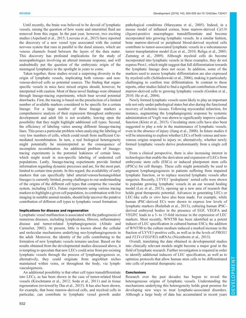
The origin of organ-specific lymphatic vesselsIn contrast to the wealth of data describing the development of earlylymphatic vessels, very little is known about the establishment oforgan-specific lymphatics at later stages, and important questions
Box 2. Heterogeneity in the origin of blood vesselsTwo models describing the formation of the great vessels in zebrafishhave been proposed. The first postulates that angioblasts in the lateralplate mesoderm are specified towards an arterial or venous fate beforethey migrate medially to generate the great vessels (Kohli et al., 2013;Zhong et al., 2001, 2000). In this model, a medial population of lateralplate mesoderm angioblasts first migrates to the midline and forms theDA, and a lateral angioblast population later gives rise to the PCV (Kohliet al., 2013). Additional support for this model came from studiesanalyzing the formation of the zebrafish lateral dorsal aortae and CCV,which also derive from medial and lateral angioblasts, respectively(Helker et al., 2013). A second model postulates that angioblasts firstmigrate medially and coalesce to form the DA primordium and then, at∼17.5 hpf, the first signs of arterial/venous differentiation are observed.Later on, a subpopulation of venous-fated angioblasts in the DA sproutsventrally to give rise to the PCV (Herbert et al., 2009; Jin et al., 2005).Most recently, a study conducted in mice demonstrated that at least∼15% of the CV cells originate in the DA (Lindskog et al., 2014; reviewedby Potente et al., 2011). In line with the findings of Herbert et al. (2009),this work demonstrated that the DA harbors a heterogeneous populationof venous and arterial ECs early during vasculogenesis, with the venousECs being those that generate the CV (Lindskog et al., 2014). The DAitself can also be considered heterogeneous, as it harbors cells of thehemogenic endothelium, the origins of which also remain controversial(reviewed by Hirschi, 2012).
930
REVIEW Development (2016) 143, 924-935 doi:10.1242/dev.132431
DEVELO
PM
ENT

about this process remain unanswered. These include whether thelymphatic vessels of all organs have similar embryonic origins,whether they respond to the same molecular cues, and whether theypossess distinct properties and unique gene expression profilesadapted to their specific functions. However, in the past year,research into the origins of organ-specific lymphatic vessels, such asthose in the skin, mesentery and heart, has begun to answer some ofthese questions. In particular, several studies have demonstratedthat, as is the case for early lymphatic vessels, cells of a non-venousorigin can contribute to the formation of these vessels in mammals(Fig. 4).Lineage-tracing experiments in mice using the venous/
endothelial cell marker Tie2 revealed that a significant portion ofdermal LECs are not labeled by the transgene, suggestingcontribution from alternative, as yet undetermined, sources. Theseresults support a dual origin mechanism in which cells of bothvenous and non-venous origins give rise to the dermal lymphaticvasculature (Martinez-Corral et al., 2015). It is important to bear inmind that this conclusion is mainly drawn from the lack of Tie2-Crelabeling in a subset of cells that were not ‘positively’ labeled byalternative markers. This lack of Tie2-Cre labeling could potentiallyhave resulted from a limited efficiency of Cre-mediatedrecombination rather than from a true absence of markerexpression. Additional experiments using alternative Cre lines thatpositively label the cells will help characterize their identity andconfirm the potential dual origin of mouse dermal lymphatics.
Another recent study explored the origins of mesentericlymphatic vessels in mice and identified a non-venous origin forthese vessels too (Stanczuk et al., 2015). It was commonly thoughtthat the mesenteric lymph sacs arise from the mesenteric vein andserve as a source for mesenteric lymphatics (Sabin, 1902; Van DerPutte, 1975). In their new study, however, Stanczuk et al. (2015)used lineage tracing followed by immunostaining for the LECmarkers Prox1 and Nrp2 to show that, although the mesenteric veinindeed represents the source for the mesenteric lymph sac, isolatedclusters of LECs are found along the mesenteric veins that do notappear to sprout from the vein. These cell clusters were shown tocoalesce at ∼E14.5 to form the mesenteric lymphatic vessels. Usinglineage-tracing analyses, a cKit+ hemogenic endothelium (HE)population was identified as the source of mesenteric LECs,indicating that non-venous lymphatics are derived from a naïvehematopoietic progenitor pool but not from definitivehematopoietic cells. Interestingly, Mahadevan et al. (2014) alsoproposed that the mesenteric lymphatics arise from two populationsof lymphatic progenitors: a venous population that originates in thewall of the subcardinal vein, and a non-venous population that arisesin the left dorsal mesentery through an arteriogenesis-dependentprocess. In this case, paired-like homeodomain 2 (Pitx2) was shownto direct asymmetric arteriogenesis in the left dorsal mesentery only.Consequently, the KO of Pitx2 resulted in specific elimination of thenon-venous LEC progenitor pool. Nonetheless, the exact lineages ofthese two subsets of lymphatic progenitors have not yet beendetermined (Mahadevan et al., 2014).
The heart is also known to possess a well-developed lymphaticvasculature, although its embryonic origins have been unclear.Expanding our knowledge of the cardiac lymphatic system might beof particular interest from a clinical point of view, since impairedlymphatic flow is associated with cardiac pathologies and has beenlinked to myocardial fibrosis and inflammation (Ullal et al., 1972).A recent study of murine embryos traced the origins of cardiac LECsto two different locations, namely cells from extra-cardiac tissuesand LECs sprouting from the CCV (Klotz et al., 2015). Both poolsof progenitors appeared to migrate to the sinus venosus and outflowtract before expanding and forming the cardiac lymphaticvasculature. Using a Tie2-Cre lineage-tracing approach, it wasshown that only 80% of cardiac lymphatic cells derive from venousECs. To further explore the source of the non-venous, Tie2-negativecells that contribute to cardiac LECs, a series of lineage-tracingexperiments using different transgenes that label a wide variety ofcell types was performed (Klotz et al., 2015). The results of theseanalyses excluded any potential contribution from the epicardium,cardiac mesoderm or cardiac neural crest populations. By contrast,extensive labeling of cells in the cardiac region was achieved whenCre driver lines under the control of Vav1 (which markshematopoietic cells), Pdgfrb (which is expressed in mesenchymalcells starting at E14) and Csf1r (a marker for myeloid cells) wereused. These transgenic drivers did not mark cells in the CCV at E10or those in the jugular vein at E12.5, but did result in the labeling ofyolk sac-derived HE cells, thus highlighting them as the putativesource of cardiac lymphatic progenitors. Interestingly, ex vivoexplants of yolk sac-derived cells isolated at E8 and treated withVegfc produced Prox1-positive LECs, providing additional supportfor the lymphatic competence of yolk sac-derived cells (Klotz et al.,2015). Unfortunately, this notion could not be corroborated in vivo,most probably owing to the lack of expression of the Cre lines usedat the relevant developmental stages. Consequently, the possibilityof alternative sources expressing similar molecular markers cannotbe excluded.
Venous (Tie2)Non-venous (?)
Mesentery
Skin
Heart
Venous (Tie2)Non-venous (Vav1, PdgfrβE17.5, Csf1r)
Venous (PdgfrβE8.5)Non-venous (cKit)
Fig. 4. The origins of organ-specific lymphatic vessels in the mouseembryo. The embryonic origins of lymphatic vessels associated with the skin,heart and mesentery have recently been studied. Tie2-based lineage-tracinganalyses in mice showed that ∼20% of skin LECs are not labeled by thetransgene, suggesting a contribution from alternative, as yet undetermined,sources. In the mesentery, some LECs are of venous origin (labeled at E8.5 bythe early EC marker Pdgfrβ), whereas others are derived from a non-venoussource traced to the cKit lineage. Similarly, cardiac lymphatics are of mixedorigins: some LECs are derived from venous ECs (Tie2 lineage), while anadditional contribution from yolk sac-derived hemogenic endothelium cells(Vav1, Pdgfrb at E17.5 and Csf1r lineages) is also observed. Pdgfrβ isexpressed early on in ECs (∼E8), whereas its expression is detected inmesenchymal cells at ∼E14.
931
REVIEW Development (2016) 143, 924-935 doi:10.1242/dev.132431
DEVELO
PM
ENT
Until recently, the brain was believed to be devoid of lymphaticvessels, raising the question of how waste and interstitial fluid areremoved from this organ. In the past year, however, two excitingstudies (Aspelund et al., 2015; Louveau et al., 2015) have reportedthe discovery of a new vessel type associated with the centralnervous system that runs in parallel to the dural sinuses, which arevenous channels found between the layers of the dura mater.This discovery has profound implications for the study ofneuropathologies involving an altered immune response, and willundoubtedly put the question of the embryonic origin of themeningeal lymphatics in the spotlight in years to come.Taken together, these studies reveal a surprising diversity in the
origin of lymphatic vessels, implicating both venous- and non-venous-derived cells as potential sources. The finding that organ-specific vessels in mice have mixed origins should, however, beinterpreted with caution. Most of these novel findings were obtainedthrough lineage-tracing experiments, which bear several potentialdrawbacks. First, the tracing is based on the preselection of a limitednumber of available markers considered to be specific for a certainlineage. For a large number of these lineage markers acomprehensive analysis of their expression throughout embryonicdevelopment and adult life is not available, leaving open thepossibility that they might highlight additional cell types. Second,the efficiency of labeling varies among different lineage-tracinglines. This poses a particular problemwhen analyzing the labeling ofvery low numbers of cells, which could result from inefficient Cre-mediated recombination. In turn, a real biological heterogeneitymight potentially be misinterpreted as the consequence ofincomplete recombination. An additional problem of lineage-tracing experiments is the potential leakiness of certain lines,which might result in non-specific labeling of undesired cellpopulations. Lastly, lineage-tracing experiments provide limitedinformation regarding dynamic processes and are, for the most part,limited to certain time points. In this regard, the availability of earlymarkers that can specifically label arterial/venous/hemangioblastlineages is currently limited, posing challenges to our understandingof the origins of the different cell types that comprise the vascularsystem, including LECs. Future experiments using various tracingmarkers to highlight a given cell population, in combination with liveimaging in suitable animal models, should help uncover the putativecontribution of different cell types to lymphatic vessel formation.
Implications for disease and regenerationLymphatic vessel malfunction is associated with the pathogenesis ofnumerous diseases, including lymphedema, fibrosis, inflammatorydisease and tumor-related lymphangiogenesis (Alitalo andCarmeliet, 2002). At present, little is known about the cellularand molecular mechanisms underlying neo-lymphangiogenesis inthe adult. Moreover, the identity of the cells contributing to theformation of new lymphatic vessels remains unclear. Based on theresults obtained from the developmental studies discussed above, itis tempting to speculate that new LECs could arise from pre-existinglymphatic vessels through the process of lymphangiogenesis or,alternatively, they could originate from angioblast nichesmaintained within veins, or throughout the body, via lymph-vasculogenesis.An additional possibility is that other cell types transdifferentiate
into LECs, as has been shown in the case of tumor-related bloodvessels (Kirschmann et al., 2012; Soda et al., 2011) and in tissueregeneration (reviewed by Das et al., 2015). It has also been shown,for example, that bone marrow-derived cells, and myeloid cells inparticular, can contribute to lymphatic vessel growth under
pathological conditions (Maruyama et al., 2005). Indeed, in amouse model of inflamed cornea, bone marrow-derived Cd11b(Itgam)-positive macrophages transdifferentiate and becomeincorporated into growing lymphatic vessels. In a similar fashion,bone marrow-derived or peripheral blood-derived myeloid cellscontribute to tumor-associated lymphatic vessels in a subcutaneoustumor transplantation model (Lee et al., 2010; Religa et al., 2005;Zumsteg et al., 2009). Although myeloid cells do becomeincorporated into lymphatic vessels in these examples, they do notexpress Prox1, which might suggest that full differentiation towardsthe lymphatic lineage does not occur. In addition, some of themarkers used to assess lymphatic differentiation are also expressedby myeloid cells (Schledzewski et al., 2006), making it particularlychallenging to confirm true differentiation. In contrast to thesereports, other studies failed to find a significant contribution of bonemarrow-derived cells to growing lymphatic vessels (Gordon et al.,2010; He et al., 2004).
Newly formed lymphatic vessels seem likely to play an importantrole not only under pathological states but also during the functionalrecovery of ischemic tissues. Following myocardial infarction, forinstance, augmenting the lymphangiogenic response by systemicadministration of Vegfc was shown to significantly improve cardiacfunction (Klotz et al., 2015). Circulating stem cells have also beensuggested to play a role in the maintenance of lymphatic vessels,even in the absence of injury (Jiang et al., 2008). In future studies itwill be interesting to explore whether LECs of both venous and non-venous origins respond to injury or, alternatively, whether newlyformed lymphatic vessels derive predominantly from a single celltype.
From a clinical perspective, there is also increasing interest intechnologies that enable the derivation and expansion of LECs fromembryonic stem cells (ESCs) or induced pluripotent stem cells(iPSCs) for cell therapy. These cells might potentially be used toaugment lymphangiogenesis in patients suffering from impairedlymphatic function, or to replace resected lymphatic vessels aftersurgeries. Recently, LYVE1+/podoplanin+ sorted cells were shownto populate growing lymphatic vessels in an ear wound healingmodel (Lee et al., 2015), opening up a new area of research thatholds great therapeutic potential. Advances in generating LECs orLEC-like cells in vitro have also been made. For example, whilehuman iPSC-derived ECs were shown to express low levels oflymphatic markers (Rufaihah et al., 2013), culturing human iPSC-derived embryoid bodies in the presence of EGF, VEGFA andVEGFC leads to a 3- to 15-fold increase in the expression of LECmarkers. Most recently, WNT5B has been identified as a potentinducer of LEC specification in cultured human ESCs; the additionof WNT5B to the culture medium induced a marked increase in thefraction of LYVE1-positive cells, as well as in the levels of PROX1and FLT4 (VEGFR3) mRNAs (Nicenboim et al., 2015).
Overall, translating the data obtained in developmental studiesinto clinically relevant models might become a major goal in thefield of lymphatic research. Further investigation is required in orderto identify additional inducers of LEC specification, as well as tooptimize protocols that allow human stem cells to be differentiatedinto cells of potential therapeutic use.
ConclusionsResearch over the past decades has begun to reveal theheterogeneous origins of lymphatic vessels. Understanding themechanisms underlying this heterogeneity holds great promise fordeveloping new ways to treat lymphatic-associated disorders.Although a large body of data has accumulated in recent years
932
REVIEW Development (2016) 143, 924-935 doi:10.1242/dev.132431
DEVELO
PM
ENT

that strongly suggests that the origin of lymphatic vessels is notexclusively venous, as previously believed, it is important to bear inmind the technical limitations of the different approaches utilized.The past 100 years of research into the lymphatic endothelium havebeen guided by the availability of animal models and experimentaltechnologies that enable lymphatic vessels to be visualized in vivo.Since its early beginning, the question of the origin of lymphaticvessels has remained controversial, in part due to biases in theinterpretation of results obtained from different systems. The initialdiscrepancy between the two key models proposed – the venous/centrifugal and non-venous/centripetal models – could be easilyexplained by the limitations imposed by each of the appliedmethodologies, namely ink injection versus histological sections,which each enable investigation of only certain aspects of theprocess. Similarly, potential bias inherent to lineage-tracingexperiments carried out in mice and zebrafish could lead toconflicting results. Complementary experiments analyzing thedistribution of endogenous proteins via immunostaining, as wellas live imaging studies analyzing the anatomical location andmigration tracks undertaken by LEC progenitors, should lead tomore definitive conclusions.The discovery of multiple origins for the lymphatic endothelium
has many implications and raises numerous important questions inthe field. For example, whether the classical specification pathwaysare equally important for the specification of non-venous cellstowards the lymphatic fate remains unclear. The anatomical regionsfrom which non-venous cells migrate to their final destination alsoremain to be determined. One possibility is that non-venous cellsreside in the embryonic veins, as is the case in zebrafish (Nicenboimet al., 2015). Alternatively, cells might transdifferentiate in situ fromsurrounding mesenchymal cells, as originally postulated by the non-venous theory. Lastly, whether the different embryonic origins oflymphatic progenitors are specifically linked to specialized functionsduring adult life remains unknown. The origin of the cells responsiblefor neo-lymphangiogenesis also requires further investigation, andunderstanding the sources of new lymphatic vessels in the adultmight be of great value in the treatment of diseases that involve thelymphatic system, such as lymphedema and cancer.
AcknowledgementsWe thank G. Brodsky (Weizmann Institute of Science) for graphic design andN. Konstantin for critical reading of the manuscript. We are grateful to all members ofthe K.Y. laboratory for many fruitful discussions. We sincerely apologize tocolleagues whose important work could not be cited owing to space limitations.
Competing interestsThe authors declare no competing or financial interests.
FundingThis work was supported in part by Marie Curie Actions-International Reintegrationgrants [FP7-PEOPLE-2009-RG 256393 to K.Y.], the European Research Council[335605 to K.Y.] and the Minerva Foundation [711128 to K.Y.]. K.Y. is supported bytheWillner Family Center for Vascular Biology; the estate of Paul Ourieff; the CarolitoStiftung; Lois Rosen, Los Angeles, CA, USA; Edith Frumin; the FondazioneHenry Krenter; the Wallach Hanna & Georges Lustgarten Fund; and the PolenCharitable Trust. K.Y. is the incumbent of the Louis and Ida Rich CareerDevelopment Chair.
ReferencesAlders, M., Hogan, B. M., Gjini, E., Salehi, F., Al-Gazali, L., Hennekam, E. A.,Holmberg, E. E., Mannens, M. M. A. M., Mulder, M. F., Offerhaus, G. J. et al.(2009). Mutations in CCBE1 cause generalized lymph vessel dysplasia inhumans. Nat. Genet. 41, 1272-1274.
Alitalo, K. and Carmeliet, P. (2002). Molecular mechanisms of lymphangiogenesisin health and disease. Cancer Cell 1, 219-227.
Aranguren, X. L., Beerens, M., Vandevelde, W., Dewerchin, M., Carmeliet, P.and Luttun, A. (2011). Transcription factor COUP-TFII is indispensable for
venous and lymphatic development in zebrafish and Xenopus laevis. Biochem.Biophys. Res. Commun. 410, 121-126.
Aspelund, A., Antila, S., Proulx, S. T., Karlsen, T. V., Karaman, S., Detmar, M.,Wiig, H. and Alitalo, K. (2015). A dural lymphatic vascular system that drainsbrain interstitial fluid and macromolecules. J. Exp. Med. 212, 991-999.
Banerji, S., Ni, J., Wang, S. X., Clasper, S., Su, J., Tammi, R., Jones, M. andJackson, D. G. (1999). LYVE-1, a new homologue of the CD44 glycoprotein, is alymph-specific receptor for hyaluronan. J. Cell Biol. 144, 789-801.
Bos, F. L., Caunt, M., Peterson-Maduro, J., Planas-Paz, L., Kowalski, J.,Karpanen, T., van Impel, A., Tong, R., Ernst, J. A., Korving, J. et al. (2011).CCBE1 is essential for mammalian lymphatic vascular development andenhances the lymphangiogenic effect of vascular endothelial growth factor-C invivo. Circ. Res. 109, 486-491.
Buttler, K., Becker, J., Pukrop, T. and Wilting, J. (2013). Maldevelopment ofdermal lymphatics in Wnt5a-knockout-mice. Dev. Biol. 381, 365-376.
Cermenati, S., Moleri, S., Cimbro, S., Corti, P., Del Giacco, L., Amodeo, R.,Dejana, E., Koopman, P., Cotelli, F. and Beltrame, M. (2008). Sox18 and Sox7play redundant roles in vascular development. Blood 111, 2657-2666.
Cermenati, S., Moleri, S., Neyt, C., Bresciani, E., Carra, S., Grassini, D. R.,Omini, A., Goi, M., Cotelli, F., Francois, M. et al. (2013). Sox18 geneticallyinteracts with VegfC to regulate lymphangiogenesis in zebrafish. Arterioscler.Thromb. Vasc. Biol. 33, 1238-1247.
Cha, Y. R., Fujita, M., Butler, M., Isogai, S., Kochhan, E., Siekmann, A. F. andWeinstein, B. M. (2012). Chemokine signaling directs trunk lymphatic networkformation along the preexisting blood vasculature. Dev. Cell 22, 824-836.
Chen, L., Mupo, A., Huynh, T., Cioffi, S., Woods, M., Jin, C., McKeehan, W.,Thompson-Snipes, L., Baldini, A. and Illingworth, E. (2010). Tbx1 regulatesVegfr3 and is required for lymphatic vessel development. J. Cell Biol. 189,417-424.
Clevers, H. (2006). Wnt/beta-catenin signaling in development and disease. Cell127, 469-480.
Covassin, L. D., Villefranc, J. A., Kacergis, M. C., Weinstein, B. M. and Lawson,N. D. (2006). Distinct genetic interactions between multiple Vegf receptors arerequired for development of different blood vessel types in zebrafish. Proc. Natl.Acad. Sci. USA 103, 6554-6559.
Das, A., Sinha, M., Datta, S., Abas, M., Chaffee, S., Sen, C. K. and Roy, S. (2015).Monocyte and macrophage plasticity in tissue repair and regeneration.Am. J. Pathol. 185, 2596-2606.
de Vinuesa, A. G., Abdelilah-Seyfried, S., Knaus, P., Zwijsen, A. and Bailly, S.(2016). BMP signaling in vascular biology and dysfunction. Cytokine GrowthFactor Rev. 27, 65-79.
Deguchi, T., Fujimori, K. E., Kawasaki, T., Ohgushi, H. and Yuba, S. (2009).Molecular cloning and gene expression of the prox1a and prox1b genes in themedaka, Oryzias latipes. Gene Expr. Patterns 9, 341-347.
Del Giacco, L., Pistocchi, A. and Ghilardi, A. (2010). prox1b Activity is essential inzebrafish lymphangiogenesis. PLoS ONE 5, e13170.
Deng, Y., Atri, D., Eichmann, A. and Simons, M. (2013). Endothelial ERKsignaling controls lymphatic fate specification. J. Clin. Invest. 123, 1202-1215.
Dieterich, L. C., Klein, S., Mathelier, A., Sliwa-Primorac, A., Ma, Q., Hong, Y.-K.,Shin, J. W., Hamada, M., Lizio, M., Itoh, M. et al. (2015). DeepCAGEtranscriptomics reveal an important role of the transcription factor MAFB in thelymphatic endothelium. Cell Rep. 13, 1493-1504.
Dumont, D. J., Jussila, L., Taipale, J., Lymboussaki, A., Mustonen, T., Pajusola,K., Breitman, M. and Alitalo, K. (1998). Cardiovascular failure in mouse embryosdeficient in VEGF receptor-3. Science 282, 946-949.
Dunworth, W. P., Cardona-Costa, J., Bozkulak, E. C., Kim, J.-D., Meadows, S.,Fischer, J. C., Wang, Y., Cleaver, O., Qyang, Y., Ober, E. A. et al. (2014). Bonemorphogenetic protein 2 signaling negatively modulates lymphatic developmentin vertebrate embryos. Circ. Res. 114, 56-66.
Duong, T., Koltowska, K., Pichol-Thievend, C., Le Guen, L., Fontaine, F., Smith,K. A., Truong, V., Skoczylas, R., Stacker, S. A., Achen, M. G. et al. (2014).VEGFD regulates blood vascular development by modulating SOX18 activity.Blood 123, 1102-1112.
Fatima, A., Culver, A., Culver, F., Liu, T., Dietz, W. H., Thomson, B. R.,Hadjantonakis, A.-K., Quaggin, S. E. and Kume, T. (2014). Murine Notch1 isrequired for lymphatic vascular morphogenesis during development. Dev. Dyn.243, 957-964.
Flores, M. V., Hall, C. J., Crosier, K. E. and Crosier, P. S. (2010). Visualization ofembryonic lymphangiogenesis advances the use of the zebrafish model forresearch in cancer and lymphatic pathologies. Dev. Dyn. 239, 2128-2135.
François, M., Caprini, A., Hosking, B., Orsenigo, F., Wilhelm, D., Browne, C.,Paavonen, K., Karnezis, T., Shayan, R., Downes, M. et al. (2008). Sox18induces development of the lymphatic vasculature in mice. Nature 456, 643-647.
Gale, N. W., Prevo, R., Espinosa, J., Ferguson, D. J., Dominguez, M. G.,Yancopoulos, G. D., Thurston, G. and Jackson, D. G. (2007). Normal lymphaticdevelopment and function in mice deficient for the lymphatic hyaluronan receptorLYVE-1. Mol. Cell. Biol. 27, 595-604.
Geudens, I., Herpers, R., Hermans, K., Segura, I., Ruiz de Almodovar, C.,Bussmann, J., De Smet, F., Vandevelde,W., Hogan, B. M., Siekmann, A. et al.
933
REVIEW Development (2016) 143, 924-935 doi:10.1242/dev.132431
DEVELO
PM
ENT

(2010). Role of delta-like-4/Notch in the formation and wiring of the lymphaticnetwork in zebrafish. Arterioscler. Thromb. Vasc. Biol. 30, 1695-1702.
Gordon, E. J., Rao, S., Pollard, J. W., Nutt, S. L., Lang, R. A. and Harvey, N. L.(2010). Macrophages define dermal lymphatic vessel calibre during developmentby regulating lymphatic endothelial cell proliferation. Development 137,3899-3910.
Haiko, P., Makinen, T., Keskitalo, S., Taipale, J., Karkkainen, M. J., Baldwin,M. E., Stacker, S. A., Achen, M. G. and Alitalo, K. (2008). Deletion of vascularendothelial growth factor C (VEGF-C) and VEGF-D is not equivalent to VEGFreceptor 3 deletion in mouse embryos. Mol. Cell. Biol. 28, 4843-4850.
Harvey, N. L., Srinivasan, R. S., Dillard, M. E., Johnson, N. C.,Witte, M. H., Boyd,K., Sleeman, M. W. and Oliver, G. (2005). Lymphatic vascular defects promotedby Prox1 haploinsufficiency cause adult-onset obesity. Nat. Genet. 37,1072-1081.
He, Y., Rajantie, I., Ilmonen, M., Makinen, T., Karkkainen, M. J., Haiko, P.,Salven, P. and Alitalo, K. (2004). Preexisting lymphatic endothelium but notendothelial progenitor cells are essential for tumor lymphangiogenesis andlymphatic metastasis. Cancer Res. 64, 3737-3740.
Helker, C. S., Schuermann, A., Karpanen, T., Zeuschner, D., Belting, H.-G.,Affolter, M., Schulte-Merker, S. and Herzog, W. (2013). The zebrafish commoncardinal veins develop by a novel mechanism: lumen ensheathment.Development 140, 2776-2786.
Hen, G., Nicenboim, J., Mayseless, O., Asaf, L., Shin, M., Busolin, G., Hofi, R.,Almog, G., Tiso, N., Lawson, N. D. et al. (2015). Venous-derived angioblastsgenerate organ-specific vessels during zebrafish embryonic development.Development 142, 4266-4278.
Herbert, S. P., Huisken, J., Kim, T. N., Feldman, M. E., Houseman, B. T., Wang,R. A., Shokat, K. M. and Stainier, D. Y. (2009). Arterial-venous segregation byselective cell sprouting: an alternative mode of blood vessel formation. Science326, 294-298.
Hirschi, K. K. (2012). Hemogenic endothelium during development and beyond.Blood 119, 4823-4827.
Hogan, B. M., Bos, F. L., Bussmann, J., Witte, M., Chi, N. C., Duckers, H. J. andSchulte-Merker, S. (2009). Ccbe1 is required for embryonic lymphangiogenesisand venous sprouting. Nat. Genet. 41, 396-398.
Hong, Y.-K., Harvey, N., Noh, Y.-H., Schacht, V., Hirakawa, S., Detmar, M. andOliver, G. (2002). Prox1 is a master control gene in the program specifyinglymphatic endothelial cell fate. Dev. Dyn. 225, 351-357.
Huntington, G. S. and McClure, C. F. w. (1912). The anatomy and development ofthe jugular lymph sacs in the domestic cat (Felis domestica). Am. J. Anat. 10,177-312.
Isogai, S., Lawson, N. D., Torrealday, S., Horiguchi, M. and Weinstein, B. M.(2003). Angiogenic network formation in the developing vertebrate trunk.Development 130, 5281-5290.
Jeltsch, M., Kaipainen, A., Joukov, V., Meng, X., Lakso, M., Rauvala, H., Swartz,M., Fukumura, D., Jain, R. K. and Alitalo, K. (1997). Hyperplasia of lymphaticvessels in VEGF-C transgenic mice. Science 276, 1423-1425.
Jeltsch, M., Jha, S. K., Tvorogov, D., Anisimov, A., Leppanen, V.-M.,Holopainen, T., Kivela, R., Ortega, S., Karpanen, T. and Alitalo, K. (2014).CCBE1 enhances lymphangiogenesis via A disintegrin and metalloprotease withthrombospondin motifs-3-mediated vascular endothelial growth factor-Cactivation. Circulation 129, 1962-1971.
Jiang, S., Bailey, A. S., Goldman, D. C., Swain, J. R., Wong, M. H., Streeter, P. R.and Fleming, W. H. (2008). Hematopoietic stem cells contribute to lymphaticendothelium. PLoS ONE 3, e3812.
Jin, S.-W., Beis, D., Mitchell, T., Chen, J.-N. and Stainier, D. Y. R. (2005). Cellularand molecular analyses of vascular tube and lumen formation in zebrafish.Development 132, 5199-5209.
Kaipainen, A., Korhonen, J., Mustonen, T., van Hinsbergh, V., Fang, G.-H.,Dumont, D., Breitman, M. and Alitalo, K. (1995). Expression of the fms-liketyrosine kinase 4 gene becomes restricted to lymphatic endothelium duringdevelopment. Proc. Natl. Acad. Sci. USA 92, 3566-3570.
Kang, J., Yoo, J., Lee, S., Tang, W., Aguilar, B., Ramu, S., Choi, I., Otu, H. H.,Shin, J. W., Dotto, G. P. et al. (2010). An exquisite cross-control mechanismamong endothelial cell fate regulators directs the plasticity and heterogeneity oflymphatic endothelial cells. Blood 116, 140-150.
Karkkainen, M. J., Haiko, P., Sainio, K., Partanen, J., Taipale, J., Petrova, T. V.,Jeltsch, M., Jackson, D. G., Talikka, M., Rauvala, H. et al. (2004). Vascularendothelial growth factor C is required for sprouting of the first lymphatic vesselsfrom embryonic veins. Nat. Immunol. 5, 74-80.
Kashiwada, T., Fukuhara, S., Terai, K., Tanaka, T., Wakayama, Y., Ando, K.,Nakajima, H., Fukui, H., Yuge, S., Saito, Y. et al. (2015). beta-Catenin-dependent transcription is central to Bmp-mediated formation of venous vessels.Development 142, 497-509.
Kim, J.-D. and Kim, J. (2014). Alk3/Alk3b and Smad5 mediate BMP signalingduring lymphatic development in zebrafish. Mol. Cells 37, 270-274.
Kim, H., Nguyen, V. P., Petrova, T. V., Cruz, M., Alitalo, K. and Dumont, D. J.(2010). Embryonic vascular endothelial cells are malleable to reprogramming viaProx1 to a lymphatic gene signature. BMC Dev. Biol. 10, 72.
Kirschmann, D. A., Seftor, E. A., Hardy, K. M., Seftor, R. E. b. and Hendrix,M. J. c. (2012). Molecular pathways: vasculogenic mimicry in tumor cells:diagnostic and therapeutic implications. Clin. Cancer Res. 18, 2726-2732.
Klotz, L., Norman, S., Vieira, J. M., Masters, M., Rohling, M., Dube, K. N., Bollini,S., Matsuzaki, F., Carr, C. A. and Riley, P. R. (2015). Cardiac lymphatics areheterogeneous in origin and respond to injury. Nature 522, 62-67.
Kohli, V., Schumacher, J. A., Desai, S. P., Rehn, K. and Sumanas, S. (2013).Arterial and venous progenitors of the major axial vessels originate at distinctlocations. Dev. Cell 25, 196-206.
Kok, F. O., Shin, M., Ni, C.-W., Gupta, A., Grosse, A. S., van Impel, A.,Kirchmaier, B. C., Peterson-Maduro, J., Kourkoulis, G., Male, I. et al. (2015).Reverse genetic screening reveals poor correlation between morpholino-inducedand mutant phenotypes in zebrafish. Dev. Cell 32, 97-108.
Koltowska, K., Lagendijk, A. K., Pichol-Thievend, C., Fischer, J. C., Francois,M., Ober, E. A., Yap, A. S. and Hogan, B. M. (2015a). Vegfc regulates bipotentialprecursor division and Prox1 expression to promote lymphatic identity inzebrafish. Cell Rep. 13, 1828-1841.
Koltowska, K., Paterson, S., Bower, N. I., Baillie, G. J., Lagendijk, A. K., Astin,J. W., Chen, H., Francois, M., Crosier, P. S., Taft, R. J. et al. (2015b). mafba is adownstream transcriptional effector of Vegfc signaling essential for embryoniclymphangiogenesis in zebrafish. Genes Dev. 29, 1618-1630.
Kuchler, A. M., Gjini, E., Peterson-Maduro, J., Cancilla, B., Wolburg, H. andSchulte-Merker, S. (2006). Development of the zebrafish lymphatic systemrequires VEGFC signaling. Curr. Biol. 16, 1244-1248.
Kukk, E., Lymboussaki, A., Taira, S., Kaipainen, A., Jeltsch, M., Joukov, V. andAlitalo, K. (1996). VEGF-C receptor binding and pattern of expression withVEGFR-3 suggests a role in lymphatic vascular development. Development 122,3829-3837.
Lawson, N. D. and Weinstein, B. M. (2002). In vivo imaging of embryonic vasculardevelopment using transgenic zebrafish. Dev. Biol. 248, 307-318.
Le Guen, L., Karpanen, T., Schulte, D., Harris, N. C., Koltowska, K., Roukens,G., Bower, N. I., van Impel, A., Stacker, S. A., Achen, M. G. et al. (2014). Ccbe1regulates Vegfc-mediated induction of Vegfr3 signaling during embryoniclymphangiogenesis. Development 141, 1239-1249.
Lee, J. Y., Park, C., Cho, Y. P., Lee, E., Kim, H., Kim, P., Yun, S. H. and Yoon, Y.-s.(2010). Podoplanin-expressing cells derived from bone marrow play a crucial rolein postnatal lymphatic neovascularization. Circulation 122, 1413-1425.
Lee, S.-J., Park, C., Lee, J. Y., Kim, S., Kwon, P. J., Kim, W., Jeon, Y. H., Lee, E.and Yoon, Y. S. (2015). Generation of pure lymphatic endothelial cells fromhuman pluripotent stem cells and their therapeutic effects on wound repair. Sci.Rep. 5, 11019.
Levet, S., Ciais, D., Merdzhanova, G., Mallet, C., Zimmers, T. A., Lee, S.-J.,Navarro, F. P., Texier, I., Feige, J.-J., Bailly, S. et al. (2013). Bonemorphogenetic protein 9 (BMP9) controls lymphatic vessel maturation andvalve formation. Blood 122, 598-607.
Lewis, F. T. (1905). The development of the lymphatic system in rabbits.Am. J. Anat.5, 95-111.
Lim, A. H., Suli, A., Yaniv, K., Weinstein, B., Li, D. Y. and Chien, C.-B. (2011).Motoneurons are essential for vascular pathfinding. Development 138,3847-3857.
Lindskog, H., Kim, Y. H., Jelin, E. B., Kong, Y., Guevara-Gallardo, S., Kim, T. N.and Wang, R. A. (2014). Molecular identification of venous progenitors in thedorsal aorta reveals an aortic origin for the cardinal vein in mammals.Development 141, 1120-1128.
Louveau, A., Smirnov, I., Keyes, T. J., Eccles, J. D., Rouhani, S. J., Peske, J. D.,Derecki, N. C., Castle, D., Mandell, J. W., Lee, K. S. et al. (2015). Structural andfunctional features of central nervous system lymphatic vessels. Nature 523,337-341.
Mahadevan, A., Welsh, I. C., Sivakumar, A., Gludish, D. W., Shilvock, A. R.,Noden, D. M., Huss, D., Lansford, R. and Kurpios, N. A. (2014). The left-rightPitx2 pathway drives organ-specific arterial and lymphatic development in theintestine. Dev. Cell 31, 690-706.
Makinen, T., Veikkola, T., Mustjoki, S., Karpanen, T., Catimel, B., Nice, E. C.,Wise, L., Mercer, A., Kowalski, H., Kerjaschki, D. et al. (2001). Isolatedlymphatic endothelial cells transduce growth, survival and migratory signals viathe VEGF-C/D receptor VEGFR-3. EMBO J. 20, 4762-4773.
Martinez-Corral, I., Ulvmar, M. H., Stanczuk, L., Tatin, F., Kizhatil, K., John,S. W. M., Alitalo, K., Ortega, S. and Makinen, T. (2015). Nonvenous origin ofdermal lymphatic vasculature. Circ. Res. 116, 1649-1654.
Maruyama, K., Ii, M., Cursiefen, C., Jackson, D. G., Keino, H., Tomita, M., VanRooijen, N., Takenaka, H., D’Amore, P. A., Stein-Streilein, J. et al. (2005).Inflammation-induced lymphangiogenesis in the cornea arises from CD11b-positive macrophages. J. Clin. Invest. 115, 2363-2372.
McClure, C. F. W. (1921). The endothelial problem. Anat. Rec. 22, 219-237.Mikels, A. J. andNusse, R. (2006). PurifiedWnt5a protein activates or inhibits beta-
catenin–TCF signaling depending on receptor context. PLoS Biol. 4, e115.Murtomaki, A., Uh,M. K., Choi, Y. K., Kitajewski, C., Borisenko, V., Kitajewski, J.
and Shawber, C. J. (2013). Notch1 functions as a negative regulator of lymphaticendothelial cell differentiation in the venous endothelium. Development 140,2365-2376.
934
REVIEW Development (2016) 143, 924-935 doi:10.1242/dev.132431
DEVELO
PM
ENT

Nguyen, P. D., Hollway, G. E., Sonntag, C., Miles, L. B., Hall, T. E., Berger, S.,Fernandez, K. J., Gurevich, D. B., Cole, N. J., Alaei, S. et al. (2014).Haematopoietic stem cell induction by somite-derived endothelial cells controlledby meox1. Nature 512, 314-318.
Nicenboim, J., Malkinson, G., Lupo, T., Asaf, L., Sela, Y., Mayseless, O., Gibbs-Bar, L., Senderovich, N., Hashimshony, T., Shin, M. et al. (2015). Lymphaticvessels arise from specialized angioblasts within a venous niche. Nature 522,56-61.
Niessen, K., Zhang, G., Ridgway, J. B., Chen, H., Kolumam, G., Siebel, C. W.and Yan, M. (2011). The Notch1-Dll4 signaling pathway regulates mousepostnatal lymphatic development. Blood 118, 1989-1997.
Ny, A., Koch, M., Schneider, M., Neven, E., Tong, R. T., Maity, S., Fischer, C.,Plaisance, S., Lambrechts, D., Heligon, C. et al. (2005). A genetic Xenopuslaevis tadpole model to study lymphangiogenesis. Nat. Med. 11, 998-1004.
Okuda, K. S., Astin, J. W., Misa, J. P., Flores, M. V., Crosier, K. E. and Crosier,P. S. (2012). lyve1 expression reveals novel lymphatic vessels and newmechanisms for lymphatic vessel development in zebrafish. Development 139,2381-2391.
Pennisi, D., Bowles, J., Nagy, A., Muscat, G. and Koopman, P. (2000). Mice nullfor sox18 are viable and display a mild coat defect.Mol. Cell. Biol. 20, 9331-9336.
Potente, M., Gerhardt, H. and Carmeliet, P. (2011). Basic and therapeutic aspectsof angiogenesis. Cell 146, 873-887.
Religa, P., Cao, R., Bjorndahl, M., Zhou, Z., Zhu, Z. and Cao, Y. (2005). Presenceof bone marrow-derived circulating progenitor endothelial cells in the newlyformed lymphatic vessels. Blood 106, 4184-4190.
Rossi, A., Kontarakis, Z., Gerri, C., Nolte, H., Holper, S., Kruger, M. and Stainier,D. Y. R. (2015). Genetic compensation induced by deleterious mutations but notgene knockdowns. Nature 524, 230-233.
Rufaihah, A. J., Huang, N. F., Kim, J., Herold, J., Volz, K. S., Park, T. S., Lee,J. C., Zambidis, E. T., Reijo-Pera, R. and Cooke, J. P. (2013). Human inducedpluripotent stem cell-derived endothelial cells exhibit functional heterogeneity.Am. J. Transl. Res. 5, 21-35.
Sabin, F. R. (1902). On the origin of the lymphatic system from the veins and thedevelopment of the lymph hearts and thoracic duct in the pig. Am. J. Anat. 1,367-389.
Sabin, F. R. (1904). On the development of the superficial lymphatics in the skin ofthe pig. Am. J. Anat. 3, 183-195.
Sabin, F. R. (1913). The Origin and Development of the Lymphatic System.Baltimore: Johns Hopkins Press.
Schledzewski, K., Falkowski, M., Moldenhauer, G., Metharom, P.,Kzhyshkowska, J., Ganss, R., Demory, A., Falkowska-Hansen, B., Kurzen,H., Ugurel, S. et al. (2006). Lymphatic endothelium-specific hyaluronan receptorLYVE-1 is expressed by stabilin-1+, F4/80+, CD11b+ macrophages in malignanttumours and wound healing tissuein vivo and in bone marrow culturesin vitro:implications for the assessment of lymphangiogenesis. J. Pathol. 209, 67-77.
Siekmann, A. F. and Lawson, N. D. (2007). Notch signalling limits angiogenic cellbehaviour in developing zebrafish arteries. Nature 445, 781-784.
Soda, Y., Marumoto, T., Friedmann-Morvinski, D., Soda, M., Liu, F., Michiue, H.,Pastorino, S., Yang, M., Hoffman, R. M., Kesari, S. et al. (2011).Transdifferentiation of glioblastoma cells into vascular endothelial cells. Proc.Natl. Acad. Sci. USA 108, 4274-4280.
Srinivasan, R. S. and Oliver, G. (2011). Prox1 dosage controls the number oflymphatic endothelial cell progenitors and the formation of the lymphovenousvalves. Genes Dev. 25, 2187-2197.
Srinivasan, R. S., Dillard, M. E., Lagutin, O. V., Lin, F.-J., Tsai, S., Tsai, M.-J.,Samokhvalov, I. M. and Oliver, G. (2007). Lineage tracing demonstrates thevenous origin of the mammalian lymphatic vasculature. Genes Dev. 21,2422-2432.
Srinivasan, R. S., Geng, X., Yang, Y., Wang, Y., Mukatira, S., Studer, M., Porto,M. P. R., Lagutin, O. and Oliver, G. (2010). The nuclear hormone receptor Coup-TFII is required for the initiation and early maintenance of Prox1 expression inlymphatic endothelial cells. Genes Dev. 24, 696-707.
Srinivasan, R. S., Escobedo, N., Yang, Y., Interiano, A., Dillard, M. E.,Finkelstein, D., Mukatira, S., Gil, H. J., Nurmi, H., Alitalo, K. et al. (2014).The Prox1–Vegfr3 feedback loop maintains the identity and the number oflymphatic endothelial cell progenitors. Genes Dev. 28, 2175-2187.
Stanczuk, L., Martinez-Corral, I., Ulvmar, M. H., Zhang, Y., Lavin a, B., Fruttiger,M., Adams, R. H., Saur, D., Betsholtz, C., Ortega, S. et al. (2015). cKit Lineagehemogenic endothelium-derived cells contribute to mesenteric lymphatic vessels.Cell Rep. 10, 1708-1721.
Tammela, T., Zarkada, G., Wallgard, E., Murtomaki, A., Suchting, S., Wirzenius,M., Waltari, M., Hellstrom, M., Schomber, T., Peltonen, R. et al. (2008).Blocking VEGFR-3 suppresses angiogenic sprouting and vascular networkformation. Nature 454, 656-660.
Tao, S., Witte, M., Bryson-Richardson, R. J., Currie, P. D., Hogan, B. M. andSchulte-Merker, S. (2011). Zebrafish prox1b mutants develop a lymphaticvasculature, and prox1b does not specifically mark lymphatic endothelial cells.PLoS ONE 6, e28934.
Tso, P. and Balint, J. A. (1986). Formation and transport of chylomicrons byenterocytes to the lymphatics. Am. J. Physiol. 250, G715-G726.
Ullal, S. R., Kluge, T. H. and Gerbode, F. (1972). Functional and pathologicchanges in the heart following chronic cardiac lymphatic obstruction. Surgery 71,328-334.
van Amerongen, R., Fuerer, C., Mizutani, M. and Nusse, R. (2012). Wnt5a canboth activate and repress Wnt/beta-catenin signaling during mouse embryonicdevelopment. Dev. Biol. 369, 101-114.
Van der Jagt, E. R. (1932). Memoirs: the origin and development of the anteriorlymph-sacs in the sea-turtle (Thalassochelys caretta). Q. J. Microsc. Sci. 2,151-163.
Van Der Putte, S. (1975). The early development of the lymphatic system in mouseembryos. Acta Morphol. Neerl. Scand. 13, 245-286.
van Impel, A., Zhao, Z., Hermkens, D. M. A., Roukens, M. G., Fischer, J. C.,Peterson-Maduro, J., Duckers, H., Ober, E. A., Ingham, P. W. and Schulte-Merker, S. (2014). Divergence of zebrafish and mouse lymphatic cell fatespecification pathways. Development 141, 1228-1238.
Walls, J. R., Coultas, L., Rossant, J. and Henkelman, R. M. (2008). Three-dimensional analysis of vascular development in the mouse embryo. PLoS ONE3, e2853.
Wang, Y., Nakayama, M., Pitulescu, M. E., Schmidt, T. S., Bochenek, M. L.,Sakakibara, A., Adams, S., Davy, A., Deutsch, U., Luthi, U. et al. (2010).Ephrin-B2 controls VEGF-induced angiogenesis and lymphangiogenesis. Nature465, 483-486.
Wang, R. N., Green, J., Wang, Z., Deng, Y., Qiao, M., Peabody, M., Zhang, Q., Ye,J., Yan, Z., Denduluri, S. et al. (2014). Bone morphogenetic protein (BMP)signaling in development and human diseases. Genes Dis. 1, 87-105.
Wigle, J. T. and Oliver, G. (1999). Prox1 function is required for the development ofthe murine lymphatic system. Cell 98, 769-778.
Wigle, J. T., Harvey, N., Detmar, M., Lagutina, I., Grosveld, G., Gunn, M. D.,Jackson, D. G. and Oliver, G. (2002). An essential role for Prox1 in the inductionof the lymphatic endothelial cell phenotype. EMBO J. 21, 1505-1513.
Wiley, D. M., Kim, J.-D., Hao, J., Hong, C. C., Bautch, V. L. and Jin, S.-W. (2011).Distinct signalling pathways regulate sprouting angiogenesis from the dorsal aortaand the axial vein. Nat. Cell Biol. 13, 687-693.
Wilting, J., Aref, Y., Huang, R., Tomarev, S. I., Schweigerer, L., Christ, B.,Valasek, P. and Papoutsi, M. (2006). Dual origin of avian lymphatics. Dev. Biol.292, 165-173.
Yang, Y., Garcia-Verdugo, J. M., Soriano-Navarro, M., Srinivasan, R. S.,Scallan, J. P., Singh, M. K., Epstein, J. A. and Oliver, G. (2012). Lymphaticendothelial progenitors bud from the cardinal vein and intersomitic vessels inmammalian embryos. Blood 120, 2340-2348.
Yaniv, K., Isogai, S., Castranova, D., Dye, L., Hitomi, J. and Weinstein, B. M.(2006). Live imaging of lymphatic development in the zebrafish. Nat. Med. 12,711-716.
Yoshimatsu, Y., Lee, Y. G., Akatsu, Y., Taguchi, L., Suzuki, H. I., Cunha, S. I.,Maruyama, K., Suzuki, Y., Yamazaki, T., Katsura, A. et al. (2013). Bonemorphogenetic protein-9 inhibits lymphatic vessel formation via activin receptor-like kinase 1 during development and cancer progression. Proc. Natl. Acad. Sci.USA 110, 18940-18945.
You, L.-R., Lin, F.-J., Lee, C. T., DeMayo, F. J., Tsai, M.-J. and Tsai, S. Y. (2005).Suppression of Notch signalling by the COUP-TFII transcription factor regulatesvein identity. Nature 435, 98-104.
Yu, M., Zhang, H., Liu, Y., He, Y., Yang, C., Du, Y., Wu, M., Zhang, G. and Gao, F.(2015). The cooperative role of S1P3 with LYVE-1 in LMW-HA-inducedlymphangiogenesis. Exp. Cell Res. 336, 150-157.
Zheng, W., Tammela, T., Yamamoto, M., Anisimov, A., Holopainen, T.,Kaijalainen, S., Karpanen, T., Lehti, K., Yla-Herttuala, S. and Alitalo, K.(2011). Notch restricts lymphatic vessel sprouting induced by vascular endothelialgrowth factor. Blood 118, 1154-1162.
Zhong, T. P., Rosenberg, M., Mohideen, M.-A. P. L., Weinstein, B. and Fishman,M. C. (2000). gridlock, an HLH gene required for assembly of the aorta inzebrafish. Science 287, 1820-1824.
Zhong, T. P., Childs, S., Leu, J. P. and Fishman, M. C. (2001). Gridlock signallingpathway fashions the first embryonic artery. Nature 414, 216-220.
Zumsteg, A., Baeriswyl, V., Imaizumi, N., Schwendener, R., Ruegg, C. andChristofori, G. (2009). Myeloid cells contribute to tumor lymphangiogenesis.PLoS ONE 4, e7067.
935
REVIEW Development (2016) 143, 924-935 doi:10.1242/dev.132431
DEVELO
PM
ENT